
Submitted:
10 January 2025
Posted:
13 January 2025
You are already at the latest version
Abstract
Purpose: Tooth regeneration research aims to bridge historical milestones with cutting-edge molecular insights and biomaterial strategies to revolutionize regenerative dentistry. It focuses on decoding the genetic underpinnings and molecular signaling pathways that govern tooth morphogenesis. This understanding is crucial for advancing the development of innovative and effective dental restoration techniques that can potentially replace conventional methods. Methods: This comprehensive review integrates historical perspectives with contemporary scientific inquiries, emphasizing the role of advanced techniques such as single-cell sequencing, induced pluripotent stem cells (iPSCs), and organoid culture in unraveling the complexities of tooth development. Additionally, this review explores the use of proteomics, phosphoproteomics, and genetic engineering systems in studying molecular signaling pathways involved in tooth development and morphogenesis. Results: This review highlights the pivotal role of induced pluripotent stem cells (iPSCs) in tooth regeneration, shedding light on their transformative applications. Furthermore, the integration of advanced computational analysis with single-cell combinatorial indexing RNA sequencing (sci-RNA-seq) has unveiled previously unknown populations of epithelial support tissues, offering profound insights into dental tissue differentiation. Additionally, this review discusses the advances in biomaterials and bioengineering approaches, emphasizing the intricacies of neurovascular integration, which is crucial for successful tooth regeneration. Conclusion: This comprehensive review provides a holistic understanding of tooth regeneration, emphasizing the historical milestones, molecular insights, and biomaterial strategies that have shaped the field. The integration of advanced techniques and innovative approaches holds promise for transformative applications in regenerative dentistry, paving the way for future advancements in tooth regeneration.
Keywords:
Tooth Regeneration
; Molecular Insights
; Stem Cell-Based Approaches
; Biomaterial Advancements
; Bioengineering Strategies
Background
Tooth regeneration stands at the forefront of modern scientific inquiry, encompassing a journey marked by historical milestones and cutting-edge advancements that have revolutionized dental medicine. The historical perspective of tooth regeneration research provides a captivating backdrop for this exploration. From pioneering studies of tooth development in embryonic models to the discovery of molecular signaling pathways orchestrating morphogenesis, the journey has been characterized by relentless curiosity and groundbreaking discoveries. Understanding this historical context not only reveals the intricacies of tooth regeneration but also highlights the collective efforts that have paved the way for the current era of scientific excellence.
In the realm of molecular insights into tooth development, genomic and epigenomic regulation are at the center stage. This section meticulously unravels the genetic underpinnings governing tooth morphogenesis, accentuating the role of cutting-edge techniques such as single cell sequencing in deciphering the complexities of molecular signaling pathways [1,2,3]. The marriage of advanced genomic exploration with innovative techniques propels our understanding of tooth development to unprecedented heights.
The emerging frontiers in stem cell-based tooth regeneration have led to a paradigm shift in dental tissue engineering. Induced pluripotent stem cells (iPSCs) have emerged as key players in regenerative dentistry [4]. Concurrently, organoid cultures offer a three-dimensional canvas for modeling tooth development [5], pushing the boundaries of in vitro research and fostering new possibilities for therapeutic interventions.
By investigating this expansive landscape, the subsequent sections comprehensively explore the advances in biomaterials, bioengineering approaches, and intricate neurovascular integration that are crucial for successful tooth regeneration. This exploration seamlessly transitions into an in-depth analysis of the intricate interplay between the immune system and host-material interactions in tooth regeneration. Triggered by the introduction of biomaterials, immune responses take center stage in shaping the unfolding integration process. This initial response is marked by inflammation, a natural reaction of the body to foreign substances. To advance the field, a crucial step is to understand and intentionally adjust these immune responses. It becomes imperative to grasp the intricacies of how the immune system reacts to these biomaterials and, from that understanding, strategically modulate these responses. This interconnected approach is indispensable for navigating the complexities of immune reactions, ensuring a well-informed and purposeful direction in the field of tooth regeneration.
Immunomodulatory strategies involving the fine-tuning of the immune response are at their center. Researchers face the dynamic challenge of selecting biomaterials with characteristics that mitigate adverse immune reactions while fostering a favorable microenvironment for regeneration. This may include incorporating anti-inflammatory agents within scaffolds or engineering biomaterial surfaces for immunomodulation, ultimately enhancing dental construct biocompatibility.
Biomaterial compatibility, a key facet, extends beyond physical integration, requiring nuanced interplay with the host's physiological environment. Materials must seamlessly integrate, actively supporting cellular proliferation and differentiation. Achieving long-term integration demands a holistic understanding of these multifaceted interactions, coupling a thorough comprehension of immune responses with strategic deployment of immunomodulatory techniques and biomaterials tailored for optimal compatibility [6]. In essence, this research review embarks on a comprehensive journey through the intricacies of tooth regeneration, weaving together historical narratives, cutting-edge molecular insights, and futuristic applications that collectively shape the landscape of modern dentistry and medicine.
Molecular Insights into Tooth Development
Genomic and Epigenomic Regulation of Tooth Morphogenesis
Recently, there have been notable advancements in the field, shedding light on the intricate processes of tooth bud formation and its shared characteristics with the early development of organs such as the lung, hair, and kidney. While significant progress has been made in unraveling the complexities of epithelial and mesenchymal cell signaling during tooth germ formation, our understanding of the precise mechanisms governing tooth morphogenesis and proper jaw positioning has been further enriched by recent research [7]. Organogenesis hinges on a meticulously planned sequence of inductive events, where molecular pathways come into play to govern the creation and arrangement of particular cell types. Within the realm of organogenesis, critical inquiries revolve around discerning the molecular mechanisms that facilitate the interaction of proteins, ultimately orchestrating precise pattern formation and the determination of cell fates. The genomic and epigenomic regulation of tooth morphogenesis is a complex process that governs the development and formation of teeth in vertebrates. Genomic factors, involving specific genes and their interactions, play a fundamental role in controlling the sequential stages of tooth development, from initiation to differentiation and mineralization. Epigenetic regulation plays a pivotal role in orchestrating the various stages of tooth development. These epigenetic modifications have considerable influence on tooth formation, growth, aging, and the intricate differentiation of stem cells originating from dental tissues. This multifaceted realm of epigenetic control comprises DNA methylation, histone modifications, and noncoding RNAs [8,9,10], which collectively form an extensive regulatory network. These modifications, capable of altering gene expression and function without modifying the underlying genetic sequence, are integral to a wide array of biological processes, including embryonic development, maintaining bone integrity, and determining the destiny of stem cells. Consequently, a growing body of research is now dedicated to revealing the impact of epigenetic modifications on the proliferation and specialization of dental-derived stem cells, which holds promise for fine-tuning the various phases of tooth development [1,2]. Over 300 genes have been linked to the development of teeth, particularly in mouse embryos. These genes primarily play roles in conserved signaling pathways that facilitate communication between epithelial and mesenchymal tissues. Many of these genes have been shown to be essential for various functions in mice, including the action of signaling molecules, receptors, and transcription factors. Moreover, mutations in several of these genes have been identified as the cause of dental defects in humans [11]. Extensive research in recent years has provided valuable insights into the conserved signaling pathways involving BMP, FGF, SHH, and WNT ligands, as well as their corresponding receptors. These pathways play pivotal and recurring roles in the intricate process of tooth development, facilitating essential interactions between epithelial and mesenchymal tissues [12]. Studies using transgenic animal models consistently demonstrate that disruptions in genes within these signaling pathways often result in severe anomalies in tooth development, ranging from the complete absence of teeth (anodontia) to the stalling of tooth development at the early stages, such as the lamina or bud stage. For instance, when FGF8 is conditionally inactivated within the dental epithelium, it leads to the arrest of tooth development at the lamina stage. Furthermore, the overexpression of BMPR1a in transgenic mice or the functional inactivation of FGFR2b or SHH leads to similar outcomes, with tooth development halting at the bud stage [1].
Cutting-Edge Techniques (e.g., Single-Cell Sequencing) in Studying Molecular Signaling Pathways
In the ever-evolving field of molecular biology and cell signaling, researchers are using advanced methods to better understand complex molecular pathways. These techniques provide unprecedented precision and detail in studying how cells work. They allow us to observe the intricate molecular processes that control cellular behavior, capture dynamic interactions, create realistic laboratory conditions, integrate various types of molecular data, and visualize molecular components in real time. These methods are driving significant advancements in our understanding and hold promise for important discoveries and medical applications in the study of molecular signaling pathways.
Single-cell sequencing has proven invaluable in dissecting intricate cell populations within human teeth, shedding light on the composition of dental pulp, periodontal tissues, and periodontium. Moreover, this technique has revealed transcriptional variations during cranial neural crest lineage diversification and evolving cellular functions throughout tooth development, offering comprehensive insights into the cellular domains involved [3]. Proteomic and phosphoproteomic techniques have revolutionized the study of molecular signaling pathways involved in tooth development and morphogenesis. These methods enable the identification and quantification of proteins and their phosphorylation states, providing a comprehensive view of the intricate signaling networks governing tooth organogenesis. Proteomics, which examines proteins at a large scale, offers insights into the structures, functions, and interactions of key molecules in tooth development. These findings can lead to a better understanding of the molecular mechanisms that drive tooth formation and maintenance.
Additionally, phosphoproteomics, a specialized branch of proteomics, delves into protein phosphorylation, a crucial posttranslational modification influencing protein function and signaling. In the context of tooth development and morphogenesis, phosphoproteomics aids in quantifying the phosphorylation states of proteins involved in signaling pathways, deepening our knowledge of the underlying molecular processes. This detailed information is instrumental in the search for potential therapeutic targets for dental diseases, ultimately contributing to improved treatments. Genetic engineering systems are integral to advancing our comprehension of the intricate molecular signaling pathways governing tooth development and morphogenesis. These systems enable researchers to manipulate specific genes and their expression, providing a pathway for exploring how particular genetic elements impact the complex process of tooth formation. Through precise gene modifications associated with tooth development, scientists can unveil the pivotal signaling molecules and pathways that are indispensable for tooth organogenesis. This approach is invaluable in mimicking genetic mutations and alterations, enhancing our grasp of the genetic underpinnings of dental abnormalities and diseases. Furthermore, genetic engineering systems provide a means to generate animal models featuring targeted genetic changes, providing crucial insights into the consequences of specific gene variations on tooth development. In addition to genetic engineering, bioengineering techniques, such as tissue culture systems and computer modeling, enable the simulation of molecular and cellular interactions inherent in tooth development and homeostasis. These techniques provide a powerful platform for studying the complex signaling networks that regulate tooth organogenesis and can be used to identify potential therapeutic targets for the treatment of dental diseases [13].
Emerging Frontiers in Stem Cell-Based Tooth Regeneration
Induced Pluripotent Stem Cells (iPSCs) and Their Application in Dental Tissue Engineering
One remarkable advancement in regenerative medicine is the use of induced pluripotent stem cells (iPSCs), which are created by reprogramming adult somatic cells with specific transcription factors. iPSCs are highly versatile and can be generated in large quantities from both dental and nondental tissues. These cells hold promise not only for regenerative medicine and dentistry but also for disease modeling and personalized drug testing. This review explores various methods for producing iPSC-derived osteogenic and odontogenic progenitors, as well as their therapeutic applications and regenerative potential in both laboratory settings and live organisms. The goal of this review is to shed light on the potential uses of iPSCs in regenerating various tissues, both dental and nondental, and to highlight the different techniques employed to generate various cell lines and tissues from iPSCs [4,14]. Induced pluripotent stem cells (iPSCs) have been successfully derived from various dental tissues, including dental pulp cells obtained from exfoliated deciduous teeth, stem cells originating from the apical papilla, dental pulp stem cells from human extracted wisdom teeth, immature human dental pulp stem cells, dental pulp cells collected from naturally lost deciduous teeth, and oral mucosa and third molar mesenchymal stem cells. This scientific advancement demonstrates the versatile potential for regenerative therapies and research applications using dental-derived cell sources [15,16]. Induced pluripotent stem cells (iPSCs) derived from dental tissues represent a cutting-edge avenue in regenerative medicine with considerable potential for personalized dental applications. These iPSCs possess the remarkable ability to differentiate into lineage-specific progenitor cells, notably neural crest cells (NCCs) and mesenchymal stem or stromal cells (MSCs). The significance of NCCs and MSCs lies in their capacity to give rise to a diverse array of cell types spanning various lineages. Neural crest cells (NCCs), for instance, are critical players in the development of craniofacial structures, making them highly relevant in the context of dental tissue engineering. They have the inherent ability to differentiate into a wide range of cell types, including those responsible for dental and oral tissues. Mesenchymal stem or stromal cells (MSCs) derived from iPSCs offer further versatility, as they can differentiate into various cell types beyond dental tissues, potentially contributing to overall oral health and beyond. The unique differentiation potential of these iPSC-derived lineage-specific progenitor cells highlights the exciting possibilities for tailoring dental therapies to individual patients, representing a paradigm shift in the field of personalized dentistry [17,18]. In their scientific experiments, researchers worked with a special type of mouse cell known as induced pluripotent stem cells, or iPSCs. These cells were placed in a unique liquid environment referred to as "ASF-CM," to which they added a substance called "BMP4". Interestingly, when this combination was used, these mouse cells underwent a remarkable transformation, turning into two distinct cell types found in our teeth: ameloblasts and odontoblasts. These cells play a crucial role in making our teeth strong and durable. The introduction of BMP4 played a significant role in making these cells more closely resemble ameloblasts and odontoblasts, which are vital for tooth development. Scientists could observe this transformation because certain genes associated with these specific cell types became more active. As a result of this transformation, these cells started to behave like ameloblasts, a phenomenon observed through the production of special proteins such as AMBN, AMGN, and CK14. These proteins are typically found in ameloblasts and are associated with tooth [19] development. This research holds great significance because it enhances our understanding of how teeth grow and develop, offering insights into the fascinating world of tooth formation [20,21]. By utilizing induced pluripotent stem cells (iPSCs) in the field of tooth regeneration, researchers have employed single-cell combinatorial indexing RNA sequencing (sci-RNA-seq) to elucidate the intricate regulatory mechanisms governing the differentiation of human ameloblasts, which are pivotal cells involved in the formation of tooth enamel. Through this approach, researchers have discerned crucial signaling pathways mediating interactions between support cells and ameloblasts during fetal tooth development. The integration of advanced computational analysis with sci-RNA-seq has revealed previously unknown populations of epithelial support tissues. This new understanding holds significant promise for contributing to the regeneration of tooth structures. Furthermore, researchers have successfully crafted AM organoids capable of maturing into mineralized structures through three-dimensional (3D) organoid culture. This comprehensive research not only provides profound insights into the intricacies of dental tissue differentiation but also opens avenues for the prospective application of regenerative dentistry [5].
Organoid Cultures and Their Potential in Modeling Tooth Development
Organoids are intricate 3D structures derived from somatic cells, adult stem cells, progenitor cells, pluripotent stem cells, or specific cell lines. They exhibit architectures and functions akin to those of in vivo organs and maintain stable genome inheritance through simulated cell‒cell and cell‒matrix interactions. Compared with conventional 2D cell cultures, organoids offer a more representative and stable system for extended cultivation and manipulation of niche components, signaling pathways, and genome editing. This makes them valuable for studying organ development, disease modeling, and applications in regenerative medicine [22]. The process of creating oral organoids involves three key elements: cells, scaffolds, and construction strategies. Stem cells from somatic cells, adult stem cells, pluripotent stem cells (PSCs), or specific cell lines are crucial for oral organoid formation. Although not mandatory for all organoids, scaffolds play a vital role by providing a 3D environment that guides self-renewal, differentiation, and seed cell assembly. Construction strategies hinge on the dynamic interaction between seed cells and the niche, creating an environment that guides self-renewal, differentiation, and assembly. Key strategies include tooth germ organoids, salivary gland organoids, lingual epithelium organoids, and taste bud organoids, each of which mimics specific interactions and processes. In summary, constructing oral organoids involves the use of suitable stem cell sources, scaffolds, and specific strategies to guide seed cells into intricate 3D structures that replicate in vivo organs and tissues in the oral and maxillofacial region [22,23]. Tooth organoids play a multifaceted role in advancing our understanding of tooth development and pathology. They provide an ethical alternative for disease modeling, utilizing organoids derived from transgenic mouse models such as amelogenesis imperfecta to explore specific tooth pathologies, thereby reducing reliance on animal experimentation. These organoids also facilitate the exploration of essential dental epithelial–mesenchymal interactions, offering insights into signaling pathways crucial for tooth development and contributing significantly to unraveling the complexities of tooth-related pathologies. In the fields of regenerative medicine and tooth bioengineering, tooth organoids serve as a cellular source, laying the foundation for strategies for tooth repair and replacement and addressing clinical challenges associated with dental issues. Coculture studies with oral microorganisms offer a unique avenue to investigate the immunological barrier function of the junctional epithelium, providing a nuanced understanding of interactions within the oral cavity. The adaptability of tooth organoids allows for tunable differentiation studies, optimizing them to enrich specific cell types and providing a versatile model for studying the differentiation of cells within the tooth organoid system, enhancing the precision and relevance of research findings, and furthering our grasp of intricate processes underlying tooth development and health [24]. Dental follicle tissue is vital in tooth biology and regenerative medicine because the epithelial cells of this tissue are Malassez (ERM), which are identified as dental epithelial stem cells that are crucial for tooth development and repair. Researchers have isolated and cultured dental follicle tissue, creating organoid cultures mirroring ERM characteristics. These organoids, derived from dental follicle tissue, undergo epithelial–mesenchymal transition, differentiating into ameloblasts responsible for enamel formation and replicating epithelium–mesenchyme interactions during tooth development. Dental mesenchymal cell inclusion enhances ameloblast differentiation, shedding light on the interplay between epithelial and mesenchymal components in tooth biology. Dental follicle tissue-derived organoid models serve as powerful tools for studying tooth biology, homeostasis, and potential regenerative therapies. Given the limited understanding of human tooth epithelial stem cells, these organoids offer a platform for investigating their phenotype, biology, and regenerative potential, paving the way for future dental regenerative perspectives, such as bioengineered tooth germs for transplantation [25]. Tooth organoids offer valuable insights into tooth biology, yet they are challenging to construct. Although they capture certain aspects of intricate tooth development, they may not fully replicate the complex microenvironment and cellular interactions observed in developing teeth in vivo. Achieving full maturation and functionality comparable to that of natural teeth poses a challenge, as the maturation timeline in tooth organoid systems may not precisely mirror in vivo tooth development. Despite their ability to recapitulate aspects of tooth development, organoids may not fully replicate all the features and functions of natural teeth, including their complex three-dimensional structure and interactions with surrounding tissues. Standardizing culture conditions and differentiation protocols for tooth organoids across different laboratories is challenging and may impact reproducibility and study comparability. Although mouse tooth organoids offer insights into mouse tooth biology, translating findings to human tooth development and disease research requires additional validation and comparison with human-derived organoid models [26]. Despite these challenges, tooth organoids remain a valuable model for studying tooth biology and disease, and ongoing research aims to enhance their utility by addressing these limitations [24,27].
Advances in Biomaterials for Tooth Tissue Engineering
Smart Materials, Nanotechnology, and 3D Printing in Scaffold Design
In recent years, significant advances have been made in the field of biomaterials for tooth tissue engineering, aiming to regenerate damaged or lost dental tissues effectively. Several biomaterials are used in tooth regeneration to facilitate the repair and restoration of damaged dental tissue. These biomaterials provide a supportive environment for cells to grow, differentiate and regenerate specific components of the tooth. For example, in laboratory settings, dental pulp stem cells can be expanded and, when placed on hydroxyapatite/tricalcium phosphate (HA/TCP), can form a dentine-pulp-like complex in mice. Similarly, stem cells from human exfoliated deciduous teeth (SHEDs) implanted in mice can generate odontoblast-like cells and dentine-like mineralized nodules, although they do not form a complete dentine-pulp-like complex as effectively as dental pulp stem cells (DPSCs) when transplanted. DPSCs, on the other hand, exhibit mineralized nodules covered by dentine-like reparative tissue when seeded onto various scaffolds, such as calcium phosphate, hexafluoropropanol silk, and polylactic acid, in animal models. Furthermore, DPSCs have been employed in periodontal regeneration. After being transplanted into immunocompromised mice, DPSCs differentiate into collagen-forming cells and have the capacity to create cementum-like tissue. The presence of specific phenotypic markers, including SCD-1, STRO-1, CD44, and CD146, on both DPSCs and SHEDs is closely associated with their role in periodontal regeneration. Additionally, both SHEDs and DPSCs have demonstrated their ability to regenerate bone, a topic that will be explored further in the following section.
Nanotechnology has revolutionized the field of biomaterials for tooth tissue engineering by enabling the development of innovative and highly efficient materials and techniques. Nanosized structures and particles offer unique properties that are beneficial for dental applications. Nanomaterials, such as nanoparticles and nanofibers, are engineered to mimic the natural components of teeth, enhancing their mechanical strength and biocompatibility. These materials provide a suitable environment for dental cells to adhere, proliferate, and differentiate. Nanotechnology also facilitates the controlled release of bioactive molecules, growth factors, and drugs, promoting tissue regeneration and minimizing side effects. In addition, nanoscale surface modifications of dental implants and scaffolds improve their integration with surrounding tissues, leading to enhanced stability and longevity. NPs can be loaded with antimicrobial agents, reducing the risk of infections and improving the overall success rate of dental procedures. Furthermore, nanotechnology has enabled the development of smart biomaterials that respond to specific triggers, such as pH or temperature changes. These materials can adapt to the local environment, providing tailored support for tissue regeneration.
Nanoscale biomaterials can be applied as control delivery systems for tissue engineering. These nanomaterials can also regulate the immune response to enhance regeneration efficiency.
Dental scaffolds are three-dimensional structures designed to mimic the extracellular matrix and provide a framework for cell attachment, proliferation, and differentiation in regenerative dentistry. These scaffolds play a crucial role in promoting the regeneration of dental tissues, including enamel, dentin, and pulp (Figure 1).
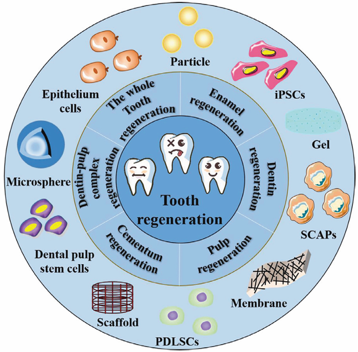
This illustration provides a visual representation of the cellular components and materials utilized in the process of regenerating teeth [28].
Numerous materials have been proposed and evaluated within the scientific literature, with instances where a blood clot has served as a physical scaffold. In the field of endodontics, various scaffolds have been introduced, including natural polymers such as fibrin, collagen, chitosan, glycosaminoglycan, and dentine scaffolds, as well as synthetic polymers such as polyglycolic acid (PGA), poly-l-lactic acid (PLLA), and polylactide-co-glycolic acid (PLGA). Additionally, inorganic and composite materials have been synthesized into porous scaffolds, nanofibrous materials, microparticles, or hydrogels.
The criteria for an ideal scaffold in tissue engineering for endodontics encompass several elements. The ideal scaffold should be biodegradable, exhibit viscoelastic properties, and have a synthesis rate akin to that of the extracellular matrix. If the degradation rate is excessively rapid, material dissolution may precede new tissue formation. Conversely, if degradation is too slow, it may disrupt the synthesis of the extracellular matrix. Crucially, the scaffold must facilitate proper interactions between cells and itself. The scaffold's design elements, including porosity, surface topography, charge, stiffness, and viscosity, can alter cellular behavior and the expression profile of cells, influencing adhesion, migration, proliferation, and extracellular scaffold synthesis.
Integration of Bioactive Molecules for Enhanced Regeneration
The integration of bioactive molecules in dentistry focuses on incorporating substances with regenerative properties into dental materials. This approach aims to enhance tissue regeneration, especially in areas such as bone and tooth structures. Bioactive molecules, such as growth factors or peptides, can stimulate cell activity and promote tissue healing, contributing to improved outcomes in dental procedures such as implant placements or periodontal treatments. This emerging field holds promise for advancing regenerative dentistry practices. In the field of regenerative dentistry, the integration of bioactive molecules involves the use of substances that actively participate in and promote natural healing and regeneration processes within oral tissues. These bioactive molecules can include growth factors, peptides, and other signaling molecules that play crucial roles in cellular communication and tissue development. For instance, in the context of dental implants, bioactive materials may be used to coat the implant surface. This coating can release growth factors or other bioactive agents over time, creating a microenvironment that stimulates the surrounding cells to differentiate into bone-forming cells, thereby enhancing osseointegration—the integration of the implant with the surrounding bone. For example, conventional methods for addressing periodontitis involve surgical and anti-inflammatory interventions, either independently or in combination. Despite their efficacy in controlling periodontitis, these approaches are limited in achieving sufficient periodontal regeneration, characterized by the reattachment of the periodontal ligament and regeneration of the alveolar bone. The utilization of bioactive molecules serves as a potential link, facilitating the connection between exogenous and endogenous mesenchymal stem cells (MSCs) within the compromised microenvironment. This interaction prompts MSCs to undergo differentiation into specific tissues essential for regeneration. Consequently, diverse bioactive molecules have been extensively investigated to optimize the outcomes of periodontal regeneration. Another example is growth factors. Growth factors (GFs) are intrinsic biological agents recognized for their ability to modulate essential cellular processes through binding to receptors on cell membranes, initiating intricate intracellular signaling pathways. Acknowledging the dissatisfaction stemming from suboptimal regenerative outcomes in conventional periodontitis treatments, GFs have emerged as a promising solution. They aim to enhance periodontal regeneration by mitigating the inflammatory microenvironment and augmenting the proliferation and differentiation capacity of transplanted stem cells. Extensive exploration through preclinical animal experiments and clinical trials has led to the FDA approval of enamel matrix derivative (EMD) and recombinant human platelet-derived growth factor-BB (rhPDGF-BB) as pharmaceuticals for treating periodontal defects. Additionally, platelet concentrates (PCs), including platelet-rich plasma (PRP) and platelet-rich fibrin (PRF), which function as natural polymeric scaffolds rich in growth factors, are widely employed in the field of periodontal regeneration. EMD, derived from purified enamel matrix proteins, has demonstrated positive preclinical and clinical results by facilitating the adhesion, proliferation, and differentiation of osteoblasts and periodontal ligament stem cells (PDLSCs). However, its broad clinical application is hindered by the fluidity of its carrier gel, necessitating a combination with various grafting materials for satisfactory outcomes. Recent innovations, such as mixing EMD with bone grafts or collagen matrices, have improved its mechanical strength and significantly enhanced periodontal regeneration. Another extensively studied FDA-approved growth factor for periodontal regeneration is rhPDGF-BB. Despite its short half-life, its combination with synthetic ceramic matrices or bone allografts has shown efficacy as a periodontal regenerative agent. The combined delivery of bioactive molecules, such as PDGF and simvastatin, has been proposed to accelerate periodontal regeneration, promoting the realignment of fibers between bone and cementum. Although platelet-derived growth factors in PCs, particularly PRF-containing leukocytes, exhibit a certain degree of tensile strength and enhance periodontal repair, their potential is limited. PRF-induced regeneration relies solely on the recruitment of endogenous cells, resulting in a majority of regenerated tissues displaying a disordered structure. Despite these challenges, ongoing research has explored innovative strategies to optimize the regenerative potential of these bioactive molecules in the context of periodontal tissue restoration [6].
Bioengineering Approaches for Tooth Replacement
Whole-Tooth Biofabrication: Progress and Challenges
Biofabricating complete teeth is a burgeoning research area with the goal of crafting implantable human teeth through stem cell-driven tissue engineering. Although embryonic and adult stem cells have been utilized previously, recent strides in employing induced pluripotent stem cells have demonstrated significant potential in rejuvenating structures resembling teeth. Nevertheless, a critical hurdle involves pinpointing suitable postnatal stem cell sources with odontogenic competence for the epithelial component, given the absence of enamel epithelial cells in adult teeth. Moreover, the extended development and differentiation of human teeth necessitate hastening the progress of the tooth germ through manipulating gene expression via locally applied inhibitory molecules and growth factors. Despite these challenges, the rapid advancement in molecular studies of tooth biology, facilitated by innovative high-throughput technologies, undoubtedly paves the way for the realization of implantable bioengineered teeth [29].
Therefore, whole tooth biofabrication (Figure 2) presents an intriguing realm, offering an alternative to conventional dental prosthetics. Within the field of bioengineering complete teeth, key hurdles involve pinpointing dependable sources of dental epithelial cells suitable for clinical use, refining techniques for constructing scaffolds that foster the systematic development of diverse dental tissues, and guaranteeing ample vascularization and seamless integration within the recipient's anatomy [30,31].
The development of teeth relies on intricate interactions between the ectoderm-derived dental epithelium and cranial neural crest-derived mesenchyme [33]. In mice, the dental epithelium exhibits odontogenic potential before embryonic day 12 (E12), initiating tooth formation when combined with mesenchymal tissue from nondental origins. Recent breakthroughs have allowed for the bioengineering of entire tooth crowns from embryonic tooth germ cells in various animal models, including mice, rats, pigs, and dogs. Notably, reaggregated mouse embryonic tooth germ cells have been demonstrated to transform into fully functional tooth replacements in adult mice, which is accompanied by revascularization and root formation at the lost tooth site. This highlights the potential of implanting bioengineered tooth germs in adults for functional tooth replacement. However, translating these findings to human teeth presents a challenge, requiring further research to identify appropriate sources of postnatal stem cells with odontogenic competence in the epithelial component (Figure 3) [29].
Moreover, promising approaches for engineering complete teeth, along with their supporting tissues, have been investigated, including the utilization of dental stem/stromal cells and the organ germ method [32,34].
While notable advancements have been made, obstacles persist in locating suitable postnatal stem cell sources and in creating biomimetic components of the tooth, including enamel, the dentin-pulp complex, and the periodontium [28,29].
Moreover, the progress in bioprinting techniques and bioinks holds great promise for regenerating dental alveolar tissues, including the complete regeneration of teeth [35]. Thus, biofabrication and regenerative dentistry offer hope for the future of dental care, holding the potential to enhance dental and oral health for individuals experiencing tooth loss [30].
Biofunctionalization and Vascularization of Tooth Constructs
Enhancing the biofunctionality and vascularization of tooth constructs is pivotal in the field of tissue engineering, with the goal of regenerating dental pulp and facilitating effective root canal therapy [36].
Biofunctionalization entails enhancing the tooth's surface to improve biocompatibility, encouraging cell adhesion and proliferation. This is achieved by immobilizing bioactive molecules such as ECM proteins, growth factors, and peptides onto the surface of a construct. Notably, ECM proteins such as fibronectin and laminin, which feature specific amino acid motifs such as the arginine-glycine-aspartic acid (RGD) sequence, play crucial roles in cell adhesion, migration, and differentiation. These proteins bind to cell surface receptors, fostering cellular attachment and signaling. Incorporating ECM proteins into tooth constructs boosts biocompatibility, fostering the adhesion and proliferation of neighboring cells, such as gingival fibroblasts, and ultimately establishing a functional interface between the tooth construct and surrounding tissues [37]. In addition, vascular endothelial growth factor (VEGF) has emerged as a powerful proangiogenic factor that is essential for fostering blood vessel formation within dental pulp tissue [38]. Furthermore, dental pulp stem cells (DPSCs) have been demonstrated to release proangiogenic factors such as angiopoietin-1 and platelet-derived growth factor, amplifying the angiogenic capabilities of engineered tooth constructs [39]. Moreover, the incorporation of biofunctionalization approaches, such as the use of GelMA hydrogels with adjustable physical and mechanical traits, has been shown to improve cell viability, spreading, and proliferation within tooth constructs [36]. Researchers have demonstrated the customization of gelatin methacrylate (GelMA) hydrogel scaffolds to emulate the specific characteristics of immature natural dental tissues. These scaffolds also facilitate the differentiation of enclosed dental cells, contributing to the development of mineralized dental tissue [40]. Furthermore, crafting prevascularized, full-length dental pulp-like tissue constructs within root canals has shown promise for prompt vascularization, tackling the issue of oxygen supply in mature teeth. This inventive method not only presents a streamlined biofabrication approach but also carries substantial clinical and translational promise, laying the groundwork for engineering vascularized dental pulp with potential positive impacts [36].
Conversely, vascularization revolves around establishing a functional network of blood vessels within the tooth construct—an imperative factor for the sustained vitality and effectiveness of engineered tissues. This process is vital for supplying nutrients and oxygen to the cells within the construct, along with expelling waste products. Diverse methods have been investigated for tooth construct vascularization, including the inclusion of endothelial cells, growth factors such as vascular endothelial growth factor (VEGF), and bioactive scaffolds emulating the extracellular matrix to foster blood vessel formation. Additionally, cutting-edge techniques such as 3D bioprinting have been applied to create intricate vascular networks within tooth constructs, enabling the seamless distribution of nutrients and oxygen throughout engineered tissues [37].
Understanding the dynamic relationship between biofunctionalization and vascularization is crucial in crafting biomimetic tooth constructs capable of seamlessly blending into the host environment, ensuring prolonged tissue survival and functionality [39].
The prospects of improving dental pulp tissue regeneration, refining tooth transplantation procedures, and bolstering the overall triumph of tissue engineering in dentistry lie in the effective biofunctionalization and vascularization of tooth constructs. This is pivotal because it signifies a crucial advancement in the journey toward creating practical and clinically feasible tooth replacements. By sustaining cell viability and tissue functionality over time, this approach emulates the natural tooth structure and function, marking a key stride in this developmental process [37].
Neurovascular Integration in Tooth Regeneration
Nerve and Blood Vessel Network Formation in Engineered Teeth
Dental pulp, a specialized mesenchymal tissue, faces limited regeneration due to anatomical constraints and the postmitotic nature of odontoblastic cells. Traditional methods, such as complete pulp amputation, often result in significant dentin loss, leaving a nonvital and weakened tooth. However, the emerging field of regenerative endodontics, a facet of modern tissue engineering, shows promise by utilizing stem cells, scaffolds, and responsive molecules to achieve positive outcomes. In parallel, the intricate challenge of coordinating nerve and blood vessel networks within engineered teeth stands as a crucial research focus in dental tissue engineering, requiring the replication of natural developmental pathways for the creation of structurally and functionally sound synthetic teeth.
Morphogens, responsive cells, and scaffolds play crucial roles in tissue engineering, yet the effectiveness of regenerative strategies hinges on the swift establishment of functional blood vessel networks. Despite its small size, regenerating pulp tissue from scratch is challenging due to anatomical constraints that impede rapid and efficient vasculogenesis. Consequently, developing vasculogenesis strategies has become a pivotal challenge in dental pulp tissue engineering. The prototypic proangiogenic factor vascular endothelial growth factor (VEGF) has been demonstrated to enhance neovascularization in severed human dental pulp. Moreover, dental pulp stem cells can differentiate into endothelial cells, contributing to the formation of functional blood vessels. The use of scaffolds as delivery systems for VEGF has emerged as a potential avenue for effectively stimulating angiogenesis [41,42].
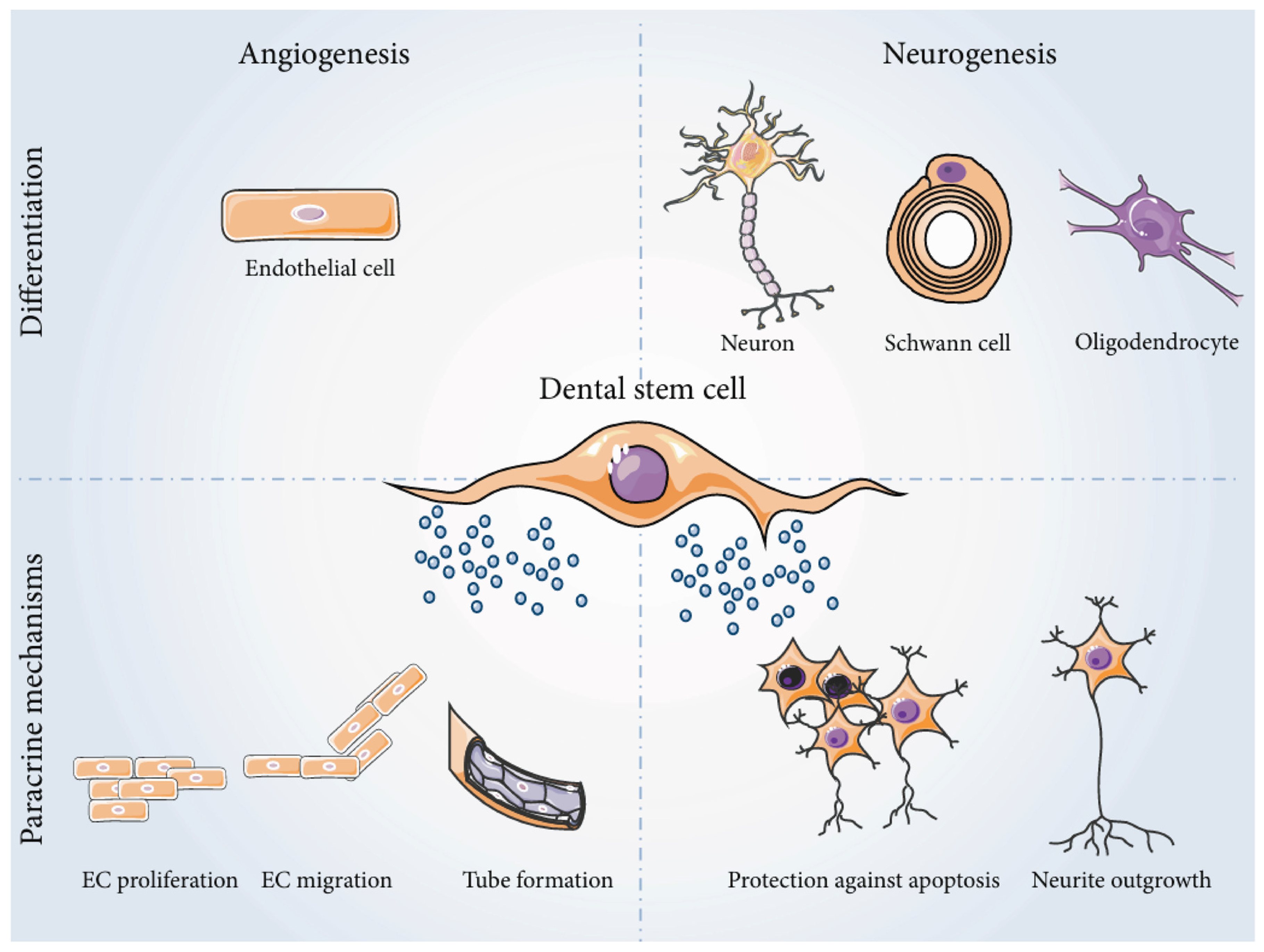
Successful tissue engineering hinges on the creation of an efficient vascular network, which is essential for supplying oxygen, nutrients, and immune cells to the tissue while eliminating byproducts and waste. Providing nutrients and oxygen is vital for sustaining the high metabolic activity of regenerating cells. Without a swift vascular network, implanted tissues are restricted to 2 to 3 mm3, or cells cannot survive the early posttransplantation stage. In the context of dental pulp tissue engineering, the anatomical features of the root canal pose a challenge, necessitating strategies to enhance neovascularization. These approaches include incorporating angiogenic factors into scaffolds, utilizing fabrication technologies for polymers with vessel-like networks, and prevascularizing matrices before cell seeding. Alternatively, leveraging the vasculogenic potential of endothelial cells (ECs) presents another avenue. Notably, vascular endothelial growth factor (VEGF) has emerged as a key proangiogenic factor that induces stem cell differentiation into endothelial cells. VEGF can prompt dental pulp stromal stem cells (DP-SCs) and SHEDs to organize into capillary-like structures, demonstrating its potential for vascular network formation [43,44]. Despite strides in understanding the angiogenic potential of dental pulp stem cells, further optimization is needed for the rapid establishment of functional vascular networks in engineered dental pulp (Figure 4) [45].
Ensuring the proper function and protection of engineered teeth requires careful consideration of their innervation. Sensory innervation, particularly the presence of nerve fibers in the odontoblast layer, is crucial for tooth function. However, the spontaneous formation of a functional nerve network in engineered teeth necessitates specific conditions. Studies indicate that immunosuppressive therapy, such as cyclosporin A, can expedite the innervation process in transplanted tissues, while immunosuppression itself stimulates dental mesenchyme innervation. Although the exact genes and proteins involved in nerve network formation are not fully understood, previous research has explored the cellular partners and networks involved in dental pulp sensory function, along with the role of glial cells and their relationship with microvascularization, emphasizing the complexity of this process. Further investigation is needed to fully elucidate the genes and proteins influencing the innervation of engineered teeth and their interaction with the dental mesenchyme and odontoblasts, which contribute to the development of functional and innervated tooth organs for regenerative purposes [46]. Such insights contribute to the development of functional and innervated tooth organs for regenerative purposes. In practical terms, the induction of neuronal differentiation in dental stem cell (DSC) monolayers often involves the use of proteins such as epidermal growth factor (EGF) and bFGF, as well as culture supplements such as B27, forskolin, and insulin-transferrin-sodium selenite (ITS). While all DSC populations can differentiate into neuron-like cells, studies often lack detailed analyses of differentiated cells, resulting in a low yield of primitive, immature neurons with limited action potential generation capacity [47].
Figure 4.
shows the angiogenic and neurogenic potentials of dental stem cells (DSCs). These cells can differentiate into various cell types, including endothelial cells, neurons, Schwann cells, and oligodendrocytes, in response to specific cues. The secretome of DSCs contains proteins that are beneficial to surrounding cells, including angiogenic factors that promote blood vessel development and neurotrophic factors that support neuron survival and growth [47].
Immune Responses and Host-Material Interactions
Immunomodulatory Strategies in Tissue Engineering for Tooth Regeneration
Immunomodulatory strategies in tissue engineering for tooth regeneration involve manipulating the immune response to enhance the success of regenerative approaches. By modulating immune reactions, researchers aim to create a favorable environment for the integration of engineered dental tissues. This includes addressing inflammation, promoting tissue healing, and mitigating immune rejection. These strategies play a crucial role in advancing the field of tooth regeneration by optimizing the interaction between engineered tissues and the host immune system, ultimately improving the outcomes of regenerative dental therapies [48,49,50].
Scientifically, this often involves the use of biomaterials with immunomodulatory properties. These materials can influence immune cell behavior and cytokine production, steering the immune response toward a more regenerative profile. For instance, hydrogels or scaffolds may be designed to release anti-inflammatory agents, promoting a balanced immune environment favorable for tissue repair. Furthermore, immunomodulation is crucial for mitigating immune rejection of implanted tissues. Strategies include surface modifications of biomaterials to reduce their immunogenicity, allowing for better integration with host tissues. This involves understanding and manipulating immune cell interactions with engineered constructs at the molecular level. Immunomodulation plays a role in promoting tissue healing and minimizing fibrotic responses. The modulation of macrophage polarization, where macrophages can adopt pro-regenerative phenotypes, is a key avenue of research. This can influence the secretion of growth factors and cytokines that support tissue regeneration while reducing scarring [51,52,53].
In recent years, there has been a notable focus on biomaterials that interact with blood and adjacent tissues to facilitate the formation of native bone. Following surgical implantation, these biomaterials contact various physiological fluids, including blood. Numerous proteins present in the blood, such as albumin, fibrinogen, fibronectin, vitronectin, and gamma-globulins, can adhere to the surface of biomaterials. The characteristics of the biomaterial surface, such as size, surface charge, fluid composition, and physicochemical properties, contribute to the formation of a complex interface termed the "protein corona." The Vroman effect, where proteins with higher affinity gradually replace those that are initially bound, plays a role in shaping this corona. The protein corona plays a critical role in recruiting and adhering proinflammatory cells, influencing blood clot formation, which defines the provisional matrix around the biomaterial. This matrix ultimately determines the type of tissue formed on the surface of the biomaterial. Variations in the macro, micro, and physicochemical compositions of materials used in guided bone regeneration and dental implantology contribute to differences in protein content, potentially explaining the variable success or failure rates of the use of PRF and L-PRF as coadjuvants for bone regeneration reported in the literature [54,55,56].
Incorporating hemoderivatives such as platelet-rich plasma (PRP), platration involves using blood components separated by centrifugation or time. This separation alters the amount and type of plasma proteins, creating a specific protein corona around bone graft materials. The development of a blood protein called corona on the surface of biomaterials will thus give this surface an immune-modulatory role.
Biomaterial Compatibility and Long-Term Integration with Host Tissues
Biomaterial compatibility and long-term integration with host tissues are critical considerations in the context of molecular insights into tooth development. When designing biomaterials for dental applications, it is essential to ensure that they not only support short-term functionality but also exhibit compatibility and integration over an extended period of time. Biomaterials are designed to emulate the complex signaling pathways and molecular events that orchestrate natural tooth development. This encompasses the precise spatial and temporal regulation of gene expression, interactions between different cell types, and the orchestrated release of growth factors and signaling molecules. Researchers aim to replicate these molecular cues in biomaterials to guide the formation of functional and durable dental tissues. Biomaterial compatibility is addressed by selecting materials with proven biocompatibility to minimize adverse immune responses. Additionally, the long-term success of biomaterial integration relies on its ability to provide a conducive microenvironment for cellular activities. This includes promoting the adhesion, proliferation, and differentiation of relevant cell types, such as odontoblasts and mesenchymal stem cells, which mirror natural cellular processes during tooth development. Several biomaterials have demonstrated promise for long-term tooth regeneration. Hydroxyapatite-based scaffolds, for instance, are a naturally occurring mineral form of calcium apatite with a composition closely resembling the inorganic component of natural tooth enamel and dentin. This similarity makes hydroxyapatite an excellent choice for scaffolds aimed at mimicking the mineral composition of teeth. These scaffolds not only provide structural support but also facilitate the deposition of mineralized tissues over time, contributing to their long-term integration with host tissues [57,58].
Moreover, the incorporation of bioactive molecules, such as growth factors and signaling proteins, into biomaterial matrices enhances their regenerative potential. Bone morphogenetic protein-2 (BMP-2) and fibroblast growth factor-2 (FGF-2) are known to play crucial roles in dental tissue development and can be integrated into biomaterials to stimulate cell differentiation and tissue formation. BMP-2 is a member of the transforming growth factor-beta (TGF-β) superfamily and is naturally found in the human body. It is known for its potent osteogenic (bone-forming) properties. BMP-2 is a powerful inducer of osteogenic differentiation in mesenchymal stem cells (MSCs) and other precursor cells. It promotes the commitment of these cells toward the osteoblastic lineage, which is crucial for the formation of bone and dental tissues. In biomaterial-based strategies, BMP-2 can be incorporated into scaffolds or carriers to enable controlled and sustained release. This controlled release ensures that the growth factor is available at the right concentration and duration, optimizing its effects on tissue regeneration and ensuring a long-term effect [6,59].
Conclusions
Researchers have made significant progress in understanding tooth development through studies on the genomic and epigenomic regulation of tooth morphogenesis, such as identifying key genes responsible for tooth formation. For instance, they have uncovered key signaling pathways through genetic engineering systems, leading to a deeper comprehension of the intricate processes involved. The incorporation of bioactive molecules such as growth factors and innovative scaffold designs like 3D-printed scaffolds has played a crucial role in promoting enhanced tissue regeneration in dental procedures. These advancements have opened promising avenues for the future of regenerative dentistry, such as the development of personalized tooth regeneration treatments. The ongoing exploration of stem cell-driven tissue engineering and biofabrication of complete teeth not only holds great promise for creating functional and clinically feasible tooth replacements, such as bioengineered teeth tailored to individual patients, but also signifies a significant leap towards personalized dental solutions. This comprehensive review illuminates the intricate processes of dental tissue differentiation and highlights the transformative potential of regenerative dentistry in shaping the future of oral healthcare, emphasizing the critical role it plays in advancing dental care.
List of Abbreviations
| iPSCs | Induced pluripotent stem cells |
| DNA | Deoxyribonucleic acid |
| RNAs | Ribonucleic acid |
| BMP | Bone Morphogenetic Protein |
| FGF | Fibroblast Growth Factor |
| SHH | Sonic Hedgehog |
| FGF8 | Fibroblast Growth Factor 8 |
| WNT | Wingless-Type MMTV Integration Site Family |
| BMPR1a | Bone Morphogenetic Protein Receptor Type 1a |
| FGFR2b | Fibroblast Growth Factor Receptor 2b |
| NCCs | Neural Crest Cells |
| MSCs | mesenchymal stem or stromal cells |
| ASF-CM | ameloblasts serum-free conditioned medium |
| BMP4 | Bone Morphogenetic Protein 4 |
| AMBN | Ameloblastin |
| AMGN | Amelogenin |
| CK14 | Cytokeratin 14 |
| sci-RNA-seq | single-cell combinatorial indexing RNA sequencing |
| isAM | Induced secretory ameloblasts |
| 3D | three-dimensional |
| 2D | two-dimensional |
| PSCs | pluripotent stem cells |
| ERM | epithelial cell rests of Malassez |
| HA/TCP | hydroxyapatite/tricalcium phosphate |
| SHED | human exfoliated deciduous teeth |
| DPSCs | dental pulp stem cells |
| SCD-1 | Stearoyl-CoA Desaturase-1 |
| STRO-1 | Stromal precursor antigen-1 |
| CD44 | Cluster of Differentiation 44 |
| CD146 | Cluster of Differentiation 146 |
| pH | power of Hydrogen |
| PGA | polyglycolic acid |
| PLLA | poly-l-lactic acid |
| PLGA | polylactide-co-glycolic acid |
| GFs | Growth factors |
| FDA | Food and Drug administration |
| EMD | enamel matrix derivative |
| rhPDGF-BB | binant human platelet-derived growth factor-BB |
| PCs | platelet concentrates |
| PRP | platelet-rich plasma |
| PRF | platelet-rich fibrin |
| PDLSCs | periodontal ligament stem cells |
| PDGF | Platelet Derived Growth Factor |
| GFSCS | Gingival Fibroblast-Derived Stem Cells |
| SCAPSCS | Stem Cells from Apical papilla |
| BMSCs | Bone Marrow Stromal Cells |
| E12 | embryonic day 12 |
| Epi | Epithelial |
| Mes | mesenchyme |
| RGD | arginine-glycine-aspartic acid |
| VEGF | vascular endothelial growth factor |
| GelMA | gelatin methacrylate |
| mm | millimeter |
| DP-SC | dental pulp stromal stem cells |
| DSC | dental stem cell |
| EGF | epidermal growth factor |
| bFGF | Basic Fibroblast Growth Factor |
| ITS | insulin-transferrin-sodium selenite |
| L-PRF | leukocyte-platelet-rich fibrin |
| BMP-2 | Bone Morphogenetic Protein-2 |
| FGF-2 | Fibroblast Growth Factor-2 |
| TGF-β | transforming growth factor-beta |
Declarations
Funding
Self-funded research.
Ethics approval
Not applicable.
Consent to participate
Not applicable.
Consent for publication
Not applicable.
Availability of data and materials
Not applicable.
Competing interests
The authors declare that they have no competing interests.
Authors' contributions
All authors have made the conception and design of the review paper, contributed significantly to the drafting and refinement of the manuscript, and provided substantial intellectual input throughout the analysis and critical review process. Additionally, All authors have actively participated in the acquisition of relevant literature and materials, ensuring the accuracy and completeness of the content. Furthermore, all authors have been involved in the critical review and revision of the manuscript, providing valuable insights and feedback. Finally, all authors have given their final approval of the version to be published and take full responsibility for the integrity and accuracy of the work.
Acknowledgments
We would like to express our sincere appreciation to our supervisor, Ali Alsuraifi, for his invaluable contributions to this research review paper. His significant input in refining the content and providing final approval of the manuscript played a crucial role in its completion. we also would like to extend our heartfelt appreciation to Abdullah Ayad for his outstanding contributions to this research review paper. His leadership in guiding the entire review process and his exceptional contributions were instrumental in shaping the quality and completeness of this work.
Declaration Regarding the Use of AI-Assisted Readability Enhancement
I hereby affirm that the utilization of AI-assisted tools in the refinement of the manuscript was strictly limited to enhancing its readability. At no point were AI technologies employed to supplant essential authorial responsibilities, including the generation of scientific, pedagogic, or medical insights, the formulation of scientific conclusions, or the issuance of clinical recommendations. The implementation of AI for readability enhancement was rigorously supervised under the discerning eye of human oversight and control.
References
- Bei, M. Molecular genetics of tooth development. Curr Opin Genet Dev. 2009; 19:504–10. [CrossRef]
- Chen, Y., Wang, X., Wu, Z., Jia, S., & Wan, M. Epigenetic regulation of dental-derived stem cells and their application in pulp and periodontal regeneration. PeerJ. 2023;11:14550. [CrossRef]
- Jing, J., Feng, J., Yuan, Y., Guo, T., Lei, J., Pei, F., Ho, T., & Chai, Y. Spatiotemporal single-cell regulatory atlas reveals neural crest lineage diversification and cellular function during tooth morphogenesis. Nat Commun. 2022; 13:4803. [CrossRef]
- Radwan, I. A., Rady, D., Abbass, M. M. S., Moshy, S. E., AbuBakr, N., Dörfer, C. E., & El-Sayed, K. M. F. Induced Pluripotent stem cells in dental and nondental tissue regeneration: a review of an unexploited potential. Stem Cells Int.2020;2020: 1941629. [CrossRef]
- Alghadeer, A., Hanson-Drury, S., Patni, A. P., Ehnes, D. D., Zhao, Y. T., Li, Z., Phal, A., Vincent, T. L., Lim, Y. B., O’Day, D. R., Spurrell, C. H., Gogate, A. A., Zhang, H., Devi, A., Wang, Y., Starita, L. M., Doherty, D., Glass, I. A., Shendure, J., . . . Ruohola-Baker, H. Single-cell census of human tooth development enables generation of human enamel. Dev Cell. 2023; 58:2163-2180,9. [CrossRef]
- Hosseinpour, S., Walsh, L. J., & Moharamzadeh, K. Regenerative Approaches in Dentistry. Springer Nature. 2021. [CrossRef]
- Tucker, A. S., & Sharpe, P. T. Molecular genetics of tooth morphogenesis and patterning: the right shape in the right place. J Dent Res. 1999; 78:826-34. [CrossRef]
- Li, Y., Zhao, X., Sun, M., Pei, D., & Li, A. (2022). Deciphering the epigenetic code of stem cells derived from dental tissues. Frontiers in Dental Medicine, 2. [CrossRef]
- Zeng, B., Liu, G., & Huang, J. (2022). DNA methylation and histone modification in dental-derived mesenchymal stem cells. Stem Cell Reviews and Reports, 18(8), 2797–2816. [CrossRef]
- Hussain, A., Tebyanian, H., & Khayatan, D. (2022). The role of epigenetic in dental and oral regenerative medicine by different types of dental stem cells: A comprehensive overview. Stem Cells International, 2022, 1–15. [CrossRef]
- Thesleff, I. The genetic basis of tooth development and dental defects. Am J Med Genet A. 2006; 140:2530-5. [CrossRef]
- Hu, X., Zhang, S., Chen, G., Lin, C., Huang, Z., Chen, Y., & Zhang, Y. (2013). Expression of SHH signaling molecules in the developing human primary dentition. BMC Developmental Biology, 13(1). [CrossRef]
- Yu, T., & Klein, O. D. Molecular and cellular mechanisms of tooth development, homeostasis and repair. Development. 2020; 147:184754. [CrossRef]
- Baena, A. R. Y., Casasco, A., & Monti, M. (2022). Hypes and Hopes of Stem Cell Therapies in Dentistry: a Review. Stem Cell Reviews and Reports, 18(4), 1294–1308. [CrossRef]
- Hynes, K., Menichanin, D., Bright, R., Ivanovski, S., Hutmacher, D. W., Gronthos, S., & Bartold, P. M. Induced pluripotent stem cells: a new frontier for stem cells in dentistry. J Dent Res. 2015; 94: 1508-15. [CrossRef]
- Hamano, S., Sugiura, R., Yamashita, D., Tomokiyo, A., Hasegawa, D., & Maeda, H. (2022). Current application of IPS cells in the dental tissue regeneration. Biomedicines, 10(12), 3269. [CrossRef]
- Gao, P., Liu, S., Wang, X., & Ikeya, M. Dental applications of induced pluripotent stem cells and their derivatives. Jpn Dent Sci Rev. 2022; 58:162-71. [CrossRef]
- Cho, Y., Kim, K., Lee, Y., & Seol, Y. (2022). Dental-derived cells for regenerative medicine: stem cells, cell reprogramming, and transdifferentiation. Journal of Periodontal & Implant Science, 52(6), 437. [CrossRef]
- Wang, J., Zhao, Z., Yang, K., & Bai, Y. (2024). Research progress in cell therapy for oral diseases: focus on cell sources and strategies to optimize cell function. Frontiers in Bioengineering and Biotechnology, 12, 1340728. [CrossRef]
- Hermans, F., Hasevoets, S., Vankelecom, H., Bronckaers, A., & Lambrichts, I. (2024). From pluripotent stem cells to organoids and bioprinting: recent advances in dental epithelium and ameloblast models to study tooth biology and regeneration. Stem Cell Reviews and Reports. [CrossRef]
- Liu, L., Liu, Y. F., Zhang, J., Duan, Y. Z., & Jin, Y. Ameloblasts serum-free conditioned medium: bone morphogenic protein 4-induced odontogenic differentiation of mouse induced pluripotent stem cells. J Tissue Eng Regen Med. 2016 Jun;10(6):466-74. [CrossRef]
- Gao, X., Wu, Y., Liao, L., & Tian, W. Oral organoids: progress and challenges. J Dent Res. 2021; 100: 454-63. [CrossRef]
- Mendes-Pinheiro, B., Campos, J., Marote, A., Soares-Cunha, C., Nickels, S. L., Monzel, A. S., ... & Salgado, A. J. (2023). Treating Parkinson’s Disease with Human Bone Marrow Mesenchymal Stem Cell Secretome: A Translational Investigation Using Human Brain Organoids and Different Routes of In Vivo Administration. Cells, 12(21), 2565. [CrossRef]
- Hermans, F., Hemeryck, L., Bueds, C., Pereiro, M. T., Hasevoets, S., Kobayashi, H., Lambrechts, D., Lambrichts, I., Bronckaers, A., & Vankelecom, H. Organoids from mouse molar and incisor as new tools to study tooth-specific biology and development. Stem Cell Reports. 2023; 18:1166–81. [CrossRef]
- Hemeryck, L., Hermans, F., Chappell, J., Kobayashi, H., Lambrechts, D., Lambrichts, I., Bronckaers, A., & Vankelecom, H. Organoids from human tooth showing epithelial stemness phenotype and differentiation potential. Cell Mol Life Sci. 2022; 79:153. [CrossRef]
- Ma, L., Rao, N., Jiang, H., Dai, Y., Wang, C., Yang, H., & Hu, J. (2022). Small extracellular vesicles from dental follicle stem cells provide biochemical cues for periodontal tissue regeneration. Stem Cell Research & Therapy, 13(1). [CrossRef]
- Bi, R., Lyu, P., Song, Y., Li, P., Song, D., Cui, C., & Fan, Y. (2021). Function of dental follicle Progenitor/Stem cells and their potential in regenerative Medicine: From mechanisms to applications. Biomolecules, 11(7), 997. [CrossRef]
- Fu, Z., Zhuang, Y., Cui, J., Sheng, R., Tomás, H., Rodrigues, J.,... & Lin, K. Development and challenges of cells-and materials-based tooth regeneration. Eng Regen.2022; 3:163-81. [CrossRef]
- Zhang, Y., & Chen, Y. Bioengineering of a human whole tooth: progress and challenge. Cell Regen.2014; 3:8. [CrossRef]
- Smith, E. E., & Yelick, P. C. Progress in bioengineered whole tooth research: from bench to dental patient chair. Curr Oral Health Rep. 2016; 3:302-8. [CrossRef]
- Horst, O. V., Chavez, M. G., Jheon, A. H., Desai, T. A., & Klein, O. D. (2012). Stem cell and biomaterials research in dental tissue engineering and regeneration. Dental Clinics of North America/the Dental Clinics of North America, 56(3), 495–520. [CrossRef]
- EzEldeen, M., Moroni, L., Nejad, Z. M., Jacobs, R., & Mota, C. Biofabrication of engineered dento-alveolar tissue. Biomater Adv. 2023; :148:213371. [CrossRef]
- Lymperi, S., Ligoudistianou, C., Taraslia, Kontakiotis, E. G., & Anastasiadou, E. (2013). Dental Stem Cells and their Applications in Dental Tissue Engineering. the Open Dentistry Journal, 7(1), 76–81. [CrossRef]
- Grawish, M. E., Grawish, L. M., Grawish, H. M., Grawish, M. M., & El-Negoly, S. A. Challenges of engineering biomimetic dental and paradental tissues. Tissue Eng Regen Med. 2020; 17:403-21. [CrossRef]
- Ostrovidov, S. Ramalingam, M., Bae, H., Orive, G., Fujie, T., Shi, X., & Kaji, H. Bioprinting and biomaterials for dental alveolar tissue regeneration. Front Bioeng Biotechnol. 2023; 14:11:991821. [CrossRef]
- Athirasala, A. Lins, F., Tahayeri, A., Hinds, M., Smith, A. J., Sedgley, C.,... & Bertassoni, L. E. A novel strategy to engineer prevascularized full-length dental pulp-like tissue constructs. Sci Rep.2017; 7:3323. [CrossRef]
- Palkowitz, A. L. Tuna, T., Bishti, S., Böke, F., Steinke, N., Müller-Newen, G., Wolfart, S., & Fischer, H. Biofunctionalization of dental abutment surfaces by crosslinked ECM proteins strongly enhances adhesion and proliferation of gingival fibroblasts. Advan Healthc Mater. 2021; 10: 2100132. [CrossRef]
- Jiang, Y., Wen, S., Li, Y., Jiang, M., Zhang, Y., Chen, X., & Zhai, Y. (2023). Research progress on nanomaterials for tissue engineering in oral diseases. Journal of Functional Biomaterials, 14(8), 404. [CrossRef]
- Rombouts, C. Giraud, T., Jeanneau, C., & About, I. Pulp Vascularization during Tooth Development, Regeneration, and Therapy. J Dent Res.2016; 96:137–44. [CrossRef]
- Smith, E. E. & Yelick, P. C. Bioengineering Tooth Bud constructs using GELMA hydrogel. In Methods mol biol.2019; 1922:139-50. [CrossRef]
- Wu, Y., Sun, J., Wang, W., Wang, Y., & Friedrich, R. E. (2024). How to make full use of dental pulp stem cells: an optimized cell culture method based on explant technology. Frontiers in Bioengineering and Biotechnology, 12. [CrossRef]
- Qiao, R., Tan, S., Guo, L., Ma, D., & Wen, J. (2023). Prevascularization techniques for dental pulp regeneration: potential cell sources, intercellular communication and construction strategies. Frontiers in Bioengineering and Biotechnology, 11. [CrossRef]
- Hashemibeni, B., & Moradi-Gharibvand, N. (2023). The effect of stem cells and vascular endothelial growth factor on cancer angiogenesis. Advanced Biomedical Research, 12(1), 124. [CrossRef]
- Zhang, Z. Oh, M. J., Sasaki, J., & Nör, J. E. (2021). Inverse and reciprocal regulation of p53/p21 and Bmi-1 modulates vasculogenic differentiation of dental pulp stem cells. Cell Death and Disease, 12(7). [CrossRef]
- Demarco, F. F., Conde, M. C. M., Cavalcanti, B. N., Casagrande, L., Sakai, V. T., & Nör, J. E. Dental pulp tissue engineering. Braz Dent J. 2011; 22:3-13. [CrossRef]
- Kökten, T., Becavin, T., Keller, L., Weickert, J. L., Kuchler-Bopp, S., & Lesot, H. Immunomodulation stimulates the innervation of engineered tooth organ. PLoS One. 2014; 9:86011. [CrossRef]
- Ratajczak, J. Bronckaers, A., Dillen, Y., Gervois, P., Vangansewinkel, T., Driesen, R. B.,... & Hilkens, P. The neurovascular properties of dental stem cells and their importance in dental tissue engineering. Stem Cells Int. 2016; 2016:9762871. [CrossRef]
- Yuan, Z., Nie, H., Wang, S., Lee, C. H., Li, A., Fu, S. Y., Zhou, H., Chen, L., & Mao, J. J. (2011). Biomaterial selection for tooth regeneration. Tissue Engineering Part B Reviews, 17(5), 373–388. [CrossRef]
- Ashtiani, R. E., Alam, M., Tavakolizadeh, S., & Abbasi, K. (2021). The role of biomaterials and biocompatible materials in Implant-Supported dental prosthesis. Evidence-based Complementary and Alternative Medicine, 2021, 1–9. [CrossRef]
- D, R. (2020). Dental stem cells- potential for tooth regeneration. Global Journal of Otolaryngology, 23(4). [CrossRef]
- Kuo, T., Sheu, S., Jiang, C., Chang, H., Chen, S., Chen, R., Hsieh, C., & Chen, M. (2015). TOOTH REGENERATION WITH DENTAL STEM CELL RESEARCH IN MINIATURE PIG MODEL. Taiwan Veterinary Journal, 41(03), 197–203. [CrossRef]
- Dal-Fabbro, R., Swanson, W. B., Capalbo, L. C., Sasaki, H., & Bottino, M. C. (2023). Next-generation biomaterials for dental pulp tissue immunomodulation. Dental Materials, 39(4), 333–349. [CrossRef]
- Nakao, K., & Tsuji, T. (2008). Dental regenerative therapy: Stem cell transplantation and bioengineered tooth replacement. Japanese Dental Science Review, 44(1), 70–75. [CrossRef]
- Mahajan, S. Singh, H., Suneja, E. S., Baweja, P. S., Sood, P., & Bajaj, K. Stem Cell Mediated Bioroot Regeneration: It’s Your Future whether you Know It or Not. [CrossRef]
- Yildirim, S., Fu, S. Y., Kim, K., Zhou, H., Lee, C. H., Li, A., Kim, S. G., Wang, S., & Mao, J. J. (2011). Tooth regeneration: a revolution in stomatology and evolution in regenerative medicine. International Journal of Oral Science, 3(3), 107–116. [CrossRef]
- Shivrayan, A. Jhajharia, K., & Sharma, P. (2014). Stem cells and their potential role in making a biotooth: A review. Indian Journal of Contemporary Dentistry, 2(1), 1-6. [CrossRef]
- Haugen, H. J., Basu, P., Sukul, M., Mano, J. F., & Reseland, J. E. (2020). Injectable biomaterials for dental tissue regeneration. International Journal of Molecular Sciences, 21(10), 3442. [CrossRef]
- Kim, J., Park, J., Kim, S., Im, G., Kim, B., Lee, J., Choi, E., Song, J., Cho, K., & Kim, C. (2013). Treatment of FGF-2 on stem cells from inflamed dental pulp tissue from human deciduous teeth. Oral Diseases, 20(2), 191–204. [CrossRef]
- Marques, N. L. a. R. V., Da Costa Júnior, N. E. A., Lotif, N. M. a. L., Neto, N. E. M. R., Da Silva, N. F. F. C., & De Queiroz Martiniano, N. C. R. (2016). Application of BMP-2 for bone graft in Dentistry. RSBO, 12(1), 88–93. [CrossRef]
Figure 2.
This schematic illustrates a tissue engineering approach for whole tooth regeneration. Various stem cell populations, including the GFSC, DPSCS, PDLSCs, SCAPSCS, and BMSCs, are denoted. The process aims to generate functional tooth structures, including the pulp-dentin complex, root, periodontal ligament, and alveolar bone. GFSCs: Gingival fibroblast-derived stem cells. DPSCs: dental pulp stem cells, PDLSCs: periodontal ligament stem cells, SCAPSCs: stem cells from apical papilla, BMSCs: bone marrow stromal cells [32].

Figure 3.
outlines a method for regenerating teeth using stem cells. Stem cells are induced into epithelial and mesenchymal tissues, combined into tooth germs, implanted into patients' missing tooth sites, and expected to grow into functional replacement teeth [29].
Figure 3.
outlines a method for regenerating teeth using stem cells. Stem cells are induced into epithelial and mesenchymal tissues, combined into tooth germs, implanted into patients' missing tooth sites, and expected to grow into functional replacement teeth [29].

Disclaimer/Publisher’s Note: The statements, opinions and data contained in all publications are solely those of the individual author(s) and contributor(s) and not of MDPI and/or the editor(s). MDPI and/or the editor(s) disclaim responsibility for any injury to people or property resulting from any ideas, methods, instructions or products referred to in the content. |
© 2025 by the authors. Licensee MDPI, Basel, Switzerland. This article is an open access article distributed under the terms and conditions of the Creative Commons Attribution (CC BY) license (http://creativecommons.org/licenses/by/4.0/).
Copyright: This open access article is published under a Creative Commons CC BY 4.0 license, which permit the free download, distribution, and reuse, provided that the author and preprint are cited in any reuse.


