Preprint
Article

This version is not peer-reviewed.

Efficacy of Colchicine in Reducing NT-proBNP, Caspase-1, TGF-β, Galectin-3 Expression and Improving Echocardiography Parameter in Acute Myocardial Infarction: A Multi-Centre, Randomized, Placebo-Controlled, Double-Blinded Clinical Trial

A peer-reviewed article of this preprint also exists.

Submitted:

05 January 2025

Posted:

09 January 2025

You are already at the latest version

Abstract

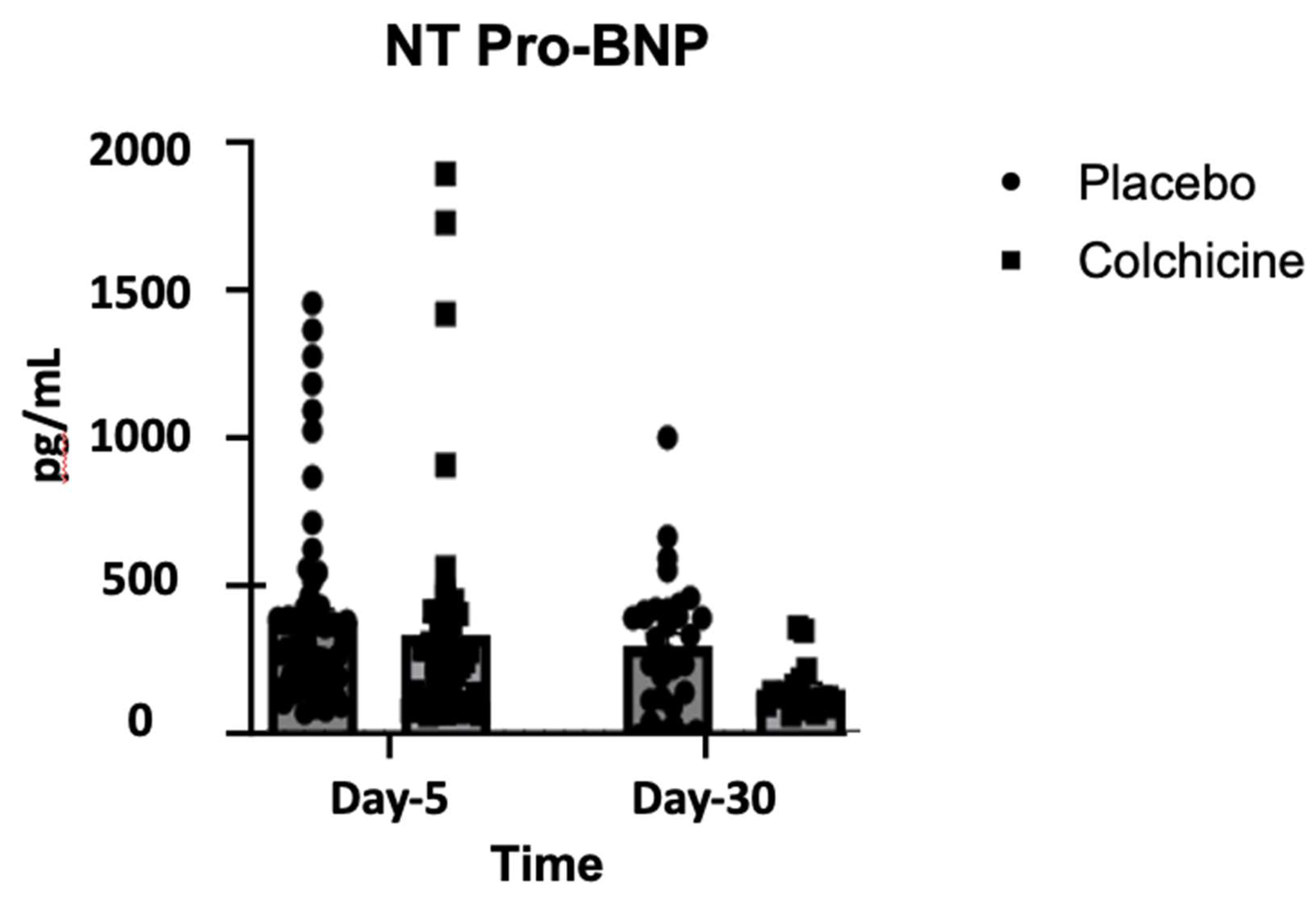
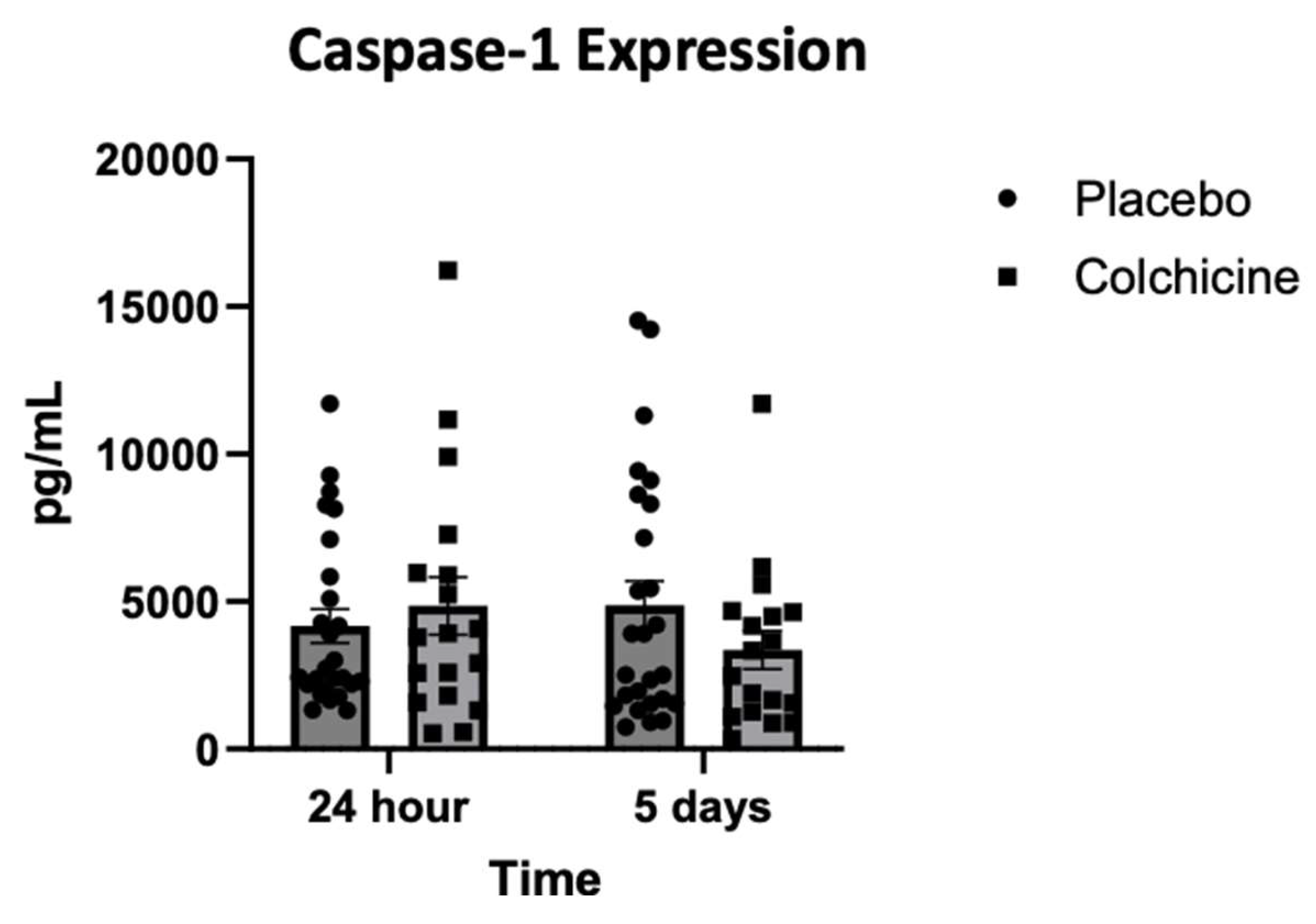
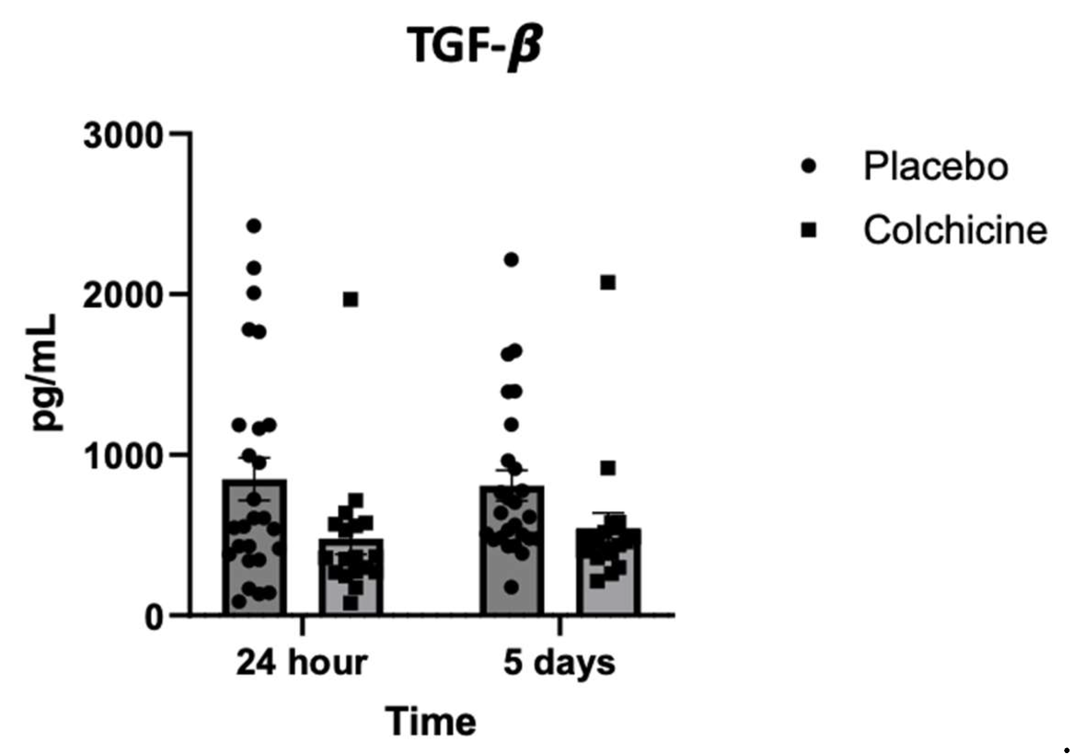
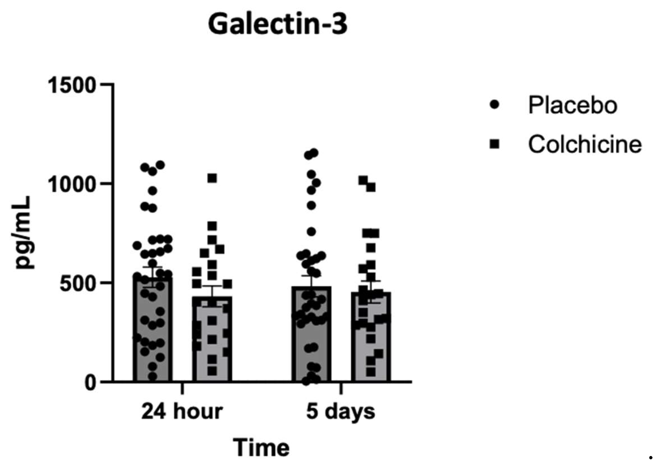
Background: Caspase-1 (reflects NOD-like receptor protein 3 inflammasomes activity), transforming growth factor-β (TGF-β), and Galectin-3 play significant role in post-AMI fibrosis and inflammation. Recently, colchicine was shown to dampen inflammation after AMI, however, its direct benefit remains controversial. Objectives. This study aimed to analyze the benefit of colchicine in reducing NT-proBNP, Caspase-1, TGF-β and Galectin-3 expression and improving echocardiography parameter among AMI patients. Methods. Double‐blinded, placebo‐controlled, randomized, multicenter clinical trial was conducted at 3 hospitals in East Java Indonesia: Dr. Saiful Anwar Hospital Malang, Dr. Soebandi Hospital Jember, Dr. Iskak Hospital Tulungagung between 1 June-31 December 2023. Total of 161 eligible AMI subjects were randomly allocated 1:1 to colchicine (0.5 mg daily) or standard treatment for 30 days. Caspase-1, TGF-β, and Galectin-3 were tested on day-1 and day-5 by ELISA, whilst NT-proBNP were tested on day 5 and 30. Transthoracic echocardiography were also tested on day 5 and day 30. Results. On day-30, no significant improvement in echocardiography parameter had been shown in colchicine group. However, colchicine reduces level of NT-proBNP on day-30 greater than placebo (ΔNT-proBNP: -273.74 ± 87.53 vs. -75.75 ± 12.44 pg/mL; p<0.001). Moreover, colchicine lower level of Caspase-1 expression on day-5, level of TGF-β and Galectin-3 expression on day-1. Conclusion. Colchicine can reduce NT-proBNP, Caspase-1, TGF-β and Galectin-3 expression significantly among AMI patients but didn’t improve echocardiography parameters (ClinicalTrials.gov identifier: NCT06426537).

Keywords: 
;  ;  ;  ;  ;  

1. Introduction

The infarcted area gives rise to necrotic and stressed cardiomyocytes that trigger inflammation by exposing damage-associated molecular patterns (DAMPs) [1]. After acute myocardial infarction (AMI), inflammation response is important and necessary for cardiac repair. takes place in the heart to remove the dead tissue resulting from ischemic injury [2]. However, persistent long-standing and excessive residual inflammatory response will cause adverse ventricular remodelling [3]. Several major inflammatory mediators in AMI are interleukin-1β (IL-1β) and NOD-like receptor protein 3 inflammasomes (NLRP3) inflammasome, which play an important role in sterile inflammation triggered by AMI [4].
Cellular injury and necrosis occur which will initiate acute inflammation through several processes including activation of the complement cascade, production of reactive oxygen species (ROS), damage associated molecular patterns (DAMP) which function as ligands for pattern recognition receptors (PRR) and formation of nucleotide-binding oligomerization domain-like receptor family of NLRP3 inflammasome [5]. This results in the release of pro-inflammatory mediators (cytokines and chemokines) that induce pro-inflammatory responses and recruitment of inflammatory cells into the infarct zone that play a role in the ventricular remodelling process [6]. The NLRP3 inflammasome is a multiprotein oligomeric complex responsible for the activation of caspase-1 and the release of inflammatory cytokines such as IL-1β and IL-18, so the NLRP3 inflammasome is considered a very important mediator in the inflammatory reaction after AMI [7]. Inhibition of the NLRP3 inflammasome is also associated with reduced infarct size in post-ischemic myocardium [8].
Fibrosis of the left ventricular wall is the main change that occurs in ventricular remodelling and there are several growth factors that play a role in the occurrence of fibrosis after AMI [9]. One of the important stages of the mechanism in the process of myocardial fibrosis is the transformation of fibroblasts into myofibroblasts [10]. Cardiac fibroblasts can trigger ventricular remodelling by producing a rigid interstitial extracellular matrix, thereby reducing myocardial compliance [11]. In this stage of fibrosis, TGF-β plays an important role in mediating the conversion of fibroblasts into myofibroblasts [12]. The phenotype of myofibroblasts is associated with the formation of TGF-β1 in its active form and activation of Smad signalling intermediates [12]. When TGF-β signalling is inhibited, Smad phosphorylation is inhibited and the myofibroblast phenotype is also lost as assessed by the expression of the myofibroblast marker α-SMA [12]. Due to the vital role of TGF-β in myocardial fibrosis and ventricular remodelling, TGF-β has received special attention and has been widely studied in recent years [13]. Several studies have shown that TGF-β expression increases in mice models of myocardial infarction [14]. As a key signalling molecules, TGF-β deactivates inflammatory macrophages, while promoting myofibroblast trans-differentiation and matrix synthesis through Smad3-dependent pathways in the infarcted heart [14].
Galectin-3 is a protein that plays an important role in the acute phase of tissue repair and ventricular remodelling after AMI [15]. Galectin-3 is mostly secreted by activated macrophages and plays an important role in the transformation of fibroblasts into myofibroblasts through TGF-β1-dependent and -independent pathways [16]. In several studies, galectin-3 has been associated with poor clinical outcomes in heart failure patients [17]. Research on the role of galectin-3 in ventricular remodelling has been conducted in anterior AMI patients receiving percutaneous coronary intervention (PCI) therapy. The study explained that increased Galectin-3 levels would increase the risk of left ventricular remodelling [18]. There have been no published studies proving the relationship between NLRP3 inflammasome and galectin-3 in the occurrence of post-myocardial infarction remodelling. Existing studies have attempted to examine the relationship between galectin-3 and NLRP3 inflammasome in models of atrial fibrillation and cholangitis, but the mechanism related to the signalling pathway is still unclear [19]. Therefore, it is necessary to conduct research that proves the relationship between NLRP3 inflammasome and galectin-3 in the occurrence of post-AMI ventricular remodelling.
The importance of inflammation in myocardial injury and ventricular remodelling after AMI has prompted studies to prove anti-inflammatory agents as potential therapies to prevent ventricular remodelling [20]. Colchicine is one of the anti-inflammatory drugs that is easily available, safe, inexpensive that has been widely used in gout arthritis, familial Mediterranean fever, acute or recurrent pericarditis for decade [21]. Colchicine has been known to affect NLRP3 inflammasome activation, in addition to its effects on microtubular function, neutrophil migration and activation [22]. At lower concentrations (50% inhibitory concentration, IC50 of 3 nM) or typical doses used as prophylaxis, colchicine alters the distribution of E-selectin on endothelial cell surfaces and eliminates the adhesiveness for neutrophils [23]. At higher concentrations (IC50 = 300 nM), colchicine induces shedding of neutrophil adhesion molecules (L-selectin) and prevents further neutrophil recruitment [23].
In vitro and clinical studies have proven the beneficial role of colchicine in modulation IL-10 expression and improves clinical outcomes in post-AMI patients [24]. In a mouse model with non-reperfusion AMI, high-dose colchicine given for 7 days can significantly inhibit the increase in NLRP3 and caspase-1 mRNA at 24 hours post-AMI [25]. Colchicine is also able to inhibit the expansion of ischemic injury in the left ventricle, reduce ventricular remodeling and mortality [25]. Administration of colchicine to mice undergoing coronary artery ligation and reperfusion can reduce infarction area and reduce TGF-β levels [26]. In a clinical trial, an initial dose of 1,5 mg colchicine, followed 1 hour later by 0.5 mg colchicine daily for 5 days showed a reduction in myocardial infarct size among patients with STEMI undergoing primary PCI [21]. Another clinical study, the Colchicine Cardiovascular Outcomes Trial (COLCOT) showed that administration of 0.5 mg colchicine for a median time of 22 months reduced major adverse cardiovascular outcomes (5.5% vs 7.1%, p = 0.02) with only minor gastrointestinal side effects such as diarrheal and nausea in the study [27].

1.1. Purposes

This study aimed to analyze the efficacy of colchicine in reducing inflammation after AMI, by measuring level of Caspase-1, TGF-β and Galectin-3 expression on day 5. This study also aimed to analyze the efficacy of colchicine in reducing ventricular remodeling and dysfunction by measuring NT-proBNP and performing serial transthoracic echocardiography on day 5 and day 30.

2. Materials and Methods

2.1. Trial Design and Trial Setting

This study was a multi-center, prospective, randomized, double-blind, parallel, placebo-controlled, investigator-initiated, superiority clinical trial. This trial was conducted in 3 sites (3 public hospitals from 3 different cities) in East Java, Indonesia: Dr. Saiful Anwar Hospital at Malang city, Dr. Soebandi Hospital at Jember district, and Dr. Iskak Hospital at Tulungagung district. This study held between 1 June - 31 December 2023. The trial was funded by the authors themselves. The trial protocol was designed by the trial steering committee and registered prospectively at ClinicalTrials.gov (Trial identifier: NCT06426537). The trial protocol was approved by the institutional review board at 3 public hospitals that participated in the trial (Ethical approval number: 400/235/K.3/302/2020). Hard clinical outcomes and potential adverse events were adjudicated by an independent clinical end-point committee composed of senior cardiologists and internists who were unaware of the trial-group assignments. The trial was overseen by a data and safety monitoring board of independent experts. The trial medication and matching placebo were provided by Laboratory of Pharmacology Brawijaya University, which had no role in the design or conduct of the trial or in the preparation or review of the manuscript.

2.2. Trial Population

Adult patients aged more than 18 years were eligible to participate if they had had an episode of acute myocardial infarction (AMI) for 24 hours, either had any primary PCI procedures or non-reperfusion, were treated according to national guidelines that included double antiplatelet and high dose statin. Diagnostic criteria for AMI were determined according to the Fourth Universal Definition of Myocardial Infarction [28]. Patients were excluded if they had severe cardiogenic shock, history of stroke within the previous 6 months, active malignancy, history of malignancy or autoimmune diseases within the previous 3 years, inflammatory bowel disease, acute liver disease or liver cirrhosis. Anemia (hemoglobin level < 11.5 g/dL), leucopenia (white blood cells count < 3,000 cells/mm3), thrombocytopenia (platelet count < 110,000 cells/mm3), and severe renal disease (with a serum creatinine level that was greater than two times the upper limit of the normal range) were excluded. Currently pregnant, breastfeeding, planning to become pregnant during the study, prior history of drug or alcohol abuse, current or planned long-term systemic glucocorticoid therapy, or a history of hypersensitivity to colchicine were also excluded. A purposive sampling was done in this study to achieve minimum subjects to participate with the following inclusion and exclusion criteria. Subjects deemed eligible at a screening visit were scheduled for daily visit to the clinic until day-30. We randomly assigned all eligible subjects in a 1:1 ratio to receive either colchicine (at a dose of 0.5 mg once daily) or placebo.

2.3. Sample Size Calculation

The sample size in this study was obtained using the formula for clinical superiority design with continuous variable [29]. The formula description is as follows:
S a m p l e   s i z e = 2   x   z 1 α +   z 1 β δ   δ 0 2   x   s 2
z 1 α : The standard normal deviation for colchicine group
z 1 β : The standard normal deviation for standard treatment group
δ : The real difference between two treatment effect
δ 0 : A clinically acceptable margin
s2: Polled standard deviation of both comparison groups
Data regarding mean change of NT-proBNP level was obtained from the previous trial (the COLICA trial), which revealed that time-averaged reduction in NT-proBNP levels for 4 weeks did not differ between the colchicine group (−57.2%, 95% CI: −64.7% to −48.1%) and the placebo group (−55.3%, 95% CI: −63.0% to −45.7%) [30].
All parameters were assumed as follows: real difference of NT-proBNP between colchicine group and standard treatment group (δ)=1.9%; δ0=1%; α=0.05; β=0.20; s=4 mmHg.
S a m p l e   s i z e = 2   x   1.645 + 0.845 1.9 1.0 2   x   4 2
Then the minimum sample size requirement is 122,67. To anticipate the problem of lost to follow-up, we decided to add 30% of sample size, therefore we recruited total. of 161 subject participants in this study.

2.4. Informed Consent

Prior to enrolment, the principal investigator will introduce the purpose, benefits, and potential risks of this study to the eligible subjects as well as their families. Subjects had 48 hours to consider whether to participate in this clinical trial or not to ensure that patients are entirely voluntary. Moreover, we will explain to the subjects that participating or not participating in the study does not affect their standard treatment. Clues to the subjects’ personal information, such as name and hospital number, will be coded instead. The personal information of all participants will always be kept confidential.

2.5. Randomization and Blinding

Randomization had been done via permuted block randomization through a web-based service (https://www.sealedenvelope.com/randomisation/simulation/). A block size of 4 was considered. Two groups with a 1:1 proportion will be formed, where one group received colchicine (at a dose of 0.5 mg once daily) besides the conventional therapy provided to both groups. The allocation of research by clinicians, subjects, research assistant, data statistical analysts, and nurses were unknown throughout the study (double-blinded).

2.6. Trial Intervention

From 161 subjects, we randomly assigned in a 1:1 ratio to receive either colchicine (at a dose of 0.5 mg once daily) or placebo. Colchicine (0.5 mg tablet) used in this study was acquired from Lapi Laboratories (Serang, Banten, Indonesia). The placebo was indistinguishable in packaging, appearance, and sensory properties from the pharmaceutical company to investigators. Total of 79 subjects in the intervention group received 0.5 mg of colchicine once daily along with the standard treatment for AMI, whilst total of 82 subjects in the control group only received standard treatment for AMI as can be seen in Figure 1. Concomitant medication was similar in the two groups. Subjects were followed-up and scheduled for daily visit to the clinic from day-1 of the outpatient until day-30. Written informed consent was obtained from all the patients before enrollment. Clinical evaluations occurred at 30 days after randomization and every 3 months thereafter. All adverse events and serious adverse events were recorded. The only adverse events recorded were gastrointestinal upset, events that were judged by data and safety monitoring board of independent experts to be related to colchicine or placebo, or laboratory abnormalities that had been judged by the independent experts to be clinically significant.

2.7. Trial Endpoints

The primary efficacy end point was change of NT-proBNP level and echocardiography parameter from day 5 to day 30 after AMI. Blood samples were drawn on day 5 and day 30 after AMI. Samples were collected in 161 prechilled tubes containing EDTA, immediately placed on ice, and promptly centrifuged at 4°C. Level of NT-pro BNP levels were measured with a two-step sandwich ELISA with 161 streptavidin coated microtitre plates. Mouse antihuman monoclonal antibody (ABCAM) 184 was used (Elabscience cat. no. E-EL-M0834, Houston, Texas, 77079, USA). Transthoracic echocardiography was performed on day 30 by Philips AFFINITI I (Koninklijke Philips N.V., the Netherlands), to measure several echocardiography parameters: Ejection Fraction (EF) by Teich methods, EF by Biplane methods, Left Ventricular End-Systolic Volume (LVESV), Left Ventricular End-Diastolic Volume (LVEDV). Tricuspid annular plane systolic excursion (TAPSE) was measured in an apical four-chamber view by placing the 2D cursor at the tricuspid lateral annulus and measuring the distance of systolic annular RV excursion along a longitudinal line defining the end of systole as the end of the T wave in the electrocardiogram. Doppler echocardiography was used to calculate ratio between early diastolic mitral inflow velocity and early diastolic mitral annular tissue velocity in the septal wall (Septal E/e’), ratio between early diastolic mitral inflow velocity and early diastolic mitral annular tissue velocity in the lateral wall (Lateral E/e’), ratio between early (E) to late (A) ventricular filling velocities (E/A ratio), cardiac output (CO), cardiac index (CI), stroke volume (SV) and pulmonary capillary wedge pressure (PCWP) using Nagueh formula [31]. All echocardiography parameters were measured using standard technique as mentioned by European Association of Cardiovascular Imaging (EACVI) [32].
The secondary end points consisted of Caspase-1, TGF-β and Galectin-3 expression on day 1 and day 5 as markers for inflammation. Sera were collected on day 1 and 5, then were centrifuged at 2000-3000 rpm for 20 minutes. Sera were diluted to 120 μl of standard solution (40 ng/ml) with 120 μl of standard diluent to a standard stock solution of 20 ng/ml. Allow the standard solution to stand for 15 minutes with gentle stirring before making dilutions. We prepared duplicate standard points by diluting 1:2 of the standard stock solution (20ng/ml) sequentially with standard diluent to produce solutions of 10ng/ml, 5ng/ml, 2.5ng/ml and 1.25ng/ml. The standard diluent serves as the zero standard (0 ng/ml). The caspase-1 expression in various groups was able to be detected by the caspase-1 activity assay kit (Catalogue number: SAB4503272, Sigma-Aldrich) according to the attached detailed instructions. Total of 40 μl of sample then added with 10 μl of the rabbit anti-CASP1 (Catalogue number: SAB4503272, Sigma-Aldrich) to the sample well, then added with 50 μl of streptavidin-HRP to the sample well and standard well to measure Caspase-1 expression. Total of 40 μl of sample then added with 10 μl of the rabbit anti-TGF-β (Catalogue number: MA1-21595; Thermo Fisher) to sample well, then added with 50 μl of streptavidin-HRP to sample well and standard well (not blank control well). After mixing the well and covering the plate with sealer, then we incubated the sample for 60 minutes at 37°C. Total of 40 μl of sample then added with 10 μl of the mouse anti-Galectin 3 (Catalogue number: MA1-40229; Thermo Fisher) to the sample well, then added with 50 μl of streptavidin-HRP to the sample well and standard well (not the blank control well). After mixing the well and covering the plate with sealer, then we incubated the sample for 60 minutes at 37°C. We determined the optical density of each well immediately using a microplate reader set to 450 nm within 10 minutes after adding the stop solution.

2.8. Withdrawal Criteria

The withdrawal rates from this trial were shown in Figure 1. Subjects will withdraw from the study for any of the following reasons:
  • Subjects who experience serious adverse events (AEs) throughout the trial
  • Subjects with poor compliance and could not cooperate with clinical examination and follow-up
  • Subjects who quit this clinical trial voluntarily. Subjects can withdraw from the study at any time for any reason. After withdrawal, the subjects’ data will be used with the patient’s consent.

2.9. Statistical Analysis

The analyses will adhere to intention-to-treat (ITT) principles. The ITT population included eligible patients who had been randomly assigned to participate in the study, regardless of whether any patients were taking a trial drug. Missing observations are accounted for using the predictive mean matching (PMM) method. Statistical analyses were performed using STATA 14.1 (College Station, TX, USA). Descriptive statistics were used to analyse the results, using mean ± SD, unless stated otherwise. Continuous variables were compared among groups using independent two-tailed t-test. The chi-squared test was used to assess the significance of differences between dichotomous variables. Pearson’s or Spearman’s correlation was used to measure correlations between continuous variables. The normality criterion was evaluated using 1-sample Kolmogorov-Smirnov test. The statistical differences were significant if p-values < 0.05.

3. Results

A total 201 subjects were selected who matched inclusion and exclusion criteria in the study, of which 161 subjects were included in the study. From 161 subjects selected to participate in this study, 79 subjects were allocated to intervention group (colchicine + standard treatment), whilst 82 subjects were allocated to control group (standard treatment only). Furthermore, 26 of the randomized patients dropped out as seen in the Figure 1.

3.1. Baseline Characteristics of Trial Participants

Demographic and baseline characteristics were similar among both groups (Table 1). No significant differences in the baseline characteristics were found among these groups.

3.2. Changes in NT-proBNP During Treatment with Colchicine

As can be seen in Figure 2, mean NT-proBNP on day 5 after AMI is 472.32 ± 90.47 pg/mL in placebo group (left bar, circle plot) and 418.56 ± 122.63 pg/mL in colchicine group (left bar, box plot). There is a significant difference between NT-proBNP level on day 5 after AMI between colchicine and placebo (p<0.001). On day-30 after AMI, mean NT-proBNP is 396.57 ± 42.18 pg/mL in placebo group (right bar, circle plot) and 144.82 ± 8.51 pg/mL in colchicine group (right bar, box plot). There is a significant difference between NT-proBNP level on day 30 after AMI between colchicine and placebo (p<0.001). There is significant reduction of NT-proBNP level between day 5 and day 30 (p<0.001), however, mean change in NT-proBNP level were greater in colchicine group compared to the control group (ΔNT-proBNP: -273.74 ± 87.53 vs. -75.75 ± 12.44 pg/mL; p<0.001). These changes are consistent when assessing intention-to-treat analysis, as per protocol analysis.

3.3. Echocardiography Parameter After 30 Days of Experiments

All subjects were evaluated with transthoracic echocardiography on 30 days after AMI. Transthoracic echocardiography was done by well-trained cardiologists and sonographers from these 3 site hospitals. Standard echocardiography parameters was taken such as Ejection fraction (EF), which is derived from the Left Ventricular End Diastolic Volume and Left Ventricular End Systolic Volume estimates. EF is measured in a 2D image by Teich methods and 3D image by Simpson / biplane method. Tricuspid annular plane systolic excursion (TAPSE) was taken to evaluate global right ventricular function which describes apex-to-base shortening. Doppler Echocardiography was used to calculate several hemodynamic parameters such as stroke volume (SV), cardiac output (CO) and cardiac index (CI). These parameters are derived from the velocity time integral (VTI) and the cross-section of the left ventricular outflow tract (LVOT). Several parameters were also taken from Doppler Echocardiography including ratio between early diastolic mitral inflow velocity and early diastolic mitral annular tissue velocity in the septal wall (Septal E/e’), ratio between early diastolic mitral inflow velocity and early diastolic mitral annular tissue velocity in the lateral wall (Lateral E/e’), ratio between early (E) to late (A) ventricular filling velocities (E/A ratio), and pulmonary capillary wedge pressure (PCWP) using Nagueh formula [31].
Table 2. Results of transthoracic echocardiography among 135 subjects on day-30 after AMI.
Echocardiography Parameters Colchicine + standard treatment (n=66) Standard treatment only (n=69) P value
EF_Biplane 50.57 ± 3.14 48.23 ± 2.47 0.908
LVESV_Biplane 50.86 ± 4.81 49.90 ± 3.61 0.990
LVEDV_Biplane 97.55 ± 5.79 101.39 ± 5.77 0.815
EF_Teich 52.84 ± 1.95 51.64 ± 2.05 0.878
LVESV_Teich 42.00 ± 2.92 39.68 ± 2.92 0.903
LVEDV_Teich 86.79 ± 4.38 84.71 ± 5.53 0.961
E/E’ Septal 10.45 ± 0.59 11.50 ± 0.53 0.527
E/E’ Lateral 8.92 ± 0.44 9.98 ± 0.87 0.874
EA 0.99 ± 0.07 1,09 ± 0.10 0.897
TAPSE 21.43 ± 0.70 20.55 ± 0.61 0.733
CO 4.29 ± 0.25 4.24 ± 0.29 1.000
CI 2.68 ± 0.18 2.56 ± 0.19 0.966
SV 54.96 ± 3.14 51.28 ± 2.91 0.797
PCWP 13.65 ± 0.56 14.82 ± 0.74 0.572
* Notes: EF: ejection fraction (%); LVESV: Left Ventricular End-Systolic Volume (ml); LVEDV: Left Ventricular End-Diastolic Volume (ml); EA: the ratio of the early (E) to late (A) ventricular filling velocities; TAPSE: tricuspid annular plane systolic excursion (mm); CO: cardiac output (L/min); CI: cardiac index (L/min/m2); SV: stroke volume (ml/beat); PCWP: pulmonary capillary wedge pressure (mmHg).

3.4. Expression of Caspase-1 During Treatment with Colchicine

Figure 3 showed the expression of Caspase-1, which reflects NLRP3 inflammasome activity after AMI isn’t different in placebo group (left bar, circle plot) compared with colchicine group (left bar, box plot) on 24 hours after AMI [4,259 ± 876 vs. 4,871 ± 1,276 pg/mL; P=0.384]. On the other hand, expression of Caspase-1 is higher significantly in placebo group (right bar, circle plot) compared with colchicine group (right bar, box plot) on 5 days after AMI [4,837 ± 1,172 vs. 3,318 ± 976 pg/mL; P=0.013]. Compared with Caspase-1 expression on 24-hours after AMI, there is a significant reduction of Caspase-1 expression on day 5 in colchicine group [4,871 ± 1,276 to 3,318 ± 976 pg/mL; Δ=-3,595 ± 1,318 pg/mL; P<0.001] but not observed in placebo group [4,259 ± 876 to 4,837 ± 1,172 pg/mL; Δ=+578 ± 118 pg/mL; P=0.235].

3.5. Level of TGF-β Expression During Treatment with Colchicine

As can be seen in Figure 4, level of TGF-β expression is lower in colchicine group (left bar, box plot) compared with placebo group (left bar, circle plot) on 24 hours after AMI [394 ± 276 vs. 863 ± 122 pg/mL; P<0.001]. Level of TGF-β expression is also lower in colchicine group (right bar, box plot) compared with placebo group (right bar, circle plot) on 5 days after AMI [437 ± 172 vs. 818 ± 116 pg/mL; P<0.001].

3.6. Level of Galectin-3 Expression During Treatment with Colchicine

Figure 5 showed that level of Galectin-3 is lower in colchicine group (left bar, box plot) compared with placebo group (left bar, circle plot) on 24 hours after AMI [417 ± 83 vs. 521 ± 82 pg/mL; P=0.028]. However, level of Galectin-3 isn’t different in colchicine group (right bar, box plot) compared with placebo group (right bar, circle plot) on 5 days after AMI [423 ± 82 vs. 451 ± 86 pg/mL; P=0.451].

4. Discussion

Future research directions may also be highlighted. In decade, the incidence of AMI has been younger, and the overall mortality has been increasing. Its morbidity and mortality are the first in cardiovascular disease [33]. At present, the main definitive treatments for AMI are thrombolysis and primary PCI, whilst the long-term treatments include β-blocker, angiotensin-converting enzyme inhibitor (ACE-I) or angiotensin receptor antagonists or angiotensin receptor-neprilysin inhibitor (ARNI), high dose statins, potent antiplatelet drugs, sodium-glucose co-transporter-2 (SGLT-2) inhibitors, and aldosterone antagonists. Although the above treatment improved the prognosis of patients, the mortality of AMI remains high [34].
Colchicine has attracted notable attention from researchers in various fields because of its powerful pharmacological activities and promising therapeutic prospects in AMI [35]. Colchicine was proved to regulate inflammatory response, oxidative stress, apoptosis as well as possessing cardioprotective effects against fibrosis and cardiac remodeling [36].
Among patients with stable coronary artery disease, Low-Dose Colchicine (LoDoCo) and LoDoCo2 trial revealed the benefit of low-dose colchicine [37,38]. From 532 patients with angiographically proven coronary artery disease who were clinically stable for at least 6 months on optimal medical therapy, colchicine may reduce the risk of the primary composite cardiovascular endpoint of cardiovascular death, MI, ischemic stroke or ischemia-driven coronary revascularization by 31% compared to placebo (6.8% vs. 9.6%; p< 0.001) driven mostly by the occurrence of spontaneous myocardial infarction and ischemia-driven revascularization with median follow-up of 29 months [37,38].
Among patients who had AMI, data regarding the benefit of colchicine is conflicting. CLEAR trial showed that treatment with colchicine, even started soon after AMI and continued for a median of 3 years, did not reduce the incidence of the composite primary outcome (death from cardiovascular causes, recurrent myocardial infarction, stroke, or unplanned ischemia-driven coronary revascularization) [39]. However, other research believe that colchicine may bolster the anti-inflammatory response post-AMI by activating IL-10 pathways in fibroblasts and in clinical settings, potentially reducing inflammation after AMI [40].
Among patients who had a previous coronary artery bypass surgery, short-term use of colchicine may have a preventive effect on reducing constrictive physiology after 1 month [41]. Among 400 subjects who underwent PCI, COLCHICINE-PCI trial revealed no differences in major adverse cardiac event, but significant decrease of inflammation 24-hours after PCI among subjects who got colchicine, as measured by level of IL-6 and hs-CRP [42].
This facts are in-line with our results which state that level of inflammation may reduce with colchicine administration, yet clinical events and echocardiography remains similar with control group. Although anti-inflammatory therapy remains a promising therapeutic option to reduce cardiovascular risk in AMI patients, however current findings doesn’t support any benefit of colchicine to reduce MACE, cardiovascular death or even echocardiography parameters. One reason is because clinical events and echocardiography parameters may require a longer time frame to appreciate, whilst level of inflammation can be significantly reduced just in 24 hours. Another reason a lack of benefit of colchicine in our study may be attributable to the pharmacodynamics of colchicine—including too short of a time period for colchicine administration and/or an insufficiently potent dosage, particularly in the setting of AMI. Prior reports have also supported our findings about discrepancies between inflammation markers and clinical events after colchicine administration [37,38,39,42].

4.1. Strength and Limitation

In the current study, we explored the efficacy of colchicine compared to standard treatment among AMI patients. Firstly, a multicentre study was conducted in 3 different hospitals in East Java with different populations, which supposed to be representative for East Java patients. We selected hospitals that have a huge number of AMI patients in this clinical setting, for quicker recruitment of the necessary number of patients. Secondly, the randomization of subjects to intervention and control groups, with concealed allocation, can minimize a selection bias. Thirdly, by keeping both the investigators and subjects remained unaware of the treatment, this trial helps to control for biases that could influence the results. This leads to more accurate and reliable results.
The study is subject to several limitations. Firstly, the analyses were limited to the subset of AMI patients with non-reperfusion treatment that in general carried more cardiovascular risk factors compared with real-world evidence. Secondly, due to the short, planned follow-up time, we are unable to understand the impact of long-term results, so we may extend the follow-up time if necessary. Thirdly, we only used single assay to analyze NT-proBNP, Caspase-1, TGF-β, Galectin-3. Given the substantial assay-specific, glycosylation-dependent, cross-reactivity of the assays, different assays could potentially detect different values. Fourthly, transthoracic echocardiography was performed by different cardiologists from different site hospital, thus inter-operator variability cannot be avoided. Fifthly, we did not obtain any echocardiography or NT-proBNP markers upon admission.

5. Conclusions

In conclusion, this randomized, placebo-controlled, double-blinded clinical trial innovatively confirmed that colchicine administration was capable to reduce post-AMI inflammation by lowering level of TGF-β and Galectin-3 expression on 24 hours after AMI and lowering Caspase-1 expression on 5 days after AMI. Colchicine administration was also capable to reduce post-AMI ventricular dysfunction and heart failure by lowering NT-proBNP level from day-5 until day-30. Unfortunately, colchicine didn’t improve echocardiography parameters until 30 days of observation.

6. Patient and Public Involvement Statement

No subjects or members of the public participated in the conception of our study. However, the results of the study will be published in the appropriate journal after complete data analysis.

Author Contributions

Conceptualization, T.A. and M.S.R.; methodology, T.A.; software, R.A.N.; validation, M.S.R., T.W. and H.S.; formal analysis, T.A.; investigation, T.A. and E.T.; resources, T.A. and M.S.R.; data curation, T.A. and E.T.; writing—original draft preparation, R.A.N.; writing—review and editing, D.O. and B.L.; visualization, A.E.; supervision, D.S. and D.O.; project administration, H.S.; funding acquisition, T.A. All authors have read and agreed to the published version of the manuscript.

Funding

This research received no external funding.

Institutional Review Board Statement

The trial protocol was designed by the trial steering committee and registered prospectively at ClinicalTrials.gov (Trial identifier: NCT06426537). The trial protocol was approved by the institutional review board at 3 public hospitals that participated in the trial (Ethical approval number: 400/235/K.3/302/2020) on January 12th, 2020. The principles of the Declaration of Helsinki will be upheld throughout this study.

Informed Consent Statement

Informed consent was obtained from all subjects involved in the study. Written informed consent has been obtained from the patient(s) to publish this paper.

Data Availability Statement

The data that support the findings of this study are available on reasonable request from the corresponding author.

Acknowledgments

We would like to express special gratitude to our subjects who actively participated in this research. We appreciated the efforts of all research staff and cardiologists participating from 3 sites: Dr. Saiful Anwar Hospital at Malang city, Dr. Soebandi Hospital at Jember district, and Dr. Iskak Hospital at Tulungagung district for their kind support and assistance.

Conflicts of Interest

The authors declare no conflicts of interest.

Abbreviations

The following abbreviations are used in this manuscript:
ACE-i angiotensin-converting enzyme inhibitor
ACS acute coronary syndrome
AEs adverse events
AMI acute myocardial infarction
ARB angiotensin II receptor blockers
ARNI angiotensin receptor-neprilysin inhibitor
BMI body mass index
BUN blood urea nitrogen
CI cardiac index
CLEAR Colchicine in Acute Myocardial Infarction
CO cardiac output
CRP C-reactive protein
DAMP damage-associated molecular patterns
E/E’ ratio between early diastolic mitral inflow velocity and early diastolic mitral annular tissue velocity in the lateral wall
EA the ratio of the early (E) to late (A) ventricular filling velocities
EACVI European Association of Cardiovascular Imaging
EF ejection fraction
eGFR estimated glomerular filtration rate
GRACE The Global Registry of Acute Coronary Events
IL interleukin
LVEDV Left Ventricular End-Diastolic Volume
LVESV Left Ventricular End-Systolic Volume
MACE major adverse cardiac events
HbA1c hemoglobin A1c
HDL high-density lipoprotein
hs-CRP High Senitivity C-reactive Protein
ITT intention-to-treat
LDL low-density lipoprotein
LVOT left ventricular outflow tract
NLRP3 NOD-like receptor protein 3
NSTEMI non-ST-elevation myocardial infarction
NT-proBNP N-terminal prohormone of brain natriuretic peptide
PCI percutaneous coronary intervention
PCWP pulmonary capillary wedge pressure
PMM predictive mean matching
PRR pattern recognition receptors
SGLT-2 sodium-glucose co-transporter-2
SGOT serum glutamic oxaloacetic transaminase
SGPT serum glutamic pyruvic transaminase
STEMI ST-elevation myocardial infarction
SV stroke volume
TAPSE tricuspid annular plane systolic excursion
TGF-β Transforming Growth Factor-β
TIMI The Thrombolysis in Myocardial Infarction
VTI velocity time integral

References

  1. Author Fang, L., Moore, X.-L., Dart, A. M., & Wang, L.-M. 2015. Systemic inflammatory response following acute myocardial infarction. Journal of Geriatric Cardiology: JGC, 2015; 12(3), 305. [CrossRef]
  2. Frangogiannis, N. G. 2014. The inflammatory response in myocardial injury, repair, and remodelling. Nature Reviews Cardiology, 2014; 11(5), 255–265. [CrossRef]
  3. Viola M, de Jager SCA, Sluijter JPG. 2021. Targeting Inflammation after Myocardial Infarction: A Therapeutic Opportunity for Extracellular Vesicles? International Journal of Molecular Sciences. 2021; 22(15):7831. [CrossRef]
  4. Abbate A, Toldo S, Marchetti C, Kron J, Van Tassell BW, Dinarello CA. Interleukin-1 and the Inflammasome as Therapeutic Targets in Cardiovascular Disease. Circ Res. 2020;126(9):1260-1280. [CrossRef]
  5. Mittal M, Siddiqui MR, Tran K, Reddy SP, Malik AB. Reactive oxygen species in inflammation and tissue injury. Antioxid Redox Signal. 2014;20(7):1126-1167. [CrossRef]
  6. Ong SB, Hernández-Reséndiz S, Crespo-Avilan GE, et al. Inflammation following acute myocardial infarction: Multiple players, dynamic roles, and novel therapeutic opportunities. Pharmacol Ther. 2018;186:73-87. [CrossRef]
  7. Chen Y, Ye X, Escames G, et al. The NLRP3 inflammasome: contributions to inflammation-related diseases. Cell Mol Biol Lett. 2023;28(1):51. Published 2023 Jun 27. [CrossRef]
  8. Marchetti C, Toldo S, Chojnacki J, et al. Pharmacologic Inhibition of the NLRP3 Inflammasome Preserves Cardiac Function After Ischemic and Nonischemic Injury in the Mouse. J Cardiovasc Pharmacol. 2015;66(1):1-8. [CrossRef]
  9. Frantz S, Hundertmark MJ, Schulz-Menger J, Bengel FM, Bauersachs J. Left ventricular remodelling post-myocardial infarction: pathophysiology, imaging, and novel therapies. Eur Heart J. 2022;43(27):2549-2561. [CrossRef]
  10. Liu M, López de Juan Abad B, Cheng K. Cardiac fibrosis: Myofibroblast-mediated pathological regulation and drug delivery strategies. Adv Drug Deliv Rev. 2021;173:504-519. [CrossRef]
  11. Humeres C, Frangogiannis NG. Fibroblasts in the Infarcted, Remodeling, and Failing Heart. JACC Basic Transl Sci. 2019;4(3):449-467. Published 2019 Jun 24. [CrossRef]
  12. Frangogiannis N. Transforming growth factor-β in tissue fibrosis. J Exp Med. 2020;217(3):e20190103. Published 2020 Feb 13. [CrossRef]
  13. Ma Y, Zou H, Zhu XX, et al. Transforming growth factor β: A potential biomarker and therapeutic target of ventricular remodeling. Oncotarget. 2017;8(32):53780-53790. Published 2017 Apr 19. [CrossRef]
  14. Dobaczewski M, Chen W, Frangogiannis NG. Transforming growth factor (TGF)-β signaling in cardiac remodeling. J Mol Cell Cardiol. 2011 Oct;51(4):600-6. [CrossRef]
  15. Seropian IM, Cassaglia P, Miksztowicz V, González GE. Unraveling the role of galectin-3 in cardiac pathology and physiology. Front Physiol. 2023;14:1304735. Published 2023 Dec 18. [CrossRef]
  16. Calver JF, Parmar NR, Harris G, Lithgo RM, Stylianou P, Zetterberg FR, Gooptu B, Mackinnon AC, Carr SB, Borthwick LA, Scott DJ, Stewart ID, Slack RJ, Jenkins RG, John AE. Defining the mechanism of galectin-3-mediated TGF-β1 activation and its role in lung fibrosis. J Biol Chem. 2024 Jun;300(6):107300. [CrossRef]
  17. Chen H, Chen C, Fang J, Wang R, Nie W. Circulating galectin-3 on admission and prognosis in acute heart failure patients: a meta-analysis. Heart Fail Rev. 2020 Mar;25(2):331-341. [CrossRef]
  18. Di Tano G, Caretta G, De Maria R, Parolini M, Bassi L, Testa S, Pirelli S. Galectin-3 predicts left ventricular remodelling after anterior-wall myocardial infarction treated by primary percutaneous coronary intervention. Heart. 2017 Jan 1;103(1):71-77. [CrossRef]
  19. Wu X, Liu Y, Tu D, Liu X, Niu S, Suo Y, Liu T, Li G, Liu C. Role of NLRP3-Inflammasome/Caspase-1/Galectin-3 Pathway on Atrial Remodeling in Diabetic Rabbits. J Cardiovasc Transl Res. 2020 Oct;13(5):731-740. [CrossRef]
  20. Huang S, Frangogiannis NG. Anti-inflammatory therapies in myocardial infarction: failures, hopes and challenges. Br J Pharmacol. 2018;175(9):1377-1400. [CrossRef]
  21. Deftereos SG, Beerkens FJ, Shah B, et al. Colchicine in Cardiovascular Disease: In-Depth Review. Circulation. 2022;145(1):61-78. [CrossRef]
  22. Leung YY, Yao Hui LL, Kraus VB. Colchicine--Update on mechanisms of action and therapeutic uses. Semin Arthritis Rheum. 2015;45(3):341-350. [CrossRef]
  23. Cronstein BN, Molad Y, Reibman J, Balakhane E, Levin RI, Weissmann G, et al. Colchicine alters the quantitative and qualitative display of selectins on endothelial cells and neutrophils. J Clin Invest. 1995;96(2):994–1002. [CrossRef]
  24. Handari SD, Rohman MS, Sargowo D, Aulanni’am, Nugraha RA, Lestari B, Oceandy D. Novel Impact of Colchicine on Interleukin-10 Expression in Acute Myocardial Infarction: An Integrative Approach. J Clin Med. 2024 Aug 7;13(16):4619. [CrossRef]
  25. Li H, Yang H, Qin Z, Wang Q, Li L. Colchicine ameliorates myocardial injury induced by coronary microembolization through suppressing pyroptosis via the AMPK/SIRT1/NLRP3 signaling pathway. BMC Cardiovasc Disord. 2024;24(1):23. Published 2024 Jan 3. [CrossRef]
  26. Bakhta O, Blanchard S, Guihot AL, Tamareille S, Mirebeau-Prunier D, Jeannin P, Prunier F. Cardioprotective Role of Colchicine Against Inflammatory Injury in a Rat Model of Acute Myocardial Infarction. J Cardiovasc Pharmacol Ther. 2018 Sep;23(5):446-455. [CrossRef]
  27. Tardif JC, Kouz S, Waters DD, Bertrand OF, Diaz R, Maggioni AP, Pinto FJ, Ibrahim R, Gamra H, Kiwan GS, Berry C, López-Sendón J, Ostadal P, Koenig W, Angoulvant D, Grégoire JC, Lavoie MA, Dubé MP, Rhainds D, Provencher M, Blondeau L, Orfanos A, L’Allier PL, Guertin MC, Roubille F. Efficacy and Safety of Low-Dose Colchicine after Myocardial Infarction. N Engl J Med. 2019 Dec 26;381(26):2497-2505. [CrossRef]
  28. Thygesen K, Alpert JS, Jaffe AS, Chaitman BR, Bax JJ, Morrow DA, White HD, Executive Group on behalf of the Joint European Society of Cardiology (ESC)/American College of Cardiology (ACC)/American Heart Association (AHA)/World Heart Federation (WHF) Task Force for the Universal Definition of Myocardial Infarction Fourth Universal Definition of Myocardial Infarction (2018) J Am Coll Cardiol. 2018;72(18):2231–64. [CrossRef]
  29. Zhong B. How to calculate sample size in randomized controlled trial? J Thorac Dis. 2009;1(1):51-54.
  30. Pascual-Figal D, Núñez J, Pérez-Martínez MT, et al. Colchicine in acutely decompensated heart failure: the COLICA trial. Eur Heart J. Published online August 30, 2024. [CrossRef]
  31. Makmun LH, Kamelia T, Yusuf PA. Pulmonary Artery Wedge Pressure Formula Using Echocardiography Finding. Acta Med Indones. 2023 Apr;55(2):219-222.
  32. Steeds RP, Garbi M, Cardim N, Kasprzak JD, Sade E, Nihoyannopoulos P, Popescu BA, Stefanidis A, Cosyns B, Monaghan M, Aakhus S, Edvardsen T, Flachskampf F, Galiuto L, Athanassopoulos G, Lancellotti P; 2014–2016 EACVI Scientific Documents Committee; 2014–2016 EACVI Scientific Documents Committee. EACVI appropriateness criteria for the use of transthoracic echocardiography in adults: a report of literature and current practice review. Eur Heart J Cardiovasc Imaging. 2017 Nov 1;18(11):1191-1204. [CrossRef]
  33. Shengshou H, Runlin G, Lisheng L, et al. Summary of the 2018 report on cardiovascular diseases in China. Chin Circ J 2019;34:209-20. [Google Scholar].
  34. Ujueta F, Weiss EN, Shah B, et al. Effect of percutaneous coronary intervention on survival in patients with stable ischemic heart disease. Curr Cardiol Rep 2017;19:17. [CrossRef]
  35. Dasgeb B, Kornreich D, McGuinn K, Okon L, Brownell I, Sackett DL. Colchicine: an ancient drug with novel applications. Br J Dermatol. 2018 Feb;178(2):350-356. [CrossRef]
  36. Zhang FS, He QZ, Qin CH, Little PJ, Weng JP, Xu SW. Therapeutic potential of colchicine in cardiovascular medicine: a pharmacological review. Acta Pharmacol Sin. 2022 Sep;43(9):2173-2190. [CrossRef]
  37. Nidorf SM, Eikelboom JW, Budgeon CA and Thompson PL. Low-dose colchicine for secondary prevention of cardiovascular disease. J Am Coll Cardiol 2013;61:404–410. [CrossRef]
  38. Nidorf SM, Fiolet ATL, Mosterd A, Eikelboom JW, Schut A, Opstal TSJ, The SHK, Xu XF, Ireland MA, Lenderink T, et al. Colchicine in Patients with Chronic Coronary Disease. The New England journal of medicine 2020;383:1838–1847. [CrossRef]
  39. Jolly SS, d’Entremont MA, Lee SF, Mian R, Tyrwhitt J, Kedev S, Montalescot G, Cornel JH, Stanković G, Moreno R, Storey RF, Henry TD, Mehta SR, Bossard M, Kala P, Layland J, Zafirovska B, Devereaux PJ, Eikelboom J, Cairns JA, Shah B, Sheth T, Sharma SK, Tarhuni W, Conen D, Tawadros S, Lavi S, Yusuf S; CLEAR Investigators. Colchicine in Acute Myocardial Infarction. N Engl J Med. 2024 Nov 17. [CrossRef]
  40. Handari SD, Rohman MS, Sargowo D, Aulanni’am, Nugraha RA, Lestari B, Oceandy D. Novel Impact of Colchicine on Interleukin-10 Expression in Acute Myocardial Infarction: An Integrative Approach. J Clin Med. 2024 Aug 7;13(16):4619. [CrossRef]
  41. Shojaeifard M, Pakbaz M, Beheshti R, Noohi Bezanjani F, Ahangar H, Gohari S, Dehghani Mohammad Abadi H, Erami S. The effect of colchicine on the echocardiographic constrictive physiology after coronary artery bypass graft surgery. Echocardiography. 2020 Mar;37(3):399-403. [CrossRef]
  42. Shah B, Pillinger M, Zhong H, Cronstein B, Xia Y, Lorin JD, Smilowitz NR, Feit F, Ratnapala N, Keller NM, Katz SD. Effects of Acute Colchicine Administration Prior to Percutaneous Coronary Intervention: COLCHICINE-PCI Randomized Trial. Circ Cardiovasc Interv. 2020 Apr;13(4):e008717. [CrossRef]
Figure 1. CONSORT flow diagram for study design of this prospective, randomized, double-blind, placebo-controlled, investigator-initiated trial.
Preprints 145265 g001
Figure 2. Level of NT-proBNP during experiments, observed on day-5 (left bar) and day-30 (right bar) after AMI. Reduction of NT-proBNP level is significantly greater in the colchicine group compared to the placebo group (P<0.001).
Preprints 145265 g002
Figure 3. Expression of Caspase-1 (which reflects activity of NLRP3 inflammasome) during experiments, observed on 24 hours (left bar) and 5 days (right bar) after AMI. Colchicine reduces Caspase-1 expression greater on day-5 (P<0.001).
Preprints 145265 g003
Figure 4. Expression of TGF-β during experiments, observed on 24 hours (left bar) and 5 days (right bar) after AMI. Colchicine lowers TGF-β expression compared with placebo on 24 hour and day-5 (P<0.001).
Preprints 145265 g004
Figure 5. Level of Galectin-3 expression during experiments, observed on 24 hours (left bar) and 5 days (right bar) after AMI. Colchicine reduces Galectin-3 expression significantly on 24 hour compared to placebo (p=0.028), but not on 5 days (P=0.451).
Preprints 145265 g005
Table 1. Demographic and baseline clinical characteristics of study patients (full analysis set).
Characteristic Colchicine + standard treatment (n=66) Standard treatment only (n=69) P value
Age (mean ± SD, years) 58.27 ± 1.307 55.32 ± 1.45 0.448
Sex (female, %) 33.15 ± 2.44 32.89 ± 2.51 0.136
Body weight (mean ± SD, kilograms) 61.35 ± 1.265 63.30 ± 1.233 0.742
Body height (mean ± SD, centimeters) 161.71 ± 1.026 163.94 ± 0.711 0.324
BMI (mean ± SD, kg/m2) 23.43 ± 0.408 23.5 ± 0.35 0.492
Smoker (%) 51,15 ± 8.44 51.27 ± 8.32 0.900
Hypertension (%) 58.66 ± 9.31 58.67 ± 9.29 0.945
Type 2 Diabetes (%) 33.56 ± 1.87 33.12 ± 1.69 0.453
Dyslipidemia (%) 72.09 ± 11.88 72.12 ± 12.51 0.895
Systolic blood pressure on ER (mean ± SD, mmHg) 130.32 ± 3.802 132.79 ± 4.136 0.783
Diastolic blood pressure on ER (mean ± SD, mmHg) 80.92 ± 3.12 82.34 ± 2.717 0.584
Heart rate on ER (mean ± SD, bpm) 79.05 ± 3.55 78.08 ± 3.01 0.661
Systolic blood pressure on ICU (mean ± SD, mmHg) 123.24 ± 2.62 124.05 ± 3.49 0.919
Diastolic blood pressure on ICU (mean ± SD, mmHg) 77.32 ± 1.74 78.34 ± 2.66 0.954
Heart rate on ICU (mean ± SD, bpm) 80.97 ±1.88 78.63 ± 2.12 0.993
Killip class (mean ± SD) 1.84 ± 0.52 1.83 ± 0.49 0.899
GRACE score (mean ± SD) 109.27 ± 3.92 104.08 ± 4.95 0.344
TIMI score (mean ± SD) 2.65 ± 0.88 2.67 ± 0.84 0.864
Hemoglobin (mean ± SD, g/dL) 15.30 ± 0.38 14.86 ± 0.46 0.983
Hematocrit (mean ± SD, %) 42.78 ± 1.50 43.50 ± 1.25 0.699
WBC (mean ± SD, cells × 109/L) 14.44 ± 1.15 12.56 ± 0.93 0.992
Platelet count (mean ± SD, cells / mm3) 296.20 ± 21.57 284.14 ± 15.79 0.992
Serum creatinine (mean ± SD, mg/dL) 1.33 ± 0.24 1.14 ± 0.07 0.990
eGFR (mean ± SD, ml/min/1,73m2) 71.60 ± 9.46 77.21 ± 5.75 0.675
BUN (mean ± SD, mg/dL) 14.99 ± 1.40 12.38 ± 1.26 0.999
SGOT (mean ± SD, U/L) 111.80 ± 28.15 86.21 ± 16.95 0.252
SGPT (mean ± SD, U/L) 33.70 ± 7.83 37.86 ± 5.76 0.999
Random blood glucose (mean ± SD, mg/dL) 147.64 ± 10.35 143.40 ± 7.49 0.229
HbA1c (mean ± SD, %) 5.97 ± 0.29 5.82 ± 0.13 0.730
Sodium level (mean ± SD, mEq/L) 136.93 ± 0.75 138.50 ± 0.97 1.000
Potassium level (mean ± SD, mEq/L) 4.13 ± 0.11 3.93 ± 0.15 0.996
Chloride level (mean ± SD, mEq/L) 103.43 ± 0.86 103.40 ± 0.69 0.982
Total bilirubin level (mean ± SD, mg/dL) 0.96 ± 0.12 0.96 ± 0.12 0.696
Troponin I (mean ± SD, pg/mL) 7775.89 ± 3640.94 7627.20 ± 4404.32 0.996
Total cholesterol (mean ± SD, mg/dL) 182.86 ± 13.80 176.80 ± 14.26 0.944
LDL (mean ± SD, mg/dL) 138.43 ± 11.97 124.20 ± 13.33 1.000
HDL (mean ± SD, mg/dL) 37.93 ± 3.10 39.70 ± 3.82 0.916
Triglyceride (mean ± SD, mg/dL) 182.29 ± 50.19 164.60 ± 32.95 1.000
* Notes: BMI: body mass index (kg/m2); GRACE: The Global Registry of Acute Coronary Events (score 1-372); TIMI: The Thrombolysis in Myocardial Infarction (score: 1-7); WBC: white blood cells count (cells × 109/L); eGFR: estimated glomerular filtration rate (MDRD GFR equation, ml/min/1,73m2); BUN: blood urea nitrogen (mg/dL); SGOT: serum glutamic oxaloacetic transaminase (U/L); SGPT: serum glutamic pyruvic transaminase (U/L); HbA1c: hemoglobin A1c (%); LDL: low-density lipoprotein (mg/dL); HDL: high-density lipoprotein (mg/dL).
Disclaimer/Publisher’s Note: The statements, opinions and data contained in all publications are solely those of the individual author(s) and contributor(s) and not of MDPI and/or the editor(s). MDPI and/or the editor(s) disclaim responsibility for any injury to people or property resulting from any ideas, methods, instructions or products referred to in the content.
Copyright: This open access article is published under a Creative Commons CC BY 4.0 license, which permit the free download, distribution, and reuse, provided that the author and preprint are cited in any reuse.
Prerpints.org logo

Preprints.org is a free preprint server supported by MDPI in Basel, Switzerland.

Subscribe

Disclaimer

Terms of Use

Privacy Policy

Privacy Settings

© 2025 MDPI (Basel, Switzerland) unless otherwise stated