INTRODUCTION
Among the most common parasite infections in camels, Platyhelminthes can cause health issues for both humans and animals worldwide. An estimated 400 million individuals are ill as a result of these illnesses, and an estimated 1.5 billion people are impacted (Albogami, (2015).
Infections with parasitic Platyhelminthes are among the most prevalent infections in both Developed and underdeveloped countries. After civil war Somalia is one of the Developing Country which needs more concentration to Platyhelminthes infection in both human and animal (Ashoor, & Wakid, (2023).
Therefore, Somalia has the most camel in Africa and Camels are utilised in traditional social customs and are a representation of wealth and prestige (Barre, A. (2023).
Camels are utilised as a kind of blood money, to compensate harmed parties in feuds, and to pay bride wealth, for instance. A camel herd's size reflects its social standing. Additionally, Somali oral literature describes them as a symbol of tradition and resiliency. Camels are employed for family relocation, transportation, and water fetching. They are also viewed as protection against sickness, drought, and other natural calamities (Ali et.al. (2023).
Camel herders are considered to be superior individuals. They know their camels by name, and use various methods to protect them from hyenas. The people who herd camels are regarded as superior. They have a name for their camels and employ a number of strategies to keep hyenas away from them. In Somali culture, Camels are extremely valuable, and people who herd them are regarded as superior. Many pieces of oral poetry illustrate the importance of camels in Somali culture, saying that Somalis are prepared to make sacrifices for them (Barre, et.al. (2023).
The Characterization of Gross and Histopathological Lesion of Platyhelminthes in liver has not been studied in the some-meat slaughterhouse Deynile, Somalia. For this reason the study was addressed major Gross and Histopathological lesions for slaughter camel, based on clinical manifestation for both infected and non-infected liver to identify the type of gross lesions at camel slaughterhouse in Deynile Somalia (Barre ,et.al. (2024).
MATERIALS AND METHODS
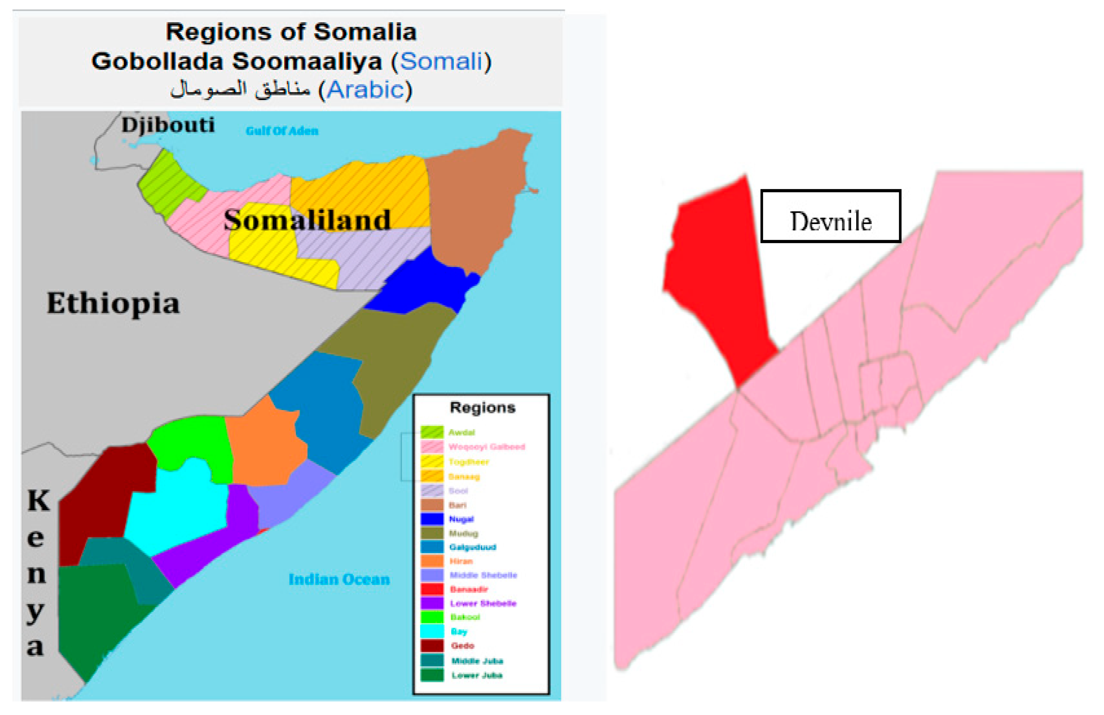
The investigation was carried out in some-meat slaughterhouse located Deynile Mogadishu, Somalia. Daynile District is located in the south-eastern Benadir region of Somalia. Daynile is the smallest administrative region in Somalia, but it has the largest population. The area is considered to be fairly safe and has good economic opportunities and resources.
Figure 1.
1: Map of Somali Showing that the Regions of Somalia and Study area.
Figure 1.
1: Map of Somali Showing that the Regions of Somalia and Study area.
TYPE OF STUDY AND ANIMAL
The study was cross-sectional, in order to determine the frequency of gross and histological lesion of Platyhelminthes in liver of slaughtered Camel at some-meat slaughterhouse in Deynile Somalia. Purposively, the study was carried out on 200 slaughtered camel From Different parts of Deynile District. All Examined Camel were include both male and female 80% were male while 20% of Female Greater than seven (7) years of age and had a good body conditions during both ante mortem and post-mortem inspection.
SAMPLING PROCESSING
After Ante-mortem process, the slaughtered Camels, were made by post-mortem examination visual examination, palpation and incision for the presence of any liver lesion. Then the gross lesion and Histopathological lesion were recorded. As well as size of the lesions and observations by Platyhelminthes made on incision were also recorded. A total of 360 camel samples were examined out of 200 liver tissue collected for histopathological manifestations.
Microscopic analysis of cells and tissues preparation were collected for every liver. The tissue were very thin up to one (1) to two (2), and high-quality sections (slices) mounted on glass slides and appropriately stained to demonstrate normal and abnormal structures.
The Most fresh tissue were very delicate and easily distorted and damaged, and thus possible to prepare thin sections unless it is chemically preserved or “fixed” and supported in some way whilst it is being cut. Broadly, the tissue used by frozen section and alternatively, used by paraffin sections which allow thin sections of the tissue specimen with a liquid agent that can subsequently be converted into a solid that has appropriate physical properties, to be cut from it. Paraffin wax is such an agent.
This produces so-called “paraffin sections”. Immediately. Each sample was fixed separately and transported to pathology laboratory for processing at the university of Nairobi Department of Veterinary Pathology, Microbiology and Parasitology. After fixing, all tissue samples collected for histopathological study were dehydrated in alcohol, and paraffin, and stained with Haematoxylin - The stained samples were examined using Electronic Microscope. Sample collection and processing was done as used by Yalew, et.al, (2018).
Appropriately. The data was entered into Microsoft excel spread sheet and coded was analysed by using SPSS 20.0 version. The Data was used descriptive analysis to determine the variance and proportion of the different lessons of Platyhelminthes in liver in both Gross and microscopic changes that was found at the slaughterhouse of some-meat company Dynile Somalia. Also Chi-square was used to test the lobular distributions of pathos-biological lesions in the slaughtered camel Liver.
RESULTS
A total of 340 slaughtered camel examined during the study, 200 camel (84.5%) were found to have Platyhelminthes in liver lesions. Considering both microscopic and gross inspection. For the gross Changes in ante-mortem Examination test included Liver Swollen 34.5% Yellowish discoloration 27.5%, Haemorrhagic lesions 11.5%, Bile duct dilation 9.5%, Necrosis 7.0%, Cirrhosis 5.5% and Exudates Cirrhosis 4.5%.
However, the most Histopathological features were Tissue Infiltration, 25.5%, Lymphocytic infiltration, 18.1%, Granulomas 13.5%, Granuloma formation 11.2%, (These can develop surrounding the parasite, particularly if the infection is being defended against by the host immune system.
The granulomas may contain lymphocytes, multinucleated giant cells, and macrophages), Necrosis, 11.2%, Abscess formation 8.3%, Inflammation 7.1% Infiltration 5.5% Fibrosis 4.2% Granulomas 3.2%, Hepatocellular necrosis 2.4%, (which is Tissue destruction due to parasitic activity, such as fluke migration through liver tissue.) Eosinophilic infiltrates 13.0%. Swelling or expansion of bile ducts 4.2%, often caused by the fluke’s impact on bile flow.
These lesions were indicated the most percentage of the study which was 84.5% while sample number was (n=85).respectively. Both Gross and Histological lesion summarized in table one (1) and two (2). Also therefore Representative images of some Platyhelminthes lesion had indicated in figure one (1), two (2), three (3) and Four (4).
However, liver enlargement or swelling brought on by fibrosis and inflammation was one of the other microscopic and gross characteristics. Yellowish discolouration brought on by necrotic tissue and bile build-up.
Haemorrhagic lesions, such as regions where tissue is being destroyed or bleeding. Chronic infections cause bile duct dilatation and cyst development. Fibrosis and granulomas surrounding the parasite or its eggs. When chronic infections cause cirrhosis, the liver develops a firm, nodular Idea Description of the Image: Cross-section of the liver as shown below
Figure 1.
Figure 1.
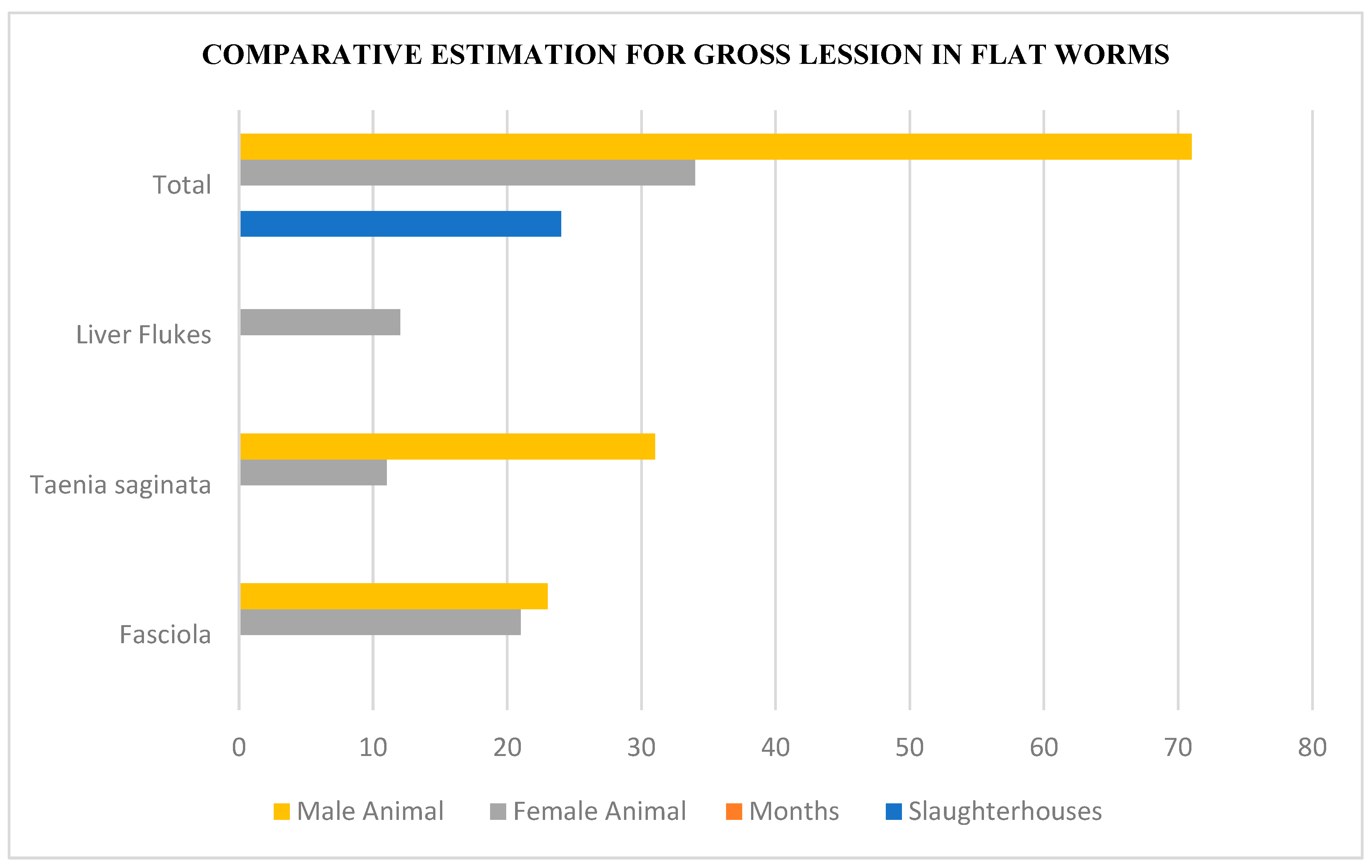
The Most gross lesions in flat worms From the Selected Slaughterhouse in Some Meat Deynile, Somalia.
Figure 1.
The Most gross lesions in flat worms From the Selected Slaughterhouse in Some Meat Deynile, Somalia.
A thorough histological section displaying the liver parenchyma with granulomas surrounding the parasites, eosinophilic infiltrates, and necrotic areas.
The flatworm parasite (such as Fasciola hepatica) or its egg inside the liver tissue is the main focus of the picture. Fibrosis: Around the afflicted tissue, there are patches of periportal fibrosis and alterations to the bile duct. Eosinophils, lymphocytes, and macrophages in close-up as examples of inflammatory cells by
Figure 2. At the following Stamens.
Though, Some of these manifestation had reported in some other studies in Africa, Asia and America but this study is Unique in Somalia especially Slaughterhouse studies in the country. Therefore I hope to fill that of study and look some further studies.
Figure 2.
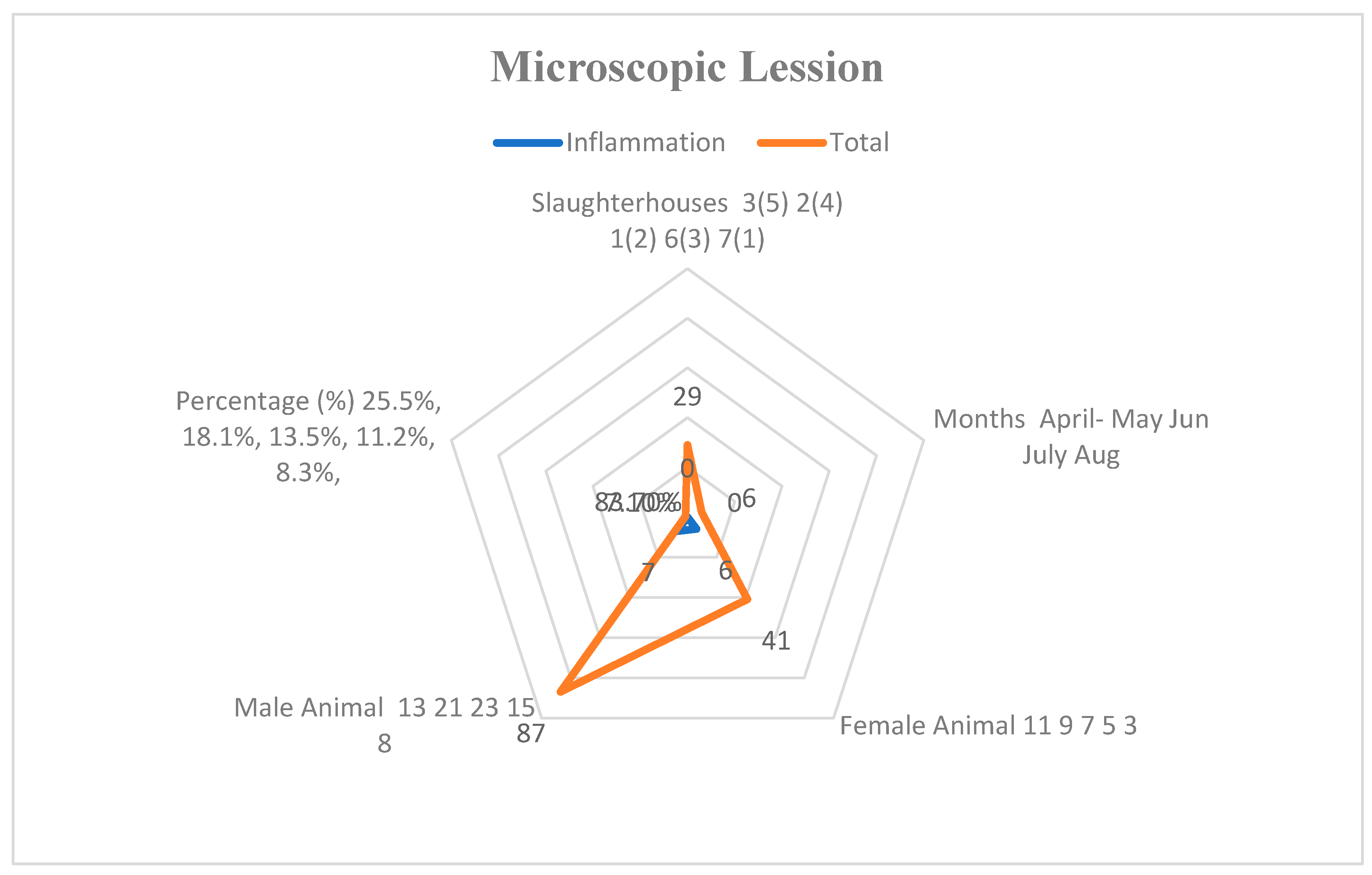
The Most Microscopically Affected Lesion for Platyhelminthes in Liver from Selected Camel Slaughterhouse in Deynile Somali.
Figure 2.
The Most Microscopically Affected Lesion for Platyhelminthes in Liver from Selected Camel Slaughterhouse in Deynile Somali.
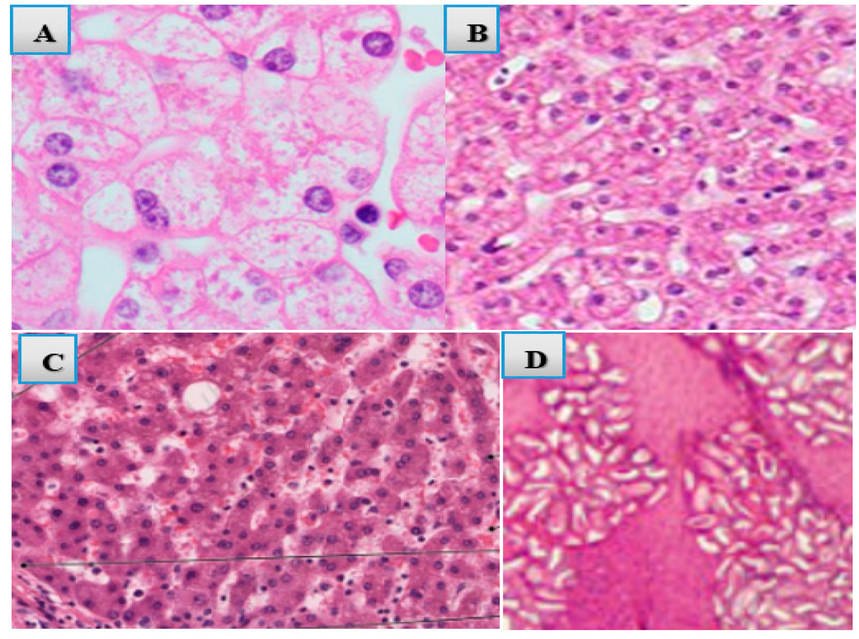
This images shows that the most Histopathological lesion and their estimation value of the study. Randomly and purposively selected by the study site.
Figure 3.
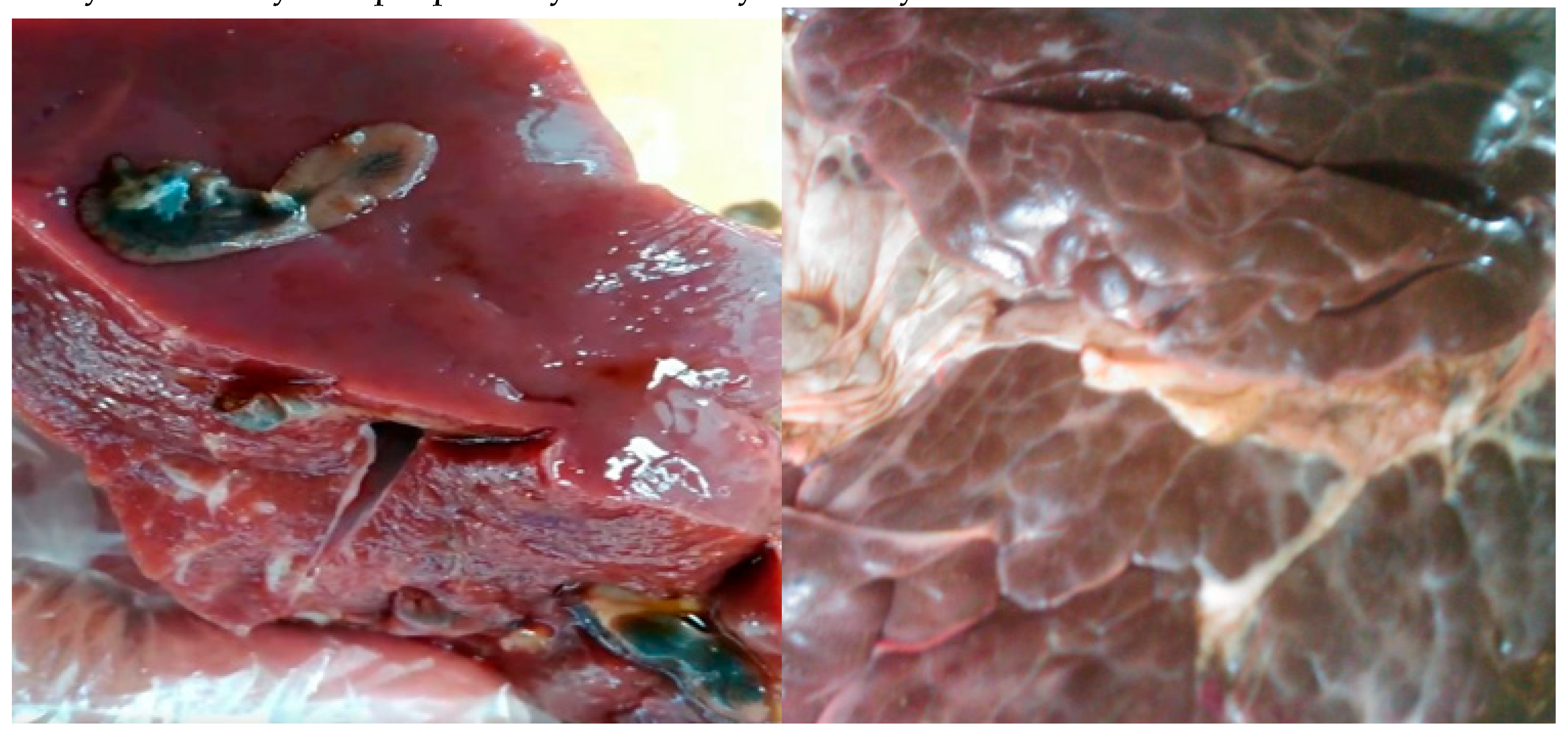
Reported Gross Lesions and Features Platyhelminthes (flat worms) in Two (2) different camel livers at selected slaughterhouse. .
Figure 3.
Reported Gross Lesions and Features Platyhelminthes (flat worms) in Two (2) different camel livers at selected slaughterhouse. .
These images are the grosses lesions in the post-mortem report analysis and clinical manifestation to Platyhelminthes of the camel live at some meat slaughterhouse
Figure 4.
Histopathological feature Necrosis(C) Lymphocytic Infiltration (B) Hyperplasia (A) Haemorrhagic areas adjacent to bile ducts (D).
Figure 4.
Histopathological feature Necrosis(C) Lymphocytic Infiltration (B) Hyperplasia (A) Haemorrhagic areas adjacent to bile ducts (D).
DISCUSSION
Gross and Microscopically Lesion for Platyhelminthes (Flat worms) associated with slaughtered camel at some meat slaughterhouse deynile Mogadishu Somalia was as shown in tables 1 and 2 and figures above of the study. Therefore, of 200 samples out of 340 selected camels examined 84.5 % had Liver Swollen 34.5%, similarly, Reported by Yalew, et.al. (2018) and Gebrehiwot et al. (2015). These researches had cited in the same results of liver swollen at the slaughterhouse of both camel and cattle in different study area.
Table 1.
Gross pathological Features in post-mortem Examination.
Table 1.
Gross pathological Features in post-mortem Examination.
| Gross Features |
Aetiology ( Cause of the Lesion) |
Frequencies (F) |
Percentage (%) |
Chi-square |
P- Value |
| Liver Swollen |
due to inflammation and fibrosis |
69 |
34.5 |
169 |
0.000 |
| Yellowish discoloration |
from bile accumulation and necrotic tissue |
55 |
27.5 |
121 |
0.000 |
| Haemorrhagic lesions |
Due to the Bleeding or tissue destruction. |
23 |
11.5 |
102 |
0.000 |
| Bile duct dilation |
Formation of cysts in chronic infections. |
19 |
9.5 |
95.1 |
0.000 |
| Necrosis |
Distinctive activities |
14 |
7.0 |
69.3 |
0.001 |
| Cirrhosis |
long-term infections, giving the liver a firm, nodular |
11 |
5.5 |
33.1 |
0.001 |
| Exudates |
Due to the parasitic migration |
9 |
4.5 |
19.2 |
0.231 |
Table 2.
microscopic Lesion at the Histopathological Features for Postpartum Examination.
Table 2.
microscopic Lesion at the Histopathological Features for Postpartum Examination.
| Histopathological Features |
Frequencies (F) |
Percentage (%) |
Chi-square (X2 ) |
P-Value |
| Tissue Infiltration |
51 |
25.5 |
139 |
0.000 |
| Lymphocytic infiltration |
37 |
18.1 |
111 |
0.000 |
| Granuloma formation |
29 |
13.5 |
103 |
0.000 |
| Necrosis |
21 |
11.2 |
97.6 |
0.000 |
| Abscess formation |
17 |
8.3 |
81.2 |
0.001 |
| Inflammation |
13 |
7.1 |
73.0 |
0.002 |
| Infiltration |
11 |
5.5 |
43 |
0.000 |
| Fibrosis |
9 |
4.2 |
27 |
0.0001 |
| Granulomas |
7 |
3.2 |
13 |
0.001 |
| Hepatocellular necrosis |
5 |
2.4 |
7 |
0.001 |
However, the result of this study was higher than the study findings of Yasine, et.al. (2019) and Xavier, et.al. (2022). an understanding of the Flat worms Lesion in camel slaughterhouses such lesion Yellowish discoloration 27.5%, is not so common in Somalia and it is some more further funding’s done in east and south Arabian countries similar study done in Iran and Saudi Arabia Tafere, et.al. (2020) and Sazmand, A. (2021).
Therefore, Haemorrhagic lesions 11.5%, Bile duct dilation 9.5%, Necrosis 7.0%, Cirrhosis 5.5% and Exudates Cirrhosis 4.5%. Although, the results of this study was higher than findings of Polinas, M. (2016) And Rajesh, et.al. (2017). whom reported Platyhelminthes lesion which is 42.2% and 39.0% respectively. Microscopic lesions were also reported similarly in this study. Therefore, the most histo-pathological lesions were found Tissue Infiltration, 25.5%, and Lymphocytic infiltration, 18.1%, as indicated the same study Odokuma, (2019) in Warri, Nigeria. And Keo, et.al. (2015). whom reported infiltration in slaughtered camel at the incidence of 36.53 and 41.77%.
Moreover, the tissue infiltration an Lymphocyte infiltration can cause some different histo-pathologic conditions including autoimmune disorders by the immune system attacking healthy cells, such that arteries and inflammation, malignant lymphomas, throughout the cells and tissue,and immune-oregulatory responses to the basis of hepatic lymphocyte and therefore, Those infiltrations in both tissue and lymphocytes had been mentioned to describe hepatic lymphocytes in the basis of hepatic Disease conditions, suggesting that these lymphocytes play an active role in hepatic damage and submits through necrosis.
Granuloma formation and Necrosis, were also mentioned in the study and reported 11.2%, 11.2%, similarly, this was higher than the results of Ibrahim, (2017) and Manga-González, M. & Ferreras, (2019). However, the result of this study was lower than the results of Islam, (2015). And Guimaraes, et.al. (2015). which was Epidemiological and Pathological Studies of one of the Platyhelminthes (Fascioliasis) in liver. Also Abscess formation were stumble upon the percentage of 8.3%, for examined liver at the slaughterhouse of some meat Mogadishu Somalia and it was lower than the findings of Gebrekrustos, et.al. (2019) in Ethiopia, therefore the study was mostly focused on Pathogenicity of Platyhelminthes especially Trypanosoma Types which is Phylum of Schistosoma.
Inflammation and Fibrosis were encountered the ratio of 7.1%, 4.2%.This result was higher than the result of Ngetich, (2019). In Kenya this result was the Thesis dissertation at the University of eldoret. Same reported the result of de-Brito, et.al. (2020). though this study focused on much as the effects on parasitic blood fluke Schistosoma infection and Antimicrobial Agents it was a similarly, resulted in liver for the flat worm lesions in our study. However, both fibrosis and inflammation are significant processes that can promote tissue regeneration, yet under some circumstances, fibrosis can result from inflammation. Although, the inflammation is a reaction to tissue damage brought on by a variety of causes, such as poisons, autoimmune reactions, infections, and traumas. While an excessive build-up of fibrous connective tissue within and surrounding injured tissue can result in fibrosis, a pathological consequence of prolonged inflammation. This may result in organ failure, lifelong scarring, or even death.
Additionally, there was three less common microscopic lesion in the study Hepatocellular necrosis 2.4%, (Tissue destruction due to parasitic activity), Eosinophilic infiltrates 13.0% and Swelling or expansion of bile ducts 4.2%, which were reported similar studies of Andersen-Ranberg, et.al (2018) and Ali, et.al. (2023).both were reported similar study results in liver infection caused by Platyhelminthes in animals.
CONCLUSION
This study presented a series of different of Platyhelminthes Features in slaughtered camels and the most common lesion in both gross and microscopic in liver parasites which may form Cells and tissue or which may induce Necrosis in the liver of slaughtered Animal or other hosts of camel.
Therefore, important new information on the effects of parasitic infections on camel health is provided by the investigation of Platyhelminthes' gross and histological lesions in camel liver. The results show that trematodes and cestodes in particular, which are Platyhelminthes, can cause fatty liver disease, chronic hepatitis, and other degenerative changes in the liver. Moreover, the study also emphasises how critical early diagnosis and intervention are. Treatment results can be greatly enhanced by promptly identifying these parasite diseases using clinical symptoms and histological examination.
The association between certain histological results and macroscopic lesions highlights the necessity of thorough veterinary examinations in camel herds, especially in areas where parasite diseases are common.
According to histopathological analyses, these parasites can result in significant liver damage that manifests as fibrosis, inflammation, and necrosis. Hepatomegaly, congestion, and the existence of nodular formations within the liver tissue are among the gross abnormalities that are frequently seen. These alterations are crucial because they impair the affected camels' general health and productivity in addition to the liver's ability to operate.
In conclusion, this study emphasises the negative effects of Platyhelminthes on the liver health of camels and the need for continued investigation and observation to lessen these effects on the productivity and health of livestock. It will take ongoing veterinary pathology research to create management plans that effectively address these parasitic diseases in camels.
Ethics:
This study was approved by the University of Nairobi and Salaam University Faculty of Veterinary Medicine Committee.
Acknowledgments:
The author sincerely would like to appreciate Some-meat Slaughterhouse (their facility to conduct the study). Salaam University and University of Nairobi Kenya University College of Veterinary Medicine Department of Pathology, Microbiology and Parasitology for provision of the laboratory and other facilities. I also extend my thanks to my wife and family.
Conflicts of Interests:
The authors have not declared any conflict of interests.
References
- Albogami, B. M. M. (2015). Study on Histopathological Injuries in Ruminants Due to The Effect of Internal Worms at Taif Province (Doctoral dissertation, king Abdul-Aziz university Jeddah).
- Ali, H., Alhamrashdi, A., El-Neweshy, M., Asi, M. N., Johnson, E. H., & Elshafie, E. I. (2023). An abattoir-based survey of helminthic liver infections and associated pathological lesions in sheep in the Sultanate of Oman.
- Andersen-Ranberg, E., Lehnert, K., Leifsson, P. S., Dietz, R., Andersen, S., Siebert, U.,... & Sonne, C. (2018). Morphometric, molecular and histopathologic description of hepatic infection by Orthosplanchnus arcticus (Trematoda: Digenea: Brachycladiidae) in ringed seals (Pusa hispida) from Northwest Greenland. Polar Biology, 41, 1019-1025.
- Ashoor, S. J., & Wakid, M. H. (2023). Prevalence and hepatic histopathological findings of fascioliasis in sheep slaughtered in Jeddah, Saudi Arabia. Scientific Reports, 13(1), 6609.
- Barre, A. (2023). Prevalence of hemorrhagic septicemia in dromedary camel (Camelus dromedarius) of some selected farms at Benadir region, Somalia. Journal of Istanbul Veterinary Sciences, 7(1), 8-14.
- Barre, A., Karanja, D. N., Bebora, L. C., & Gitao, C. G. (2023). Comparative prevallance and pathological changes on camel brucelosis at the selected slaughterhouses in Garissa County, Kenya. Veterinary Sciences: Research and Reviews, 9(1), 1-17.
- Barre, A., Mohamed, S. A., Hassan, A. S., & Mohamed, A. N. (2024). Anthrax: Distribution, Knowledge, Attitude, and Practice among Pastoralists In Jawhar, Qalimow, and Adan Yabal, Somalia. Journal of Applied Veterinary Science and Technology, 5(1).
- Barre, A., Mohamed, S. A., Mohamed, A. A., & Zakaria, I. I. (2024). Foot and Mouth Disease: Farmers, Knowledge, Attitude and Practice Direction to Pastoral Community in Lower Shabelle, Somalia. Journal of Veterinary Research and Clinical Care, 1(1), 1-8.
- de Brito, M. G., Mengarda, A. C., Oliveira, G. L., Cirino, M. E., Silva, T. C., de Oliveira, R. N.,... & de Moraes, J. (2020). Therapeutic effect of diminazene aceturate on parasitic blood fluke Schistosoma mansoni infection. Antimicrobial Agents and Chemotherapy, 64(11), 10-1128.
- de Moraes, J., Nascimento, C., Lopes, P. O., Nakano, E., Yamaguchi, L. F., Kato, M. J., & Kawano, T. (2011). Schistosoma mansoni: in vitro schistosomicidal activity of piplartine. Experimental Parasitology, 127(2), 357-364.
- Gebrekrustos, M., Weldu, K., Awol, N., & Hadush, B. (2019). Pathogenicity of Ethiopian Trypanosoma evansi Type A and B in Swiss Albino Mice Model. East African Journal of Veterinary and Animal Sciences, 3(1), 39-46.
- Guimaraes, M. A., de Oliveira, R. N., Véras, L. M., Lima, D. F., Campelo, Y. D., Campos, S. A.,... & Leite, J. R. S. (2015). Anthelmintic activity in vivo of epiisopiloturine against juvenile and adult worms of Schistosoma mansoni. PLoS neglected tropical diseases, 9(3), e0003656.
- Ibrahim, S. O. A. (2017). Gastrointestinal helminth parasites of ruminants slaughtered in shendi abattoir, river nile state, sudan (Doctoral dissertation, Yasser Abdelmaged Suleiman). Keo, T., Leung, J., & Weinstock, J. V. (2015). Parasitic diseases: helminths. Yamada's Textbook of Gastroenterology, 2337-2377.
- Islam, K. M. (2015). Epidemiological and Pathological Studies of Fascioliasis in Goats in Sylhet Region of Bangladesh and investigation on Effects of Different Liver Tonic on Pathology of Fascioliasis (Doctoral dissertation, University of Rajshahi).
- Keo, T., Leung, J., & Weinstock, J. V. (2015). Parasitic diseases: helminths. Yamada's Textbook of Gastroenterology, 2337-2377.
- Manga-González, M. Y., & Ferreras, M. C. (2019). Dicrocoeliidae family: Major species causing veterinary diseases. Digenetic Trematodes, 279-319.
- Meharie, B. G., & Tunta, T. A. (2021). Phytolacca dodecandra (phytolaccaceae) root extract exhibits antioxidant and hepatoprotective activities in mice with CCl4-induced acute liver damage. Clinical and Experimental Gastroenterology, 59-70.
- Murshed, M., Al-Quraishy, S., Mares, M. M., Mohammed, O. B., & Aljawdah, H. M. (2022). Survey of Dicrocoelium dendriticum infection in imported Romani and local sheep (Ovis aries), and potential epidemiological role in Saudi Arabia. Journal of Animal Science and Technology, 64(6), 1215.
- Ngetich, E. C. (2019). Prevalence and the effect of parasites on haematological parameters of livestock in the upper kerio valley, kenya (doctoral dissertation, university of eldoret).
- Odokuma, E. I. (2019). A 10-Year Histopathologic Audit of Uterine Cervical Biopsies in Warri, Nigeria. Galician medical journal, 26(1).
- Oyugi, A. M., Kibet, J. K., & Adongo, J. O. (2021). A review of the health implications of heavy metals and pesticide residues on khat users. Bulletin of the National Research Centre, 45, 1-22.
- Polinas, M. (2016). Histopathological and immunohistochemical characterization of host-parasite interaction in visceral organs of mullets (Osteichthyes: Mugilidae) from Sardinian lagoons.
- Rajesh, K. D., Subramani, V., Annamalai, P., Nakulan V, R., Narayanaperumal, J., Ponraj, P., & Durai, R. (2017). Gastrothylax crumenifer: Ultrastructure and histopathology study of in vitro trematodicidal action of Marattia fraxinea (Sm.). Clinical Phytoscience, 3, 1-17.
- Sanjari, A., Davari, S. A., & Rasekh, M. (2018). Macroscopic and histopathological examinations of liver lesions in slaughtered cattle in Zabol City, Iran.
- Sazmand, A. (2021). Paleoparasitology and archaeoparasitology in Iran: A retrospective in differential diagnosis. International Journal of Paleopathology, 32, 50-60.
- Tafere, G. G., Tuem, K. B., Gebre, A. K., & Balasubramaniam, R. (2020). In vitro antioxidant and in vivo hepatoprotective activities of root bark extract and solvent fractions of Croton macrostachyus Hochst. Ex Del. (Euphorbiaceae) on paracetamol-induced liver damage in mice. Journal of experimental pharmacology, 301-311.
- Xavier, E. S., de Souza, R. L., Rodrigues, V. C., Melo, C. O., Roquini, D. B., Lemes, B. L.,... & de Moraes, J. (2022). Therapeutic efficacy of carvacrol-loaded nanoemulsion in a mouse model of schistosomiasis. Frontiers in Pharmacology, 13, 917363.
- Yalew, K. W., Awol, N., Tsegay, Y., & Abraha, H. (2018). A study on gross and histopathological pulmonary lesions of cattle slaughtered at Abergelle Abattoir, Mekelle, Tigray, Ethiopia. Journal of Veterinary Medicine and Animal Health, 10(6), 148-152.
- Yasine, A., Daba, M., Ashenafi, H., Geldhof, P., Van Brantegem, L., Vercauteren, G., & Govaere, J. (2019). Tissue (re) distribution of Trypanosoma equiperdum in venereal infected and blood transfused horses. Veterinary parasitology, 268, 87-97.
|
Disclaimer/Publisher’s Note: The statements, opinions and data contained in all publications are solely those of the individual author(s) and contributor(s) and not of MDPI and/or the editor(s). MDPI and/or the editor(s) disclaim responsibility for any injury to people or property resulting from any ideas, methods, instructions or products referred to in the content. |
© 2025 by the authors. Licensee MDPI, Basel, Switzerland. This article is an open access article distributed under the terms and conditions of the Creative Commons Attribution (CC BY) license (http://creativecommons.org/licenses/by/4.0/).