Preprint
Article

This version is not peer-reviewed.

Endoplasmic Reticulum Proteins Impact Penetrance in a Pink1-Mutant Drosophila Model

A peer-reviewed article of this preprint also exists.

Submitted:

18 December 2024

Posted:

19 December 2024

You are already at the latest version

Abstract

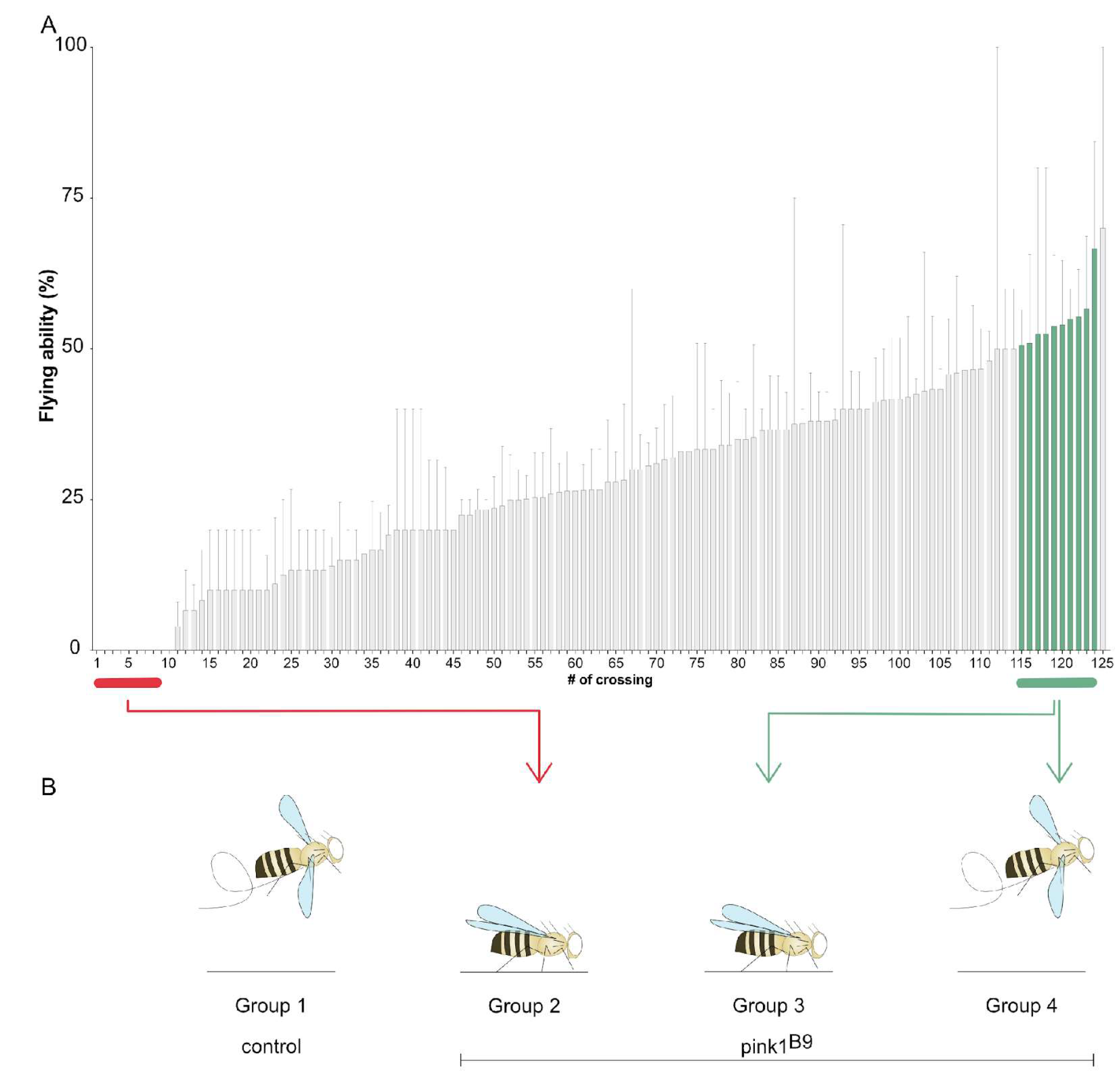
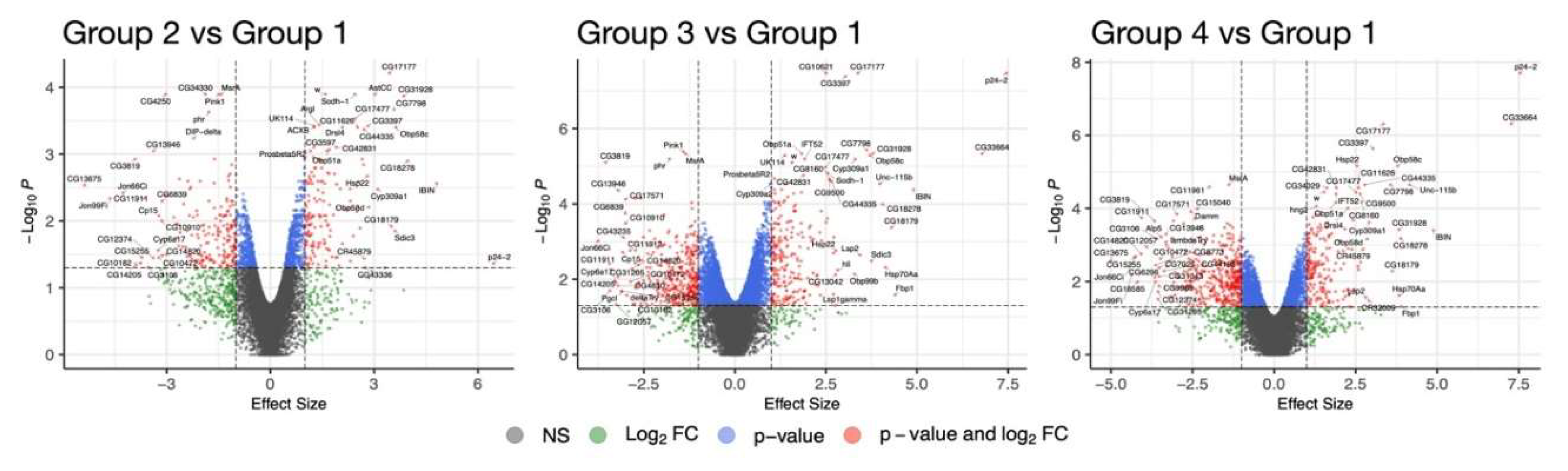
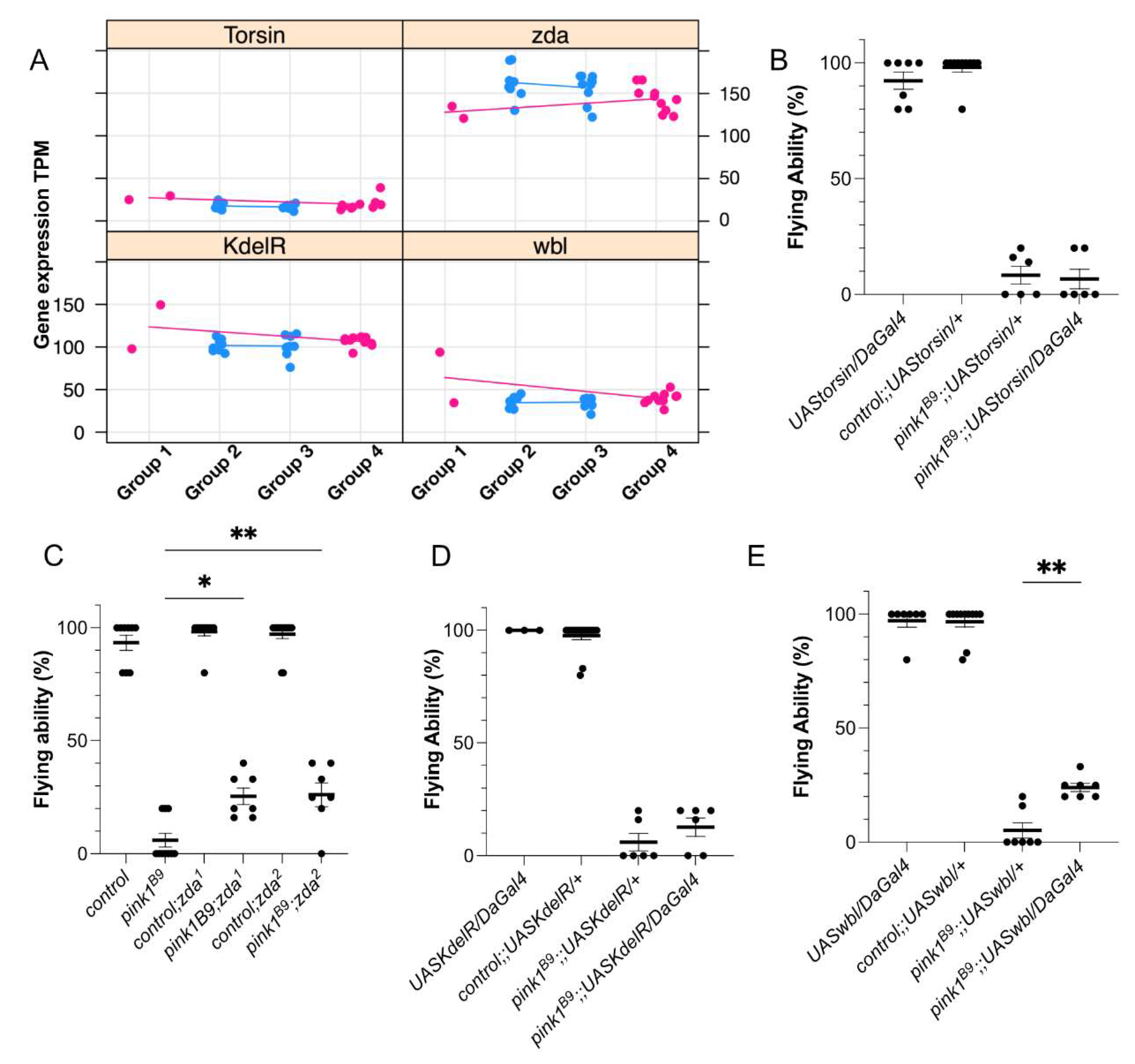
Parkinson’s disease (PD) is a neurodegenerative disorder with a high variability of age at onset, disease severity, and progression. This suggests that other factors, including genetic, environ-mental, or biological factors, are at play in PD. Loss of PINK1 causes a recessive form of PD and is typically fully penetrant; however, it features a wide range in disease onset, further supporting the existence of protective factors, endogenous or exogenous, to play a role. Loss of Pink1 in Drosophila melanogaster results in locomotion deficits, which are also observed in PINK1-related PD in humans. In flies, Pink1 deficiency induces defects in the ability to fly; none-theless, around ten percent of the mutant flies are still capable of flying, indicating that advanta-geous factors affecting penetrance also exist in flies. Here, we aimed to identify the mechanisms underlying this reduced penetrance in Pink1-deficient flies. We performed genetic screening in pink1-mutant flies to identify RNA expression alterations affecting the flying ability. The most important biological processes involved were transcription-al and translational activities, endoplasmic reticulum (ER) regulation, and flagellated movement and microtubule organization. We validated 2 ER-related proteins, zonda and windbeutel, to positively affect the flying ability of Pink1-deficient flies. Thus, our data suggest that these pro-cesses are involved in the reduced penetrance and that influencing them may be beneficial for Pink1 deficiency.

Keywords: 
;  ;  ;  ;  

1. Introduction

Parkinson’s disease (PD) features a wide variability in disease signs and symptoms, severity, progression, and age at onset [1], suggesting elements exist that alter disease development. One such factor is genetic; for instance, mutations in specific genes associated with PD have incomplete penetrance, meaning that not all carriers of these gene mutations will develop PD [2]. The recessively inherited forms of PD, caused by mutations in PINK1 and Parkin, are typically highly (to fully) penetrant, yet they come with an extensive range of ages at onset, supporting external factors to be pivotal [3,4]. For instance, studies show that smoking or caffeine intake reduces the risk of PD [5,6].
Several mechanisms have been identified to play a role in the pathogenesis of PD, including mitochondrial dysfunction, defects at the endo-lysosomal pathway, and, more recently also, a defective sphingolipids metabolism [7,8,9]. Thus, elements affecting the penetrance in PD are most likely involved in these cellular processes. For example, a recent study on homozygous twins discordant for idiopathic PD links mitochondrial integrity to altered disease penetrance [10]. However, the underlying mechanisms remain elusive. Hence, additional studies are required to understand the mechanisms that result in reduced penetrance.
Studies in Drosophila melanogaster have significantly contributed to our current knowledge of the mechanisms implicated in PD [11]. Research using the fruit fly was the first to highlight a common pathway between PINK1 and Parkin [12,13]. This finding was confirmed in human cellular systems [14,15]. Interestingly, in Drosophila melanogaster, reduced penetrance is observed concerning the flying phenotype in pink1-mutant flies. Previous studies show that the loss of Pink1 in flies results in a variable flying ability with a mean of around 10% [9,16,17], even though all these flies carry the same loss of function deletion in pink1. Thus, the fruit fly is a suitable model to study mechanisms underlying altered penetrance.
Here, we performed a transcriptome analysis in pink1-mutant flies to identify alterations in RNA expression levels that affect the flying ability. The most significant upregulation of biological processes was observed primarily in pathways associated with transcriptional and translational activities and the endoplasmic reticulum (ER) regulation. Conversely, downregulated pathways were linked to flagellated movement and microtubule organization. In addition, four genes were tested for validation, of which two, Zonda (Zda) and windbeutel (wbl), were confirmed to be beneficial and involved in autophagy and ER stress, supporting a role of reduced penetrance for these cellular mechanisms.

2. Results

2.1. Lack of Flying Ability of Pink1-Deficient Flies Shows a Pattern of Reduced Penetrance

The average flying ability of a general pink1B9-mutant fly stock is around ten percent [16], suggesting that a protective mechanism allows these ten percent of flies to fly. To analyze genetic factors that play a role in this reduced penetrance, we set up a group of 200 parent pairs consisting of one male and one virgin female fly to reduce the genetic variability of the offspring. One hundred twenty-five parent pairs resulted in Pink1-deficient offspring and were tested for their flying ability. The offspring showed substantial variability in the ability to fly, ranging from a complete inability to fly to a flying ability of 60% (Figure 1A). Although we reduced the genetic variability by combining only one male and one female, the offspring of these parent pairs are not genetically identical, explaining that not all offspring of a specific parent pair display the same flying ability. This provides a direct tool to analyze the existing reduced penetrance, and our screen allows the identification of genetic factors impacting the penetrance of the flying phenotype.

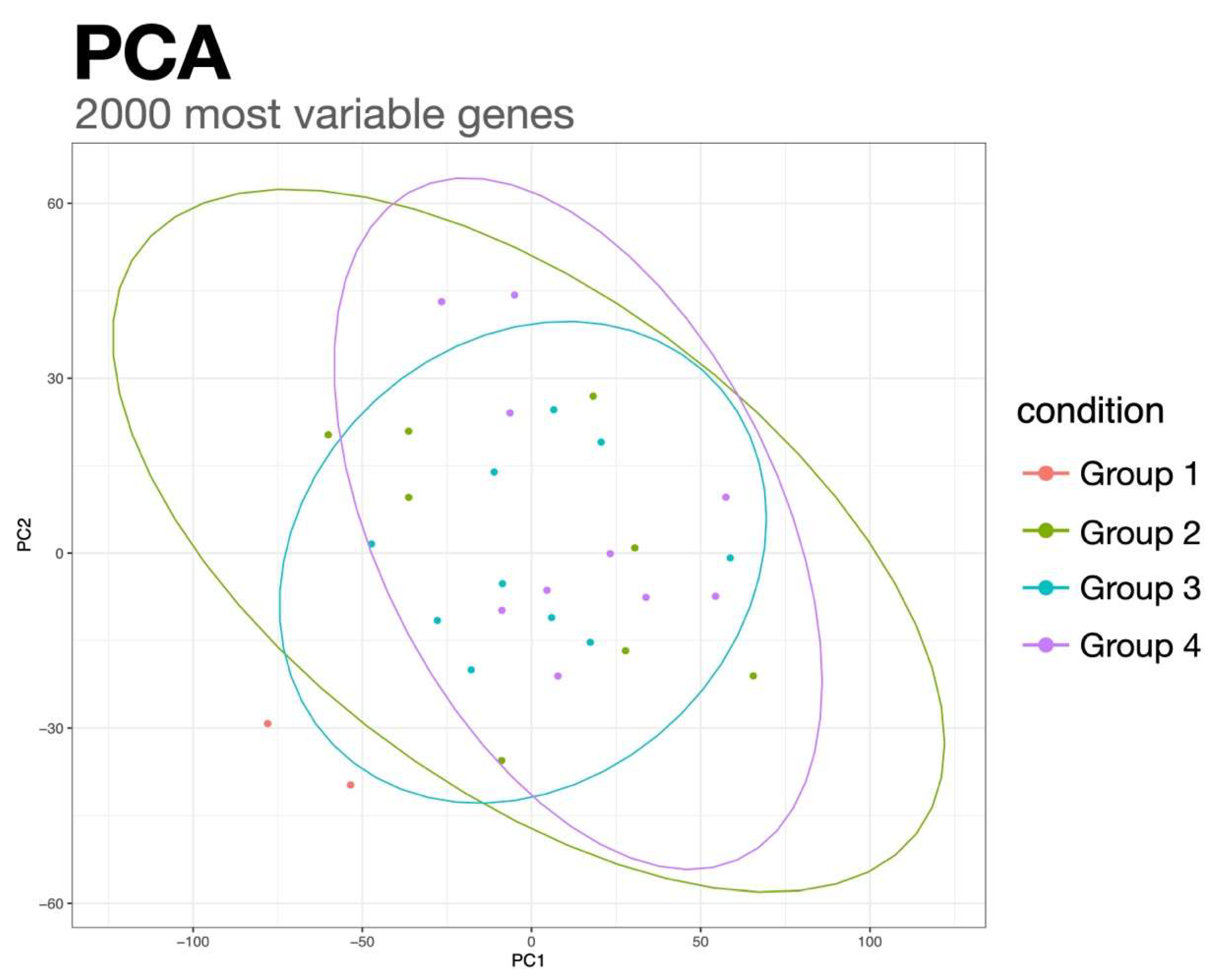
2.2. RNA Sequencing Analysis Identifies Genes Involved in Reduced Penetrance

To identify genes that play a role in reduced penetrance, we performed RNA sequencing analyses on four different groups (Figure 1B): 1. control flies (pink1RV); 2. pink1B9-mutant flies of which none of the offspring can fly; 3. pink1B9-mutant flies that cannot fly but with 4. ‘siblings’ that can fly (group 4). Thus, groups 3 and 4 are offspring of the same parent pair and have the lowest genetic variability; hence, differential RNA expression between those two groups may be the most relevant in the context of reduced penetrance. We identified 12 genes that displayed significant RNA expression differences between groups 3 and 4, i.e., Pink1-deficient flies that cannot fly and that can fly coming from the same parent pair.
In our group analysis, genes were designated differentially expressed with an absolute effect size (beta-value) > 1 and an adjusted p-value < 0.05. Notably, we identified 350, 611, and 650 differentially expressed genes (DEGs) when comparing the control group against groups two, three, and four, respectively (cf. volcano plots in Figure 2). When considering genes with human orthologs only, the gene count was substantially reduced, resulting in 144, 226, and 261 DEGs for the control comparison against groups two, three, and four.
Significant DEGs were not identified when correcting for multiple testing in the comparisons between groups two, three, and four. The proximity of mutated samples was evident from the first two principal components of principal component analysis (PCA) (Figure 3). Notably, the three groups of mutated flies clustered closely, with overlapping ellipses. Interestingly, when exclusively considering the p-value, group 4 exhibited more significantly altered gene hits than group three, compared to non-flying parents.
To assess the penetrance of the loss of Pink1, we examined genes differentially regulated in controls and mutants that retained their flying ability compared to non-flying mutants. The comparison of the established groups (Flying, MutatedFlying, and Mutant) with the multiple regression model from limma revealed 3396 genes that were significantly reduced to 1692 genes when filtered for human orthologs. Additionally, we refined our gene selection criteria to include only those that exhibited either up- or down-regulation in both groups when compared to mutated, non-flying fruit flies. To enhance specificity and eliminate non-penetrance-related noise, we restricted the magnitude of expression changes between these groups to a factor of 3. Consequently, we successfully identified 124 genes, as detailed in Table 2.
2.3 Validation of the Protective Effects of Single Hits
Pathway analyses revealed a significant enrichment of transcriptional and translational activities and regulation of (endo)membranes and ER and a decrease in pathways related to flagellated movement and microtubule organization. The ER is known to be linked to neurodegeneration [18]; however, the underlying processes are not fully understood. Hence, we selected four ER-linked genes, which were also significant in the ANOVA test: Torsin, KdelR, and winbeutel (wbl) that show increased differential expression levels, and zonda (zda) that displayed lower differential expression levels (Figure 4A), approaching control expression levels. Overexpression of KdelR and Torsin in Pink1-deficient flies failed to elicit an effect on the flying ability of pink1-mutant flies (Figure 4B, D), while overexpression of wbl and two independent alleles of heterozygous loss of Zda improved the flying ability (Figure 4C, E).

3. Discussion

In this work, we showed the presence of reduced penetrance in Pink1-deficient flies and performed RNA expression analyses to elucidate possible mechanisms underlying this observation. Via gene ontology and pathway analyses, we identified several molecular mechanisms that appear to be involved in the penetrance of loss of Pink1, including ER-related activity. Finally, we confirmed a beneficial effect of lower zda and higher wbl levels on the flying phenotype of pink1-mutant flies, suggesting that ER-linked autophagy and immunity play an essential role in the penetrance of a PD-like phenotype in our Drosophila model.
The low genetic variability between the parents and offspring groups is to be expected since we assume that the genetic background should still be relatively close to its progenitor individuals. Additionally, the offspring groups were sampled from the same time point, resulting in an even higher genetic resemblance. In an attempt to circumvent this, we decided that a multiple linear regression would be the most fitting model, only focusing on the flying ability of the different sample groups. This also enabled us to work with a rather small control group of two sequencing samples since limma is fit for handling small sample sizes, and the multiple linear regression grouped the controls with the flying mutants, allowing us to pinpoint potential genetic variables that could affect the penetrance of the flying phenotype. Notably, by applying our set criteria for candidate genes (relevant Pathways, significant ANOVA hits, same direction of logFC, human ortholog), we identified 124 genes that approach control expression levels in our mutants. Most genes in Table 2 still need to be identified, which makes them potential candidates for modulation of penetrance that are challenging to evaluate experimentally.
The comparison of DEGs between groups 3 (pink1-mutant flies that cannot fly) and 4 (pink1-mutant flies that can fly and are ‘siblings’ of group 3) revealed twelve candidate genes to be altered, suggesting that these genes affect the reduced penetrance of the flying ability. Nonetheless, we did not validate these genes due to the assumption that it is unlikely that one gene can regulate penetrance. Instead, (minor) alterations in biological processes or cellular mechanisms are more likely to affect the penetrance, and hence, we decided to pick four genes that revealed significantly altered expression as part of an affected biological process to be validated. The ER is one of these biological processes that play a role in the penetrance of the flying ability of Pink1-deficient flies. Thus, we tested four genes separately and will discuss these candidates in more detail below.
The ER has previously been linked to neurodegeneration via its function in protein quality control. ER stress, because of unfolded proteins, can lead to neuronal cell death and, thus, neurodegeneration, such as Alzheimer’s disease and PD [18]. One of the proteins involved in ER stress is the KDELR [19]. KDELR is upregulated upon ER stress [20,21], resulting in autophagy induction [21]. Interestingly, autophagy is a process that is affected in (PINK1-related) PD [7,9,22], suggesting KDELR upregulation can be beneficial to overcome defective autophagy upon PD. This is in line with the findings of our initial screen, in which higher expression levels improve the flying ability. However, we were not able to reproduce these findings by overexpressing KDELR. One possible explanation could lie in the limitations of the applied GAL4/UAS system. This system overexpresses a protein beyond physiological levels [23] and is temperature-sensitive [24]. In our experimental setup, we grew the flies at 25°C, which supports an efficient activation of the GAL4/UAS system, while the increased expression levels identified are only tenfold higher. These data suggest that too much KDELR abolishes the positive effect in pink1-mutant flies and that there is a small therapeutic window.
Interestingly, wbl is linked to KDELR, further supporting the involvement of KDELR and its processes in the penetrance of PD symptoms. Wbl is a protein disulfide isomerase (PDI)-related chaperone [25,26], which contains a C-terminal ER retention motif KDEL [27]. This motif interacts with KDELR to retrieve these ER chaperones from the Golgi [28,29]. The human ortholog for wbl is the endoplasmic reticulum protein of 29 kDA (ERp29) that is ubiquitously expressed [29]. Similar to the KDELR, ERp29 is upregulated upon ER stress. Furthermore, ERp29 is elevated following a dopamine exposure-related PD model, suggesting that ER stress occurs in the early stages of PD [30]. Our data show that increased ERp29 levels benefit the Pink1-deficient flying phenotype, supporting the notion that ER stress and its associated elevated ER proteins are a compensatory mechanism in this Pink1-related PD model.
Torsin A, the human ortholog for Torsin, is an AAA ATPase localized to the ER and encoded by the TOR1A gene, in which mutations cause early-onset dystonia [31]. Previously, Torsin has been linked to PD via its presence in the Lewy bodies [32,33]. Furthermore, in C. elegans, increased Torsin exerts a protective effect in a 6-hydroxydopamine (6-OHDA) PD model [34], supporting the findings from our screen where higher Torsin expression is beneficial for the flying phenotype in Pink1-deficient flies. However, via the GAL4/UAS system, we were unable to confirm these results, while overexpression in C. elegans did lead to a rescue. Unfortunately, these data are difficult to directly compare due to the unknown expression efficiency and the difference in the human disease and phenotypes observed in animal models. Hence, while increased Torsin levels have the potential to be protective, further investigations are required to determine the exact effect of Torsin, its relationship to the ER function, and its effect on PD.
Zda is an immunophilin localized at the ER. Zda is required for starvation-induced autophagy and interacts with the autophagy genes Atg1, Atg6, and Vps34 (vacuolar sorting protein) to activate the autophagy-specific activation of Vps34 and thus the initial stages of autophagy [35]. The human ortholog of zda is FKBP8, which contains a microtubule-associated protein 1 light chain 3 (LC3)-interacting region (LIR) motif. It recruits LC3 to the mitochondria to induce Parkin-independent mitophagy while escaping degradation [36]. Autophagy or mitochondrial-specific mitophagy are processes that are affected in PD. Namely, PINK1 functions in mitophagy [7,9,14,37], suggesting that the beneficial effect of zda lies in its mitophagy function. Previous studies showed that lowering mitophagy activity is beneficial for Pink1-dependent PD [9,22]. Thus, decreased zda expression provokes lower mitophagy levels in an attempt to improve the pink1-mutant flying phenotype. In addition, under stress conditions, FKBP8 equally plays a role in mitochondrial fragmentation. Via its LIR-motif-like sequence (LIRL), FKBP8 binds to OPA1, mediating fragmentation and enabling mitophagy [38]. Reversely, the knockdown of FKBP8 enlarges the mitochondria to form its natural tubular network. Interestingly, the loss of Pink1 induces mitochondrial accumulation and loss of the tubular network [13,16]. Thus, decreased zda levels could induce a more tubular mitochondrial network, allowing the mitochondria to function more efficiently. Therefore, the beneficial role of reducing zda levels may extend to a dual role in inhibiting mitophagy and mitochondrial dynamics.
This study is the first to identify possible cellular mechanisms involved in the penetrance of the flying ability in a PD model. The fly model proved to be of excellent value for investigating and identifying biological processes involved in reduced penetrance. Nonetheless, with this setup, we could not validate if these biological processes equally affect other phenotypes in pink1-mutant flies, nor did we test their relevance to patients. In conclusion, our data further point to the ER implicated in PD and its penetrance in a genetic PD fly model that needs further validation in a patient-relevant context. In addition, further investigation into the exact underlying mechanism is necessary so that this pathway can be modulated to alter the penetrance for PD.

4. Materials and Methods

Fly Genetics

w pink1B9 null mutants and controls (w pink1RV) were kindly provided by Jeehye Park and Jongkyeong Chung (Korea Advanced Institute of Science and Technology) [12]. y[1] w[*]; M{RFP[3xP3.PB] w[+mC]=UAS-KDELR-RFP}ZH-22A (UASKdelR) were generously shared by the Katanaev laboratory (Department of Pharmacology and Toxicology, University of Lausanne) [39]. Two independent lines of Zda (w1118; PBac{IT.GAL4}zda0818-G4 (Zda1) and y1 w67c23; P{EPgy2}zdaEY08359 (Zda2)), and daughterless Gal4 (DaGal4) were purchased from the Bloomington Stock Center (Indianapolis, United States of America). M[UAS-Torsin.ORF.3xHA.GW]ZH-86Fb (UASTorsin) and M[UAS-Wbl.ORF.3xHA.GW] (UASWbl) were purchased from the FlyORF stock center (Zurich, Switzerland).

Flight Assay

One-day-old male flies were collected and tested for their flying ability. Flies were placed in an empty vial that was gently tapped. Flies able or unable to fly were scored one and zero, respectively [16]. For the sequencing analyses, the offspring of each parent pair that was able to fly (group 4) was separately collected from those that were not able to fly (group 3).

RNA Sequencing and Analyses

RNA sequencing was performed on four different groups. Group 1 are control flies (pink1RV), group 2 are pink1B9-mutant flies of which none of the offspring was able to fly, groups 3 and 4 are pink1B9-mutant flies that are offspring from the same parent, but in group 3, none of the flies were able to fly, while in group 4 all the flies were able to fly. RNA was isolated from 5 flies via a standard procedure (Qiagen), and RNA sequencing was performed by the genomics core UZ Leuven (Belgium) using an Illumina Hiseq system.
The fastq files underwent preprocessing using fastp (version 0.23.4) [40], wherein reads with less than 30% of bases possessing a quality PHRED score under 15 were selectively retained. Subsequently, reads were pseudoaligned to the Drosophila melanogaster genome assembly BDGP6.46 through kallisto (version 0.44.0). The alignment metrics are detailed in Table 1.
Differential expression and pathway analyses were executed in R (version 4.2.2), primarily employing sleuth (version 0.30.1)[41] or limma (version 3.54.2) [42]. Sleuth was utilized for straightforward group comparisons, while limma and its extension of ANOVA tests were applied for more intricate model computations. Gene annotation was derived from the Genome wide annotation for Drosophila melanogaster package org.Dm.eg.db (version 3.16.0), encompassing entries for HGNC and flybase nomenclature, GO terms, and Homo sapiens gene homology.
Expression data analysis with limma was performed on kallisto's abundance levels, which were transformed into length-scaled count values using tximport (version 1.26.1) [43]. Pathway analysis was conducted using gage (version 2.48.0) [44], incorporating either the log2 fold changes from differential expression or sleuth's beta values. We corrected differential gene expression and pathway significance for multiple testing using the BH correction. The cutoff for the adjusted p-value was set to 0.05 in all our analyses.
To investigate penetrance, we combined the sample groups into flying, mutated, and non-flying and looked for co-regulation between the two flying groups when compared to the mutated non-flying group with multiple comparison testing. Specifically, the group Flying consisted of samples from the controls (group 1) and the offspring with flying ability (group 4). The MutatedFlying group only encapsulated the flying offspring (group 4), while the Mutated group combined the samples from offspring that were unable to fly (group 2 and 3). Using this design, we calculated the DEGs between Flying versus Mutated and MutatedFlying versus Mutated and subsequently called the limma equivalent of an ANOVA, and these multiple comparisons.

Author Contributions

Conceptualization: M.V., H.B. C.K.; methodology: M.V., F.O., H.G., G.C., S.M.; software: F.O. and H.B.; validation: M.V., H.B., C.K.; formal analysis: M.V. and F.O.; data curation: H.B.; writing – original draft preparation: M.V. and F.O.; writing – review and editing: H.G., G.C., S.M., H.B., C.K.; visualization: M.V. and F.O.; supervision: M.V., H.B., C.K.; project administration: M.V. All authors have read and agreed to the published version of the manuscript.

Funding

F.O. and H.B. acknowledge funding through the German Science Foundation (BU 2487/6-2) and Germany`s Excellence Strategy (EXC 2167-390884018).

Institutional Review Board Statement

Not Applicable

Data Availability Statement

The data will be made available

Conflicts of Interest

CK has served as a consultant for Centogene, Retromer Therapeutics, and Lundbeck and received speakers’ honoraria from Desitin and Bial and research funding from DFG, MJFF, and ASAP.

References

  1. B.R. Bloem, M.S. Okun, C. Klein, Parkinson’s disease, Lancet 397 (2021) 2284–2303.
  2. G.U. Höglinger, C.H. Adler, D. Berg, C. Klein, T.F. Outeiro, W. Poewe, R. Postuma, A.J. Stoessl, A.E. Lang, A biological classification of Parkinson’s disease: the SynNeurGe research diagnostic criteria, Lancet Neurol 23 (2024) 191–204. [CrossRef]
  3. V.A. Morais, M. Vos, Reduced penetrance of Parkinson’s disease models, Med Genet 34 (2022) 117–124. [CrossRef]
  4. M. Kasten, C. Hartmann, J. Hampf, S. Schaake, A. Westenberger, E.J. Vollstedt, A. Balck, A. Domingo, F. Vulinovic, M. Dulovic, I. Zorn, H. Madoev, H. Zehnle, C.M. Lembeck, L. Schawe, J. Reginold, J. Huang, I.R. König, L. Bertram, C. Marras, K. Lohmann, C.M. Lill, C. Klein, Genotype-Phenotype Relations for the Parkinson’s Disease Genes Parkin, PINK1, DJ1: MDSGene Systematic Review, Movement Disorders (2018). [CrossRef]
  5. C. Gabbert, I.R. König, T. Lüth, B. Kolms, M. Kasten, E.J. Vollstedt, A. Balck, A. Grünewald, C. Klein, J. Trinh, Coffee, smoking and aspirin are associated with age at onset in idiopathic Parkinson’s disease, J Neurol 269 (2022) 4195–4203. [CrossRef]
  6. T. Lüth, I.R. König, A. Grünewald, M. Kasten, C. Klein, F. Hentati, M. Farrer, J. Trinh, Age at Onset of LRRK2 p.Gly2019Ser Is Related to Environmental and Lifestyle Factors, Mov Disord 35 (2020) 1854–1858. [CrossRef]
  7. M. Vos, C. Klein, A.A. Hicks, Role of Ceramides and Sphingolipids in Parkinson’s Disease, J Mol Biol 435 (2023). [CrossRef]
  8. F. Mandik, M. Vos, Neurodegenerative Disorders: Spotlight on Sphingolipids, Int J Mol Sci 22 (2021). [CrossRef]
  9. M. Vos, M. Dulovic-Mahlow, F. Mandik, L. Frese, Y. Kanana, S.H. Diaw, J. Depperschmidt, C. Böhm, J. Rohr, T. Lohnau, I.R. König, C. Klein, Ceramide accumulation induces mitophagy and impairs β-oxidation in PINK1 deficiency, Proceedings of the National Academy of Sciences 118 (2021) e2025347118. [CrossRef]
  10. M. Dulovic-Mahlow, I.R. König, J. Trinh, S.H. Diaw, P.P. Urban, E. Knappe, N. Kuhnke, L.C. Ingwersen, F. Hinrichs, J. Weber, P. Kupnicka, A. Balck, S. Delcambre, T. Vollbrandt, A. Grünewald, C. Klein, P. Seibler, K. Lohmann, Discordant Monozygotic Parkinson Disease Twins: Role of Mitochondrial Integrity, Ann Neurol 89 (2021) 158–164. [CrossRef]
  11. M. Vos, C. Klein, The Importance of Drosophila melanogaster Research to UnCover Cellular Pathways Underlying Parkinson’s Disease, Cells 10 (2021) 579. [CrossRef]
  12. J. Park, S.B. Lee, S. Lee, Y. Kim, S. Song, S. Kim, E. Bae, J. Kim, M. Shong, J.-M. Kim, J. Chung, Mitochondrial dysfunction in Drosophila PINK1 mutants is complemented by parkin, Nature 441 (2006) 1157–1161. [CrossRef]
  13. I.E. Clark, M.W. Dodson, C. Jiang, J.H. Cao, J.R. Huh, J.H. Seol, S.J. Yoo, B.A. Hay, M. Guo, Drosophila pink1 is required for mitochondrial function and interacts genetically with parkin, Nature 441 (2006) 1162–1166. [CrossRef]
  14. D. Narendra, A. Tanaka, D.F. Suen, R.J. Youle, Parkin-induced mitophagy in the pathogenesis of Parkinson disease, Autophagy 5 (2009) 706–708. http://www.ncbi.nlm.nih.gov/entrez/query.fcgi?cmd=Retrieve&db=PubMed&dopt=Citation&list_uids=19377297.
  15. D.P. Narendra, S.M. Jin, A. Tanaka, D.F. Suen, C.A. Gautier, J. Shen, M.R. Cookson, R.J. Youle, PINK1 is selectively stabilized on impaired mitochondria to activate Parkin, PLoS Biol 8 (2010) e1000298. http://www.ncbi.nlm.nih.gov/entrez/query.fcgi?cmd=Retrieve&db=PubMed&dopt=Citation&list_uids=20126261.
  16. M. Vos, G. Esposito, J.N. Edirisinghe, S. Vilain, D.M. Haddad, J.R. Slabbaert, S. Van Meensel, O. Schaap, B. De Strooper, R. Meganathan, V.A. Morais, P. Verstreken, Vitamin K2 is a mitochondrial electron carrier that rescues pink1 deficiency, Science (1979) 336 (2012) 1306–1310. http://www.ncbi.nlm.nih.gov/entrez/query.fcgi?cmd=Retrieve&db=PubMed&dopt=Citation&list_uids=22582012.
  17. M. Vos, A. Geens, C. Böhm, L. Deaulmerie, J. Swerts, M. Rossi, K. Craessaerts, E.P. Leites, P. Seibler, A. Rakovic, T. Lohnau, B. De Strooper, S.-M. Fendt, V.A. Morais, C. Klein, P. Verstreken, Cardiolipin promotes electron transport between ubiquinone and complex I to rescue PINK1 deficiency, J Cell Biol 216 (2017) 695–708.
  18. D. Lindholm, H. Wootz, L. Korhonen, ER stress and neurodegenerative diseases, Cell Death Differ 13 (2006) 385–392. [CrossRef]
  19. K. Yamamoto, H. Hamada, H. Shinkai, Y. Kohno, H. Koseki, T. Aoe, The KDEL receptor modulates the endoplasmic reticulum stress response through mitogen-activated protein kinase signaling cascades, J Biol Chem 278 (2003) 34525–34532. [CrossRef]
  20. E.S. Wires, K.A. Trychta, L.M. Kennedy, B.K. Harvey, The Function of KDEL Receptors as UPR Genes in Disease, Int J Mol Sci 22 (2021). [CrossRef]
  21. P. Wang, B. Li, L. Zhou, E. Fei, G. Wang, The KDEL receptor induces autophagy to promote the clearance of neurodegenerative disease-related proteins, Neuroscience 190 (2011) 43–55. [CrossRef]
  22. M. Vos, C. Klein, Ceramide-induced mitophagy impairs ß-oxidation-linked energy production in PINK1 deficiency, Https://Doi.Org/10.1080/15548627.2022.2027193 18 (2022) 703–704. [CrossRef]
  23. Y. Her, D.M. Pascual, Z. Goldstone-Joubert, P.C. Marcogliese, Variant functional assessment in Drosophila by overexpression: what can we learn?, Genome 67 (2024) 158–167. [CrossRef]
  24. S. Nagarkar-Jaiswal, P.T. Lee, M.E. Campbell, K. Chen, S. Anguiano-Zarate, M.C. Gutierrez, T. Busby, W.W. Lin, Y. He, K.L. Schulze, B.W. Booth, M. Evans-Holm, K.J.T. Venken, R.W. Levis, A.C. Spradling, R.A. Hoskins, H.J. Bellen, A library of MiMICs allows tagging of genes and reversible, spatial and temporal knockdown of proteins in Drosophila, Elife 4 (2015). [CrossRef]
  25. J. Sen, J.S. Goltz, M. Konsolaki, T. Schüpbach, D. Stein, Windbeutel is required for function and correct subcellular localization of the Drosophila patterning protein Pipe, Development 127 (2000) 5541–5550. [CrossRef]
  26. M. Konsolaki, T. Schüpbach, windbeutel, a gene required for dorsoventral patterning in Drosophila, encodes a protein that has homologies to vertebrate proteins of the endoplasmic reticulum, Genes Dev 12 (1998) 120–131. [CrossRef]
  27. H.R.B. Pelham, The retention signal for soluble proteins of the endoplasmic reticulum, Trends Biochem Sci 15 (1990) 483–486. [CrossRef]
  28. K. Yamamoto, R. Fujii, Y. Toyofuku, T. Saito, H. Koseki, V.W. Hsu, T. Aoe, The KDEL receptor mediates a retrieval mechanism that contributes to quality control at the endoplasmic reticulum, EMBO J 20 (2001) 3082–3091. [CrossRef]
  29. M. Brecker, S. Khakhina, T.J. Schubert, Z. Thompson, R.C. Rubenstein, The Probable, Possible, and Novel Functions of ERp29, Front Physiol 11 (2020). [CrossRef]
  30. A.A. Dukes, V.S. Van Laar, M. Cascio, T.G. Hastings, Changes in endoplasmic reticulum stress proteins and aldolase A in cells exposed to dopamine, J Neurochem 106 (2008) 333–346. [CrossRef]
  31. P.M.E.S.M.I.S.M.N.L.S.I.-S.I. Slominsky, A common 3-bp deletion in the DYT1 gene in Russian families with early-onset torsion dystonia, Hum Mutat 3 (1999) 269. [CrossRef]
  32. P. Shashidharan, P.F. Good, A. Hsu, D.P. Perl, M.F. Brin, C.W. Olanow, TorsinA accumulation in Lewy bodies in sporadic Parkinson’s disease, Brain Res 877 (2000) 379–381. [CrossRef]
  33. N. Sharma, J. Hewett, L.J. Ozelius, V. Ramesh, P.J. McLean, X.O. Breakefield, B.T. Hyman, A close association of torsinA and alpha-synuclein in Lewy bodies: a fluorescence resonance energy transfer study, Am J Pathol 159 (2001) 339–344. [CrossRef]
  34. S. Cao, C.C. Gelwix, K.A. Caldwell, G.A. Caldwell, Torsin-Mediated Protection from Cellular Stress in the Dopaminergic Neurons of Caenorhabditis elegans, (2005). [CrossRef]
  35. M. Melani, A. Valko, N.M. Romero, M.O. Aguilera, J.M. Acevedo, Z. Bhujabal, J. Perez-Perri, R. V. De La Riva-Carrasco, M.J. Katz, E. Sorianello, C. D’Alessio, G. Juhász, T. Johansen, M.I. Colombo, P. Wappner, Zonda is a novel early component of the autophagy pathway in Drosophila, Mol Biol Cell 28 (2017) 3070. [CrossRef]
  36. Z. Bhujabal, Å.B. Birgisdottir, E. Sjøttem, H.B. Brenne, A. Øvervatn, S. Habisov, V. Kirkin, T. Lamark, T. Johansen, FKBP8 recruits LC3A to mediate Parkin-independent mitophagy, EMBO Rep 18 (2017) 947–961. [CrossRef]
  37. D. Narendra, A. Tanaka, D.-F. Suen, R.J. Youle, Parkin is recruited selectively to impaired mitochondria and promotes their autophagy, J Cell Biol 183 (2008) 795–803. [CrossRef]
  38. S.M. Yoo, S. ichi Yamashita, H. Kim, D.H. Na, H. Lee, S.J. Kim, D.H. Cho, T. Kanki, Y.K. Jung, FKBP8 LIRL-dependent mitochondrial fragmentation facilitates mitophagy under stress conditions, FASEB J 34 (2020) 2944–2957. [CrossRef]
  39. G.P. Solis, O. Bilousov, A. Koval, A.M. Lüchtenborg, C. Lin, V.L. Katanaev, Golgi-Resident Gαo Promotes Protrusive Membrane Dynamics, Cell 170 (2017) 939-955.e24. [CrossRef]
  40. S. Chen, Y. Zhou, Y. Chen, J. Gu, fastp: an ultra-fast all-in-one FASTQ preprocessor, Bioinformatics 34 (2018) i884–i890. [CrossRef]
  41. H. Pimentel, N.L. Bray, S. Puente, P. Melsted, L. Pachter, Differential analysis of RNA-seq incorporating quantification uncertainty, Nature Methods 2017 14:7 14 (2017) 687–690. [CrossRef]
  42. M.E. Ritchie, B. Phipson, D. Wu, Y. Hu, C.W. Law, W. Shi, G.K. Smyth, limma powers differential expression analyses for RNA-sequencing and microarray studies, Nucleic Acids Res 43 (2015) e47–e47. [CrossRef]
  43. C. Soneson, M.I. Love, M.D. Robinson, Differential analyses for RNA-seq: Transcript-level estimates improve gene-level inferences, F1000Res 4 (2016). [CrossRef]
  44. W. Luo, M.S. Friedman, K. Shedden, K.D. Hankenson, P.J. Woolf, GAGE: Generally applicable gene set enrichment for pathway analysis, BMC Bioinformatics 10 (2009) 1–17. [CrossRef]
Figure 1. Reduced penetrance of the lack of flying ability of Pink1-deficient flies. (A) The flying ability of the offspring of 125 parent pairs of pink1-mutant flies. (B) scheme to identify the different groups on which RNA sequencing analyses were performed. Data are percentages with sem; n>5.
Preprints 143357 g001
Figure 2. Volcano Plots showing groupwise differentially regulated genes. The x- and y-axis denote the effect size (beta-value) and negative log10 values of the p-value corrected for multiple testing, respectively. Values are based on the likelihood-ratio test using sleuth. Significantly regulated genes (adj. p-value < 0.05; |effect size| > 1.0 are shown in red and annotated with their gene symbol.
Preprints 143357 g002
Figure 3. Scatterplot of the dataset’s first and second principal components, using only the 25% most variable genes. The colors represent the different groups, as shown in the legend. The ellipses encapsulate the several groups and assume a multivariate t-distribution of the data.
Preprints 143357 g003
Figure 4. Validation of the protective effects of selected positive hits. (A) Gene expression of relevant hits from the multiple regression analysis. Normalized gene expression is plotted for the different groups, where the points for control and the mutated flyable offspring are colored in pink, while the data points of parents and non-flyable offspring are colored in blue. The genes selected showed annealing of mean gene expression for flyable offspring to the control level, while the mutated group remained higher/lower than the expression of the controls. (B-E) Flying ability of over-expression using the ubiquitous driver Daughterless Gal4 (DaGal4) of Torsin (B), KdelR (D), and wbl (E) or heterozygous loss of zda (C) in a pink1-mutant background to validate the identified hits. Data are single data points of percentages with sem; n≥ 3. *: p< 0.05; **: p< 0.01.
Preprints 143357 g004
Disclaimer/Publisher’s Note: The statements, opinions and data contained in all publications are solely those of the individual author(s) and contributor(s) and not of MDPI and/or the editor(s). MDPI and/or the editor(s) disclaim responsibility for any injury to people or property resulting from any ideas, methods, instructions or products referred to in the content.
Copyright: This open access article is published under a Creative Commons CC BY 4.0 license, which permit the free download, distribution, and reuse, provided that the author and preprint are cited in any reuse.
Prerpints.org logo

Preprints.org is a free preprint server supported by MDPI in Basel, Switzerland.

Subscribe

Disclaimer

Terms of Use

Privacy Policy

Privacy Settings

© 2025 MDPI (Basel, Switzerland) unless otherwise stated