Preprint
Article

This version is not peer-reviewed.

Responsivity of Two Pea Genotypes to the Symbiosis with Rhizobia and Arbuscular Mycorrhiza Fungi – a Proteomics Aspect of the “Efficiency of Interactions with Beneficial Soil Microoganisms” Trait

A peer-reviewed article of this preprint also exists.

Submitted:

11 December 2024

Posted:

11 December 2024

You are already at the latest version

Abstract

It is well-known that individual pea (Pisum sativum L.) cultivars differ in their symbiotic responsivity. This trait is typically manifested with an increase in seed weights due to inoculation with rhizobial bacteria and arbuscular mycorrhizal fungi. The aim of this work was to characterize the alterations in root proteome of highly responsive pea genotype k-8274 and low-responsive genotype k-3358 grown in non-sterile soil, which were associated with root colonization with rhizobial bacteria and arbuscular mycorrhiza fungi in comparison to proteome shifts caused by soil supplementation with mineral nitrogen salts. Our results clearly indicate that supplementation of the soil with mineral nitrogen-containing salts switched the root proteome of both genotypes to assimilation of the available nitrogen, whereas the processes associated with nitrogen fixation were suppressed. Surprisingly, inoculation with rhizobial bacteria had only a minor effect on root proteomes of the both genotypes. The most pronounced response was observed for highly responsive k-8274 genotype inoculated simultaneously with rhizobial bacteria and arbuscular mycorrhizal fungi. This response involved activation of the proteins related to redox metabolism and suppression of excessive nodule formation. In turn, the low-responsive genotype k-3358 demonstrated a pronounced inoculation-induced suppression of protein metabolism and enhanced diverse defense reactions in pea roots under the same soil conditions. The results of the study shed light on the molecular basis of differential symbiotic responsivity in different pea cultivars.

Keywords: 
;  ;  ;  ;  

1. Introduction

Mineral fertilizers represent an essential component of global agriculture, but their overuse can negatively affect soil health and biodiversity [1]. Currently, attempts are being done to reduce the use of mineral fertilizers while maintaining the same yields and quality of the final agricultural product [2]. Legumes (Fabaceae) are of particular interest in this regard, as they can be independent from mineral nitrogen sources due to mutualistic endosymbiosis with nitrogen-fixing nodule bacteria (rhizobia) and arbuscular mycorrhizal (AM) fungi of the phylum Glomeromycota [3,4]. In terms of these plant-microbial interactions, rhizobia supply the plant with metabolically accessible nitrogen, whereas AM fungi provide water and hardly soluble phosphates. On the other hand, the microorganisms gain access to plant photosynthetic assimilates and find a well-balanced habitat within the plant rhizosphere.
Multiple studies confirmed that these symbioses can improve the overall fitness of the plant and, ultimately positively impact on seed productivity and quality [5,6]. However, the overall outcomes of symbiotic relationships are not predetermined and depend on multiple factors, in particular on the genotypes of plant and microorganism partners [7,8,9]. Thus, it is well-known that different pea genotypes are featured with different responsivity to inoculation with rhizobia and/or AM fungi. This diversity in plant response to inoculation is underlied with a genetic trait known as EIBSM (Efficiency of Interactions with Beneficial Soil Microorganisms) and manifested as an increase in the seed biomass productivity (seed weights) upon inoculation with rhizobia and/or AM fungi [10,11]. In agreement with this, ‘responsive’ and ‘non-responsive’ genotypes were described in pea (Pisum sativum L.). This responsivity trait was manifested with high or low gain of seed weight upon inoculation [12,13]. Despite the fact that the molecular mechanisms behind the establishment and functioning of the symbioses in legumes are well-studied [14,15,16], only few information about the molecular mechanisms underlying the plant responsivity to inoculation is available [13]. Filling this knowledge gap would give access to powerful tools for marker-assisted breeding and genomic selection to establish new legume cultivars with high symbiotic responsivity.
Pea (Pisum sativum L.) is worldwide recognized as one of the most important crops (FAOSTAT 2021). Besides, it represents a convenient model to address the mechanisms behind establishment and functioning of the nitrogen-fixing symbiosis and arbuscular mycorrhiza. Recent achievements in the field of pea genomics and high throughput transcriptomics and proteomics make it possible to uncover the molecular bases of the traits of interest [17], including the EIBSM trait [13,18]. Our earlier proteomics study, accomplished with the seeds of two pea genotypes featured with high and low responsivity to symbiosis, showed that the high responsivity to combined inoculation with rhizobia and AM fungi was associated with prolongation of the seed filling period [18]. The aim of the present work was to investigate the molecular mechanisms of the EIBSM trait by description of the proteomic changes in the roots of pea genotypes, the ‘responsive’ k-8274 and the ‘non-responsive’ k-3358, after inoculation with nodule bacteria (NB), nodule bacteria plus AM fungi (NB+AMF), and under mineral nutrition (MN), as compared to a non-treated control.

2. Results

2.1. Protein Isolation and Tryptic Digestion

To ensure efficient extraction of proteins and the maximal possible coverage of the pea proteome, we decided for the method of phenol extraction for isolation of the total protein fraction. Determination of the protein concentrations in the obtained isolates revealed extraction yields in the range of 0.9–3.5 mg/g fresh weight (Supplementary Table S1-3). The assay precision was determined by SDS-PAGE (5 µg of protein loaded per lane) followed with the cross-validation procedure based on the densitometric assessment of the total lane signal intensities in the Coomassie-stained gels (Supplementary Figure S1-1). The precision test revealed the overall lane densities of 1.5x10⁴ – 1.9 x10⁴ arbitrary units (AU) yielding the relative standard deviation (RSD) of 6.9% (Supplementary Figure S1-1). This value was sufficient for sample amount normalization and did not require additional recalculation of the digest loads on the LC column.
The signal patterns observed in the electropherograms showed low intra-group variability and were similar between the lanes corresponding to different treatment groups (Supplementary Figure S1-1). Tryptic digestion of the obtained protein isolates was considered to be complete, as no bands could be detected in the electropherograms of the corresponding hydrolysates acquired with the same load (Supplementary Figure S1-2).

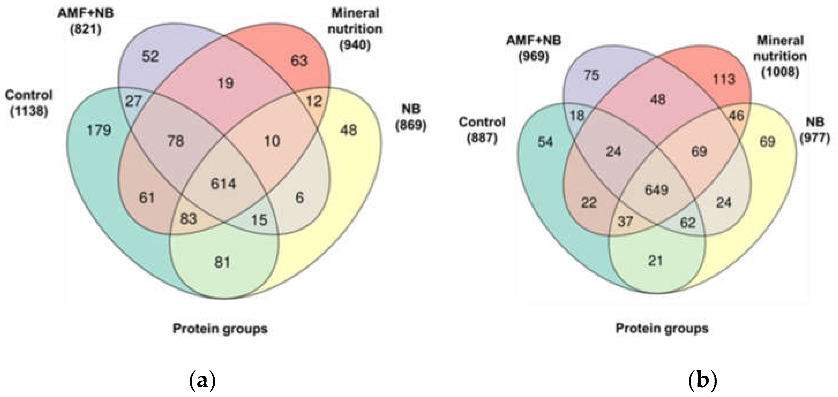
2.2. Protein Annotation

In total, 5382 and 5356 peptides could be identified by the MS/MS-based database search in the datasets acquired for the P. sativum genotypes k-8274 and k-3358, respectively (Supplementary Figure S1-3A and S1-4A, Table S2-1). Among these, for k-8274 genotype 2003 peptides occurred in all four groups, while 699, 199, 233 and 377 peptides were unique for the control plants (C), those grown in the presence of rhizobial symbiosis (nodule bacteria, NB), in the presence of NB and arbuscular mycorrhiza (NB+AMF) and in the presence of mineral nitrogen source in soil without inoculation with microorganisms (mineral nutrition, MN), respectively. Analogously, in total 2167 peptides were identified in all experimental plants of the k-3358 genotype, while 233, 343, 294 and 575 peptides were unique for C, NB, NB+AMF and MN groups, respectively.
Based on these identifications, altogether 2606 possible proteins (2237, 1746, 1637 and 1899 in C, NB, NB+AMF and MN groups) could be annotated for the k-8274 genotype (Supplementary Figure S1-3B and Table S2-2). They represented altogether 1348 non-redundant proteins (protein groups) with 1138, 869, 821 and 940 annotated to the controls, NB, AMF+NB and MN groups, respectively (Figure 1a, Supplementary Table S2-3). Among these, 1281 non-redundant proteins were common for the all four groups, while 312, 75, 75 and 128 proteins were unique for controls, NB, NB+AMF and MN groups, respectively. Analogously, in total 2570 probable proteins could be annotated in the plants of the k-3358 genotype (with 1699, 1948, 1866 and 2062 accessions annotated to the controls, NB, NB+AMF and MN groups, respectively, Supplementary Figure S1-4B and Table S2-2). Thereby, 1327 proteins were common for the all four groups, while 89, 111, 108 and 241 polypeptides were unique for the controls, NB, NB+AMF and MN groups, respectively (Supplementary Figure S1-4B). These annotations represented altogether 1331 protein groups, 887, 977, 969 and 1008 of which were identified in the controls, NB, NB+AMF and MN groups, respectively (Figure 1b, Supplementary Table S2-3). Thereby, 89, 111, 108 and 241 non-redundant proteins were unique in the controls, NB, NB+AMF and MN groups, respectively.
It is important to note that the MS/MS-based assignment of individual proteins as unique totally relies on confident prediction of the peptide sequences without any consideration of the inter-group alignments at the MS1 level. Obviously, such alignments (typically employed as a part of the label-free quantification (LFQ) protocol), would, on one hand, reduce the numbers of treatment-specific unique proteins (i.e., identification of individual proteins will be independent from the growing conditions), on the other – they would allow precise assessment of the inter-group abundance differences for individual “common” proteins. Therefore, this strategy was employed as the next step.

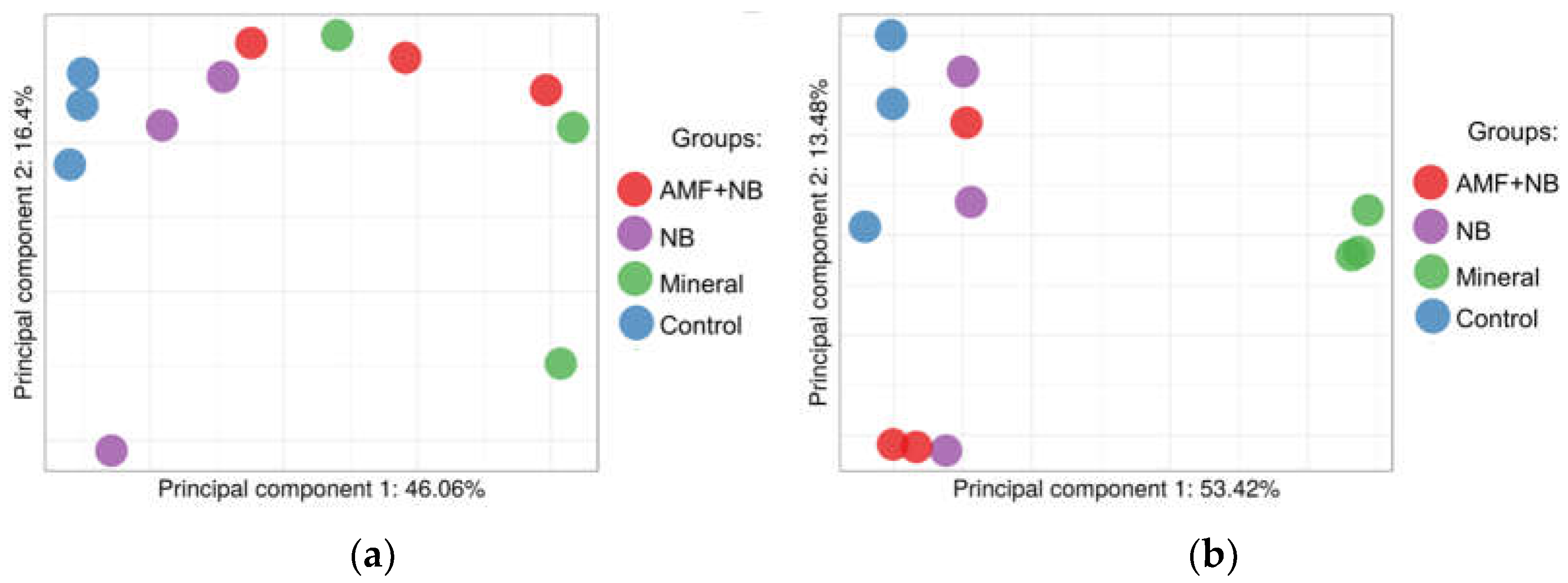
2.3. Label-Free Relative Quantification of the Annotated Proteins

At the first step, the inter-group differences were assessed by the methods of multivariative statistics. Thus, principal component analysis (PCA) accomplished for all samples obtained from the k-8274 plants revealed poor separation of the four treatment groups with 46.1 and 16.4% of the total variance, explained by the principal component 1 (PC1) and the principal component 2 (PC2), respectively. As can be seen from the corresponding scores plot (Figure 2a), the controls and NB plants were to some extent separated from each other and from the other two groups, that is in agreement with the known high responsivity of this genotype to inoculation with rhizobia [18]. The AMF+NB group and the plants grown in the absence of inoculations but in presence of mineral salts (external nitrogen supply) remained unseparated, that might indicate similar effect of these two conditions on the plant proteome.
The PCA accomplished for all samples obtained from the k-3358 plants revealed only limited separation of the four treatment groups with 53.4 and 2.1% of the total variance, explained by the principal component 1 (PC1) and the principal component 2 (PC2), respectively. As can be seen from the corresponding scores plot (Figure 2B), the MN group was well-separated from the others. The controls could be also distinguished from the two treatments with microorganisms (which remained unseparated), although the degree of underlying differences was lower than that observed for k-8274. This pattern was in a good agreement with low responsivity of k-3358 to rhizobia and AMF [19].
To get better insight in the molecular mechanisms underlying the observed inter-line differences in the responses to symbiotic microorganisms and supplemented mineral nutrition, we decided to dissect this complex picture in a series of paired inter- and intra-genotype comparisons. Specifically, we compared the protein expression patterns in the roots of the control k-8274 and k-3358 plants along with three intra-group comparisons (MN vs. control, NB vs. control and AMF+NB vs. control) for each of two genotypes. For this, the t-test was employed with the cut-off criteria FC (protein abundance fold change) ≥ 1.5 and padjusted ≤ 0.05. In these seven paired comparisons altogether 491 differentially abundant proteins could be found. The top three up- and down-regulated proteins identified in each comparison - which appeared to be the most responsive to different soil conditions (i.e., legume-rhizobial symbiosis, arbuscular mycorrhiza and supplementation with mineral sources of nitrogen vs control plants grown in the absence of soil supplementation), are listed in Table 1. The complete patterns of differential protein expression are described in more detail below.
Thus, 44 proteins could be identified as differentially abundant, when the genotypes k-8274 and k-3358 were compared in the absence of microorganism inoculations and soil supplementation with mineral salts. Thereby, 24 and 20 proteins were more abundant in the roots of the k-8274 and k-3358 plants, respectively (Supplementary Figure S3A, Tables S3-1 and S3-2). Thereby, dirigent protein and histone 2B showed the most pronounced inter-genotype differences (5.3- and 3.7-fold, respectively) and were more represented in the roots of k-8274 plants, whereas a representative of the peptidase C1 family (Q40993), isoforms X1 (G7JCJ5) and MYB-CC type transcription factor were more abundant in the roots of k-3358 (34.8-, 18.4- and 16-fold inter-genotype differences, respectively).
Soil complementation with mineral salts resulted in different responses of the genotypes k-8274 and k-3358 to external nitrogen supplementation. Thus, the k-3358 plants appeared to be more responsive to these conditions with 179 differentially ex-pressed proteins represented with 98 and 81 up- and down-regulated (in comparison to the corresponding controls) accessions, respectively (Supplementary Tables S3-3 and S3-4). The up-regulation pattern dominated with histone H2B, protein G7JCJ5 isoform X1 and Luc7-like protein (11.4-, 5.2- and 4.9-fold, respectively). Although the down-regulation pattern was less rich, it was featured with much more pronounced group of symbio-sis-related proteins with associated enzymatic and regulatory machinery the most af-fected. The top-responsive proteins of this group were the heat shock protein DnaJ (A2Q3G0), leghemoglobin K, carbonic anhydrase EC4.2.1.1 and leghemoglobin B with 1390.8-, 150.9-, 129.2- and 98.9-fold decrease in relative abundances, respectively.
The genotype k-8274 showed clearly less pronounced response to the soil supple-mentation with external nitrogen. Thereby only 111 proteins appeared to be affected with 55 and 56 up- and down-regulated accessions, respectively (Supplementary Tables S3-5 and S3-6). The up-regulation pattern was different from the counterpart genotype and dominated with the uncharacterized protein LOC101501292 (A0A1S2YAH3), mono-copper oxidase SKS1-like protein (putative L-ascorbate oxidase, EC 1.10.3.3) and lipoxygenase (EC 1.13.11. Q14ST8) showing 9.0-, 6.4- and 4.1-fold relative abundance shifts (Supplementary Tables S3-5). The down-regulation pattern showed less pronounced decrease in abundancies of the symbiosis-related proteins (Supplementary Table S3-6). Moreover, not only these proteins belonged to the top-affected. Indeed, β-fructofuranosidase cell wall isozyme 2-like protein (A0A1U9X403), unknown protein and putative chromatin CoxE-like regulator PHD family were the most regulated (246.3, 153.9 and 121.1-fold, respectively).
When the k-8274 and k-3358 plants were inoculated with rhizobia, the proteome response appeared to be principally different. Thus, the proteome of the former genotype did not respond to the inoculation with rhizobia, i.e., no changes in protein dynamics could be observed when the plants of the k-8274 genotype were inoculated with the microorganism culture. In contrast, 13 and ten proteins were identified as up- and down-regulated when rhizobia colonized the roots of the k-3358 plants (Supplementary Tables S3-7 and S3-8). Thereby, 60S ribosomal protein L5-2, protein G7JCJ5 isoform X1 and glutelin type-B-like protein (putative 11-S seed storage protein) most strongly contributed to the up-regulation pattern with 3.9-, 3.2- and 2.5-fold alterations, respectively (Supplementary Table S3-7). Although the down-regulation pattern included only ten proteins, it generally resembled the response of this genotype to soil complementation with external nitrogen with CBS/octicosapeptide/phox/Bemp1 (PB1) domain protein, putative ser-ine/threonine-protein kinase (A0A392LZA9) and leghemoglobin most strongly affected (16.4-, 2.4- and 2.3-fold, respectively, Supplementary Table S3-8).
Finally, combined inoculation of the k-3358 and k-8274 plants with arbuscular mycorrhiza fungi and rhizobia (AMF+NB) yielded clearly different responsivity of the corresponding root proteomes (Supplementary Tables S3-9 – S3-12). Thus, the former genotype re-sponded to the combined inoculation with 16 up- and 17 down-regulated proteins. Thereby, uncharacterized protein A0A2Z6PHW6, caffeic acid O-methyltransferase (putative methyltransferases EC 2.1.1.-) and histone H2B.1 appeared to be most strongly up-regulated species (3.5-, 2.8- and 2.5-fold, Supplementary Table S3-9), whereas putative wall-associated serine/threonine-protein kinase (A0A392LZA9), uncharacterized pro-tein A0A396ITY0 and carboxypeptidase EC 3.4.16.- (G7IT50) were the most down-regulated proteins (3.6- 3.4- and 3.3-fold, respectively, Supplementary Table S3-10).
In comparison to the genotype k-3358, the root proteome of the k-8274 plants proved to be much more responsive to the combined inoculation with two microor-ganisms (AMF+NB). This response was manifested with 39 up- and 59 down-regulated proteins (Supplementary Tables S3-11 and S3-12). Thereby, monocopper oxidase SKS1-like protein (putative L-ascorbate oxidase EC 1.10.3.3), HTH myb-type domain-containing protein (MYB-CC type transcription factor) and mitochondrial-like arginase 1 showed the highest increase in relative abundancies (5.5-, 4.2- and 3.4-fold). The down-regulation pattern was much more represented with the heat shock protein DnaJ, early nodulin ENOD18 and putative wall-associated serine/threonine-protein kinase as the most strongly affected proteins (67.8-, 44.9- and 26.3-fold, respectively).

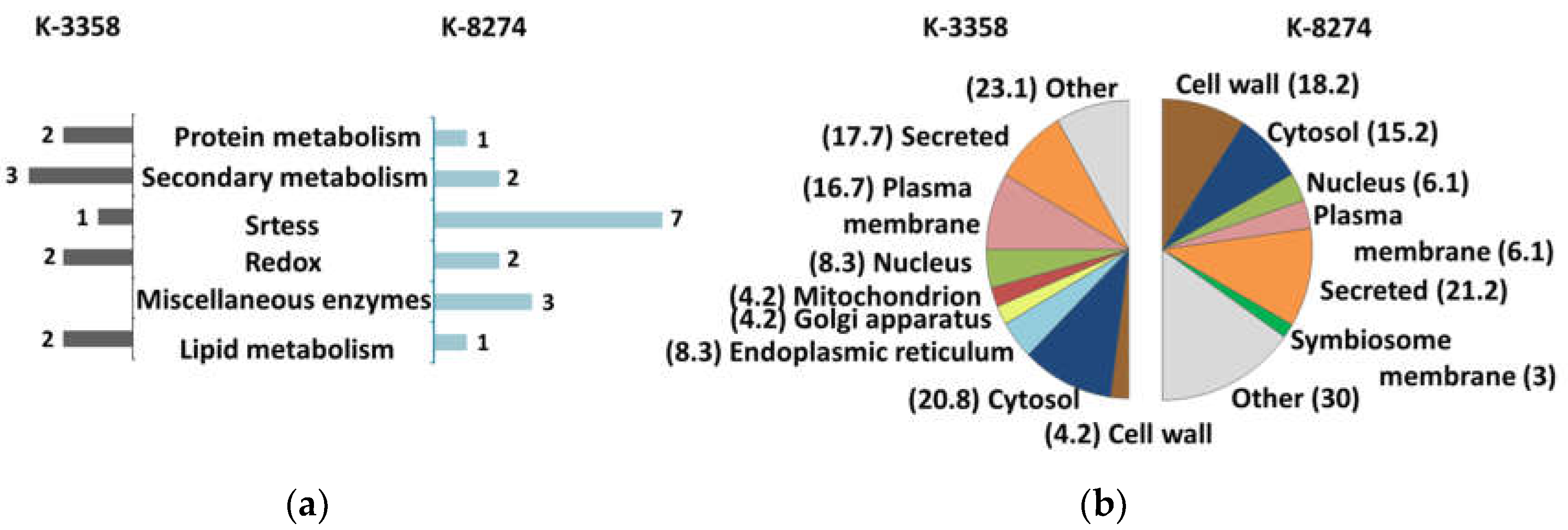
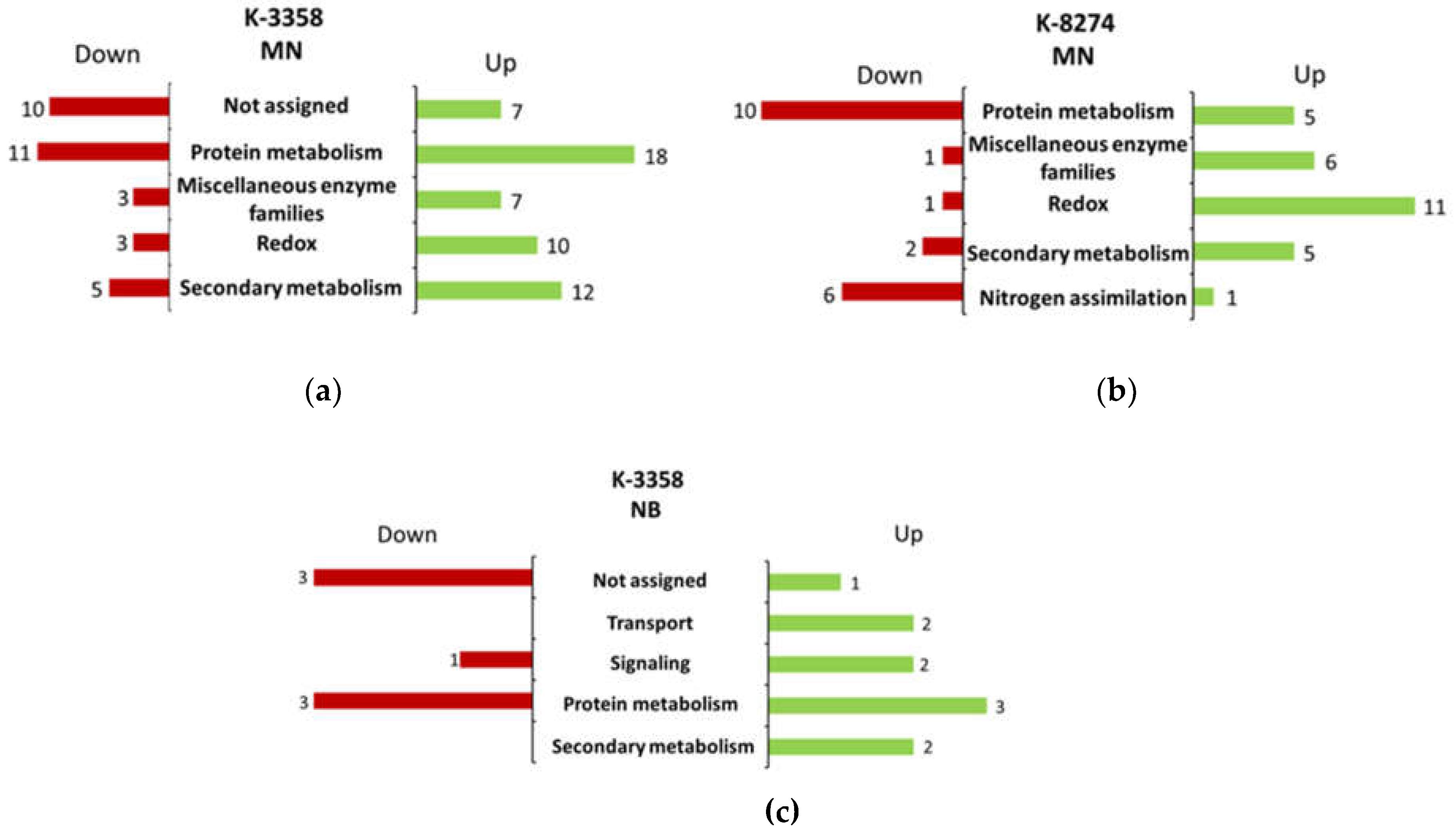
2.4. Functional Annotation of Differentially Expressed Proteins

Functional annotation of the differentially expressed P. sativum proteome relied on the Mercator 4 v.2 software. The annotated proteins represented altogether 31 functional classes (bins) among the 35 available (Figure 3 and Figure 4, Supplementary Table S4 and Supplementary Table S1-6 - S1-11).
Comparison of the root proteomes obtained from the control k-8274 and k-3358 plants revealed 14 and 13 functional classes, characteristic for the former and latter genotypes, respectively. Thereby, the most of these classes were represented with one or two proteins (Figure 3a, Figure S1-5). Among the proteins, more abundant in the roots of the genotype k-8274, the polypeptides involved in redox and secondary metabolism were the best represented (seven and three accessions, respectively, Supplementary Table S4-1). These groups included three peroxidases, two cytochromes b5, non-symbiotic leghemoglobin and three proteins involved in polymerization of lignins in cell walls. In agreement with this, cell wall (along with the group of secreted polypeptides) appeared to be one of the major predicted compartments for localization of the proteins, which were more abundant in the roots of k-8274, Figure 3b).
On the other hand, bins 20 (stress), 26 (enzymatic families), 29 (protein metabolism) and 30 (signaling) were the most represented among the proteins, which were more expressed in the roots of the k-3358 plants (Supplementary Table S4-2). The first group included a lipoxygenase PLAT-plant-stress protein and a late embryogenesis abundant protein, while the second one comprised three enzymes involved in secondary metabolism – two methyl transferases and a representative of the dienelactone hydrolase family. The bin 29 was represented with a cysteine peptidase C1 and serine carboxypeptidase S10, which are known to be involved in regulation of plant development, seed maturation, senescence and stress response [18,20,21]. Finally, the group of signaling proteins was represented with the RAB GTPase-like protein A1D (Ras-related protein Rab7) and a cell
Complementation of the soil with mineral nitrogen induced strong proteome response in the roots of the k-3358 plants. This response was manifested with pronounced up-regulation of 24 and down-regulation of 25 functional classes (Supplementary Figure S1-7). The former dominated with the polypeptides involved in protein metabolism, secondary and redox metabolism, as well as those annotated as “miscellaneous enzyme families” with 18, 12, 10 and 7 accessions, respectively (Figure 4a, Supplementary Table S4-3).
The up-regulated part of the protein metabolism-related proteome strongly dominated with its catabolic (degradation-related) component, including peptidases, pro-teases and elements of the proteasomal degradation machinery. The up-regulation pattern of the bin 16 (secondary metabolism) clearly indicated enhancement of several specific pathways. The most represented among these were phenylpropanoid pathway (with four enzymes - phenylalanine ammonia-lyase, cinnamyl alcohol dehydrogenase-like protein and two caffeic acid O-methyltransferases) and flavonoid biosynthesis pathway (with four enzymes - 2-hydroxyisoflavanone dehydratase, 2-hydroxyisoflavanone dehydratase and two chalcone-flavonone isomerase family proteins). Besides, phospho-2-dehydro-3-deoxyheptonate aldolase (the first enzyme in the chorismate biosynthesis pathway) and cytochrome P450 family monooxygenase G7KQK8 (the enzyme involved in the biosynthesis of xantophylls) were identified as up-regulated in the presence of the source of mineral nitrogen. Further, enhanced expression of the FAD-binding berberine family protein (an oxidase involved in biosynthesis of benzylisoquinoline alkaloids) was associated with these soil conditions. Finally, dirigent protein - the enzyme involved in the coupling of phenoxyl radicals, which is associated with all these pathways, was found to be more abundant when mineral salts were complemented to the soil. The enzyme families (bin 26) were represented by secretory proteases (plant basic secretory protein (BSP) family protein), stress tolerance (carboxylesterase), oxidoreductase of furanone biosynthesis (2-methylene-furan-3-one reductase) and others. Finally, the up-regulated redox-related pathways were represented with peroxidases, glutathione reductases and oxidoreductases.
The down-regulation pattern of the k-3358 root proteome dominated with protein metabolism (bin 29), secondary (bin 16) and redox metabolism (bin 21, Figure 4a, Supplementary Figure S1-7). The first group appeared to be heterogeneous: along with three polypeptides involved in proteasomal degradation and one S10 family carboxypeptidase, several players of translation (elongation factor Tu and SRP9/SRP14 subunit of the signal recognition particle), protein translocation (three different signal peptidases) and folding (peptide methionine sulfoxide reductase and protein disulfide-isomerase) machinery were discovered. The nitrogen assimilation bin (six accessions) represented the most strongly down-regulated group. This alteration could explain a strong enhancement of the protein degradation machinery described in the previous paragraph. The five down-regulated enzymes of the bin 16 represent five diverse pathways of secondary metabolism: phenylpropanoid pathway (caffeic acid/5-hydroxyferulic acid O-methyltransferase), metabolism of oximes (cytochrome P450 83B1) that might con-tribute to metabolism of auxins [22], alkaloid biosynthesis (3’-hydroxy-N-methyl-(S)-coclaurine 4’-O-methyltransferase), detoxification of α-dicarbonyls in the glyoxalase cycle (lactoylglutathione lyase-like protein) and oxida-tive polymerization of phenolics (dirigent protein). Moreover, aspartate and cysteine metabolism were clearly suppressed (Supplementary Table S4-4).
The pattern of the differential proteome response of the k-8274 roots to external mineral nitrogen supplementation (Figure S1-8) differed from that of k-3358. Thus, redox metabolism (bin 21), miscellaneous enzyme families (bin 26) protein metabolism (bin 29) and secondary metabolism (16) appeared to be the most responsive to these conditions (Supplementary Table S5). Among them, the bin 21 was the best represented and accounted 11 entries, being similar to the response of k-3358 by both protein numbers and diversity. The bin 26 was represented with six entries, which included an oxidoreductase, a glycosyl hydrolase, α/β-hydrolase and carboxypeptidase (Supplementary Table S4-5). In contrast to the roots of the k-3358 genotype, no up-regulation of the proteasome protein degradation machinery was observed. On the other hand, peptidases, potentially involved in lyzosomal degradation (family C1 peptidase) and stress induced programmed cell death (metacaspase-4-like protein) were observed as up-regulated, as well as the proteins involved in protein biosynthesis and maturation (aspartate-tRNA ligase 2, cytoplasmic-like, acyl-peptide hydrolase-like protein). Finally, two unique proteins (phenylcoumaran benzylic ether reductase-like protein Fi1 and vestitone reductase-like protein), along with three those detected in k-3358, could be found among the five up-regulated proteins representing the bin 16 (secondary metabolism).
The down-regulation pattern of the k-8274 root proteome was less abundant as the up-regulation one and dominated with the polypeptides involved in protein metabolism (bin 29), nitrogen assimilation (bin 12) and signaling (bin 30, Figure 5b and Supplementary Table S1-8). The down-regulated part of the bin 29 was more abundant and diverse in com-parison to the up-regulated one. Thus, among the ten accessions, three proteins were involved in protein degradation (one proteasome protein, one associated regulatory protein and one peptidase S10 type), whereas five proteins appeared to be involved in protein synthesis and translocation and further two – in protein folding (Supplementary Table S4-6). The proteins involved in nitrogen metabolism were represented with six leghemoglo-bin accessions, which were the most strongly down-regulated species (although at least three times less affected in comparison to the root proteome of the k-3358). Finally, DNA-binding regulatory element (general regulatory factor 2 of the 14-3-3 family) was detected among the four down-regulated signaling proteins. Moreover, similarly to k-3358, cysteine and aspartate metabolism was affected in the roots of the k-8274 grown in the presence of mineral nitrate, although this response was much less metabolically diverse in comparison to k-3358. On the other hand, in contrast to k-3358, here threonine metabolism was affected as well.
The nitrogen assimilation bin (six accessions) represented the most strongly down-regulated group. This alteration could explain a strong enhancement of the protein degradation machinery described in the previous paragraph. The five down-regulated enzymes of the bin 16 represent five diverse pathways of secondary metabolism: phenylpropanoid pathway (caffeic acid/5-hydroxyferulic acid O-methyltransferase), metabolism of oximes (cytochrome P450 83B1) that might contribute to metabolism of auxins [22], alkaloid biosynthesis (3’-hydroxy-N-methyl-(S)-coclaurine 4’-O-methyltransferase), detoxification of α-dicarbonyls in the glyoxalase cycle (lactoylglutathione lyase-like protein) and oxidative polymerization of phenolics (dirigent protein). Moreover, aspartate and cysteine metabolism were clearly suppressed (Supplementary Table S4-4).
When the k-3358 plants were grown in the absence of mineral nitrogen sources, but in the presence of legume-rhizobial symbiosis, the response of the root proteome was minimal and covered nine up- and five down-regulated proteins (Figure 5C and Supplementary Figures S1-9, Supplementary Tables S4-7 and S4-8). Thus, bin 29 (protein metabolism) showed the most pro-nounced up- and down-regulation response (three accessions in each case). The all three up-regulated proteins represented the biosynthetic branch of the protein metabo-lism – two ribosomal proteins and one storage polypeptide - glutelin type-B-like pro-tein. In contrast, the down-regulation pattern of this bin was heterogeneous – one cat-abolic protein (carboxypeptidase of the S10 family), one repairing enzyme (F-box plant-like protein) and one regulatory polypeptide (peptide methionine sulfoxide re-ductase-like protein). The latter pattern was accompanied with one leghemoglobin – a protein involved in nitrogen fixation, which decreased its abundance. The up-regulation pattern included enzymes involved in secondary metabolism (phenyl-alanine ammonia-lyase and dirigent protein) transport (Transmembrane amino acid transporter family protein and drug resistance transporter-like ABC domain protein) and signaling (B-5-like nuclear transcription factor and vacuolar-sorting receptor-like protein).
In agreement with our earlier observations done at the level of seeds [18] , the pro-teome responses were essentially enhanced when the roots of the k-3358 and k-8274 plants were inoculated with rhizobial culture in parallel to the infection with arbuscu-lar mycorrhiza fungi (AMF+NB, Figure 5d,e, Supplementary Figures S1-10 and S1-11, Supplementary Tables S4-9 – S4-12). Thus, functional annotation of the k-3358 root proteome, which appeared to be highly responsive to such combined inoculation, revealed 10 (16 accessions) and 12 bins (17 accessions) representing up-regulation and down-regulation patterns, respec-tively (Figure 5D and Figure S1-10). Among the former, protein metabolism (bin 29) and miscellaneous enzyme families (bin 26) were the best represented with four and three accessions, respectively (Supplementary Table S4-9). The polypeptides representing the bin “protein metabolism” were mostly involved in protein biosynthesis (leucine tRNA ligase, cyto-plasmic isoform X1) and folding (TCP-1/cpn60 chaperonin family protein), while the annotated enzymes (bin 26) were the players of phenolic metabolism - caffeic acid O-methyltransferase (phenylpropanoid pathway), 2-hydroxyisoflavanone dehydratase (flavonoid biosynthesis pathway) and putative secoisolariciresinol dehydrogenase (lignin biosynthesis). Although the down regulated proteins were most represented in terms of bins, only three annotated functional classes could be observed with more than one differentially abundant protein. The first of these – bin 29, included five accessions, three of which were catabolic proteins (26S proteasome regulatory particle non-ATPase subunit 8 and two S10 family peptidases). The other two down-regulated proteins of this bin represented regulatory (plant F-box-like protein) and folding (70 kDa heat shock protein chaperone DnaK) functions. Bins 30 (signaling) and 11 (lipid metabolism) were represented with two proteins each. Thus, two signaling proteins (calmodulin and putative serine/threonine-protein kinase) and two enzymes of lipid metabolism (fatty acyl-CoA synthetase family protein and phospholipase A1-IIgamma-like isoform X1) were annotated to these two protein functional classes, respectively.
In comparison to k-3358, the k-8274 plants demonstrated approximately 2.5 – 3.5-fold more abundant response to combined inoculation with two microorganisms, which was featured also with different bin distribution. Indeed, the up-regulation pat-tern included 39 proteins distributed in 22 functional classes, while the down-regulation one had 59 accessions in 26 bins (Figure 5e and Supplementary Figure S1-11). Thereby, re-dox metabolism comprised the most representative up-regulated functional class. All seven proteins contributing to this bin appeared to be protein antioxidants and ROS-detoxification agents: ascorbate oxidase, monodehydroascorbate reductase, glutathione S-transferase and several peroxidases (Supplementary Table S4-11). Further four bins - protein metabolism, miscellaneous enzyme families, stress response and mitochondrial energy metabolism were represented by three up-regulated proteins. The latter group included three FAD- and NAD(P)H-dependent oxidoreductases (quinine reductases) of the mitochondrial electron-transport chain. The stress response proteins included one late embryogenesis abundant (LEA) protein, and one heat shock protein that might indicate (in addition to the above listed redox proteins) activation several mechanisms of stress response. Importantly, one of these proteins - PLAT-plant stress protein, plays an important role in successful nodule formation [23]. The enzyme families were rep-resented with one O-methyltransferase, glycoside hydrolase 18 domain-containing protein (with chitinase and/or ENGase activity) and 3-ketoacyl-CoA thiolase peroxi-somal-like protein, which is known as an important actor of fatty acid peroxisomal beta-oxidation cleaving straight chain 3-oxoacyl-CoAs of different length. Finally, the bin 29 (protein metabolism) was represented by three proteins, including one heat shock protein and Kunitz type trypsin inhibitor.
The down-regulation pattern was more abundant and diverse E and Supplementary Figures S1-11). Although it was dominated with bin 29 (protein metabolism), the proteins in-volved in stress response, amino acid and nitrogen metabolism demonstrated a strong abundance decrease as well. The down-regulated polypeptides (eight accessions) of the bin 29 were mostly represented by the anabolic branch of the protein metabolism, i.e., proteins involved in translation (elongation factor Tu and an S40 ribosomal protein), translocation (two signal peptidases) and folding (one mitochondrial heat shock 70 kDa protein and a peptide methionine sulfoxide reductase-like protein), with only minimal impact of the catabolic pathways (proteasome subunit alpha type, Supplementary Table S4-12). The next abundant down-regulated protein group was bin 20 (stress) which was represented with two heat shock proteins, early nodulin ENOD18 (which is known as a stress-responsive protein, which is over-expressed e.g., under drought condi-tions [24]), chloroplastic plastoglobulin-1 (which is involved in transport of lipids across tilacoid membrane [25]) and carboxylesterase 15 – enzyme involved in catabo-lism of strigolactones [26]. Decrease in the abundance of the latter enzyme (which is known to degrade strigolactones [26]) might indicate the success of the arbuscular my-corrhiza symbiosis, as strigolactonts are directly involved in its establishment. Interestingly, the pathways of arginine, aspartate and cysteine biosynthesis appeared to be suppressed in presence of the combined root inoculation with two microorganisms. Analogously, four leghemoglobins were strongly down-regulated under these conditions.

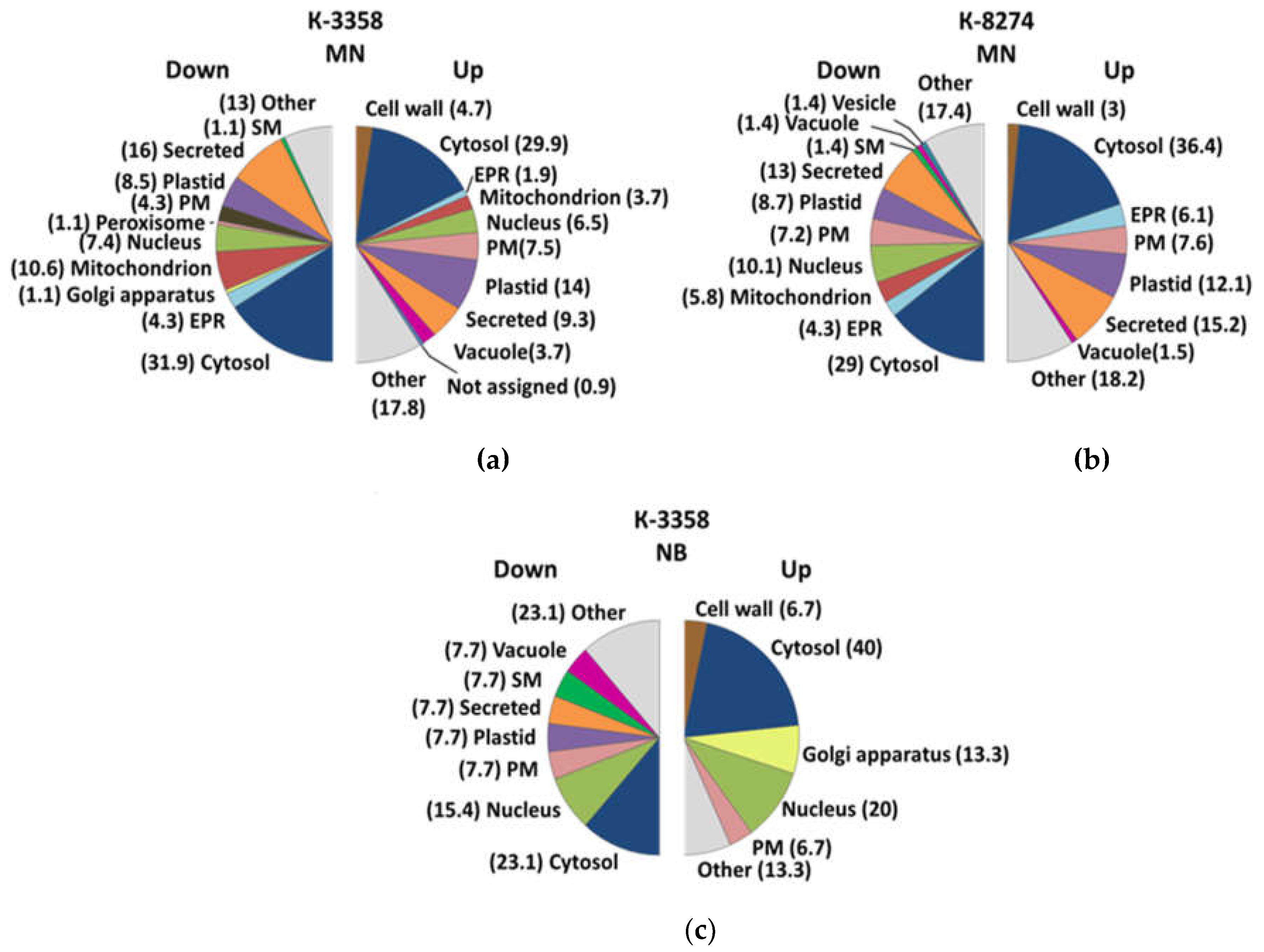
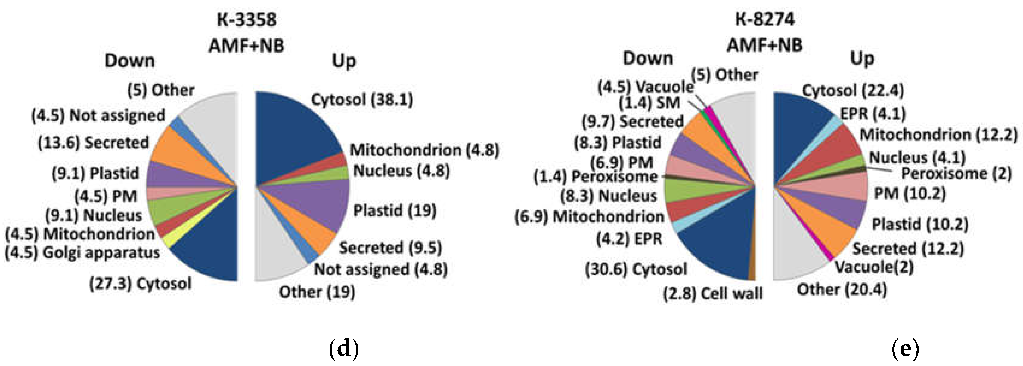
2.5. Subcellular Localization

The prediction of subcellular localization relied on LocTree3 with subsequent manual verification based on UniProt database and literature data (Supplementary Information S5). The results demonstrated rather similar patterns of differentially abundant proteins, changing their localization in all experimental groups of plants grown in the presence of different soil supplementations. For the both genotypes, cytosol, plastids and extracellular space appeared to be the most strongly contributing compartments (Figure 5a–d). On the other hand, although it could be noted that proteins localized in nucleus and Golgi apparatus were also highly susceptible to changes in the roots of the k-3358 plants, it was much less the case for the genotype k-8274 (Figure 5b).

3. Discussion

The principal objective of this work was to study the root response of two pea (P. sativum) genotypes, namely k-3358 and k-8274, to inoculation with beneficial soil microorganisms. Based on dynamics of multiple productivity parameters (including morphological traits [27] and seed proteome shifts [18]) related to associations with beneficial soil microorganisms (BSMs), these genotypes are known to be low- and high-performing, respectively. Indeed, in a three-year field trial k-3358 did not increase the seed productivity due to inoculation with BSMs, whereas for k-8274 the increment of seed weight due to inoculation exceeded 100% [19]. The genetic basis of these differences in symbiotic responsivity of garden pea was investigated earlier, and some related genes could be identified [12].

3.1. Inter-Genotype Differences in Proteome Signatures

Here we are making the next step and address the changes in the root proteome associated with inoculation of the pea k-3358 and k-8274 plants with rhizobia in the absence (nodule bacteria, NB) and presence of simultaneous infection with arbuscular mycorrhiza fungi (AMF+NB) in parallel to the plants grown in the absence (controls) and presence of mineral nitrogen supplementation (mineral nutrition, MN). We believe that this experimental setup might give better access to the molecular mechanisms involved in the response to interaction with both microorganisms. Thereby, both the differences in the effect of the legume-rhizobial symbiosis on the root proteome and the impact of AM fungi on its dynamics could be efficiently addressed. Obviously, it will result in pronounced success in establishment of fundamentally new pea cultivars capable of efficient interaction with beneficial soil microorganisms.
The first step to understand the responsivity of two genotypes to symbiosis with BSMs was their comparison in the absence of microbial inoculation and supplementation of mineral nitrogen. Although this comparison revealed relatively low numbers of differentially expressed proteins (that could indicate minimal inter-genotype differences visible in the absence of symbiosis, Supplementary Tables S3-1 and S3-2), their functional annotation could give access to some genotype-specific features (Supplementary Tables S4-1 and S4-2).
Thus, in the absence of supplemental nitrogen source, both genotypes are expected to suffer from nutritional stress. However, this state appeared to be much more pronounced in the roots of the k-8274 plants. Indeed, the latter showed essentially higher diversity of the stress induced polypeptides in comparison to the k-3358 plants (seven vs. two accessions) with strong domination of redox proteins. This response pattern was in agreement with higher expression of the proteins involved in biosynthesis of phenylpropanoids and their polymerization in the cell wall that was clearly characteristic for the k-8274 roots. This reaction is characteristic for abiotic stress response [28].
In contrast, no substantial activation of the redox metabolism (and, specifically, no up-regulation of peroxidases) could be seen in the roots of the k-3358 plants, while the only two dominating here stress-associated proteins were related to abiotic stress tolerance [29,30]. In agreement with this, MYB-CC type transcription factor, which is known to be involved in regulation of cell cycle, cell morphogenesis, responses to biotic and abiotic stresses [31], was found to be most expressed in k-3358 roots (Supplementary Table S4-2). Analogously, cysteine peptidase C1 and serine carboxypeptidase S10, which are involved in regulation of plant development, seed maturation, senescence and stress response [21,32] , were more abundant in the k-3358 roots. Most likely, this protein response pattern indicates higher stress tolerance and ability to sustain cell integrity of this genotype.

3.2. Subcellular Root Proteome Response of the k-3358 and k-8274 Plants to Soil Complementation with Mineral Nitrogen Localization

The inter-genotype differences became more striking when soil was supplemented with mineral nitrogen-containing salts. The analyzed genotypes demonstrated essential differences in their sensitivity to this factor with the genotype k-3358 being essentially more responsive. The main feature was an essential decrease in expression of leghemoglobins and several other regulatory proteins and enzymes related to nitrogen fixation. Such suppression of symbiotic nitrogen fixation in presence of mineral nitrogen in soil was reported for bean [33], soybean [34] and pea [35] (in the experiment from which the material for this study was collected, the suppression of nodulation under mineral nutrition was also noted for both k-3358 and k-8274 [27]). However, the relative abundance of leghemoglobins demonstrated at least a 7-fold stronger decrease in presence of mineral nitrogen in the roots of k-3358 in comparison to k-8274 (Supplementary Figures S3-4 and S3-6). Interestingly, the roots of k-3358 showed the most strong expression suppression for the heat shock protein DnaJ (1391-fold). To some extent it can be explained by its role in improvement of symbiosis performance (reviewed by Da Silva and co-workers [36]).
In general, complementation of inorganic nitrogen to the soil resulted in strong down-regulation of multiple proteins critically impacting on the success and efficiency of legume-rhizobial symbiosis. Thus, among the proteins strongly down-regulated in the roots of k-3358 in presence of mineral nitrogen were the proteins involved in initiation of symbiosis - early nodulins 7 (which is typically expressed in pea root nodules induced by Rhizobium leguminosarum bv. viciae [37]) and 18 (which can act as a carbohydrate transporter [38] or might impact on salt stress tolerance [39]). Following this logic, a pronounced decrease in the relative abundances of VWA domain containing CoxE-like protein, putative chromatin regulator PHD family A0A396JLK8 (both 89-fold), FAM91A1-like protein (10-fold) and pentatricopeptide repeat-containing protein At1g31920 (53-fold) might indicate in regulation of nodulation.
Another strongly down-regulated protein was Acyl-CoA-binding protein, ACBP (75.5-fold). This protein is a key player in oxygen sensing in plants (which relies on the N-end rule pathway, NERP), it binds group VII ethylene response factor and, thereby, directly involved in the regulation of their stability [40]. A slightly lower, but pronounced (49-fold) nitrogen-induced decrease in the abundance of the vegetative lectin Q41069 – the molecule known to impact in success of root inoculation with rhizobia [41] was observed as well. Finally, several enzymes, which are critical for symbiosis establishment, were also strongly down-regulated. Most pronounced it was for carbonic anhydrase (129-fold). The gene of this enzyme is known to be induced in the nodules of different origin, and the activity of this enzyme might have clear anabolic role [42]. Another important actor – cell wall invertase, which degrades sucrose to make sugars accessible for the symbiotic microorganisms [43] , was also strongly down-regulated in k-3358 roots upon N complementation (89-fold). Interestingly, although multiple enzymes of central metabolism were down-regulated as well (e.g., fructose-bisphosphate aldolase, isocitrate dehydrogenase and cytochrom c), the degree of this expression suppression was relatively low. Finally, in agreement with the published data, clear down-regulation of heme biosynthesis (oxygen-dependent coproporphyrinogen-III oxidase), ammonia metabolism (amine oxidase and aspartate aminotransferase) and cysteine metabolism (cysteine synthase and lactoylglutathione lyase-like protein) was observed (Supplementary Table S3-4).
Obviously, the described down-regulation pattern observed in the k-3358 roots was the result of the strong enhancement in biosynthesis of enzymes and regulators primarily associated with protein degradation (amino- and carboxypeptidases, proteinases, components of proteasomes, Supplementary Tables S3-3 and S4-3). This was, however, most likely accompanied with enhanced transcription. Indeed, two up-regulated proteins act at the chromatin level: the chromatin CoxE-like regulator PHD family (PHD-containing proteins also mediate adaptive responses of the root to environmental factors - a part of the chromatin remodeling system, mediating activation of gene expression [44]) and histone H2B, one of the main components of the nucleosome. Importantly, the synthesis of core histones is strictly coupled to DNA replication in S-phase. This pattern of up- and down-regulated proteins may indicate the intensification of processes related to maturation.
The comparison of the above described responses with those of the k-8274 plants showed that the whole pattern of the nitrogen-responsive proteins was less representative and the observed changes were less pronounced, although in further details similar (Supplementary Tables S3-5 and S3-6). Thus, the genotypes showed similar degree of stress response – both in terms of the types and numbers of redox enzymes and in terms of fold change (although k-3358 showed more abundant up-regulation pattern of stress-induced proteins).
However, despite this similarity, in contrast to the genotype k-3358, the k-8274 plants did not show any activation of protein degradation in response to complementation of mineral nitrogen (although down-regulation of three proteins involved in this process, including one proteasome component, was observed). Another important difference was the presence of the N-induced responses at the level of secondary metabolism. Thus, though the both genotypes showed up-regulation of phenolic metabolism (specifically, phenylpropanoid pathway and flavonoid biosynthesis), it was essentially more pronounced for the k-3358 (Supplementary Table S4-3). In line with this, dirigent protein which plays an important role in lignan and lignin biosynthesis by exercising the spatial control of lignin deposition, especially under stressed conditions [45] was more strongly up-regulated in the k-3358 roots (2.5- vs 1.5-fold). In contrast, the k-8274 plants showed more pronounced decrease in the abundance of lactoylglutathione lyase, that might indicate less activity of glyoxalase cycle and, hence, higher amenability of this genotype to oxidative stress under these conditions.
Thus, it can be concluded, that the k-3358 plants appeared much more responsive to complementation with mineral nitrogen salts, demonstrating higher adaptation potential in to this factor. It showed more pronounced redirection of metabolism from symbiotic nitrogen nutrition to the mineral nitrogen supply. Thus, this genotype has higher (in comparison to k-8274) efficiency when nitrogen was supplied from mineral sources. In contrast, k-8274 showed compromised metabolic efficiency under such nutritional conditions that was in line with its lower productivity gain when mineral nitrogen sources were used.

3.3. Root Proteome Response of the k-3358 and k-8274 Plants to Inoculation with Symbiotic Rhizobia

When the nitrogen supply was provided only via the symbiotic route (i.e., upon establishment of legume-rhizobial symbiosis in the absence of soil complementation with mineral nitrogen sources), no changes in the proteome of the k-8274 roots could be observed. The absence of detectable response at the level of metabolic pathway, regulatory systems and stress response might indicate god compatibility of the plant and microbial partners. This fact was in a good agreement with good responsivity of this genotype to inoculation with R. leguminosarum [18].
In contrast, the roots of the k-3358 plants showed detectable response at the proteome level, although it was minimal (Supplementary Tables S3-7 and S3-8). The up-regulation pattern was represented with a small number of bins, with not more than one to two proteins in each. We can pay attention to the increase in glutelin type-B-like protein - one of the main reserve proteins that are combined into protein bodies in the endosperm, which may indicate an acceleration of plant development. This fact in a good agreement with the seed productivity data [18], indicating low responsivity of this genotype to inoculation with symbiotic rhizobia and lower biomass gain due to accelerated development.

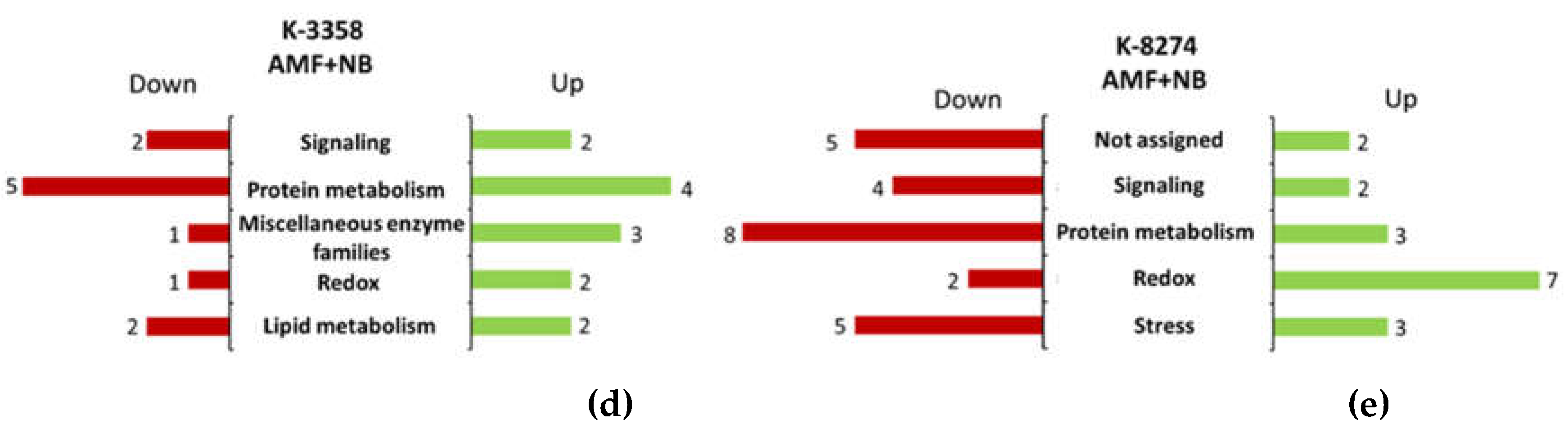
3.4. Root Proteome Response of the k-3358 and k-8274 Plants to Combined Inoculation with Nodule Bacteria and AM Fungi

At the last step, we addressed the response of the root proteome of k-3358 and k-8274 pea genotypes to combined inoculation with arbuscular mycorrhiza fungi and rhizobia. Based on previous data [18] this setup results in the maximal productivity gain of the genotype k-8274, which responsivity to rhizobial inoculation increased when the roots were colonized with AM fungi.
Our data indicated that both genotypes demonstrated clear proteome responses, although it was more pronounced for the k-8274 plants. Indeed, the latter showed approximately two times richer up- and three times more representative down-regulation response (Supplementary Tables S3-9 – S3-12). Also, the response of the k-8274 plants was featured with much higher functional diversity - 22 and 26 bins were up- and down-regulated, respectively, while only 10 bins contributed to the up-regulation pattern in the k-3358 roots. This fact reflects the higher responsivity of k-8274 to inoculation with beneficial microorganisms, as compared to k-3358.
In the roots of k-3358, histone H2B.1 (variant form of histone H2B) was detected. Also, polypeptides involved in protein synthesis and maturation (leucine tRNA ligase, TCP-1/cpn60 chaperonin family protein) were up-regulated. On the other hand, in the k-8274 plants this functional group is represented by only three proteins - heat shock protein with ATPase domain, Kunitz type trypsin inhibitor and uncharacterized protein.
Also in k-3358 enzymes of phenylpropanoid pathway, flavonoid biosynthesis pathway and lignin biosynthesis were up-regulated. This may indicate the activation of defense reactions as a response to mycorrhization. Putative 3-oxoacyl- [acyl-carrier-protein] reductase, an enzyme involved in fatty acid biosynthesis (a process accompanying the arbuscular mycorrhiza development in plant roots) [46], was also found to be up-regulated. Down-regulated proteins belong to almost all functional groups, but most of the groups are represented by a single protein. Bin 29 (Protein metabolism), represented by Carboxypeptidases, 26S proteasome regulatory particle non-ATPase subunit 8, HSP DNA K and F-box plant-like regulatory protein, deserves attention. Then, a down-regulation of isocitrate dehydrogenase [NADP], an enzyme participating in antioxidant system [47], and 1-aminocyclopropane-1-carboxylate oxidase, an enzyme catalyzing the last step of ethylene biosynthesis, was noted. This may indicate that biological processes in k-3358 roots are less active under NB+AMF inoculation than in control conditions.
As for the genotype k-8274, the greatest changes affected the group of proteins of redox metabolism: protein antioxidants and ROS-detoxification agents, which contributes to the overall activation of metabolism and increased adaptive capacity of the plant. Up-regulation of the antistress proteins Late embryogenesis abundant protein (LEA), PLAT-plant-stress protein and lipoxygenase may have the same result. Although a number of up-regulated functional protein classes are represented by one or two proteins, these changes indicate increased catabolism, primarily carbohydrate catabolism: glucan endo-1,3-beta-glucosidase (polysaccharide cleavage), fructose-bisphosphate aldolase (glycolysis), isocitrate dehydrogenase, and malic enzyme (tricarboxylic acid cycle). The up-regulation of components of the respiratory complexes of the mitochondrial inner membrane and ferredoxin-NADP reductase of chloroplasts is also consistent with these data. Among the up-regulated enzymes is arginase, whose function in plants is to mobilize nitrogen by arginine accumulation, which contributes to the synthesis of other amino acids and polyamines during seed germination.
The down-regulation pattern is even richer in both diversity and quantity of proteins. Similar to the proteome of the roots of the former variety, the most representative is the functional group “protein metabolism”, represented by factors of translation, folding and processing. Also, a number of stress-induced proteins represented with two heat shock proteins, early nodulin ENOD18, leghemoglobins, together with proteins from the pathways of arginine, aspartate and cysteine biosynthesis, were down-regulated. The observed down-regulation of nodule-specific proteins matches the fact that the number of nodules in k-8274 under NB+AMF inoculation was lower than in control conditions [27]; perhaps, the control over the nodules number contributes to the manifestation of the high EIBSM trait in k-8274 genotype.

4. Materials and Methods

4.1. Reagents

Unless stated otherwise, the following materials were obtained from Merck KGaA (Darmstadt, Germany): ethylenediaminetetraacetic acid, phenol, dithiothreitol, iodoacetamide, Coomassie Brilliant Blue G-250; SERVA Electrophoresis GmbH (Heidelberg, Germany): β-mercaptoethanol; AppliChem (Darmstadt, Germany): phenylmethylsulfonyl fluoride; Hollywell (Charlotte, North Carolina, USA): formic acid (LC-MS Grade), acetonitrile (LC-MS Grade); Progenta™ (Radnor, Pennsylvania, USA): Anionic Acid Labile Surfactant II (AALS II). All other chemicals were purchased from Carl Roth GmbH and Co (Karlsruhe, Germany). Water was purified in house on a water conditioning and purification system Millipore Milli-Q Gradient A10 system (resistance 18 mΩ/cm, Merck Millipore, Darmstadt, Germany).

4.2. Plant Experiment

In this work, we used plant material from the previously accomplished vegetation experiment described in detail earlier [27]. Pea plants of the genotypes k-8274 (cv. Vendevil, France) and k-3358 (unnamed cultivar from Saratov region, Russia) (from the collection of cultivated peas, N. I. Vavilov All-Russian Institute of Plant Genetic Resources (VIR) St. Petersburg, Russia) were grown in non-sterile soil (three plants per a five-liter pot) in a vegetation house during summer period at ARRIAM, St. Petersburg, Russia. The experiment included the following variants: non-inoculated plants (control), the plants treated with full mineral nutrition (MN) – a mineral nitrogen reference group, the plants inoculated with R. leguminosarum bv. viciae strain RCAM 1026 (NB), the plants simultaneously inoculated with R. leguminosarum bv. viciae strain RCAM 1026 and with a mixture of arbuscular mycorrhizal fungi Rhizophagus irregularis BEG144, R. irregularis BEG53 and Glomus sp. ST3 (NB+AMF). After four weeks of growth, the plants were harvested; root systems were rinsed with water, immediately frozen in liquid nitrogen and stored in -80° C until further analysis. All experiments were performed in three biological replicates. Thereby, three plants in one pot were pooled per replicate.

4.3. Protein Extraction

Protein extraction was accomplished according to the protocol of Frolov and co-workers with minor modifications [48]. In detail, 650 µL of ice cold (4°C) phenol extraction buffer (0.7 mol/L sucrose, 0.1 mol/L KCl, 5 mmol/L ethylenediaminetetraacetic acid (EDTA), 2% (v/v) β-mercaptoethanol and 1 mmol/L phenylmethylsulfonyl fluoride (PMSF) in 0.5 mol/L Tris-HCl buffer, pH 7.5) was added to approximately 500 mg of grinded frozen root material. After short vortexing (30 s), 650 µL of ice cold phenol (4°C) pre-saturated with 0.5 mol/L Tris-HCl buffer (pH 7.5) were added and vortexed under the same conditions. Afterwards, the samples were shaken (30 min, 900 rpm, 4°C) and centrifuged (5000 g, 15 min, 4°C), the lower aqueous phase was discarded, and the phenolic phase was washed two times with equal volumes of the phenol extraction buffer. Each buffer addition was followed by vortexing 30 s, shaking (30 min, 900 rpm, 4°C) and centrifugation (5000 g, 15 min, 4°C). Finally, the aq. phase was discarded, the phenol one was supplemented with five volumes of cold (-20°C) ammonium acetate in methanol (0.1 mol/L), and the proteins were let to precipitate under -20°C overnight. Next morning, the total protein fraction was pelleted by centrifugation (5000 g, 15 min, 4°C), and the pellets were washed twice with two volumes (relative to the volume of the phenol phase before precipitation) of cold (4°C) methanol, and once with the same volume of cold (4°C) acetone. Each time after re-suspending, the samples were centrifuged (5000 g, 10 min, 4°C). Finally, cleaned pellets were dried under air flow in a fume hood for 1 h and then re-constituted in the shotgun buffer (8 mol/L urea, 2 mol/L thiourea in 0.1 mol/L Tris-HCl buffer, pH 7.5) containing 0.1 % (w/v) anionic acid-labile surfactant II (AALS II, (Protea, USA).

4.4. Determination of Protein Concentration

Determination of protein concentration relied on the 2-D Quant Kit (GE Healthcare, USA), according to the protocol of the manufacturer. The precision of the assay was cross-verified by SDS-PAGE according to Greifenhagen et al. with some modifications [49]. In brief, after the gels were stained overnight with 0.1 % (w/v) Coomassie Brilliant Blue G-250 for 12 h, the average densities across individual lanes (expressed in arbitrary units) were determined by Bio-5000plus Microtek imaging system controlled by BioDoc analysis software (SERVA Electrophoresis, Germany). For calculation of the relative standard deviations (RSDs), the densities of individual lines were normalized to the gel average value by ImageJ software.

4.5. Tryptic Digestion

The aliquots of the tryptic digests (50 µg) were supplemented with the AALS II-containing shotgun buffer to obtain the final sample volumes of 90 µL. Afterwards, 10 µL of 50 mmol/L tris(2-carboxyethyl)phosphine) in AALS-free shotgun buffer were added. After a 30-min incubation at 37°C under continuous shaking (450 rpm), 11 µL of 100 mmol/L iodoacetamide in the same buffer were added and the samples were further incubated (1h, 450 rpm, 4°C) before 875 µL of aq. 50 mmol/L ammonium bicarbonate (ABC) and freshly prepared 0.5 µg/µL trypsin in ABC (enzyme-to protein-ratio of 1:20 w/w) were added. After the following incubation for 5 hours (450 rpm, 37 °C), digestion was repeated at the enzyme-to protein-ratio of 1:50 (w/w) overnight. Next morning the digestswere frozen and the samples were stored at -20°C. The completeness of the digestion was verified by SDS-PAGE as described by Greifenhagen and co-workers [49].

4.6. Solid Phase Extraction

After decomposition of ALLS (10 % v/v TFA, 20 min at 37 °C), the proteolytic hydrolysates were pre-cleaned by reversed phase solid phase extraction (RP-SPE) using the elution scheme of Spiller, et al. [50] with minor modifications. Stage-Tips with six layers of C-18 Extraction Disks (66883-U) (Sigma-Aldrich) were prepared in 200 µL polypropylene pipette tips and inserted in 2 mL tubes via plastic tube adaptors. The Stage-Tips were conditioned with 100 µL of MeOH (2000x g, 5 min, 25°C) and equilibrated with two portions of 200 µL of 0.1% (v/v) formic acid (FA, 2000x g, 5 min, 25°C), before the individual tryptic digests were applied and the stage tips were centrifuged (2000x g, 5 min, 25°C). After the washing (2 x 200 µL of 0.1% (v/v) FA) with centrifugation after each step (2000x g, 5 min, 25°C), the peptides were eluted by sequential application of 150 µL of 60% (v/v) acetonitrile and 150 µL of 80% (v/v) acetonitrile, 0.1% (v/v) FA. Finally, the pooled eluate was transferred to 0.5 mL polypropylene tubes and dried (4°C) under the reduced pressure in the vacuum concentrator Concentrator Plus (Eppendorf, Germany).

4.7. Nano LC-MS/MS

The protein hydrolysates were loaded on PepMap 100 C18 trap column (300 μm ×5 mm, 3 μm particle size) and separated on a PepMap RSLC C18 column (75 μm ×250 mm, 2 μm particle size) separation column (both Thermo Fisher Scientific, Bremen, Germany) using a nLC 1000 nanoflow chromatography system (Thermo Fisher Scientific, Bremen, Germany) equipped with EASY-Spray™ Source and coupled online to LTQ-Orbitrap XL hybrid mass spectrometer (Thermo Fisher Scientific, Bremen, Germany). The details of the chromatographic separation method and MS-settings, including the applied data-dependent acquisition (DDA) parameters, are summarized in Supplementary Information (Supplementary Table S1-1).

4.8. Data Processing and Post-Processing

Identification of peptides and annotation of proteins relied on a combined sequence database, designed FASTA files of Lotus japonicus, Medicago and Pisum sativum from UniProtKB, combined into a single file (Pisum sativum V1r port). The duplicated sequences were recognized and deleted using the Cd-hit program. Proteins searching relied with SEQUEST engine operated in the framework of the Proteome Discoverer v. 2.2 software (Thermo Fisher Scientific, Bremen, Germany), for the search settings see Table S1-2. Quantitative protein analysis (label-free quantification) was accomplished with the Progenesis QI software with FDR correction 0.05. Further, Mercator4 (v. 2) was used to accomplish functional annotation of differentially expressed proteins [51,52]. Prediction of sub-cellular localization of those proteins relied on the LocTree3 tool. The annotation and prediction data were manually verified to curate possible missidentifications.

5. Conclusions

The EIBSM (Effectiveness of Interaction with Beneficial Soil Microorganisms) trait in pea is manifested with an increase in seed weights when the plants are grown in symbiosis with nitrogen-fixing rhizobia and arbuscular mycorrhiza fungi. As we showed before [18], this effect can be (at least, partly) explained by prolongation of the seed filling period. Here we addressed the features of pea root proteome, which accompany the EIBSM trait. Using the proteomics approach, we found that the high-EIBSM genotype k-8274 demonstrated more pronounced response to inoculation with NB+AMF than the low-EIBSM genotype k-3358. This response, most likely associated with the high- EIBSM trait, was featured with pronounced enhancement in expression of the proteins related to redox metabolism. One remarkable protein, which abundance in k-8274 root proteome dramatically increased under NB+AMF treatment, was annotated as L-ascorbate oxidase. Most likely, the function of this protein is related to stress tolerance [53,54]. Earlier, suppression of this gene was shown in senescent nodules of Lotus japonicas [55]. Intriguingly, expression of this gene was activated here in the nodules of k-8274 upon combined inoculation with NB+AMF. On the other hand, the low-EIBSM genotype k-3358 responded to NB+AMF inoculation with suppression of root protein metabolism and enhancement of stress response manifested with increased levels of phenolic/lignin biosynthesis. These findings should be verified in further experiments targeting other post-genomic aspects, i.e., at the transcriptome, metabolome and enzyme activity levels. The data on the EIBSM-responsivity at the level of the root proteome were in a good agreement with the plant response to mineral nitrogen salts supplemented to the soil. This response was much stronger manifested in the roots of the k-3358 plants, which showed an increase in abundance of a Ferredoxin-nitrite reductase probably reflecting the activation of available nitrate assimilation. In general, our results will be helpful for further breeding work aimed at creation of new legume varieties with high responsivity to inoculation with beneficial soil microorganisms.

Supplementary Materials

The following supporting information can be downloaded at the website of this paper posted on Preprints.org. Supplementary Information S1 tables and figures; Supplementary Information S2: Peptides, proteins, and protein groups, identified in the roots of P.sativum plants of the genotypes K-3358 and K-8274 in the absence of soil supplementations (controls) and after inoculation with nodule bacteria (NB), combined inoculation with nodule bacteria with arbuscular micorrhiza fungi (AMF+NB) and soil supplementation of mineral salts; Supplementary Information S3: Proteins, differentially expressed in the roots of P.sativum plants of the genotypes K-3358 and K-8274 in the absence of soil supplementations (controls) and after inoculation with nodule bacteria (NB), combined inoculation with nodule bacteria with arbuscular micorrhiza fungi (AMF+NB) and soil supplementation with mineral salts; Supplementary Information S4: Functional classes (bins) annotated to the proteins, differentially expressed in the roots of P.sativum plants of the genotypes K-3358 and K-8274 in the absence of soil supplementations (controls) and after inoculation with nodule bacteria (NB), combined inoculation with nodule bacteria with arbuscular micorrhiza fungi (AMF+NB) and soil supplementation with mineral salts. Supplementary Information S5: Proteins, differentially expressed in P.sativum root cells (genotypes K-3358 and K-8274) in complex with arbuscular micorrhiza; in complex with nodule bacteria; in complex with arbuscular micorrhiza and nodule bacteria; treated with mineral nutrition; intact root cells

Author Contributions

Conceptualization, I.A.T., O.Y.S. and V.A.Z.; methodology, J.S., A.S.S., and C.I.; software, S.E.A., A.T. and E.R.; formal analysis, E.L., A.S. and V.M., investigation, A.S., A.S.S. and S.E.A.; supervision, A.F. and V.A.Z.; funding acquisition, V.A.Z.; writing—original draft preparation, V.M. and J.S.; writing—review and editing, A.F, V.A.S. and I.A.T. All co-authors red the manuscript

Funding

The study was supported by the Russian Science Foundation grant # 22-16-00109 (for the setup of the plant experiments) and #20-16-00086 (for proteomics analysis)

Data Availability Statement

The data are in the author disposal and are available by request

Acknowledgments

The work was infrastructurally supported by the Ministry of Science and Higher Education of the Russian Federation (theme № 122042700043-9).

Conflicts of Interest

The authors declare no conflicts of interest.

References

  1. Iqbal, S.; Riaz, U.; Murtaza, G.; Jamil, M.; Ahmed, M.; Hussain, A.; Abbas, Z. Chemical Fertilizers, Formulation, and Their Influence on Soil Health. In Microbiota and Biofertilizers; Springer International Publishing: Cham, 2021; pp. 1–15. ISBN 9783030487706. [Google Scholar]
  2. Yu, X.; Li, H.; Doluschitz, R. Towards Sustainable Management of Mineral Fertilizers in China: An Integrativ Analysis and Review. Sustainability 2020, 12, 7028. [Google Scholar] [CrossRef]
  3. Herridge, D.F.; Peoples, M.B.; Boddey, R.M. Global Inputs of Biological Nitrogen Fixation in Agricultural Systems. Plant Soil 2008, 311, 1–18. [Google Scholar] [CrossRef]
  4. Liu, A.; Ku, Y.-S.; Contador, C.A.; Lam, H.-M. The Impacts of Domestication and Agricultural Practices on Legume Nutrient Acquisition through Symbiosis with Rhizobia and Arbuscular Mycorrhizal Fungi. Front. Genet. 2020, 11, 583954. [Google Scholar] [CrossRef] [PubMed]
  5. Noceto, P.-A.; Bettenfeld, P.; Boussageon, R.; Hériché, M.; Sportes, A.; van Tuinen, D.; Courty, P.-E.; Wipf, D. Arbuscular Mycorrhizal Fungi, a Key Symbiosis in the Development of Quality Traits in Crop Production, Alone or Combined with Plant Growth-Promoting Bacteria. Mycorrhiza 2021, 31, 655–669. [Google Scholar] [CrossRef]
  6. Harman, G.; Khadka, R.; Doni, F.; Uphoff, N. Benefits to Plant Health and Productivity from Enhancing Plant Microbial Symbionts. Front. Plant Sci. 2020, 11, 610065. [Google Scholar] [CrossRef]
  7. Allito, B.B.; Ewusi-Mensah. , N.; Alemneh, A.A. Rhizobia Strain and Host-Legume Interaction Effects on Nitrogen Fixation and Yield of Grain Legume: A Review. Mol. Soil Biol. 2015, 6. [Google Scholar]
  8. Bourion, V.; Heulin-Gotty, K.; Aubert, V.; Tisseyre, P.; Chabert-Martinello, M.; Pervent, M.; Delaitre, C.; Vile, D.; Siol, M.; Duc, G.; et al. Co-Inoculation of a Pea Core-Collection with Diverse Rhizobial Strains Shows Competitiveness for Nodulation and Efficiency of Nitrogen Fixation Are Distinct Traits in the Interaction. Front. Plant Sci. 2017, 8, 2249. [Google Scholar] [CrossRef]
  9. Tawaraya, K. Arbuscular Mycorrhizal Dependency of Different Plant Species and Cultivars. Soil Sci. Plant Nutr. 2003, 49, 655–668. [Google Scholar] [CrossRef]
  10. Shtark O., Y. , Zhukov V. A., Sulima A. S., Singh R., Naumkina T. S., Akhtemova G. A., Borisov A.Y. Prospeсts for the Use of Multi-Component Symbiotic Systems of the Legumes. Ekol. Genet. 2015, 13. [Google Scholar]
  11. Shtark, O.Y.; Borisov, A.Y.; Zhukov, V.A.; Tikhonovich, I.A. Mutually Beneficial Legume Symbioses with Soil Microbes and Their Potential for Plant Production. Symbiosis 2012, 58, 51–62. [Google Scholar] [CrossRef]
  12. Zhukov, V.A.; Zhernakov, A.I.; Sulima, A.S.; Kulaeva, O.A.; Kliukova, M.S.; Afonin, A.M.; Shtark, O.Y.; Tikhonovich, I.A. Association Study of Symbiotic Genes in Pea (Pisum Sativum L.) Cultivars Grown in Symbiotic Conditions. Agronomy (Basel) 2021, 11, 2368. [Google Scholar] [CrossRef]
  13. Zorin, E.A.; Sulima, A.S.; Zhernakov, A.I.; Kuzmina, D.O.; Rakova, V.A.; Kliukova, M.S.; Romanyuk, D.A.; Kulaeva, O.A.; Akhtemova, G.A.; Shtark, O.Y.; et al. Genomic and Transcriptomic Analysis of Pea (Pisum Sativum L.) Breeding Line “Triumph” with High Symbiotic Responsivity. Plants 2023, 13, 78. [Google Scholar] [CrossRef]
  14. Roy, S.; Liu, W.; Nandety, R.S.; Crook, A.; Mysore, K.S.; Pislariu, C.I.; Frugoli, J.; Dickstein, R.; Udvardi, M.K. Celebrating 20 Years of Genetic Discoveries in Legume Nodulation and Symbiotic Nitrogen Fixation. Plant Cell 2020, 32, 15–41. [Google Scholar] [CrossRef] [PubMed]
  15. Yang, J.; Lan, L.; Jin, Y.; Yu, N.; Wang, D.; Wang, E. Mechanisms Underlying Legume-Rhizobium Symbioses. J. Integr. Plant Biol. 2022, 64, 244–267. [Google Scholar] [CrossRef] [PubMed]
  16. Shumilina, J.; Soboleva, A.; Abakumov, E.; Shtark, O.Y.; Zhukov, V.A.; Frolov, A. Signaling in Legume-Rhizobia Symbiosis. Int. J. Mol. Sci. 2023, 24, 17397. [Google Scholar] [CrossRef]
  17. Pandey, A.K.; Rubiales, D.; Wang, Y.; Fang, P.; Sun, T.; Liu, N.; Xu, P. Omics Resources and Omics-Enabled Approaches for Achieving High Productivity and Improved Quality in Pea (Pisum Sativum L.). Züchter Genet. Breed. Res. 2021, 134, 755–776. [Google Scholar] [CrossRef]
  18. Mamontova, T.; Afonin, A.M.; Ihling, C.; Soboleva, A.; Lukasheva, E.; Sulima, A.S.; Shtark, O.Y.; Akhtemova, G.A.; Povydysh, M.N.; Sinz, A.; et al. Profiling of Seed Proteome in Pea (Pisum Sativum L.) Lines Characterized with High and Low Responsivity to Combined Inoculation with Nodule Bacteria and Arbuscular Mycorrhizal Fungi. Molecules 2019, 24, 1603. [Google Scholar] [CrossRef] [PubMed]
  19. Shtark, O.Y.; Danilova, T.N.; Naumkina, T.S.; Vasilchikov, A.G.; Chebotar, V.K.; Kazakov, A.E.; Zhernakov, A.I.; Nemankin, T.A.; Prilepskaya, N.A.; Borisov, A.U.; et al. Analysis of pea (Pisum sativum L.) source material for breeding of cultivars with high symbiotic potential and choice of criteria for its evaluation. Ekol. Genet. 2006, 4, 22–28. [Google Scholar] [CrossRef]
  20. Wang, Y.; Zhao, J.; Deng, X.; Wang, P.; Geng, S.; Gao, W.; Guo, P.; Chen, Q.; Li, C.; Qu, Y. Genome-Wide Analysis of Serine Carboxypeptidase-like Protein (SCPL) Family and Functional Validation of Gh_SCPL42 Unchromosome Conferring Cotton Verticillium Der Verticillium Wilt Stress in Gossypium Hirsutum. BMC Plant Biol. 2022, 22, 421. [Google Scholar] [CrossRef] [PubMed]
  21. Martínez, M.; Cambra, I.; González-Melendi, P.; Santamaría, M.E.; Díaz, I. C1A Cysteine-Proteases and Their Inhibitors in Plants. Physiol. Plant. 2012, 145, 85–94. [Google Scholar] [CrossRef]
  22. Barlier, I.; Kowalczyk, M.; Marchant, A.; Ljung, K.; Bhalerao, R.; Bennett, M.; Sandberg, G.; Bellini, C. The SUR2 Gene of Arabidopsis Thaliana Encodes the Cytochrome P450 CYP83B1, a Modulator of Auxin Homeostasis. Proc. Natl. Acad. Sci. U. S. A. 2000, 97, 14819–14824. [Google Scholar] [CrossRef]
  23. Trujillo, D.I.; Silverstein, K.A.T.; Young, N.D. Nodule-Specific PLAT Domain Proteins Are Expanded in the Medicago Lineage and Required for Nodulation. New Phytol. 2019, 222, 1538–1550. [Google Scholar] [CrossRef] [PubMed]
  24. Lim, J.-H.; Kim, S.-D. Induction of Drought Stress Resistance by Multi-Functional PGPR Bacillus Licheniformis K11 in Pepper. Plant Pathol. J. 2013, 29, 201–208. [Google Scholar] [CrossRef]
  25. Kessler, F.; Schnell, D.; Blobel, G. Identification of Proteins Associated with Plastoglobules Isolated from Pea (Pisum Sativum L.) Chloroplasts. Planta 1999, 208, 107–113. [Google Scholar] [CrossRef] [PubMed]
  26. Xu, E.; Chai, L.; Zhang, S.; Yu, R.; Zhang, X.; Xu, C.; Hu, Y. Catabolism of Strigolactones by a Carboxylesterase. Nat. Plants 2021, 7, 1495–1504. [Google Scholar] [CrossRef]
  27. Zhukov, V.A. , Akhtemova G.A., Zhernakov A.I., Sulima A.S., Shtark O.Yu., Tikhonovich I.A. Evaluation of the Symbiotic Effectiveness of Pea (Pisum Sativum L.) Genotypes in Pot Experiment. Sel’skokhozyaistvennaya Biologiya 2017, 52, 607–614. [Google Scholar] [CrossRef]
  28. Sharma, A.; Shahzad, B.; Rehman, A.; Bhardwaj, R.; Landi, M.; Zheng, B. Response of Phenylpropanoid Pathway and the Role of Polyphenols in Plants under Abiotic Stress. Molecules 2019, 24, 2452. [Google Scholar] [CrossRef] [PubMed]
  29. Hsiao, A.-S. Protein Disorder in Plant Stress Adaptation: From Late Embryogenesis Abundant to Other Intrinsically Disordered Proteins. Int. J. Mol. Sci. 2024, 25, 1178. [Google Scholar] [CrossRef] [PubMed]
  30. Hyun, T.K.; Albacete, A.; van der Graaff, E.; Eom, S.H.; Großkinsky, D.K.; Böhm, H.; Janschek, U.; Rim, Y.; Ali, W.W.; Kim, S.Y.; et al. The Arabidopsis PLAT Domain Protein1 Promotes Abiotic Stress Tolerance and Growth in Tobacco. Transgenic Res. 2015, 24, 651–663. [Google Scholar] [CrossRef] [PubMed]
  31. Roy, S. Function of MYB Domain Transcription Factors in Abiotic Stress and Epigenetic Control of Stress Response in Plant Genome. Plant Signal. Behav. 2016, 11, e1117723. [Google Scholar] [CrossRef] [PubMed]
  32. Xu, X.; Zhang, L.; Zhao, W.; Fu, L.; Han, Y.; Wang, K.; Yan, L.; Li, Y.; Zhang, X.-H.; Min, D.-H. Genome-Wide Analysis of the Serine Carboxypeptidase-like Protein Family in Triticum Aestivum Reveals TaSCPL184-6D Is Involved in Abiotic Stress Response. BMC Genomics 2021, 22, 350. [Google Scholar] [CrossRef]
  33. Reinprecht, Y.; Schram, L.; Marsolais, F.; Smith, T.H.; Hill, B.; Pauls, K.P. Effects of Nitrogen Application on Nitrogen Fixation in Common Bean Production. Front. Plant Sci. 2020, 11, 1172. [Google Scholar] [CrossRef] [PubMed]
  34. Saito, A.; Tanabata, S.; Tanabata, T.; Tajima, S.; Ueno, M.; Ishikawa, S.; Ohtake, N.; Sueyoshi, K.; Ohyama, T. Effect of Nitrate on Nodule and Root Growth of Soybean (Glycine Max (L.) Merr.). Int. J. Mol. Sci. 2014, 15, 4464–4480. [Google Scholar] [CrossRef] [PubMed]
  35. Cowan, M. The Influence of Nitrate, Water Potential and Oxygen Tension on Nitrogen Fixation in Detached Pea Roots. Plant Food Hum Nutr 1978, 28, 65–69. [Google Scholar] [CrossRef]
  36. da-Silva, J.R.; Alexandre, A.; Brígido, C.; Oliveira, S. Can Stress Response Genes Be Used to Improve the Symbiotic Performance of Rhizobia? AIMS Microbiol. 2017, 3, 365–382. [Google Scholar] [CrossRef] [PubMed]
  37. Kozik, A.; Matvienko, M.; Scheres, B.; Paruvangada, V.G.; Bisseling, T.; van Kammen, A.; Ellis, T.H.; LaRue, T.; Weeden, N. The Pea Early Nodulin Gene PsENOD7 Maps in the Region of Linkage Group I Containing Sym2 and Leghaemoglobin. Plant Mol. Biol. 1996, 31, 149–156. [Google Scholar] [CrossRef]
  38. Denancé, N.; Szurek, B.; Noël, L.D. Emerging Functions of Nodulin-like Proteins in Non-Nodulating Plant Species. Plant Cell Physiol. 2014, 55, 469–474. [Google Scholar] [CrossRef] [PubMed]
  39. Pandey, M.; Paladi, R.K.; Srivastava, A.K.; Suprasanna, P. Thiourea and Hydrogen Peroxide Priming Improved K+ Retention and Source-Sink Relationship for Mitigating Salt Stress in Rice. Sci. Rep. 2021, 11, 3000. [Google Scholar] [CrossRef]
  40. Kosmacz, M.; Weits, D.A. Oxygen Perception in Plants. In Low-Oxygen Stress in Plants; Plant cell monographs; Springer Vienna: Vienna, 2014; pp. 3–17. ISBN 9783709112533. [Google Scholar]
  41. De Hoff, P.L.; Brill, L.M.; Hirsch, A.M. Plant Lectins: The Ties That Bind in Root Symbiosis and Plant Defense. Mol. Genet. Genomics 2009, 282, 1–15. [Google Scholar] [CrossRef]
  42. Coba de la Peña, T.; Frugier, F.; McKhann, H.I.; Bauer, P.; Brown, S.; Kondorosi, A.; Crespi, M. A Carbonic Anhydrase Gene Is Induced in the Nodule Primordium and Its Cell-Specific Expression Is Controlled by the Presence of Rhizobium during Development. Plant J. 1997, 11, 407–420. [Google Scholar] [CrossRef] [PubMed]
  43. Rich, M.K.; Schorderet, M.; Reinhardt, D. The Role of the Cell Wall Compartment in Mutualistic Symbioses of Plants. Front. Plant Sci. 2014, 5, 238. [Google Scholar] [CrossRef] [PubMed]
  44. Mouriz, A.; López-González, L.; Jarillo, J.A.; Piñeiro, M. PHDs Govern Plant Development. Plant Signal. Behav. 2015, 10, e993253. [Google Scholar] [CrossRef] [PubMed]
  45. Paniagua, C.; Bilkova, A.; Jackson, P.; Dabravolski, S.; Riber, W.; Didi, V.; Houser, J.; Gigli-Bisceglia, N.; Wimmerova, M.; Budínská, E.; et al. Dirigent Proteins in Plants: Modulating Cell Wall Metabolism during Abiotic and Biotic Stress Exposure. J. Exp. Bot. 2017, 68, 3287–3301. [Google Scholar] [CrossRef] [PubMed]
  46. Luginbuehl, L.H.; Menard, G.N.; Kurup, S.; Van Erp, H.; Radhakrishnan, G.V.; Breakspear, A.; Oldroyd, G.E.D.; Eastmond, P.J. Fatty Acids in Arbuscular Mycorrhizal Fungi Are Synthesized by the Host Plant. Science 2017, 356, 1175–1178. [Google Scholar] [CrossRef]
  47. Marino, D.; González, E.M.; Frendo, P.; Puppo, A.; Arrese-Igor, C. NADPH Recycling Systems in Oxidative Stressed Pea Nodules: A Key Role for the NADP+ -Dependent Isocitrate Dehydrogenase. Planta 2007, 225, 413–421. [Google Scholar] [CrossRef]
  48. Frolov, A.; Didio, A.; Ihling, C.; Chantzeva, V.; Grishina, T.; Hoehenwarter, W.; Sinz, A.; Smolikova, G.; Bilova, T.; Medvedev, S. The Effect of Simulated Microgravity on the Brassica Napus Seedling Proteome. Funct. Plant Biol. 2018, 45, 440–452. [Google Scholar] [CrossRef] [PubMed]
  49. Greifenhagen, U.; Frolov, A.; Blüher, M.; Hoffmann, R. Plasma Proteins Modified by Advanced Glycation End Products (AGEs) Reveal Site-Specific Susceptibilities to Glycemic Control in Patients with Type 2 Diabetes. J. Biol. Chem. 2016, 291, 9610–9616. [Google Scholar] [CrossRef] [PubMed]
  50. Spiller, S.; Frolov, A.; Hoffmann, R. Quantification of Specific Glycation Sites in Human Serum Albumin as Prospective Type 2 Diabetes Mellitus Biomarkers. Protein Pept. Lett. 2017, 24, 887–896. [Google Scholar] [CrossRef]
  51. Lohse, M.; Nagel, A.; Herter, T.; May, P.; Schroda, M.; Zrenner, R.; Tohge, T.; Fernie, A.R.; Stitt, M.; Usadel, B. Mercator: A Fast and Simple Web Server for Genome Scale Functional Annotation of Plant Sequence Data: Mercator: Sequence Functional Annotation Server. Plant Cell Environ. 2014, 37, 1250–1258. [Google Scholar] [CrossRef] [PubMed]
  52. Schwacke, R.; Ponce-Soto, G.Y.; Krause, K.; Bolger, A.M.; Arsova, B.; Hallab, A.; Gruden, K.; Stitt, M.; Bolger, M.E.; Usadel, B. MapMan4: A Refined Protein Classification and Annotation Framework Applicable to Multi-Omics Data Analysis. Mol. Plant 2019, 12, 879–892. [Google Scholar] [CrossRef] [PubMed]
  53. Smirnoff, N.; Wheeler, G.L. The Ascorbate Biosynthesis Pathway in Plants Is Known, but There Is a Way to Go with Understanding Control and Functions. J. Exp. Bot. 2024, 75, 2604–2630. [Google Scholar] [CrossRef] [PubMed]
  54. Stevens, R.; Truffault, V.; Baldet, P.; Gautier, H. Ascorbate Oxidase in Plant Growth, Development, and Stress Tolerance. In Ascorbic Acid in Plant Growth, Development and Stress Tolerance; Springer International Publishing: Cham, 2017; pp. 273–295. ISBN 9783319740560. [Google Scholar]
  55. Chungopast, S.; Hirakawa, H.; Sato, S.; Handa, Y.; Saito, K.; Kawaguchi, M.; Tajima, S.; Nomura, M. Transcriptomic Profiles of Nodule Senescence in Lotus Japonicus and Mesorhizobium Loti Symbiosis. Plant Biotechnol. (Tsukuba) 2014, 31, 345–349. [Google Scholar] [CrossRef]
Figure 1. The numbers of non-redundant proteins (protein groups), identified in the P. sativum genotype k-8274 (a) and k-3358 (b) plants grown in the absence of soil supplementations (controls), plants grown with supplementation of mineral nitrogen salts in the absence of microorganism complementation (mineral nutrition, MN), plants inoculated with rhizobia (nodule bacteria, NB), and plants inoculated with rhizobia and arbuscular mycorrhizal fungi (NB+AMF).
Preprints 142562 g001
Figure 2. Principal component analysis (PCA) accomplished for the P.sativum plants of the genotypes k-8274 (a) and k-3358 (b) grown in different soil supplementation condition: AMF+NB – plants inoculated with arbuscular mycorrhizal fungi and rhizobia, NB – plants inoculated with rhizobia, plants grown in presence of mineral nutrition supplementation (in the absence of microorganism complementation), controls - plants grown in the absence of soil supplementations.
Preprints 142562 g002
Figure 3. The most represented functional groups (A) and prediction of sub-cellular localization (B) for the proteins identified as differentially abundant in the roots of the k-3358 and k-8274 P.sativum plants. The individual proteins comprising each functional group (as well as all related information) are listed in Supplementary Figure S1-6 – S1-11 and Supplementary Table S4.
Preprints 142562 g003
Figure 4. Top five functional groups (bins) representing the proteins identified as differentially abundant in P.sativum genotypes k-3358 (a, c, d) and k-8274 (b and e) under different soil supplementation conditions - external mineral nitrogen nutrition (MN, A, B), symbiotic rhizobia (nodule bacteria - NB, c), combined complementation of arbuscular mycorrhiza and nodule bacteria (AMF+NB, d and e). Numerical values indicate the numbers of proteins constituting individual up- (green) or down-regulated (red) functional classes. The individual proteins comprising each functional group (in addition to all related information) are listed in Supplementary Information S1, Figure S1-6 – S1-11 and Supplementary Information S4.
Preprints 142562 g004aPreprints 142562 g004b
Figure 5. Prediction of the sub-cellular localization of proteins identified as up- and down-regulated in P.sativum genotypes k-3358 (panels a, c and d) and K 8274 (b and e) under external mineral nitrogen nutrition conditions (MN, panels a and b), upon inoculation with nodule bacteria (NB, panel c), upon the combined inoculation with arbuscular mycorrhiza and nodule bacteria (AMF+NB, panels d and e). The individual proteins annotated to specific predicted compartments are listed in Supplementary information S5. SM - symbiosome membrane, EPR - endoplasmic reticulum, PM - plasma membrane.
Preprints 142562 g005aPreprints 142562 g005b
Table 1. The most strongly regulated (i.e., those) differentially expressed protein with the highest impact on the inter-group differences observed in the principal component analysis performed for 486 differentially expressed proteins.
Description Accessiona Directionof alterationsb FCс padj d Prediction of localization Functional annotation
Control of K-8274 in comparison with control of K-3358
1 Dirigent protein Psat7g248720.1 up 5.3 1.1E-02 Sc Secondary metabolism
2 data Histone H2B Psat0s3083g0040.1 up 3.7 3.8E-03 3.8E-03 DNA metabolism
3 Membrane steroid-binding protein 2-like Psat6g199920.1 up 3.6 5.7E-03 PM Redox
4 HTH myb-type domain-containing protein Psat5g257320.1 down 16 2.8E-02 Nc RNA metabolism
5 Phospholipase D Psat5g302040.1 down 6.4 2.5E-03 Ct, PM, Pl, EPR, Mt, Nc, Pd, Vc Lipid metabolism
6 Carboxypeptidase Psat1g016320.1 down 4.1 3.9E-03 Sc Protein metabolism
K-3358: Control in comparison with mineral nutrition
7 Histone H2B Psat0s3083g0040.1 up 11.4 2.5E-05 Nc DNA metabolism
8 Non-symbiotic hemoglobin Psat7g205800.1 up 4.3 1.8E-02 Ct, CW, Pd, PM Redox
9 Ferredoxin-nitrite reductase Psat7g123960.1 up 4.2 2.0E-03 Pl Nitrogen assimilation
10 Heat shock protein DnaJ Psat5g156720.1 down 1390.8 2.5E-05 Pl Stress
11 Leghemoglobin K Psat7g013000.1 down 150.9 5.4E-05 Ct Nitrogen assimilation
12 Carbonic anhydrase Psat0s2720g0080.1 down 129.2 7.8E-04 Mt Minor carbohydrate metabolism
K-8274: Control in comparison with mineral nutrition
13 Putative L-ascorbate oxidase Psat4g070480.1 up 6.4 3.7E-02 Sc, CW, Vc, PM, Pd Development
14 Lipoxygenase Psat0s1212g0080.1 up 4.1 2.5E-02 Ct Hormones
15 Ferredoxin-nitrite reductase Psat7g123960.1 up 3.3 1.6E-02 Pl N-metabolism
16 Beta-fructofuranosidase cell wall isozyme 2-like protein Psat4g188080.1 down 246.3 2.1E-03 Sc Major carbohydrate metabolism, Minor carbohydrate metabolism
17 Putative chromatin regulator PHD family Psat5g278840.1 down 121.1 9.6E-05 Nc Protein metabolism
18 Early noduline 7 Psat0s240g0040.1 down 85.5 6.3E-04 SM Nitrogen assimalation
K-3358: Control in comparison with inoculation by nodule bacteria
19 60S ribosomal protein L5-2 Psat0s1164g0040.1 up 3.9 2.5E-02 Ct Protein metabolism
20 Glutelin type-B-like protein Psat7g061840.2 up 2.5 2.1E-02 Ct Protein metabolism
21 Phenylalanine ammonia-lyase Psat6g072360.1 up 2.5 1.7E-02 Ct Secondary metabolism
22 CBS/octicosapeptide/phox/Bemp1 (PB1) domain protein Psat5g058880.1 down 16.4 2.1E-02 Pl, Mt Amino acid metabolism, Protein metabolism
23 Putative serine/threonine-protein kinase Psat1g036280.1 down 2.4 2.5E-02 PM Protein metabolism
24 Leghemoglobin Psat0s241g0080.1 down 2.3 2.0E-02 Ct Nitrogen assimalation
K-3358: Control in comparison with inoculation by arbuscular micorrhiza and nodule bacteria
25 Caffeic acid O-methyltransferase Psat3g198600.1 up 2.8 1.6E-02 Ct, Nc, Pd, Pl Secondary metabolism
26 Histone H2B Psat0s3083g0040.1 up 2.5 8.2E-03 Nc DNA metabolism
27 Leucine-tRNA ligase, cytoplasmic isoform X1 Psat5g170080.1 up 2.4 4.0E-02 Ct Protein metabolism
28 Putative serine/threonine-protein kinase Psat1g036280.1 down 3.6 8.2E-03 PM Protein metabolism
29 Carboxypeptidase Psat1g016320.1 down 3.3 8.2E-03 Sc Protein metabolism
30 Fatty acyl-CoA synthetase family protein Psat7g253640.1 down 2.9 8.2E-03 Ct, Nc Lipid metabolism
K-8274: Control in comparison with mineral nutrition
31 Monocopper oxidase SKS1-like protein Psat4g070480.1 up 5.5 1.0E-02 Sc, CW Development
32 HTH myb-type domain-containing protein Psat5g257320.1 up 4.2 1.4E-02 Nc RNA metabolism
33 Arginase 1 Psat0s13993g0040.1 up 3.4 2.7E-02 Mt Amino acid metabolism, Stress
34 Heat shock protein DnaJ Psat5g156720.1 down 67.8 4.4E-03 Mt Stress
35 Early nodulin ENOD18 Psat3g134480.1 down 44.9 9.5E-03 Ct Nitrogen assimalation
36 Putative serine/threonine-protein kinase Psat1g036280.1 down 26.3 2.9E-03 PM Protein metabolism
Disclaimer/Publisher’s Note: The statements, opinions and data contained in all publications are solely those of the individual author(s) and contributor(s) and not of MDPI and/or the editor(s). MDPI and/or the editor(s) disclaim responsibility for any injury to people or property resulting from any ideas, methods, instructions or products referred to in the content.
Copyright: This open access article is published under a Creative Commons CC BY 4.0 license, which permit the free download, distribution, and reuse, provided that the author and preprint are cited in any reuse.
Prerpints.org logo

Preprints.org is a free preprint server supported by MDPI in Basel, Switzerland.

Subscribe

Disclaimer

Terms of Use

Privacy Policy

Privacy Settings

© 2026 MDPI (Basel, Switzerland) unless otherwise stated