Introduction
The osteochondral interface (OCI) refers to the graded region between the cartilage layer and the underlying bone in the joints. It is seen in articulating joints, including articular cartilage, growth plates, and intervertebral discs [
1]. The OCI plays an important role in providing mechanical stability to the joint as well as preventing the vascularization and mineralization of uncalcified cartilage [
2,
3]. Osteochondral interface lesions are more prevalent in countries with a high prevalence of high-impact sports, including football, basketball, and soccer [
4]. Additionally, if not treated, osteoarthritis can lead to the initiation and progression of osteochondral interface lesions. Due to its avascular and hypocellular nature, injuries related to articular cartilage do not heal themselves and require surgical treatments, including microfracture, mosaicplasty, and subchondral drilling [
2]. Despite reported encouraging results, these currently available treatment options seem far from optimal outcomes, and therefore, alternative strategies are being investigated [
5]. Despite the growing research on the osteochondral interface, its pathogenesis and natural history are not fully understood, especially the contribution of the selective membrane called tidemark. Engineering the osteochondral interface using regenerative approaches has the potential to overcome these limits; however, our recent review on the topic demonstrated a need to draw the community's attention to examine the composition and structure of the OCI effectively. Therefore, further research must be performed to recapitulate the native OCI's structure, composition, and function. In this regard, the trends in research and the most impactful studies in the field need to be systematically identified to make plausible recommendations for better outcomes. Bibliometric analysis is used to statistically and visually evaluate quantitative and qualitative trends in research. This study aims to analyze the scientific publications on the osteochondral interface using bibliometric methods to reveal highly cited publications and analyze the trends in OCI related area.
Material and Methods
Searching WSCC
The data was generated by collecting articles from the WSCC on August 25, 2023. The keywords are obtained using Medical SubjectHeading terms from Pub Med. The search was designed as follows: ((osteochondral interface) [Title]) OR ((osteochondral tissue engineering) [Title]) OR ((osteochondral regenerative engineering) [Title]) OR ((osteochondral tissue regeneration) [Title]) OR (tidemark [Title]) AND Document type (article) AND Period 2000 to 2023.
Results
Publication Trend
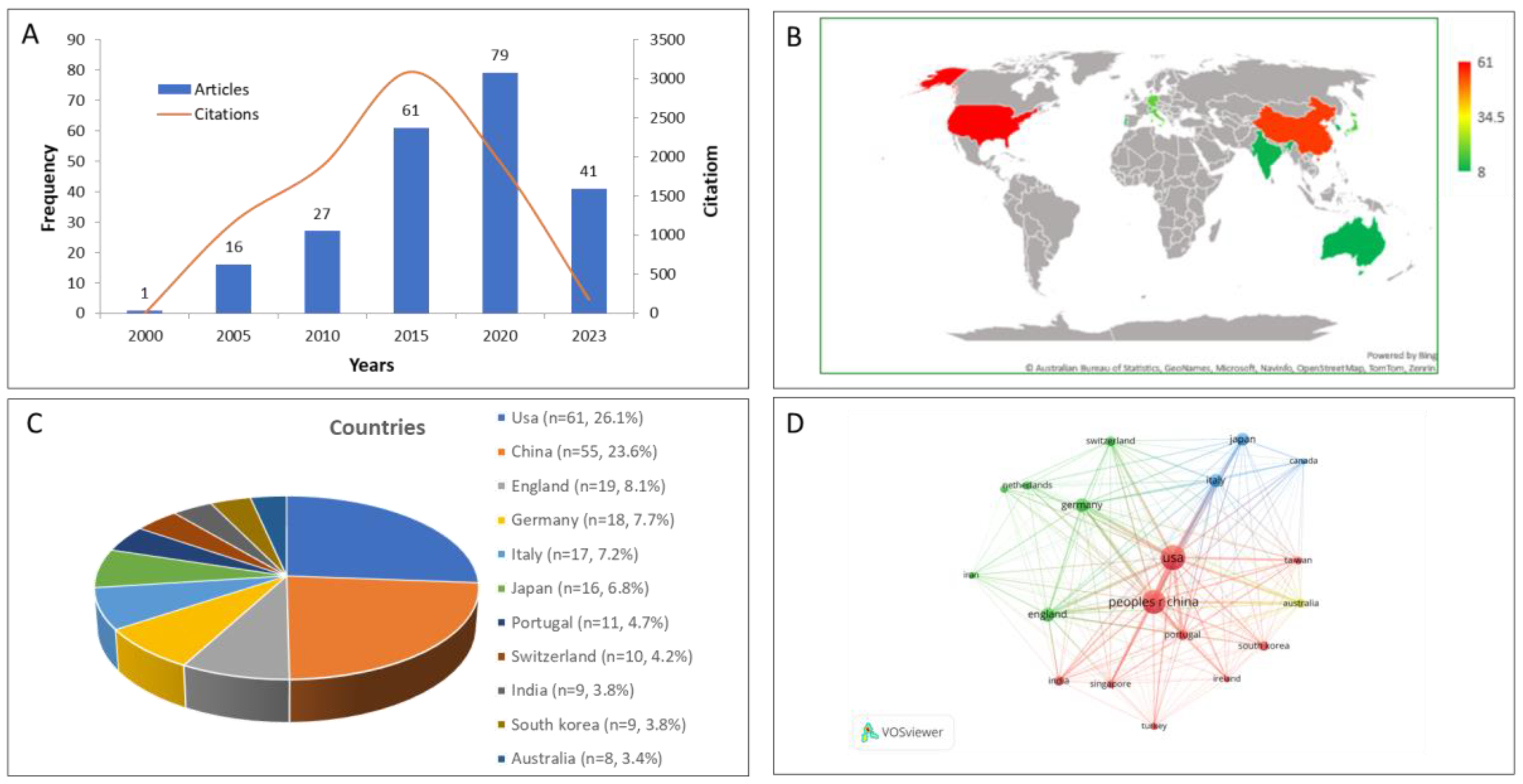
A total of 225 publications were identified on OCI research included to the database between 2000 and 2023. The total publications related to OCI increased from 27 between 2000 and 2010 to 79 between 2015 and 2020. In the years after 2020, 41 articles related to OCI were published. (
Figure 1A).
Country Distribution
The research published in articles was performed in 31 different countries. The USA published the most articles (n=61), followed by China (n=55), England (n=19), and Germany (n=18) (
Figure 1B and C). A total of 22 countries were the home for 2 articles. The network analysis revealed that the USA served as the hub of Osteochondral interface research, while China, England, and Germany were seen as additional centers (
Figure 1D).
Institution Distribution
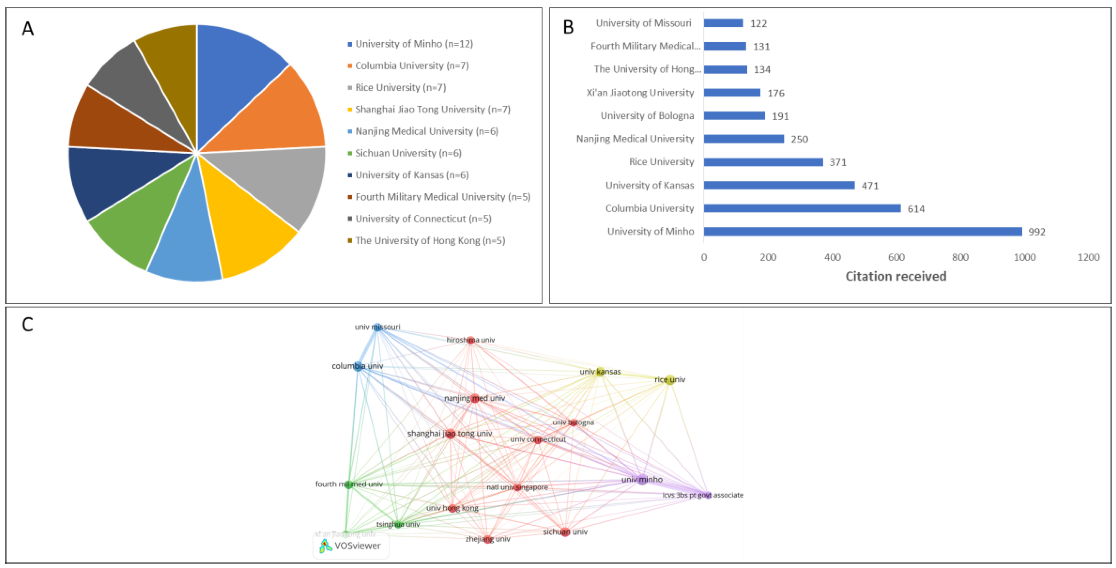
A total of 473 universities or research centers were involved in the extracted publications, with the highest contribution coming from the Chinese institutions (
Figure 2). The highest ranking 10 institutions were the University of Minho (Portugal; n=8), Columbia University (USA; n=7), Rice University (USA; n=7), Shanghai Jiao Tong University (China; n=7), Nanjing Medical University (China; n=6), Sichuan University (China; n=6), University of Kansas (USA; n=6), Fourth Military Medical University (China; n=5), University of Connecticut (USA; n=5), and The University of Hong Kong (China; n=5) (
Figure 2A). Publications from the University of Minho were the most cited, with a total of 753 times, followed by Columbia University with 614 citations and the University of Kansas with 471 citations (
Figure 2B). Regarding co-authorship relationships between institutions, the University of Minho has the strongest network (n=37), followed by Columbia University (n=33), University of Kansas (n=24), and ICVS/3B's—PT Government Associate Laboratory (n=17) (
Figure 2C). The number of authorship share between institutions is depicted by the thickness of the line connecting the institutions.
Journal of Publication
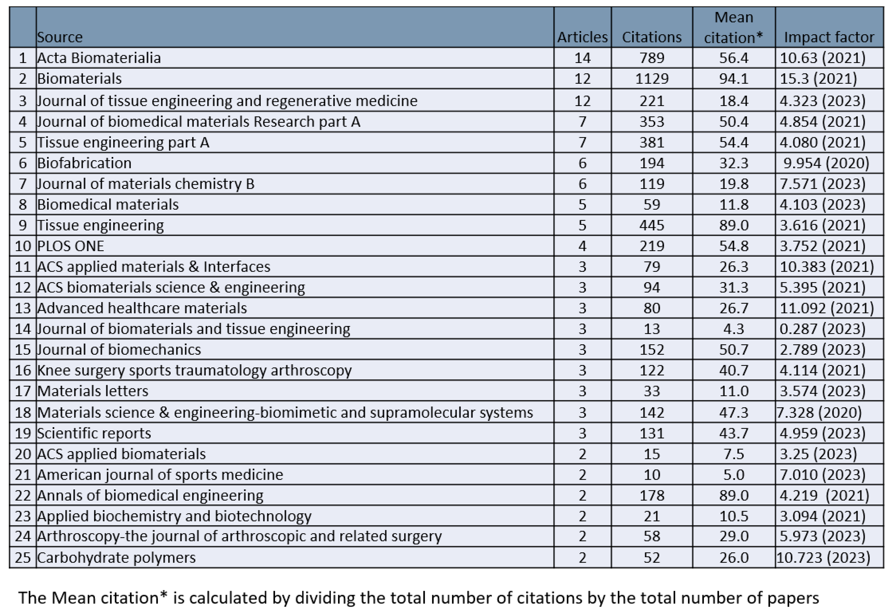
The 225 extracted publications appeared in 118 different journals. The top 25 journals published 47.5% of the publications (
Table 1). The most publishing 5 journals were Acta Biomaterialia, Biomaterials, Journal of Tissue Engineering and Regenerative Medicine, Journal of BiomedicalMaterials Research-PartA, and TissueEngineering-PartA. The articles published in Biomaterials had the highest total citations (n=1129). In contrast, the Journal of Controlled Release had the highest mean citations (217, not shown in the table of top 25). The impact factor of the journals with greater than 10 publications averaged to 8.44.
Keywords and Research Clusters
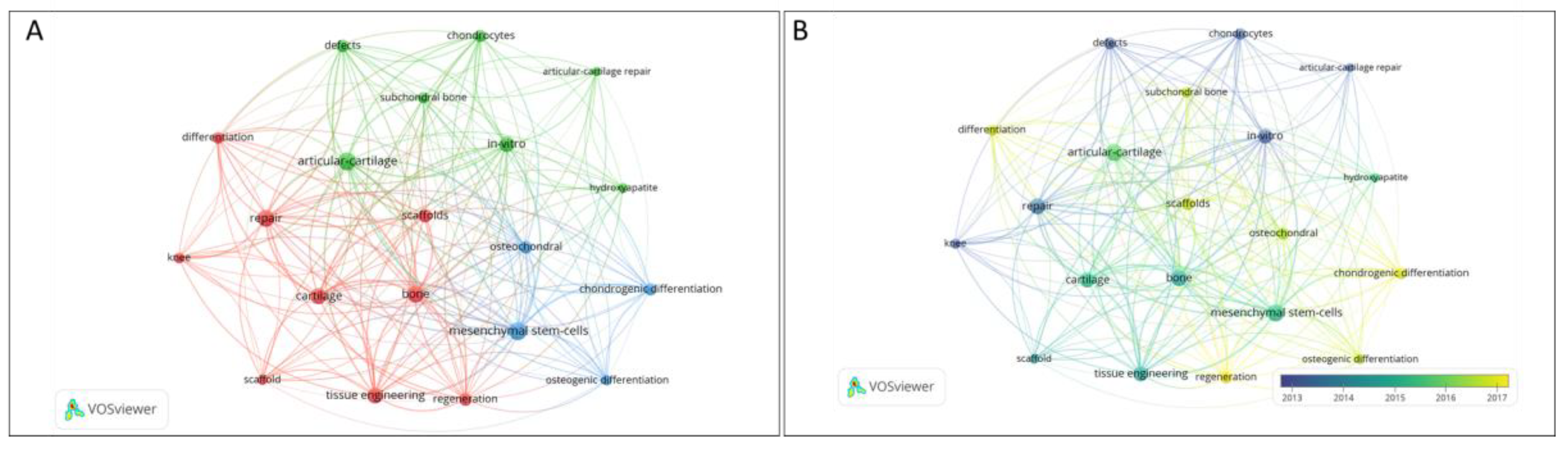
The co-occurrence network analysis tool analyzed keywords from articles related to the osteochondral interface. The number of total occurrences was set at 20, and a total of 15 keywords were used and they were classified into 3 different clusters: "Basic research," "articular cartilage," and "cell culture." In the primary research cluster, the most frequently seen keywords were "bone," "cartilage," "differentiation," "knee," and "regeneration" (
Figure 3A). The frequency of "bone" was high during the period selected. In the “articular cartilage” cluster, "articular cartilage repair," "chondrocytes," "defects," "hydroxyapatite," "in-vitro," and "subchondral bone" were the keywords that were most popular. In the cell culture cluster, the most popular keywords were "chondrogenic differentiation," "mesenchymal stem cells," "osteochondral," and "osteogenic differentiation.” To determine variation in research focus over the period examined, we evaluated the trend of the keywords with highest-frequency (
Figure 3B). Colors represent the average time in years the keywords appeared in articles. For instance, the purple color shows that the keywords appeared earlier than the keywords in yellow. This analysis demonstrated that “basic science research” attracted more interest early on, which later shifted in favor of “cell culturing”. In recent years, it is seen that the shift was toward keywords "mesenchymal stem cells", "osteogenic differentiation" and "chondrogenic differentiation".
The Most-Cited 100 Publications
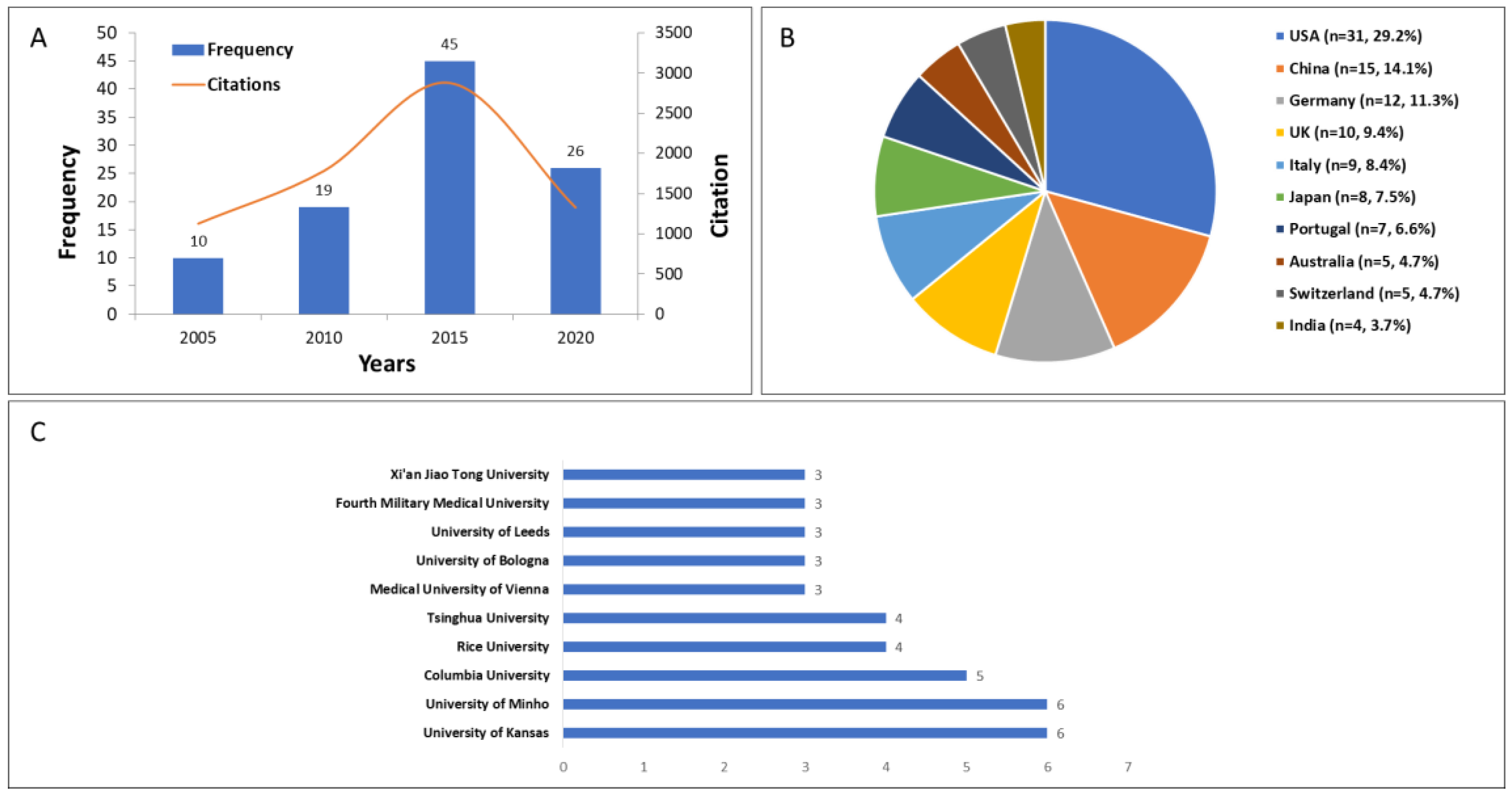
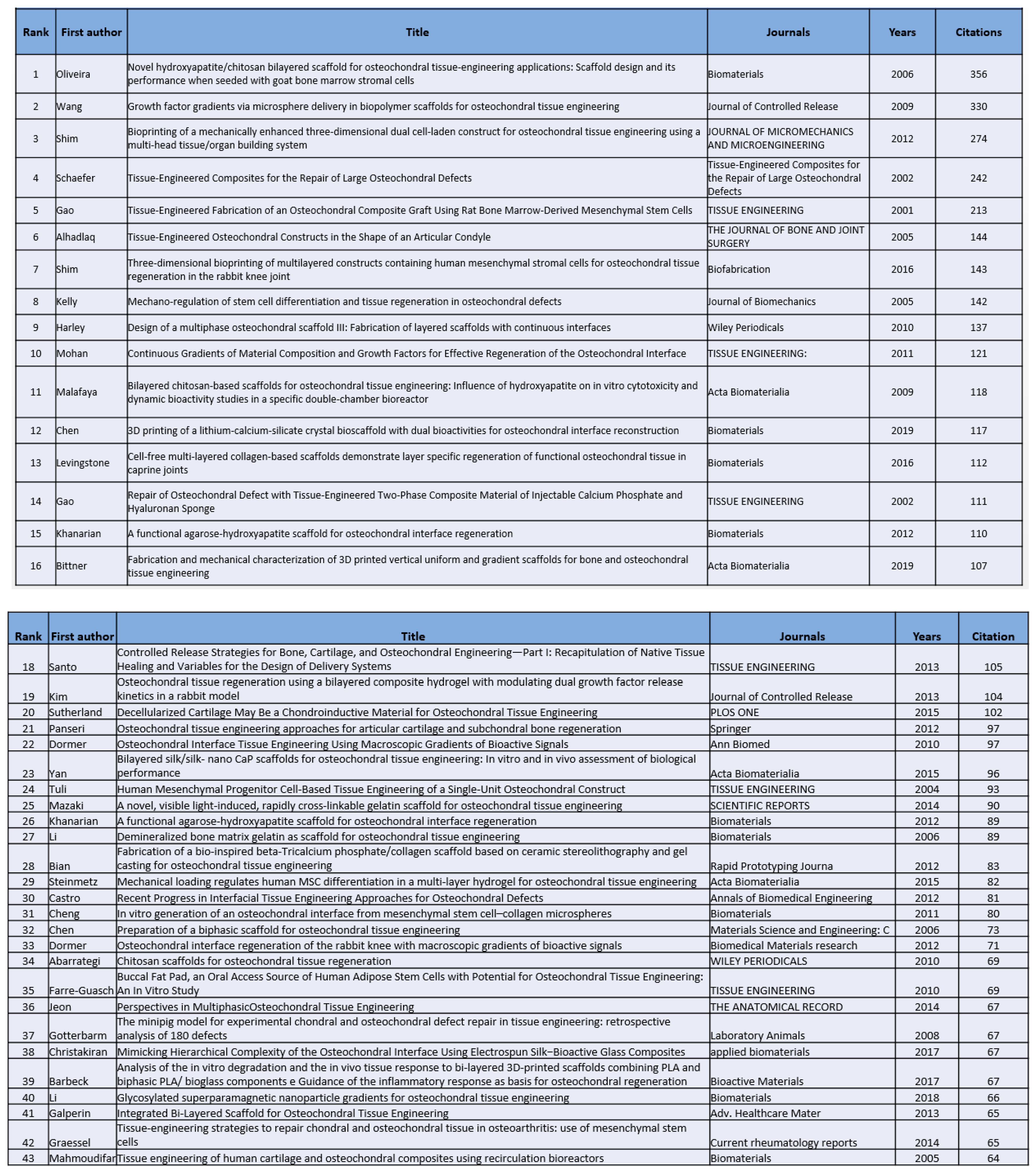
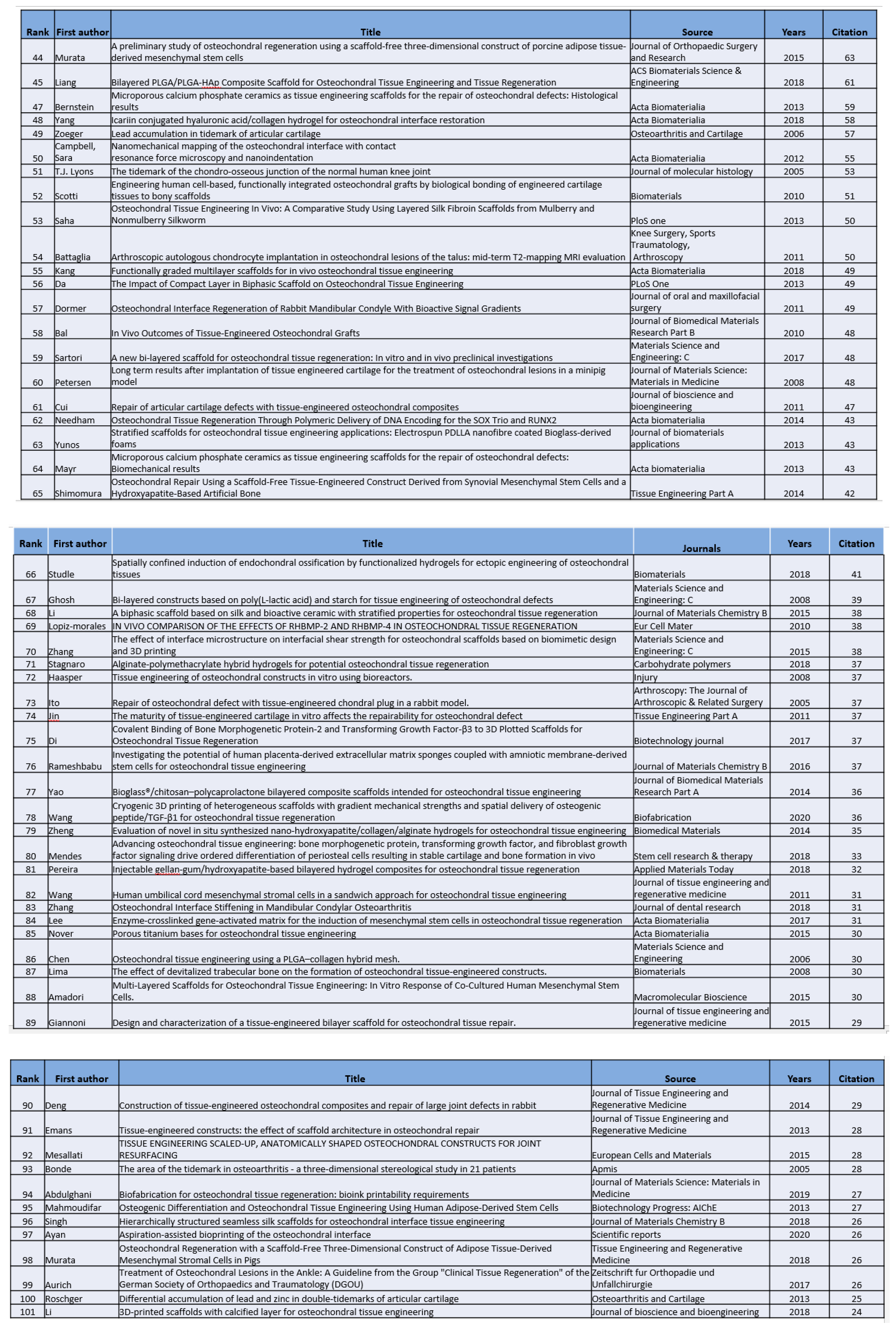
The most highly-cited 100 publications on osteochondral interface appeared between 2000 and 2023 (
Table 2). The largest number of articles published on the topic was between 2011 and 2015 (45 articles), followed by 2016 and 2020 (26 articles) (
Figure 4A). These 100 articles were produced by twenty countries. The USA contributed 30 articles, followed by China with 14 articles; Germany with 12 articles; the UK with 10 articles; Italy with 9 articles; Japan with 8 articles; and Portugal with 7 articles (
Figure 4B). The University of Kansas and Minho contributed 6 articles each to the top 100 most cited articles, followed by Columbia University, with 5 articles, and Rice University and Tsinghua University, with 4 articles each (
Figure 4C). The other 23 institutions produced two articles, and the remaining institutions produced only one article in the most-cited 100 groups.
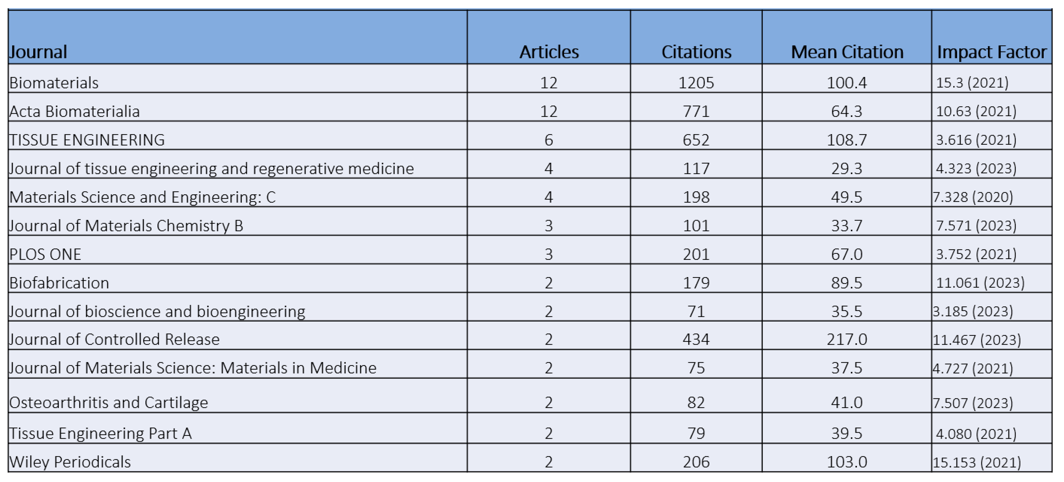
The most highly-cited studies were published in 52 journals. Biomaterials was at the top of the list, with 12 publications and 1205 citations that was followed by Acta Biomaterialia with the same number of articles (771 citations), and Tissue Engineering, which published 6 articles (652 citations) (
Table 3).
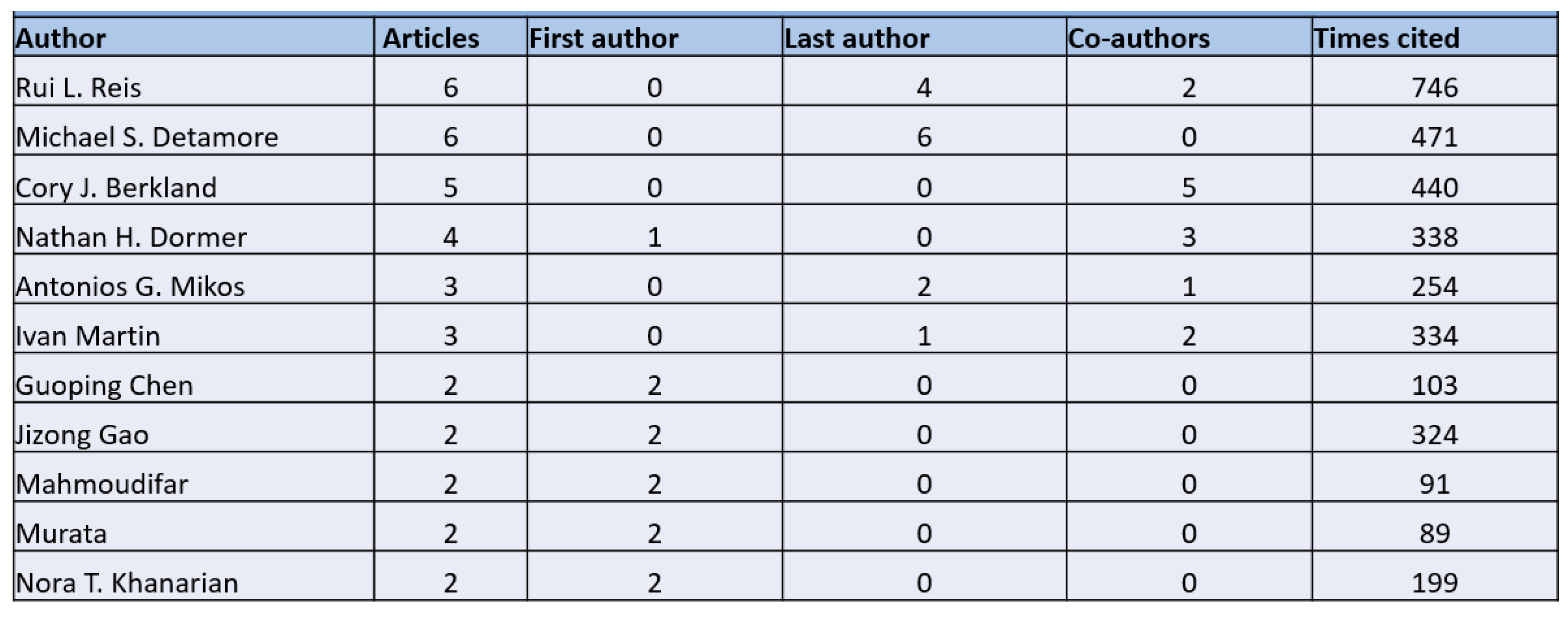
Concerning authorship in most-cited articles, Detamore and Berkland contributed 5 articles, followed by Dormer and Reis. They contributed 4 articles each, followed by Mikos and Martin, who had 3 articles each (
Table 4).
Discussion
Kroner first reported the osteochondral defect of the patella in 1905 and described in detail by HM Coleman in 1948 [
6]. The primary function of the cartilage as load-bearing tissue is to resist and absorb the load exerted on the joint. Thus, osteochondral defects resulting from diseases, trauma, and aging may lead to severe consequences that cannot heal themselves due to their avascular nature.[
7] We have detected significant interest in osteochondral interface-related research. This study reports the first bibliometric analysis of osteochondral literature to demonstrate OCI-related research data over time and to determine the articles with the highest impact.
Publication Trends in the Osteochondral Interface Research
Recently, the number of osteochondral interface-related articles has increased rapidly. The articles originated from 31 countries, with the highest contribution coming from the USA. This can be due to high prevalence of osteochondral defects in the United States. Concerning institutional contributions, the University of Minho (Portugal) produced the most articles and topped the list of total citations. Analysis of joint work between countries and institutions showed a regional cluster formation. However, despite the geographic separation, the highest frequency of collaborations occurred between China and the United States. England also significantly contributed to the OCI research, ranking number 3 after China. In addition, Actabiomaterilia, Biomaterials, Tissue Engineering, Journal of Controlled Release, and Tissue Engineering Part A published the most articles on the osteochondral interface.
Research Focus
Our analysis determined that articular cartilage, biomaterials, and chondrogenic differentiation were the cluster centers. Essential science keywords were seen more frequently at the beginning, with more focus on in-vitro investigation keywords later. In this regard, keywords used more frequently in earlier included mesenchymal stem cells, scaffolds, differentiations, and design.
The Most Impactful Publications
The study with the most citations was "Novel hydroxyapatite/chitosan bilayered scaffold for osteochondral tissue-engineering applications: Scaffold design and its performance when seeded with goat bone marrow stromal cells," authored by Joaquim M. Oliveira et al. in 2006 [
8]. In this article, the authors aimed at evaluating hydroxyapatite / chitosan (HA / CS) bi-layered construct, and goat marrow stromal cells were tested for their differentiation capacity on these scaffolds. It was demonstrated that the scaffold may provide an adequate environment for the regeneration of osteochondral defects.
The earliest publication in 100 most-cited list was authored by Jizong Gao et al. in 2001 and titled "Tissue-Engineered Fabrication of an Osteochondral Composite Graft Using Rat Bone Marrow-Derived Mesenchymal Stem Cells" [
9]. These researchers demonstrated the possibility of constructing a tissue-engineered composite osteochondral graft using MSCs, biomaterials and biofactors.
The latest publication in the list of top 100 most-cited was authored by Chong Wang et al. in 2020. It was titled "Cryogenic 3D Printing of Heterogeneous Scaffolds with Gradient Mechanical Strengths and Spatial Delivery of Osteogenic Peptide/TGF-β1 for Osteochondral Tissue Regeneration" [
10]. These investigators found that osteochondral scaffolds obtained gradient rBMSC osteogenic/chondrogenic differentiation.
Limitations
In this study, there may be impactful articles in other databases excluded from our study. Second, the most recent date of publication was August 25, 2023. Although the numbers are unlikely to differ dramatically since then, the findings are subject to change slightly.
Conclusions
Findings revealed that research efforts on the OC interface have dramatically increased in the last 23 years, despite limited success in the field. Our search results indicated that only 225 papers have been published in this field, with the specified keywords, between 2000 and 2023. Characterization of the OC interface, designing biomimetic biomaterials, and applying these to regenerate the OC interface appear to be the priorities on this topic. This bibliometric analysis is expected to serve as a helpful guide for clinicians and researchers in portraying the most impactful literature in the field.
Author Contributions
SK and BK performed the search, analyzed the data, and wrote the manuscript draft. CE designed the study, supervised the research, and finalized the manuscript. All authors have read and agreed to the published version of the manuscript.
Acknowledgments
This research was funded by a Faculty Development Competitive Research grant [No 021220FD1951] from Nazarbayev University awarded to Cevat Erisken.
Conflicts of Interest
The authors declare no conflict of interest.
References
- Cheng H wa, Luk KDK, Cheung KMC, Chan BP. In vitro generation of an osteochondral interface from mesenchymal stem cell–collagen microspheres. Biomaterials. 2011;32(6):1526-1535. [CrossRef]
- Yildirim N, Amanzhanova A, Kulzhanova G, Mukasheva F, Erisken C. Osteochondral Interface: Regenerative Engineering and Challenges. ACS Biomater Sci Eng. 2023;9(3):1205-1223. [CrossRef]
- Lyons TJ, McClure SF, Stoddart RW, McClure J. The normal human chondro-osseous junctional region: evidence for contact of uncalcified cartilage with subchondral bone and marrow spaces. BMC Musculoskelet Disord. 2006;7(1):52. [CrossRef]
- Dvorak J, Junge A, Derman W, Schwellnus M. Injuries and illnesses of football players during the 2010 FIFA World Cup. Br J Sports Med. 2011;45(8):626-630. [CrossRef]
- Martin I, Miot S, Barbero A, Jakob M, Wendt D. Osteochondral tissue engineering. J Biomech. 2007;40(4):750-765. [CrossRef]
- Coleman HM. RECURRENT OSTEOCHONDRAL FRACTURE OF THE PATELLA. J Bone Joint Surg Br. 1948;30-B(1):153-157. [CrossRef]
- Liu J, Li L, Suo H, Yan M, Yin J, Fu J. 3D printing of biomimetic multi-layered GelMA/nHA scaffold for osteochondral defect repair. Mater Des. 2019;171. [CrossRef]
- Oliveira JM, Rodrigues MT, Silva SS, et al. Novel hydroxyapatite/chitosan bilayered scaffold for osteochondral tissue-engineering applications: Scaffold design and its performance when seeded with goat bone marrow stromal cells. Biomaterials. 2006;27(36):6123-6137. [CrossRef]
- Gao J, Dennis JE, Solchaga LA, Awadallah AS, Goldberg VM, Caplan AI. Tissue-Engineered Fabrication of an Osteochondral Composite Graft Using Rat Bone Marrow-Derived Mesenchymal Stem Cells. Tissue Eng. 2001;7(4):363-371. [CrossRef]
- Wang C, Yue H, Huang W, et al. Cryogenic 3D printing of heterogeneous scaffolds with gradient mechanical strengths and spatial delivery of osteogenic peptide/TGF-β1 for osteochondral tissue regeneration. Biofabrication. 2020;12(2):025030. [CrossRef]
|
Disclaimer/Publisher’s Note: The statements, opinions and data contained in all publications are solely those of the individual author(s) and contributor(s) and not of MDPI and/or the editor(s). MDPI and/or the editor(s) disclaim responsibility for any injury to people or property resulting from any ideas, methods, instructions or products referred to in the content. |
© 2024 by the authors. Licensee MDPI, Basel, Switzerland. This article is an open access article distributed under the terms and conditions of the Creative Commons Attribution (CC BY) license (http://creativecommons.org/licenses/by/4.0/).