
Submitted:
27 November 2024
Posted:
28 November 2024
You are already at the latest version
Abstract
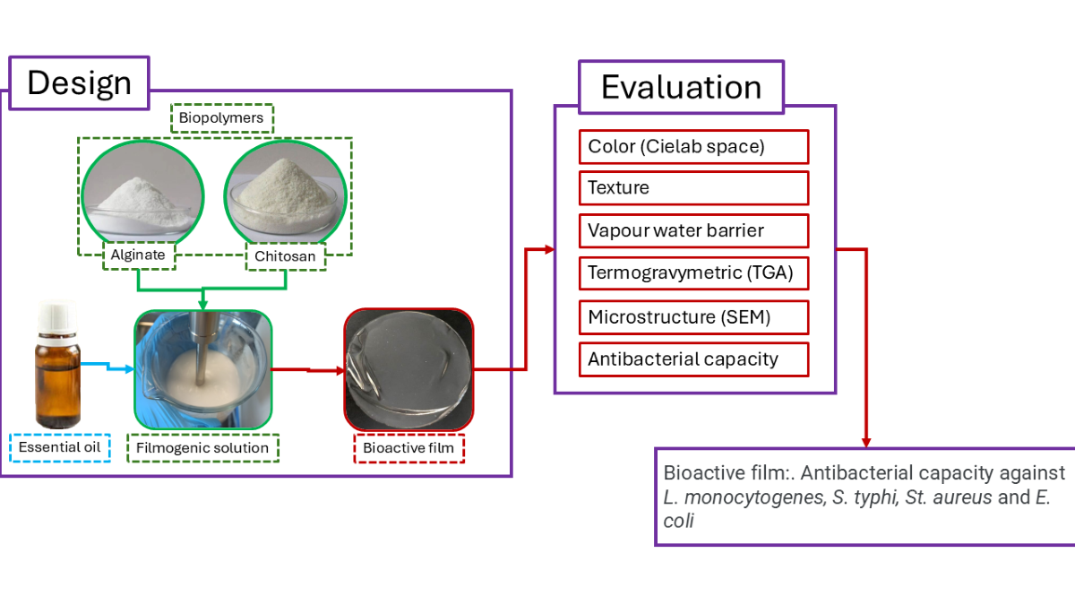
The effect on the physical, mechanical, and antibacterial properties of films composed of alginate-chitosan with the incorporation of oregano (EOO) or thyme (EOT) essential oils was evaluated. The results indicated that the incorporation of essential oil increased the thickness of the films, in addition to evidencing a significant effect on the colour variation towards the yellow tone, especially in the b* factor. On the other hand, the incorporation of essential oil significantly decreased the tensile strength, simultaneously increasing elasticity. Regarding antibacterial capacity, as the concentration of essential oil increases, the antibacterial capacity also increases. On average, the increase from 1% to 3% of EOO increased the antimicrobial capacity against Gram-negative and Gram-positive bacteria. These results suggest that films with the addition of oregano and thyme essential oils can be promising for food packaging applications with the ability to improve food safety and increase product shelf life by achieving functional packaging characteristics.
Keywords:
chitosan
; alginate
; essential oil
; antimicrobial films
; edible films
1. Introduction
The potential presence of pathogens on food surfaces, such as Salmonella sp., E. coli, L. monocytogenes, or S. aureus, generates a loss of food safety, presenting a high health risk to the consumer [1]. The reduction of microbial loads in foods is mainly achieved by adding several synthetic or semisynthetic preservatives. Also, consumers are looking for foods with minimal processing, which do not involve high temperatures. Therefore, natural antimicrobial agents that do not affect the nutritional and sensory quality of the food could be relevant in minimally processed fresh foods. Among these antimicrobial agents, essential oils (EOs) stand out [2,3]. EOs are a variable mixture of different biochemical agents of a volatile nature, standing out in the terpenoid family of compounds, such as carvacrol or thymol [4]. EOs have a recognized antimicrobial capacity, but their main advantage is the synergistic effect between their components [5]. This synergy is responsible for EOs’ high antimicrobial performance (low minimum inhibitory and bactericidal/fungicidal concentrations), a broad spectrum of action, and a low risk of microbial resistance development [6]. In addition, many essential oils are considered GRAS (Generally Recognized as Safe) by the Food and Drug Administration (FDA).
The EOs are found in various plant species, characterized as being “aromatic,” and usually used as spices in gastronomy [7]. Examples are oregano (Origanum vulgare), thyme (Thymus vulgaris), and rosemary (Rosmarinus officinalis) [8]. In particular, oregano and thyme EOs have a broad antimicrobial spectrum (bacteria and molds) and could be a natural alternative to control food pathogens and spoilage microorganisms [9]. Several investigations demonstrate the ability to increase the foods’ shelf life by applying EOs emulsions or dispersions through spraying or immersion [10]. However, the effectiveness of these application forms limits their antimicrobial efficiency because EOs can volatilize or oxidize by chemicals or ultraviolet light, among other environmental factors. On the other hand, its antimicrobial activity is intense at the beginning of the application and decreases to ineffective levels over time [11].
One way to preserve the EOs’ bioactivity and control their release is to incorporate them into a biopolymeric matrix. This matrix can be shaped in films, scaffolds, nanofibers, or nano/microcapsules [12,13]. The films containing EOs can be considered active packaging since they isolate food from the environment and simultaneously show an antimicrobial effect, preventing food deterioration and pathogens proliferation [14]. The active films can be made from various types of biopolymers (e.g., polysaccharides, proteins, PLA, PHBV), taking into account their physicochemical, mechanical, and barrier properties against oxygen and water vapor [15]. Thus, it is vital to choose the best option according to the active ingredient and the final application [16]. On the other hand, the incorporated antimicrobial agent could also act as a plasticizing agent that affects mechanical or barrier properties [15,16]. In summary, the biopolymeric matrix and the bioactive agent determine the adequate performance of the film [17]. Some of these films combine more than one polymer, generating so-called composite films designed to generate improved functional properties by adding or generating a synergistic effect between constituent polymers [18].
Chitosan and alginate are the most common biopolymeric materials for food packaging. Chitosan is a natural polycationic polysaccharide chemically composed of D-glucosamine and D-N-acetylglucosamine units β-(1-4)-linked, having a predominance (>50%) of the former units that are found in nature in fungal and sponge species but on a commercial scale, mainly isolated from the chitin of the crustaceans’ shells [9,17,19]. The chitosan antimicrobial activity has been confirmed. However, chitosan application in the food packaging industry has limited oxygen barrier properties and is sensitive to environmental humidity [20]. On the other hand, alginate or 1,4-β-d-mannuronic and α-l-guluronic acid is a water-soluble anionic polysaccharide obtained from brown algae [21]. The L-guluronic group can crosslink with polyvalent cations, which allows it to form a polymeric network in the form of a gel [22]. This property allows alginate to form films with good mechanical and barrier properties.
Film design relies on the structural interaction based on ionic charges between alginate and chitosan. In this context, one of the most significant challenges is the rapid interaction between alginate and polycations, generating films with non-homogeneous characteristics, some resistant and others more fragile. This lack of uniformity gives rise to films with a varied microtopography [23], including rough, smooth and agglomerated areas. In this line, this study focuses on investigating and improving the interactions between alginate, chitosan and essential oils to enhance the physical, mechanical and antibacterial properties of bioactive films applicable in food packaging.
2. Materials and Methods
2.1. Materials
The essential oils used in this study were oregano (Origanum vulgare) and thyme (Thymus vulgaris) obtained by steam distillation from company Sigma-Aldrich (St. Louis, MO, USA). Sodium alginate (CAS number: 9005-38-3, medium viscosity), Chitosan (CAS number: 9012-76-4; high molecular weight, and degree of deacetylation 75%), calcium chloride, acetic acid, glycerol, and Tween-80 were purchased from Sigma-Aldrich (St. Louis, MO, USA). To determine the antibacterial characteristics of different essential oils, the following bacterial cultures Escherichia coli (ATCC 25922), Salmonella enteritidis (ATCC 13076), Staphylococcus aureus (ATCC 6538), and Listeria monocytogenes (ATCC 7644) obtained from the culture collection of the Food Microbiology Laboratory of the Department of Food Engineering of the Universidad del Bio Bio, Chile.
2.2. Methods
2.2.1. Determination of Antibacterial Properties of Essential Oils
The estimation of the minimum inhibitory concentration (MIC) and minimum bactericidal concentration (CMB) was carried out using dilution in broth [24]. EOs dilutions from 10% v/v to 0.01% v/v were used. The bacterium culture was seeded in trypticase soy broth (TSB, GranuCult®, Merck, Darmstadt, Germany) and cultivated in an oven (Memmert 55 L, UN 55 model) for 24 h at 35±1 ºC until cell concentration was reached. of 106 CFU/mL that was confirmed with the McFarland scale [25]. The MIC values were taken as the lowest concentration of oil that prevents visible bacterial growth after 24 h of incubation at 37±1 °C, and MBC as the lowest concentration that completely inhibits bacterial growth. TSB with methanol and without essential oils was used as a negative control, and inoculated TBS was used as a positive control. Each experiment was performed in triplicate.
2.2.2. Preparation of Alginate-Chitosan Films
For the preparation of the chitosan solution, the methodology indicated by Lijun, et al., [26] was applied, with modifications: 4 g of chitosan were dissolved in 200 mL of distilled water acidified with acetic acid (1.0% v/v) stirring for 24 h at 1,000 rpm at 20±1°C. Finally, the chitosan solution was sonicated for 5 min to remove bubbles and filtered through cellulose paper (Whatman, 3 µm). The plasticizer used was glycerol (0.243 g glycerol/g chitosan). In parallel, a 1.0% w/v sodium alginate solution was prepared; the alginate dilution was acidified with 1.0% v/v acetic acid to prevent the insolubilization of the chitosan in the sodium alginate solution. The chitosan-alginate mixture was made in a 1:1 ratio in a 500 mL beaker under constant stirring at 200 rpm for 10 min [27].
2.2.3. Preparation of Alginate-Chitosan Films with Antimicrobial Agents
Different amounts of essential oils (EOs) were added to a freshly prepared chitosan/alginate solution (1:1 ratio) under gentle stirring. Oregano oil (EOO) or thyme oil (EOT), previously mixed with Tween-80 emulsifier (0.250 g/g oil), were added to the biopolymer mixture at final concentrations of 1.0; 2.0 and 3.0% w/v respectively. The solutions were homogenized with Ultra Turrax equipment (IKA® Werke, Germany) at 13,500 rpm for 3 min. Subsequently, the solutions were ultrasonicated in an ultrasonic bath (Biobase, Mod. UC-30A) for 20 minutes to eliminate bubbles. The emulsions were transferred to Petri dishes (at a rate of 30 mL per capsule) and placed in an oven (55 L Memmert, UN 55 model) to dry at 35±1 °C until constant weight (48 to 62 h). After that, One milliliter of CaCl2 (0.5% w/v) solution was added to the film surface and left in contact for 5 min. The excess solution was removed. Petri dishes with biocomposite films were dried for 3 h at 35±1 °C. Finally, the films were removed from the Petri dishes and placed in a desiccator at 20 °C with 30% humidity until use.
2.2.4. Determination of Physical Properties
2.2.4.1. Film Thickness
The film thicknesses were measured with a digital micrometer (Electronic Outside Micrometer, Fowler, USA) in different areas of the film. The thickness was considered as the average of 5 measurements.
2.2.4.2. Color, Yellowness Index, and Whiteness Index
The film’s color was measured with a CR-300 colorimeter (Minolta Camera Co., Ltd., Osaka, Japan) through a calibrated standard plate (a = 0.20; b = 1.84; L = 97,14). The CIELab color space was used, and lightness (L*), and color parameters a* (red-green) and b* (yellow-blue) were measured using a D65/10° illuminant observer. Total color difference (ΔE) was defined between the alginate-chitosan control film (L*0, a*0, b*0) and the alginate-chitosan films with EOs (L^*, a^*, b^*), where the values L^* (black 0 to white 100), a^* (red 120 to green -120), and b^* (yellow 120 to blue -120) correspond to whiteness, redness, and yellowness, respectively (Equation (1)). Furthermore, the yellowness index (YI) and whiteness index (WI) were calculated using Equations (2) and (3), respectively [28,29].
2.2.4.3. Water Vapor Permeability (WVP)
Water vapor permeability (WVP) was determined using the modified ASTM E96-95 gravimetric method proposed for hydrophilic films [30,31]. Film discs were mounted in permeability dishes (Fisher Scientific, USA), to which 10 mL of distilled water was previously added. The dishes were placed in a desiccator containing saturated sodium chloride solution (75% RH) to generate a 75/100% RH humidity gradient between both sides of the film. The air around the film was homogenized using a small fan inside the desiccator. Finally, the system was placed in an air circulation oven at 25±1 °C. The weight of the capsules was checked every 2 hours for a period of 24 h. The WVP was estimated using the regression analysis of the Equation. (4):
where X is the average thickness of the edible films, A is the permeation area (5.5*10-3 m2). ΔP is the difference in the partial vapor pressure of the atmosphere (2337 Pa at 20 ºC), and the w/t term was calculated by linear regression from the points of weight and time increase in the constant velocity period [30]. Three replicates were obtained for each sample.
2.2.5. Determination of Mechanical Properties
The tensile strength (TS) and the percentage elongation to break (%E) of the films were measured in a texture analyzer (TA-XT2, TA Instruments, United Kingdom) according to the standardized method ASTM D882-95 (ASTM nineteen ninety-five). The films were cut into 20 mm by 90 mm strips and conditioned at 25 °C and 50% relative humidity in a desiccator with saturated Mg(NO3)2 solution for 72 h. A set of jaws with a separation of 50 mm and an operating speed of 1 mm/s were used for mechanical properties measurement. The TS was read directly from the equipment in units of MPa, and %E was calculated by Equation (5):
where L is the length at the breaking point (mm), and L0 is the original length (mm). All measurements were performed in triplicate.
2.2.6. Scanning Electron Microscopy (SEM)
The microstructures of the microtopographic and cross-sect films were obtained and examined using a scanning electron microscope (SEM, JEOL JSM-6380LV). Previously, the films were frozen with liquid nitrogen and fractured into approximately 5*3 mm pieces. The pieces were fixed in a sample holder and coated with a nanometric layer of gold using sputtering equipment (Aname, SC7620. Madrid, Spain). The samples were observed using an accelerating voltage of 15 kV.
2.2.7. Thermogravimetry Analysis
For the TGA evaluation of the samples, the methodology described by Jinman et al., [28] was used: The measurements were carried out in a thermogravimetric analyzer (Netzsch TG 209-F3, Germany) and the thermal increase was from 30 to 600°C at a rate of 10°C/min.
2.2.8. Antibacterial Effect of the Alginate-Chitosan Films
The agar diffusion method was used to determine the antibacterial effect according to the method proposed by Chen et al. [32]. Sterile 21 mm diameter film discs were placed on Muller Hinton Agar (MHA), previously inoculated with 0.1 ml of trypticase soy broth (TSB) 105-106 CFU/mL bacterial strains. The Petri dishes (90*15 mm) were incubated at 37±1 °C for 24 h. Finally, the diameter of inhibition generated from the edge of bacterial growth around the film disc was measured. These tests were performed in triplicate.
2.3. Statistical Analysis
Statistical analyses were performed using IBM SPSS version 21 software. The effects of each variable on the functional properties and antibacterial properties of the films were analyzed using a one-way ANOVA test. Differences between mean film property values were determined using Dunnett’s T3 Least Significant Difference test for multiple comparisons. Data were represented by mean ± standard deviation. Statistical significance was considered with p < 0.05.
3. Results and Discussion
3.1. Determination of the Minimum Inhibitory Concentration (MIC) and Minimum Bactericidal Concentration (MBC) of Essential Oils
EOs have generally shown broad antimicrobial activity, particularly against spoilage and pathogenic bacteria [33]. Previous studies have observed a higher concentration of essential oils of thyme and oregano in edible films for meat, resulting in significant antibacterial activity [34]. Similarly, EOT and EOO enhanced the helpful life of the Trout [35]. To evaluate the antimicrobial capacity of essential oils, it is necessary to determine the minimum inhibitory concentration (MIC) and the minimum bactericidal concentration (MBC). Both parameters previously indicate the antibacterial power of each oil. The results of this stage are shown in Table 1. The results showed that Gram-positive bacteria (S. aureus and L. monocytogenes) were more vulnerable to essential oils, requiring low concentrations to cause an inhibitory or bactericidal effect. On the other hand, Table 1 also shows that the EOO generally showed a greater antibacterial capacity than EOT. The antimicrobial capacity of essential oils depends on their active compounds, predominantly monoterpenes (thymol, carvacrol, among others). The antibacterial mechanism of action of these active compounds is based on the alteration of the cytoplasmic membrane through proton interactions and electron flow, which alters the transport mechanisms at the cell membrane level and can generate cytoplasmic coagulation effects [22]. Therefore, the lower susceptibility observed in Gram-negative bacteria (E. coli and S. enterica) could be explained by the more complex structure of these bacteria due to an additional outer membrane [24]. It is important to note that EOO showed a significantly higher inhibitory power (p < 0.05) than EOT against the same microorganisms. On the other hand, the concentrations of oil to achieve bacterial lysis were lower, demonstrating that EOO has a greater bactericidal power. These results approved previously reported observations [36]. However, variations in values are probably because of environmental conditions, the types of active ingredients, the geographic origin of the plant, and the extraction methods.
3.2. Determination of Physical Properties
3.2.1. Film Thickness
The films’ thickness varied between 37.7 to 38.2 µm (Table 2). The thicknesses of all EOs formulated films (regardless of the type) increased compared to the blank films because incorporating EOs in the films decreases the density of the emulsion. Consequently, the same volume has fewer solids than a solution without essential oils. However, this film thickness increase was not statistically significant [21,27,37]. The mass and volume variations generated in filmogenic matrices fundamentally depend on the constitutive components, their properties (density, for example), and the interactions between them in terms of electrostatic repulsions or attractions, formation of hydrogen bonds or disulfide bonds of more cohesive or looser structures. On the other hand, the molecular structure of alginate contains -COO- groups, which under acidic conditions become -COOH. This contracts the molecular chain, reducing hydrophobicity. Due to the mentioned reason, incorporating a particular agent into a filmogenic matrix, such as the case of EOs in the alginate/chitosan matrices, could increase, decrease, or maintain the same thickness when dosed at a constant mass.
3.2.2. Color Measurement, Yellowness Index (YI), and Whiteness Index (WI)
Film color can negatively affect consumer acceptability. That is why the evaluation of the color of a film is a crucial requirement, particularly when structural components influence this parameter. Essential oils often give a yellow hue to the films. However, this effect depends on the type and concentration of the essential oils, which are directly influenced by the required amount for antimicrobial activity [38]. Table 2 shows the effect of incorporating EOs (oregano or thyme) on the color parameters. The result showed that increasing the EOs content from 1 to 3% increased ΔE and reduced the lightness (L* value) from 98 in blank films to 86 for 3% oregano-formulated films. In the case of EOT, similar reductions were obtained from 98.5 (control) to 86.3 (p<0.05), a result that agrees with previous research where the lightness value tends to decrease when emulsions are prepared filmogenic based on essential oils [39,40]. Our results indicated that both oregano and thyme essential oils significantly reduced the luminosity of the films. However, these differences became evident as the EOs concentration in the film increased. On average, the blank films’ L value was 98.5, while the films containing 1% essential oils were 87.8 and 88.3 for oregano and thyme, respectively. This effect is due to the preparation of the emulsion before the formation of the film since the essential oils are arranged as a dispersed phase in the matrices, which affects the refractive index of light and increases its dispersion [41]. On the other hand, it was possible to show a significant variation in the color parameter a* (red-green). In this sense, the blank film gave a value of -1.52, while the film with EOO varied between -3.46 to -3.56 and thyme between -3.35 to -3.70. It was also possible to show a slight displacement of the color towards the greener zone of the CIELAB colour space. Regarding the parameter b* (yellow-blue), it was observed that, regardless of the type of EOs, the presence and increase in the concentration of these compounds resulted in a shift in colour towards the yellowest area of the CIELAB space. Besides, the blank film gave a b* value of 5.47, while for alginate/chitosan-EOO films, the b* value varied between 11.63 and 11.69, and for alginate/chitosan-TEO films, between 11.56 and 11. 96. Comparing the blank film, L, a*, and b* parameters have been varied in the EOs incorporated films, especially in the b* parameter, where significant statistical differences were evident. Consistent with this, the total color variation ΔE, including the effect of all parameters (Eq 1), also showed significant differences between the blank (4.26) and the EOs formulated films (12.1 and 14), mainly due to the change in the b*. Finally, the yellowness index (YI) indicated a significant change between 7.94 for the blank film and around 20, regardless of the type of oils for formulated films, confirming the yellower color of incorporated films. Sharma et al. [42], who worked with thyme and clove essential oils, also observed the effect of incorporating EOs on the variation of the b* parameter. They evidenced the change from 1.34 to 1.89 by increasing the thyme essential oil from 0 to 10% in PLA/PBAT films. In the case of clove essential oil, this effect was more noticeable, reaching 7.78 [42]. Regarding the whiteness index (WI), a significant decrease could be due to the incorporation of essential oils that led to a darkening of the film. For the same reason, the change in L could result from increased essential oils in the filmogenic emulsion.
3.2.3. Water Vapor Permeability
Food packaging films require good barrier capabilities, particularly with ambient humidity. Limiting water migration between the medium and the feed maintains effective control over unwanted hydration in the feed, which can increase water activity and promote microbial growth. Consequently, the film must cancel or minimize moisture transfer between the environment and the food surface [43]. For this reason, evaluating water vapor permeability is necessary for food packaging. As shown in Table 2, regardless of the type of oils, the increase in EOs content significantly decreases water vapor permeability (p<0.05). It can be due to the EOs’ hydrophobicity and can confirm the homogeneous and even distribution of EOs as a dispersed phase throughout the films. In several studies, the water vapor barrier (WVP) effect has been reported in biopolymeric films that incorporate essential oils in their formulation.
3.2.4. Determination of Mechanical Properties (Tensile Strength and Elongation at Break)
The tensile strength (TS) and the elongation at break (%E) represent the mechanical properties of the films, which depend on their microstructural characteristics. These variables are crucial to define the behavior of the polymeric matrices in terms of plasticity and resistance of the material, which ultimately determine the quality of the packaging material. The results of this analysis are shown in Figure 1 (TS) and Figure 2 (%E). The results indicated that the incorporation of essential oils (oregano or thyme) in concentrations of 1, 2, or 3% significantly decreased the TS (p<0.05).
On the other hand, the flexibility of the films is represented by the elongation at break (%E). It was observed that the incorporation of essential oils (oregano or thyme) generated a significant increase in E% compared to the control without essential oils (p<0.05). The increase in the concentration of oregano or thyme oil from 0 (control) at 2,0% increased E% from 4.12% to 12.50% on average (Figure 2), which can be explained as a plasticizing effect of the film by the essential oils. Indeed, incorporating essential oils in the films can weaken the internal polymer bonds resulting from replacing polymer-polymer interaction with polymers-EOs interaction through hydrogen bonds and hydrophilic-hydrophobic interactions [44]. For this reason, EOs-incorporated films showed an increase in elongation and flexibility, leading to more fragile films with excellent stretchability [42,45]. However, a 3.0% concentration of essential oils (oregano or thyme) makes the film less elastic by over-plasticizing the film [27].
A similar result was reported by Karami et al., [46] where the effect of yarrow essential oil (EOY) in gelatine/sodium alginate films was investigated. The result showed that the increasing EOY from 0 to 3% decreased TS by 59.8%, while E% increased by 60.8% [46]. In addition, Zhou et al. [15] worked with starch films and the incorporation of cinnamon essential oil and concluded that an increase in essential oil from 0 to 2.5% managed to reduce TS by 43.7%, while the E% increased by 99.1% [15]. In this work, when the essential oils content was increased from 0 to 3% w/v, the TS decreased to 47.1 and 44.6%, while the E% increased to 132 and 145% in EOO and EOT, respectively.
3.2.5. Microstructural Features
The SEM microphotographs show the formation of microstructures between both biopolymer matrices and the incorporation of the essential oils. Figure 3 and Figure 4 show the alginate-chitosan films’ superficial and cross-sectional area. There were no microstructural differences in cross-sectional areas or surfaces according to the type of essential oils (oregano or thyme). Generally, the control film (Figure 3a or 4a) without EOs was smooth, without pores or cracks, with little irregular agglomerated microstructures or impurities, possibly during drying [47]. The cross-sectional area of blank films (Figure 3d or 4d) is smooth and compact, without reticulate structures. Incorporating oregano and thyme essential oils generated a heterogeneous surface with gelled areas that increased proportionally with the concentration of essential oils. The arrangement of the alginate molecules and the aggregation of the oil droplets during the formation of the film can give rise to some concave or convex irregularities on the surface [48]. The cross-section of the films showed a spongy-type reticulate structure. The pores in the matrix increased with the concentration of essential oils, probably due to the oil mobilization from the interior structure towards the surface due to the temperature during drying, which may affect the microstructure [49].
3.2.6. Thermogravimetry Analysis
Figure 5 shows that all samples have various thermal decomposition processes. It is known that the first thermal effect occurring at a maximum decomposition rate (Tpeak) below 100 °C is due to the evaporation of free and bound water from the samples [50]. In this process, films shown an associated mass losses ranging from 5.1-10.6 %. Some studies point out that the method used for chitosan-alginate polyelectrolyte complex drying could influence their supramolecular structure, and hence the amount of water inside them once dried [51].
The thermogram of chitosan (Figure 5A) indicates a second thermal degradation effect within the range of 251–402 °C, with Tpeak at 310 °C, and associated mass loss of 50.4 %. This process is associated with the first stage of pyrolysis of polysaccharides, where simultaneous sugar ring dehydration, chain depolymerization, and decomposition of the acetylated and deacetylated units of chitosan occur [50,52]. At higher temperatures a continuos polymer degradation occurs, which is due to residual polysaccharide decomposition. This behaviour is similar to previously observed during chitosan thermal degradation by other authors [53].
On the other side, the TG-DTG curve of alginate (Figure 5B) shows four thermal effects. The second stage shows the major polymer degradation and occurs withing a temperatura range 191-266ºC, with Tpeak 244ºC and a mass loss 24%. It is due to the polymeric chain hydrolysis, decarboxylation, decarbonilation and dehydration of the sugar ring [53]. Another two process shown a maximum decomposition rate at 284ºC and 399ºC, with an associated mass losses around 11% each. These processes must be associated with polymer pyrolysis and residual decomposition of the polysaccharide, respectively.
In calcium-crosslinked chitosan-alginate polyelectrolyte complex (CHI-ALG) films, thermogravimetric curves (Figure 5) reveal two thermal effects with maximum decomposition temperatures at 216ºC and 265°C, respectively. The weight losses associated with the second decomposition effect (42.4%) is 1.7-times higher than the first one (24.6%). It is worth noting that both Tpeak temperatures are lower than the maximum decomposition shown by neat polysaccharides (Table 3, Figure 5 A,B). The above results sugested that the polyelectrolyte complex formation leads to a decrease in the thermal stability of the obtained film. This could be due to the catalytic effect of Ca2+ ions on thermal decomposition of the complex, like previously observed with Ba2+ ion [53]. In this complex, residual decomposition begins above 500 °C, indicating the formation of a more complex and thermally stable structures than in single polysaccharides. In previous work, it was reported the composition of the chitosan–alginate polyelectrolyte complex is independent of the alginate chemical composition as well as the chitosan molecular weight [55]. Additionally, the degree of complexation between them had been found to be 0.51 regardless of the sodium alginate composition. In this sense, the study of thermal stability of CHI-ALG complex with different polymer ratio shown similar shape than TG-DTG curves from present work [53]. Moreover, these authors reported a first Tpeak near 180ºC and a second Tpeak in the range 218-275ºC, depending on the chitosan content. It is worth noting that the second temperature effect of the CHI-ALG films in this research occurs within the temperature range previously reported.
Finally, a comparable thermal breakdown pattern is seen between the samples containing essential oils of thyme and oregano. In contrast to the CHI-ALG crosslinked films, these samples exhibit five thermal degradation steps. The initial effect in each group of oil-containing films occurs within temperature ranges of 179-240ºC and 204-241ºC, respectively, with Tpeak approximately at 220ºC. The recorded mass losses varied with the increase in oil concentration. The simultaneous breakdown of the volatile components of the essential oil and the first step of the polyelectrolyte complex decomposition may have caused this effect. The third and largest thermal effect of these films exhibited a Tpeak ranging from 260 to 274ºC, with associated mass losses of 24.8% to 34.4%. This result resembles the third decomposition step of CHI-ALG films (Tpeak 265ºC) but exhibits reduced mass losses. A fourth and five thermal degradation stage in oil-loaded films revealed Tpeak at 400 and 476ºC, respectively. It is possible that both effects could be due to the residual decomposition of the mixture, which is generated by novel compounds. Such products were produced through a reaction between the polymeric matrix and the chemical constituents of the essential oil. Films with analogous polymeric compositions, loaded with aromatic compounds, have exhibited similar shapes [56].
3.2.7. Antibacterial Effect of Alginate-Chitosan Films
The antibacterial effect of incorporated EOs within the biopolymer matrices depends on migrating the EOs to the film’s surface. Figure 3c and Figure 4c show porous structures when the oil concentration increases. These pores facilitate the migration of essential oils through the matrices. As discussed in the SEM segment, the number of porosity is directly influenced by the concentration of EOs. Consequently, the antibacterial capacity of the films depends not only on the oil concentration but also on the surface migration capacity. Table 4 shows the antibacterial results of the inhibition halos generated by the films. As the oil concentration in the film increases, the antibacterial capacity also increases, demonstrated by the generation of the inhibition halo (Figure 5). On average, the increase from 1% to 3% of EOO increased the antimicrobial capacity against Gram-negative bacteria from 24 to 37 mm (154% increase), while for EOT, it was from 28 to 39 mm (139% increase). In the case of Gram-positive bacteria, 1 to 3% of oregano oil increased the inhibition halos between 26 to 46 mm (177% increase), while for EOT, it was from 34 to 55 mm (154% increase). This situation had already been verified when testing the antibacterial efficacy of the pure oils, which is consistent with the results observed in previous research [57].
The essential oils (oregano or thyme) incorporated in films showed an interesting antibacterial effect that was more intense in higher concentrations of EOs. The alginate/chitosan-EOT film was particularly effective against L. monocytogenes, showing significantly larger inhibition halos than other bacterial strains. In most cases, the inhibition halos against E. coli, S. aureus, and L. monocytogenes were considerably lower in alginate/chitosan-EOO compared to alginate/chitosan-EOT films with the same concentration.
4. Conclusions
The alginate-chitosan-essential oils interaction significantly affected the films’ physical, mechanical, and antibacterial properties. The films with the highest essential oils content were more elastic (E%) but with lower tensile strength (TS). On the other hand, the water vapor permeability (WVP) decreased with EOs’ proportion. The antimicrobial activity of the EOs incorporated alginate/chitosan films was observed in the presence of 1.0% w/v of EOs. However, the highest inhibition was observed at alginate/chitosan-3.0% EOT. Therefore, this film can be a good alternative for bioactive packaging applications.
Acknowledgments
The authors are grateful for the financial support of the Research Department of the Bio Bio University (DIUBB) through Project 085922 3/R.
Disclosure Statement
No potential conflict of interest was reported by the author(s).
References
- Luchansky, J.B., Barlow, K., Webb, B., Beczkiewicz, A., Merrill, B., Vinyard, B.T., Shane, L.E., Shoyer, B.A., Osoria, M., Campano, S.G., Porto-Fett, A.C.S. (2024). Inactivation of Listeria monocytogenes and Salmonella spp. During Cooking of Country Ham and Fate of L. monocytogenes and Staphylococcus aureus During Storage of Country Ham Slices. Journal of Food Protection, 87. [CrossRef]
- Movahedi, F., Nirmal, N., Wang, P. Fatemeh, Jin, H., Grøndahl1, L., and Li, L. (2024). Recent advances in essential oils and their nanoformulations for poultry feed. Journal Animal Sciences Biotechnology 15, 110. [CrossRef]
- Ribeiro Cerqueira de Oliveira. L., Monteiro de Barros da Cruz Machado, A.C., de Freitas Guimarães Filho, C.E., Almeida Esmerino, E., Andrade Calixto, F.A. Marques de Mesquita, E.F., Holanda Duarte, M.C.K. (2025). Evaluation of the antimicrobial effect of oregano essential oil (Origanum vulgare) on cooked mussels (Perna perna) experimentally contaminated with Escherichia coli and Salmonella Enteritidis. Food Control, Volume 167. [CrossRef]
- Chroho, M., Youssef Rouphael, Y., Petropoulos, S.A. and Bouissane, L. (2024). Carvacrol and Thymol Content Affects the Antioxidant and Antibacterial Activity of Origanum compactum and Thymus zygis Essential Oils. Antibiotics 13, no. 2: 139. [CrossRef]
- Hajibonabi, A., Yekani, M., Sharifi, S., Sadri Nahad, J., Maleki Dizaj, S., Yousef Memar, M. (2023). Antimicrobial activity of nanoformulations of carvacrol and thymol: New trend and applications. OpenNano, 13. [CrossRef]
- Basavegowda N and Baek K.H. (2021). Synergistic antioxidant and antibacterial advantages of essential oils for food packaging applications. Biomolecules, 11(9),1267. [CrossRef]
- Micić, D.; Đurović, S.; Riabov, P.; Tomić, A.; Šovljanski, O.; Filip, S.; Tosti, T.; Dojčinović, B.; Božović, R.; Jovanović, D.; Blagojevićet, S. (2021). Rosemary Essential Oils as a Promising Source of Bioactive Compounds: Chemical Composition, Thermal Properties, Biological Activity, and Gastronomical Perspectives. Foods, 10, 2734. [CrossRef]
- Yemiş G.P, Candoğan K. (2017). Antibacterial activity of soy edible coatings incorporated with thyme and oregano essential oils on beef against pathogenic bacteria. Food Sciences. Biotechnol, 26(4). [CrossRef]
- Amadio C, Farrando S, Zimmermann M. (2019). Effect of chitosan coating enriched with oregano essential oil on the quality of refrigerated meat hamburgers. Rev. Fac. Ciencias Agrar, 51(1), 173–189. https://api.semanticscholar.org/CorpusID:204810268.
- Kim J.H, Lee E.S, Song K.J, Kim B.M, Ham J.S, Oh M.H. (2022). Development of Desiccation-Tolerant Probiotic Biofilms Inhibitory for Growth of Foodborne Pathogens on Stainless Steel Surfaces. Foods, 11(6), 831. [CrossRef]
- Chawla R, Sivakumar S, Kaur H. (2021). Antimicrobial edible films in food packaging: Current scenario and recent nanotechnological advancements- a review. Carbohydr. Polym. Technol. Appl, 2,100024. [CrossRef]
- Benavides, S., Mariotti-Celis, M.S., Paredes, M.J.C., Parada, J.A., Franco, W. V., 2021. Thyme essential oil loaded microspheres for fish fungal infection: microstructure, in vitro dynamic release and antifungal activity. J. Microencapsul. 38, 11–21. [CrossRef]
- Ghosh, V., Ranjha, R., Gupta, A.K., 2023. Polymeric encapsulation of anti-larval essential oil nanoemulsion for controlled release of bioactive compounds. Inorg. Chem. Commun. 150, 110507. [CrossRef]
- Tong, W.Y., Ahmad Rafiee, A.R., Leong, C.R., Tan, W.N., Dailin, D.J., Almarhoon, Z.M., Shelkh, M., Nawaz, A., Chuah, L.F., 2023. Development of sodium alginate-pectin biodegradable active food packaging film containing cinnamic acid. Chemosphere 336, 139212. [CrossRef]
- Zhou Y, Wu X, Chen J, He J. (2021). Effects of cinnamon essential oil on the physical, mechanical, structural and thermal properties of cassava starch-based edible films. Int. J. Biol. Macromol, 184, 574–583. [CrossRef]
- Sharma S, Barkauskaite S, Jaiswal A.K, Jaiswal S. (2021). Essential oils as additives in active food packaging. Food Chem, 343, 128403. [CrossRef]
- Azadbakht E, Maghsoudlou Y, Khomiri M, Kashiri M. (2018). Development and structural characterization of chitosan films containing Eucalyptus globulus essential oil: Potential as an antimicrobial carrier for packaging of sliced sausage. Food Packag. Shelf Life, 17(2018), 65–72. [CrossRef]
- Chawla R, Sivakumar S, Kaur H. (2021). Antimicrobial edible films in food packaging: Current scenario and recent nanotechnological advancements- a review. Carbohydr. Polym. Technol. Appl, 2(2021),100024. [CrossRef]
- Moeini A., Germann N., Malinconico M., Santagata G. (2021). Formulation of secondary compounds as additives of biopolymer-based food packaging: A review, Trends in Food Science & Technology, 114(1), 342-354. [CrossRef]
- Gan H, Lv M, Lv C, Fu Y, Ma H. (2021). Inhibitory effect of chitosan-based coating on the deterioration of muscle quality of Pacific white shrimp at 4°C storage. J. Food Process. Preserv, 45(2), e15167. [CrossRef]
- Benavides S, Cortés P, Parada J, Franco W. (2016). Development of alginate microspheres containing thyme essential oil using ionic gelation. Food Chem. 204(2016), 77–83. [CrossRef]
- Pan J, Li Y, Chen K, Zhang Y, Zhang H. (2021). Enhanced physical and antimicrobial properties of alginate/chitosan composite aerogels based on electrostatic interactions and noncovalent crosslinking. Carbohydr. Polym, 266, 118102. [CrossRef]
- Karimi-Khorrami N, Radi M, Amiri S, Abedi E, McClements D.J. (2022). Fabrication, characterization, and performance of antimicrobial alginate-based films containing thymol-loaded lipid nanoparticles: Comparison of nanoemulsions and nanostructured lipid carriers. Int. J. Biol. Macromol, 207, 801–812. https://api.semanticscholar.org/CorpusID:247831372.
- Gedikoğlu A, Sökmen M, Çivit A. (2019). Evaluation of Thymus vulgaris and Thymbra spicata essential oils and plant extracts for chemical composition, antioxidant, and antimicrobial properties. Food Sci. Nutr, 7(5), 1704–1714. [CrossRef]
- Kiehlbauch J.A, Hannett G.E, Salfinger M, Archinal W, Monserrat C, Carlyn C. (2000). Use of the National Committee for Clinical Laboratory Standards guidelines for disk diffusion susceptibility testing in New York State Laboratories. J. Clin. Microbiol, 38, 3341–3348. [CrossRef]
- Lijun Sun, Jiaojiao Sun, Lei Chen, Pengfei Niu, Xingbin Yang, Yurong Guo (2017). Preparation and characterization of chitosan film incorporated with thinned young apple polyphenols as an active packaging material, Carbohydrate Polymers, 163, Pages 81-91. [CrossRef]
- Benavides S, Villalobos-Carvajal R, Reyes J.E. (2012). Physical, mechanical and antibacterial properties of alginate film: Effect of the crosslinking degree and oregano essential oil concentration. J. Food Eng, 110(2012), 232–239. [CrossRef]
- Jinman He, Wanli Zhang, Gulden Goksen, Mohammad Rizwan Khan, Naushad Ahmad, Xinli Cong (2024). Functionalized sodium alginate composite films based on double-encapsulated essential oil of wampee nanoparticles: a green preservation material, Food Chemistry: X, Volume 24. [CrossRef]
- Łopusiewicz Ł, Drozlowska E, Trocer P, Kostek M, Śliwiński M, Henriques M.H.F, Bartkowiak A, Sobolewski P. (2020). Whey protein concentrate/isolate biofunctional films modified with melanin from watermelon (Citrullus lanatus) seeds. Materials (Basel), 13(17), 3876. [CrossRef]
- Mchugh H, Bustillos A, Krochta J. (1993). Hydrophilic Edible Films: Modified Procedure for Water Vapor Permeability and Explanation of Thickness Effects. J. Food Sci, 58(4), 899–903. [CrossRef]
- Gennadios, A., Hanna, M.A., Kurth, L.B., 1997. Application of edible coatings on meats, poultry, and seafood. Lebensmittel-Wissenschaft Und-Technologie 30, 337–350. [CrossRef]
- Chen M.H., Yeh G.H., Chiang B.H. (1996) Antimicrobial and physicochemical properties of methylcellulose and chitosan films containing a preservative. Journal of Food Processing and Preservation, (20), pp. 379-390. [CrossRef]
- Li Y. xin, Erhunmwunsee F, Liu M, Yang K, Zheng W, Tian J. (2022). Antimicrobial mechanisms of spice essential oils and application in food industry. Food Chem, 382, 132312. [CrossRef]
- Yemiş G.P, Candoğan K. (2017). Antibacterial activity of soy edible coatings incorporated with thyme and oregano essential oils on beef against pathogenic bacteria. Food Sci. Biotechnol, 26(4),1113–1121. [CrossRef]
- Jouki M, Yazdia F.T, Mortazavia S.A, Koocheki A, Khazaei N. (2014). Effect of quince seed mucilage edible films incorporated with oregano or thyme essential oil on shelf life extension of refrigerated rainbow trout fillets. Int. J. Food Microbiol, 174, 88–97. [CrossRef]
- Irianto H.E, Marpaung D.B, Ggiyatmi Fransiska D, Basriman I. (2021). Anti-bacterial Activity of Alginate Based Edible Coating Solution Added with Lemongrass Essential Oil against Some Pathogenic Bacteria. IOP Conf. Ser. Earth Environ. Sci, 934. [CrossRef]
- Fakhouri F.M, Martelli S.M, Caon T, Velasco J.I, Buontempo R.C, Bilck A.P, Innocentini Mei L.H. (2018). The effect of fatty acids on the physicochemical properties of edible films composed of gelatin and gluten proteins. LWT—Food Sci. Technol, 87,293–300. [CrossRef]
- Vieira T.M, Moldão-Martins M, Alves V.D. (2021). Design of chitosan and alginate emulsion-based formulations for the production of monolayer crosslinked edible films and coatings. Foods, 10(7),1654. [CrossRef]
- Tan L.F, Elaine E, Pui L.P, Nyam K.L, Yusof Y.A. (2021). Development of chitosan edible film incorporated with Chrysanthemum morifolium essential oil. Acta Sci. Pol. Technol. Aliment, 20(1), 55–66. https://api.semanticscholar.org/CorpusID:231612621.
- Kporwodu., F., Garcia, C.V., Shin, G.H., Kim, J.T. (2018). Alginate biocomposite films incorporated with cinnamon essential oil nanoemulsions: Physical, mechanical, and antibacterial properties. Int. J. Polym. Sci, 2018. [CrossRef]
- Sánchez-González L, González-Martínez C, Chiralt A, Cháfer M. (2010). Physical and antimicrobial properties of chitosan-tea tree essential oil composite films. J. Food Eng, 98(4), 443–452. [CrossRef]
- Sharma S, Barkauskaite S, Duffy B, Jaiswal A.K, Jaiswal S. (2020). Characterization and Antimicrobial Activity of Biodegradable Active Packaging Enriched with Clove and Thyme Essential Oil for Food Packaging Application. Foods, 9(8), 1117. [CrossRef]
- Radev R.S, Dimitrov G.A. (2017). Water vapor permeability of edible films with different composition. Sci. Work. Univ. Food Technol, 64, 96–102. https://uft-plovdiv.bg/site_files/file/scienwork/scienworks_2017/docs/2-15.pdf.
- Moeini A, Cimmino A, Masi M, Evidente A, Van Reenen A. (2020) The incorporation and release of ungeremine, an antifungal Amaryllidaceae alkaloid, in poly(lactic acid)/poly(ethylene glycol) nanofibers. J Appl Polym Sci. 137:e49098. [CrossRef]
- Bonilla J, Atarés L, Vargas M, Chiralt A. (2012). Effect of essential oils and homogenization conditions on properties of chitosan-based films. Food Hydrocoll, 26(2012), 9–16. [CrossRef]
- Karami P, Zandi M, Ganjloo A. (2022). Evaluation of physicochemical, mechanical, and antimicrobial properties of gelatin-sodium alginate-yarrow (Achillea millefolium L.) essential oil film. J. Food Process. Preserv, 46(7),1–14. [CrossRef]
- Cofelice M, Cuomo F, Chiralt A. (2019). Alginate films encapsulating lemongrass essential oil as affected by spray calcium application. Colloids and Interfaces, 3(58), 1–15. [CrossRef]
- Nur Hanani Z.A, Aelma Husna A.B. (2018). Effect of different types and concentrations of emulsifier on the characteristics of kappa-carrageenan films. Int. J. Biol. Macromol, 114, 710–716. [CrossRef]
- Zhang Y, Ma Q, Critzer F, Davidson P.M, Zhong Q. (2015). Physical and antibacterial properties of alginate films containing cinnamon bark oil and soybean oil. LWT—Food Sci. Technol, 64(1), 423–430. [CrossRef]
- Cabrera-Barjas, G., Jimenez, R., Romero, R., Valdes, O., Nesic, A., Hernández-García, R., Neira, A., Alejandro-Martín, S., de la Torre, A. F. (2023). Value-added long-chain aliphatic compounds obtained through pyrolysis of phosphorylated chitin. International Journal of Biological Macromolecules, 238, 124130. [CrossRef]
- Conzatti, G., Faucon, D., Castel, M., Ayadi, F., Cavalie, S., Tourrette, A. (2017). Alginate/chitosan polyelectrolyte complexes: A comparative study of the influence of the drying step on physicochemical properties. Carb. Polym. 72, 142-151 (2017). [CrossRef]
- Cárdenas, G., Paredes, J. C., Cabrera, G., Casals, P. (2002). Synthesis and characterization of chitosan alkyl carbamates. Journal of Applied Polymer Science, 86(11), 2742-2747. [CrossRef]
- Kulig, D.; Zimoch-Korzycka, A.; Jarmoluk, A.; Marycz, K. (2016). Study on Alginate–Chitosan Complex Formed with Different Polymers Ratio. Polymers, 8 (5), 167. [CrossRef]
- Liu, Y., Zhang, C-J., Zhao, J-C., Guo, Y., Zhu, P., Wang, D-Y. (2016). Bio-based barium alginate film: Preparation, flame retardancy and thermal degradation behavior, Carbohydrate Polymers 139(30), 106-114 (2016). [CrossRef]
- Becheran-Maron L., Peniche C., Arguelles-Monal W. (2004). Study of the inter-polyelectrolyte reaction between chitosan and alginate: influence of alginate composition and chitosan molecular weight. Int. J. Biol. Macromol; 34: 127–133. [CrossRef]
- Wang, S., Ren, Z., Li, H., Xue, Y., Zhang, M., Li, R., & Liu, P. (2024). Preparation and sustained-release of chitosan-alginate bilayer microcapsules containing aromatic compounds with different functional groups. International Journal of Biological Macromolecules, 271 (2), 132663. [CrossRef]
- Qian-Jun Shen, Jinyue Sun, Jia-Neng Pan, Ting Yu, Wen-Wen Zhou (2024) Synergistic antimicrobial potential of essential oil nanoemulsion and ultrasound and application in food industry: A review. Innovative Food Science & Emerging Technologies, Volume 98. [CrossRef]
Figure 1.
Tensile strength (TS) of the film with (A) oregano oil and (B) thyme oil. Data are reported as mean + standard deviation (n = 5). Mean values with different letters indicate a significant difference (p<0.05).
Figure 1.
Tensile strength (TS) of the film with (A) oregano oil and (B) thyme oil. Data are reported as mean + standard deviation (n = 5). Mean values with different letters indicate a significant difference (p<0.05).
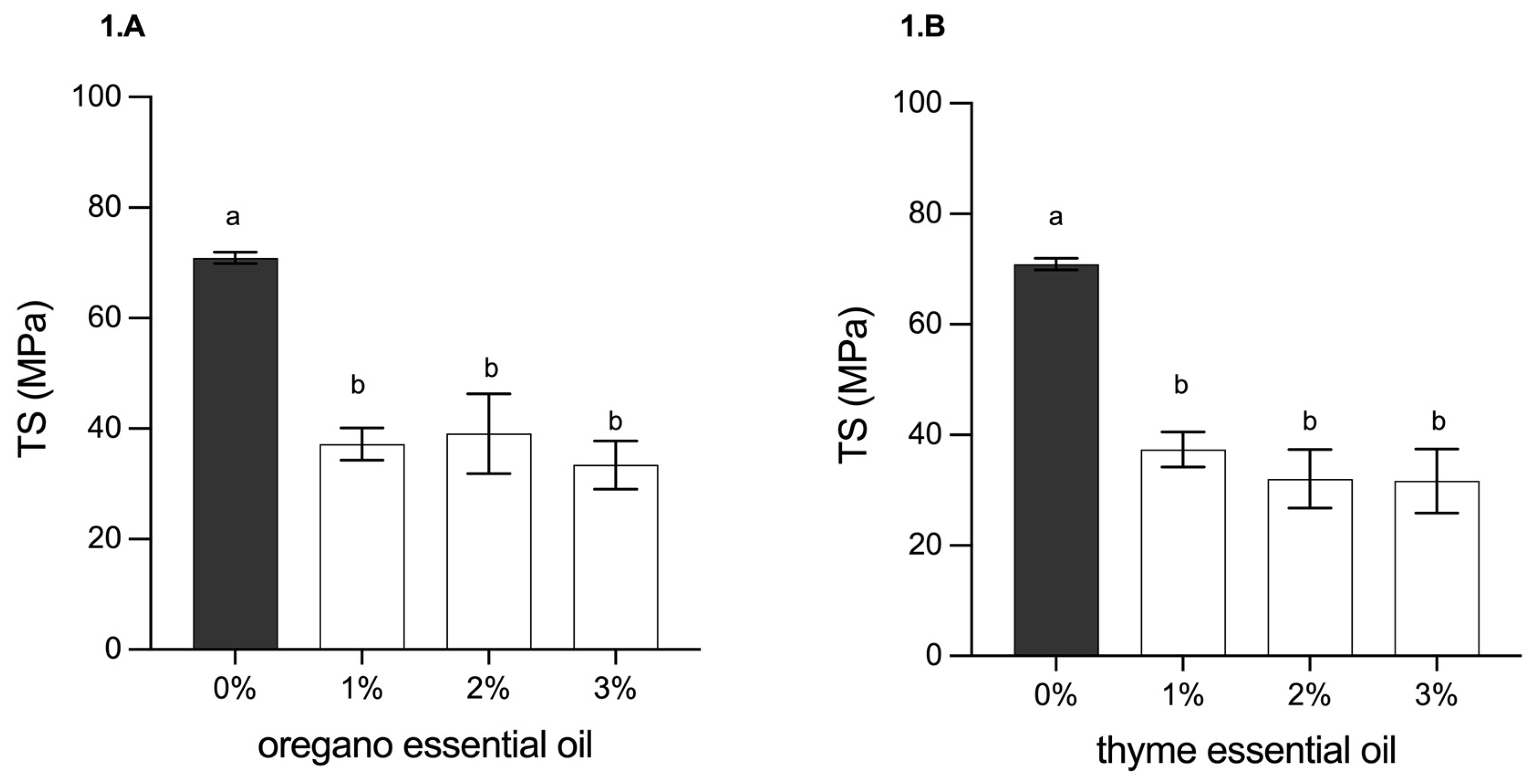
Figure 2.
Elongation at break (%E) of the film with (A) oregano oil and (B) thyme oil. Data are reported as mean + standard deviation (n = 5). Mean values with different letters indicate a significant difference (p<0.05).
Figure 2.
Elongation at break (%E) of the film with (A) oregano oil and (B) thyme oil. Data are reported as mean + standard deviation (n = 5). Mean values with different letters indicate a significant difference (p<0.05).
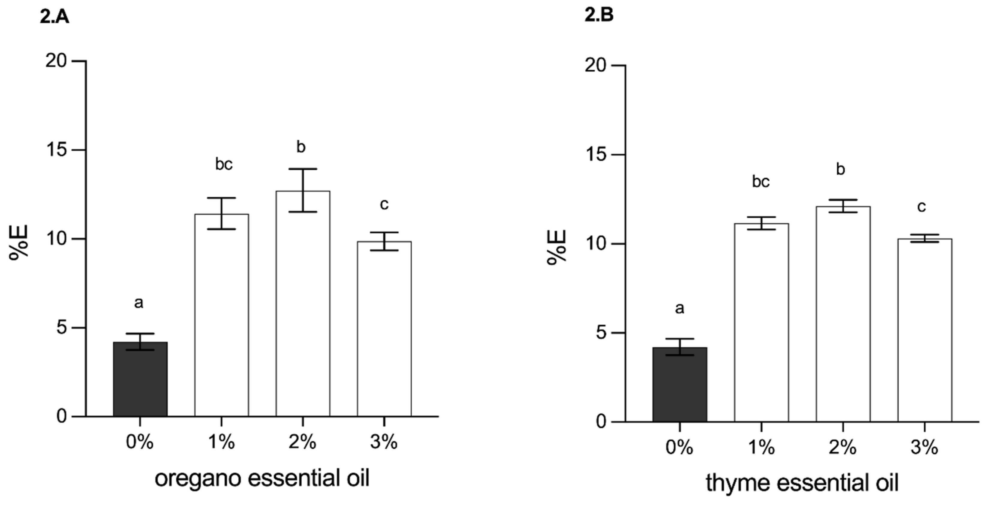
Figure 3.
SEM micrographs showing the surface structure of (a) control alginate-chitosan film, and with the addition of (b) EOO 2%, (c) EOO 3%, and cross-section of (d) control alginate-chitosan film and with addition of (e) EOO 2% (f) EOO 3%. EOO: oregano oil.
Figure 3.
SEM micrographs showing the surface structure of (a) control alginate-chitosan film, and with the addition of (b) EOO 2%, (c) EOO 3%, and cross-section of (d) control alginate-chitosan film and with addition of (e) EOO 2% (f) EOO 3%. EOO: oregano oil.
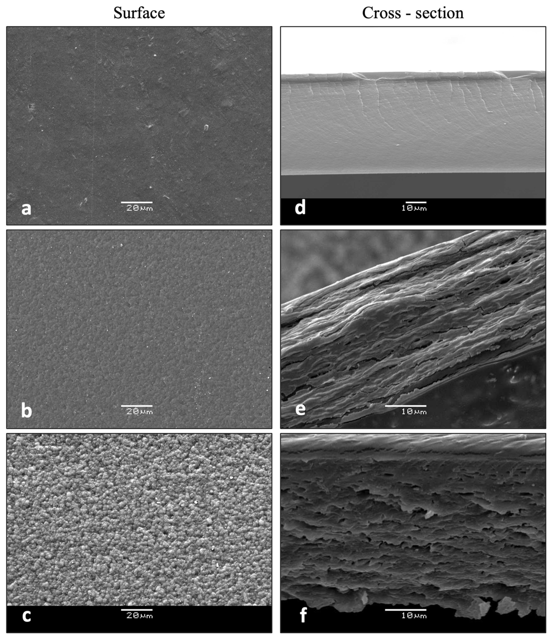
Figure 4.
SEM micrographs showing the surface structure of (a) control alginate-chitosan film and with the addition of (b) EOT 2%, (c) EOT 3%, and cross-section of (d) control alginate-chitosan film and with the addition of (e) EOT 2% (f) EOT 3%. EOT: Essential oil thyme.
Figure 4.
SEM micrographs showing the surface structure of (a) control alginate-chitosan film and with the addition of (b) EOT 2%, (c) EOT 3%, and cross-section of (d) control alginate-chitosan film and with the addition of (e) EOT 2% (f) EOT 3%. EOT: Essential oil thyme.
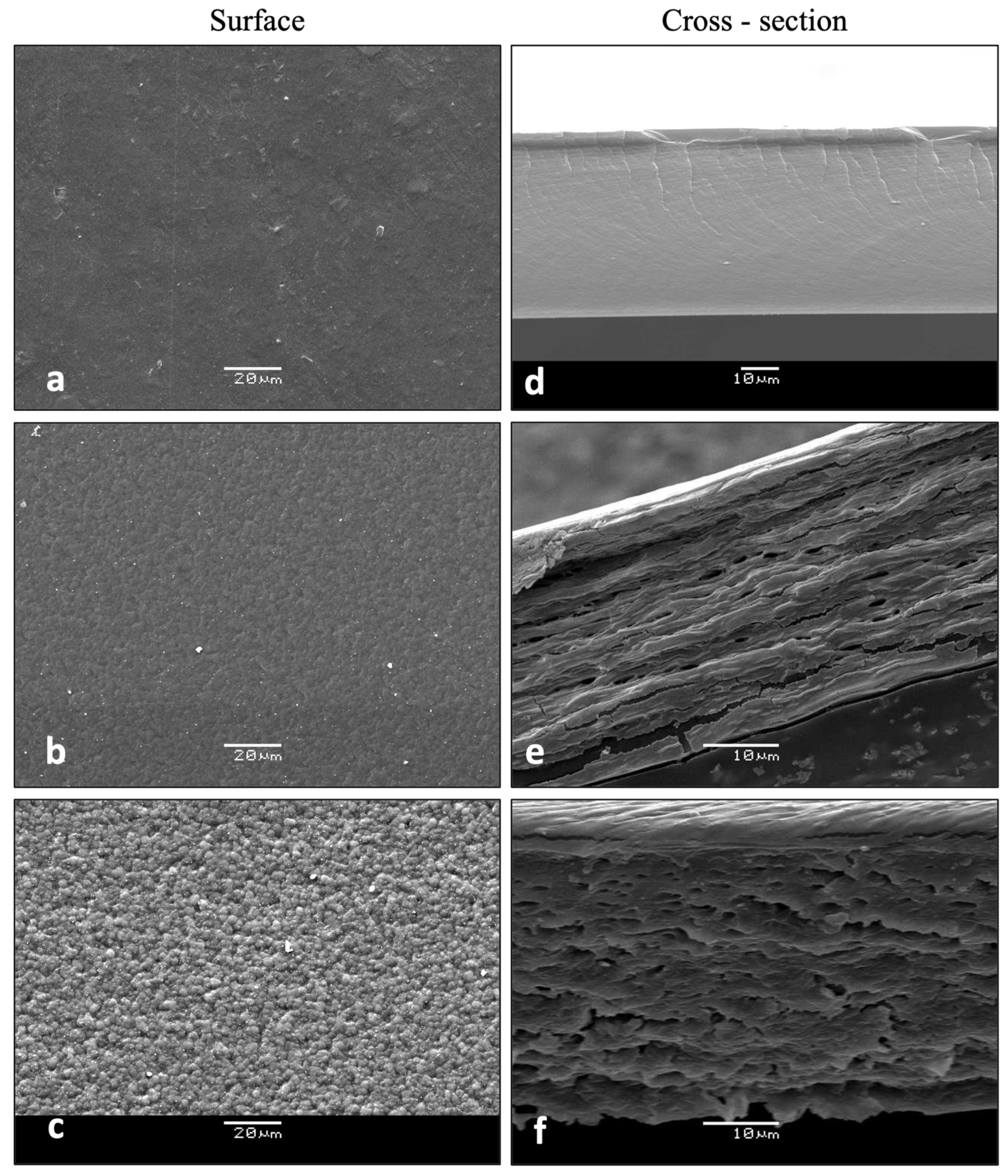
Figure 5.
Thermogravimetric curves (TG-DTG) of chitosan (CHI)-alginate (ALG) based films loaded with different concentrations of Origanum vulgare (A-B) and Thymus vulgaris (C-D) essential oils.
Figure 5.
Thermogravimetric curves (TG-DTG) of chitosan (CHI)-alginate (ALG) based films loaded with different concentrations of Origanum vulgare (A-B) and Thymus vulgaris (C-D) essential oils.
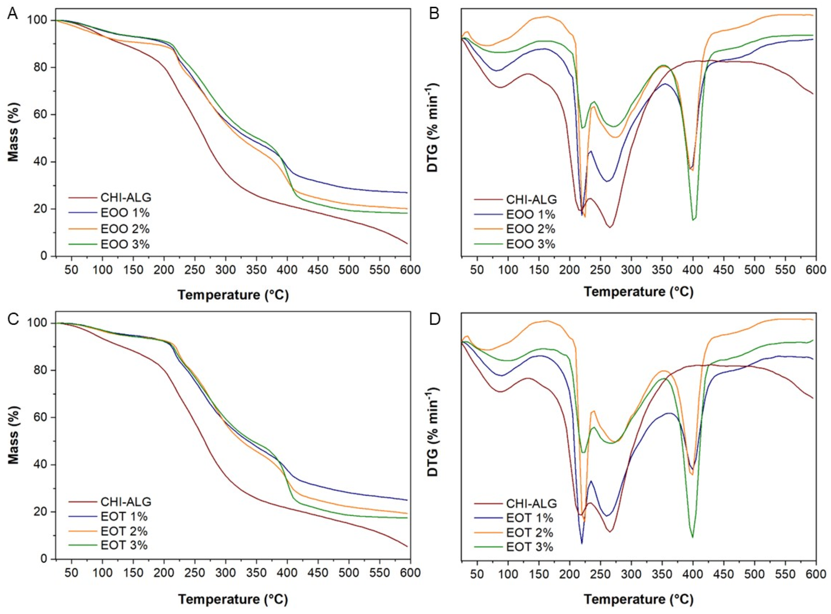
Figure 5.
Inhibition halo of films.
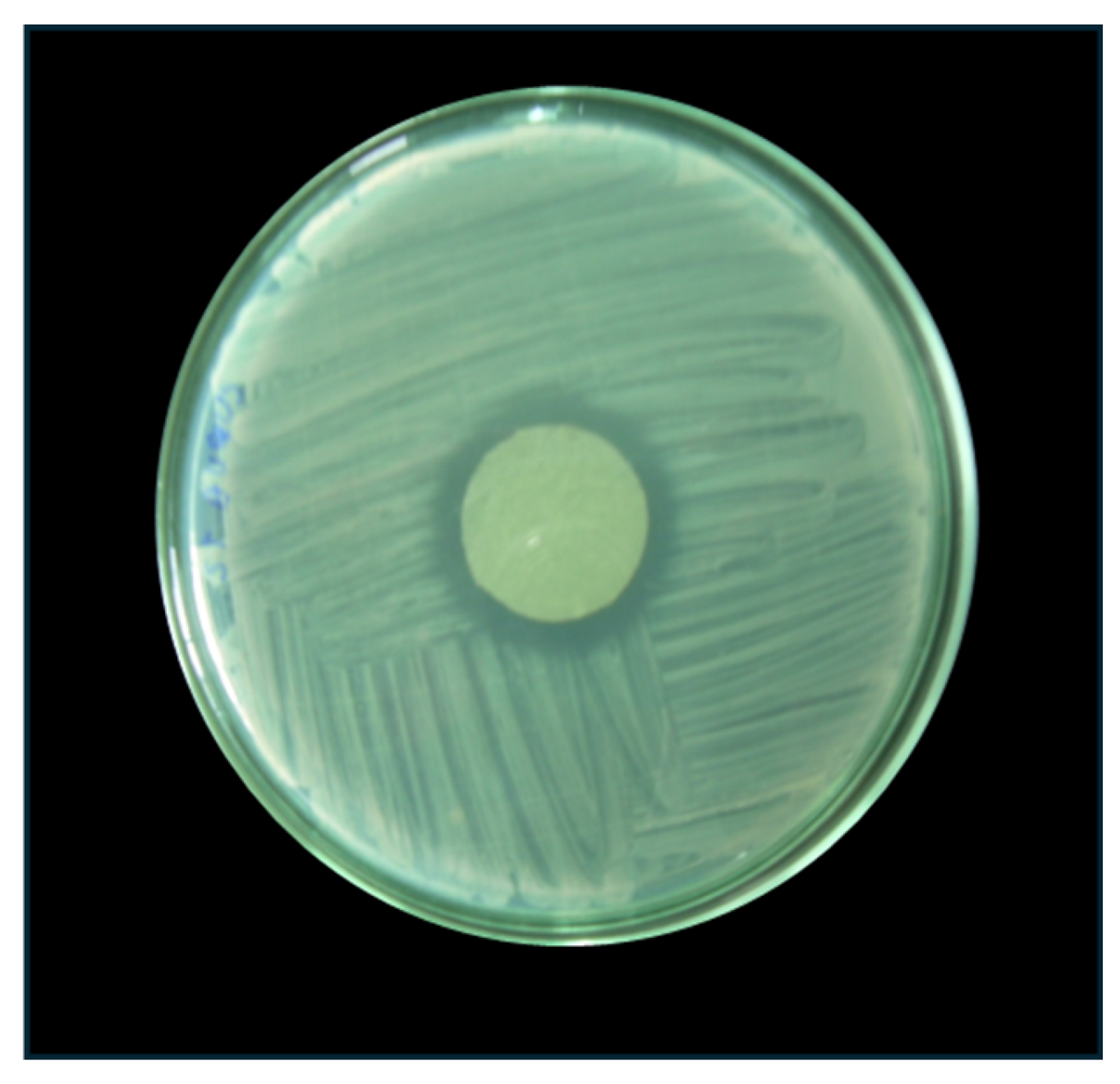

Table 1.
MIC and MBC of oregano and thyme essential oils for the different strains used.
| Strains | Antibacterial Parameter | |||
|---|---|---|---|---|
| MIC (mg/mL) | MBC (mg/mL) | |||
| EOT | EOO | EOT | EOO | |
| E. coli. | 1.39±0.03a | 0.69±0.03b | 2.78±0.04a | 2.78±0.04a |
| S. enterica | 1.39±0.03a | 0.69±0.03b | 5.56±0.04ª | 2.78±0.04b |
| S. aureus | 0.69±0.03a | 0.35±0.03b | 2.78±0.04a | 1.39±0.04b |
| L. monocytogenes | 0.35±0.03a | 0.17±0.03b | 2.78±0.04a | 1.39±0.04b |
MIC: minimum inhibitory concentration; MBC: minimum bactericidal concentration; EOO: oregano oil; EOT: thyme oil. Data are reported as mean + standard deviation (n=5). Mean values with different letters indicate a significant difference (p<0.05).
Table 2.
Effect of adding oregano and thyme oils in different concentrations on physical properties of alginate-chitosan films.
Table 2.
Effect of adding oregano and thyme oils in different concentrations on physical properties of alginate-chitosan films.
| Essential Oil Type | %EO | Thickness (x10-2 mm) |
Color (CIELAB Space) | WVP (g/m s Pa)x10-9 |
|||||
|---|---|---|---|---|---|---|---|---|---|
| L* | a* | b* | ΔE | YI | WI | ||||
| Control | 0.0 | 3.49±0.37a | 98.5±0.17a | -1.52±0.18a | 5.47±0.18a | 4.26±0.21a | 7.94±0.27a | 94.12±0.20a | 4.03±0.47ab |
| EOO | 1.0 | 3.82±0.07a | 88.3±0.20b | -3.46±0.22b | 11.69±0.16b | 12.1±0.11b | 18.9±0.23b | 83.10±0.10b | 3.19±0.15a |
| 2.0 | 3.77±0.12a | 87.2±0.47c | -3.56±0.08b | 11.65±0.13b | 13.0±0.37c | 19.1±0.18b | 82.37±0.29c | 2.47±1.00b | |
| 3.0 | 3.79±0.09a | 86.4±0.09c | -3.52±0.15b | 11.63±0.07b | 13.6±0.06c | 19.2±0.11b | 81.83±0.05c | 1.80±0.39c | |
| EOT | 1.0 | 3.78 ± 0.05a | 87.8±0.10b | 3.35±0.14b | 11.52±0.07b | 12.3±0.06b | 18.7±0.11b | 82.94±0.04b | 3.04±0.35a |
| 2.0 | 3.82 ± 0.08a | 86.8±0.04c | -3.70±0.03c | 11.64±0.03b | 13.3±0.04c | 19.1±0.05c | 82.05±0.04c | 2.45±0.16a | |
| 3.0 | 3.80 ± 0.05a | 86.3±0.03d | -3.43±0.02b | 11.96±0.14c | 13.9±0.05d | 19.7±0.22d | 81.54±0.07d | 1.65±0.69b | |
ΔE: colour delta; YI: yellowness index; WI: whiteness index; WVP: water vapor permeability; OEO: oregano oil; TEO: thyme oil. Data are reported as mean + standard deviation (n=5). Mean values with different letters in the same column indicate a significant difference (p<0.05).
Table 3.
Thermal analysis of alginate-chitosan films loaded with Thymus vulgaris and Origanum vulgare essential oil.
Table 3.
Thermal analysis of alginate-chitosan films loaded with Thymus vulgaris and Origanum vulgare essential oil.
| Sample | Temperature (°C) | Weight Loss (%) | ||
|---|---|---|---|---|
| Onset | Peak | End | ||
| Chitosan (CHI) | 24 | 66 | 135 | 3.8 |
| 251 | 310 | 402 | 50.4 | |
| Alginate (ALG) | 25 | 69 | 185 | 10.6 |
| 191 | 244 | 266 | 24.0 | |
| 266 | 284 | 319 | 10.9 | |
| 319 | 399 | 544 | 11.0 | |
| CHI-ALG | 30 | 87 | 133 | 10.1 |
| 134 | 216 | 234 | 24.6 | |
| 235 | 265 | 385 | 42.4 | |
| EOT 1% | 24 | 90 | 156 | 6.4 |
| 179 | 220 | 235 | 12.9 | |
| 236 | 260 | 360 | 34.9 | |
| 361 | 400 | 442 | 14.7 | |
| 443 | 477 | 525 | 4.2 | |
| EOT 2% | 26 | 73 | 157 | 5.9 |
| 195 | 225 | 239 | 11.1 | |
| 240 | 274 | 351 | 37.0 | |
| 352 | 399 | 443 | 18.8 | |
| 444 | 476 | 523 | 4.0 | |
| EOT 3% | 28 | 102 | 156 | 5.9 |
| 204 | 223 | 240 | 11.4 | |
| 241 | 267 | 352 | 41.3 | |
| 353 | 399 | 443 | 27.5 | |
| 444 | 477 | 524 | 3.6 | |
| EOO 1% | 24 | 81 | 149 | 7.0 |
| 205 | 219 | 235 | 10 | |
| 236 | 261 | 355 | 32.2 | |
| 356 | 396 | 438 | 15.5 | |
| 439 | 476 | 523 | 4.2 | |
| EOO 2% | 24 | 70 | 145 | 9.2 |
| 205 | 224 | 239 | 12.0 | |
| 240 | 273 | 353 | 32.0 | |
| 354 | 399 | 438 | 19.2 | |
| 439 | 476 | 523 | 4.0 | |
| EOO 3% | 26 | 83 | 149 | 6.8 |
| 204 | 221 | 241 | 18.3 | |
| 242 | 273 | 353 | 30.9 | |
| 354 | 400 | 438 | 26.8 | |
| 439 | 476 | 522 | 3.8 | |
Table 4.
Antibacterial effects of chitosan/alginate films with essential oils of oregano and thyme.
| Concentration (%) | Inhibition Zone by Type of Essential Oil (Diameter in mm) | |||||||
|---|---|---|---|---|---|---|---|---|
| E. coli | S. enterica | S. aureus | L. monocytogenes | |||||
| EOO | EOT | EOO | EOT | EOO | EOT | EOO | EOT | |
| 0.0 | 0Aa | 0Aa | 0Aa | 0Aa | 0Aa | 0Aa | 0Aa | 0Aa |
| 1.0 | 26±1.2Ba | 30±0.9Bb | 22±0.2Ba | 26±0.8Ba | 24±1.4Ba | 27±0.09Ba | 27±1.8Ba | 40±0.9Bc |
| 2.0 | 25±0.9Ba | 39±1.5Cb | 28±1.6Ca | 28±2.1Ba | 30±0.1Ba | 38±0.13Cb | 37±1.3Cb | 46±1.5Bc |
| 3.0 | 39±1.3Ca | 41±1.3Ca | 35±0.8Da | 36±0.9Ca | 43±1.2Ca | 49±0.09Db | 49±1.3Db | 55±1.3Cb |
EOO: oregano oil; EOT: thyme oil. Capital letters indicate differences between concentrations for the same oil; small letters indicate differences between oils for the same concentration (p<0.05).
Disclaimer/Publisher’s Note: The statements, opinions and data contained in all publications are solely those of the individual author(s) and contributor(s) and not of MDPI and/or the editor(s). MDPI and/or the editor(s) disclaim responsibility for any injury to people or property resulting from any ideas, methods, instructions or products referred to in the content. |
© 2024 by the authors. Licensee MDPI, Basel, Switzerland. This article is an open access article distributed under the terms and conditions of the Creative Commons Attribution (CC BY) license (http://creativecommons.org/licenses/by/4.0/).
Copyright: This open access article is published under a Creative Commons CC BY 4.0 license, which permit the free download, distribution, and reuse, provided that the author and preprint are cited in any reuse.


