
Submitted:
27 November 2024
Posted:
28 November 2024
You are already at the latest version
Abstract
Metal organic frameworks (MOFs) have become a highly usable system in various sectors because of their highly ordered structure and high porosity providing them with high storage capacity. But their use is sometimes forbidden in the food industry due to the presence of some organic compounds which have undesirable effects. Cyclodextrins which are considered GRAS (Generally Recognized as Safe) by FDA comes as a very good alternative to previously used compounds for the development of the MOF’s to be used in food packaging industry especially in the packaging sector. These cyclodextrin MOF does possess edible, biocompatible as well as biodegradable characteristics and due to these reasons they have gained attentions from researchers in the food industry. In this review we focus on the recent advancements in the field of CD MOF’s. We have emphasized on the synthesis of these MOF’s through different techniques, formations of their inclusion complex with bioactive compounds and their characterization. Finally, we discussed on the use of CD MOF as a carrier for various highly volatile bioactive compounds and their ability to increase the solubility and stability of these bioactive compounds.
Keywords:
cyclodextrins
; MOF
; antibacterial food
Introduction
Metal Organic Frameworks are crystalline hybrid porous network materials formed by self-assembly of inorganic metal ions/nodes and organic bridging ligands [1]. These coordination polymers have high surface area and their shape and size can be controlled and modified [2,3,4]. Metal organic frameworks possess versatile applications such as gas storage and adsorption [5,6], catalysis, electrochemistry, drug delivery, sensors molecular recognition. MOF’s based on cyclodextrins (CDs) have been used in pharmaceuticals but in recent years, CD MOF’s have received attention as a potential candidate to be used in various food applications because of the biocompatibility and non-toxic nature of CDs. Although, only bio-friendly and bio-compatible metal ions such as K+, Ca2+and Ti4+ can be used as these are considered to be non-toxic in nature and can be used in food industry within acceptable ranges [7].
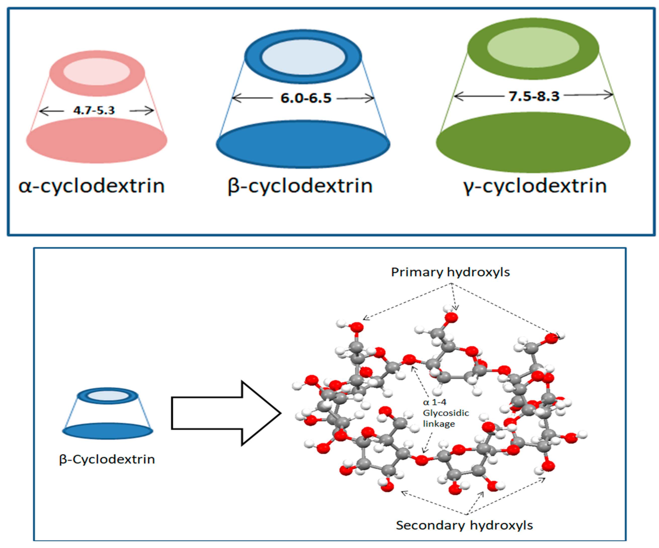
Cyclodextrins are cyclic oligosaccharide composed of glucose monomeric units which are linked through alpha 1-4 glycosidic linkages [8,9,10]. CDs are produced as a result of intramolecular transglycosylation (cyclization) reaction during degradation of starch by CGTase enzyme [9,10]. The three types of cyclodextrins that are naturally obtained are α (hexasaccharide), β (heptasaccharide) and γ (octasaccharide). The nature of the enzyme used while production decides the distribution of α-, β-, and γ-CDs [10]. CDs possess truncated cone structure with their outer surface being hydrophilic while the inner cavity being lipophilic in nature. The cavity, lined with non-polar groups—C3, O4 glycosidic bond (ether like), and C5–CH (aliphatic-like) groups attributed to its hydrophobicity [11] Their hydrophobic cavity acts as an excipient for various active molecules to form complexes in order to achieve better solubilization, better physical & chemical stability and better delivery [12]. The diameter for a α-, β-, and γ-CDs range from 4.7 to 5.3, 6.0 to 6.5, and 7.5 to 8.3 Å, respectively which shows the difference in their suitability to form inclusion complex with compounds based on their molecular size. β-CD is stated as the most suitable molecule due to its higher ability to form inclusion complex with food based additives but due to its O2-O3 intramolecular hydrogen bond it has a very low solubility in water [13] while its counterparts α- and γ-CDs , possess higher water solubility because in α-cyclodextrin, the hydrogen bond belt is incomplete because of the distorted position of one glucose unit in the structure, whereas γ-cyclodextrins have a non-coplanar structure that makes it the most soluble of the native cyclodextrins [10]. CDs with larger cavity size also exist but both their production is difficult and they possess limited ability to form inclusion complex [14]. Table 1 compares the different properties of the three native CDs [15].
As discussed above the three naturally occurring cyclodextrins namely α-, β-, and γ-CDs possess a hydrophobic inner cavity. In aqueous solution, the CD cavity is filled with the water molecules [16]. These water molecules are not able to complete the hydrogen bonding network as in bulk medium and are energetically frustrated [11,17]. These frustrated water molecules facilitates the inclusion of guest molecule through their replacement by the less polar molecule thus decreasing the strain on the cyclodextrin ring, resulting in a more stable lower energy state [10,16,18]. Due to their cyclic structure CDs are less susceptible towards enzymatic degradation than the linear dextrins and CDs are better complexing agents and solubilizers [14]
Figure 1.
General structure of cyclodextrin (top), View into the molecular structure of β-cyclodextrin (bottom).
Figure 1.
General structure of cyclodextrin (top), View into the molecular structure of β-cyclodextrin (bottom).
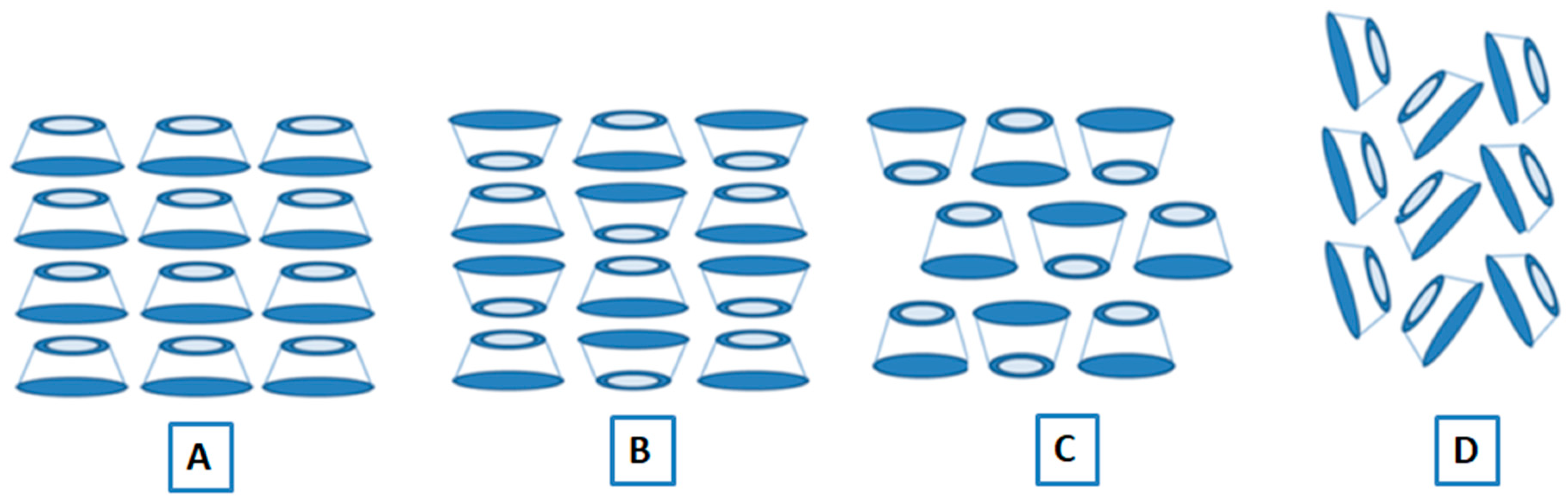
Native cyclodextrins and their inclusion complex exist in two crystal structures: channels and cages [13,19]. In channel type the molecules are placed exactly on top of each other which create a channel due to the alignment of their cavities in symmetry. The stacking of the cyclodextrin units can be head to head or head to tail depending on various factors. In case of cage type crystal, the stacking of the CD units is such that all the cavities are blocked by each other, therefore the guest molecules remains isolated. The packing in cage type crystal structure depends on the complexed guest compound and can be of two types: ‘Herringbone’ and ‘Brick-type’.
Figure 2.
Crystal structure of cyclodextrins: A) channel structure(head to tail), B) Channel structure(mixed), C) cage structure (Brick-Type), D) cage structure (Herringebone).
Figure 2.
Crystal structure of cyclodextrins: A) channel structure(head to tail), B) Channel structure(mixed), C) cage structure (Brick-Type), D) cage structure (Herringebone).
Toxicological Profile of Cyclodextrins
The basic concern regarding the use of cyclodextrin in food based applications is the toxicity. The toxicity of cyclodextrins depends on the route of administration [20,21,22]. α and β CDs are resistant to β-amylases that hydrolyse starch from the non-reducing end, but they are slowly hydrolysed by α-amylases that hydrolyse starch from within the carbohydrate chain. The only CD that is readily hydrolysed by α-amylase is the unsubstituted γ-CD [23,24,25] . Cyclodextrins are either not or only partially absorbable in the human gastrointestinal tract [26,27,28]. Undigested cyclodextrins can be metabolized by microbiota in the lower part of the gastrointestinal tract [27,28,29] after oral administration, γ-CD is completely digested in the gastrointestinal tract, whereas αCD and βCD, as well as the CD derivatives, are predominantly digested by bacteria in the colon. α-CD is digested more slowly than βCD. After parenteral administration CDs are mainly (> 90%) excreted unchanged in the urine via glomerular filtration with the rest eliminated by other excretion pathways, such as liver metabolism and biliary excretion [29]. When the administration is oral, CDs are practically nontoxic because of their hydrophilic and bulky nature [22]. The bioavailability of natural CDs and some derivatives are also very low, making them safe when administered orally [27,29]
In the USA, they have obtained the GRAS (Generally Recognized as Safe) status as approved by the FDA (US Food and Drug Administration). In Europe, they are considered food additives and labelled as E-457 (αCDs), E-458 (γ-CDs), and E-459 (β-CDs) [30,31]. The FAO/WHO (Food and Agriculture Organization of the United Nations/World Health Organization), Joint Expert Committee on Food Additives (JECFA) set the maximum advisable level of β-CD in food at 5 mg/kg of body weight per day; however, α-CD and γ-CD do not have an established Acceptable Daily Intake (ADI), due to their benign toxicological profiles [27]. Current regulations suggest that the migration of cyclodextrins from packaging to food does not have harmful effects on consumers if they are part of the packaging; this is why cyclodextrins are recognized as safe additives for use in food [15,27]
Synthesis of Cycloedxtrin MOFs
Various techniques for synthesis of CD MOFs like Vapor diffusion, Solvothermal, Hydrothermal, Microwave mediated and Ultrasound mediated have been reported in literature but the most favorable technique in applications pertaining to food industry is the Vapor diffusion method. This method provides crystal formation with high yield along with an advantage of being easy to setup and control. Although to control crystal growth rate and make the process rapid, vapor diffusion has been often modified with ultrasound assistance as observed from various literature. In this section we have focused on the synthesis of MOFs using the three native CDs.
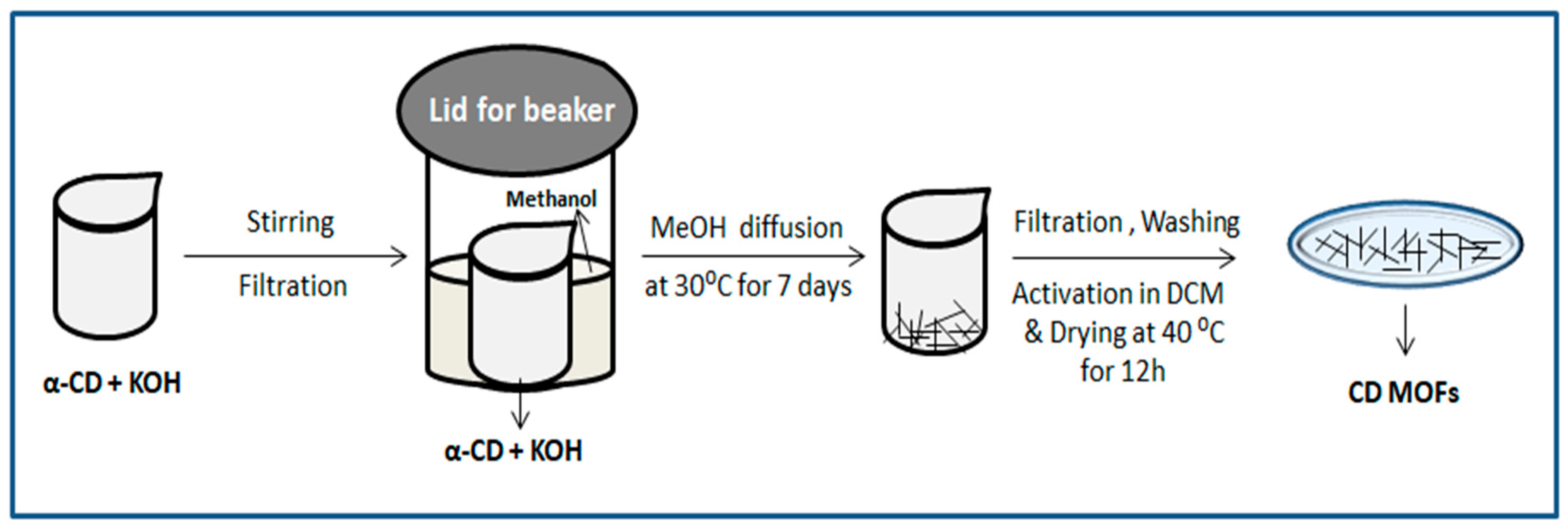
Synthesis of α-CD MOF
α-CD-MOF using sodium salt was synthesized by dissolving α-CD (1.46 g) and NaOH (0.48 g) in deionized water (20 mL), and then MeOH (50 mL) was added to vapour diffuse into the solution for 7 days at 30°C. The resulting crystals were filtered and washed three times with MeOH. To remove interstitial solvent, the α-CD-MOF crystals were immersed in dichloromethane for 3 days. The sample was dried in a 40°C vacuum oven for 12 h. The method of preparing α-CD-MOF using potassium salt was the same, except that KOH (0.673 g) was used [32].
Figure 3.
Schematic route of formation of MOFs by Vapour Diffusion Method.
Synthesis of β-CD MOF
In different literatures β-CD MOF has been mostly prepared with K+ ion due to its biocompatibility and non-toxic nature which makes it easier to be used in food industry. Purified β-CD (1.1349 g) and KOH (0.4488g) were dissolved in distilled water. The solution was then filtered and transferred to a beaker. The beaker was sealed in a tank containing 100ml of methanol and kept for one week. In this time period vapors from methanol diffused into the solution and formed K-β-CD MOF [33]. A more defined method is stated by Jiang W et.al, where β-CD (0.1418 g, 0.125 mmol) and KOH (0.056 g, 1mmol) dissolved in H2O (5 ml). The molar ratio in most of the studies is maintained at β-CD: KOH =1:8. This solution is then filtered through an organic membrane having pore size of 0.45μm into a small beaker containing 0.5ml MeOH. This small beaker was kept inside a big container with 60ml of MeOH, followed by vapour diffusion into aqueous solution at 50°C for 12h. After 12h a colourless and transparent liquid was obtained to which 5ml MeOH solution (including 60mg of PEG 20000) was added and kept in a closed container. This colourless liquid was incubated overnight leading to the formation of K-β-CD MOF crystals. The crystals were collected after separation, washed with 15ml ethanol twice and then suspended in dichloromethane for 3 days for activation. Ultimately the crystals were desiccated overnight at 50°C under vacuum conditions [4].
Synthesis of γ-CD-MOF
Majorly, the synthesis of γ-CD-MOFs has also been performed using vapor diffusion technique due to its simple procedure and effective results. One of the first works mentioned in literature was reported by Smaldone et.al. γ-CD and KOH were taken in a 1:8 mmol ratio and dissolved in 20ml water. The solution was filtered and MeOH was allowed to vapor diffuse into the solution over a period of one week. The result was colorless cubic crystals which were isolated, filtered and washed with MeOH and then being left to dry in the air [34].
Another method explained the use of ultrasonication for the production of γ-CD-MOF. In a work by Shen et.al. γ-CD (648 mg) and KOH (256 mg) were dissolved in ultra-pure water (20 mL). The solution was filtered with a 0.45μm filter membrane and stirred at room temperature. MeOH (12 mL) and the above solution were added in the tube to form a white solution. The tube was then transferred to a water bath at 60°C for 10 min, a clear and transparent solution was obtained. A probe ultrasonicator was used and the solutions were ultrasonically processed at a frequency of 20 kHz and a power of 540 W, reaction under intermittent action for 10 min, the intermittent ultrasonic action mode is on for 2 s, off for 2 s, while PEG-8000 (256 mg) was quickly added after the start of the ultrasonic treatment to trigger the deposition of crystalline materials. In addition, the rapid synthesis and the regular morphology of γ-CD-MOF crystal are inseparable from PEG-8000 as the excipient which was used as the size modulator for controlling the size and morphology of MOF crystals in aqueous systems. The crude product was obtained after the reaction. The crude product was allowed stand for 1h to obtain the white precipitate. The precipitate was centrifuged at 5000 rpm for 5 min and washed with MeOH for 3 times, then the precipitate was dispersed in MeOH and dried at 50°C under vacuum for 12 h [35].
Figure 4.
Schematic route of formation of MOFs by Ultrasonication Method.
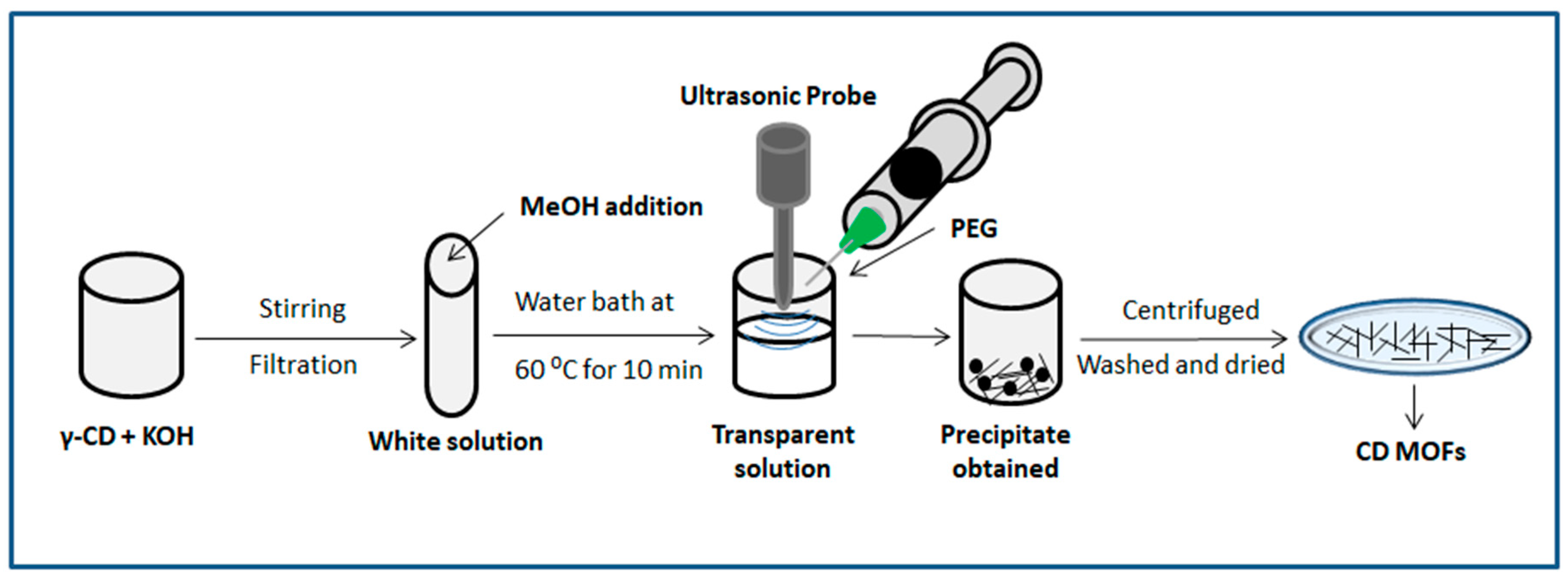

Table 2.
Common ways of synthesis of Cyclodextrin based Metal Organic frameworks.
| Metal Ion (Salt used) | Ligand | Synthesis technique | Conditions | Reference | ||
|---|---|---|---|---|---|---|
| Mixing / Ultrasonication time |
Temperature for vapour diffusion (°C) | Vapour diffusion time | ||||
| K+ ( C7H5KO2) | α-CD | Vapour diffusion | 6-8h | R.T | 3-7 days | [36] |
| K+ (KOH) | α-CD | Ultrasonication | 30 min | *After ultrasonication, solution was mixed with MeOH, heated at 60°C, cooled to r.t and PEG and MeOH was added to obtain crystals | [37] | |
| K+ (KOH) | β-CD | Vapour diffusion | - | R.T | One week | [38] |
| K+ (KOH) | β-CD | Vapour diffusion | - | 50 | 12h | [39] |
| K+ (KOH) | β-CD | Vapour diffusion | 3h | 25 | - | [40] |
| K+ (KOH) | β-CD | Vapour diffusion | 3.5h | R.T | 3-5 weeks | [41] |
| K+ (KOH) | γ-CD | Ultrasonication | 30 min | - | - | [37] |
| Rb+ (RbOH) | γ-CD | Vapour diffusion | - | R.T | One week | [34] |
| K+ (KOH) | γ-CD | Vapour diffusion | 6-12h at 500pm | 23 | One week | [42] |
| K+ (KOH) | γ-CD | Vapour diffusion | - | 60 | 2h | [43] |
| K+ (KOH) | γ-CD | Vapour diffusion | - | 50 | 5h | [44] |
| K+ (KOH) | γ-CD | Ultrasound assisted vapour diffusion | 5 min | 50 | 6h | [45] |
| K+ (KOH) | γ-CD | Ultrasound assisted vapour diffusion | Different time (0, 5, 10, or 15 min) | 50 | 6h | [46] |
| K+ (KOH) | γ-CD | Seed mediated methanol vapour diffusion | - | 50 | 1h | [47] |
| K+ (KOH) | γ-CD | Ultrasonication | 30min | *After ultrasonication solution was heated at 60°C for 1 h and the PEG 20,000 was added to obtain crystals | [48] | |
| K+ (KOH) | γ-CD | Vapour diffusion | 6h 500rpm | R.T | 3-7 days | [49] |
| K+ ( C7H5KO2) | γ-CD | Vapour diffusion | 6h 500rpm | R.T | 3-7 days | [49] |
| K+ (KOH) | γ-CD | Ultrasonication | 30 min | *After ultrasonication, solution was mixed with MeOH, heated at 60°C, cooled to r.t and PEG and MeOH was added to obtain crystals | [37] | |
| K+ (KOH) | γ-CD | Vapour diffusion | - | 50 | 24h | [50] |
| K+ (KOH) | γ-CD | Ultrasonication | 30min | *After ultrasonication, solution was mixed with MeOH, heated & cooled to r.t and PEG and MeOH was added to obtain crystals | [51] | |
| K+ (KOH) | γ-CD | Seed mediated methanol vapour diffusion | - | 50 | 6 | [52] |
| K+ (KOH) | γ-CD | Seed mediated Ultrasonication | Different time (0, 3, 5, 10, and 15 min) | After ultrasonication, solution was mixed with MeOH to obtain crystals | [53] | |
CD MOFs Applications in Food Industry
The direct usage of highly volatile compounds like essential oils is difficult in any application. There is a need to provide them with the stability required during the process of product manufacturing without the loss of their inherent properties. Encapsulation is an easier and highly effective process while dealing with compounds like essential oils. The active molecule (guest molecule) can be entrapped inside the cavities of CD-MOFs overcoming the issues that exist in the direct usage of active molecule. Some of the improvements that are observed are as follows [54]-
- The diffusion and volatility (in the case of volatile substances) of the included guest can decrease strongly.
- The complexed substances, even gaseous substances can be entrapped in a carbohydrate matrix forming a microcrystalline or amorphous powder.
- The complexed substance can be effectively protected against heat decomposition, oxidation, and any other type of reaction, except against those with the hydroxyl groups of cyclodextrin, or reactions catalyzed by them.
The above-mentioned positive changes give the liberty of using different active principles for various applications in the food-based industry; some of them are mentioned in Table 3. However, it is also worth noticing that for the MOFs to be a useful system, it is essential to characterize them as well as the inclusion complex formed by them. The next section focuses on various characterization techniques that have been mostly used.
Figure 5.
A schematic diagram of CD-MOFs synthesis and their potential food industry applications.
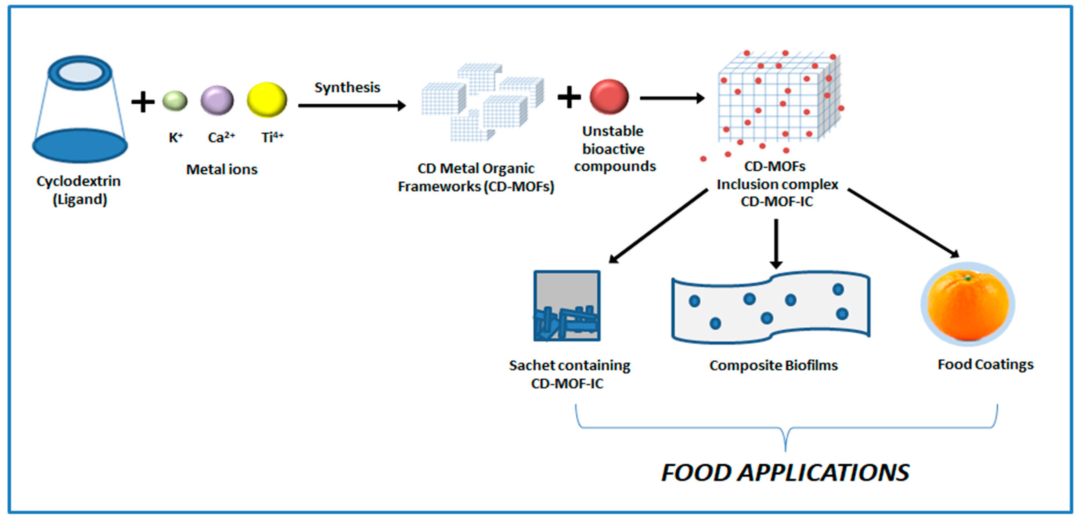
Table 3.
Overview of the existing and potential application of CD-MOFs in food industry.
| MOF | ACTIVE COMPOUND | APPLICATION | IMPORTANT OBSERVATIONS | REFERENCE |
|---|---|---|---|---|
| α-CD MOF | Ethylene gas | Accelerated fruit ripening | MOF- ethylene complexes had controlled ethylene-release for accelerated fruit ripening | [32] |
| α-CD MOF | Catechin | Potential application in Food packaging | CD-MOFs protected catechin against light, oxygen and temperature, thus improving its storage stability. Catechin encapsulated within CD-MOFs exhibited superior bioavailability | [37] |
| β -CD MOF | - | Herbicide adsorption and potassium replenishment | The maximum adsorption capacities of four herbicides were in the range of 261.21-343.42 mg.g-1. The herbicide removal percentage was in the order: MET>PRE>ALA>ACE. | [55] |
| β-CD MOF | Hexanal | Preservation of Mangoes | Treated fruit remains fresh until 2 weeks after storage. They possessed higher firmness and had lower weight loss. | [56] |
| β-CD MOF | Catechin | Zein based packaging film | Zein films with Catechin loaded β-CD MOFs possessed better physical properties, antibacterial characteristics and more steady release profile for catechin as compared to normal Zein film containing catechin. | [57] |
| β-CD MOF | Clove essential oil (CEO) | Preservation of Chinese Bacon | Decrease in the lipid oxidation of bacon due to the increasing inhibitory effect of CEO after encapsulation in β-CD-MOF. Apart from that, the free radical scavenging activities and thermal and pH stabilities were also better in case of CEO/ β-CD-MOF’s than just CEO | [58] |
| β-CD MOF | Lavender essential oil (LEO) | Potential application in Food packaging | LEO/K-βCD-MOFs were proved to be more thermally and acid-base stable than LEO, and its intracellular antioxidant effect was also significantly improved by encapsulation. | [33] |
| β-CD MOF | Thymol (THY) | Preservation of Cherry Tomatoes | The decay index of whole cherry tomatoes treated with γ-CD-MOF-THY decreased from 67.5% (control group) to less than 20% during storage at room temperature for 15 days. | [40] |
| β-CD MOF | Polyphenols | Potential application in Food packaging | The stabilities and solubility’s of ALP were significantly improved compared when encapsulated in β - CD-MOFs as compared to β -CD, suggesting the potential of β-CD-MOFs as better carriers than β -CD for polyphenols in food industry applications. | [38] |
| β-CD MOF | Origanum Compactum essential oil (OCEO) | Potential application in Food packaging | Compared to βCD, K-βCD-MOFs displayed higher encapsulation efficiency. Antioxidant capacity of OCEO was significantly enhanced in the presence of K-βCD-MOFs | [59] |
| β-CD MOF | - | Extraction of Organochlorine pesticides from Honey samples | CD-MOF/TiO2 has good selective enrichment ability for OCP and is suitable for the D-SPE pre-treat of honey sample analysis. | [60] |
| γ-CD MOF |
Anthocyanins | Grape preservation | Grapes coated with Sodium alginate + CD-MOFs containing anthocyanin showed gradual decrease in weight loss after 10 days. The firmness and epidermal puncture value of the grapes was also high with this coating. Brix value was found to be less as compared to others. | [50] |
| γ-CD MOF |
Ethylene gas | Accelerated ripening as well as preservation of bananas | Polycaprolactone nanofibers containing γ-CD-MOF and TiO2 were used. The γ-CD-MOF were encapsulated with ethylene and helped in the accelerated ripening of bananas while TiO2 under the action of UV helped to degrade ethylene prolonging the shelf life of bananas | [61] |
| γ-CD MOF |
Carvacol | Chitosan-Cellulose(CS-CEL) Active packaging film | CS-CEL films containing Carvacol- γ-CD MOF showed the lowest weight loss in strawberries as compared to other conditions. Also, Carvacol-γ-CD-MOFs/CS-CEL composite film showed the lowest firmness loss, highest TSS value and lowest pH change. |
[62] |
| γ-CD MOF |
Cinnamaldehyde | Preservation of fresh cut cantaloupes | CD/MOF containing cinnamaldehyde (CA) and carbon dots improved the shelf life of the fresh cut cantaloupes was and maintained the quality of the fruit. It was observed that CD/MOF-0.5(amount of carbon dots)/CA exhibited a strong and long-lasting antibacterial activity when tested against E. coli in vitro and on fresh-cut cantaloupes. | [45] |
| γ-CD MOF |
Curcumin | Preservation of Centennial Seedless grapes (CSg) through Pullulan and trehalose (Pul/Tre) composite film containing Curcumin- γ-CD-MOF | The naturally placed CSg began to rot on the 4th day, while the CSg coated with Pul/Tre film rot on the 8th day with a shrunken surface and severe dehydration. However, the appearance of CSg coated with Cur-CD-MOFs-Pul/Tre film was still largely unaltered on day 10. | [44] |
| γ-CD MOF |
- | Ethylene absorber for improving postharvest quality of kiwi fruit | The fruit in the γ-CDMOF-K group did not decay over the whole storage period, maintained a good appearance, and remained edible. | [46] |
| γ-CD MOF |
Octadecenylsuccinic anhydride (ODSA) | Pickering emulsions coating and package paper for fruit preservation | The uncoated bananas experienced a 27.5% weight loss after 9 days, whereas the sample coated with a 10% ODSA emulsion had just 15.6% weight loss. Similarly, the weight loss also reduced in ODSA emulsions containing ODSA modified γ-CD-MOFs. | [63] |
| γ-CD MOF |
β-carotene | Development of High internal phase emulsion(HIPEs) | CD-MOF offers a safeguarding matrix for β-carotene , reducing the degradation and enabling a modulated release profile. | [64] |
| γ-CD MOF |
Vitamin A palmitate | Encapsulation of Vitamin A palmitate (VAP) for delivery as a food supplement | The half-life (t1/2) vitamin A in γ-CD-MOFs/VAP was recorded to be 20.5 days which is a 1.6 time increase compared to BASF vitamin A powder (t1/2 = 13.0 days) and a 2.6 time increase compared to physical mixture (t1/2 = 7.9 days), respectively | [65] |
Characterization of CD-MOFs
In this section we have focused on some of the most useful techniques that are being used for the analysis of MOF structure, especially for the characterization of CD-MOF inclusion complex. These techniques help us to confirm the formation of inclusion complex by showing the presence of active principles like essential oils inside the MOF cavities.
1. Thermal Analysis
a. Thermogravimetric Analysis (TGA)
TGA is an important characterization technique to understand the thermo-stability of any type of sample. It helps us to identify the degradation temperature of the compound. We can also analyze the presence of various compounds inside a sample and also know the quantities of different compound present. Changes in weight loss and shifts in degradation temperature or evaporation temperature indicate towards the formation of an inclusion complex. In a study by Hu et.al., a lower degradation temperature for CD-MOFs was observed as compared to native CD’s which attributed to the weaker metal coordination bond is and porous structure of the MOFs, the results indicated the formation of K-CD framework [41]. The thermal stability of CEO was found to be better when encapsulated in K- β-CD MOFs as compared to just β-CD. After the heating of 10 days, the rates of CEO/β-CDMOFs and CEO/β-CD remained at 63.35 and 50.23% respectively, however that of the free CEO was only 24.03% [58]. Similarly, in case of LEO, higher preservation rate was observed for LEO/K-βCD-MOFs (66.8%) as compared to LEO/β-CD (31.50%) and native LEO (18.00%) after 10 days.
b. Differential Scanning Calorimetry (DSC)
Another technique reported to clarify the interaction between guest and host is differential scanning calorimetry (DSC). It is generally observed that the cyclodextrin cavity affects the thermal behaviour of inclusion complexes, thus, leading to a shift in their endothermic peaks as compared to pure compounds. Shifting, broadening and appearance of new peaks or disappearance of certain peaks in the host may be due to evaporation, oxidation, decomposition, melting, or polymorphic transition, suggesting complex formation. Inclusion complex formation generally causes the reduction or absence of endothermic peaks of the host (at the temperature of its boiling and melting point). A work confirmed the formation of inclusion complex CA-β-CD through the shift in the peak for different CD-MOFs. It can be seen in Figure 6(b), the α-CD-MOFs, β-CD-MOFs and γ-CD-MOFs displayed a wide endothermic peak at 106.49, 107.76 and 87.84 ℃ which were shifted to 120.79 ℃ for CA/α-CD-MOFs, 101.84 ℃ for CA/β-CD-MOFs and 92.3 ℃ for CA/γ-CD-MOFs after CA loading. This change in the peak value can be reasoned with the encapsulation of CA in the cavities of porous MOF structure which altered the thermal properties [37]. Similarly, the formation of Curcumin- γ-CD-MOFs was also evident from the disappearance of the melting peak of curcumin (174.35) when encapsulated in γ-CD-MOFs [48].
2. Microscopy
Microscopic techniques like scanning electron microscopy (SEM) and transmission electron microscopy (TEM) are the most initial techniques to use for MOFs characterization. These techniques enable us to evaluate the particle size and shape of the formed framework. Both of the techniques can be further extended for better analysis of samples. For example, SEM can be used along with EDX for elemental mapping of the MOFs. The techniques are also able to provide an instant visual representation of change in the morphology of MOFs due to variations in different factors responsible for the final structure of MOFs. Shen et.al showed the effect of varying ultrasonic power, ultra-sonication time, and incubation temperature on the final structure of γ-CD-MOF. It was observed that the change in incubation temperature resulted in the formation of MOFs with different sizes. When the temperature increased, the particle size of the samples also increased, this phenomenon may be attributed to the fact that the nucleation rate was inversely proportional to the temperature during the crystallization process [35]. In another work, it was detected that β-CD-MOF and γ-CD-MOF were highly crystalline and possessed cuboid and cubic shapes respectively, while α-CD-MOF had flaky crystals of uniform size (Figure 7b). Elemental mapping of CD-MOFs (Figure 7c) also provided information about the homogenous distribution of C, O, and K [41]. TEM was also performed to confirm that γ-CD-MOFs displayed a uniform cubic shape (Figure 7a), and the mean particle size was approximately 190 nm [47].
3. X-Ray Diffraction
X-ray diffraction is considered to be one of the best techniques for studying the structure of the Metal Organic Frameworks. The technique detects the crystallinity of the formed MOF. The sharpness of the peaks indicates the crystalline nature of the sample. The shifts in the peaks also help us to understand the formation of inclusion complexes with the MOF. Jiang et.al found that both K- β-CD-MOF MOFs and Cs-β-CD-MOFs possessed high crystallinity [66] with diffraction peaks at 4.6 , 6.36 , 9.24 , 10.42 , 12.2 , 18.54 and 6.68 , 9.0 , 11.7 , 13.2 , 16.38 , 18.8 respectively [4]. Their comparison of the experimental diffraction pattern with the simulated implied that the MOFs were formed. These data are consistent with a previous report which was also verified with the XRD spectra of K-β-CD-MOF [39,67]. XRD analysis of γ-CD-MOFs and curcumin-loaded γ-CD-MOFs (Cur-CD-MOF) are shown in Figure 8. It can be observed that the characteristic diffraction peaks of curcumin were present when it was physically mixed with γ-CD-MOF but the peaks disappeared when compared with Cur-CD-MOF indicating the successful encapsulation of curcumin in the MOF cavities [51].
4. Spectroscopy
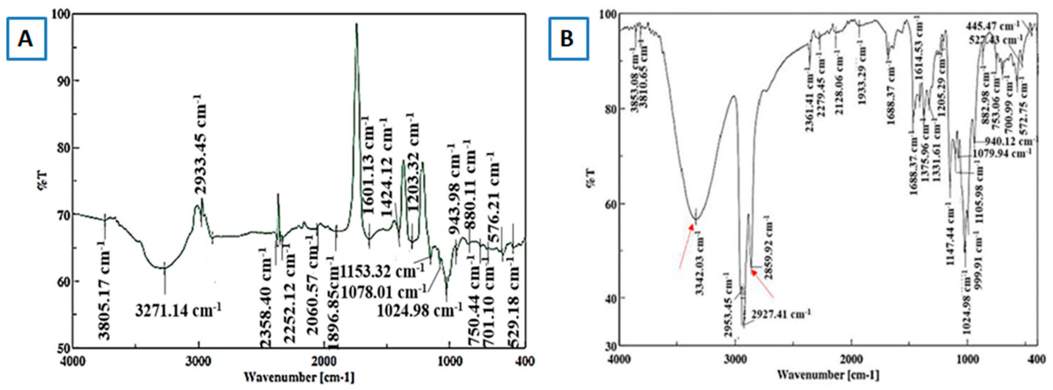
a. Fourier Transform Infrared (FT-IR)
Fourier Transform Infrared (FTIR) spectroscopy is, typically used to identify functional groups. FTIR is thus used initially during MOF synthesis to identify impurities and residual reactants on external surfaces and pores in the interior. Non-reacted organic linkers and coordinated solvents in the MOF structures can easily be identified through FTIR [68]. The formation of an intra-molecular hydrogen bond/complex formation between the cyclodextrin and host moieties can be confirmed with the aid of this technique [66]. Pan and his group reported the shifts in the wavelength when the native γ-CD was converted to γ-CD-MOF and also when the γ-CD-MOF was used to form an inclusion complex with thymol. Compared with those in γ-CD, the O–H stretching vibration peak (3750–3000 cm−1 ) and C–H stretching vibration peak (2900–3000 cm−1 ) in γ-CD-MOFs moved slightly to a low wavenumber indicating that the –OCCO–coordination group of CDs formed a good topological structure with K+ , and the hydrogen bond between molecules was strengthened. Compared with those of γ-CD-MOFs, the O–H and C–H stretching vibrations of the inclusion-complex (IC) shifted slightly to the lower wavenumber. Compared with those of free thymol, the O–H and C–H stretching vibrations of IC shifted to a higher wavenumber. Alternatively, for ICs, the aromatic-ring characteristic peak (1600–1400 cm−1 ) of thymol and the characteristic peak of the 1,2,4-tri-benzene ring (900–650 cm−1 ) weakened, indicating that thymol was trapped in the hydrophobic cavity, which increased the hydrogen bond strength of γ-CD-MOFs during the composite process [69]. Results based on similar phenomena were established by Jiang et.al. working with catechin (CAT) and β-cyclodextrin inclusion complexes. It was observed that after the inclusion of catechin in the β-CD-MOF, the O-H peak which was previously at 3382 cm− 1 shifted to a lower wavelength of 3373 cm− 1 indicating the formation of hydrogen bonding interactions between CAT and β-CD-MOFs. Moreover, the peaks at 1520 and 1466 cm− 1 , representing the alkene part of CAT, disappeared in CAT-β-CD-MOFs, indicating the CAT was completely trapped in the pores of β-CD-MOFs [57].
Figure 9.
FT-IR spectra of MOF crystals A) before hexanal encapsulation and B) after hexanal encapsulation (red arrows represent the hexanal related peaks). Reprinted with permission [56] Copyright 2021, American Chemical Society.
Figure 9.
FT-IR spectra of MOF crystals A) before hexanal encapsulation and B) after hexanal encapsulation (red arrows represent the hexanal related peaks). Reprinted with permission [56] Copyright 2021, American Chemical Society.
b. Raman Spectroscopy
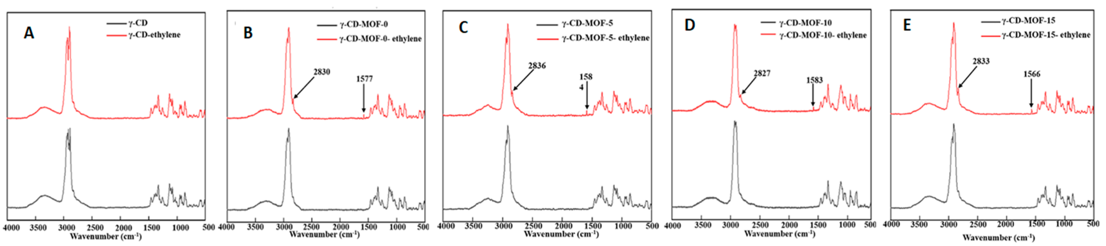
Raman spectroscopy is a technique complementary to IR. Raman spectroscopy is an inelastic scattering technique, while IR spectroscopy is an absorption-based and most often, a mode that is IR active (where there is a change in the dipole moment) is Raman inactive (no change in polarizability) and vice versa. Hence, to get a complete understanding, it is best to employ both methods. Raman spectroscopy can help us to evaluate the structural changes in MOFs which involve the displacement of lightweight atoms such as hydrogen, carbon, and oxygen [70]. It can also provide evidence regarding the formation of inclusion complexes with MOFs and other porous compounds. Liu et.al. used Raman spectroscopy and detected a great degree of disordered carbon in the calcined β-CD-MOF as compared to β-CD-MOF which indicated the loss of crystalline structure due to carbonization of β-CD-MOF at high temperature [55]. In another work, Raman spectra provided information about the successful encapsulation of the ethylene gas in the γ-CD-MOF which was impossible in γ-CD. Characteristic peaks were observed at 1570 cm− 1 resulting from C=C bonds and 2826 cm− 1 from the C–H bonds in ethylene loaded γ-CD-MOF, which were previously absent in γ-CD-MOF [46].
Figure 10.
Comparison of Raman spectra of γ-CD-MOF before and after ethylene absorption A) γ-CD shows no sign of ethylene absorption, B-E) Absorption of ethylene in γ-CD-MOF prepared at different ultrasonic times. Reprinted with permission [46] Copyright 2023, Elsevier.
Figure 10.
Comparison of Raman spectra of γ-CD-MOF before and after ethylene absorption A) γ-CD shows no sign of ethylene absorption, B-E) Absorption of ethylene in γ-CD-MOF prepared at different ultrasonic times. Reprinted with permission [46] Copyright 2023, Elsevier.
5. UV-Visible
While the former techniques focus mainly on the characterization of MOFs and provide necessary information about the formation of inclusion complex (encapsulation of active principle inside the MOF), UV-Vis plays a crucial role in providing information about the release kinetics of the compound loaded in the MOF cavities. The rate at which the active molecules leave the MOF cavities is an important factor in applications where delivery has to be maintained for a longer time, UV-Vis helps clarify that delivery pattern for an active molecule. Apart from that, the adsorption efficiency and kinetics of systems like MOFs can also be studied and compared with other systems. It was found that the adsorption peak of (-)-epigallocatechin gallate (EGCG) continuously showed a drop for the time when samples of γ-CD-MOF were present in its solution (Figure 11). These hypochromic shifts indicated the inclusion of ECGC into the MOF cavities [71].
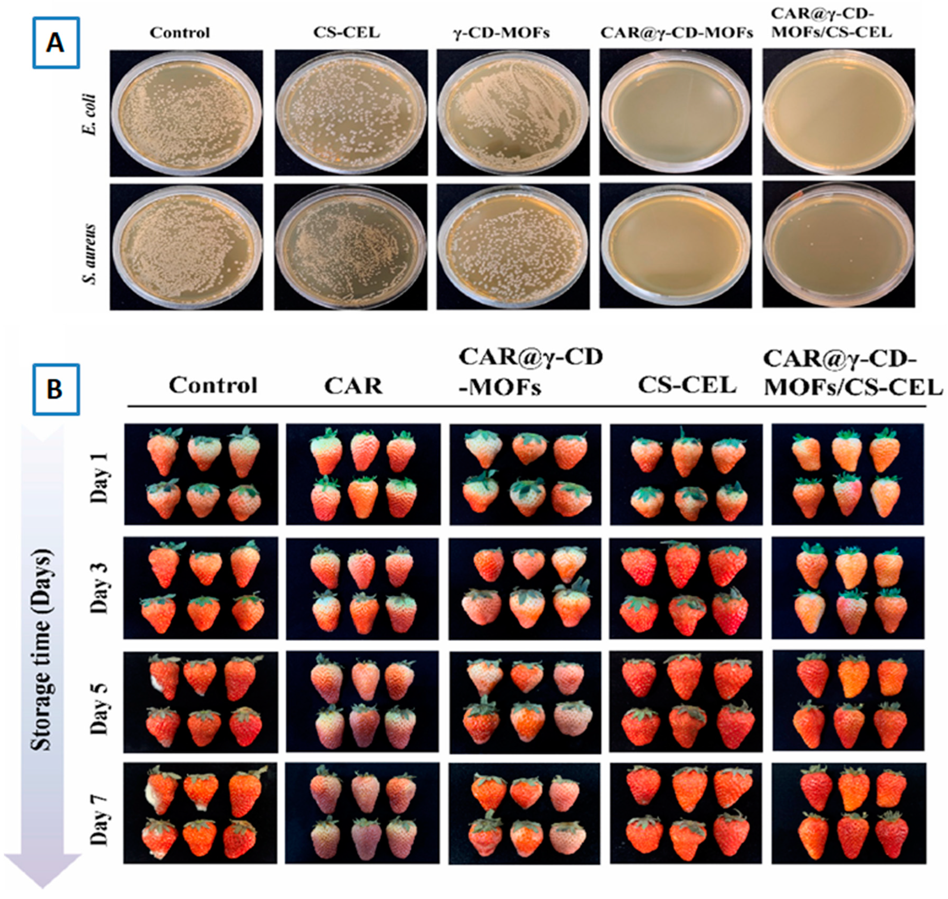
6. Antibacterial Studies
It is one of the most important tests to be carried out for the MOF inclusion complex that is intended to be used in food-based applications. The antibacterial activity of loaded MOF can be evaluated by calculating Minimum Inhibitory Concentration (MIC) and Minimum Bactericidal Concentration (MBC). It was reported that without caffeic acid (CA), loading the γ-CD-MOF had no antibacterial effects but CA-CD-MOF showed MIC at 25 mg⋅mL− 1 for E. coli O157:H7 and S. aureus. The MBC for CA-γ -CD-MOF was 25 mg⋅mL− 1 in E. coli O157:H7 and no value could be observed in the measurement range in S. aureus [35]. In a different work colony-forming unit (CFU) method was used to evaluate the antibacterial activity of Curcumin (Cur) loaded γ-CD-MOFs. The results showed that initially, γ-CD-MOFs had almost no antibacterial effect but after the addition of curcumin, Cur-CD-MOFs showed excellent antibacterial properties in a time-dependent manner, and its lethality of 100% to E. coli and S. aureus at 4 h and 8 h [44].
Figure 12.
A) Antibacterial activity of mechanism of Carvacrol (CAR)-γ-CD-MOFs/Chitosan-Celluslose (CS-CEL) composite film B) Preservation effects of pure CAR, CAR@γ-CD-MOFs, CS-CEL and CAR@γ-CD-MOFs/CS-CEL film on strawberries. Reprinted with permission [62] Copyright 2024, Elsevier.
Figure 12.
A) Antibacterial activity of mechanism of Carvacrol (CAR)-γ-CD-MOFs/Chitosan-Celluslose (CS-CEL) composite film B) Preservation effects of pure CAR, CAR@γ-CD-MOFs, CS-CEL and CAR@γ-CD-MOFs/CS-CEL film on strawberries. Reprinted with permission [62] Copyright 2024, Elsevier.
Institutional Review Board Statement
Not applicable.
Informed Consent Statement
Not applicable.
References
- Liang, S.; et al. Metal-organic frameworks as novel matrices for efficient enzyme immobilization: An update review. Coordination Chemistry Reviews 2020, 406, 213149. [Google Scholar] [CrossRef]
- Xu, Y.; et al. Synthesis and potential applications of cyclodextrin-based metal–organic frameworks: a review. Environmental Chemistry Letters 2023, 21, 447–477. [Google Scholar] [CrossRef] [PubMed]
- Magri, A.; Petriccione, M.; Gutiérrez, T.J. Metal-organic frameworks for food applications: A review. Food Chemistry 2021, 354, 129533. [Google Scholar] [CrossRef] [PubMed]
- Jiang, W.; et al. Preparation of two metal organic frameworks (K-β-CD-MOFs and Cs-β-CD-MOFs) and the adsorption research of myricetin. Polyhedron 2021, 196, 114983. [Google Scholar] [CrossRef]
- Furukawa, H.; et al. The Chemistry and Applications of Metal-Organic Frameworks. Science 2013, 341, 1230444. [Google Scholar] [CrossRef]
- Wu, D.; et al. Direct Calorimetric Measurement of Enthalpy of Adsorption of Carbon Dioxide on CD-MOF-2, a Green Metal–Organic Framework. Journal of the American Chemical Society 2013, 135, 6790–6793. [Google Scholar] [CrossRef]
- Shen, M.; Liu, D.; Ding, T. Cyclodextrin-metal-organic frameworks (CD-MOFs): main aspects and perspectives in food applications. Current Opinion in Food Science 2021, 41, 8–15. [Google Scholar] [CrossRef]
- Poulson, B.G.; et al. Cyclodextrins: Structural, Chemical, and Physical Properties, and Applications. Polysaccharides 2021, 3, 1–31. [Google Scholar] [CrossRef]
- Sandilya, A.A.; Natarajan, U.; Priya, M.H. , Molecular View into the Cyclodextrin Cavity: Structure and Hydration. ACS Omega 2020, 5, 25655–25667. [Google Scholar] [CrossRef]
- Szejtli, J. , Introduction and General Overview of Cyclodextrin Chemistry. Chemical Reviews 1998, 98, 1743–1754. [Google Scholar] [CrossRef]
- Connors, K.A. , The Stability of Cyclodextrin Complexes in Solution. Chemical Reviews 1997, 97, 1325–1358. [Google Scholar] [CrossRef] [PubMed]
- Dodziuk, H. Molecules with Holes – Cyclodextrins, in Cyclodextrins and Their Complexes. 2006. p. 1-30.
- Saenger, W.; et al. Structures of the Common Cyclodextrins and Their Larger AnaloguesBeyond the Doughnut. Chemical Reviews 1998, 98, 1787–1802. [Google Scholar] [CrossRef] [PubMed]
- Jansook, P.; Ogawa, N.; Loftsson, T. , Cyclodextrins: structure, physicochemical properties and pharmaceutical applications. Int J Pharm 2018, 535, 272–284. [Google Scholar] [CrossRef] [PubMed]
- Velazquez-Contreras, F.; et al. Cyclodextrins in Polymer-Based Active Food Packaging: A Fresh Look at Nontoxic, Biodegradable, and Sustainable Technology Trends. Polymers (Basel) 2021, 14. [Google Scholar] [CrossRef] [PubMed]
- Pereva, S.; et al. Water inside beta-cyclodextrin cavity: amount, stability and mechanism of binding. Beilstein J Org Chem 2019, 15, 1592–1600. [Google Scholar] [CrossRef]
- Liu, L.; Guo, Q.-X. , The Driving Forces in the Inclusion Complexation of Cyclodextrins. Journal of inclusion phenomena and macrocyclic chemistry 2002, 42, 1–14. [Google Scholar] [CrossRef]
- Loftsson, T.; Brewster, M.E. , Pharmaceutical applications of cyclodextrins. 1. Drug solubilization and stabilization. (0022-3549 (Print)).
- He, Y.; et al. Cyclodextrin-based aggregates and characterization by microscopy. Micron 2008, 39, 495–516. [Google Scholar] [CrossRef]
- Muankaew, C.; Loftsson, T. Cyclodextrin-Based Formulations: A Non-Invasive Platform for Targeted Drug Delivery. Basic Clin Pharmacol Toxicol 2018, 122, 46–55. [Google Scholar] [CrossRef]
- Gadade, D.D.; Pekamwar, S.S. Cyclodextrin Based Nanoparticles for Drug Delivery and Theranostics. Adv Pharm Bull 2020, 10, 166–183. [Google Scholar] [CrossRef]
- Gidwani, B.; Vyas, A. A Comprehensive Review on Cyclodextrin-Based Carriers for Delivery of Chemotherapeutic Cytotoxic Anticancer Drugs. BioMed Research International 2015, 2015, 1–15. [Google Scholar] [CrossRef]
- Munro, I.C.; et al. Safety assessment of gamma-cyclodextrin. Regul Toxicol Pharmacol 2004, 39 (Suppl. 1), S3–S13. [Google Scholar] [CrossRef] [PubMed]
- Saokham, P.; Loftsson, T. gamma-Cyclodextrin. Int J Pharm 2017, 516, 278–292. [Google Scholar] [CrossRef] [PubMed]
- Cid-Samamed, A.; et al. Cyclodextrins inclusion complex: Preparation methods, analytical techniques and food industry applications. Food Chemistry 2022, 384, 132467. [Google Scholar] [CrossRef] [PubMed]
- Carneiro, S.B.; et al. Cyclodextrin(-)Drug Inclusion Complexes: In Vivo and In Vitro Approaches. Int J Mol Sci 2019, 20. [Google Scholar] [CrossRef] [PubMed]
- Matencio, A.; et al. Applications of cyclodextrins in food science. A review. Trends in Food Science & Technology 2020, 104, 132–143. [Google Scholar]
- Van Ommen, B.; De Bie, A.T.; Bar, A. Disposition of 14C-alpha-cyclodextrin in germ-free and conventional rats. Regul Toxicol Pharmacol 2004, 39 (Suppl. 1), 57–66. [Google Scholar] [CrossRef]
- Stella, V.J.; He, Q. Cyclodextrins. Toxicologic Pathology 2008, 36, 30–42. [Google Scholar] [CrossRef]
- Mortensen, A.; et al. Re-evaluation of β-cyclodextrin (E 459) as a food additive. EFSA Journal 2016, 14. [Google Scholar]
- Giancarlo, C.; et al. Cyclodextrins as Food Additives and in Food Processing. Current Nutrition & Food Science 2006, 2, 343–350. [Google Scholar]
- Li, H.; et al. Metal-Organic Framework Based on alpha-Cyclodextrin Gives High Ethylene Gas Adsorption Capacity and Storage Stability. ACS Appl Mater Interfaces 2020, 12, 34095–34104. [Google Scholar] [CrossRef]
- Wang, Y.; et al. Enhancement of the Stabilities and Intracellular Antioxidant Activities of Lavender Essential Oil by Metal-Organic Frameworks Based on β-Cyclodextrin and Potassium Cation. Polish Journal of Food and Nutrition Sciences 2021, 39–50. [Google Scholar] [CrossRef]
- Smaldone, R.A.; et al. Metal–Organic Frameworks from Edible Natural Products. Angewandte Chemie International Edition 2010, 49, 8630–8634. [Google Scholar] [CrossRef] [PubMed]
- Shen, M.; et al. Cyclodextrin metal-organic framework by ultrasound-assisted rapid synthesis for caffeic acid loading and antibacterial application. Ultrason Sonochem 2022, 86, 106003. [Google Scholar] [CrossRef] [PubMed]
- Kathuria, A.; et al. Multifunctional Ordered Bio-Based Mesoporous Framework from Edible Compounds. Journal of Biobased Materials and Bioenergy 2018, 12, 449–454. [Google Scholar] [CrossRef]
- Jiang, L.; et al. Encapsulation of catechin into nano-cyclodextrin-metal-organic frameworks: Preparation, characterization, and evaluation of storage stability and bioavailability. Food Chemistry 2022, 394, 133553. [Google Scholar] [CrossRef]
- Wang, Y.; et al. Improvement of the stabilities and antioxidant activities of polyphenols from the leaves of Chinese star anise (Illicium verum Hook. f.) using beta-cyclodextrin-based metal-organic frameworks. J Sci Food Agric 2021, 101, 287–296. [Google Scholar] [CrossRef]
- Wang, S.; et al. Covering soy polysaccharides gel on the surface of β-cyclodextrin-based metal–organic frameworks. Journal of Materials Science 2020, 56, 3049–3061. [Google Scholar] [CrossRef]
- Li, Z.; et al. Controlled Release of Thymol by Cyclodextrin Metal-Organic Frameworks for Preservation of Cherry Tomatoes. Foods 2022, 11. [Google Scholar] [CrossRef]
- Hu, Z.; et al. Encapsulation of menthol into cyclodextrin metal-organic frameworks: Preparation, structure characterization and evaluation of complexing capacity. Food Chem 2021, 338, 127839. [Google Scholar] [CrossRef]
- Moussa, Z.; et al. Encapsulation of curcumin in cyclodextrin-metal organic frameworks: Dissociation of loaded CD-MOFs enhances stability of curcumin. Food Chem 2016, 212, 485–94. [Google Scholar] [CrossRef]
- Wang, Z.; et al. Ethanol-mediated synthesis of gamma-cyclodextrin-based metal-organic framework as edible microcarrier: performance and mechanism. Food Chem 2023, 418, 136000. [Google Scholar] [CrossRef] [PubMed]
- Kang, L.; et al. Preparation technology and preservation mechanism of gamma-CD-MOFs biaological packaging film loaded with curcumin. Food Chem 2023, 420, 136142. [Google Scholar] [CrossRef] [PubMed]
- Che, J.; et al. Fabrication of gamma-cyclodextrin-Based metal-organic frameworks as a carrier of cinnamaldehyde and its application in fresh-cut cantaloupes. Curr Res Food Sci 2022, 5, 2114–2124. [Google Scholar] [CrossRef] [PubMed]
- Li, S.; et al. Synthesis of γ-cyclodextrin metal-organic framework as ethylene absorber for improving postharvest quality of kiwi fruit. Food Hydrocolloids 2023, 136, 108294. [Google Scholar] [CrossRef]
- Qiu, C.; et al. Characterization and Mechanisms of Novel Emulsions and Nanoemulsion Gels Stabilized by Edible Cyclodextrin-Based Metal-Organic Frameworks and Glycyrrhizic Acid. J Agric Food Chem 2019, 67, 391–398. [Google Scholar] [CrossRef]
- Chen, Y.; et al. Cyclodextrin-based metal–organic framework nanoparticles as superior carriers for curcumin: Study of encapsulation mechanism, solubility, release kinetics, and antioxidative stability. Food Chemistry 2022, 383, 132605. [Google Scholar] [CrossRef]
- Al-Ghamdi, S.; et al. Synthesis of nanoporous carbohydrate metal-organic framework and encapsulation of acetaldehyde. Journal of Crystal Growth 2016, 451, 72–78. [Google Scholar] [CrossRef]
- Su, Q.; et al. Enhanced stability of anthocyanins by cyclodextrin–metal organic frameworks: Encapsulation mechanism and application as protecting agent for grape preservation. Carbohydrate Polymers 2024, 326, 121645. [Google Scholar] [CrossRef]
- Chen, Y.; et al. Novel γ-cyclodextrin-metal–organic frameworks for encapsulation of curcumin with improved loading capacity, physicochemical stability and controlled release properties. Food Chemistry 2021, 347, 128978. [Google Scholar] [CrossRef]
- Qiu, C.; et al. Green Synthesis of Cyclodextrin-Based Metal–Organic Frameworks through the Seed-Mediated Method for the Encapsulation of Hydrophobic Molecules. Journal of Agricultural and Food Chemistry 2018, 66, 4244–4250. [Google Scholar] [CrossRef]
- Qiu, C.; et al. Development of nanoscale bioactive delivery systems using sonication: Glycyrrhizic acid-loaded cyclodextrin metal-organic frameworks. Journal of Colloid and Interface Science 2019, 553, 549–556. [Google Scholar] [CrossRef] [PubMed]
- Szente, L. and E. Fenyvesi, Cyclodextrin-Enabled Polymer Composites for Packaging (dagger). Molecules 2018, 23. [Google Scholar]
- Liu, C.; et al. Multifunctional β-Cyclodextrin MOF-Derived Porous Carbon as Efficient Herbicides Adsorbent and Potassium Fertilizer. ACS Sustainable Chemistry & Engineering 2019, 7, 14479–14489. [Google Scholar]
- Nagarajan, V.; et al. Encapsulation of a Volatile Biomolecule (Hexanal) in Cyclodextrin Metal–Organic Frameworks for Slow Release and Its Effect on Preservation of Mangoes. ACS Food Science & Technology 2021, 1, 1936–1944. [Google Scholar]
- Jiang, L.; et al. Development and characterization of zein-based active packaging films containing catechin loaded β-cyclodextrin metal-organic frameworks. Food Packaging and Shelf Life 2022, 31, 100810. [Google Scholar] [CrossRef]
- Wang, Y.; et al. Enhanced preservation effects of clove (Syzygium aromaticum) essential oil on the processing of Chinese bacon (preserved meat products) by beta cyclodextrin metal organic frameworks (beta-CD-MOFs). Meat Sci 2023, 195, 108998. [Google Scholar] [CrossRef]
- Ez-zoubi, A.; et al. Encapsulation of Origanum compactum Essential Oil in Beta-Cyclodextrin Metal Organic Frameworks: Characterization, Optimization, and Antioxidant Activity. Journal of Food Processing and Preservation 2023, 2023, 1–10. [Google Scholar] [CrossRef]
- Sun, X.; et al. Application of beta-Cyclodextrin metal-organic framework/titanium dioxide hybrid nanocomposite as dispersive solid-phase extraction adsorbent to organochlorine pesticide residues in honey samples. J Chromatogr A 2022, 1663, 462750. [Google Scholar] [CrossRef]
- Qin, Z.; et al. Food packaging for ripening and preserving banana based on ethylene-loaded nanofiber films deposited with nanosized cyclodextrin metal-organic frameworks and TiO2 nanoparticles. Food Packaging and Shelf Life 2024, 45, 101332. [Google Scholar] [CrossRef]
- Min, T.; et al. Highly efficient anchoring of γ-cyclodextrin-MOFs on chitosan/cellulose film by in situ growth to enhance encapsulation and controlled release of carvacrol. Food Hydrocolloids 2024, 150, 109633. [Google Scholar] [CrossRef]
- Zhang, Y.; et al. A functional Pickering emulsion coating based on octadecenylsuccinic anhydride modified γ-cyclodextrin metal-organic frameworks for food preservation. Food Hydrocolloids 2024, 150, 109668. [Google Scholar] [CrossRef]
- Zhang, Y.; et al. Enhanced stability and biocompatibility of HIPEs stabilized by cyclodextrin-metal organic frameworks with inclusion of resveratrol and soy protein isolate for β-carotene delivery. International Journal of Biological Macromolecules 2024, 274, 133431. [Google Scholar] [CrossRef]
- Zhang, G.; et al. Enhanced stability of vitamin A palmitate microencapsulated by gamma-cyclodextrin metal-organic frameworks. J Microencapsul 2018, 35, 249–258. [Google Scholar] [CrossRef]
- Wadhwa, G.; et al. Essential oil–cyclodextrin complexes: an updated review. Journal of Inclusion Phenomena and Macrocyclic Chemistry 2017, 89, 39–58. [Google Scholar] [CrossRef]
- Yang, A.; et al. Green synthesis of β-cyclodextrin metal–organic frameworks and the adsorption of quercetin and emodin. Polyhedron 2019, 159, 116–126. [Google Scholar] [CrossRef]
- Abid, H.R.; et al. Physiochemical characterization of metal organic framework materials: A mini review. Heliyon 2024, 10, e23840. [Google Scholar] [CrossRef]
- Pan, X.; et al. Effect of potassium salts on the structure of gamma-cyclodextrin MOF and the encapsulation properties with thymol. J Sci Food Agric 2022, 102, 6387–6396. [Google Scholar] [CrossRef]
- Sunil, J.; et al. Raman spectroscopy, an ideal tool for studying the physical properties and applications of metal–organic frameworks (MOFs). Chemical Society Reviews 2023, 52, 3397–3437. [Google Scholar] [CrossRef]
- Ke, F.; et al. Synergistic antioxidant activity and anticancer effect of green tea catechin stabilized on nanoscale cyclodextrin-based metal–organic frameworks. Journal of Materials Science 2019, 54, 10420–10429. [Google Scholar] [CrossRef]
Figure 6.
A) TGA curve of raw OCEO and after its encapsulation in β-CD and β-CD-MOF, B) DSC curves for (a) catechin, (b) α-CD-MOFs, (c) CA/α-CD-MOFs (d) β-CD-MOFs, (e) CA/β-CD-MOFs and (f) γ-CD-MOFs and (g) CA/γ-CD-MOFs. Reprinted with permission [37] Copyright 2022, Elsevier.
Figure 6.
A) TGA curve of raw OCEO and after its encapsulation in β-CD and β-CD-MOF, B) DSC curves for (a) catechin, (b) α-CD-MOFs, (c) CA/α-CD-MOFs (d) β-CD-MOFs, (e) CA/β-CD-MOFs and (f) γ-CD-MOFs and (g) CA/γ-CD-MOFs. Reprinted with permission [37] Copyright 2022, Elsevier.
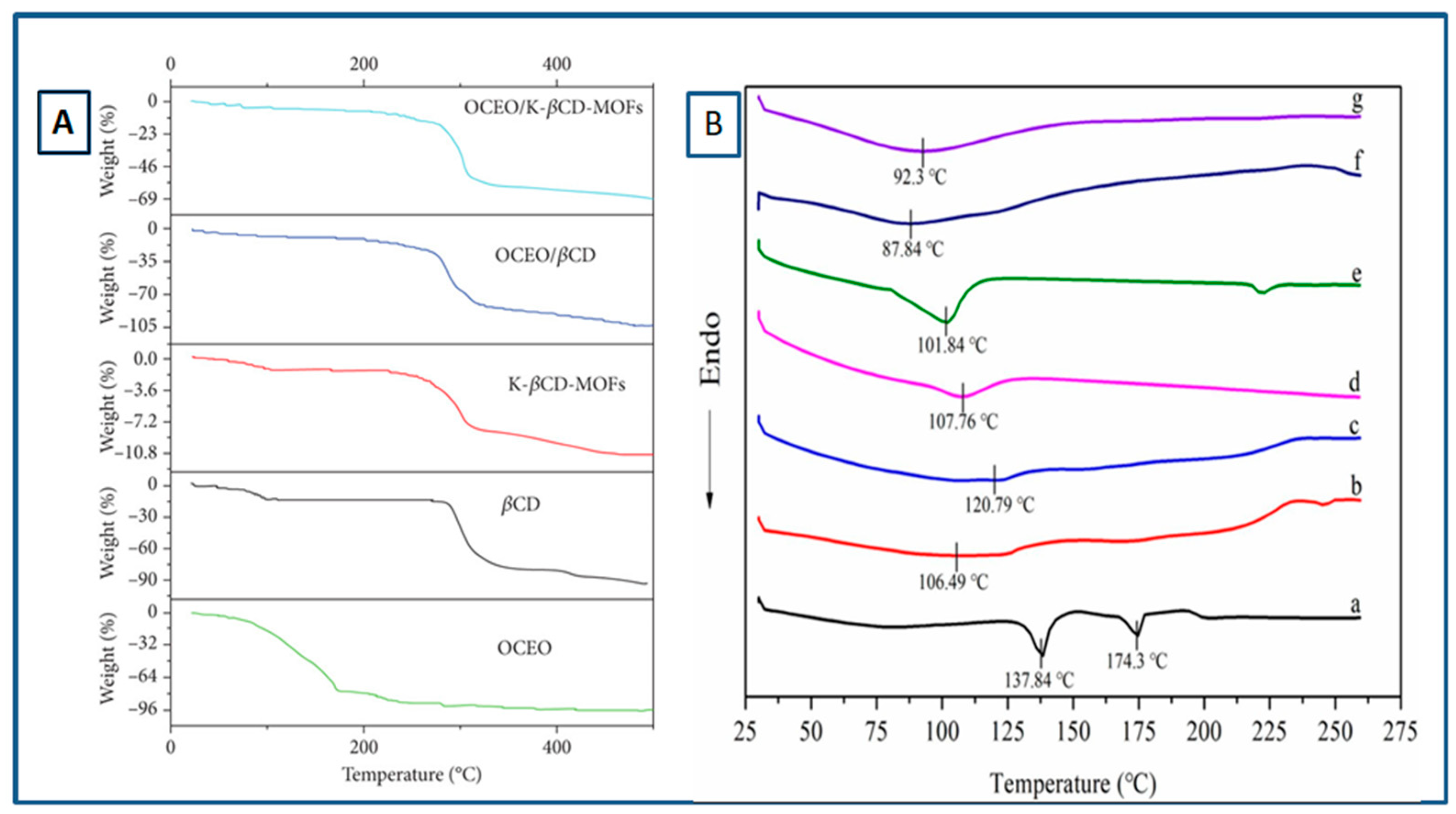
Figure 7.
A) TEM images of γ-CD-MOFs. Reprinted with permission [47] Copyright 2019, American chemical society. B) SEM images of that α-CD, β-CD, γ-CD and their MOFs Reprinted with permission [41] Copyright 2021, Elsevier C) EDX data of α, β and γ CD MOFs. Reprinted with permission [41] Copyright 2021, Elsevier.
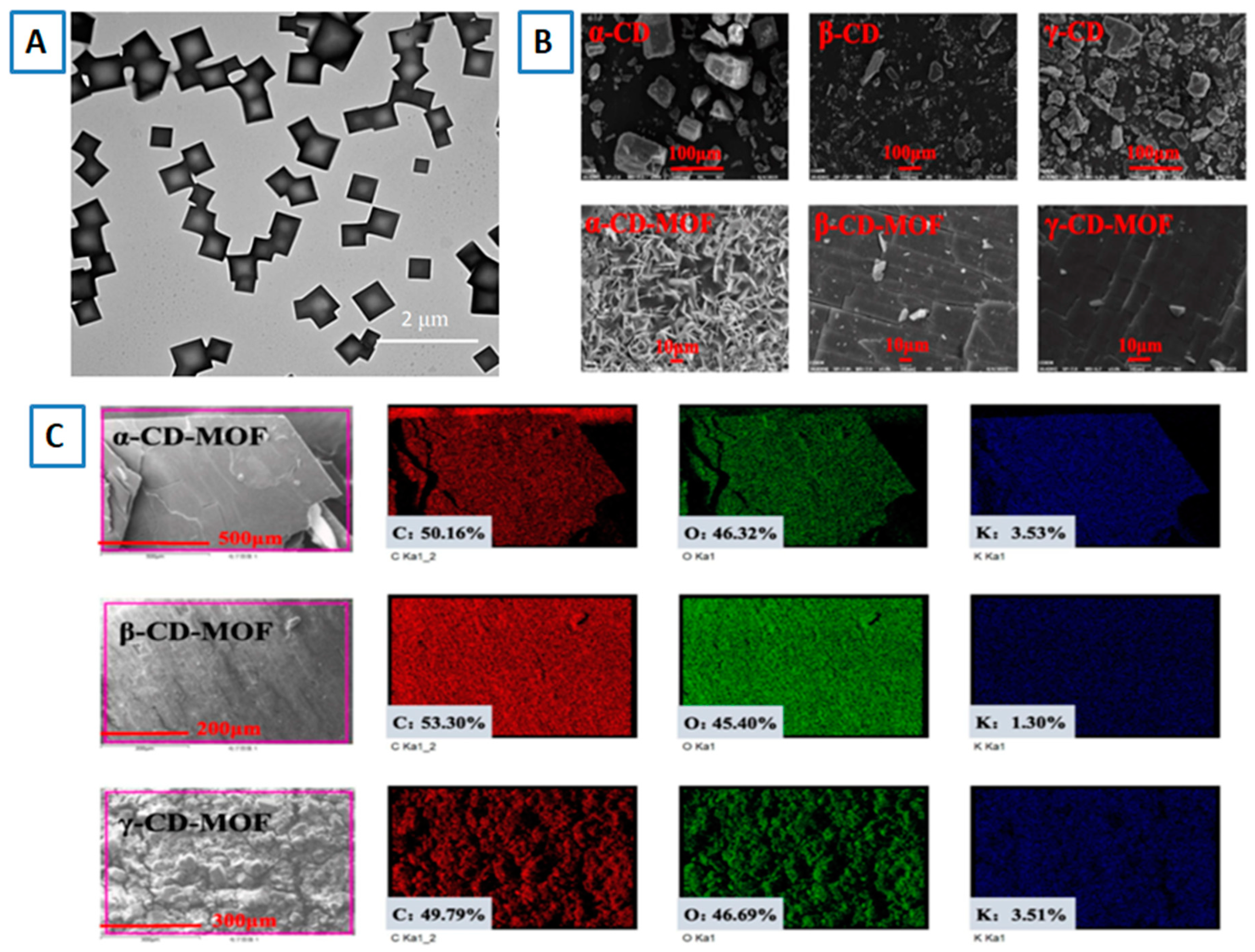
Figure 8.
XRD pattern of Curcumin and its inclusion complexes with γ-CD-MOFs. Reprinted with permission [51] Copyright 2021, Elsevier.
Figure 8.
XRD pattern of Curcumin and its inclusion complexes with γ-CD-MOFs. Reprinted with permission [51] Copyright 2021, Elsevier.
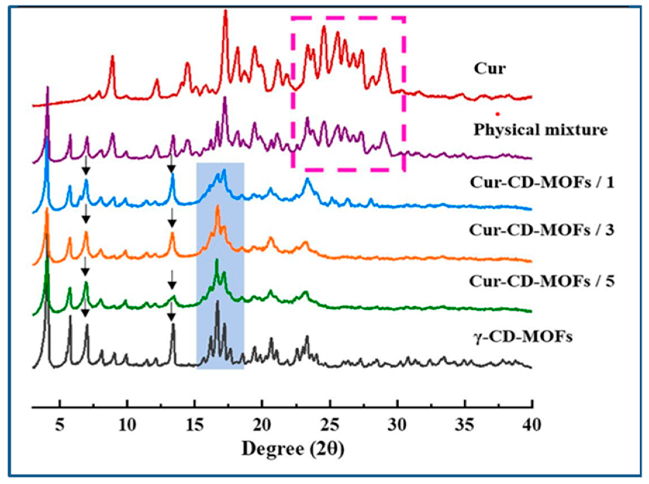
Figure 11.
UV–Vis absorption spectra of EGCG in the presence (a) and absence (b) of CD-MOF in ethanol with time. (c) Variation of absorbance at 276 nm of EGCG in the presence of CD-MOF and (d) the calibration curve of EGCG in ethanol [71].
Figure 11.
UV–Vis absorption spectra of EGCG in the presence (a) and absence (b) of CD-MOF in ethanol with time. (c) Variation of absorbance at 276 nm of EGCG in the presence of CD-MOF and (d) the calibration curve of EGCG in ethanol [71].
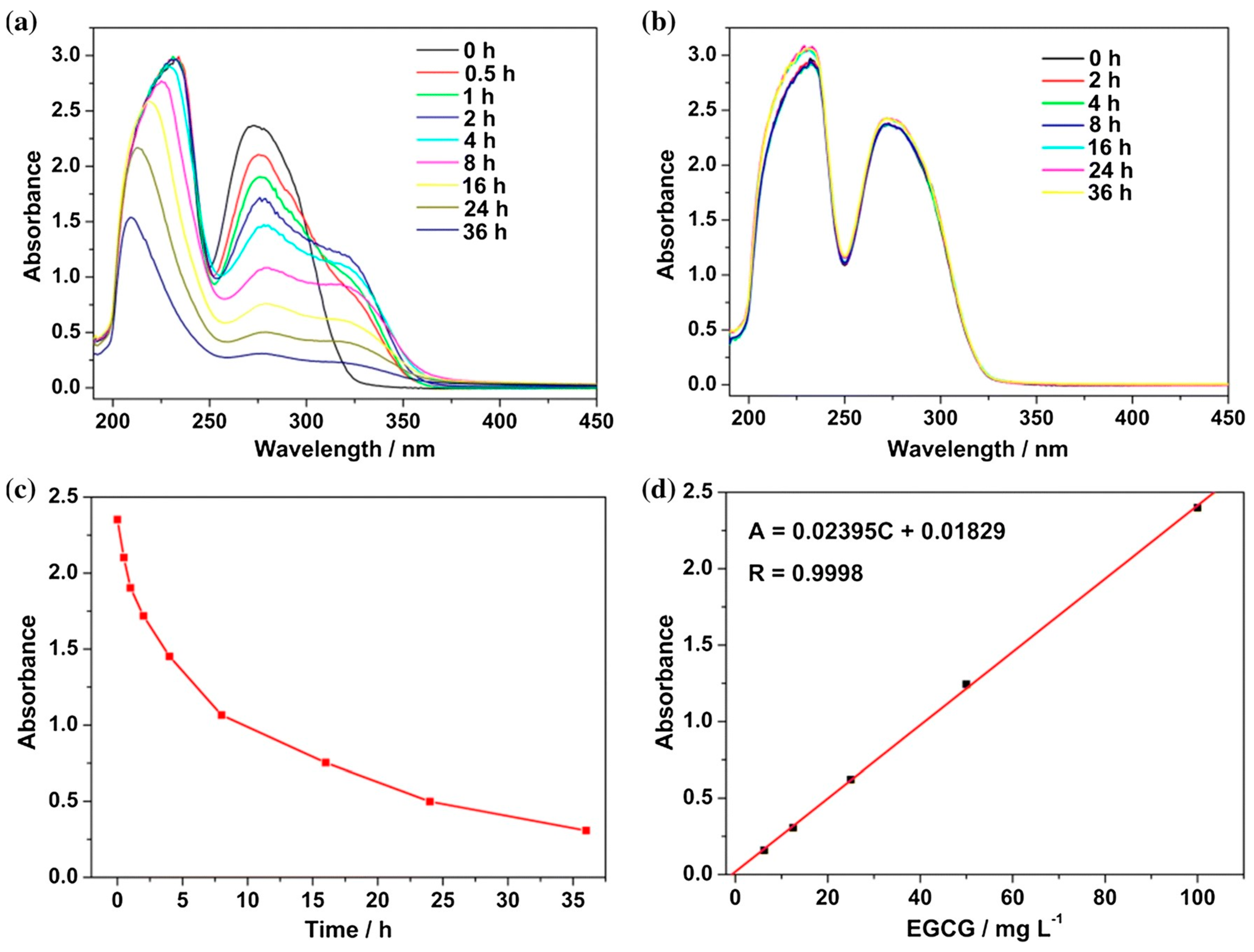
Table 1.
Properties of the native cyclodextrins.
| Properties | α-CD | β-CD | γ-CD |
|---|---|---|---|
| Number of glucose units | 6 | 7 | 8 |
| Molecular weight (g/mol) | 972 | 1135 | 1297 |
| Solubility in water at 25 ◦C (%, w/v) | 14.5 | 1.9 | 23.2 |
| Melting point (◦C) | 275 | 280 | 275 |
| Cavity diameter (Å) | 4.7–5.3 | 6.0–6.5 | 7.5–8.3 |
| External diameter (Å) | 14.6 | 15.4 | 17.5 |
| Crystal forms (from water) | Hexagonal plates | Monoclinic parallelograms | Quadratic prisms |
| European trade name as food additives | E-457 | E-459 | E-458 |
Disclaimer/Publisher’s Note: The statements, opinions and data contained in all publications are solely those of the individual author(s) and contributor(s) and not of MDPI and/or the editor(s). MDPI and/or the editor(s) disclaim responsibility for any injury to people or property resulting from any ideas, methods, instructions or products referred to in the content. |
© 2024 by the authors. Licensee MDPI, Basel, Switzerland. This article is an open access article distributed under the terms and conditions of the Creative Commons Attribution (CC BY) license (http://creativecommons.org/licenses/by/4.0/).
Copyright: This open access article is published under a Creative Commons CC BY 4.0 license, which permit the free download, distribution, and reuse, provided that the author and preprint are cited in any reuse.


