Submitted:
26 November 2024
Posted:
27 November 2024
You are already at the latest version
Abstract
Nanoparticles (NPs) have emerged as a potent choice for various applications, serving as an al-ternative and promising methodology for future advancements. They have been explored in nu-merous emerging applications, demonstrating immense promise and high efficiency. NPs can be utilized with various biomolecules for delivery or as surface materials to enhance their capabili-ties. Lactoferrin (Lf) is a glycoprotein recognized for its significant multifunctional properties, such as antimicrobial, antioxidant, anti-inflammatory, anticancer and neuroprotective effects. Its activity has a broad distribution in the human body, with Lf receptors present in multiple regions. Current research shows that Lf is utilized in NP technology as a surface material, encapsulated biomolecule, and even as a NP itself. Due to the abundance of Lf receptors in various regions, Lf can be employed as a surface material in NPs for targeted delivery strategies, particularly in crossing the BBB and targeting specific cancers. Furthermore, Lf can be synthesized in a NP structure, positioning it as a strong candidate in future NPs related applications. In this article, we explored the highlighted and underexplored areas of Lf applications in NPs research.
Keywords:
Lactoferrin
; Nanoparticles
; Antimicrobial activity
; Drug delivery
; Anticancer
; Agriculture
; Neuroprotection
; Toxicity
1. Introduction
Lf is a multifunctional iron-binding protein found in mammalian milk and various biological fluids, such as saliva and tears [1]. Lf’s N-terminal and C-terminal lobes bind iron ions, retaining this ability even at low pH (~3.5) [2]. Lf’s molecular structure changes depending on its binding status, found in iron-free (Apo-Lf) and iron-bound (Holo-Lf) forms. Lf's iron-binding ability is particularly significant because it can retain binding at low pH levels (~3.5) [3]. Thanks to their strong iron-binding characteristic, Lf exhibits immense antioxidant activity by inhibiting the Fenton reaction by reducing the iron ions that are needed for reactive oxygen species (ROS) generation [4]. This feature supports ongoing research into Lf's potential neuroprotective activity. In addition to its antioxidant and immunomodulatory activities, Lf is considered a potential iron chelator that may help reduce the progression of neurodegenerative diseases [5]. Moreover, several mechanisms have been suggested for Lf's neuroprotective activity. One of the most discussed is the interaction of Lf with surface receptors, which initiates intracellular pathways that protect dopaminergic cells against apoptosis [6].
Lf initiates its antimicrobial activity by reducing the essential iron needed for bacterial growth [7]. Additionally, it can directly interact with cells with its cation-binding residue in the anionic region of the outer membrane, leading to their destruction [8]. The same activities are also mediated with Lf-derived peptides.
Lf is known for binding to various receptor types, enhancing its multifunctionality. Some of these receptors, such as heparan sulfate proteoglycans (HSPGs), are also targeted by many viruses during the initiation of infection [9]. Leveraging this characteristic, various strategies have been developed to use Lf to disrupt the initiation of viral infection by targeting both viral and host receptors, such as cellular glycosaminoglycan [10]. These strategies include applications against viruses like SARS CoV-2 [11], human papillomavirus [12], hepatitis B [13], hepatitis C [14], and dengue virus [15].
Moreover, many studies highlight the significant antifungal activity of both Lf and Lf-derived peptides against both fungal and human fungal pathogens [16]. Compared to antibacterial activity, Lf exhibits iron-independent mechanisms against fungus, directly targeting cell wall proteins [17]. Moreover, the antifungal activity of Lf-derived peptides can vary significantly depending on their peptide chain composition and their source [18]. As an example, Biasibetti et al. demonstrated the significant antifungal activity of lactoferrin-derived peptides (lactoferricin and lactoferrampin) [19]. The study revealed that lactoferrin exhibited higher antimicrobial activity against a bacterial strain and yeast, whereas lactoferrampin demonstrated strong activity against the well-known fungal species C. albicans. Most importantly, both peptides demonstrated synergistic activity against all tested strains, significantly enhancing their antimicrobial potency. This demonstrates that Lf-derived peptides differ in their antimicrobial activities and can be used together to achieve higher efficacy where potent antimicrobial action is required. Similar studies have investigated the antifungal activity of these peptides [20,21].
In addition, Lf is regarded as an excellent immunoregulator and enhancer of host defense, achieved through the modulation of iron homeostasis, stimulation of immune signals, recruitment of immune cells, regulation of proinflammatory secretion, and other mechanisms (Figure 1) [22]. Lf's immunoregulatory activity is also linked to its anticancer properties. In addition to influencing cytokine levels, Lf can inhibit cellular growth and metastasis of cancer cells, indirectly induce apoptosis by enhancing caspase cleavage, and potentially alter gene expression [23].
Considering these well-known functions, Lf is commonly used in many areas, such as in agriculture to maintain food preservation, dietary supplements to promote gut health and immune modulation, supplements in infant formulas for developing the immune system, and so on [24]. Recently, Lf has been involved in nanotechnology as a supportive molecule and material for NPs to utilize the mentioned functions of the protein [25,26,27].
NPs are structures with small sizes, ranging from 1 to 100 nm, a high surface area-to-volume ratio, and tunable surface chemistry, and they are classified based on their material composition, such as organic or inorganic [28]. NPs are found in diverse shapes, such as spherical, spheroid, and nanorod, which significantly affect their properties [29,30]. Moreover, the properties of NPs, such as size and shape, are greatly influenced by the chosen synthesis method [31]. Various methods are preferred for NP synthesis, including chemical, physical, and green synthesis, using sources such as bacteria, fungi, algae, and plant extracts [32].
NPs have distinct applications in the current literature depending on their physicochemical properties. Their wide-ranging applications include drug delivery, biomolecule, and pathogen detection, photothermal-based cancer cell destruction, bioimaging, biosensor development, chemical catalysis, materials for food packaging and fertilizers, environmental applications, and more [33,34,35].
In this review, we have analyzed the use of Lf in NP technology over the past few years. Lf demonstrates notable potential in NP research as a surface material for targeted delivery strategies. Moreover, Lf can be delivered using various types of NPs for site-specific applications and can also be utilized in NP synthesis to expand the range of applications. Numerous materials are integrated with NPs to enhance therapeutic efficacy and optimize application outcomes. Given its significant potential, Lf is an excellent candidate for integration into NP applications, paving the way for impactful developments and innovative approaches. Considering the substantial potential of both Lf and NP applications in emerging fields, reviews that highlight the current integration of these two areas are essential to drive future innovations.
2. Lf-Coating and Lf NPs in Delivery Applications
In recent years, Lf has received increasing attention in NP-based drug delivery systems, both as a surface coating and an active NP component (Table 1). Drug delivery applications are one of the most common and highlighted types of applications in NP research [36]. Since Lf is a multifunctional glycoprotein with a high affinity for multiple receptors and diverse surface components, Lf shows significant potential as an agent in nanomedicine.
Surface modification of NPs with Lf enhances bioavailability and stability while enabling efficient, targeted cellular uptake, which can improve therapeutic outcomes. Furthermore, multiple studies have demonstrated the potential of Lf-NPs in drug delivery systems focusing on their synthesis and characterization [37,38,39]. Thus, Lf can initiate its multifunctional properties while ensuring a controlled drug release for the biomolecule. Moreover, some approaches use various types of NPs to deliver Lf itself, aiming to enhance the biological properties of the protein. Still, considering the usage of Lf as a material, this approach has rarely been conducted in the last years.

Table 1.
NP-based Drug Delivery Systems with Surface Modification and Lf NPs.
| Application | Study Type | Main Results | Reference |
|---|---|---|---|
| Deliveries With Lf NPs | |||
| Targeted lung delivery of antibiotic with Lf-included nanocomplex | In vivo In vitro |
-Sustained drug release profile. -Minimum inhibitory concentration (MIC) value of 0.5 μg/mL against Pseudomonas Aeruginosa. -100% bacterial reduction and zone of inhibition (ZOI) by 29 ± 1.45 mm at the highest concentration (5 μg/mL). -Bacterial reduction in kidneys of infected mice treated with dual drug-loaded PEX, with counts dropping to 3.24 ± 0.067 log₁₀ CFU/mL, compared to 18.22 ± 0.194 log₁₀ CFU/mL in untreated mice. -Insignificant activity at the lowest concentrations (0.05 and 0.1 μg/mL). -Significant inhibition of bacterial accumulation (6-fold reduction) in kidney and lung tissue of mice. -Reduced oxidative stress in mice, with PEX increasing glutathione (GSH) and catalase (CAT) levels while decreasing malondialdehyde (MDA) levels. -Improved antioxidant parameters and maintained the body weight of mice during the infection. -Improved hemocompatibility with minimal toxicity to hepatic and renal functions |
[40] |
| Delivery of antibiotics and natural compounds with Lf NPs | In vitro | -Increased uptake of drug-loaded Lf NPs, up to 90%, by THP-1 cells, surpassing that of free Lf. -Complete inhibition of Staphylococcus Aureus (S. aureus) strain Newman, at concentrations of 25 and 50 μg/mL. -Retained stability of Lf NPs following storage for over 30 days at 4 °C. |
[41] |
| Curcumin-loaded Lf NPs for ulcerative colitis treatment | In vivo In vitro |
-Improvement of the loading efficiency of curcumin, up to 95.08%, following incorporation of Lf in the nanosystem. -Increased tight junction protein expression (ZO-1, Occludin, Claudin-1) levels in colon tissues by Lf-included nanosystem, compared to the free curcumin and curcumin-NP groups. -Suppression of TLR4, MyD88, and NF-κB protein levels in colon tissues of UC mice treated with the Lf-included nanosystem. -Restoration of microbial flora diversity, with increased Bacteroidetes and decreased Firmicutes, following treatment with the Lf-included nanosystem in UC mice. |
[42] |
| Microencapsulated Lf NPs for docetaxel and atorvastatin delivery in the oral treatment of colorectal cancer | In vivo In vitro |
-Effective internalization of drug-loaded Lf NPs by Caco-2 cells along with lower half maximal inhibitory concentration (IC50) values compared to free drug samples. -Sustained release of NPs in rat cecal content without degradations observed in the upper gastrointestinal tract. -Suppression of p-AKT, p-ERK1/2, and NF-κB levels and activation of caspase enzymes |
[43] |
| Production of Lf NP encapsulated gold complexes, (Lf-C2), to cross blood-brain barrier (BBB) in glioma treatment | In vivo In vitro |
-Successful crossing of the BBB by Lf-C2 NPs compared to C2 alone. -Increased inhibition rates on glioma growth with Lf-C2 NPs (68.6%), compared to the free C2 (21.6%). -Achievement of higher, 88.2%, apoptosis rate in LF-C2 NP-treated tumor tissues compared to 21.2% and 6.9% for C2 and NaCl, respectively. |
[44] |
| Production of zein-glycosylated Lf NPs for improved stability and bioaccessibility of 7,8-dihydroxyflavone (7,8-DHF) | In vitro | -High encapsulation efficiency (above 98.50%) with zein-glycosylated Lf NPs. -Improved bioaccessibility with the existence of Lf, reaching up to a maximum of 84.05%, while free 7,8-DHF achieved only 18.06%. -Increased retention percentage with the addition of Lf, rising from 12.35% to 43.21% under dark conditions at 50°C. -Enhanced stability over 30 days of storage compared to zein NPs alone. |
[45] |
| Fabrication of zein-Lf NPs for encapsulation of 7,8-DHF | In vitro | -Approximately 30 times higher water solubility with zein-Lf NPs (231.60 μg/mL) than that of 7,8-DHF alone (7.12 μg/mL). -Improved bioaccessibility with zein-Lf NPs (63.51%) in comparison to free 7,8-DHF, (18.06%) and zein-DHF (31.85%). -Enhanced chemical stability with zein/LF NPs, retaining 27.4% of 7,8-DHF, while free 7,8-DHF was nearly degraded after 15 days at 25 °C under light. |
[46] |
| Development of disulfiram-loaded Lf NPs (DSF-LF-NPs) for the treatment of inflammatory diseases | In vitro In vivo |
-Protection against LPS-induced sepsis in mice. -Protection against DSS-induced colitis supported by improved disease activity index (DAI), reduced body weight loss, preserved colon length, and minimized epithelial damage and inflammatory cell infiltration. -Reliable safety profile that enables further use. |
[47] |
| NP Modification With Lf | |||
| Anticancer Reserach | |||
| Production of Lf-coated mesoporous maghemite NPs for the delivery of anticancer drug Doxorubicin. | In vivo In vitro |
-Improved inhibition of cancer cell proliferation and targeted delivery into desired areas. -Enhanced toxicity towards breast cancer cells with Lf-Doxo-MMNPs, supported by IC50 value of 20 μg/mL. -Increased tumor growth inhibition (TGI) in mice treated with Lf-Doxo-MMNPs compared to formulations without Lf and Doxo alone. -Increased TNF-α, Fas, Bax, and caspase-3 expression levels with Lf-Doxo-MMNPs at a concentration of 20 μg/mL. |
[48] |
| Synthesis of mesoporous silica NPs, coated with Lf shell, for breast cancer therapy | In vitro | -Highest cytotoxicity towards MCF-7 breast cancer cell lines, supported by the lowest combination index (CI) of 0.885 in comparison to free drugs. -Improved cellular uptake of NPs to MCF-7 cells with formulations containing Lf as a targeting ligand. |
[49] |
| Development of Lf-containing nanosystem to mitigate Doxorubicin-induced hepatotoxicity | In vitro In vivo |
-Prevention of gastric degradation following Lf included double coating. -Alleviation of doxorubicin-induced hepatotoxic effects along with maintenance of body weight in mice models. |
[50] |
| Brain Targeted Deliveries | |||
| Lf-functionalized resveratrol-loaded cerium dioxide NPs (LMC-RES) with neuroprotective activity against Alzheimer’s Disease | In vivo In vitro |
-Successful penetration into BBB, leading to neuronal protection. -Sustained release of resveratrol with high biocompatibility. -Improved drug release rate with LMC-RES, reaching up to 80.9 ± 2.25% after 24 hours. -Inhibition of oxidative stress in SH-SY5Y cells through the Nrf-2/HO-1 signaling pathway. |
[51] |
| Development of Lf-modified berberine nanoliposomes (BR-Lf) against Alzheimer’s Disease | In vivo In vitro |
-High entrapment efficiency following Lf modification -Inhibition of acetylcholinesterase (AChE) activity and apoptosis in the hippocampus. -Significant improvement in spontaneous alternation behavior in the BR-Lf group of mice. -Inhibition of tau-over phosphorylation in the cerebral cortex. |
[52] |
| Synthesis of Lf included polymeric nanocarriers (F-PMBN-Lf) for the delivery of frankincense against Alzheimer’s Disease | In vivo In vitro |
-Inhibition of scopolamine-induced increases in AChE and GSH. -Sustained release of frankincense by the incorporation of Lf, with a release rate of 18.2% after 48 hours. -Alleviation of depression and stress by F-PMBN-Lf. -Improvements in short-term memory. |
[53] |
| Other Deliveries | |||
| Lf-decorated nanoconjugates for targeted curcumin delivery | In vitro | -Controlled release of 78.12% of curcumin under acidic conditions (pH 5.8). -Increased anticancer effects through functionalization with Lf, supported by cell viability results ranging from 98.02 ± 1.19% to 94.23 ± 1.45%. -Improvement in bioavailability following Lf coating. |
[54] |
| Development of Lf-modified ternary NPs for the delivery of curcumin | In vitro | -Increased cellular uptake of curcumin up to 89.5% after 12 hours of treatment, compared to sole curcumin treatment at 61.9%. -Improvement in bioaccessibility of curcumin through encapsulation, from 22.1% to 53.6%. -Increased anticancer effects on HT-29 and CT-26 cells in a dose-dependent manner. |
[55] |
| Development of Lf-bearing gold nanocages as gene delivery systems against prostate cancer | In vitro | -Increased gene expression levels with the incorporation of Lf into nanoconjugate, evidenced by a 1.71-fold increase compared to conditions without Lf. -Increased DNA cellular uptake (reaching up to 8.65-fold) compared to naked DNA. |
[56] |
| Lf-decorated nanostructured lipid carriers (NLCs) for leukemia treatment | In vivo In vitro Ex vivo |
-Increased stability of NLCs over 120-day period with negligible changes in particle size -Antileukemic cytotoxicity and induction of apoptosis in K562 cells. -Increased cellular uptake by K562 cells following Lf coating. -Enhanced cytotoxic effects with Lf coating, with an IC50 value of 19.81 ± 1.01 μg/mL, compared to uncoated particles, at 35.01 ± 2.23 μg/mL. |
[57] |
| Development of curcumin-loaded Lf nanohydrogels against food stimulants | In vitro | -Improvement in stability; up to 35 days for storage at 4°C and 14 days for storage at 25°C. -Increased release rates of curcumin from Lf nanohydrogels in lipophilic compounds compared to hydrophilic ones. -Successful incorporation into a gelatin matrix without degradation over 7 days of storage. |
[58] |
| Lf Delivery With NPs | |||
| Liposomal-Lf Based Eye Drops | In vivo (Clinical Trial) | -Reduction in the proportion of potentially pathogenic bacteria, from 36% pre-treatment to 9% post-treatment. -Reliable safety profile with no adverse effects reported. -Higher chance of maintaining the saprophytic flora with eyes treated with Lf. |
[59] |
| Liposomal Lf Delivery For Dry Eye Disease | In vitro In vivo |
-Maintained stability over 60 days at both 4 °C and 25 °C. -Sustained release of Lf from liposomes, reaching 71.44% over 72 hours. -No signs of toxicity against HCE-2 cells, with cell viability remaining above 80%. -Increased aqueous tear secretion in the Lf-treated group, showing a 6.25-fold increase after 5 days compared to baseline and a 4.5-fold increase over the saline-treated group. |
[60] |
| Synthesis of Lf-loaded Chitosan NPs to alleviate oxidative damage in rats | -Suppression of oxidative stress and inflammation after treatment with Lf-containing nanocomplex. -Achieved approximately 58% loading capacity and 88% encapsulation efficiency. -Significant reduction in hepatic MDA and nitric oxide (NO) levels, along with increased GSH and antioxidant enzyme activities (GPx, CAT, GST). -Reduced caspase-3 immunoreactivity in the Lf-treated group, compared to the control group. |
[61] | |
| Development of Lf-incorporated mesoporous glass scaffolds to enhance osteoblastic cell cultures | In vitro | -High biocompatibility, achieved through the integration of Lf, supporting cell proliferation. -Enhanced biomineralization and osteoblast proliferation following incorporation of Lf. -Increased levels of ALP and Runx2, contributing to osteoblastic differentiation. |
[62] |
2.1. Drug Delivery with Lf NPs
Protein-based NPs offer high biodegradability and tunable structures that enhance drug loading and enable surface modifications, making various proteins suitable as nanocarriers in drug delivery systems [63]. Due to its multifunctional protein structure, Lf is a strong candidate as a nanocarrier for a variety of drugs (Figure 2) [64].
A recent study used Lf NPs to deliver rifampicin for infection therapy against bacteria [66]. Lf NPs showed encapsulation efficiency by 28.9% with a mean size of 123.6 nm. The in vitro antibacterial activity was tested against Mycobacterium marinum, S. aureus, and Escherichia coli (E. coli). The results showed that delivery of rifampicin with Lf NPs decreased MIC values (12.5, 8.0, and 0.3125 µg/ml, respectively) by nearly 2-fold compared to solo delivery. A decrease in survival rates of the referred bacteria with Lf NPs treatment was directly correlated with the antibacterial activity of Lf. In addition, the Lf NP treatment observed notable membrane disruption and deformation, potentially due to the increased permeability through the NP structure of the protein. While the free antibiotic group reduced biofilm formation by 25%, Lf NPs-mediated treatment increased this value to 60%. Moreover, the targeted activity of Lf NPs was demonstrated with enhanced drug accumulation through interaction between Lf and activated macrophages. In vivo mice study revealed that Lf NPs-mediated treatment nearly restored all wound areas compared to the control group. Lf NPs group furtherly reduced fibroblast migration and inflammatory cell infiltration. Finally, Lf NPs reduced bacterium numbers in various organs and blood, thus increasing the survival rate of mice. The mice model also shows the potential therapeutic activity of Lf NPs against bacterial keratitis for corneal infection.
Another study delivered anti-HIV drugs with sulfonate-modified Lf NPs [67]. Spherical-shaped Lf NPs exhibited 68% drug loading efficiency and 7.2% loading capacity. Cellular internalization and targeting capability of Lf NPs against HIV-1 viral surface protein of gp160 were demonstrated with chitosan encapsulation. While sole treatment of Lf NPs inhibited cell fusion by 20%, drug-loaded particles increased this value to 50%. The direct antiviral activity of unloaded and loaded Lf NPs was shown to be nearly 20% and 90%, respectively.
A study modified Lf NPs for dual-targeting treatment of ulcerative colitis [68]. Coating the Lf NPs prevented undesired interactions in the small intestine, thereby enhancing targeted delivery to inflamed regions of the colon. The results from the in vivo experiments showed improvement in colitis and reduced the severity of colonic inflammation symptoms. Additionally, Lf NPs-mediated treatment decreased the expression of inflammatory cytokines and triggered anti-inflammatory responses by inhibiting the toll-like receptor 4-linked NF-κB signaling pathway. The NP treatment also protected the intestinal barrier in mice.
Lf is a promising nanostructure for targeted drug delivery systems. Given its multifunctional properties, Lf NPs can serve as drug carriers in antimicrobial and anti-inflammatory applications. Furthermore, Lf's inherent ability to perform these activities independently from the carried drug enhances its potential in delivery-based therapeutic applications. Still, when the current literature is analyzed, Lf is commonly used as a therapeutic agent and delivered with other types of nanostructures, rather than using it as a nanocarrier.
2.2. Surface Modification of NPs with Lf for Targeted Drug Delivery in Anticancer and Neurological Applications
One of the most preferred approaches for using Lf in drug delivery systems is through modifying the surface of nanocarriers with Lf (Figure 3) [64].
The main strategy is to increase drug internalization through utilizing overexpression of Lf receptors in certain cases. In the current literature, based on the existence of highly expressed Lf receptors, targeted drug deliveries into cancer cells and brain regions commonly use Lf-modified NPs [69].
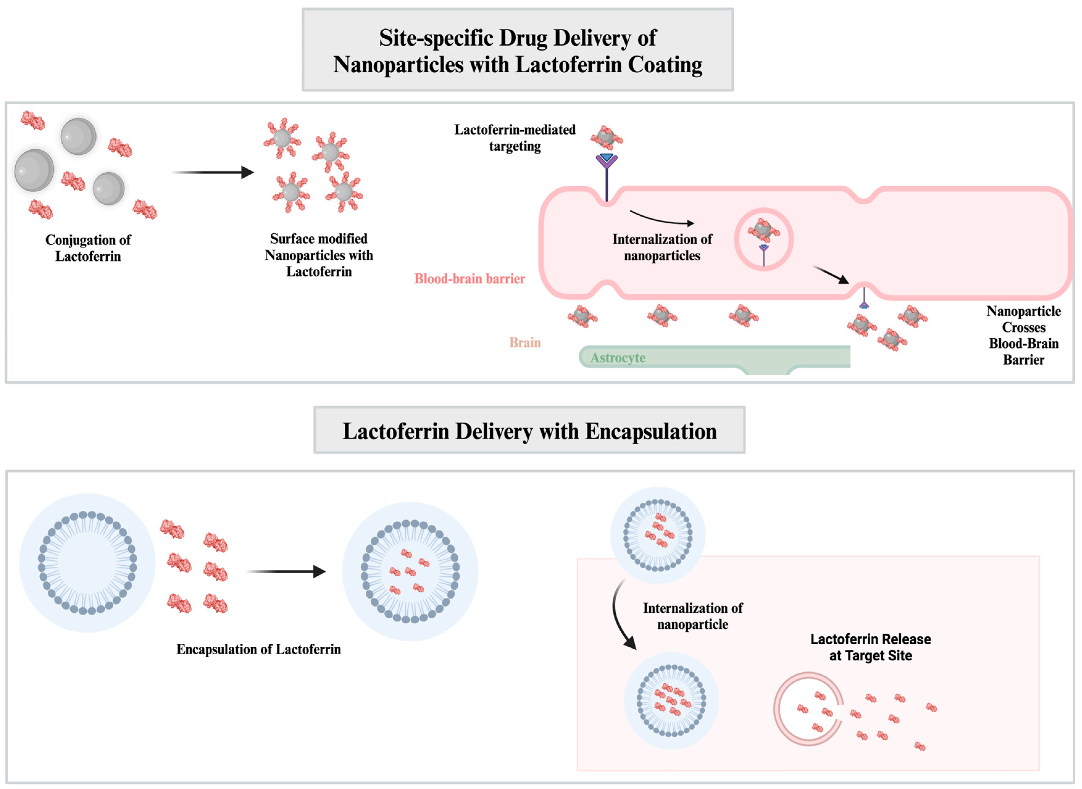
Figure 3.
Drug delivery with Lf conjugated NPs. Lf-conjugated NPs can cross over BBB with receptor-mediated transcytosis, delivering both NPs and encapsulated biomolecules at the target location. Lf can also be delivered with encapsulation into NPs, enhancing its biological activities [70,71].
- Anticancer Applications
In anticancer applications, NPs are frequently utilized for receptor-mediated targeting of cancer cells, including transferrin receptors, growth factor receptors, and cluster of differentiation (CD) receptors [72]. These receptors play a critical role in the tumor cell cycle or are overexpressed on the cell surface. For example, various NPs are employed for targeted drug delivery against cancer cells via the highly expressed CD44 receptors [73]. The desired receptor-specific targeting can be enhanced with surface modification of NPs. Lf possesses a strong affinity towards low-density lipoprotein receptors (LDLRs), transferrin binding receptors, and CD receptors [64,74,75]. Considering the following examples that are given below, Lf modification can further enhance receptor-specific targeting of NPs in drug delivery strategies.
For example, one study coated poly(lactic-co-glycolic) (PLGA) NPs with Lf and hyaluronic acid for targeting CD44 receptors in lung cancer [76]. Lf-modified PLGA NPs were decorated with polydatin and established sustained release for up to 24 hours. In vitro cellular uptake study revealed that Lf coating onto PLGA NPs increased fluorescence intensity from 325.39 ± 11.03% to 1741.13 ± 65.50%, compared to the control group. Moreover, polydatin decoration increased the intensity to the highest by 3130.66 ± 228.53%.
A similar study also conducted targeted anticancer activity with chondroitin-Lf dual-modified solid lipid NPs (SLNPs) against breast cancer [77]. In vitro drug release profiles showed that both dual-modification and only Lf modification led to controlled drug release, compared to unmodified SLNPs. The anticancer activity was determined by using cell line studies (MDA-MB-231 cells). Similar to the previously mentioned study, the authors highlighted that the cell lines were preferred due to their high expression of transferrin, CD44, and LDL receptors. The selective cytotoxicity towards cancer cells was shown during the experiments, whereas only LF modification showed the highest selectivity index, followed by the dual modification. Determining intracellular uptake through fluorescence intensity further supported the selectivity with high values. It was discussed that there is a great possibility that Lf-modified particles demonstrated the highest internalization through receptor-mediated endocytosis. Finally, in vivo anticancer studies revealed the following results: the highest reduction in tumor volume compared to the control group (from 1056 ± 93.34% to 274 ± 62.22%), strongest antiangiogenic activity with a 2.33-fold decrease in vascular endothelial growth factor, lowest expression levels of cyclin D1, apoptosis induction and having the highest area of necrosis.
- Targeted Brain Delivery Applications
Another common approach for Lf-modified NPs in drug delivery systems is the targeted delivery into the brain regions. Lf exhibits significant neuroprotective activity against neurodegenerative diseases [4]. It has been proposed that Lf can initiate intracellular activity through receptor binding, such as with heparan sulfate proteoglycans, lipoprotein receptor-related proteins, and other Lf receptors [4,6]. Dopaminergic neurons, which are found in decreased whereas the neurodegeneration is severe, have overly expressed Lf receptors [78]. Moreover, the fact that Lf can initiate receptor-mediated transcytosis through BBB makes Lf a great choice as a protein for NP modification in drug delivery applications for reducing the progression of neurodegenerative diseases [64].
Although there are only a few recent examples, Lf NPs can still be utilized to induce neuroprotective activity in brain regions. As discussed, Lf exhibits strong neuroprotective activity and can cross through BBB. Therefore, on an NP basis, various types of NPs are modified with Lf for targeted delivery, rather than using Lf as the NP itself or delivering it directly into specific brain regions. A recent example demonstrated the internasal administration of curcumin-Lf NPs for determining the capacity of neuroprotective effects [79]. The characterized NPs showed ideal properties as drug carriers, with an encapsulation efficiency of 91.2% ± 3.6% and a drug loading of 9.6% ± 0.8%. In vitro experiments demonstrated sustained drug release, high cellular penetration, and significant cellular uptake. A PC-12 cell-mediated nerve damage model was employed to determine the protective effect of Lf-curcumin NPs. Although Lf NPs managed to show notable neuroprotective effects when administered alone, their combined application with free curcumin and Lf-curcumin NPs resulted in even higher protective effects, with cell viability improving by over 90%. Furthermore, the NP treatment significantly reduced induced apoptosis and oxidative stress. Most importantly, in vivo pharmacokinetic studies revealed that Lf NPs successfully accumulated in brain regions and enhanced the bioavailability of curcumin.
To give a few examples of Lf coating on NPs, an in vivo study utilized Lf-functionalized manganese-doped silica hollow mesoporous NPs to deliver resveratrol for treating ischemic stroke [80]. In vivo bioimaging studies demonstrated that Lf-modified NPs successfully accumulated in brain regions exhibiting high fluorescence intensity in neurons and microglia, whereas unmodified NPs were mainly observed in the liver. Additionally, an in vitro BBB model demonstrated the significant Lf-mediated crossing of the NPs through BBB. The modified NPs significantly exhibited two crucial activities: antioxidant and anti-inflammatory effects. Most importantly, a notable reduction in neuronal apoptosis was observed in mice, with a decrease in pro-apoptotic factor expression by up to 64.1% ± 7.83%.
Another similar study modified nanoliposomes with Lf and borneol to mediate targeted delivery of crocetin in mice [81]. In vitro tests on HT22 cells demonstrated high cellular uptake and internalization of the NPs. The neuroprotective activity of Lf-modified NPs was determined by an increased cell survival rate of 15-33%, depending on the source of injury. In vivo experiments demonstrated the biodistribution of the NPs, highlighting the Lf-mediated brain targeting of liposomes.
The current literature suggests that Lf is an effective agent for surface modification of NPs in targeted drug delivery applications. Lf exhibits a multifunctional protein structure and unique interactions with various receptors, expanding the range of targets in drug delivery. Given the emergence of alternative drug carriers for both anticancer and neurodegenerative applications, Lf could be one of the molecules that precisely fits into NP-based treatments.
2.3. Lf and NPs in Delivery Systems for Hepaprotective, Antioxidant, and Anti-Inflammatory Applications
Lf demonstrates both antioxidant and anti-inflammatory activities in several applications, such as in inflammatory bowel diseases [75] and antioxidant biofilms in food packaging [82]. Moreover, oxidative stress and neuroinflammation levels are directly linked to the progression of neurodegenerative diseases [83], and Lf has the potential to reduce these levels significantly [6]. Additionally, several studies have demonstrated the hepatoprotective activity of Lf against induced hepatotoxicity (including NP-induced toxicity) [84,85,86]. As a result, various nanostructures including Lf have been employed with various types of approaches to mitigate the aforementioned toxic effects, including surface modification with Lf, direct delivery of Lf, and incorporation of Lf into nanocomplexes.
An in vivo study used selenium NPs to deliver Lf for its Hepatic and immune modulatory activity for drug-induced hepatic injury [87]. The biochemical analysis from mice demonstrated a significant reduction in lipid peroxidation and NO levels, oxidative stress regulation through increased GSH levels, and reduction in pro-inflammatory cytokines, apoptotic, and pro-fibrotic markers. Moreover, Lf-loaded NP treatment significantly reduced caspase-3 expression in liver tissue nearly to the control levels, lowering the drug-induced hepatotoxicity. The histopathological analysis supported the reduced hepatotoxicity with improved hepatic structures.
A similar in vivo study also treated drug-induced hepatoxicity but through Lf-coated zein NPs as a dual oral carrier [40]. The involvement of Lf as one of the coating materials was thought to be increase the gastric digestion tolerance, considering the molecular structure of the protein. This was supported by the lowered and sustained drug release of the nanostructure. While observing the distribution of the administered NPs, it was shown that coated NPs exhibited less off-target effect by low fluorescence intensity in kidneys and spleen. The oxidative stress parameters were normalized similarly to the previous study, through increased GSH levels and reduced levels of lipid peroxidation byproduct. Undesired changes in the parameters of both the liver and kidney were changed nearly to the control levels through coated-NP treatment. It is worth mentioning that all of these outcomes were not significant with the solo treatment of compounds or uncoated forms of NPs.
2.4. Lf Delivery with NPs
In addition to its use in NP-based delivery systems, Lf's biological properties are applied in various fields by directly delivering it to NPs. Lf exhibits multifunctional activity in various places [5], and is known for initiating both covalent and non-covalent interactions with various biomolecules [71]. Site-specific delivery of Lf in high efficiency can enhance the therapeutic application of Lf.
- Ocular Delivery of Lf with NPs
Lf plays a crucial role in tear fluid due to its multifunctional properties. It is widely expressed in various parts of human eye tissue [88]. Lf shows significant potential as a therapeutic agent for treating dry eye disease and ocular surface infections [89]. Furthermore, ocular surface diseases can significantly reduce Lf concentrations in tear fluid, [90], which highlights its potential as a biomarker for dry eye disease [91].
Considering the role of Lf, various delivery systems, including nanostructures as a transporter, have been used for the ocular delivery of Lf. Primarily polymer-based, such as PLGA, and lipid-based, such as lipid nanocarriers and liposomes, have been used in topical administration of Lf in ocular delivery applications [92].
For example, a study demonstrated the potential of administering Lf topically to the eye using PLGA NPs in the form of nanospheres and nanocapsules [93]. The nanostructures exhibited efficient, controlled drug release and showed no significant cytotoxicity. In vivo study revealed that the nanostructures could initiate interaction with the ocular surface for up to 5 hours. A study showed further evaluation of PLGA-based NPs for delivering Lf, indicating its effectiveness in treating ocular inflammation in vivo [94]. Compared to free Lf, NP-carried Lfs exhibited a prolonged and controlled drug release profile in an in vitro release study. Ex vivo corneal permeation indicated that Lf’s encapsulation improved penetration speed and administration concentration, resulting in approximately 1.5-fold higher permeability coefficient and permeated amount than free Lf. The anti-inflammatory and cytotoxicity status of Lf-loaded PLGA NPs were evaluated in vitro using HCE-2 cells. Due to the biodegradable nature of the PLGA polymer, the delivery system did not induce cytotoxicity and reduced the expression of cytokines (IL-8 and TNF-α). In vivo studies confirmed similar anti-inflammatory activity, with a slight increase in recovery speed and reduction in inflammation, and demonstrated that the treatment passed ocular tolerance tests without irritating.
- Lf Delivery for Bone Engineering
Lf exhibits significant potential in regeneration of bone structures through induction of proliferation and differentiation of tissues [95]. Despite its potential in bone engineering applications, meta-analyses from recent years highlight the need for further research on Lf in in vivo and clinical studies [96]. Although limited, some studies have investigated NP-mediated Lf delivery for bone formation applications.
For instance, an in vitro study used alginate chitosan NPs to deliver bovine Lf, aiming to perform detailed characterization and enhance the growth of the MG-63 cell line [97]. The study primarily focused on NP characterization and analyzed changes following Lf loading. The NPs exhibited an encapsulation efficiency of 89.94% and showed the highest interaction rate with Lf at pH 8, with no structural changes after Lf loading. MTT tests demonstrated that Lf delivery increased cell viability by 8.5-fold.
Noh et al. developed Lf-anchored silica NPs for enhancing osteo-differentiation for bone healing and regeneration applications [98]. Lf release patterns exhibited sustained release profiles for up to 28 days. Osteo-differentiation marker changes were analyzed. An increase in alkaline phosphatase was first observed on day 3, with a significant difference noted by day 9. By day 14, the concentration of accumulated calcium levels showed a similar increase. The most significant change was observed in the mRNA expression of osteocalcin and osteopontin, with up to a 3-fold increase.
The same research group used an in vivo rat model to demonstrate the induced bone fusion with the developed Lf carrier NPs [99]. The following results were obtained that indicate the enhanced bone fusion in rats: increased frequency of blood vessels (2-fold increase), a significant increase in osteocalcin concentrations (up to 3-fold increase), and increased mRNA expression levels of osteocalcin and osteopontin (nearly 4 and 3-fold, respectively).
3. Antimicrobial Applications of Lf-NPs
A vital component of milk, Lf is an iron-binding glycoprotein that is essential to the innate immune response. Iron homeostasis, immunological response, antioxidant, anticancer, and anti-inflammatory qualities are among its physiological roles; its antimicrobial action has been the subject of the most research. Lf demonstrates efficacy against a range of bacteria, fungi, viruses, and protozoa by sequestering iron, an essential resource for pathogens, while its structural integrity is vital for preserving its bioactivity during processing [100].
- Antibacterial
As an initial line of defense, many species generate antimicrobial proteins and peptides. For animals, including humans, milk is an essential source of nourishment. It is also a rich supply of proteins including Lf, a multipurpose protein that has antimicrobial properties due to its capacity to bind iron. By targeting bacterial pathogenicity mechanisms, iron sequestration, membrane instability, and host cell invasion tactics, Lf has antibacterial effectiveness against a variety of bacterial pathogens [101]. Its broad-spectrum antibacterial inhibitory action has been demonstrated against both Gram-positive and Gram-negative bacteria, successfully preventing the development of Salmonella typhi (S. typhi), Streptococcus, Legionella pneumophila, S. aureus, and E. coli [102].
The physicochemical characteristics of bovine Lf and the resulting silver-Lf complexes, the technique of immobilizing silver onto and/or inside LTF, and the antibacterial activity of these complexes were all examined by Pomastowski et al. The antibacterial activity study's findings prospectively imply that synthesized silver-Lf may find application as a novel, commercially and ecologically viable antimicrobial agent in the food and medical industries [103]. Similarly, Alhadide et al. have used NPs with whey protein films to manufacture Lf packed with iron oxide Fe2O3-NPs (IONPs) utilizing CONCARPUS extraction, which includes complete three-dimensional structural stability. Using the well diffusion approach, the antibacterial activity of the generated NPs in conjunction with the created whey protein films was examined against Salmonella enterica, S. aureus, S. galactiae, and E. coli. As a consequence, they discovered that Lf nanovesicles in whey films produced under optimal conditions were also successfully employed in antibacterial research facilities [104].
A novel strategy for combating pathogenic-resistant bacteria is provided by hybrid NPs, which are made to have various antibacterial activity mechanisms. Silver NPs were created by Abdalla et al. and have antibacterial and anti-biofilm qualities. The silver NPs were surface functionalized with either GO (AgGO) or LTF (Ag-LTF) after being produced using chitosan and mushroom waste. A surface plasmon resonance band was observed at 430 nm for the silver NPs, whereas AgGO and Ag-LTF exhibited absorption at 402 and 441 nm, respectively. Gram-positive and Gram-negative bacteria were suppressed by silver NPs, Ag-LTF, and AgGO with similar antibacterial efficacy. Ag-LTF did not impact cell survival or migration rate, indicating non-toxicity, however, AgGO and Ag-LTF showed synergistic efficacy against P. aeruginosa. According to these results, silver NPs, Ag-LTF, and AgGO could be useful antibacterial agents in the future [105].
- Antiviral
Numerous naturally occurring antimicrobial proteins and peptides have shown promise in preventing viral infection, and the virus from entering the host cell, or influencing the virus at a later stage of development [106]. Lf is a glycoprotein found in external secretions, with many roles, such as antiviral properties and the enhancement of immune response [107]
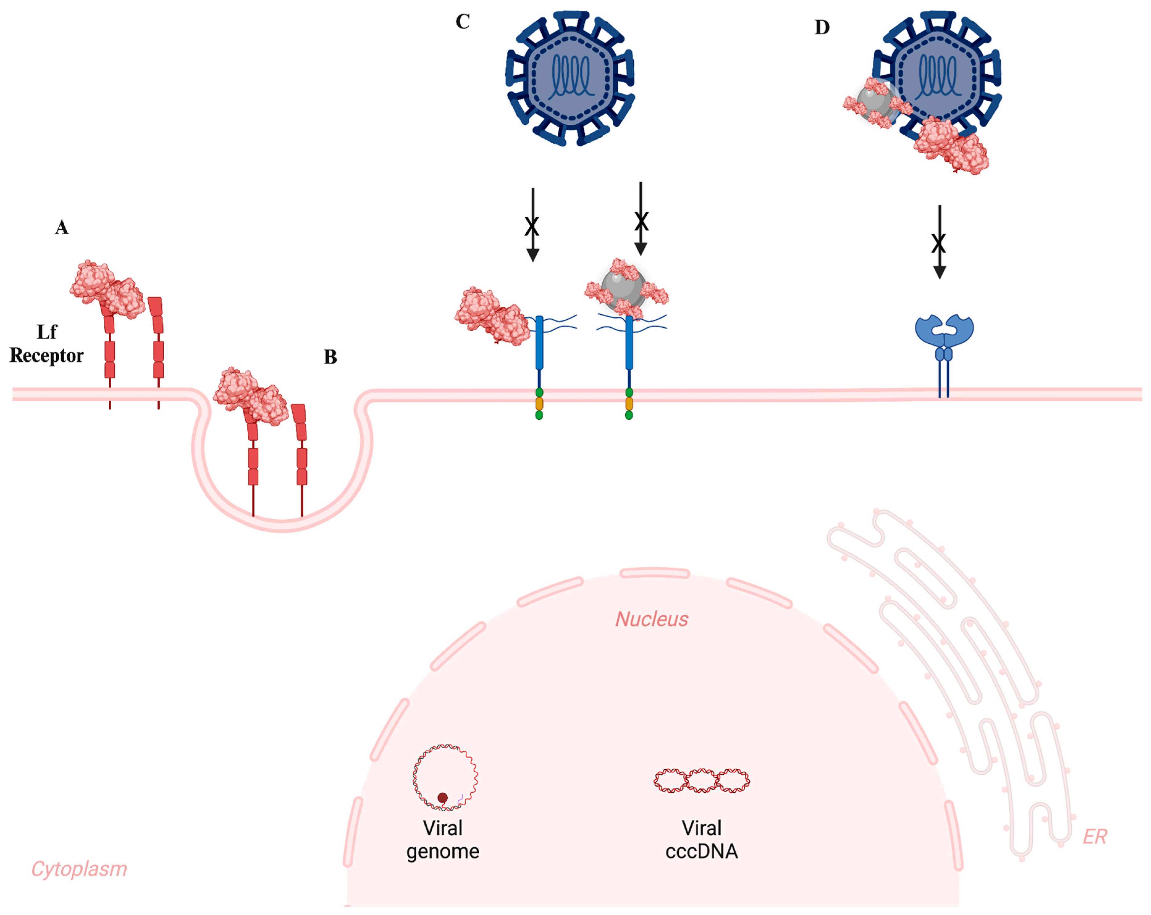
Within the concept of antiviral mechanisms of Lf, the cell surface and extracellular matrix include heparan sulfate proteoglycans (HSPGs), which Lf can bind to. On the surface of the majority of eukaryotic cell types, these proteins are expressed and consist of a core protein with GAG chains. The negatively charged sulfated groups of heparan sulfate (HS) present in the glycocalix of the cell surface and viral attachment ligands (VALs) are what bind viruses to HSPGs. Certain viruses can enter cells more easily when HS helps them make contact with the cell surface and fuse with the host membrane [108,109]. Lf may disrupt heparan sulfate proteoglycans on the cell surface, directly attach to viruses or their receptors, or enhance the antiviral response of the immune system. Nevertheless, several scientists suggest its potential mechanism of action by disrupting the endocytic route of viral infection (Figure 4) [109].
Hepatitis C virus (HCV), herpes virus, respiratory syncytial virus, rotavirus, and HIV are among the viruses against which LF has demonstrated antiviral efficacy [110]. By preventing viral attachment and penetration into the host, Lf has an anti-HSV impact [111]. Both HSV-1 and HSV-2 have been shown to be susceptible to Lf's antiviral action, albeit the precise antiviral process varies slightly because of the two viruses' differing initial attachment to target cells [106,107]
Figure 4.
Schematic illustration depicting the potential pathways of antiviral activity by Lf and Lf-modified NPs. (A) Lf can be swallowed by endocytosis; the interaction of Lf-modified NPs need validation. (B) The endocytosis of Lf-modified NPs need validation. (C) Lf or Lf-modified NPs can inhibit HSV-1/2 infection by binding to heparan sulfate moieties found in the glycoproteins of the cell surface and extracellular matrix. (D) Lf or Lf-modified NPs can obstruct viral binding by attaching to the virus attachment ligands (VALs), thus preventing the virus from connecting to its particular receptor(s) [112].
Proteins and NPs can interact to create the so-called interfacial protein corona, which gives the particles particular biological activity. Van der Waals interactions and hydrogen bonds allow bovine Lf to be absorbed into the silver NP surface without changing the stability and shape of the protein, as shown by the work of Nayak et al. [113]. Additionally, bovine Lf (bLf) reduced the cytotoxicity mediated by silver NPs [113]. By directly inhibiting viral attachment, penetration, and infection, Krzyzowska et al. showed that pretreatment with human Lf functionalized gold and silver NPs sized 10 or 30 nm reduced HSV-2 infection. According to the study, Lf bonds to HSPGs on the cell surface more efficiently in nanometal conjugates, forming a barrier that prevents HSV-2. Lf conjugates had an extra immunomodulatory impact during vaginal infection, however, all tested Lf conjugates reduced HSV-2 titers in in vivo tests [114]. In a different study, as an HIV-microbicide, Yeruva et al. developed Lf NPs (TCNPs) loaded with tenofovir and curcumin. HIV-1 replication was successfully suppressed by TCNPs, which had a diameter of 74.31 ± 2.56 nm and an IC50 of 1.75 μM for curcumin and 2.8 μM for tenofovir. They demonstrated spermicidal action, produced low cytotoxicity and inflammation in vaginal epithelium, and offered drug release for up to 8–12 hours, making them a promising anti-HIV microbicidal agent [115].
Zidovudine + efavirenz + lamivudine loaded Lf NPs (FLART-NP) were produced and assessed by Kumar et al. for their physicochemical characteristics, bioactivity, and pharmacokinetic profile in order to improve the efficacy, bioavailability, and decrease the toxicity of the first-line highly active antiretroviral regimen. Thus, three anti-HIV medications have been effectively encapsulated in NPs by researchers, producing a stable, evenly distributed colloidal solution. The regulated and prolonged intracellular release of medications by the NPs improves their pharmacokinetics without endangering vital organs [116].
The current investigation posited a synergistic antiviral effect of Zn-NPs coated with LF protein against SARS-CoV-2 infection and the resultant pulmonary fibrosis. This application innovatively enhances cellular absorption and maximizes the antiviral and anti-inflammatory effects of LF by the saturation of LF with biosynthesized Zn-NPs. The superior effectiveness of biosynthesized Zn-NPs and LF-coated Zn-NP nanocomplexes may serve as a potential treatment for SARS-CoV-2 and its associated problems [117].
- Antifungal
The escalating prevalence of serious fungal infections, mostly attributed to a growing population of immunocompromised individuals, has rendered the identification of novel compounds with potent antifungal properties, or those that enhance the efficacy of existing antifungal medicines, increasingly imperative. Milk proteins and other natural products are a major source of new antifungal agents [16].
Numerous studies in the literature have demonstrated the promise of Lf, a multifunctional protein, as a broad-spectrum natural antifungal agent due to its strong antifungal activity against a variety of fungi (Table 2). The antifungal efficacy of Lf (both human and bovine sources) arises from its capacity to sequester iron, so depriving Candida species of this essential nutrient, which results in membrane breakdown and leakage through iron-independent mechanisms [118].
Antimicrobial materials have gained significance for localized therapy to avert microbial resistance caused by systemic antibiotic treatments. The fabrication of electrospun poly(lactic acid) nanofiber membranes infused with bovine Lf at concentrations up to 20% was performed by Machado et. al. The resulting fibers are characterized by a smooth, defect-free morphology, with average diameters ranging from 717 ± 197 nm to 495 ± 127 nm, and an overall porosity of around 80%. The synthesized membranes exhibit antifungal properties against Aspergillus nidulans by obstructing spore germination and mycelial proliferation [100].
Plasmonic magnetoliposomes (PMLs) are advantageous nanocarriers for simultaneous hyperthermia and localized chemotherapy, owing to their integration of magnetic and gold NPs, which facilitates targeted drug delivery. From this perspective, gold NPs (5–7.5 nm size) and manganese ferrite NPs (28 nm size) functionalized with 11-mercaptoundecanoic acid or octadecanethiol were added to PMLs and loaded with bLf. Using fluorescence microscopy and colony-forming unit counts, the antifungal potential of bLf-loaded PMLs and their internalization process were evaluated in Saccharomyces cerevisiae. Thus, PMLs containing bLf exhibit cytotoxicity efficacy comparable to that of free bLf, suggesting that they may be used to deliver bLf in antifungal therapeutic treatments [119].
Table 2.
Antimicrobial applications of Lf-NPs and Lf-incorporated nanomaterial systems.
| Antimicrobial activity of Lf with NP | Study Type | Main Results | Reference |
|---|---|---|---|
| Antibacterial | |||
| Development of Silver-Lf NP incorporated hydrogels | In vitro | -Silver-Lf NPs demonstrated antibacterial activity against both Gram-positive (S. aureus) and Gram-negative (E. coli and P. aeruginosa) bacteria. -Largest zones of inhibition were 11.3 ± 7.5 mm, 10.3 ± 1.5 mm and 7.3 ± 0.6 mm for E. coli, S. aureus, and P. aeruginosa, respectively, at a NP concentration of 125 µg/mL. |
[120] |
| Development of antibiotic loaded Lf NPs | In vitro In vivo |
-NPs demonstrated bactericidal activity against E. coli, Mycobacterium marinum (MM) and methicillin-resistant S. aureus (MRSA). -Superior antibacterial effects, in comparison to free antibiotic and antibiotic loaded bovine serum albumin NPs, were observed. -MIC values were determined as 12.5 µg/mL for E. coli, 0.3125 µg/mL for MM, and 8.0 µg/mL for MRSA. -In vivo assays on mice model highlighted that antibiotic loaded Lf NPs can promote wound healing by improving intracellular bacteria elimination. |
[66] |
| Synthesis of Lf functionalized gold NPs | In vitro In vivo |
-Antibacterial activity against both non-pathogenic bacteria, such as Bacillus subtilis (B. subtilis) and E. coli, and pathogenic strains including S. aureus, Enterococcus faecalis (E. faecalis), and S. typhi. -Enhanced antibacterial effects, through functionalization with Lf, compared to NPs alone. -Lowest MIC values were determined as 10 µg/mL and 15 µg/mL for B. subtilis and E. faecalis, respectively. -Largest zones of inhibitions were observed as 8.35 mm for E. faecalis and 8.45 mm for B. subtilis. -Increased biocompatibility and hemocompatibility in Wistar rats, following incorporation of Lf functionalized gold NPs. |
[121] |
| Development of Lf-functionalized silver NP incorporated gelatin hydrogels | In vitro | -Dose-dependent antibacterial activity of Lf-Silver NPs against S. aureus and P. aeruginosa. -Increased inhibition zones, from 10.7 ± 3.6 to 12.7 ± 2.3 for S. aureus and 10.8 ± 1.4 to 11.9 ± 3.2 for P. aeruginosa, when Lf-silver NP concentration in hydrogels was increased from 62.5 μg/mL to 125 μg/mL. |
[122] |
| Preservation of strawberry samples through antibacterial Lf NPs | In vitro | -Significant antibacterial activity against S. aureus 0.3 mg/mL MIC value -Lf-NP coating on strawberries with carboxymethylcellulose reduced weight loss from 85% to 60% at day 6. -Significant reduction in counted aerobic mesophilic bacteria. -Reduced physiological changes of strawberries during storage. |
[123] |
| Lf-included Nanocomposite for Packaging | in vitro | -High antioxidant activity by 67.6 ± 1.4 % DPPH radical scavenging -Significant antibacterial activity against E.coli and S. aureus with 18.5 mm ZOI. -Increased decomposition. |
[124] |
| Antiviral | |||
| Development of Zn-NPs coated with bLf using green synthesis | In vitro | -LF-Zn-NPs contained larger particles that measured up to 98 ± 6.40 nm, whereas the biosynthesized Zn-NPs were white, oval to spherical in form, and had an average size of 77 ± 5.50 nm. -The negatively charged surfaces of the biosynthesized Zn-NPs and LF-Zn-NPs were found to have zeta-potentials of -20.25 ± 0.35 and -44.3 ± 3.25 mV, respectively. -By attaching to the ACE2-receptor and spike protein receptor binding domain with IC50 values of 59.66 and μg/mL, respectively, LF-Zn-NPs showed a notable in vitro delay of SARS-CoV-2 entrance to host cells. |
[117] |
| Development zidovudine + efavirenz + lamivudine loaded Lf-NPs (FLART -NP) against HIV therapy | In vivo In vitro |
-Encapsulation efficiency, cellular localization, release kinetics, safety analysis, biodistribution, and pharmacokinetics have all been investigated in vitro and in vivo. -For each medication, FLART-NP was produced with an encapsulation effectiveness of >58% and a mean diameter of 67 nm (FE-SEM). -With little burst release, low erythrocyte damage, and enhanced anti-HIV effectiveness in in vitro experiments, FLART-NP delivers the maximal payload at pH5. |
[116] |
| Development Coencapsulated Lf-NPs with tenofovir and curcumin enhance vaginal protection against HIV-1 infection. | In vitro In vivo |
TCNPs | [115] |
| Antifungal | |||
| Development nanofiber membranes loaded with bLf to display antifungal activity against Aspergillus nidulans | In vitro | -The membranes had an overall porosity of around 80% and smooth, nondefective fibers with mean diameters ranging from 717 ± 197 to 495 ± 127 nm. -The presence of bLf decreases the hydrophobicity of the PLLA membranes. -Human fibroblasts were not cytotoxically affected by the bLf–PLLA membranes that were created; interestingly, after 24 hours of indirect contact, the 20-weight percent bLf–PLLA membrane was even capable of inducing cell growth. |
[100] |
| In vitro | -PMLs have been produced with gold NPs and manganese ferrite, functionalized with either octadecanethiol or 11-mercaptoundecanoic acid, and then loaded with bLf. -PMLs were formed when both plasmonic and magnetic NPs were enclosed in DPPC and Egg-PC liposomes. -PMLs loaded with bLf are around 200 nm in size, exhibit a positive zeta potential, and remain stable for at least five days. -Because PMLs are non-cytotoxic and retain their antifungal function, they exhibit encouraging potential for delivering bLf into yeast cells. |
[119] | |
4. Agriculture Applications of Lf-NPs
Utilization of natural components in the field of agriculture, especially for current food packaging and food preservation applications, has significantly increased [82]. This is mostly attributed to the latest research on commercially employed chemical additives, which are known to cause several negative side effects, including carcinogenic effects and allergies, on consumer health [125].
To address this, researchers are using Lf as a natural additive in food storage and protection applications. Lately, many studies have been conducted in this field to enhance the shelf life of meat, fish, and fruits [126,127,128].
Being a less-toxic, biodegradable, and biocompatible alternative, Lf has also been employed in either its NP form or as a coating material for the development of reinforced preservation systems [129,130]. The main reason behind this is the superior antimicrobial activity of Lf, which effectively inhibits the proliferation of a wide array of pathogens to extend shelf life, prevent food spoilage, and increase crop yields.
4.1. Food Packaging
One of the main focuses of NP employment in the food industry is on the packaging systems. NPs are used to enhance the mechanical, barrier, and thermal properties of matrices, which paves the way for the production of strengthened food packaging materials [131]. However, their applicability is hindered by toxicity concerns, regarded as one of the main drawbacks associated with nanomaterials [132].
Hence, protein-based NPs emerged with their sustainable, renewable, and biodegradable nature to replace commercially used NPs [133]. Also, their relative abundance, oxygen barrier ability, film-formation capacity and nutritional value further support their use in this area [134]. Lf, as an antimicrobial milk glycoprotein, possesses great potential for use in these applications. For example, it can be integrated into packaging films, either alone or combined with other proteins, to enhance the antibacterial activity and physicochemical properties of the final products [126,135].
Khezerlou et al. synthesized Lf-included NPs (chromium-based metal-organic frameworks) to develop gelatin–κ-carrageenan films as packaging materials to overcome limitations associated with perishable foods. Incorporation of Lf enhanced the functional properties of films as evidenced by decreases from 105% to 70.8%, 61% to 34.63%, and 2.46 to 2.19 x 10⁻¹¹ g. m/m2.s, in swelling index, water solubility, and water permeability, respectively. Also, at increasing concentrations of Lf, antibacterial effects were observed against S. aureus and E. coli with inhibition zones of 20.2 and 19.7 mm. In packaging assays over 6 days, at 50% humidity and 25 °C, softening and mold growth on strawberries was observed with uncoated films, while the addition of Lf prevented these side effects and maintained the freshness [136].
Similarly, Tavassoli et al. developed bioactive packaging materials that contain Lf-encapsulated silver-metal organic framework NPs. Incorporation of these NPs improved the tensile strength, from 41.1 ± 2.4 MPa to 56.1 ± 3.2 MPa, and thermal stability while decreasing water vapor and oxygen permeability. Besides, bactericidal assays showed these films effectively inhibited the growth of both Gram-positive and Gram-negative bacteria, S. aureus and E. coli, with a maximum ZOI of 20.1 ± 3.2 mm and 19.8 ± 5.2 mm, in comparison to the films alone, 10.8 ± 6.1 and 10.6 ± 1.5 mm. Researchers examined the effect of Lf concentration (0.5, 1, and 2%) on the storage of fresh apples at 25 °C and 50 % relative humidity for 7 days. Samples containing 2% of Lf effectively extended the shelf life of apples and resulted in minimum visible changes, while their counterparts led to softness, dehydration, and change of color. Also, these films exhibited strong UV-protective properties, which indicated their further applicability in the protection of materials that are sensitive to photodegradation [137].
4.2. Food Preservation
Multiple studies in current literature highlight the use of nanomaterials, especially inorganic ones, for effective food preservation [138,139]. Specifically, metallic and metal oxide NPs (including silver, zinc oxide, and titanium dioxide) have gained attention in these systems with their strong antimicrobial activity [131,140].
However, similar to food packaging, there are rising concerns about the toxicity potential of these NPs due to their inherent non-biodegradability [141,142]. Considering these, researchers are utilizing organic-based food contact materials, like proteins, to create non-toxic and novel approaches. Lf stands out in this aspect owing to its strong antibacterial efficiency and previously demonstrated strong food preservation capability [143,144,145]. Its ease of functionalization and combined effects with NPs further support their use in this field and underscore its promising potential in future studies.
A recent study involved the combined use of Lf NPs with other biomolecules, chitosan, and gellan gum, to extend the shelf life of fresh strawberries. It has been observed that coating with NPs significantly enhanced the physicochemical properties of strawberries in comparison to their uncoated counterparts, which showed shrinkage, mold, and darker color following 96 hours of storage at 25 °C. These ternary NPs exhibited noteworthy antibacterial activity against S. aureus (at a concentration of 103 CFU), with MIC values down to 0.0117 mg/mL. Also, researchers conducted aerobic bacteria counts in natura to assess overall fruit quality. After 144 hours of storage, bacterial concentration increased from 2.47 Log CFU/mL to 4.2 Log CFU/mL for the untreated (control) sample, while it remained at 3.74 Log CFU/mL for NP treated fruits [146].
Similarly, Duarte et al. focused on producing natural preservatives to improve the shelf life of strawberries. Aiming to exploit the combined antimicrobial effect of bovine Lf and chitosan, researchers synthesized Lf-chitosan-Sodium tripolyphosphate NPs. These NPs demonstrated potent antibacterial activity against S. aureus (with a MIC value of 0.0370 mg/mL) indicating a three times lower concentration than the pure materials. Further, coated strawberries were stored at 25 °C and 50% relative humidity for 6 days. Results revealed darker colors, shrinking, and water loss in uncoated samples, while NPs coating led to delays in food ripening and degradation [129].
5. Toxicity
Lf is a primary antimicrobial component found abundantly in milk and colostrum [147], making it a preferred, safe agent for various therapeutic applications. Depending on its source, Lf may vary in molecular structure due to differences in amino acid sequences and the structure of glycans [7]. Consequently, its effectiveness in specific applications can vary, as demonstrated by previous studies [148,149]. Therefore, selecting the appropriate type of Lf is crucial to prevent any undesired outcomes in the application.
bLf represents the largest scale in industrial production due to its abundant availability, making it suitable for therapeutic and supplementary uses [150]. Consequently, bLf has received approval by the United States Food and Drug Administration and is recognized as a safe dietary supplement by European Food Safety Authority [75]. These approvals place Lf in a reassuring position regarding toxicity concerns. However, it remains necessary to evaluate optimal dosing approaches to prevent any toxic outcomes.
One study investigated bovine Lf’s optimum dose of administration for preterm infants [151]. 31 patients were divided into three groups, receiving 100, 200, and 300 mg/kg of daily bovine Lf for 10 months. At the end of the supplementation, none of the patients showed any Lf-based adverse effects, showing notable tolerance at the dose of 300 mg/kg. In the past few years, studies that involved investigating the effects of bovine Lf supplementation used similar doses during the experimentation. One study used 200 mg/kg daily bovine Lf to investigate its effect against sepsis and neurodevelopment impairment in infants [152]. Although the trial did not show any effect, the supplementation did not create any adverse effects. Another study used 200 mg to 1000 mg daily bovine Lf for COVID-19 patients, showing reduced symptoms and no adverse effects [153].
The latest experiments on Lf toxicity and clinical trials using naturally sourced Lf primarily focus on determining the optimal dose for administration. To investigate Lf toxicity further, recombinant synthesis of Lf can be centered since it holds significant potential for future research on Lf.
Recombinant Lfs (rLf) holds promise as an alternative source for advancing Lf-based NP applications. It exhibits a great alternative to obtain high amounts of Lf for conducting wide-scale applications and possible productization. This is why recombinant technology is commonly preferred for human Lfs, given the challenges of obtaining it from natural sources for large-scale production. Still, given the current uncertainties regarding safety conditions, rLfs face significant challenges in their application. One of the main reasons behind this problem is the alterations in the biological activity of Lfs due to the recombinant synthesis, given the possible variables on the glycosylation patterns including monosaccharide composition and connection with glycan structures [154,155]. As a result, current research increasingly focuses on assessing the toxicological characteristics of recombinant human Lfs for future applications.
One of the most recent developments in the investigation of rLf was mediated with human Lf (hLf) which is synthesized from glycoengineered yeast, komatagaella phaffii. On this issue, a workshop report has been published that discussed the roadmap to achieve the safety standard of the hLf [156]. Certain safety factors need to be evaluated in the report: determination of any potential immunotoxicity from rLf ingestion, potential effects of rLf in iron homeostasis, any trace of alloimmunization, and the pathway that rLf potentially goes through during the digestion.
Recently, recombinant hLf was tested in an in vivo study to determine the dose range for further research [157]. Rats were separated into 3 different groups, each receiving a different dose of recombinant hLf for 14 days: 200, 1000, and 2000 mg/kg. A fourth control group consumed 2000 mg/kg of bovine Lf daily. The toxicological results indicated that rats demonstrated high tolerance to 2000 mg/kg daily consumption of recombinant hLf, allowing a reference dose for further studies. Followingly, the same research group tested this recombinant hLf for potential immunogenicity, comparing it to bovine Lf in a randomized, double-blind, controlled trial [158]. The trial consisted of three main groups (66 healthy individuals), whereas two groups consumed human rLf, taking 0.34 and 3.4 grams per day, and the third group consumed 3.4 grams of bLf per day. At day 56, it was shown that intake of human rLf did not induce any alloimmunization or increase the levels of hLf-antibodies.
Given Lf’s features and the glycan structure variations that can significantly impact its activity, similar safety concerns must be thoroughly investigated to include rLfs in products intended for supplements or therapeutic applications. Recombinant Lf production could provide a strong foundation for future Lf-based NP applications and be a basis for upcoming clinical trials. Further research could support the safe use of rLfs in NP-based applications. However, certain types of NPs carry higher health risks and safety concerns compared to the Lf protein
Regarding the characteristics of the NPs, particular types have been viewed as having high toxicity potential (Figure 5). With their small size and high reactivity, NPs can exhibit high toxicity depending on their concentration, exposure time, and route of administration [159]. One of the main mechanisms behind NPs toxicity is ROS synthesis. NPs can increase the oxidative stress levels that lead to DNA damage, protein denaturation, lipid peroxidation, and/or mitochondrial dysfunction [28,160,161].
We have discussed some types of NPs that have been used with Lf in various applications. Excluding the organic-based structures, metal-based NPs have been commonly used in these applications. Many metallic NPs, such as silver, gold and iron oxide, have shown high efficiency in delivering various natural products [163]. Moreover, these NPs exhibit significant potential in future clinical trials, such as in antimicrobial therapy, antitumor application, additive material for bone healing, and so on [164]. These applications are mostly common with the biological activities of Lf, indicating the possibility of future therapies based on Lf-based NPs applications. It needs to be highlighted that most of the common metal-based NPs are known to have high potential for inducing oxidative stress, depending on their physicochemical properties [165].
6. Conclusions
Thanks to its multifunctional properties, Lf exhibits significant applicability across various fields. Nanotechnology is an emerging field that offers promising approaches in various applications. Considering the characteristics of Lf, there is a great possibility that Lf will have a major interest in NP technology. Lf demonstrates strong antibacterial properties, immunomodulatory activity, and potential use in agricultural applications with minimal adverse effects. Moreover, due to the rising demand for alternative therapeutic approaches and carriers, Lf’s properties can significantly be utilized for developing drug delivery strategies, particularly in NPs technology. Lf can not only be conjugated and modified on NPs for various therapeutic applications but can also function as NPs itself, showcasing its versatility. As Lf receptors are present in key human tissues, particularly the BBB, Lf has the potential to drive the development of novel delivery strategies that address several existing challenges in the field.
Still, despite the multifunctionality of the protein, certain areas of Lf-based NPs applications remain quite limited. One of the most overlooked areas where Lf holds significant potential is agriculture. As covered in the agriculture section, only a limited number of studies have been published in recent years where Lf is combined with NPs technology in agriculture-based applications. Interestingly, current literature indicates that both Lf and NPs have been individually utilized in agricultural applications with a wide-ranging approach. It is somewhat surprising that these two approaches are not combined more frequently. In the future, with sufficient research support, Lf-based NPs applications could be utilized in various agricultural areas, particularly in food preservation.
Another deficient part that holds great potential is the NP-based delivery of Lf. Beyond its role as a supportive molecule, the significance of Lf's biological activity has been extensively discussed in recent years. Lf exerts its activity in various parts of the body, with effects that can vary depending on the specific location. With the necessary optimizations, site-directed delivery of Lf could have a significant impact on various therapeutic applications, such as anticancer, anti-inflammatory, and neuroprotective therapies. However, the range of these applications in recent years remains quite limited, despite the proven efficacy of Lf in these areas. Various types of NPs have been demonstrated to be excellent carriers for therapeutic agents, highlighting them for advanced delivery methodologies. Conducting further research on NPs-based Lf delivery could yield significant outcomes in numerous therapies that require improvements and novel alternatives.
Taken together, Lf demonstrates key characteristics that position it as a potential therapeutic agent in NPs-based nanomedicine applications. The current literature highlights several significant and promising sub-areas of Lf-based NPs applications. With sufficient research, additional sub-areas are likely to emerge as Lf is further integrated into NPs technology. Future research could not only expand the range of Lf applications but also enhance existing NPs applications in various emerging fields.
Author Contributions
Conceptualization, S.K.; writing—original draft preparation, E.A., F.E., H.D. writing—review and editing, S.K., M.B., P.S. visualization, H.D., E.A., F.E. All authors have read and agreed to the published version of the manuscript.
Funding
This research received no external funding.
Data Availability Statement
Not applicable.
Conflicts of Interest
The authors declare no conflicts of interest.
References
- García-Montoya, I.A.; Cendón, T.S.; Arévalo-Gallegos, S.; Rascón-Cruz, Q. Lactoferrin a Multiple Bioactive Protein: An Overview. Biochimica et Biophysica Acta (BBA) - General Subjects 2012, 1820, 226–236. [CrossRef]
- Baker, E.N.; Baker, H.M. Molecular Structure, Binding Properties and Dynamics of Lactoferrin. Cellular and Molecular Life Sciences 2005, 62, 2531–2539. [CrossRef]
- Baker, H.M.; Baker, E.N. Lactoferrin and Iron: Structural and Dynamic Aspects of Binding and Release. BioMetals 2004, 17, 209–216. [CrossRef]
- Eker, F.; Bolat, E.; Pekdemir, B.; Duman, H.; Karav, S. Lactoferrin: Neuroprotection against Parkinson’s Disease and Secondary Molecule for Potential Treatment. Front Aging Neurosci 2023, 15, 1204149. [CrossRef]
- Coccolini, C.; Berselli, E.; Blanco-Llamero, C.; Fathi, F.; Oliveira, M.B.P.P.; Krambeck, K.; Souto, E.B. Biomedical and Nutritional Applications of Lactoferrin. Int J Pept Res Ther 2023, 29, 1–23. [CrossRef]
- Yong, S.J.; Veerakumarasivam, A.; Lim, W.L.; Chew, J. Neuroprotective Effects of Lactoferrin in Alzheimer’s and Parkinson’s Diseases: A Narrative Review. ACS Chem Neurosci 2022. [CrossRef]
- Karav, S.; German, J.B.; Rouquié, C.; Le Parc, A.; Barile, D. Studying Lactoferrin N-Glycosylation. International Journal of Molecular Sciences 2017, Vol. 18, Page 870 2017, 18, 870. [CrossRef]
- Cao, X.; Ren, Y.; Lu, Q.; Wang, K.; Wu, Y.; Wang, Y.H.; Zhang, Y.; Cui, X.S.; Yang, Z.; Chen, Z. Lactoferrin: A Glycoprotein That Plays an Active Role in Human Health. Front Nutr 2023, 9, 1018336. [CrossRef]
- Kell, D.B.; Heyden, E.L.; Pretorius, E. The Biology of Lactoferrin, an Iron-Binding Protein That Can Help Defend Against Viruses and Bacteria. Front Immunol 2020, 11, 550441. [CrossRef]
- Eker, F.; Duman, H.; Ertürk, M.; Karav, S. The Potential of Lactoferrin as Antiviral and Immune-Modulating Agent in Viral Infectious Diseases. Front Immunol 2024, 15, 1402135. [CrossRef]
- Bolat, E.; Eker, F.; Kaplan, M.; Duman, H.; Arslan, A.; Saritaş, S.; Şahutoğlu, A.S.; Karav, S. Lactoferrin for COVID-19 Prevention, Treatment, and Recovery. Front Nutr 2022, 9, 992733. [CrossRef]
- Kaplan, M.; Baktıroğlu, M.; Kalkan, A.E.; Canbolat, A.A.; Lombardo, M.; Raposo, A.; de Brito Alves, J.L.; Witkowska, A.M.; Karav, S. Lactoferrin: A Promising Therapeutic Molecule against Human Papillomavirus. Nutrients 2024, Vol. 16, Page 3073 2024, 16, 3073. [CrossRef]
- Jose-Abrego, A.; Rivera-Iñiguez, I.; Torres-Reyes, L.A.; Roman, S. Anti-Hepatitis B Virus Activity of Food Nutrients and Potential Mechanisms of Action. Ann Hepatol 2023, 28, 100766. [CrossRef]
- Mancinelli, R.; Rosa, L.; Cutone, A.; Lepanto, M.S.; Franchitto, A.; Onori, P.; Gaudio, E.; Valenti, P. Viral Hepatitis and Iron Dysregulation: Molecular Pathways and the Role of Lactoferrin. Molecules 2020, Vol. 25, Page 1997 2020, 25, 1997. [CrossRef]
- Chen, J.M.; Fan, Y.C.; Lin, J.W.; Chen, Y.Y.; Hsu, W.L.; Chiou, S.S. Bovine Lactoferrin Inhibits Dengue Virus Infectivity by Interacting with Heparan Sulfate, Low-Density Lipoprotein Receptor, and DC-SIGN. International Journal of Molecular Sciences 2017, Vol. 18, Page 1957 2017, 18, 1957. [CrossRef]
- Fernandes, K.E.; Carter, D.A. The Antifungal Activity of Lactoferrin and Its Derived Peptides: Mechanisms of Action and Synergy with Drugs against Fungal Pathogens. Front Microbiol 2017, 8, 238609. [CrossRef]
- Stella, M.M.; Soetedjo, R.; Tandarto, K.; Arieselia, Z.; Regina, R. Bovine Lactoferrin and Current Antifungal Therapy Against Candida Albicans: A Systematic Review and Meta-Analysis. Indian J Dermatol 2024, 68, 725. [CrossRef]
- Gruden, Š.; Poklar Ulrih, N. Diverse Mechanisms of Antimicrobial Activities of Lactoferrins, Lactoferricins, and Other Lactoferrin-Derived Peptides. Int J Mol Sci 2021, 22, 11264. [CrossRef]
- Biasibetti, E.; Rapacioli, S.; Bruni, N.; Martello, E. Lactoferrin-Derived Peptides Antimicrobial Activity: An in Vitro Experiment. Nat Prod Res 2021, 35, 6073–6077. [CrossRef]
- Brouwer, C.P.J.M.; Theelen, B.; van der Linden, Y.; Sarink, N.; Rahman, M.; Alwasel, S.; Cafarchia, C.; Welling, M.M.; Boekhout, T. Combinatory Use of HLF(1-11), a Synthetic Peptide Derived from Human Lactoferrin, and Fluconazole/Amphotericin B against Malassezia Furfur Reveals a Synergistic/Additive Antifungal Effect. Antibiotics 2024, 13. [CrossRef]
- Ostrówka, M.; Duda-Madej, A.; Pietluch, F.; Mackiewicz, P.; Gagat, P. Testing Antimicrobial Properties of Human Lactoferrin-Derived Fragments. Int J Mol Sci 2023, 24. [CrossRef]
- Ohradanova-Repic, A.; Praženicová, R.; Gebetsberger, L.; Moskalets, T.; Skrabana, R.; Cehlar, O.; Tajti, G.; Stockinger, H.; Leksa, V. Time to Kill and Time to Heal: The Multifaceted Role of Lactoferrin and Lactoferricin in Host Defense. Pharmaceutics 2023, Vol. 15, Page 1056 2023, 15, 1056. [CrossRef]
- Ashraf, M.F.; Zubair, D.; Bashir, M.N.; Alagawany, M.; Ahmed, S.; Shah, Q.A.; Buzdar, J.A.; Arain, M.A. Nutraceutical and Health-Promoting Potential of Lactoferrin, an Iron-Binding Protein in Human and Animal: Current Knowledge. Biological Trace Element Research 2023 202:1 2023, 202, 56–72. [CrossRef]
- Conesa, C.; Bellés, A.; Grasa, L.; Sánchez, L. The Role of Lactoferrin in Intestinal Health. Pharmaceutics 2023, Vol. 15, Page 1569 2023, 15, 1569. [CrossRef]
- Rajput, H.; Nangare, S.; Khan, Z.; Patil, A.; Bari, S.; Patil, P. Design of Lactoferrin Functionalized Carboxymethyl Dextran Coated Egg Albumin Nanoconjugate for Targeted Delivery of Capsaicin: Spectroscopic and Cytotoxicity Studies. Int J Biol Macromol 2024, 256. [CrossRef]
- Abdel-Wahab, M.M.; Taha, N.M.; Lebda, M.A.; Elfeky, M.S.; Abdel-Latif, H.M.R. Effects of Bovine Lactoferrin and Chitosan Nanoparticles on Serum Biochemical Indices, Antioxidative Enzymes, Transcriptomic Responses, and Resistance of Nile Tilapia against Aeromonas Hydrophila. Fish Shellfish Immunol 2021, 111, 160–169. [CrossRef]
- Aslam Saifi, M.; Hirawat, R.; Godugu, C. Lactoferrin-Decorated Cerium Oxide Nanoparticles Prevent Renal Injury and Fibrosis. Biol Trace Elem Res 2023, 201, 1837–1845. [CrossRef]
- Eker, F.; Duman, H.; Akdaşçi, E.; Bolat, E.; Sarıtaş, S.; Karav, S.; Witkowska, A.M. A Comprehensive Review of Nanoparticles: From Classification to Application and Toxicity. Molecules 2024, Vol. 29, Page 3482 2024, 29, 3482. [CrossRef]
- Zahmatkesh, I.; Sheremet, M.; Yang, L.; Heris, S.Z.; Sharifpur, M.; Meyer, J.P.; Ghalambaz, M.; Wongwises, S.; Jing, D.; Mahian, O. Effect of Nanoparticle Shape on the Performance of Thermal Systems Utilizing Nanofluids: A Critical Review. J Mol Liq 2021, 321, 114430. [CrossRef]
- Wang, W.; Gaus, K.; Tilley, R.D.; Gooding, J.J. The Impact of Nanoparticle Shape on Cellular Internalisation and Transport: What Do the Different Analysis Methods Tell Us? Mater Horiz 2019, 6, 1538–1547. [CrossRef]
- Punia, P.; Bharti, M.K.; Chalia, S.; Dhar, R.; Ravelo, B.; Thakur, P.; Thakur, A. Recent Advances in Synthesis, Characterization, and Applications of Nanoparticles for Contaminated Water Treatment- A Review. Ceram Int 2021, 47, 1526–1550. [CrossRef]
- Pandit, C.; Roy, A.; Ghotekar, S.; Khusro, A.; Islam, M.N.; Emran, T. Bin; Lam, S.E.; Khandaker, M.U.; Bradley, D.A. Biological Agents for Synthesis of Nanoparticles and Their Applications. J King Saud Univ Sci 2022, 34, 101869. [CrossRef]
- Ijaz, I.; Gilani, E.; Nazir, A.; Bukhari, A. Detail Review on Chemical, Physical and Green Synthesis, Classification, Characterizations and Applications of Nanoparticles. Green Chem Lett Rev 2020, 13, 59–81. [CrossRef]
- Aghebati-Maleki, A.; Dolati, S.; Ahmadi, M.; Baghbanzhadeh, A.; Asadi, M.; Fotouhi, A.; Yousefi, M.; Aghebati-Maleki, L. Nanoparticles and Cancer Therapy: Perspectives for Application of Nanoparticles in the Treatment of Cancers. J Cell Physiol 2020, 235, 1962–1972. [CrossRef]
- Ali, A.; Shah, T.; Ullah, R.; Zhou, P.; Guo, M.; Ovais, M.; Tan, Z.; Rui, Y.K. Review on Recent Progress in Magnetic Nanoparticles: Synthesis, Characterization, and Diverse Applications. Front Chem 2021, 9, 629054. [CrossRef]
- Eker, F.; Akdaşçi, E.; Duman, H.; Bechelany, M.; Karav, S. Gold Nanoparticles in Nanomedicine: Unique Properties and Therapeutic Potential. Nanomaterials 2024, 14, 1854. [CrossRef]
- Mofidian, R.; Barati, A.; Jahanshahi, M.; Shahavi, M.H. Optimization on Thermal Treatment Synthesis of Lactoferrin Nanoparticles via Taguchi Design Method. SN Appl Sci 2019, 1, 1–9. [CrossRef]
- Xia, X.; Liu, H.; Lv, H.; Zhang, J.; Zhou, J.; Zhao, Z. Preparation, Characterization, and in Vitro/Vivo Studies of Oleanolic Acid-Loaded Lactoferrin Nanoparticles. Drug Des Devel Ther 2017, 11, 1417–1427. [CrossRef]
- Kumar, P.; Lakshmi, Y.S.; Kondapi, A.K. An Oral Formulation of Efavirenz-Loaded Lactoferrin Nanoparticles with Improved Biodistribution and Pharmacokinetic Profile. HIV Med 2017, 18, 452–462. [CrossRef]
- Mohamed, S.A.; Mahmoud, H.E.; Embaby, A.M.; Haroun, M.; Sabra, S.A. Lactoferrin/Pectin Nanocomplex Encapsulating Ciprofloxacin and Naringin as a Lung Targeting Antibacterial Nanoplatform with Oxidative Stress Alleviating Effect. Int J Biol Macromol 2024, 261, 129842. [CrossRef]
- Andima, M.; Boese, A.; Paul, P.; Koch, M.; Loretz, B.; Lehr, C.M. Targeting Intracellular Bacteria with Dual Drug-Loaded Lactoferrin Nanoparticles. ACS Infect Dis 2024, 10, 1696–1710. [CrossRef]
- Ye, N.; Zhao, P.; Ayue, S.; Qi, S.; Ye, Y.; He, H.; Dai, L.; Luo, R.; Chang, D.; Gao, F. Folic Acid-Modified Lactoferrin Nanoparticles Coated with a Laminarin Layer Loaded Curcumin with Dual-Targeting for Ulcerative Colitis Treatment. Int J Biol Macromol 2023, 232, 123229. [CrossRef]
- Elmorshedy, Y.M.; Teleb, M.; Sallam, M.A.; Elkhodairy, K.A.; Bahey-El-Din, M.; Ghareeb, D.A.; Abdulmalek, S.A.; Abdel Monaim, S.A.H.; Bekhit, A.A.; Elzoghby, A.O.; et al. Engineered Microencapsulated Lactoferrin Nanoconjugates for Oral Targeted Treatment of Colon Cancer. Biomacromolecules 2023, 24, 2149–2163. [CrossRef]
- Zhang, J.; Fang, R.; Li, Y.; Jin, J.; Yang, F.; Chen, J. Encapsulation of Au(III) Complex Using Lactoferrin Nanoparticles to Combat Glioma. Mol Pharm 2023, 20, 3632–3644. [CrossRef]
- Chen, Y.; Gao, X.; Liu, S.; Cai, Q.; Wu, L.; Sun, Y.; Xia, G.; Wang, Y. Establishment and Characterization of Stable Zein/Glycosylated Lactoferrin Nanoparticles to Enhance the Storage Stability and in Vitro Bioaccessibility of 7,8-Dihydroxyflavone. Front Nutr 2022, 8, 806623. [CrossRef]
- Chen, Y.; Zhao, Z.; Xia, G.; Xue, F.; Chen, C.; Zhang, Y. Fabrication and Characterization of Zein/Lactoferrin Composite Nanoparticles for Encapsulating 7,8-Dihydroxyflavone: Enhancement of Stability, Water Solubility and Bioaccessibility. Int J Biol Macromol 2020, 146, 179–192. [CrossRef]
- Ou, A. te; Zhang, J. xin; Fang, Y. fei; Wang, R.; Tang, X. ping; Zhao, P. fei; Zhao, Y. ge; Zhang, M.; Huang, Y. zhuo Disulfiram-Loaded Lactoferrin Nanoparticles for Treating Inflammatory Diseases. Acta Pharmacol Sin 2021, 42, 1913–1920. [CrossRef]
- Sharifi, M.; Sharifi, M.; Jafari, S.; Hasan, A.; Hasan, A.; Paray, B.A.; Gong, G.; Zheng, Y.; Falahati, M. Antimetastatic Activity of Lactoferrin-Coated Mesoporous Maghemite Nanoparticles in Breast Cancer Enabled by Combination Therapy. ACS Biomater Sci Eng 2020, 6, 3574–3584. [CrossRef]
- Ali, O.M.; Bekhit, A.A.; Khattab, S.N.; Helmy, M.W.; Abdel-Ghany, Y.S.; Teleb, M.; Elzoghby, A.O. Synthesis of Lactoferrin Mesoporous Silica Nanoparticles for Pemetrexed/Ellagic Acid Synergistic Breast Cancer Therapy. Colloids Surf B Biointerfaces 2020, 188, 110824. [CrossRef]
- Mohamed, S.A.; Helmy, M.W.; Mahmoud, H.E.; Embaby, A.M.; Haroun, M.; Sabra, S.A. Cinnamaldehyde /Naringin Co-Loaded into Lactoferrin/ Casienate-Coated Zein Nanoparticles as a Gastric Resistance Oral Carrier for Mitigating Doxorubicin-Induced Hepatotoxicity. J Drug Deliv Sci Technol 2024, 96, 105688. [CrossRef]
- Hu, Y.; Guo, H.; Cheng, S.; Sun, J.; Du, J.; Liu, X.; Xiong, Y.; Chen, L.; Liu, C.; Wu, C.; et al. Functionalized Cerium Dioxide Nanoparticles with Antioxidative Neuroprotection for Alzheimer’s Disease. Int J Nanomedicine 2023, 18, 6797–6812. [CrossRef]
- Wang, L.; Zhou, B.Q.; Li, Y.H.; Jiang, Q.Q.; Cong, W.H.; Chen, K.J.; Wen, X.M.; Wu, Z.Z. Lactoferrin Modification of Berberine Nanoliposomes Enhances the Neuroprotective Effects in a Mouse Model of Alzheimer’s Disease. Neural Regen Res 2023, 18, 226–232. [CrossRef]
- Moazzam, F.; Hatamian-Zarmi, A.; Ebrahimi Hosseinzadeh, B.; Khodagholi, F.; Rooki, M.; Rashidi, F. Preparation and Characterization of Brain-Targeted Polymeric Nanocarriers (Frankincense-PMBN-Lactoferrin) and in-Vivo Evaluation on an Alzheimer’s Disease-like Rat Model Induced by Scopolamine. Brain Res 2024, 1822, 148622. [CrossRef]
- Nangare, S.; Ramraje, G.; Patil, P. Formulation of Lactoferrin Decorated Dextran Based Chitosan-Coated Europium Metal-Organic Framework for Targeted Delivery of Curcumin. Int J Biol Macromol 2024, 259, 129325. [CrossRef]
- Li, X.; He, Y.; Zhang, S.; Gu, Q.; McClements, D.J.; Chen, S.; Liu, X.; Liu, F. Lactoferrin-Based Ternary Composite Nanoparticles with Enhanced Dispersibility and Stability for Curcumin Delivery. ACS Appl Mater Interfaces 2023, 15, 18166–18181. [CrossRef]
- Almowalad, J.; Somani, S.; Laskar, P.; Meewan, J.; Tate, R.J.; Mullin, M.; Dufès, C. Lactoferrin-Bearing Gold Nanocages for Gene Delivery in Prostate Cancer Cells in Vitro. Int J Nanomedicine 2021, 16, 4391–4407. [CrossRef]
- Abou-Elnour, F.S.; El-Habashy, S.E.; Essawy, M.M.; Abdallah, O.Y. Alendronate/Lactoferrin-Dual Decorated Lipid Nanocarriers for Bone-Homing and Active Targeting of Ivermectin and Methyl Dihydrojasmonate for Leukemia. Biomaterials Advances 2024, 162, 213924. [CrossRef]
- Araújo, J.F.; Bourbon, A.I.; Simões, L.S.; Vicente, A.A.; Coutinho, P.J.G.; Ramos, O.L. Physicochemical Characterisation and Release Behaviour of Curcumin-Loaded Lactoferrin Nanohydrogels into Food Simulants. Food Funct 2020, 11, 305–317. [CrossRef]
- Giannaccare, G.; Comis, S.; Jannuzzi, V.; Camposampiero, D.; Ponzin, D.; Cambria, S.; Santocono, M.; Pallozzi Lavorante, N.; Del Noce, C.; Scorcia, V.; et al. Effect of Liposomal-Lactoferrin-Based Eye Drops on the Conjunctival Microflora of Patients Undergoing Cataract Surgery. Ophthalmol Ther 2023, 12, 1315–1326. [CrossRef]
- López-Machado, A.; Díaz-Garrido, N.; Cano, A.; Espina, M.; Badia, J.; Baldomà, L.; Calpena, A.C.; Souto, E.B.; García, M.L.; Sánchez-López, E. Development of Lactoferrin-Loaded Liposomes for the Management of Dry Eye Disease and Ocular Inflammation. Pharmaceutics 2021, Vol. 13, Page 1698 2021, 13, 1698. [CrossRef]
- Abdel-Wahhab, K.G.; Ashry, M.; Hassan, L.K.; Gadelmawla, M.H.A.; Elqattan, G.M.; El-Fakharany, E.M.; Mannaaa, F.A. Nano-Chitosan/Bovine Lactoperoxidase and Lactoferrin Formulation Modulates the Hepatic Deterioration Induced by 7,12-Dimethylbenz[a]Anthracene. Comp Clin Path 2023, 32, 981–991. [CrossRef]
- Arias-Rodríguez, L.I.; Pablos, J.L.; Vallet-Regí, M.; Rodríguez-Mendiola, M.A.; Arias-Castro, C.; Sánchez-Salcedo, S.; Salinas, A.J. Enhancing Osteoblastic Cell Cultures with Gelatin Methacryloyl, Bovine Lactoferrin, and Bioactive Mesoporous Glass Scaffolds Loaded with Distinct Parsley Extracts. Biomolecules 2023, Vol. 13, Page 1764 2023, 13, 1764. [CrossRef]
- Martínez-López, A.L.; Pangua, C.; Reboredo, C.; Campión, R.; Morales-Gracia, J.; Irache, J.M. Protein-Based Nanoparticles for Drug Delivery Purposes. Int J Pharm 2020, 581, 119289. [CrossRef]
- Elzoghby, A.O.; Abdelmoneem, M.A.; Hassanin, I.A.; Abd Elwakil, M.M.; Elnaggar, M.A.; Mokhtar, S.; Fang, J.Y.; Elkhodairy, K.A. Lactoferrin, a Multi-Functional Glycoprotein: Active Therapeutic, Drug Nanocarrier & Targeting Ligand. Biomaterials 2020, 263, 120355. [CrossRef]
- Tran, T.H.; Tran, P.T.T.; Truong, D.H. Lactoferrin and Nanotechnology: The Potential for Cancer Treatment. Pharmaceutics 2023, 15, 1362. [CrossRef]
- Wang, W.; Mo, W.; Xiao, X.; Cai, M.; Feng, S.; Wang, Y.; Zhou, D. Antibiotic-Loaded Lactoferrin Nanoparticles as a Platform for Enhanced Infection Therapy through Targeted Elimination of Intracellular Bacteria. Asian J Pharm Sci 2024, 19, 100926. [CrossRef]
- Senapathi, J.; Bommakanti, A.; Mallepalli, S.; Mukhopadhyay, S.; Kondapi, A.K. Sulfonate Modified Lactoferrin Nanoparticles as Drug Carriers with Dual Activity against HIV-1. Colloids Surf B Biointerfaces 2020, 191, 110979. [CrossRef]
- Luo, R.; Lin, M.; Fu, C.; Zhang, J.; Chen, Q.; Zhang, C.; Shi, J.; Pu, X.; Dong, L.; Xu, H.; et al. Calcium Pectinate and Hyaluronic Acid Modified Lactoferrin Nanoparticles Loaded Rhein with Dual-Targeting for Ulcerative Colitis Treatment. Carbohydr Polym 2021, 263, 117998. [CrossRef]
- Sabra, S.; Agwa, M.M. Lactoferrin, a Unique Molecule with Diverse Therapeutical and Nanotechnological Applications. Int J Biol Macromol 2020, 164, 1046–1060. [CrossRef]
- Kondapi, A.K. Targeting Cancer with Lactoferrin Nanoparticles: Recent Advances. Nanomedicine 2020, 15, 2071–2083. [CrossRef]
- Liu, F.; Zhang, S.; Li, J.; McClements, D.J.; Liu, X. Recent Development of Lactoferrin-Based Vehicles for the Delivery of Bioactive Compounds: Complexes, Emulsions, and Nanoparticles. Trends Food Sci Technol 2018, 79, 67–77. [CrossRef]
- Tian, H.; Zhang, T.; Qin, S.; Huang, Z.; Zhou, L.; Shi, J.; Nice, E.C.; Xie, N.; Huang, C.; Shen, Z. Enhancing the Therapeutic Efficacy of Nanoparticles for Cancer Treatment Using Versatile Targeted Strategies. Journal of Hematology & Oncology 2022 15:1 2022, 15, 1–40. [CrossRef]
- Kesharwani, P.; Chadar, R.; Sheikh, A.; Rizg, W.Y.; Safhi, A.Y. CD44-Targeted Nanocarrier for Cancer Therapy. Front Pharmacol 2022, 12, 800481. [CrossRef]
- Cutone, A.; Rosa, L.; Ianiro, G.; Lepanto, M.S.; Di Patti, M.C.B.; Valenti, P.; Musci, G. Lactoferrin’s Anti-Cancer Properties: Safety, Selectivity, and Wide Range of Action. Biomolecules 2020, Vol. 10, Page 456 2020, 10, 456. [CrossRef]
- Sienkiewicz, M.; Jaśkiewicz, A.; Tarasiuk, A.; Fichna, J. Lactoferrin: An Overview of Its Main Functions, Immunomodulatory and Antimicrobial Role, and Clinical Significance. Crit Rev Food Sci Nutr 2022, 62, 6016–6033. [CrossRef]
- Nashaat Alnagar, A.; Motawea, A.; Elamin, K.M.; Abu Hashim, I.I. Hyaluronic Acid/Lactoferrin–Coated Polydatin/PLGA Nanoparticles for Active Targeting of CD44 Receptors in Lung Cancer. Pharm Dev Technol 2024. [CrossRef]
- Aly, S.; El-Kamel, A.H.; Sheta, E.; El-Habashy, S.E. Chondroitin/Lactoferrin-Dual Functionalized Pterostilbene-Solid Lipid Nanoparticles as Targeted Breast Cancer Therapy. Int J Pharm 2023, 642, 123163. [CrossRef]
- Qian, Z.M.; Li, W.; Guo, Q. Lactoferrin/Lactoferrin Receptor: Neurodegenerative or Neuroprotective in Parkinson’s Disease? Ageing Res Rev 2024, 101, 102474. [CrossRef]
- Li, L.; Tan, L.; Zhang, Q.; Cheng, Y.; Liu, Y.; Li, R.; Hou, S. Nose-to-Brain Delivery of Self-Assembled Curcumin-Lactoferrin Nanoparticles: Characterization, Neuroprotective Effect and in Vivo Pharmacokinetic Study. Front Bioeng Biotechnol 2023, 11, 1168408. [CrossRef]
- Zou, Z.; Wen, S.; Li, Y.; An, J.; Wu, Q.; Tong, L.; Mei, X.; Tian, H.; Wu, C. Novel Lactoferrin-Functionalized Manganese-Doped Silica Hollow Mesoporous Nanoparticles Loaded with Resveratrol for the Treatment of Ischemic Stroke. Mater Today Adv 2022, 15, 100262. [CrossRef]
- Tong, Z.; Jie, X.; Chen, Z.; Deng, M.; Li, X.; Zhang, Z.; Pu, F.; Xie, Z.; Xu, Z.; Wang, P. Borneol and Lactoferrin Dual-Modified Crocetin-Loaded Nanoliposomes Enhance Neuroprotection in HT22 Cells and Brain Targeting in Mice. Eur J Med Chem 2024, 276, 116674. [CrossRef]
- Tavassoli, M.; Bahramian, B.; Abedi-Firoozjah, R.; Ehsani, A.; Phimolsiripol, Y.; Bangar, S.P. Application of Lactoferrin in Food Packaging: A Comprehensive Review on Opportunities, Advances, and Horizons. Int J Biol Macromol 2024, 273, 132969. [CrossRef]
- Teleanu, D.M.; Niculescu, A.G.; Lungu, I.I.; Radu, C.I.; Vladâcenco, O.; Roza, E.; Costăchescu, B.; Grumezescu, A.M.; Teleanu, R.I. An Overview of Oxidative Stress, Neuroinflammation, and Neurodegenerative Diseases. International Journal of Molecular Sciences 2022, Vol. 23, Page 5938 2022, 23, 5938. [CrossRef]
- Zhang, W.; Sun, J.; Liu, F.; Li, S.; Wang, X.; Su, L.; Liu, G. Alleviative Effect of Lactoferrin Interventions Against the Hepatotoxicity Induced by Titanium Dioxide Nanoparticles. Biol Trace Elem Res 2024, 202, 624–642. [CrossRef]
- Aoyama, Y.; Naiki-Ito, A.; Xiaochen, K.; Komura, M.; Kato, H.; Nagayasu, Y.; Inaguma, S.; Tsuda, H.; Tomita, M.; Matsuo, Y.; et al. Lactoferrin Prevents Hepatic Injury and Fibrosis via the Inhibition of NF-ΚB Signaling in a Rat Non-Alcoholic Steatohepatitis Model. Nutrients 2022, 14, 42. [CrossRef]
- Farid, A.S.; El Shemy, M.A.; Nafie, E.; Hegazy, A.M.; Abdelhiee, E.Y. Anti-Inflammatory, Anti-Oxidant and Hepatoprotective Effects of Lactoferrin in Rats. Drug Chem Toxicol 2021, 44, 286–293. [CrossRef]
- Abdel-Wahhab, K.G.; Ashry, M.; Hassan, L.K.; El-Azma, M.H.; Elqattan, G.M.; Gadelmawla, M.H.A.; Mannaa, F.A. Hepatic and Immune Modulatory Effectiveness of Lactoferrin Loaded Selenium Nanoparticles on Bleomycin Induced Hepatic Injury. Scientific Reports 2024 14:1 2024, 14, 1–12. [CrossRef]
- Rageh, A.A.; Ferrington, D.A.; Roehrich, H.; Yuan, C.; Terluk, M.R.; Nelson, E.F.; Montezuma, S.R. Lactoferrin Expression in Human and Murine Ocular Tissue. Curr Eye Res 2016, 41, 883–889. [CrossRef]
- Vagge, A.; Senni, C.; Bernabei, F.; Pellegrini, M.; Scorcia, V.; Traverso, C.E.; Giannaccare, G. Therapeutic Effects of Lactoferrin in Ocular Diseases: From Dry Eye Disease to Infections. International Journal of Molecular Sciences 2020, Vol. 21, Page 6668 2020, 21, 6668. [CrossRef]
- Regueiro, U.; López-López, M.; Varela-Fernández, R.; Otero-Espinar, F.J.; Lema, I. Biomedical Applications of Lactoferrin on the Ocular Surface. Pharmaceutics 2023, Vol. 15, Page 865 2023, 15, 865. [CrossRef]
- Ponzini, E.; Scotti, L.; Grandori, R.; Tavazzi, S.; Zambon, A. Lactoferrin Concentration in Human Tears and Ocular Diseases: A Meta-Analysis. Invest Ophthalmol Vis Sci 2020, 61, 9–9. [CrossRef]
- Ponzini, E.; Astolfi, G.; Grandori, R.; Tavazzi, S.; Versura, P. Development, Optimization, and Clinical Relevance of Lactoferrin Delivery Systems: A Focus on Ocular Delivery. Pharmaceutics 2024, Vol. 16, Page 804 2024, 16, 804. [CrossRef]
- Varela-Fernández, R.; García-Otero, X.; Díaz-Tomé, V.; Regueiro, U.; López-López, M.; González-Barcia, M.; Lema, M.I.; Otero-Espinar, F.J. Mucoadhesive PLGA Nanospheres and Nanocapsules for Lactoferrin Controlled Ocular Delivery. Pharmaceutics 2022, Vol. 14, Page 799 2022, 14, 799. [CrossRef]
- López-Machado, A.; Díaz, N.; Cano, A.; Espina, M.; Badía, J.; Baldomà, L.; Calpena, A.C.; Biancardi, M.; Souto, E.B.; García, M.L.; et al. Development of Topical Eye-Drops of Lactoferrin-Loaded Biodegradable Nanoparticles for the Treatment of Anterior Segment Inflammatory Processes. Int J Pharm 2021, 609, 121188. [CrossRef]
- Antoshin, A.A.; Shpichka, A.I.; Huang, G.; Chen, K.; Lu, P.; Svistunov, A.A.; Lychagin, A. V.; Lipina, M.M.; Sinelnikov, M.Y.; Reshetov, I. V.; et al. Lactoferrin as a Regenerative Agent: The Old-New Panacea? Pharmacol Res 2021, 167, 105564. [CrossRef]
- Trybek, G.; Jedliński, M.; Jaroń, A.; Preuss, O.; Mazur, M.; Grzywacz, A. Impact of Lactoferrin on Bone Regenerative Processes and Its Possible Implementation in Oral Surgery - A Systematic Review of Novel Studies with Metanalysis and Metaregression. BMC Oral Health 2020, 20, 1–12. [CrossRef]
- Reyhani, V.; Zibaee, S.; Mokaberi, P.; Amiri-Tehranizadeh, Z.; Babayan-Mashhadi, F.; Chamani, J. Encapsulation of Purified Lactoferrin from Camel Milk on Calcium Alginate Nanoparticles and Its Effect on Growth of Osteoblasts Cell Line MG-63. Journal of the Iranian Chemical Society 2022, 19, 131–145. [CrossRef]
- Noh, S.H.; Jo, H.S.; Choi, S.; Song, H.G.; Kim, H.J.; Kim, K.N.; Kim, S.E.; Park, K. Lactoferrin-Anchored Tannylated Mesoporous Silica Nanomaterials for Enhanced Osteo-Differentiation Ability. Pharmaceutics 2021, Vol. 13, Page 30 2020, 13, 30. [CrossRef]
- Noh, S.H.; Sung, K.; Byeon, H.E.; Kim, S.E.; Kim, K.N. Lactoferrin-Anchored Tannylated Mesoporous Silica Nanomaterials-Induced Bone Fusion in a Rat Model of Lumbar Spinal Fusion. International Journal of Molecular Sciences 2023, Vol. 24, Page 15782 2023, 24, 15782. [CrossRef]
- Machado, R.; da Costa, A.; Silva, D.M.; Gomes, A.C.; Casal, M.; Sencadas, V. Antibacterial and Antifungal Activity of Poly(Lactic Acid)–Bovine Lactoferrin Nanofiber Membranes. Macromol Biosci 2018, 18, 1700324. [CrossRef]
- Jenssen, H.; Hancock, R.E.W. Antimicrobial Properties of Lactoferrin. Biochimie 2009, 91, 19–29. [CrossRef]
- Xu, Y.; Wang, Y.; He, J.; Zhu, W. Antibacterial Properties of Lactoferrin: A Bibliometric Analysis from 2000 to Early 2022. Front Microbiol 2022, 13, 947102. [CrossRef]
- Pomastowski, P.; Sprynskyy, M.; Žuvela, P.; Rafińska, K.; Milanowski, M.; Liu, J.J.; Yi, M.; Buszewski, B. Silver-Lactoferrin Nanocomplexes as a Potent Antimicrobial Agent. J Am Chem Soc 2016, 138, 7899–7909. [CrossRef]
- Alhadide, L.T.; Nasif, Z.N.; Sultan, M.S. Green Synthesis of Iron Nanoparticles Loaded on Bovine Lactoferrin Nanoparticles Incorporated into Whey Protein Films in Food Applications. Egypt J Chem 2023, 66, 159–169. [CrossRef]
- Suleman Ismail Abdalla, S.; Katas, H.; Chan, J.Y.; Ganasan, P.; Azmi, F.; Fauzi Mh Busra, M. Antimicrobial Activity of Multifaceted Lactoferrin or Graphene Oxide Functionalized Silver Nanocomposites Biosynthesized Using Mushroom Waste and Chitosan. RSC Adv 2020, 10, 4969–4983. [CrossRef]
- Jenssen, H.; Sandvik, K.; Andersen, J.H.; Hancock, R.E.W.; Gutteberg, T.J. Inhibition of HSV Cell-to-Cell Spread by Lactoferrin and Lactoferricin. Antiviral Res 2008, 79, 192–198. [CrossRef]
- Jenssen, H. Anti Herpes Simplex Virus Activity of Lactoferrin/Lactoferricin - An Example of Antiviral Activity of Antimicrobial Protein/Peptide. Cellular and Molecular Life Sciences 2005, 62, 3002–3013. [CrossRef]
- Raman, R.; Tharakaraman, K.; Sasisekharan, V.; Sasisekharan, R. Glycan–Protein Interactions in Viral Pathogenesis. Curr Opin Struct Biol 2016, 40, 153–162. [CrossRef]
- Berlutti, F.; Pantanella, F.; Natalizi, T.; Frioni, A.; Paesano, R.; Polimeni, A.; Valenti, P. Antiviral Properties of Lactoferrin—A Natural Immunity Molecule. Molecules 2011, Vol. 16, Pages 6992-7018 2011, 16, 6992–7018. [CrossRef]
- Groot, F.; Geijtenbeek, T.B.H.; Sanders, R.W.; Baldwin, C.E.; Sanchez-Hernandez, M.; Floris, R.; van Kooyk, Y.; de Jong, E.C.; Berkhout, B. Lactoferrin Prevents Dendritic Cell-Mediated Human Immunodeficiency Virus Type 1 Transmission by Blocking the DC-SIGN--Gp120 Interaction. J Virol 2005, 79, 3009–3015. [CrossRef]
- Andersen, J.H.; Jenssen, H.; Sandvik, K.; Gutteberg, T.J. Anti-HSV Activity of Lactoferrin and Lactoferricin Is Dependent on the Presence of Heparan Sulphate at the Cell Surface. J Med Virol 2004, 74, 262–271. [CrossRef]
- Krzyzowska, M.; Janicka, M.; Tomaszewska, E.; Ranoszek-Soliwoda, K.; Celichowski, G.; Grobelny, J.; Szymanski, P. Lactoferrin-Conjugated Nanoparticles as New Antivirals. Pharmaceutics 2022, Vol. 14, Page 1862 2022, 14, 1862. [CrossRef]
- Nayak, P.S.; Borah, S.M.; Gogoi, H.; Asthana, S.; Bhatnagar, R.; Jha, A.N.; Jha, S. Lactoferrin Adsorption onto Silver Nanoparticle Interface: Implications of Corona on Protein Conformation, Nanoparticle Cytotoxicity and the Formulation Adjuvanticity. Chemical Engineering Journal 2019, 361, 470–484. [CrossRef]
- Krzyzowska, M.; Chodkowski, M.; Janicka, M.; Dmowska, D.; Tomaszewska, E.; Ranoszek-Soliwoda, K.; Bednarczyk, K.; Celichowski, G.; Grobelny, J. Lactoferrin-Functionalized Noble Metal Nanoparticles as New Antivirals for HSV-2 Infection. Microorganisms 2022, Vol. 10, Page 110 2022, 10, 110. [CrossRef]
- Yeruva, S.L.; Kumar, P.; Deepa, S.; Kondapi, A.K. Lactoferrin Nanoparticles Coencapsulated with Curcumin and Tenofovir Improve Vaginal Defense Against HIV-1 Infection. Nanomedicine 2021, 16, 569–586. [CrossRef]
- Kumar, P.; Lakshmi, Y.S.; Kondapi, A.K. Triple Drug Combination of Zidovudine, Efavirenz and Lamivudine Loaded Lactoferrin Nanoparticles: An Effective Nano First-Line Regimen for HIV Therapy. Pharm Res 2017, 34, 257–268. [CrossRef]
- El-Fakharany, E.M.; El-Maradny, Y.A.; Ashry, M.; Abdel-Wahhab, K.G.; Shabana, M.E.; El-Gendi, H. Green Synthesis, Characterization, Anti-SARS-CoV-2 Entry, and Replication of Lactoferrin-Coated Zinc Nanoparticles with Halting Lung Fibrosis Induced in Adult Male Albino Rats. Scientific Reports 2023 13:1 2023, 13, 1–19. [CrossRef]
- Liao, H.; Liu, S.; Wang, H.; Su, H.; Liu, Z. Enhanced Antifungal Activity of Bovine Lactoferrin-Producing Probiotic Lactobacillus Casei in the Murine Model of Vulvovaginal Candidiasis. BMC Microbiol 2019, 19, 1–13. [CrossRef]
- Pereira, M.; Rodrigues, A.R.O.; Amaral, L.; Côrte-Real, M.; Santos-Pereira, C.; Castanheira, E.M.S. Bovine Lactoferrin-Loaded Plasmonic Magnetoliposomes for Antifungal Therapeutic Applications. Pharmaceutics 2023, Vol. 15, Page 2162 2023, 15, 2162. [CrossRef]
- Fathil, M.A.M.; Katas, H. Antibacterial, Anti-Biofilm and Pro-Migratory Effects of Double Layered Hydrogels Packaged with Lactoferrin-DsiRNA-Silver Nanoparticles for Chronic Wound Therapy. Pharmaceutics 2023, Vol. 15, Page 991 2023, 15, 991. [CrossRef]
- Polash, S.A.; Hamza, A.; Hossain, M.M.; Dekiwadia, C.; Saha, T.; Shukla, R.; Bansal, V.; Sarker, S.R. Lactoferrin Functionalized Concave Cube Au Nanoparticles as Biocompatible Antibacterial Agent. OpenNano 2023, 12, 100163. [CrossRef]
- Suleman Ismail Abdalla, S.; Katas, H.; Chan, J.Y.; Ganasan, P.; Azmi, F.; Fauzi, M.B. Gelatin Hydrogels Loaded with Lactoferrin-Functionalized Bio-Nanosilver as a Potential Antibacterial and Anti-Biofilm Dressing for Infected Wounds: Synthesis, Characterization, and Deciphering of Cytotoxicity. Mol Pharm 2021, 18, 1956–1969. [CrossRef]
- Duarte, L.G.R.; Alencar, W.M.P.; Iacuzio, R.; Silva, N.C.C.; Picone, C.S.F. Synthesis, Characterization and Application of Antibacterial Lactoferrin Nanoparticles. Curr Res Food Sci 2022, 5, 642–652. [CrossRef]
- Tavassoli, M.; Sani, M.A.; Khezerlou, A.; Ehsani, A.; McClements, D.J. Multifunctional Nanocomposite Active Packaging Materials: Immobilization of Quercetin, Lactoferrin, and Chitosan Nanofiber Particles in Gelatin Films. Food Hydrocoll 2021, 118, 106747. [CrossRef]
- Silva, M.; Lidon, F. Food Preservatives - An Overview on Applications and Side Effects. Emir J Food Agric 2016, 28, 366. [CrossRef]
- Barbiroli, A.; Bonomi, F.; Capretti, G.; Iametti, S.; Manzoni, M.; Piergiovanni, L.; Rollini, M. Antimicrobial Activity of Lysozyme and Lactoferrin Incorporated in Cellulose-Based Food Packaging. Food Control 2012, 26, 387–392. [CrossRef]
- Rollini, M.; Nielsen, T.; Musatti, A.; Limbo, S.; Piergiovanni, L.; Munoz, P.H.; Gavara, R. Antimicrobial Performance of Two Different Packaging Materials on the Microbiological Quality of Fresh Salmon. Coatings 2016, Vol. 6, Page 6 2016, 6, 6. [CrossRef]
- Wang, J.; Shi, X.G.; Wang, H.Y.; Xia, X.M.; Wang, K.Y. Effects of Esterified Lactoferrin and Lactoferrin on Control of Postharvest Blue Mold of Apple Fruit and Their Possible Mechanisms of Action. J Agric Food Chem 2012, 60, 6432–6438. [CrossRef]
- Duarte, L.G.R.; Ferreira, N.C.A.; Fiocco, A.C.T.R.; Picone, C.S.F. Lactoferrin-Chitosan-TPP Nanoparticles: Antibacterial Action and Extension of Strawberry Shelf-Life. Food Bioproc Tech 2023, 16, 135–148. [CrossRef]
- Quintieri, L.; Pistillo, B.R.; Caputo, L.; Favia, P.; Baruzzi, F. Bovine Lactoferrin and Lactoferricin on Plasma-Deposited Coating against Spoilage Pseudomonas Spp. Innovative Food Science & Emerging Technologies 2013, 20, 215–222. [CrossRef]
- Dash, K.K.; Deka, P.; Bangar, S.P.; Chaudhary, V.; Trif, M.; Rusu, A. Applications of Inorganic Nanoparticles in Food Packaging: A Comprehensive Review. Polymers 2022, Vol. 14, Page 521 2022, 14, 521. [CrossRef]
- Zhou, X.; Zhou, X.; Zhou, L.; Jia, M.; Xiong, Y. Nanofillers in Novel Food Packaging Systems and Their Toxicity Issues. Foods 2024, Vol. 13, Page 2014 2024, 13, 2014. [CrossRef]
- Oymaci, P.; Altinkaya, S.A. Improvement of Barrier and Mechanical Properties of Whey Protein Isolate Based Food Packaging Films by Incorporation of Zein Nanoparticles as a Novel Bionanocomposite. Food Hydrocoll 2016, 54, 1–9. [CrossRef]
- Calva-Estrada, S.J.; Jiménez-Fernández, M.; Lugo-Cervantes, E. Protein-Based Films: Advances in the Development of Biomaterials Applicable to Food Packaging. Food Engineering Reviews 2019 11:2 2019, 11, 78–92. [CrossRef]
- Brown, C.A.; Wang, B.; Oh, J.H. Antimicrobial Activity of Lactoferrin against Foodborne Pathogenic Bacteria Incorporated into Edible Chitosan Film. J Food Prot 2008, 71, 319–324. [CrossRef]
- Khezerlou, A.; Tavassoli, M.; Alizadeh-Sani, M.; Hashemi, M.; Ehsani, A.; Bangar, S.P. Multifunctional Food Packaging Materials: Lactoferrin Loaded Cr-MOF in Films-Based Gelatin/κ-Carrageenan for Food Packaging Applications. Int J Biol Macromol 2023, 251, 126334. [CrossRef]
- Tavassoli, M.; Khezerlou, A.; Sani, M.A.; Hashemi, M.; Firoozy, S.; Ehsani, A.; Khodaiyan, F.; Adibi, S.; Noori, S.M.A.; McClements, D.J. Methylcellulose/Chitosan Nanofiber-Based Composites Doped with Lactoferrin-Loaded Ag-MOF Nanoparticles for the Preservation of Fresh Apple. Int J Biol Macromol 2024, 259, 129182. [CrossRef]
- Badawy, M.E.I.; Lotfy, T.M.R.; Shawir, S.M.S. Preparation and Antibacterial Activity of Chitosan-Silver Nanoparticles for Application in Preservation of Minced Meat. Bulletin of the National Research Centre 2019 43:1 2019, 43, 1–14. [CrossRef]
- Wan, X.; Xu, Y.; Li, Y.; Liao, Q.; Tao, H.; Wang, H. Photodynamic Inactivation of Staphylococcus Aureus in the System of Titanium Dioxide Nanoparticles Sensitized by Hypocrellin B and Its Application in Food Preservation. Food Research International 2022, 156, 111141. [CrossRef]
- Eker, F.; Duman, H.; Akdaşçi, E.; Witkowska, A.M.; Bechelany, M.; Karav, S. Silver Nanoparticles in Therapeutics and Beyond: A Review of Mechanism Insights and Applications. Nanomaterials 2024, 14, 1618. [CrossRef]
- Hoseinnejad, M.; Jafari, S.M.; Katouzian, I. Inorganic and Metal Nanoparticles and Their Antimicrobial Activity in Food Packaging Applications. Crit Rev Microbiol 2018, 44, 161–181. [CrossRef]
- Duman, H.; Akdaşçi, E.; Eker, F.; Bechelany, M.; Karav, S. Gold Nanoparticles: Multifunctional Properties, Synthesis, and Future Prospects. Nanomaterials 2024, 14, 1805. [CrossRef]
- Soyer, F.; Keman, D.; Eroğlu, E.; Türe, H. Synergistic Antimicrobial Effects of Activated Lactoferrin and Rosemary Extract in Vitro and Potential Application in Meat Storage. J Food Sci Technol 2020, 57, 4395–4403. [CrossRef]
- Qasim, S.M.; Taki, T.M.; Badawi, S.K. Effect of Lactoferrin in Reducing the Growth of Microorganisms and Prolonging the Preservation Time of Cream. IOP Conf Ser Earth Environ Sci 2024, 1371, 062041. [CrossRef]
- Quintieri, L.; Caputo, L.; Monaci, L.; Deserio, D.; Morea, M.; Baruzzi, F. Antimicrobial Efficacy of Pepsin-Digested Bovine Lactoferrin on Spoilage Bacteria Contaminating Traditional Mozzarella Cheese. Food Microbiol 2012, 31, 64–71. [CrossRef]
- Duarte, L.G.R.; Picone, C.S.F. Antimicrobial Activity of Lactoferrin-Chitosan-Gellan Nanoparticles and Their Influence on Strawberry Preservation. Food Research International 2022, 159, 111586. [CrossRef]
- Eker, F.; Akdaşçi, E.; Duman, H.; Yalçıntaş, Y.M.; Canbolat, A.A.; Kalkan, A.E.; Karav, S.; Šamec, D. Antimicrobial Properties of Colostrum and Milk. Antibiotics 2024, Vol. 13, Page 251 2024, 13, 251. [CrossRef]
- El-Fakharany, E.M.; Sánchez, L.; Al-Mehdar, H.A.; Redwan, E.M. Effectiveness of Human, Camel, Bovine and Sheep Lactoferrin on the Hepatitis C Virus Cellular Infectivity: Comparison Study. Virol J 2013, 10, 1–10. [CrossRef]
- Avery, T.M.; Boone, R.L.; Lu, J.; Spicer, S.K.; Guevara, M.A.; Moore, R.E.; Chambers, S.A.; Manning, S.D.; Dent, L.; Marshall, D.; et al. Analysis of Antimicrobial and Antibiofilm Activity of Human Milk Lactoferrin Compared to Bovine Lactoferrin against Multidrug Resistant and Susceptible Acinetobacter Baumannii Clinical Isolates. ACS Infect Dis 2021, 7, 2116–2126. [CrossRef]
- Krolitzki, E.; Schwaminger, S.P.; Pagel, M.; Ostertag, F.; Hinrichs, J.; Berensmeier, S. Current Practices with Commercial Scale Bovine Lactoferrin Production and Alternative Approaches. Int Dairy J 2022, 126, 105263. [CrossRef]
- Kaufman, D.A.; Berenz, A.; Itell, H.L.; Conaway, M.; Blackman, A.; Nataro, J.P.; Permar, S.R. Dose Escalation Study of Bovine Lactoferrin in Preterm Infants: Getting the Dose Right. Biochemistry and Cell Biology 2020, 99, 7–13. [CrossRef]
- Ochoa, T.J.; Zegarra, J.; Bellomo, S.; Carcamo, C.P.; Cam, L.; Castañeda, A.; Villavicencio, A.; Gonzales, J.; Rueda, M.S.; Turin, C.G.; et al. Randomized Controlled Trial of Bovine Lactoferrin for Prevention of Sepsis and Neurodevelopment Impairment in Infants Weighing Less Than 2000 Grams. J Pediatr 2020, 219, 118-125.e5. [CrossRef]
- Rosa, L.; Tripepi, G.; Naldi, E.; Aimati, M.; Santangeli, S.; Venditto, F.; Caldarelli, M.; Valenti, P. Ambulatory Covid-19 Patients Treated with Lactoferrin as a Supplementary Antiviral Agent: A Preliminary Study. J Clin Med 2021, 10, 4276. [CrossRef]
- Parc, A. Le; Karav, S.; Rouquié, C.; Maga, E.A.; Bunyatratchata, A.; Barile, D. Characterization of Recombinant Human Lactoferrin N-Glycans Expressed in the Milk of Transgenic Cows. PLoS One 2017, 12. [CrossRef]
- Karav, S. Selective Deglycosylation of Lactoferrin to Understand Glycans’ Contribution to Antimicrobial Activity of Lactoferrin. Cell Mol Biol 2018, 64, 52–57. [CrossRef]
- Malinczak, C.A.; Burns Naas, L.A.; Clark, A.; Conze, D.; DiNovi, M.; Kaminski, N.; Kruger, C.; Lönnerdal, B.; Lukacs, N.W.; Merker, R.; et al. Workshop Report: A Study Roadmap to Evaluate the Safety of Recombinant Human Lactoferrin Expressed in Komagataella Phaffii Intended as an Ingredient in Conventional Foods – Recommendations of a Scientific Expert Panel. Food and Chemical Toxicology 2024, 190, 114817. [CrossRef]
- Peterson, R.; Crawford, R.B.; Blevins, L.K.; Kaminski, N.E.; Sass, J.S.; Ferraro, B.; Vishwanath-Deutsch, R.; Clark, A.J.; Malinczak, C.A. Dose Range-Finding Toxicity Study in Rats With Recombinant Human Lactoferrin Produced in Komagataella Phaffii. Int J Toxicol 2024, 43, 407–420. [CrossRef]
- Peterson, R.D.; Guarneiri, L.L.; Adams, C.G.; Wilcox, M.L.; Clark, A.J.; Rudemiller, N.P.; Maki, K.C.; Malinczak, C.-A. A Randomized, Double-Blind, Controlled Trial to Assess the Effects of Lactoferrin at Two Doses vs. Active Control on Immunological and Safety Parameters in Healthy Adults. Int J Toxicol 2024. [CrossRef]
- Yang, W.; Wang, L.; Mettenbrink, E.M.; Deangelis, P.L.; Wilhelm, S. Nanoparticle Toxicology. Annu Rev Pharmacol Toxicol 2021, 61, 269–289. [CrossRef]
- Yu, Z.; Li, Q.; Wang, J.; Yu, Y.; Wang, Y.; Zhou, Q.; Li, P. Reactive Oxygen Species-Related Nanoparticle Toxicity in the Biomedical Field. Nanoscale Research Letters 2020 15:1 2020, 15, 1–14. [CrossRef]
- Duman, H.; Eker, F.; Akdaşçi, E.; Witkowska, A.M.; Bechelany, M.; Karav, S. Silver Nanoparticles: A Comprehensive Review of Synthesis Methods and Chemical and Physical Properties. Nanomaterials 2024, 14, 1527. [CrossRef]
- Vishwanath-Deutsch, R.; Dallas, D.C.; Besada-Lombana, P.; Katz, L.; Conze, D.; Kruger, C.; Clark, A.J.; Peterson, R.; Malinczak, C.-A. A Review of the Safety Evidence on Recombinant Human Lactoferrin for Use as a Food Ingredient. Food and Chemical Toxicology 2024, 189, 114727. [CrossRef]
- Meena, J.; Gupta, A.; Ahuja, R.; Singh, M.; Bhaskar, S.; Panda, A.K. Inorganic Nanoparticles for Natural Product Delivery: A Review. Environmental Chemistry Letters 2020 18:6 2020, 18, 2107–2118. [CrossRef]
- Huang, H.; Feng, W.; Chen, Y.; Shi, J. Inorganic Nanoparticles in Clinical Trials and Translations. Nano Today 2020, 35, 100972. [CrossRef]
- Horie, M.; Tabei, Y. Role of Oxidative Stress in Nanoparticles Toxicity. Free Radic Res 2021, 55, 331–342. [CrossRef]
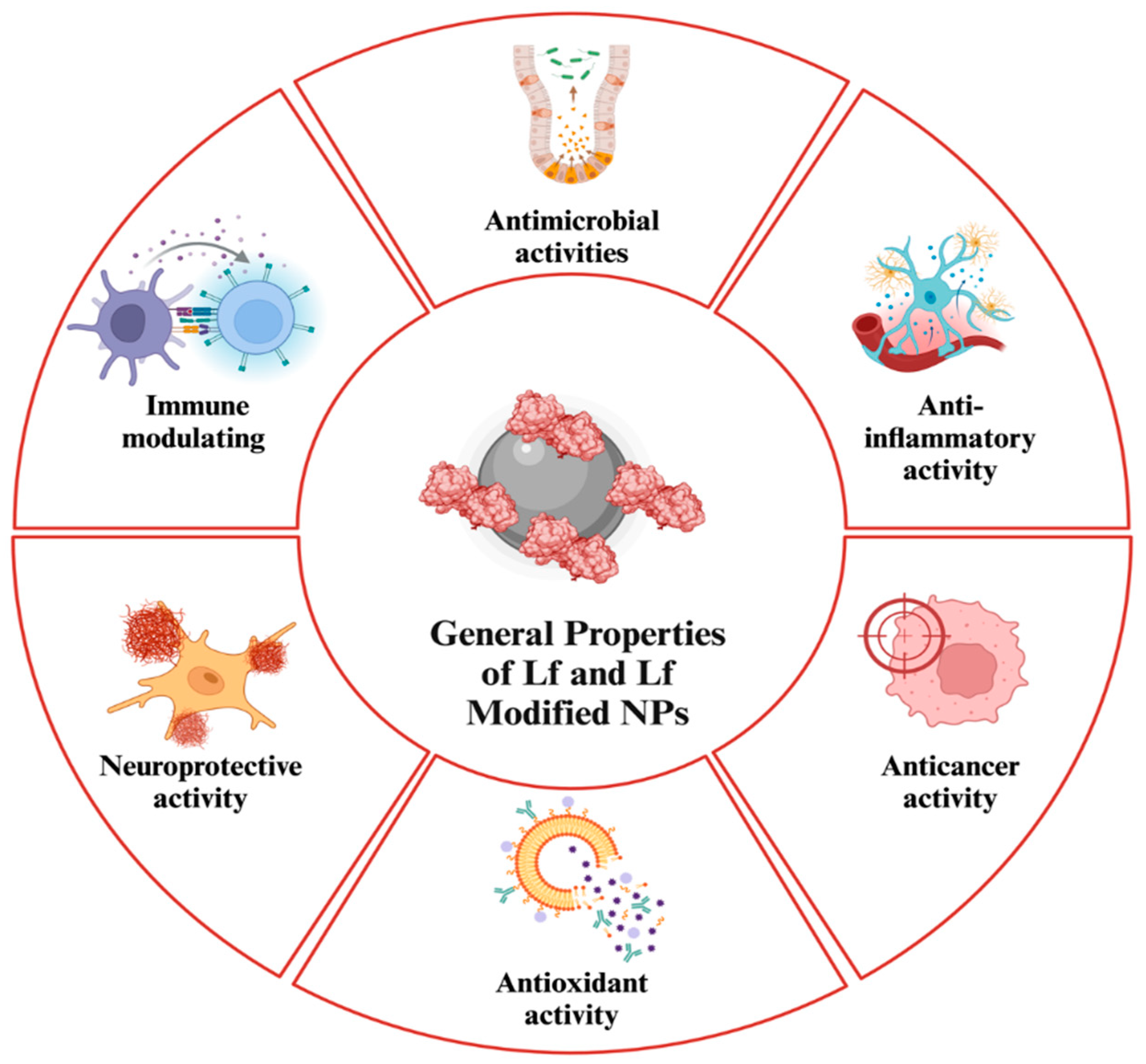
Figure 2.
General Mechanism of Drug Delivery of Lf NPs [65].
Figure 2.
General Mechanism of Drug Delivery of Lf NPs [65].
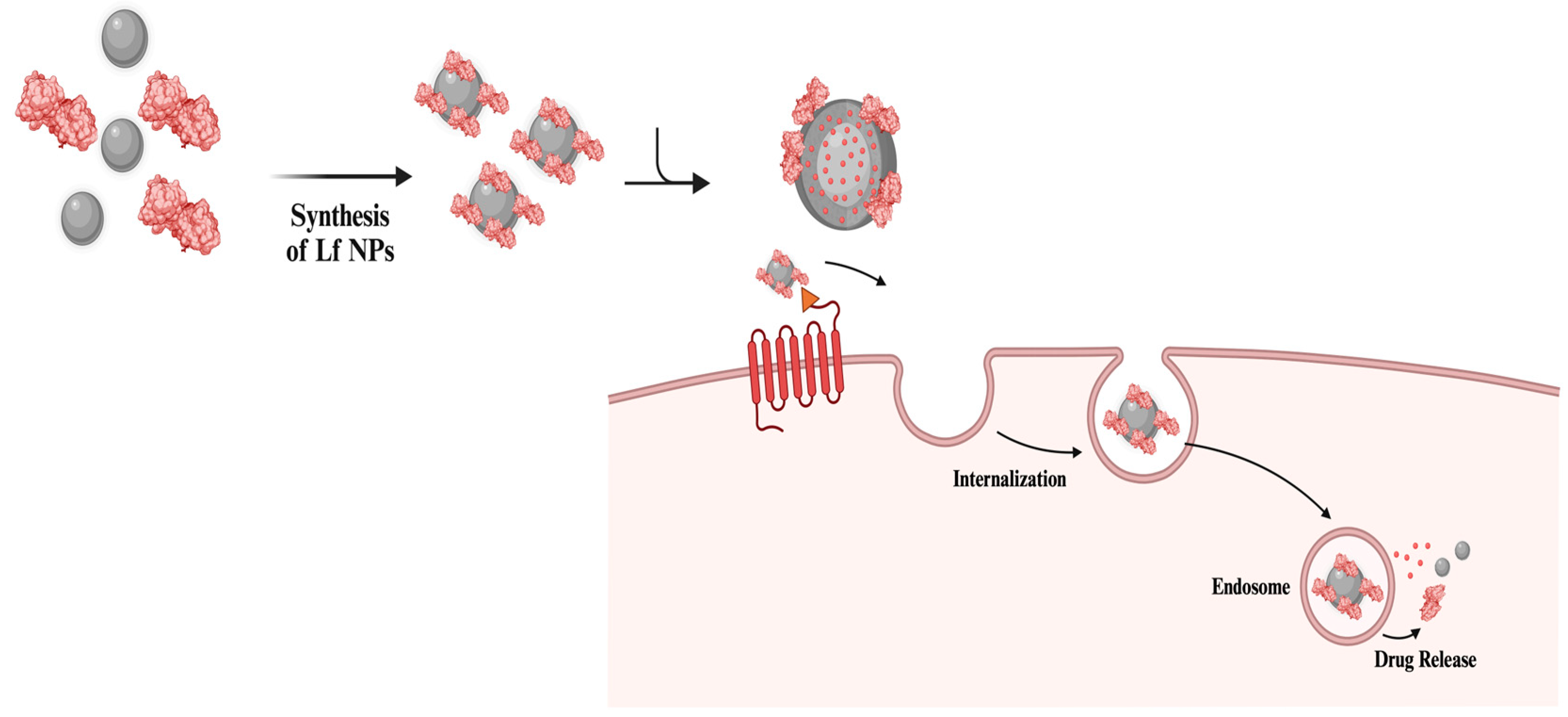
Figure 5.
Potential toxicity of NPs and Lf. The toxicity capacity of NPs depends on not only the type of the NPs, but also the exposure time, concentration and physicochemical properties, specifically their size. The high concentration and recombinantly produced Lf can cause potential adverse effects on the immune system. As a result, similar to most NP-based applications, following toxicity mechanisms can be observed in Lf-based NP applications: ROS-mediated DNA damage, protein denaturation, lipid peroxidation and mitochondrial dysfunction [28,162].
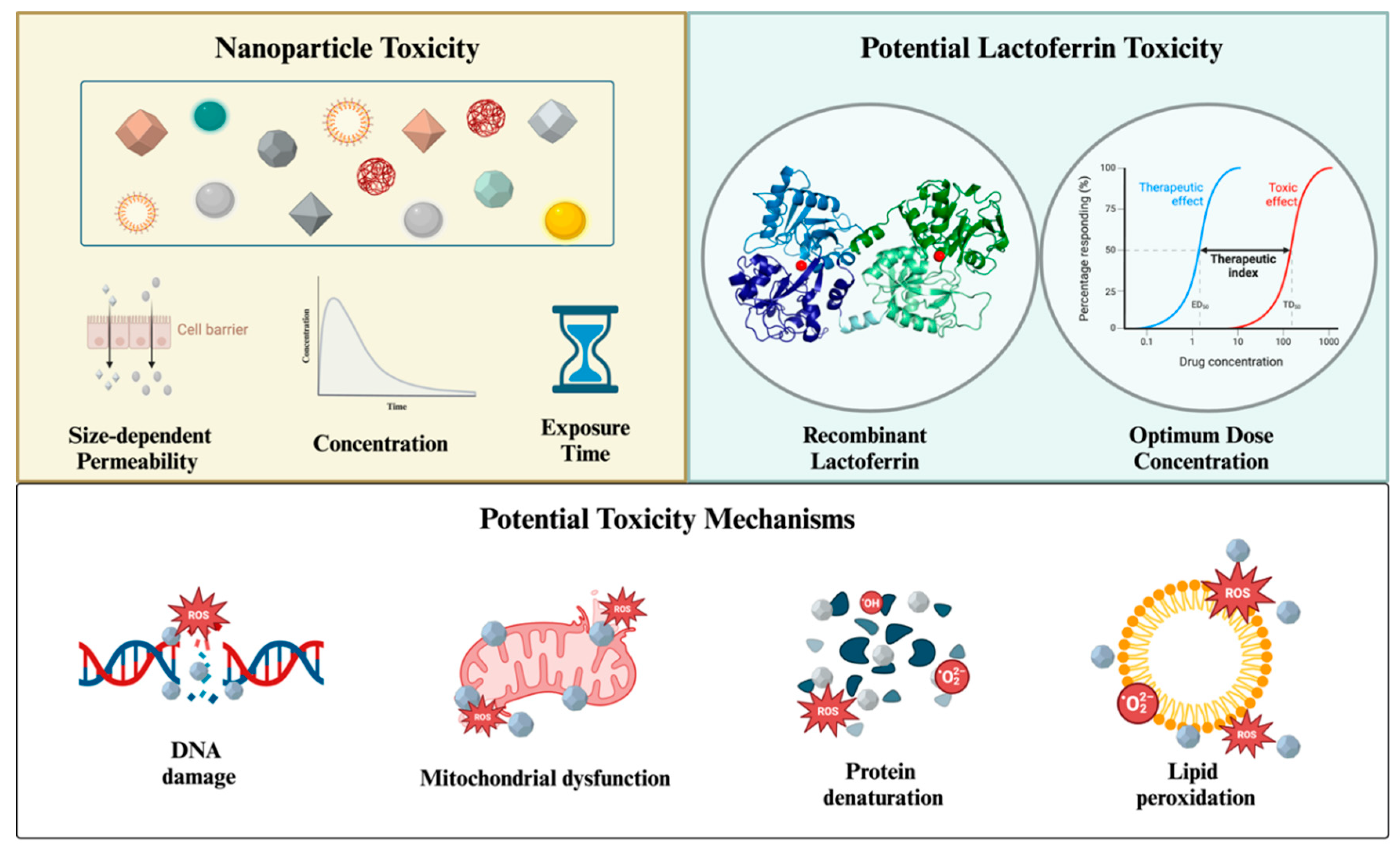
Disclaimer/Publisher’s Note: The statements, opinions and data contained in all publications are solely those of the individual author(s) and contributor(s) and not of MDPI and/or the editor(s). MDPI and/or the editor(s) disclaim responsibility for any injury to people or property resulting from any ideas, methods, instructions or products referred to in the content. |
© 2024 by the authors. Licensee MDPI, Basel, Switzerland. This article is an open access article distributed under the terms and conditions of the Creative Commons Attribution (CC BY) license (http://creativecommons.org/licenses/by/4.0/).
Copyright: This open access article is published under a Creative Commons CC BY 4.0 license, which permit the free download, distribution, and reuse, provided that the author and preprint are cited in any reuse.


