
Submitted:
24 November 2024
Posted:
25 November 2024
You are already at the latest version
Abstract
In the last years, awareness has grown regarding micro-nanoplastics (MNPs) effects on human health. Despite a large body of evidence about the origin and distribution of MNPs in the environment, knowledge regarding their impact on human health remains limited. In this context, there is a significant need to address the potential carcinogenic risk of MNPs, both intrinsic and mediated by their ability to transport carcinogenic chemicals. Currently, evidence of carcinogenicity of MNPs is scarce and heterogeneous, but the reported increase in the incidence of malignant tumors among younger populations, together with the increasing environmental abundance of MNPs, is rising a global concern about the possible role of MNPs in the induction and promotion of cancer. In this review, we aim to provide an overview of the current evidence regarding MNPs in terms of eco-toxicological evidence, methods for identification and characterization of environmental particulate, and health-associated risks, particularly for cancer development. In addition, we will suggest possible routes for future research in order to unravel the carcinogenetic potential of MNP exposure and to dissect prognostic and preventive implications of intratumoral MNPs.
Keywords:
microplastics
; nanoplastics
; ecotoxicology
; tumor
; cancer
; carcinogenic
; carcinogenicity
; cancerogenesis
; cancerogenicity
1. Introduction
Pollution is a main environmental cause of disease in the world [1]. In recent years, concerns on plastic pollution in seas stimulated multiple researches that reported evidence of widespread presence of micro-nanoplastics (MNPs) both in marine and terrestrial environments [2,3,4,5]. Historically, the rise of plastics began in the mid-1900s with the large-scale extraction and utilization of oil-based products. Plastic is a synthetic, carbon-based class of different polymers developed through the manipulation of oil components. Plastics revolutionized modern industry by replacing several natural materials that were previously used, with a continuous expansion since the 1960s and a 20-fold increase in its production in 50 years [ref]. Nonetheless, the ubiquitous diffusion of plastics utilization led to severe environmental consequences, as a great amount of mismanaged plastics has been abandoned in the environment. Over time, environmental weathering processes such as ultraviolet radiation (UV photo-oxidation), wind, and sea waves can cause plastic items (macroplastics) to progressively fragment into microscopic particles [6]. The formation of this particulate, defined as MNP particles, constitute an environmental debris that permeate the terrestrial and marine environments and that infiltrated multiple ecosystems. It also recirculates in the water cycle through atmosphere suspension, increasing diffusion, interaction with living beings and humans and continuous ultra-fragmentation via continuous recirculation across biologic systems [7] (see Figure 1).
Microplastics (MPs) and nanoplastics (NPs), overall recognized as MNP debris, are already known as environmental pollutants, although definitive conclusions on their impacts are yet to be drawn. MPs are defined as plastic particles ranging between 1 millimetre and 1 micrometre, while NPs range between 1 micrometre and 1 nanometre [8]. Plastic production and mismanagement exponentially increased during the last decades, paralleled by health-risk concerns related to the increasing detection of MNPs in the environment [9]. In 2019 the Scientific Advice Mechanism Group of Scientific Advisors provided the European Commission with a Scientific Opinion on microplastics, concluding that, while MNP pollution does not currently represent a widespread risk, stable emissions at the present rate will potentially cause serious damages within a century, warranting the introduction of preventive measures [10]. Increasing evidence points towards a possible role of MNPs in the etiology of multiple human disease, including cancer [11]. At present, there is still lack of sound epidemiological data to support the association between MNPs and cancer, and MNPs have not been classified for their carcinogenic potential by the International Agency for Research on Cancer (Agents classified by the IARC Monographs, Volumes 1–136)[12]. However, multiple preclinical and clinical studies have recently pointed to a potential link between MNP exposure and cancer development. In this narrative review, we will offer an overall prospect of the current available literature, offering general considerations about eco-toxicology, risks for human health and potential carcinogenic effects of MNPs, providing a future perspective on human health research from a medical oncologist point-of-view.
2. Micro-Nanoplastics: Definition and Eco-Toxicology
2.1. Definitions and General Considerations
MNP debris is a highly complex entity, originating from multiple sources. Primary MNPs are manufactured as such for specific purposes, such as plastic micro-beads added to cosmetics; conversely, secondary MNPs originate from mismanaged environmental macroplastics [13]. Secondary MNPs represent the greatest bulk of environmental MNPs, originating from the abrasion or degradation of multiple plastic-based materials, such as synthetic textiles and clothing, car-tyres, plastic coatings and paints, as well as other materials used for insulation that contribute to city dust, in addition to abrasion of marine vessels and fishing gear [13]. Despite this, a quantitative estimate of the proportional contribution of different sources to MNPs spread into the environment is a current challenge, owing to the lack of reliable methods to measure MNPs and to track their origin [13]. Similarly, while MNPs have been found in all environments – including seas, freshwater and land – and are known to recirculate between them, there is no full understanding of their whole “life-cycle” [13]. However, data suggest that the increasing prevalence of plastic use in multiple aspects of daily life in the last decades has been paralleled by a progressive spread of MNPs in the environment. Multiple sources of human exposure have been demonstrated: such as via inhalation of particulate matter and other airborne particles, via exposure to beauty and human care products, and via consumption of contaminated foods and beverages [11,13,14,15,16,17]. Indeed, MNPs have been isolated across most marine environments – both in the abyss and on the coastline, accumulating on the surface of sandy beaches – and in freshwaters, the latter with proportional concentrations in correlation with intensity of human activities in the area [18,19,20,21,22,23,24,25]. MNPs have also been detected in soils across most Continents, both in agricultural fields and in areas not subject to human activities, probably coming mainly from fertilizers and sewage debris, although the study of MNPs in soils is largely hampered by technical limitations in MNP isolation [3,26,27,28,29,30]. Moreover, MNPs are present both in indoor and outdoor air, with 2-fold higher concentrations reported for the former, and higher concentrations in urban environments [31,32,33,34,35,36]. Finally, wastewaters of urban areas are largely contaminated with MNPs – coming from domestic sources, such as textiles and cosmetics, and to a less known extent from industrial sources – and enter thereby aquatic ecosystems [37,38,39,40]. Considering such a widespread diffusion of MNPs, it is not surprising that they have been largely isolated also in water and food intended for human consumption. In fact, MNPs have been isolated both in bottled and tap drinking water, as well as in beer, milk, honey, rice, sea salt and seafood and fish, while some sources point to a possible uptake of MNPs via crops contamination from soils or fertilizers [30,41].
While these data seem to outline an alarming scenario, it should be emphasized that they have been gathered from scattered independent studies focusing on single geographical areas or on single dietary items, providing a highly fragmented picture of the problem. For example, a minority of studies on MNPs diffusion in freshwater come from Asia, while South America and Africa have been hardly considered at all, leaving significant knowledge gaps about some major geographical areas [13]. As a matter of fact, both the Scientific Opinion provided to the European Commission in 2019 and the more recent report on dietary sources of MNPs issued by the World Health Organization in 2022 highlighted the limited amount of available data and the questionable quality of most of these studies, concluding that available evidence is still too limited to draw conclusions about human MNP exposure from environmental sources and to assess related risks [13,41]. However, it is known that persistence and recirculation of MNPs in the environment cause the formation of a biochemical layered structure called eco-corona [42]. These layers develop when MNPs encounter environmental natural organic matter, generally organic polymeric substances such as DNA, proteins, carbohydrates and humus substances, through chemical interactions as hydrogen bonds, Van der Waals forces, and hydrophobic interactions. The eco-corona can enrich the spectrum of bio-physic-chemical interactions between MNPs and living organisms [43]. Given the nature of eco-coronated MNPs, these strucutres account for additional possible biological hazards of MNP debris, such as formation of micro-organism colonies on the surface, entrapment of toxic chemicals and release of toxic additives [13,44,45]. Thus, predicting MNPs effect in different environmental and biologic systems, as well as providing a general risk assessment, is extremely complex according to current knowledge [13].
2.2. Routes of Human Exposure
MNPs can enter the human body primarily through ingestion, inhalation and dermal absorption.
2.2.1. Ingestion
MNPs have been found in drinking water, food such as fish and shellfish, table salt and in other numerous foods. When ingested, these particles accumulate in the gastrointestinal tract lumen, where they adhere to the mucous layer. Major size particles interact with M-cells, while particles smaller than 50 µm can be absorbed through enterocytes. Moreover, small particles of various size can cross intercellular spaces [11,46,47]. Penetrating the mucosal layer, MNPs can reach the bloodstream and the lymphatic system [48]. In addition, accumulation of MNPs in the digestive lumen can lead to microbiome disruption, leading to indirect systemic toxicity trough gut-organ axes perturbation [49].
2.2.2. Inhalation
airborne MNPs can be inhaled as suspended particles, especially in urban and industrial environments. Particles below 2.5 µm have a higher probability to reach alveolar sacs, depositing in lung tissue or crossing the alveolar-capillary barrier, entering the bloodstream. Alternatively, MNPs are engulfed by alveolar macrophages. Moreover, airborne MNPs can be absorbed through the olfactory bulb, potentially reaching the central nervous system [50].
2.2.3. Dermal Absorption
airborne MNPs can be absorbed through dermal contact. Indeed, in vitro evidence of active micropinocytosis by keratinocytes of keratin-coated MNPs suggests that skin could represent a locus minoris resistentiae through which MNP particles can penetrate the body [51].
3. MNPs: Analytical Methods in Biological Studies
To date, methods of detection, identification and characterization of MNPs in biological studies are heterogeneous, and analytical processes are far from being standardized. Differences in MNPs size definitions and limitations in analytical power hamper the reproducibility and reliability of results, and methods need to be refined. Currently, different approaches could be informative of diverse characteristics of MNPs: for instance cell models to describe cellular uptake and intracellular localization, animal models in order to dissect bioaccumulation tendencies and systemic and local effects, in vivo imaging models to track MNP translocations in blood circulation and tissue penetration, and finally ex vivo studies to earn real-life information. Here we provide an overview of analytical methods used to characterize the biology of MNPs.
3.1. Surrogate Model Studies
MNPs can primarily recirculate through the bloodstream and lymphatic system, potentially reaching various organs and tissues. To date, human body circulation, distribution, and tissue accumulation are estimated mainly using surrogate in vivo animal models, in vitro cell cultures, and computational models. In vivo animal studies are used to evaluate how MNPs enter the body through ingestion or inhalation. The particles can be tracked using fluorescent dyes, allowing researchers to observe their distribution in various organs like the lungs, liver, and kidneys [52]. On the other hand, cell cultures give a significant opportunity to examine direct interaction of MNPs and human cells. These studies can elucidate different mechanisms of active cell uptake and intracellular processes of MNPs [53]. In recent years, computational models have gained attention for their capability to predict biological and chemical behaviors. Simulations of MNP distribution throughout the body offer reliable predictions based on available data. These models could inspire the design of more informative future studies that will help to dissect MNP recirculation and accumulation in human tissues.
3.2. In Vivo Imaging Techniques
Magnetic Resonance Imaging (MRI) and Positron Emission Tomography (PET) are used to monitor the distribution of traceable MNPs in the body in real-time. PET ability to provide real-time imaging enables to track MNP passage through the bloodstream, absorption by organs, and tissue accumulation. This helps distinguish between acute (short-term) and chronic (long-term) exposures, assessing timings of MNP persistence in the body, sites of primary accumulation, and main ways of elimination [54]. Another recent method that aims to dissect MNP interactions with cells and tissues is fluorescent labelling, a technique that can track real-time MNP movements [52].
3.3. EX Vivo Studies
Techniques like electron microscopy, fluorescence, Raman spectroscopy and Fourier-transform infrared (FTIR) spectroscopy are employed to visualize and identify MNPs in ex-vivo biological tissues, enabling direct observation of the particles within organs [55,56,57,58]. FTIR spectroscopy and Raman spectroscopy are vibrational spectroscopy methods widely used to identify MNPs in human tissues [59,60,61]. They can assess polymer type, size, shape, and distribution. FTIR spectroscopy is particularly useful to analyse particles larger than 10 µm, while Raman spectroscopy can be used for even smaller particles, down to 1.2 µm. Electron microscopy, as scanning electron microscopy (SEM) and transmission electron microscopy (TEM) are employed to visualize and measure nanoplastics in ex-vivo samples [58]. These methods provide detailed images of particles, allowing researchers to assess their morphology and distribution at the cellular level.
4. Human Health-Risk of MNP Exposure
Evidence is accumulating regarding MNP-related health risks, suggesting multiple molecular mechanisms as drivers, mainly through induced persistent chronic inflammation, oxidative stress, and disruption of other physiological processes. MNPs can induce a variety of cytotoxic effects, mainly through reactive oxygen species (ROS) generation, which can lead to oxidative stress, inflammation, and cellular damage [62]. ROS play a significant role in the disruption of cellular homeostasis, contributing to inflammatory responses by activating multiple molecular pathways [63]. Excessive production of ROS causes oxidative damage to lipids, proteins, and DNA [64]. Studies in both in vitro and in vivo models have shown that exposure to MNPs significantly increases ROS levels, leading to cellular dysfunction and apoptosis [65,66,67]. Indeed, the imbalance between ROS production and the antioxidant defense system can activate various signalling pathways associated with inflammation, such as the nuclear factor-kappa B (NF-κB) pathway [68]. This pathway is known to regulate the expression of pro-inflammatory cytokines and can be activated in response to cellular stress caused by MNPs [69]. Prolonged activation of inflammatory pathways may lead to chronic inflammation, which has been linked to a range of pathological conditions, including fibrosis and tissue damage [70]. In addition, MNPs can perturb mitochondrial metabolism, until apoptosis occurs [71]. Furthermore, research showed that MNPs can interfere with autophagy, causing potentially harmful cell damages [72,73,74].
4.1. Digestive Health
Ingested MNPs tend to be processed by stomach and gut secretion, going through progressive fragmentation [75]. Systemic toxicity of ingested MNPs is mainly related to the disruption of the gut microbiome, leading to perturbation of gut-organ axes and of systemic immunological modulation [49]. Locally, MNPs accumulation in mucosal cells and lymphoid-associated structures of digestive tissue can induce chronic inflammation and direct cell death [76].
4.2. Respiratory Health
Evidence regarding lung toxicity of MNPs in human mainly derives from occupational exposure studies. Workers of the plastic industry exposed to plastic flocks have a reportedly higher incidence of pulmonary fibrosis and pneumoconiosis [77,78,79,80]. Preclinical models showed that persistent alveolar macrophage activation and alveolar cell damage are the main underlying mechanisms [81].
4.3. Cardiovascular Health
Recent studies have linked MNPs to cardiovascular diseases. Rats exposed to polystyrene MNPs at incremental environmental concentrations developed proportional grades of cardiac fibrosis through oxidative stress mechanisms [82]. In another mouse model study, inhaled NPs accumulated preferentially in inflamed vascular lesions [83]. A recent pivotal prospective analysis of resected atheromas from carotid endarterectomies showed that the presence of MNPs in macrophages or in the plaques is a negative predictive factor for secondary cardiovascular events and mortality. This was the first study to indirectly prove that MNPs could induce or foster inflammation in humans [58].
5. MNPs’ Role in Cancer Development and Progression
The current evidence on the potential role of MNPs in cancer initiation and progression emerge from mainly preclinical studies of in vitro models. These studies have focused on the capability of MNPs to elicit a number of different biological effects potentially linked to carcinogenesis. Key biological processes that influence the induction and promotion of carcinogenesis include tumor-promoting inflammation, oxidative stress, alterations in cellular metabolism, genotoxicity, cytotoxicity, induction of invasive cellular phenotypes, and changes in the local microbiota [84].
5.1. Tumor-Promoting Inflammation
A body of studies assessed MNPs capability of causing a sustained inflammatory response, as a surrogate of their carcinogenicity potential, according to the hypothetic carcinogenetic capacity of chronic inflammation in mammals. Human cancer cell lines significantly increased their production of inflammatory cytokines when exposed to polystyrene MNPs [85,86,87,88]. In vivo evidence in mammalian models confirmed the capacity of polystyrene MNPs to induce variegated mucosal inflammatory response [89,90,91,92,93].
5.2. Cancerogenesis Initiation
Different types of alterations in determined cellular processes could initiate the multi-step process of carcinogenesis, generally through loss of apoptotic capacity and anchorage-dependent growth inhibition. Regarding the latter, previous studies demonstrated that exposure of human cancer cell lines to MNPs can lead to anchorage-independent outgrow [94,95]. Immune surveillance is another fundamental mechanism in the prevention of cancerogenesis initiation in mammals. Regarding this, MNPs have shown the potential to polarize immune responses in pregnant mice towards immune tolerance, through a decrease of NK cells and M2 polarization of macrophages [96]. In a similar manner, when Type-1-diabetic mice were exposed to MNPs, apoptosis of CD4+ T-lymphocyte increased in favor of an expansion of Tregs and complete reversion of autoimmune response [97]. In addition, MNPs showed genotoxic potential in human blood cells and mammalian cell models in vitro [98,99,100,101].
5.3. Cancerogenesis Promotion
Once pre-cancerous cells gained immortalization and escaped the immune system, they need to acquire determined genetic mutations in order to progress in the carcinogenesis process. This phase, called “promotion”, involves a series of alterations in cell proliferation and invasiveness that ultimately result in the development of invasive disease. Different cellular effects of MNP exposure are related to the dysregulation of cell proliferation [85,94,95,102], potentially fostering the development of cancer. Moreover, in vitro models showed that MNP exposure can induce epithelial-mesenchymal transition in human cell lines [103,104,105,106], a process that enhances invasiveness and migration capability of cancer cells.
5.4. MNPs’ Presence in Cancer Tissue
To date, available studies showed that MNPs in human cancer tissues are generally more abundant compared to non-cancerous tissues. In an historical study, MNPs were more commonly detected in lung cancer compared to normal lung tissue [107]. In a more recent case-control study, MNPs were significantly more concentrated in colorectal cancer tissue compared to non-tumoral tissues [108]. In another recent study, the abundance of MNPs in tumor and para-tumoral tissues of human prostate cancer were compared, showing that particles were significantly more concentrated in cancer [109]. Underlying mechanisms should be unravelled, and realistic hypotheses could be hard to propose. Since MNPs can be inherited by cell division and actively absorbed by cancer cells, the higher concentration in tumor tissue could be related to a higher absorption rate of circulating and tissue MNPs. Common alterations in the intratumoral circulation and microenvironment could be responsible for an increased diffusion of MNPs within the tumor, together with a possible fostered pinocytosis of cancer cells. In this scenario, the abundance of MNPs in the tumor tissue is merely representative of their specific bioaccumulation tendencies and overall individual exposure. On the other hand, MNPs’ capability to foster carcinogenic events could influence clonal evolution, favoring the proliferation of cells with more effective pinocytosis processes. Extensive characterizations of MNP presence and concentration in tumor, para-tumoral and normal tissues are needed in order to solve these interpretation issues.
6. Discussion
The increasing abundance of environmental MNPs and their capability to recirculate, infiltrate and deposit into various ecosystems, including the human body, has raised significant concerns about their long-term health effects in the recent years. Although the related body of evidence has progressively expanded in the last decade, knowledge about MNP-related health risks, especially as a carcinogen, remains limited to date. Researchers have pointed out a potential to foster inflammation and oxidative stress in living beings and humans, indicating a hypothetical role in cancer initiation and progression. Fragmented and rather initial evidence suggests that MNPs can trigger several biological processes that are strongly linked to carcinogenesis, as tumor-promoting inflammation, oxidative stress and ROS production, direct and indirect genotoxicity (being MNPs carriers of genotoxic chemicals), alterations in immune response, and disruption of gut microbiota (Figure 2). On the other hand, there is an important lack of large-scale epidemiological studies evaluating the impact of human MNP exposure on cancer incidence and mortality. In fact, the IARC has not ever mentioned MNPs as a potential carcinogen for humans, and most specific plastic polymers that have the potential to give origin to secondary MNPs have been listed in group 3, i.e. “not classifiable as to its carcinogenicity to humans”, according to IARC monographs (see https://monographs.iarc.who.int/list-of-classifications).
Current basic research is hampered by the incomplete refinement of analytical methods, and heterogeneity of MNPs in their sizes, shapes, polymer structure, chemical properties, nature and composition of the eco-corona further complicate experimental result interpretations. At the eco-toxicological level, human exposure potentially depends on geographical location, weather and soil properties, density of MNP emissions, type of diet and daily habits. Reasonably, dissecting the abundance of different MNPs in indoor environments could refine MNP exposure estimation.
At the current state of knowledge, health risks related to MNP exposure, in particular as a potential carcinogen, remain poorly understood. Preclinical evidence showed major biological effects of MNP exposure, but dose expositions are significantly higher in experimental models compared to real-life cellular exposures, thus results are seldom reliable.
In our view, in order to expand the knowledge regarding MNPs effects, some major areas of further development can be implemented:
- First, standardization in analytical methods for MNP detection in biological specimens will be essential to achieve reproducible and reliable results, and to provide the basis for building consistent evidence from different scientific groups.
- Studies employing mammalian model should evaluate MNP bioaccumulation in tissues over time, thanks to innovative in vivo techniques, as well as unravel the real genotoxic potential of MNPs. In this regard, human organ-specific models could further elucidate tissue-specific effects, and provide information on risks specific to different routes of exposure.
- Translational research should effortlessly characterize the presence, distribution and abundance of MNPs in normal and cancer tissues, aiming at investigating potential prognostic and molecular implications. In the near future, the integration of advanced analytical techniques, computational tools, and multi-omics approaches could unravel MNPs role as a carcinogen.
- At a more complex level, MNPs could have a range of interactions with fundamental body-wide entities, such as microbiota, systemic immunity, and nervous signals, which have to be further elucidated.
- To date, there is an urgent need of robust epidemiological data evaluating the correlation of MNPs exposure and disease development at a population level. Longitudinal epidemiologic studies should determine the cancer risk for populations at higher probability of MNP exposure, as in industrial and metropolitan areas or in coastal locations. Moreover, epidemiological data would be extremely valuable in order to generate hypotheses in basic science and further elucidate the underlying mechanisms.
5. Conclusions
In the last years, MNPs raised public health concerns since their ubiquitous nature as environmental pollutants. Currently, the scarcity of knowledge regarding MNP-related health risks hampers the development of regulatory policies and public health strategies aimed at controlling human MNP exposure.
In the field of oncology, many significant advances were achieved in the last decades, especially in the characterization of cancer (epi)genic, metabolic, immunological and microenvironmental features. Meanwhile, environmental and behavioral causative factors are rarely identified, since the complex interplay between different risk factors in multifactorial carcinogenesis. Indeed, in recent years, epidemiological data showed a potential increase in early onset cancers [110,111], and this could be paired with the potentially increased human MNP exposure for new generations. For these reasons, the scientific community will have to put intensive research efforts in order to unravel the real carcinogenic role of MNPs. This knowledge will be fundamental to inform global regulatory ecological and health policies.
Author Contributions
conceptualization: LR, OA, CM; methodology: LR, OA, CM; data curation: LR, OA, CM; writing—original draft preparation: LR, OA, CM; writing—review and editing: NLV, DD, MSC, AG, MN, ER; visualization: LR, OA; supervision, NLV, ER; project administration: NLV. All authors have read and agreed to the published version of the manuscript.
Funding
this research received no external funding.
Acknowledgments
we thank Joanna Landi for her technical support. Figure 1 created in BioRender. Ruggieri, L. (2024) https://BioRender.com/h88g872. Figure 2 was created in BioRender. Ruggieri, L. (2024) https://BioRender.com/u69b909.
Conflicts of Interest
the authors declare no conflicts of interest.
References
- Landrigan, P.J.; Fuller, R.; Acosta, N.J.R.; Adeyi, O.; Arnold, R.; Basu, N. (Nil); Baldé, A.B.; Bertollini, R.; Bose-O’Reilly, S.; Boufford, J.I.; et al. The Lancet Commission on Pollution and Health. The Lancet 2018, 391, 462–512. [CrossRef]
- Thompson, R.C.; Olsen, Y.; Mitchell, R.P.; Davis, A.; Rowland, S.J.; John, A.W.G.; McGonigle, D.; Russell, A.E. Lost at Sea: Where Is All the Plastic? Science 2004, 304, 838–838. [Google Scholar] [CrossRef] [PubMed]
- He, D.; Bristow, K.; Filipović, V.; Lv, J.; He, H. Microplastics in Terrestrial Ecosystems: A Scientometric Analysis. Sustainability 2020, 12, 8739. [Google Scholar] [CrossRef]
- Debnath, R.; Prasad, G.S.; Amin, A.; Malik, M.M.; Ahmad, I.; Abubakr, A.; Borah, S.; Rather, M.A.; Impellitteri, F.; Tabassum, I.; et al. Understanding and Addressing Microplastic Pollution: Impacts, Mitigation, and Future Perspectives. Journal of Contaminant Hydrology 2024, 266, 104399. [Google Scholar] [CrossRef] [PubMed]
- Osman, A.I.; Hosny, M.; Eltaweil, A.S.; Omar, S.; Elgarahy, A.M.; Farghali, M.; Yap, P.-S.; Wu, Y.-S.; Nagandran, S.; Batumalaie, K.; et al. Microplastic Sources, Formation, Toxicity and Remediation: A Review. Environ Chem Lett 2023, 21, 2129–2169. [Google Scholar] [CrossRef]
- Thompson, R.C.; Courtene-Jones, W.; Boucher, J.; Pahl, S.; Raubenheimer, K.; Koelmans, A.A. Twenty Years of Microplastics Pollution Research—What Have We Learned? Science 2024, 0, eadl2746. [Google Scholar] [CrossRef]
- Allen, D.; Allen, S.; Abbasi, S.; Baker, A.; Bergmann, M.; Brahney, J.; Butler, T.; Duce, R.A.; Eckhardt, S.; Evangeliou, N.; et al. Microplastics and Nanoplastics in the Marine-Atmosphere Environment. Nat Rev Earth Environ 2022, 3, 393–405. [Google Scholar] [CrossRef]
- Koelmans, A.A.; Redondo-Hasselerharm, P.E.; Nor, N.H.M.; de Ruijter, V.N.; Mintenig, S.M.; Kooi, M. Risk Assessment of Microplastic Particles. Nat Rev Mater 2022, 7, 138–152. [Google Scholar] [CrossRef]
- Jambeck, J.R.; Geyer, R.; Wilcox, C.; Siegler, T.R.; Perryman, M.; Andrady, A.; Narayan, R.; Law, K.L. Plastic Waste Inputs from Land into the Ocean. Science 2015, 347, 768–771. [Google Scholar] [CrossRef]
- European Commission, Directorate-General for Research and Innovation Environmental and Health Risks of Microplastic Pollution 2019.
- Krause, S.; Ouellet, V.; Allen, D.; Allen, S.; Moss, K.; Nel, H.A.; Manaseki-Holland, S.; Lynch, I. The Potential of Micro- and Nanoplastics to Exacerbate the Health Impacts and Global Burden of Non-Communicable Diseases. Cell Reports Medicine 2024, 5, 101581. [Google Scholar] [CrossRef]
- IARC, List of Classifications. Available online: https://monographs.iarc.who.int/list-of-classifications (accessed on 27 July 2024).
- SAPEA A Scientific Perspective on Microplastics in Nature and Society: Evidence Review Report (1.1).; SAPEA, 2019; ISBN 978-3-9820301-0-4.
- Geyer, R.; Jambeck, J.R.; Law, K.L. Production, Use, and Fate of All Plastics Ever Made. Sci. Adv. 2017, 3, e1700782. [Google Scholar] [CrossRef]
- Ostle, C.; Thompson, R.C.; Broughton, D.; Gregory, L.; Wootton, M.; Johns, D.G. The Rise in Ocean Plastics Evidenced from a 60-Year Time Series. Nat Commun 2019, 10, 1622. [Google Scholar] [CrossRef] [PubMed]
- Borrelle, S.B.; Ringma, J.; Law, K.L.; Monnahan, C.C.; Lebreton, L.; McGivern, A.; Murphy, E.; Jambeck, J.; Leonard, G.H.; Hilleary, M.A.; et al. Predicted Growth in Plastic Waste Exceeds Efforts to Mitigate Plastic Pollution. Science 2020, 369, 1515–1518. [Google Scholar] [CrossRef] [PubMed]
- Ageel, H.K.; Harrad, S.; Abdallah, M.A.-E. Occurrence, Human Exposure, and Risk of Microplastics in the Indoor Environment. Environ. Sci.: Processes Impacts 2022, 24, 17–31. [Google Scholar] [CrossRef] [PubMed]
- Arthur, Courtney; Baker, Joel E; Bamford, Holly A Proceedings of the International Research Workshop on the Occurrence, Effects and Fate of Microplastic Marine Debris.; 2009.
- Woodall, L.C.; Sanchez-Vidal, A.; Canals, M.; Paterson, G.L.J.; Coppock, R.; Sleight, V.; Calafat, A.; Rogers, A.D.; Narayanaswamy, B.E.; Thompson, R.C. The Deep Sea Is a Major Sink for Microplastic Debris. R. Soc. open sci. 2014, 1, 140317. [Google Scholar] [CrossRef] [PubMed]
- Eriksen, M.; Lebreton, L.C.M.; Carson, H.S.; Thiel, M.; Moore, C.J.; Borerro, J.C.; Galgani, F.; Ryan, P.G.; Reisser, J. Plastic Pollution in the World’s Oceans: More than 5 Trillion Plastic Pieces Weighing over 250,000 Tons Afloat at Sea. PLoS ONE 2014, 9, e111913. [Google Scholar] [CrossRef]
- Van Sebille, E.; Wilcox, C.; Lebreton, L.; Maximenko, N.; Hardesty, B.D.; Van Franeker, J.A.; Eriksen, M.; Siegel, D.; Galgani, F.; Law, K.L. A Global Inventory of Small Floating Plastic Debris. Environ. Res. Lett. 2015, 10, 124006. [Google Scholar] [CrossRef]
- Lots, F.A.E.; Behrens, P.; Vijver, M.G.; Horton, A.A.; Bosker, T. A Large-Scale Investigation of Microplastic Contamination: Abundance and Characteristics of Microplastics in European Beach Sediment. Marine Pollution Bulletin 2017, 123, 219–226. [Google Scholar] [CrossRef]
- Eerkes-Medrano, D.; Thompson, R.C.; Aldridge, D.C. Microplastics in Freshwater Systems: A Review of the Emerging Threats, Identification of Knowledge Gaps and Prioritisation of Research Needs. Water Research 2015, 75, 63–82. [Google Scholar] [CrossRef]
- Li, J.; Liu, H.; Paul Chen, J. Microplastics in Freshwater Systems: A Review on Occurrence, Environmental Effects, and Methods for Microplastics Detection. Water Research 2018, 137, 362–374. [Google Scholar] [CrossRef]
- Rezania, S.; Park, J.; Md Din, M.F.; Mat Taib, S.; Talaiekhozani, A.; Kumar Yadav, K.; Kamyab, H. Microplastics Pollution in Different Aquatic Environments and Biota: A Review of Recent Studies. Marine Pollution Bulletin 2018, 133, 191–208. [Google Scholar] [CrossRef] [PubMed]
- Zubris, K.A.V.; Richards, B.K. Synthetic Fibers as an Indicator of Land Application of Sludge. Environmental Pollution 2005, 138, 201–211. [Google Scholar] [CrossRef]
- Scheurer, M.; Bigalke, M. Microplastics in Swiss Floodplain Soils. Environ. Sci. Technol. 2018, 52, 3591–3598. [Google Scholar] [CrossRef] [PubMed]
- Zhang, S.; Yang, X.; Gertsen, H.; Peters, P.; Salánki, T.; Geissen, V. A Simple Method for the Extraction and Identification of Light Density Microplastics from Soil. Science of The Total Environment 2018, 616–617, 1056–1065. [CrossRef]
- Fuller, S.; Gautam, A. A Procedure for Measuring Microplastics Using Pressurized Fluid Extraction. Environ. Sci. Technol. 2016, 50, 5774–5780. [Google Scholar] [CrossRef] [PubMed]
- Tian, L.; Jinjin, C.; Ji, R.; Ma, Y.; Yu, X. Microplastics in Agricultural Soils: Sources, Effects, and Their Fate. Current Opinion in Environmental Science & Health 2022, 25, 100311. [Google Scholar] [CrossRef]
- Dris, R.; Gasperi, J.; Rocher, V.; Saad, M.; Renault, N.; Tassin, B. Microplastic Contamination in an Urban Area: A Case Study in Greater Paris. Environ. Chem. 2015, 12, 592. [Google Scholar] [CrossRef]
- Dris, R.; Gasperi, J.; Mirande, C.; Mandin, C.; Guerrouache, M.; Langlois, V.; Tassin, B. A First Overview of Textile Fibers, Including Microplastics, in Indoor and Outdoor Environments. Environmental Pollution 2017, 221, 453–458. [Google Scholar] [CrossRef]
- Gasperi, J.; Wright, S.L.; Dris, R.; Collard, F.; Mandin, C.; Guerrouache, M.; Langlois, V.; Kelly, F.J.; Tassin, B. Microplastics in Air: Are We Breathing It In? Current Opinion in Environmental Science & Health 2018, 1, 1–5. [Google Scholar] [CrossRef]
- Chen, Y.; Li, X.; Zhang, X.; Zhang, Y.; Gao, W.; Wang, R.; He, D. Air Conditioner Filters Become Sinks and Sources of Indoor Microplastics Fibers. Environmental Pollution 2022, 292, 118465. [Google Scholar] [CrossRef]
- Brahney, J.; Mahowald, N.; Prank, M.; Cornwell, G.; Klimont, Z.; Matsui, H.; Prather, K.A. Constraining the Atmospheric Limb of the Plastic Cycle. Proc. Natl. Acad. Sci. U.S.A. 2021, 118, e2020719118. [Google Scholar] [CrossRef]
- Boccia, P.; Mondellini, S.; Mauro, S.; Zanellato, M.; Parolini, M.; Sturchio, E. Potential Effects of Environmental and Occupational Exposure to Microplastics: An Overview of Air Contamination. Toxics 2024, 12, 320. [Google Scholar] [CrossRef] [PubMed]
- Koelmans, A.A.; Mohamed Nor, N.H.; Hermsen, E.; Kooi, M.; Mintenig, S.M.; De France, J. Microplastics in Freshwaters and Drinking Water: Critical Review and Assessment of Data Quality. Water Research 2019, 155, 410–422. [Google Scholar] [CrossRef] [PubMed]
- Murphy, F.; Ewins, C.; Carbonnier, F.; Quinn, B. Wastewater Treatment Works (WwTW) as a Source of Microplastics in the Aquatic Environment. Environ. Sci. Technol. 2016, 50, 5800–5808. [Google Scholar] [CrossRef] [PubMed]
- Prata, J.C. Microplastics in Wastewater: State of the Knowledge on Sources, Fate and Solutions. Marine Pollution Bulletin 2018, 129, 262–265. [Google Scholar] [CrossRef] [PubMed]
- Van Wezel, A.P.; Van Den Hurk, F.; Sjerps, R.M.A.; Meijers, E.M.; Roex, E.W.M.; Ter Laak, T.L. Impact of Industrial Waste Water Treatment Plants on Dutch Surface Waters and Drinking Water Sources. Science of The Total Environment 2018, 640–641, 1489–1499. [CrossRef]
- World Health Organization Dietary and Inhalation Exposure to Nano- and Microplastic Particles and Potential Implications for Human Health. 2022.
- Liu, S.; Junaid, M.; Liao, H.; Liu, X.; Wu, Y.; Wang, J. Eco-Corona Formation and Associated Ecotoxicological Impacts of Nanoplastics in the Environment. Science of The Total Environment 2022, 836, 155703. [Google Scholar] [CrossRef]
- Liu, S.; Zhang, X.; Zeng, K.; He, C.; Huang, Y.; Xin, G.; Huang, X. Insights into Eco-Corona Formation and Its Role in the Biological Effects of Nanomaterials from a Molecular Mechanisms Perspective. Science of The Total Environment 2023, 858, 159867. [Google Scholar] [CrossRef]
- Campanale, C.; Massarelli, C.; Savino, I.; Locaputo, V.; Uricchio, V.F. A Detailed Review Study on Potential Effects of Microplastics and Additives of Concern on Human Health. Int J Environ Res Public Health 2020, 17, 1212. [Google Scholar] [CrossRef]
- Molina, E.; Benedé, S. Is There Evidence of Health Risks From Exposure to Micro- and Nanoplastics in Foods? Front. Nutr. 2022, 9. [Google Scholar] [CrossRef]
- Ali, N.; Katsouli, J.; Marczylo, E.L.; Gant, T.W.; Wright, S.; Serna, J.B. de la The Potential Impacts of Micro-and-Nano Plastics on Various Organ Systems in Humans. eBioMedicine 2024, 99. [Google Scholar] [CrossRef]
- Ensign, L.M.; Cone, R.; Hanes, J. Oral Drug Delivery with Polymeric Nanoparticles: The Gastrointestinal Mucus Barriers. Advanced Drug Delivery Reviews 2012, 64, 557–570. [Google Scholar] [CrossRef]
- Dong, X.; Liu, X.; Hou, Q.; Wang, Z. From Natural Environment to Animal Tissues: A Review of Microplastics(Nanoplastics) Translocation and Hazards Studies. Science of The Total Environment 2023, 855, 158686. [Google Scholar] [CrossRef] [PubMed]
- Pan, Y.; Zhang, H.; Zhu, L.; Tan, J.; Wang, B.; Li, M. The Role of Gut Microbiota in MP/NP-Induced Toxicity. Environmental Pollution 2024, 359, 124742. [Google Scholar] [CrossRef] [PubMed]
- Amato-Lourenço, L.F.; Dantas, K.C.; Júnior, G.R.; Paes, V.R.; Ando, R.A.; de Oliveira Freitas, R.; da Costa, O.M.M.M.; Rabelo, R.S.; Soares Bispo, K.C.; Carvalho-Oliveira, R.; et al. Microplastics in the Olfactory Bulb of the Human Brain. JAMA Network Open 2024, 7, e2440018. [Google Scholar] [CrossRef] [PubMed]
- Gopinath, P.M.; Twayana, K.S.; Ravanan, P. ; John Thomas; Mukherjee, A. ; Jenkins, D.F.; Chandrasekaran, N. Prospects on the Nano-Plastic Particles Internalization and Induction of Cellular Response in Human Keratinocytes. Particle and Fibre Toxicology 2021, 18, 35. [Google Scholar] [CrossRef]
- Villacorta, A.; Cazorla-Ares, C.; Fuentes-Cebrian, V.; Valido, I.H.; Vela, L.; Carrillo-Navarrete, F.; Morataya-Reyes, M.; Mejia-Carmona, K.; Pastor, S.; Velázquez, A.; et al. Fluorescent Labeling of Micro/Nanoplastics for Biological Applications with a Focus on “true-to-Life" Tracking. Journal of Hazardous Materials 2024, 476, 135134. [Google Scholar] [CrossRef]
- Hua, X.; Wang, D. Cellular Uptake, Transport, and Organelle Response After Exposure to Microplastics and Nanoplastics: Current Knowledge and Perspectives for Environmental and Health Risks. Reviews Env.Contamination (formerly:Residue Reviews) 2022, 260, 12. [Google Scholar] [CrossRef]
- Keinänen, O.; Dayts, E.J.; Rodriguez, C.; Sarrett, S.M.; Brennan, J.M.; Sarparanta, M.; Zeglis, B.M. Harnessing PET to Track Micro- and Nanoplastics in Vivo. Sci Rep 2021, 11, 11463. [Google Scholar] [CrossRef]
- Lee, S.; Kim, D.; Kang, K.-K.; Sung, S.-E.; Choi, J.-H.; Sung, M.; Shin, C.-H.; Jeon, E.; Kim, D.; Kim, D.; et al. Toxicity and Biodistribution of Fragmented Polypropylene Microplastics in ICR Mice. International Journal of Molecular Sciences 2023, 24, 8463. [Google Scholar] [CrossRef]
- Arribas Arranz, J.; Villacorta, A.; Rubio, L.; García-Rodríguez, A.; Sánchez, G.; Llorca, M.; Farre, M.; Ferrer, J.F.; Marcos, R.; Hernández, A. Kinetics and Toxicity of Nanoplastics in Ex Vivo Exposed Human Whole Blood as a Model to Understand Their Impact on Human Health. Science of The Total Environment 2024, 948, 174725. [Google Scholar] [CrossRef]
- Braun, T.; Ehrlich, L.; Henrich, W.; Koeppel, S.; Lomako, I.; Schwabl, P.; Liebmann, B. Detection of Microplastic in Human Placenta and Meconium in a Clinical Setting. Pharmaceutics 2021, 13, 921. [Google Scholar] [CrossRef]
- Marfella, R.; Prattichizzo, F.; Sardu, C.; Fulgenzi, G.; Graciotti, L.; Spadoni, T.; D’Onofrio, N.; Scisciola, L.; Grotta, R.L.; Frigé, C.; et al. Microplastics and Nanoplastics in Atheromas and Cardiovascular Events. New England Journal of Medicine 2024, 390, 900–910. [Google Scholar] [CrossRef] [PubMed]
- Qian, N.; Gao, X.; Lang, X.; Deng, H.; Bratu, T.M.; Chen, Q.; Stapleton, P.; Yan, B.; Min, W. Rapid Single-Particle Chemical Imaging of Nanoplastics by SRS Microscopy. Proceedings of the National Academy of Sciences 2024, 121, e2300582121. [Google Scholar] [CrossRef] [PubMed]
- Ao, J.; Xu, G.; Wu, H.; Xie, L.; Liu, J.; Gong, K.; Ruan, X.; Han, J.; Li, K.; Wang, W.; et al. Fast Detection and 3D Imaging of Nanoplastics and Microplastics by Stimulated Raman Scattering Microscopy. CR-PHYS-SC 2023, 4. [Google Scholar] [CrossRef]
- Xie, J.; Gowen, A.; Xu, W.; Xu, J. Analysing Micro- and Nanoplastics with Cutting-Edge Infrared Spectroscopy Techniques: A Critical Review. Analytical Methods 2024, 16, 2177–2197. [Google Scholar] [CrossRef]
- Kadac-Czapska, K.; Ośko, J.; Knez, E.; Grembecka, M. Microplastics and Oxidative Stress—Current Problems and Prospects. Antioxidants 2024, 13, 579. [Google Scholar] [CrossRef]
- Sies, H.; Belousov, V.V.; Chandel, N.S.; Davies, M.J.; Jones, D.P.; Mann, G.E.; Murphy, M.P.; Yamamoto, M.; Winterbourn, C. Defining Roles of Specific Reactive Oxygen Species (ROS) in Cell Biology and Physiology. Nat Rev Mol Cell Biol 2022, 23, 499–515. [Google Scholar] [CrossRef]
- Das, A. The Emerging Role of Microplastics in Systemic Toxicity: Involvement of Reactive Oxygen Species (ROS). Science of The Total Environment 2023, 895, 165076. [Google Scholar] [CrossRef]
- Feng, M.; Luo, J.; Wan, Y.; Zhang, J.; Lu, C.; Wang, M.; Dai, L.; Cao, X.; Yang, X.; Wang, Y. Polystyrene Nanoplastic Exposure Induces Developmental Toxicity by Activating the Oxidative Stress Response and Base Excision Repair Pathway in Zebrafish (Danio Rerio). ACS Omega 2022, 7, 32153–32163. [Google Scholar] [CrossRef]
- Babaei, A.A.; Rafiee, M.; Khodagholi, F.; Ahmadpour, E.; Amereh, F. Nanoplastics-Induced Oxidative Stress, Antioxidant Defense, and Physiological Response in Exposed Wistar Albino Rats. Environ Sci Pollut Res 2022, 29, 11332–11344. [Google Scholar] [CrossRef]
- Sun, R.; Liu, M.; Xiong, F.; Xu, K.; Huang, J.; Liu, J.; Wang, D.; Pu, Y. Polystyrene Micro- and Nanoplastics Induce Gastric Toxicity through ROS Mediated Oxidative Stress and P62/Keap1/Nrf2 Pathway. Science of The Total Environment 2024, 912, 169228. [Google Scholar] [CrossRef]
- Lingappan, K. NF-κB in Oxidative Stress. Current Opinion in Toxicology 2018, 7, 81–86. [Google Scholar] [CrossRef] [PubMed]
- Liu, S.; Li, H.; Wang, J.; Wu, B.; Guo, X. Polystyrene Microplastics Aggravate Inflammatory Damage in Mice with Intestinal Immune Imbalance. Science of The Total Environment 2022, 833, 155198. [Google Scholar] [CrossRef] [PubMed]
- Jeong, C.-B.; Won, E.-J.; Kang, H.-M.; Lee, M.-C.; Hwang, D.-S.; Hwang, U.-K.; Zhou, B.; Souissi, S.; Lee, S.-J.; Lee, J.-S. Microplastic Size-Dependent Toxicity, Oxidative Stress Induction, and p-JNK and p-P38 Activation in the Monogonont Rotifer (Brachionus Koreanus). Environ. Sci. Technol. 2016, 50, 8849–8857. [Google Scholar] [CrossRef] [PubMed]
- Lee, S.E.; Yi, Y.; Moon, S.; Yoon, H.; Park, Y.S. Impact of Micro- and Nanoplastics on Mitochondria. Metabolites 2022, 12, 897. [Google Scholar] [CrossRef] [PubMed]
- Lu, Y.-Y.; Li, H.; Ren, H.; Zhang, X.; Huang, F.; Zhang, D.; Huang, Q.; Zhang, X. Size-Dependent Effects of Polystyrene Nanoplastics on Autophagy Response in Human Umbilical Vein Endothelial Cells. Journal of Hazardous Materials 2022, 421, 126770. [Google Scholar] [CrossRef]
- Chen, Y.-C.; Chen, K.-F.; Lin, K.-Y.A.; Chen, J.-K.; Jiang, X.-Y.; Lin, C.-H. The Nephrotoxic Potential of Polystyrene Microplastics at Realistic Environmental Concentrations. J Hazard Mater 2022, 427, 127871. [Google Scholar] [CrossRef]
- Lim, S.L.; Ng, C.T.; Zou, L.; Lu, Y.; Chen, J.; Bay, B.H.; Shen, H.-M.; Ong, C.N. Targeted Metabolomics Reveals Differential Biological Effects of Nanoplastics and nanoZnO in Human Lung Cells. Nanotoxicology 2019, 13, 1117–1132. [Google Scholar] [CrossRef]
- Ritchie, M.W.; Provencher, J.F.; Allison, J.E.; Muzzatti, M.J.; MacMillan, H.A. The Digestive System of a Cricket Pulverizes Polyethylene Microplastics down to the Nanoplastic Scale. Environmental Pollution 2024, 343, 123168. [Google Scholar] [CrossRef]
- von Moos, N.; Burkhardt-Holm, P.; Köhler, A. Uptake and Effects of Microplastics on Cells and Tissue of the Blue Mussel Mytilus Edulis L. after an Experimental Exposure. Environ. Sci. Technol. 2012, 46, 11327–11335. [Google Scholar] [CrossRef]
- Ghio, A.J.; Funkhouser, W.; Pugh, C.B.; Winters, S.; Stonehuerner, J.G.; Mahar, A.M.; Roggli, V.L. Pulmonary Fibrosis and Ferruginous Bodies Associated with Exposure to Synthetic Fibers. Toxicol Pathol 2006, 34, 723–729. [Google Scholar] [CrossRef]
- Kern, D.G.; Kern, E.; Crausman, R.S.; Clapp, R.W. A Retrospective Cohort Study of Lung Cancer Incidence in Nylon Flock Workers, 1998-2008. Int J Occup Environ Health 2011, 17, 345–351. [Google Scholar] [CrossRef] [PubMed]
- Studnicka, M.J.; Menzinger, G.; Drlicek, M.; Maruna, H.; Neumann, M.G. Pneumoconiosis and Systemic Sclerosis Following 10 Years of Exposure to Polyvinyl Chloride Dust. Thorax 1995, 50, 583–589. [Google Scholar] [CrossRef] [PubMed]
- Atis, S.; Tutluoglu, B.; Levent, E.; Ozturk, C.; Tunaci, A.; Sahin, K.; Saral, A.; Oktay, I.; Kanik, A.; Nemery, B. The Respiratory Effects of Occupational Polypropylene Flock Exposure. Eur Respir J 2005, 25, 110–117. [Google Scholar] [CrossRef] [PubMed]
- Vasse, G.F.; Melgert, B.N. Microplastic and Plastic Pollution: Impact on Respiratory Disease and Health. Eur Respir Rev 2024, 33, 230226. [Google Scholar] [CrossRef] [PubMed]
- Li, Z.; Zhu, S.; Liu, Q.; Wei, J.; Jin, Y.; Wang, X.; Zhang, L. Polystyrene Microplastics Cause Cardiac Fibrosis by Activating Wnt/β-Catenin Signaling Pathway and Promoting Cardiomyocyte Apoptosis in Rats. Environmental Pollution 2020, 265, 115025. [Google Scholar] [CrossRef]
- Miller, M.R.; Raftis, J.B.; Langrish, J.P.; McLean, S.G.; Samutrtai, P.; Connell, S.P.; Wilson, S.; Vesey, A.T.; Fokkens, P.H.B.; Boere, A.J.F.; et al. Inhaled Nanoparticles Accumulate at Sites of Vascular Disease. ACS Nano 2017, 11, 4542–4552. [Google Scholar] [CrossRef]
- Hanahan, D. Hallmarks of Cancer: New Dimensions. Cancer Discovery 2022, 12, 31–46. [Google Scholar] [CrossRef]
- Forte, M.; Iachetta, G.; Tussellino, M.; Carotenuto, R.; Prisco, M.; De Falco, M.; Laforgia, V.; Valiante, S. Polystyrene Nanoparticles Internalization in Human Gastric Adenocarcinoma Cells. Toxicol In Vitro 2016, 31, 126–136. [Google Scholar] [CrossRef]
- Xu, Y.; Huang, D.; Liu, P.; Ouyang, Z.; Jia, H.; Guo, X. The Characteristics of Dissolved Organic Matter Release from UV-Aged Microplastics and Its Cytotoxicity on Human Colonic Adenocarcinoma Cells. Science of The Total Environment 2022, 826, 154177. [Google Scholar] [CrossRef]
- Shi, J.; Yu, X.; Zhao, J.; Wang, T.; Li, N.; Yu, J.; Yao, L. Integrated Transcriptomics and Metabolomics Reveal the Mechanism of Polystyrene Nanoplastics Toxicity to Mice. Ecotoxicology and Environmental Safety 2024, 284, 116925. [Google Scholar] [CrossRef]
- Cheng, W.; Li, X.; Zhou, Y.; Yu, H.; Xie, Y.; Guo, H.; Wang, H.; Li, Y.; Feng, Y.; Wang, Y. Polystyrene Microplastics Induce Hepatotoxicity and Disrupt Lipid Metabolism in the Liver Organoids. Science of The Total Environment 2022, 806, 150328. [Google Scholar] [CrossRef] [PubMed]
- K, L.; Kp, L.; T, S.; S, J.; Z, L.; X, L.; Tf, C.; Jk, F.; M, L.; L, G.; et al. Detrimental Effects of Microplastic Exposure on Normal and Asthmatic Pulmonary Physiology. Journal of hazardous materials 2021, 416. [Google Scholar] [CrossRef]
- Choi, Y.J.; Kim, J.E.; Lee, S.J.; Gong, J.E.; Jin, Y.J.; Seo, S.; Lee, J.H.; Hwang, D.Y. Inflammatory Response in the Mid Colon of ICR Mice Treated with Polystyrene Microplastics for Two Weeks. Lab Anim Res 2021, 37, 31. [Google Scholar] [CrossRef] [PubMed]
- Li, B.; Ding, Y.; Cheng, X.; Sheng, D.; Xu, Z.; Rong, Q.; Wu, Y.; Zhao, H.; Ji, X.; Zhang, Y. Polyethylene Microplastics Affect the Distribution of Gut Microbiota and Inflammation Development in Mice. Chemosphere 2020, 244, 125492. [Google Scholar] [CrossRef] [PubMed]
- Li, L.; Xu, M.; He, C.; Wang, H.; Hu, Q. Polystyrene Nanoplastics Potentiate the Development of Hepatic Fibrosis in High Fat Diet Fed Mice. Environmental Toxicology 2022, 37, 362–372. [Google Scholar] [CrossRef]
- Deng, Y.; Zhang, Y.; Lemos, B.; Ren, H. Tissue Accumulation of Microplastics in Mice and Biomarker Responses Suggest Widespread Health Risks of Exposure. Sci Rep 2017, 7, 46687. [Google Scholar] [CrossRef]
- Barguilla, I.; Domenech, J.; Ballesteros, S.; Rubio, L.; Marcos, R.; Hernández, A. Long-Term Exposure to Nanoplastics Alters Molecular and Functional Traits Related to the Carcinogenic Process. Journal of Hazardous Materials 2022, 438, 129470. [Google Scholar] [CrossRef]
- Barguilla, I.; Domenech, J.; Rubio, L.; Marcos, R.; Hernández, A. Nanoplastics and Arsenic Co-Exposures Exacerbate Oncogenic Biomarkers under an In Vitro Long-Term Exposure Scenario. International Journal of Molecular Sciences 2022, 23, 2958. [Google Scholar] [CrossRef]
- Hu, J.; Qin, X.; Zhang, J.; Zhu, Y.; Zeng, W.; Lin, Y.; Liu, X. Polystyrene Microplastics Disturb Maternal-Fetal Immune Balance and Cause Reproductive Toxicity in Pregnant Mice. Reproductive Toxicology 2021, 106, 42–50. [Google Scholar] [CrossRef]
- Liu, M.; Feng, D.; Liang, X.; Li, M.; Yang, J.; Wang, H.; Pang, L.; Zhou, Z.; Yang, Z.; Kong, D.; et al. Old Dog New Tricks: PLGA Microparticles as an Adjuvant for Insulin Peptide Fragment-Induced Immune Tolerance against Type 1 Diabetes. Mol. Pharmaceutics 2020, 17, 3513–3525. [Google Scholar] [CrossRef]
- Gopinath, P.M.; Saranya, V.; Vijayakumar, S.; Mythili Meera, M.; Ruprekha, S.; Kunal, R.; Pranay, A.; Thomas, J.; Mukherjee, A.; Chandrasekaran, N. Assessment on Interactive Prospectives of Nanoplastics with Plasma Proteins and the Toxicological Impacts of Virgin, Coronated and Environmentally Released-Nanoplastics. Sci Rep 2019, 9, 8860. [Google Scholar] [CrossRef] [PubMed]
- Ballesteros, S.; Domenech, J.; Barguilla, I.; Cortés, C.; Marcos, R.; Hernández, A. Genotoxic and Immunomodulatory Effects in Human White Blood Cells after Ex Vivo Exposure to Polystyrene Nanoplastics. Environ. Sci.: Nano 2020, 7, 3431–3446. [Google Scholar] [CrossRef]
- Poma, A.; Vecchiotti, G.; Colafarina, S.; Zarivi, O.; Aloisi, M.; Arrizza, L.; Chichiriccò, G.; Di Carlo, P. In Vitro Genotoxicity of Polystyrene Nanoparticles on the Human Fibroblast Hs27 Cell Line. Nanomaterials 2019, 9, 1299. [Google Scholar] [CrossRef] [PubMed]
- Rubio, L.; Barguilla, I.; Domenech, J.; Marcos, R.; Hernández, A. Biological Effects, Including Oxidative Stress and Genotoxic Damage, of Polystyrene Nanoparticles in Different Human Hematopoietic Cell Lines. Journal of Hazardous Materials 2020, 398, 122900. [Google Scholar] [CrossRef]
- Xu, M.; Halimu, G.; Zhang, Q.; Song, Y.; Fu, X.; Li, Y.; Li, Y.; Zhang, H. Internalization and Toxicity: A Preliminary Study of Effects of Nanoplastic Particles on Human Lung Epithelial Cell. Science of The Total Environment 2019, 694, 133794. [Google Scholar] [CrossRef]
- Halimu, G.; Zhang, Q.; Liu, L.; Zhang, Z.; Wang, X.; Gu, W.; Zhang, B.; Dai, Y.; Zhang, H.; Zhang, C.; et al. Toxic Effects of Nanoplastics with Different Sizes and Surface Charges on Epithelial-to-Mesenchymal Transition in A549 Cells and the Potential Toxicological Mechanism. Journal of Hazardous Materials 2022, 430, 128485. [Google Scholar] [CrossRef]
- Kim, H.; Zaheer, J.; Choi, E.-J.; Kim, J.S. Enhanced ASGR2 by Microplastic Exposure Leads to Resistance to Therapy in Gastric Cancer. Theranostics 2022, 12, 3217–3236. [Google Scholar] [CrossRef]
- Traversa, A.; Mari, E.; Pontecorvi, P.; Gerini, G.; Romano, E.; Megiorni, F.; Amedei, A.; Marchese, C.; Ranieri, D.; Ceccarelli, S. Polyethylene Micro/Nanoplastics Exposure Induces Epithelial–Mesenchymal Transition in Human Bronchial and Alveolar Epithelial Cells. International Journal of Molecular Sciences 2024, 25, 10168. [Google Scholar] [CrossRef]
- Shao, W.; Weng, Z.; Liang, J.; Liu, Q.; Zhang, H.; Xu, J.; Li, G.; Zhang, Z.; Song, Y.; Xing, H.; et al. Enterohepatic Circulation of Nanoplastics Induced Hyperplasia, Epithelial-Mesenchymal Transition, and Neutrophil Extracellular Traps in Gallbladder. Nano Today 2024, 57, 102353. [Google Scholar] [CrossRef]
- Pauly, J.L.; Stegmeier, S.J.; Allaart, H.A.; Cheney, R.T.; Zhang, P.J.; Mayer, A.G.; Streck, R.J. Inhaled Cellulosic and Plastic Fibers Found in Human Lung Tissue. Cancer Epidemiology, Biomarkers & Prevention 1998, 7, 419–428. [Google Scholar]
- Cetin, M.; Demirkaya Miloglu, F.; Kilic Baygutalp, N.; Ceylan, O.; Yildirim, S.; Eser, G.; Gul, H.İ. Higher Number of Microplastics in Tumoral Colon Tissues from Patients with Colorectal Adenocarcinoma. Environ Chem Lett 2023, 21, 639–646. [Google Scholar] [CrossRef]
- Deng, C.; Zhu, J.; Fang, Z.; Yang, Y.; Zhao, Q.; Zhang, Z.; Jin, Z.; Jiang, H. Identification and Analysis of Microplastics in Para-Tumor and Tumor of Human Prostate. eBioMedicine 2024, 108. [Google Scholar] [CrossRef] [PubMed]
- Sinicrope, F.A. Increasing Incidence of Early-Onset Colorectal Cancer. N Engl J Med 2022, 386, 1547–1558. [Google Scholar] [CrossRef] [PubMed]
- Koh, B.; Tan, D.J.H.; Ng, C.H.; Fu, C.E.; Lim, W.H.; Zeng, R.W.; Yong, J.N.; Koh, J.H.; Syn, N.; Meng, W.; et al. Patterns in Cancer Incidence Among People Younger Than 50 Years in the US, 2010 to 2019. JAMA Network Open 2023, 6, e2328171. [Google Scholar] [CrossRef]
Figure 1.
MNP environmental recirculation. Once emitted in the environment, MNPs have the capability to continuously recirculate through air suspension, soil penetration and sea immission, entering in the water cycle. Sea natant MNPS can be ingested by marine organisms, persisting in the marine foodchain. In addition, airborn MNPs can precipitate within rain, penetrating in soil and reaching underwater, rivers and lakes. Created in BioRender. Ruggieri, L. (2024) https://BioRender.com/h88g872.
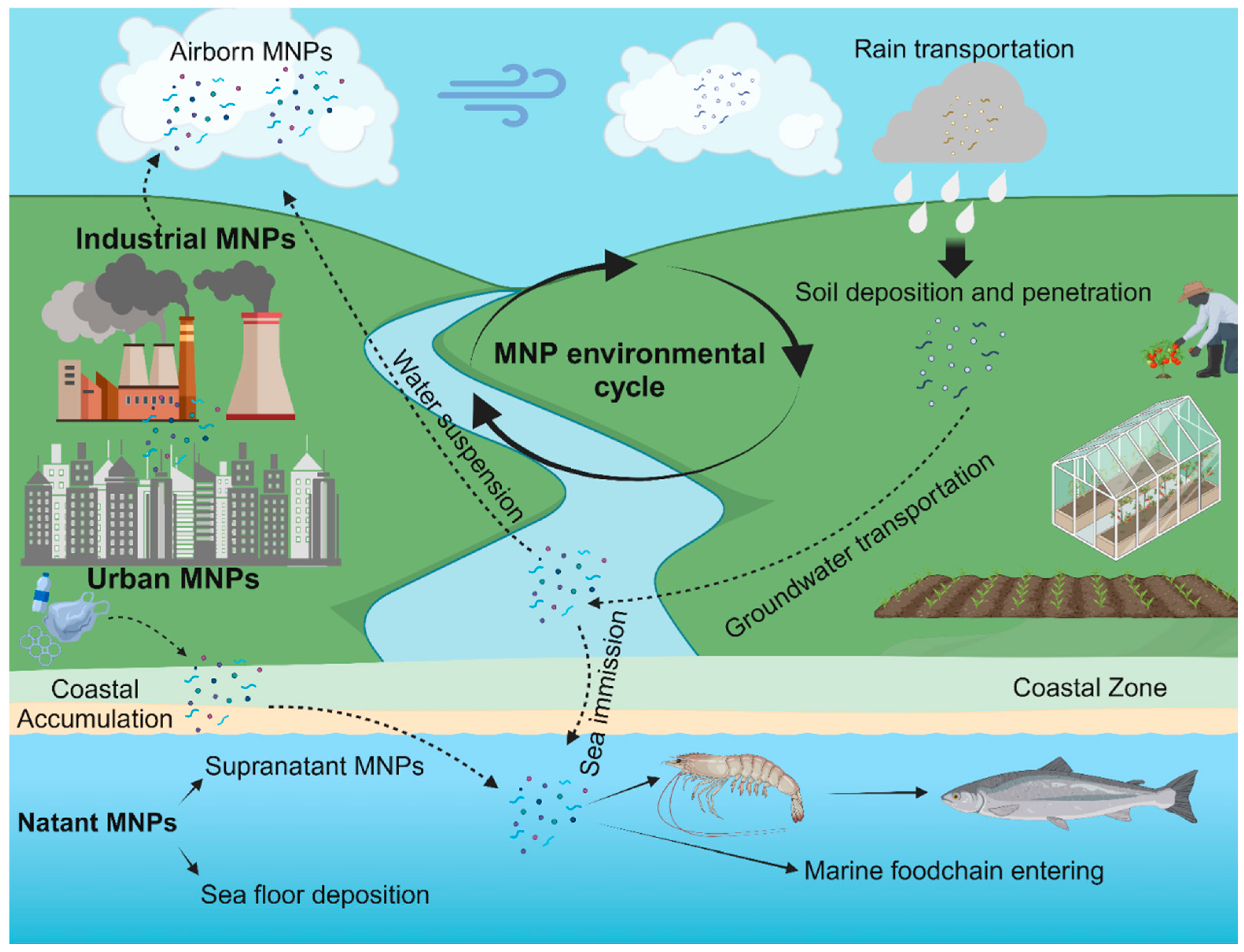
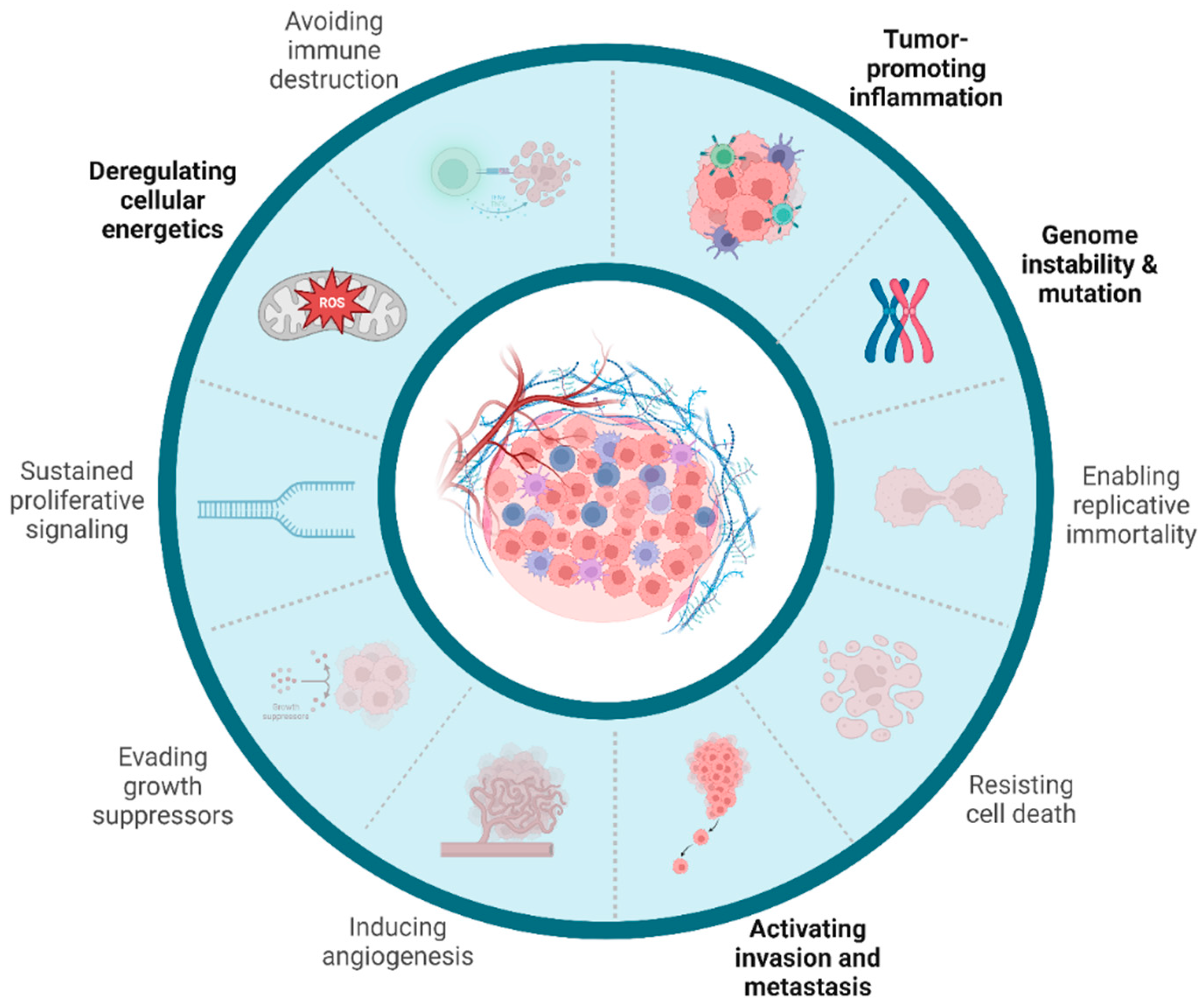
Figure 2.
Hallmarks of cancer related to MNPs. Current evidence regarding the role of MNPs in cancer initiation and promotion is linked mainly to the capability of MNPs to induce metabolic stress through the induction of ROS, fostering immune infiltration and chronic inflammation since their persistence in cancer cells and macrophages. The inability of intracellular litic enzymes of mononuclear phagocytes to process MNPs induces a “frustrated” phenotype that can bring to uncontrolled cell death, further sustaining inflammatory processes. Moreover, MNPs exposed cancer cell showed augmented capcity of invasion and metastatization in preclinical models. Finally, genomic instability could be the results of intricated cytotoxic and genotoxic damage triggered by the presence of MNPs, that need to be further elucidated.
Disclaimer/Publisher’s Note: The statements, opinions and data contained in all publications are solely those of the individual author(s) and contributor(s) and not of MDPI and/or the editor(s). MDPI and/or the editor(s) disclaim responsibility for any injury to people or property resulting from any ideas, methods, instructions or products referred to in the content. |
© 2024 by the authors. Licensee MDPI, Basel, Switzerland. This article is an open access article distributed under the terms and conditions of the Creative Commons Attribution (CC BY) license (http://creativecommons.org/licenses/by/4.0/).
Copyright: This open access article is published under a Creative Commons CC BY 4.0 license, which permit the free download, distribution, and reuse, provided that the author and preprint are cited in any reuse.


