Preprint
Article

This version is not peer-reviewed.

Efficacy of Fat Supplements with Different Unsaturated/Saturated FA Ratios Undergoing First Postpartum Ovulation in Lactating Anovulatory Goats

A peer-reviewed article of this preprint also exists.

Submitted:

20 November 2024

Posted:

21 November 2024

You are already at the latest version

Abstract

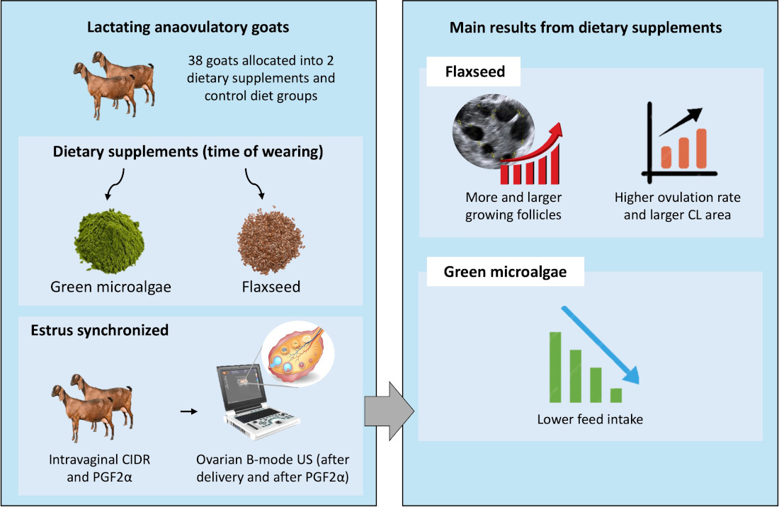
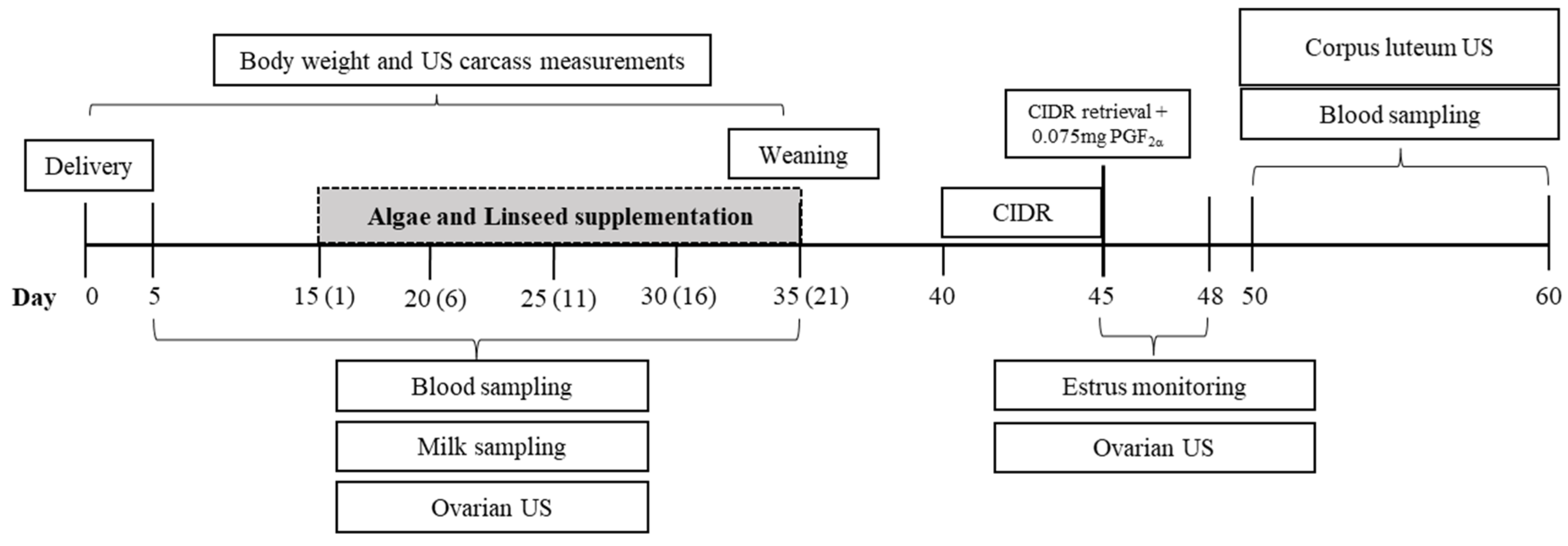
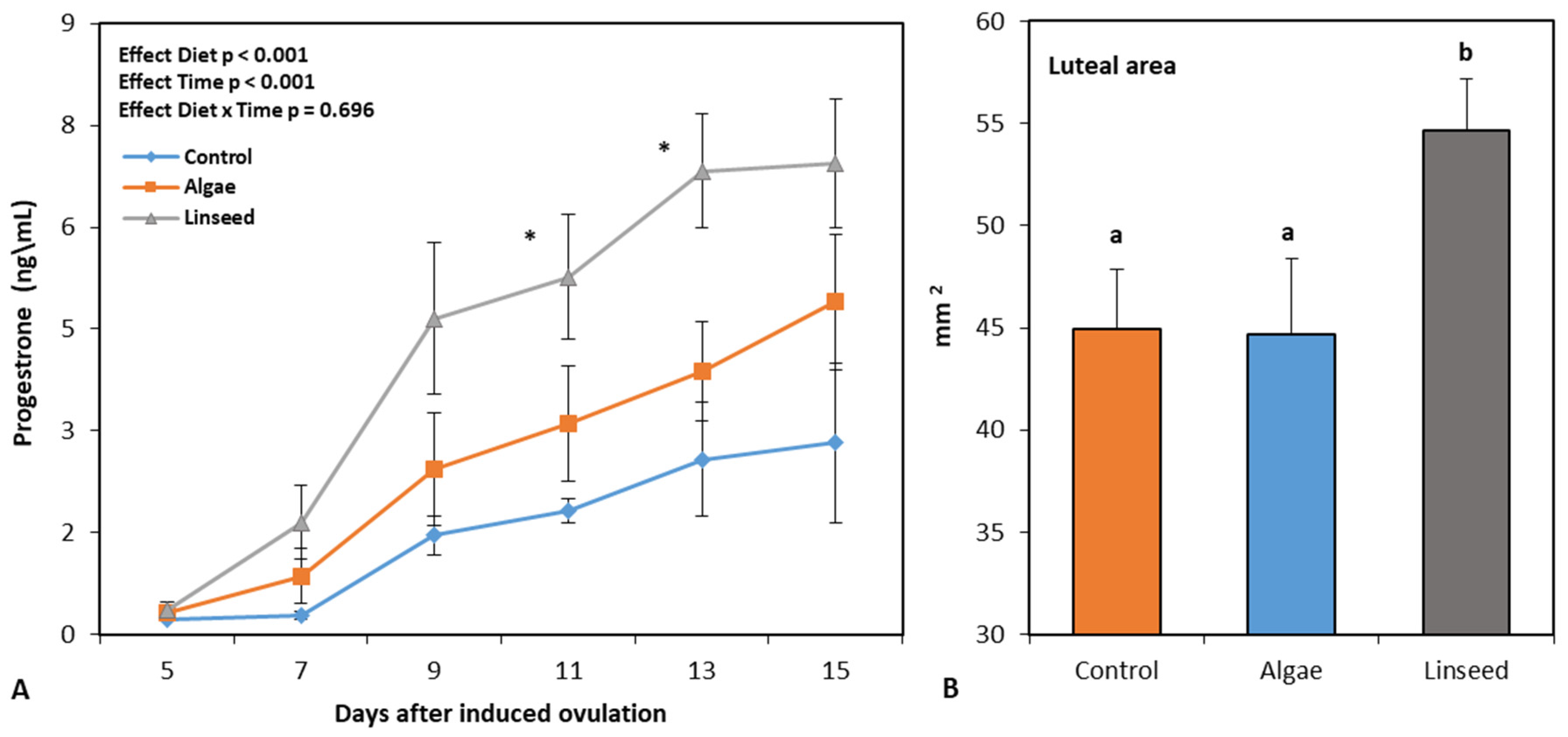
We investigated whether microalgae or linseed supply during the early postpartum period affects ovarian restimulation and supports the first postpartum ovulation in lactating anovulatory goats. Thirty-eight An-glo-Nubian-crossbred adult goats were allocated into three groups: con-trol diet (n=12), fed a total mixed ration (TMR) comprising chopped elephant grass and concentrate; algal diet (n=13), fed TMR+green microalgae (1% dry matter); and linseed diet (n=13), TMR+linseed (12% dry matter). Supplements were furnished from the 2nd to 5th week (time of weaning). Goats were estrus synchronized on day 40 by insertion of an intravaginal CIDR device for 5 days, after which 0.075mg PGF2α was applied to in-duce ovulation, and estrus was monitored for 72 hours. From the 5th-15th day of ovulation induction, the corpus luteum (CL) area and progesterone rate were monitored. The algal and linseed groups showed lower feed intake (P<0.001) and higher (P<0.001) triglyceride levels/follicle numbers, respectively. After estrus induction, no differences were ob-served in estrus response; however, the linseed group showed more and larger growing follicles (P=0.016 and P<0.01), a higher ovulation rate (P<0.05), larger CL area (P<0.05), and higher progesterone levels (P<0.001). Linseed after delivery stimulates follicular growth before and after ovulation induction, favoring better CL quality during the first ovulation.

Keywords: 
;  ;  ;  ;  ;  

1. Introduction

In ruminants, the first ovulation and the presence of a functional corpus luteum in the postpartum period are indispensible to ensure pregnancy after insemination [1], as well as the production of quality oocytes [2] in in vitro embryo culture. In dairy cows, postpartum ovarian function is strictly associated with metabolic conditions before and after calving, which in turn are as-sociated with the appearance of pro-inflammatory pa-thologies in periparturients [1]. Accentuated lipolysis, due to a negative energy balance, promotes oxidative stress that damages DNA, proteins, and lipids, thus favoring immune system dysfunction [3]. Oxidative stress also increases the proinflammatory phenotype of immune cells, which act on redox-regulated proinflammatory factors [4]. In addition, changes in the molecular profile also occur, due to the upregulation of pro-inflammatory genes, such as the cytokines IL-1β and TNF-α [5].
Data on pregnancy losses at the first service in goats are limited, largely because of the conditions of management in an extensive system [6]. In tropical and subtropical regions, the main goat breeders and high environmental temperatures make the postpartum re-productive management of herds even more difficult due to the difficulty in meeting nutritional demands with adequate food consumption [7]. Consequently, nutritional strategies play a fundamental role in this field, and the use of lipid food sources is widely recognized owing to their high energy content [8], their ability to optimize rumen fermentations with lower methane production [9], and the impact of polyunsaturated fatty acids (PUFA) on the reproductive response [10].
In one study in goats, [11] achieved a resumption of upper follicular activity on day 5 postpartum, and re-duction of serum prostaglandin F2α on day 7 post-partum through diet supplementation with PUFA-rich fish oil (−3 to +3 weeks postpartum). The metabolism of prostaglandins can be influenced by the dietary in-take of n-6:n-3 PUFAs, which are directly associated with embryonic survival both in vivo and in vitro [12]. In addition to the positive effects of n-3 PUFA supplementation on different reproductive processes such as follicular turnover and growth, development of the corpus luteum and steroidogenesis modulate ovarian dynamics [12,13].
Despite these results, the primary challenge in this field is to quantify the lipid requirements in reproductive processes, such as breeding time [14]. High levels of fat in ruminant feed (> 5%) have limitations due to the negative effects on feed utilization and digestibility [8]. Recent studies in goats have further shown that higher fat inclusion (7%) for short periods (11 days) stimulates follicular growth without harming the animals [15]; however, modulate lipid supplementation to promote reproductive response at longer intervals, such as postpartum and in the presence of high metabolic stress, remains a challenge. Recently, several authors have focused on products, such as green micro-algae [16,17,18] and flaxseeds [19,20], both of which have a high content of unsaturated fatty acids and the advantage of being used in smaller quantities in the diet, allowing for control of the baseline dry matter fat. Although their use is promising, inconsistencies re-main regarding the effectiveness of long-term supple-mentation [19,21], as well as the actual efficacy of un-saturated fatty acids on oocyte quality and embryonic development [22,23].
Thus, we hypothesized that the inclusion of green microalgae or flaxseed in diets for lactating goats during the postpartum period would effectively promote follicular growth and sustain the conditions for the first ovulation. Therefore, the present study aimed to investigate the impact of green microalgae (Chlorella pyrenoidosa) and flaxseed supplementation during the first month postpartum in the diets of lactating goats on follicular growth, oxidative and metabolic stress, and ovarian and luteal responses before and after ovulation induction using a hormonal synchronization protocol.

2. Materials and Methods

2.1. Animals and Diet Management

This study was conducted at the School of Veterinary Medicine farm, Ceará State University (UECE), Brazil. All procedures in this study were approved by the Ethics Committee on Animal Experimentation of UECE (No. 38257569). The model animals included 38 Anglo-Nubian crossbred adult and pluriparous goats with 33.6 ± 6.6 kg body weight, 2.7 ± 0.2 (score from 1 to 5) body condition score, and age of 42.0 ± 8.6 months (overall mean ± SD). All animals belonging to the school farm herd had their estrus synchronized and were naturally mated with Anglo-Nubian males. Pregnancies were detected using ultrasonography 45 days after mating, when they were clinically followed up by a trained veterinarian.
All goats received the same diet, comprising a total mixed ration (TMR) of chopped elephant grass and concentrate. The TMR was prepared in a water solution, and furnished to satisfy the nutritional requirements of adult non-dairy goats [24] for each phase of gestation (initial and late) and early lactation. The TMR was prepared immediately before feeding and offered twice, at 08:00 and 15:00. Food refusals were collected and weighed daily to estimate diet intake.

2.2. Nutritional Treatments and Experimental Design

At delivery, goats were distributed homogeneously according to the feeding group as follows: control diet (n = 12) fed with a baseline TMR, algal diet (n = 13), and linseed diet (n = 13). In the latter two groups, the TMR was supplemented with 1% DM of powdered green microalgae (Chlorella pyrenoidosa) or ground linseed on a 12% DM basis, respectively. Supplements were added to the TMR concentrate and offered from the 2th week to the 5th week after delivery (Figure 1). Goats were distributed to maintain a similar ratio between pregnancy types (six singletons and seven twins in the Algae and Linseed groups; five singletons and seven twins in the control diet) and were kept in open collective shed stalls, grouped according to feeding and pregnancy type, with free access to mineral supplements and water. After delivery, the kids were kept with goats and weaned at the 5th week postpartum.
The foodstuff ingredients and chemical compositions of the diets and supplements are presented in Table 1. For microalgae, lipids were quantified gravimetrically according to [25] To evaluate the acid profiles, samples were analyzed using a gas-liquid chromatograph coupled to a mass spectrometer (model GCMS-QP2010S, SHIMADZU, Japan). Non-fibrous carbohydrate (NFC) values were calculated as described by [26]. The me-tabolizable energy (ME) diet content was calculated according to [24] recommendations: ME (Mcal/kg of DM) = TDN (kg) × 4.4 × 0.82.

2.3. Synchronization Program

At 40 days postpartum, five days after weaning of kids (Figure 1), all goats underwent synchronization of the estrus and follicular wave through the insertion of an intravaginal device impregnated with progesterone (CIDR®, Controlled intravaginal drug release; InterAg, Hamilton, New Zealand). After five days, the device was removed, and 1 mL (0.075 mg) of prostaglandin (PGF2α) (Prolise®; ARSA S.R.L., Buenos Aires, Argen-tina) was applied to induce ovulation. Followign CIDR removal, estrus was monitored by a trained observer for 72 h using a vasectomized buck introduced into the pen four times daily (06:00, 10:00, 14:00, and 18:00).

2.4. In Vivo Performance and Adipose Carcass Markers Measurements

Every five days, from delivery to the 5th week postpartum (Figure 1), goats were weighed, and adipose mass was verified by ultrasonography by measuring the thickness of the subcutaneous lumbar fat deposits [27]. To estimate the levels of visceral fat, we measured the thickness of the kidney fat behind the 13th rib using a previously described methodology [28]. A convex transducer with a frequency of 3.5 MHz (Z5 Vet; Mindray Bio-Medical Electronics Co., Shenzhen, China) was used for kidney imaging. Images were captured in triplicate, and analyzed using the previously calibrated ImageJ program (ImageJ, National Institutes of Health, Bethesda, MD, USA).

2.5. Ovarian Function

2.5.1. Follicular Dynamics, Ultrasonography Analysis, and Ovulatory Rate

Ultrasonography was performed daily from the 5th to the 35th day after delivery, and during the 72 h after prostaglandin administration, corresponding to ovulation induction. Ovarian images were obtained with B-mode ultrasound equipment using a 5 MHz linear transrectal probe (model Z5 Vet; Mindray Bio-Medical Electronics Co., Shenzhen, China). Images were captured and analyzed using Image J software (National Institutes of Health, Millersville, MA, USA), which were previously calibrated.
An ovarian follicular wave was defined as the emergence of a group of small follicles (< 3 mm) that gave rise to one or more large follicles (≥ 3 mm). The day of wave emergence was defined as the day when the largest follicle of the wave reached 3 mm in diameter. The growth phase was defined as the period during which a large follicle grows from 3 mm to its maximum diameter. The regression phase was defined as the period from the maximum follicle diameter to a diameter of 3 mm. Six days after the injection of the PGF2 alfa analog (Figure 1), the ovulatory response was quantified by laparoscopy through a morphological classification of the corpus luteum as described previously [29].

2.6. Luteal Function

2.6.1. Progesterone Assay

Blood samples were collected every five days from the fifth day after parturition until weaning and every two days from the fifth to fifteenth day after ovulation in-duction (Figure 1). Sample were taken by jugular venipuncture into heparinized vacuum tubes (BD Vacutainer®, Franklin Lakes, NJ, USA), performed before the morning feed was provided. Samples were then centrifuged at 3000 rpm for 15 min to separate the plasma, from which three aliquots were obtained and stored at -20°C until progesterone analysis. Progesterone analyses was performed on an automatic analyzer (IMMULITE® 2000, Siemens Healthcare Diagnostics LTDA, Llanberis, United Kingdom) using a specific commercial kit (IMMULITE® 2000 progesterone, Siemens Healthcare Diagnostics LTDA, Llanberis, United Kingdom), with a sensitivity of 1ng/mL, as indicated by the manufacturer's recommendations.

2.6.2. Postpartum Luteal Activity

Luteal activity was monitored from the fifth day after parturition until weaning by measuring the plasma progesterone concentrations, which indicated the presence of corpora lutea. The corpus luteum was considered functional when progesterone concentrations were greater than 1 ng/mL in at least two successive samplings, non-functional when progesterone concentrations were greater than 1 ng/mL in less than two consecutive samplings, and absent when progesterone concentrations were less than 1 ng/mL.

2.6.3. Corpus Luteum Ultrasonography

Luteal activity after ovulation induction was measured daily using B-mode ultrasonography from the fifth day after ovulation induction (Figure 1). Evaluations were performed using B-mode ultrasound equipped with a 5.0 MHz linear transrectal probe for the identification and counting of the corpus luteum (CL), as described previously [30]. In brief, B-mode- and Doppler-mode videos of both ovaries were recorded for subsequent evaluation. In the B-mode, frozen images were measured for CL count and luteal diameter (mm). Images corresponding to the cross-section of the CL at its greatest diameter were collected to obtain the total ovarian area [30].

2.6.4. Metabolites, β-Hydroxybutyrate (BHB), Glutathione Peroxidase (GPx) Assays, and Fat-Protein Milk Ratio

Blood samples were collected every five days from the fifth day after parturition until weaning, and at fifth day after ovulation induction using heparinized vacutainer tubes (BD Vacutainer®, Franklin Lakes, NJ, USA) before morning feeding. The samples were subsequently centrifuged at 3000 rpm for 15 min to separate the plasma, from which three aliquots were obtained and stored at -20°C for further quantification of metabolites. Plasma concentrations of cholesterol and triglycerides were determined using an automated biochemical analyzer (Mindray BS 120, Mindray®) and commercial kits (Bioclin, Quibasa, Minas Gerais, Brazil). The sensitivity of the assay kit was 1.472 mg/dL and 2.58 mg/dL, for cholesterol and triglycerides. GPx and BHB were analyzed using automated biochemical analyzer (Mindray BS 120, Mindray®) and commercial kits (Randox Laboratories, Crumlin, UK) with a sensitivity of 75U/L for GPx and 0.100 mmol/L for BHB.
Every five days from the fifth day after parturition until weaning milk samples were taken and analyzed for fat and protein concentrations (Lactoscan SA®, Entelbra, Londrina, Brazil). The fat-to-protein ratio (FPR) of milk was calculated as the milk fat content (g/kg) divided by the milk protein content (g/kg).

2.7. Statistical Analysis

Statistical analyses were performed using Statistica Software version 13.4.0.14 (2018; TIBCO Software, Inc., Palo Alto, CA, USA). All data were initially verified for mathematical assumptions using the Shapiro-Wilk test. If these conditions were not met, log10x transformation was applied. All descriptive data were analyzed using the GLM procedure analysis of variance (ANOVA). The factors used in the model included diet (Control, Algae, Linseed), interval between samples used (time), and interactions. The type of parturition (single or twin) effect and diet × type of parturition interaction were tested for measurements before hormonal induction of ovulation and were not significant (p > .05) for most of the parameters; thus, only the main effects of diet and time were presented. All pairwise comparisons were performed using the Newman–Keuls post hoc test. The number of CL classes, number of does in estrus, and number of does that ovulated were compared using the chi-square test.

3. Results

3.1. Response During Supplementation

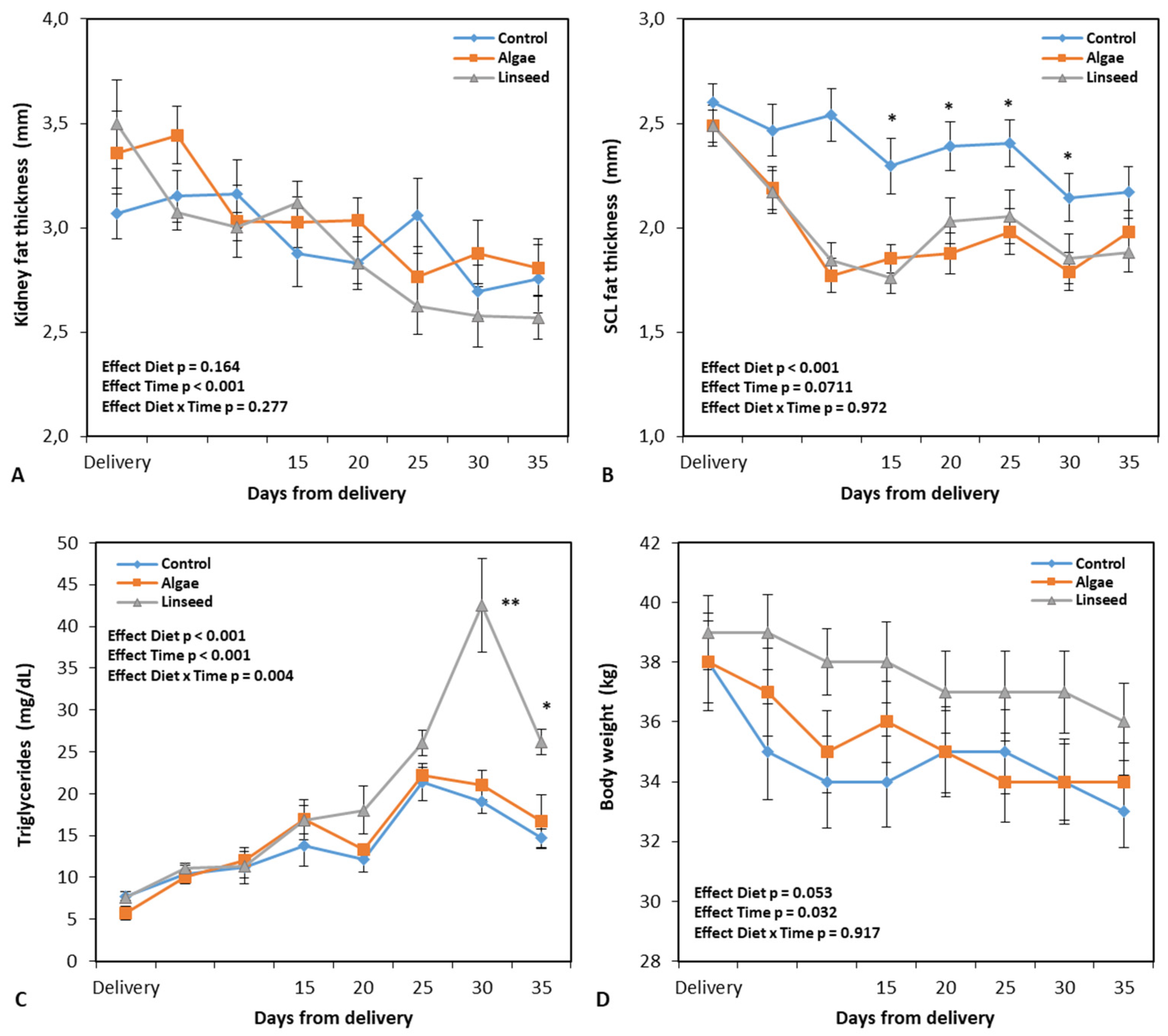
3.1.1. Changes in Feed Intake, Body-Weight, and Carcass Markers

The group supplemented with microalgae showed a reduced (P < 0.001) dry matter intake (Table 2) compared to the control group and animals supplemented with linseed. When intake was expressed as a percentage of body weight, the microalgae intake was 2.9%, whereas in the other treatments, it reached 3.4%. Figure 2 illustrates the changes after delivery for body weight and carcass adipose tissue marker levels after delivery. For the measurements performed during the supplementation interval, no differences were observed between the nutritional groups in terms of body weight (P = 0.053) (Figure 2D). All three groups lost an average of 1.5 kg of mass, which was equivalent to 4.2% of their own weight. If the total interval between delivery and weaning of the kids was considered (Figure 2D), the average losses were 4.6 kg, equivalent to 12% of the initial weight of the females.
All animals showed a reduction in kidney fat thickness (time effect: P < 0.001; Figure 2A). This reduction was on average 0.3 mm for the supplementation interval and 0.6 mm between delivery and weaning, respectively. Regarding lumbar subcutaneous fat (Figure 2B), both supplemented groups recorded a smaller thickness than the control (P < 0.001) during dietary treatment. None of the groups showed any significant effect on the measurement interval (time effect, P = 0.0711). Figure 2B presents how the most consistent reduction in adipose deposits occurred between delivery and the beginning of supplementation.

3.1.2. Metabolic and Oxidative Damage Markers

No differences were recorded between the groups regarding the plasma concentrations of cholesterol and Gpx (Table 2). An increase in cholesterol levels was observed between days 6 and 11 of supplementation (effect time: P < 0.001; Table 2). Regarding plasma β-hydroxybutyrate, no differences were also evidenced between the groups (Table 2).
The plasma concentrations of triglycerides are presented in Figure 2C. Overall, our analysis revealed a significant interaction between diet and time (P = 0.004), owing to the substantial increase in metabolites in the animals supplemented with linseed from the second week of supplementation (Figure 2C), which did not occur in the other groups that showed a decrease in triglyceride levels in the same interval. The milk fat/protein ratio (Table 2) was higher in the supplemented groups than in the control group.

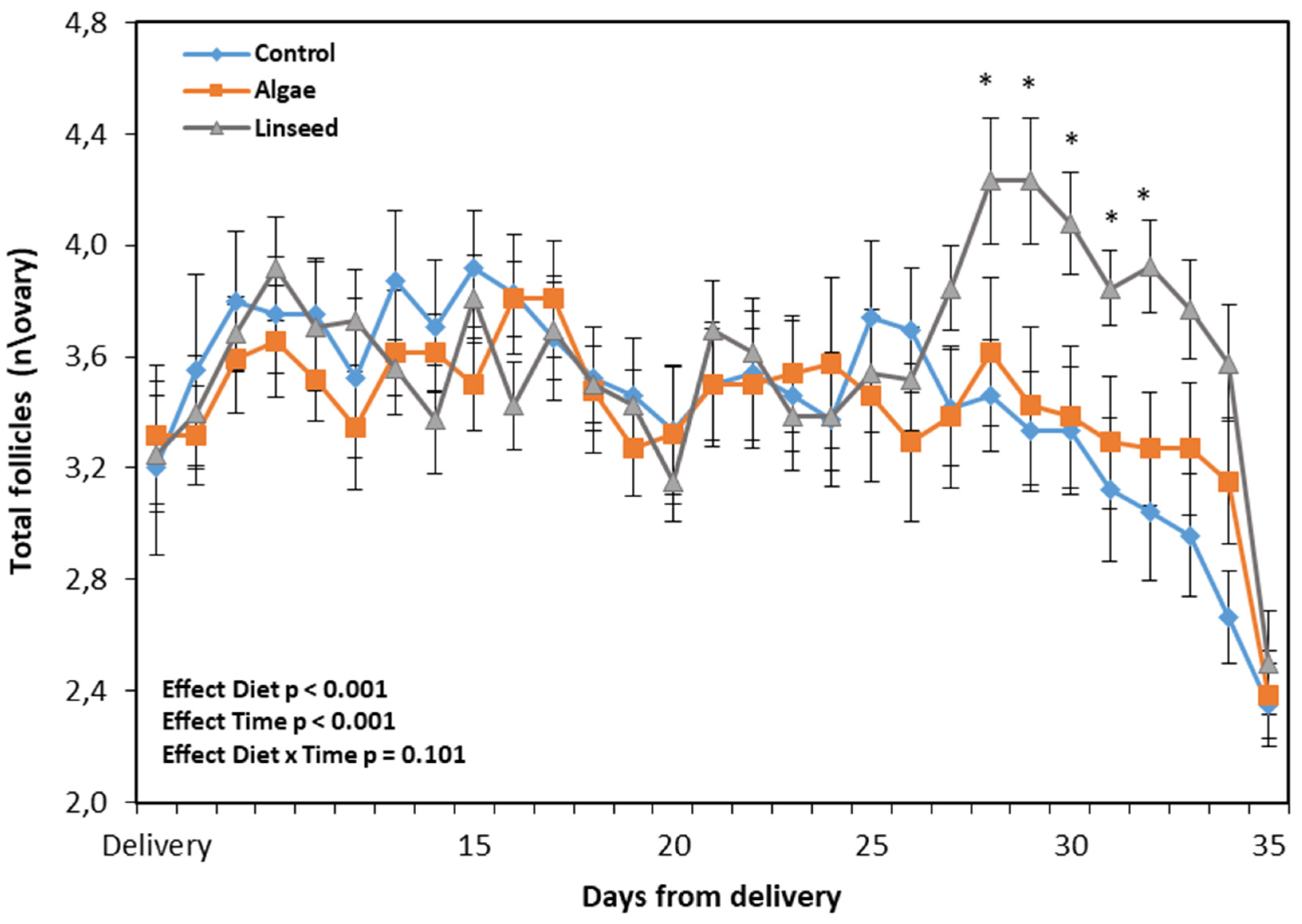
3.1.3. Ovarian and Luteal Function

Figure 3 presents the number of follicles counted using ultrasound analysis after delivery and supplementation. There was a diet effect (P < 0.001) owing to a significant increase in the ovarian follicular population in the linseed group between the 14th day and the 18th day of supplementation (Figure 3).
The presence of corpora lutea was verified using peripheral progesterone concentrations (Table 2). During the supplementation period, no functional corpora lutea were observed, and one non-functional CL was recorded in the linseed group (Table 2). This result justified the higher progesterone levels (P < 0.05; Table 2).

3.2. Response After Ovulation Induction

3.2.1. Responsiveness to Estrus Synchronization

The groups did not differ in terms of their responses to hormonal treatment for estrus synchronization (Table 3). However, in the control group, we observed a significant difference between the number of goats in estrus and the total number of synchronized animals (Table 3). The overall mean number of animals in estrus was 29% (11/38), with the onset of estrus at 34 h following CIDR withdrawal. Although the supplemented groups recorded an estrus duration twice that of the control group, the difference was not statistically significant (Table 3). Thus, the average estrus duration was 28 h.

3.2.2. Follicles, Ovulatory Response, BHB, and GPx

During the measurement interval, all groups showed a significant increase (Table 3) in the number of large follicles (time effect, P = 0.009) and the maximum follicular diameter (time effect, P = 0.006). However, comparison of the groups revealed that animals supplemented with linseed showed higher values for both parameters (Table 3). At the same time, in the same nutritional treatment, a significant reduction in the number of small follicles (< 3 mm) was also recorded. There were no differences in the number of ovulating does between the diets (Table 3); however, in the control group, a significant difference (P < 0.05) was observed between the number of ovulated animals and the total number of synchronized batches. Regarding the ovulation rate (Table 3), the supplemented groups showed values twice as high as those of the control, with a significant difference observed between the linseed and control group (P < 0.05).
No differences were observed between nutritional treatments in relation to the concentrations of β-hydroxybutyrate in glutathione peroxidase (Table 3).

3.2.3. Luteal Function

Progesterone concentrations in the CL measured after estrus induction revealed that only one CL was classified as nonfunctional in the linseed group. Figure 4 presents the progesterone levels recorded following ovulation induction in the functional CL (Figure 4A) and luteal areas (Figure 4B). The linseed group had a larger luteal area than the control and microalgal groups. All three nutritional treatments showed an in-crease in peripheral progesterone during the measurement interval (time effect, P < 0.001; Figure 4A); however, the linseed group had higher values starting from day 11 after ovulation induction.

4. Discussion

Overall, the results of the present study partially confirm our initial hypothesis, showing that the inclusion of linaça in the diet between the second and fifth weeks postpartum stimulated ovarian and luteal responses in goats. The data also showed that among the two sources of lipids chosen, only flaxseed was successful, whereas the use of microalgae, at least at the proposed dosages, did not present advantages in terms of re-productive responses.
These two supplements have specific nutritional characteristics and different inclusion intervals in ruminant diets. However, in the case of microalgae, the results were not as promising asexpected. This is surprising as prior studies have shown it is possible to obtain a significant follicular growth response using low dosages of microalgae [16,17,18], which allows maintainence of the baseline dry matter fat of the diet for ruminants between 2%-3%.
Microalgae are a potential source of lipid supple-mentation due to their ability to produce biomass rich in polyunsaturated fatty acids (PUFAs), such as ω-3 eicosapentaenoic acid (EPA; 20:5 n-3) and docosahexaenoic acid (DHA; 22:6 n-3) [31]. Chlorella sp. is a species of green microalgae that has been widely used as a supplement in the diet of ruminants, not only because of the beneficial action of PUFAs on milk quality [32,33], but also to improve reproductive parameters, such as ovarian response [16] and embryo production [18]. This supplement at the cell membrane level, affecting the transfer of nutrients to reproductive tissues [34], as well as precursors of steroids and prostaglandins. [18] observed that the administration of 10 g of Chlorella in a short time interval (7 days) induced a lower diet consumption, but simultaneously promoted greater intraovarian blood perfusion and an increase in the number of large follicles. This phenomenon can be explained by the higher plasma glucose levels pro-moted by the greater availability of AF [35], since glucose is an intrafollicular mediator that, at higher concentrations in the blood, promotes an increase in insulin like growth factor 1 (IGF1), and inhibits the action of neuropeptide Y, which acts on the metabolic hormonal axis and stimulates the release of GnRH in the hypothalamus, which in turn increases FSH and LH pulses, thereby stimulating follicular growth [36].
Conversely, research has shown that supplementation of diets with products rich in PUFAs for a longer period can promote a decrease in feed intake due to a reduction in the palatability and digestibility of fiber, which can alter the pH and biohydrogenation during rumen fermentation, causing toxic effects in the rumen [32]. This phenomenon may have been responsible for the lower use of the microalgal diet and, ultimately, for the change in the milk protein/fat ratio in the two supplemented groups. The use of high doses of microalgae, for example, leads to substantial changes in the use and digestibility of feed; however, a reduction in dry matter intake has also been recorded with brief supplementation [17,18], without having an impact on the ovarian response.
In our study, flaxseed supplementation induced an increase in plasma triglycerides between days 25 and 30 postpartum, which corresponded to the third week of diet application. In the same time interval, an increase in the ovarian follicular population was also observed through ultrasound analysis. Flaxseed diet also resulted in more efficient follicular growth following hormonal synchronization. Ultrasound analysis revealed more intense follicular depletion (a reduction in the number of small follicles) in this group, in parallel with a greater increase in the number of large follicles and follicular size. This phenomenon did not occur at the same intensity in the other treatments.
As previously anticipated, the lack of efficacy of the microalgal diet was not an expected result; the literature shows that the dosage used in shorter application times is sufficient to induce an increase in lipid metabolites [37] and to stimulate follicular dynamics [18]. The discrepancy between these results is potentially attributable to the low nutritional status of the animals in the present study as a consequence of prolonged metabolic imbalance in the postpartum period. At the supplementation interval, all experimental groups showed renal adipose mass mobilization, with an average weight loss of 130 g/day, values that were in ac-accordance with those reported by [38] and [15]. In addi-tion, the results showed that the concentrations of peripheral GPx, an indicator of oxidative stress, and BHB remained high in all groups at the measurement intervals.
After calving, energy needs are met through adipose tissue catabolism, an adaptation mechanism that in-creases the plasma levels of ketone bodies such as β-hydroxybutyric acid and non-esterified fatty acids. In turn, the increase in lipolysis is strictly associated with oxidative stress and the establishment of inflammatory states in immune cells, with the consequent production of cytokines that act on pro-inflammatory factors regulated by redox reactions. During this phase, cytokines diffuse in a paracrine/autocrine manner to regulate ovarian function, and are involved in the control of follicular development and ovulation, acting on follicular development, steroidogenesis, ovulation, corpus luteum function, and the regulation and differentiation of granulosa cells [39]. TNF-α is a mediator of ovulation in terms of oocyte release, granulosa cell autophagy in ovarian tissue remodeling, and favoring luteinizing hormone action have shown that luteinizing hormone (LH) induces ovulation via TNF-α-dependent increases [40,41]. As such, the BEN nadir is temporarily associated with a marked increase in LH pulse rate, which facilitates the first postpartum ovulation [1].
The two supplements also behaved differently with respect to ovulation and functionality of the corpus luteo induced by the synchronization protocol. Although both groups expressed a low response to the synchronization protocol in terms of animals in estrus, the flaxseed diet displayed a higher rate of ovulation and luteal area, and consequently, higher secretion of progesterone during the development of the CL. The latter two parameters are usually used as indicators of CL functionality and quality [30], which in turn represent key factors for fertilization and embryonic development during breeding [30].
In this context, flaxseed supplementation promoted a positive effect on the ovarian response. The inclusion level used in the diet was allowed to remain within the fat level limit range indicated for ruminants [8], and simultaneously provided a higher energy density in the diet. Thus, it is plausible that the difference in the flaxseed diet in terms of ovarian response was an effect of the availability of an extra share of food energy and the greater presence of n-3 fatty acids, which are rich in flaxseed. Flaxseed has been used in ruminant supplementation because it contains approximately 40-45% PUFAs in its composition, mainly alpha-linolenic, a precursor of EPA and DHA [42], which modulates the activation of the primordial follicle and stimulates folliculogenesis by promoting the increase in IGF1 levels [43]. In addition, IGF1 induces the expression of P450 cholesterol desmolase and aromatase, both of which are enzymes essential for the production of cholesterol, a precursor of progesterone, directly influencing ovarian metabolic processes [44,45,46]. In goats, supplementation with flaxseed at 8-12% DM promoted an increase in the levels of fatty acids in milk [47,48,49], and quality embryos [50], whereas in cows, it resulted in the accumulation of linolenic acid and its metabolites in milk and plasma [37], increased follicular growth and oocyte quality, and reduced pregnancy losses [42,51].
However, there are contrasting results regarding the effect of PUFA supplementation on oocyte chemical properties, embryo quality, and fertility, as the composition of follicular fluid depends on the reproductive age of the animal, follicular size, and type of PUFAs administered [2,23]. According to [23], the follicular fluid of adult and prepubertal goats, percentages of linolenic acid (ALA; n-3 PUFA), total saturated fatty acids (SFAs), and total n-3 PUFAs were significantly higher in adults, and the percentages of Linoleic Acid (LA; n-6 PUFA), total MUFAs, total PUFAs, total n-6 PUFAs, and n-6: n-3 PUFAs were higher in prepubertal goats than in adults. However, in prepubertal goats, comparing large (≥3 mm) and small (<3 mm) follicles, the concentrations of total SFAs, MUFAs, PUFAs, and n-6 PUFAs were higher in the small ones. In in vitro studies, the concentrations of linolenic and linoleic acids added as supplements to the IVM medium affect the competence of oocytes for embryo development [52]. Other studies have indicated that pregnancy rates increase in cattle fed diets containing high amounts of n-3 fatty acids [53]. In addition, n-3-enriched diets may inhibit PGF2α synthesis, preventing the regression of the corpus luteum [54], and as a result, there are sustained concentrations of progesterone required for embryo survival and pregnancy maintenance.

5. Conclusions

The addition of linseed to the diet in the first few weeks following parturition in goats induced effective follicular growth from the second week of supplementation, and the synchronization protocol was applied at 40 days postpartum. Furthermore, the use of this food allowed for the expression of efficient ovarian activity and better quality of the corpus luteum in hormonally induced ovulation. From these results, we concluded that under the experimental conditions of the present study, dietary supplementation with linseed is an efficient strategy to support the first ovulation in goats.

Author Contributions

C.P.S.: Investigation, Methodology; CCLF: Supervision, Investigation, Methodology; Writing-original draft; JPMA: Supervision, Investigation, Methodology, Writing-original draft; C.M.C.: Writing-original draft; F.B.B.O: Investigation, Methodology; A.J.H.C.: Writ-ing-original draft; D.C.S.P.: Resources, Investigation; A.C.R.: Funding acquisition, Resources; DIAT: Resources; DR: Conceptualization, Funding acquisition, Formal Analysis, Supervision, Writing-original draft.

Funding

This research was funded by Fundação Cearense de Apoio ao Desenvolvimento Científico e Tecnológico, FUNCAP (Grant No. 11839627/2022 and grant No. 09564039\2022).

Institutional Review Board Statement

All procedures used in this study were approved by the Ethics Committee in Animal Experimentation of Ceará State University (number 38257569/2016).

Data Availability Statement

The data presented in this study are available on request from the corresponding author due to legal reason.

Acknowledgments

This study was part of a project supported by the Fundação Cearense de Apoio ao Desenvolvimento Científico e Tecnológico (FUNCAP; Grant Nos. 11839627/2022 and 09564039\2022). Silva C.P. received a doctoral scholarship from CNPq, Brazil. This paper is part of Silva’s doctoral thesis supervised by Rondina D.. A.C. Rego and D. Rondina are senior investigators at CNPq, Brazil.

Conflicts of Interest

The authors declare that there is no conflict of interest regarding the publication of this article.

References

  1. Banuelos, S.; Stevenson, J. S. Transition cow metabolites and physical traits influence days to first postpartum ovulation in dairy cows. Theriogenology. 2021, v. 173, p. 133–143. [CrossRef]
  2. Maranesi, M.; Castellini, C.; Dall’Aglio, C.; Petrucci, L.; Mattioli, S.; Boiti, C.; Zerani, M. Effects of PUFAs on animal reproduction: Male and female performances and endocrine mechanisms. Phytochemistry Reviews. 2018, v. 17, p. 801-814. [CrossRef]
  3. Sordillo, L. M. Nutritional strategies to optimize dairy cattle immunity. Journal of dairy science. 2016, v. 99, n. 6, p. 4967–4982. [CrossRef]
  4. Mezzetti, M.; Bionaz, M.; Trevisi, E. Interaction between inflammation and metabolism in periparturient dairy cows. Journal of Animal Science. 2020, v. 98, n. Supplement_1, p. S155-S174, 98(Supplement_1), S155–S174. [CrossRef]
  5. Kuhla, B. Pro-inflammatory cytokines and hypothalamic inflammation: Implications for insufficient feed intake of transition dairy cows. Animal. 2020, v. 14, n. S1, p. s65-s77. [CrossRef]
  6. Robertson, S. M.; Atkinson, T.; Friend, M. A.; Allworth, M. B.; Refshauge, G. Reproductive performance in goats and causes of perinatal mortality: A review. Animal Production Science. 2020, v. 60, n. 14, p. 1669-1680. [CrossRef]
  7. Menezes, L. M.; Sousa, W. H.; Cavalcanti-Filho, E. P.; Gama, L. T. Genetic parameters for reproduction and growth traits in Boer goats in Brazil. Small Ruminant Research. 2016, v. 136, p. 247-256. [CrossRef]
  8. Palmquist, D. L.; Jenkins, T. C. A 100-Year Review: Fat feeding of dairy cows. Journal of Dairy Science. 2017, v. 100, n. 12, p. 10061-10077. [CrossRef]
  9. Hristov, A. N.; Melgar, A.; Wasson, D.; Arndt, C. Symposium review: Effective nutritional strategies to mitigate enteric methane in dairy cattle. Journal of dairy Science.2022, v. 105, n. 10, p. 8543-8557. [CrossRef]
  10. Roque-Jiménez, J. A.; Rosa-Velázquez, M.; Pinos-Rodríguez, J. M.; Vicente-Martínez, J. G.; Mendoza-Cervantes, G.; Flores-Primo, A.; Relling, A. E. Role of long chain fatty acids in developmental programming in ruminants. Animals. 2021, v. 11, n. 3, p. 762. [CrossRef]
  11. Yadav, D.; Singh, A. K.; Kumar, B.; Mahla, A. S.; Singh, S. K.; Patra, M. K.; Krishnaswamy, N. Effect of n-3 PUFA-rich fish oil supplementation during late gestation on kidding, uterine involution and resumption of follicular activity in goat. Reproduction in domestic animals. 2019, 54(12), 1651-1659. [CrossRef]
  12. Mahla, A. S.; Bunkar, S. K.; Kumawat, B. L.; Saxena, V. K.; Selvaraju, S.; Bhatt, R. S.; Kumar, A. Dietary n-3 PUFA augments pre-ovulatory follicle turnover and prolificacy in well-fed ewes. Animal Reproduction Science. 2023, v. 252, p. 107231. [CrossRef]
  13. Elis, S.; Freret, S.; Desmarchais, A.; Maillard, V.; Cognié, J.; Briant, E.; Dupont, J. Effect of a long chain n-3 PUFA-enriched diet on production and reproduction variables in Holstein dairy cows. Animal reproduction Science. 2016, v. 164, p. 121-132. [CrossRef]
  14. Ribeiro, E. S.; Greco, L. F.; Bisinotto, R. S.; Lima, F. S.; Thatcher, W. W.; Santos, J. E. Biology of Preimplantation Conceptus at the Onset of Elongation in Dairy Cows1. Biology of Reproduction. 2016, v. 94, n. 4, p. 97, 1-18. [CrossRef]
  15. Cavalcanti, C. M.; Silva, M. R. L.; Conde, A. J. H.; Bezerra, A. F.; Alves, J. P. M.; Fernandes, C. C. L.; Rondina, D. Effect of peri-conception high-fat diets on maternal ovarian function, foetal and placentome growth, and vascular umbilical development in goats. Reproduction in Domestic Animals, 2022, v. 57, n. 12, p. 1481-1492. [CrossRef]
  16. Senosy, W.; Kassab, A.Y.; Mohammed, A.A. Effects of feeding green microalgae on ovarian activity, reproductive hormones and metabolic parameters of Boer goats in arid subtropics. Theriogenology. 2017, v. 96, p. 16-22. [CrossRef]
  17. Silva, M. R. L.; Alves, J. P. M.; Fernandes, C. C. L.; Cavalcanti, C. M.; Conde, A. J. H.; Bezerra, A. F.; Rondina, D. Use of green microalgae Chlorella as a nutritional supplement to support oocyte and embryo production in goats. Animal Reproduction Science. 2023a, v. 256, p. 107296. [CrossRef]
  18. Silva, M. R. L.; Alves, J. P. M.; Fernandes, C. C. L.; Cavalcanti, C. M.; Conde, A. J. H.; Bezerra, A. F.; Rondina, D. Effect of short-term nutritional supplementation of green microalgae on some reproductive indicators of Anglo-Nubian crossbred goats. Veterinary World. 2023b, v.16, n.3, p. 464. [CrossRef]
  19. Nudda, A.; Bee, G.; Correddu, F.; Lunesu, M. F.; Cesarani, A.; Rassu, S. P. G.; Battacone, G. Linseed supplementation during uterine and early post-natal life markedly affects fatty acid profiles of brain, liver and muscle of lambs. Italian Journal of Animal Science. 2022, v. 21, n.1, p. 361-377. [CrossRef]
  20. Akhtar, P.; Rajoriya, J. S.; Singh, A. K.; Ojha, B. K.; Jha, A. K.; Bisen, A.; Singh, M. Effects of dietary supplementation with omega-3 fatty acid-rich linseed on the reproductive performance of ewes in subtropical climates. Frontiers in Veterinary Science. 2024, v. 11, p. 1398961. [CrossRef]
  21. Till, B. E.; Huntington, J. A.; Posri, W.; Early, R.; Taylor-Pickard, J.;Sinclair, L. A. Influence of rate of inclusion of microalgae on the sensory characteristics and fatty acid composition of cheese and performance of dairy cows. Journal of dairy Science. 2019, v.102, n.12, p.10934-10946. [CrossRef]
  22. Ribeiro, E. S. Symposium review: Lipids as regulators of conceptus development: Implications for metabolic regulation of reproduction in dairy cattle 1. Journal of Dairy Science. 2018, v. 101, n.4, p. 3630–3641. [CrossRef]
  23. Izquierdo, D.; Roura, M.; Pérez-Trujillo, M.; Soto-Heras, S.; Paramio, M. T. Fatty acids and metabolomic composition of follicular fluid collected from environments associated with good and poor oocyte competence in goats. International journal of molecular sciences. 2022, v. 23, n.8, p. 4141. [CrossRef]
  24. Council, N. R. Nutrient requirements of small ruminants: Sheep, goats, cervids, and new world camelids. Nutrient requirements of small ruminants. 2007.
  25. Bligh, E.G.; Dyer, W.J. A rapid method of total lipid extraction and purification. Can. J. Biochem Physiol. 1959, v. 39, p. 911-917. [CrossRef]
  26. Sniffen, C. J.; O'connor, J. D.; Van Soest, P. J.; Fox, D. G.; Russell, J. B. A net carbohydrate and protein system for evaluating cattle diets: II. Carbohydrate and protein availability. Journal of animal Science. 1992, v.70, n.11, p. 3562-3577. [CrossRef]
  27. Teixeira, A.; Joy, M., Delfa, R. In vivo estimation of goat carcass composition and body fat partition by real-time ultrasonography. J. Anim. Sci. 2008, v.86, n.9, p.369–2376. [CrossRef]
  28. Harter, C.J.; Silva, H.G.O.; Lima, L.D.; Castagnino, D.S.; Rivera, A.R.; Neto, O.B.; Gomes, R.A.; Canola, J.C.; Resende, K.T.; Teixeira, I.A.M.A. Ultrasonographic measurements of kidney fat thickness and Longissimus muscle area in predicting body composition of pregnant goats. Anim. Prod. Sci. 2014, v. 54, n. 9, p. 1481–1485. [CrossRef]
  29. Guido, S.I.; Andrade, J.C.O.; Guido, F.C.L.; Oliveira, M.A.L.; Lima, P.F.; Moura, R.T.D.; Santos, V.F.; Cavalcanti, C.C. Avaliação de corpos lúteos de receptoras caprinas. Revista Brasileira de Reprodução Animal. 2023, v. 27, n. 3, p. 491-492.
  30. Balaro, M.F.A,; Santos, A.S,; Moura, L.F.G.M,; Fonseca, J.F,; Brandão, F.Z. Luteal dynamic and functionality assessment in dairy goats by luteal blood flow, luteal biometry, and hormonal assay. Theriogenology. 2017, v. 95, p. 118-126. [CrossRef]
  31. Batista, A. P.; Gouveia, L.; Bandarra, N. M.; Franco, J. M.; Raymundo, A. Comparison of microalgal biomass profiles as novel functional ingredient for food products. Algal Research. 2013, v. 2, n. 2, p. 164-173. [CrossRef]
  32. Boeckaert, C.; Vlaeminck, B.; Dijkstra, J.; Issa-Zacharia, A.; Van Nespen, T.; Van Straalen, W.; Fievez, V. Effect of dietary starch or micro algae supplementation on rumen fermentation and milk fatty acid composition of dairy cows. Journal of Dairy Science. 2008, v. 91, n. 12, p. 4714-4727. [CrossRef]
  33. Pajor, F.; Egerszegi, I.; Szűcs, Á.; Póti, P., Bodnár, Á. Effect of marine algae supplementation on somatic cell count, prevalence of udder pathogens, and fatty acid profile of dairy goats’ milk. Animals. 2021, v.11, n.4, p. 1097. [CrossRef]
  34. Zeron, Y.; Sklan, D.; Arav, A. Effect of polyunsaturated fatty acid supplementation on biophysical parameters and chilling sensitivity of ewe oocytes. Molecular reproduction and development. 2002, v.61, n.2, p.271-278. [CrossRef]
  35. Zarrin, M.; De Matteis, L.; Vernay, M. C. M. B.; Wellnitz, O.; Van Dorland, H. A.; Bruckmaier, R. M. Long-term elevation of β-hydroxybutyrate in dairy cows through infusion: Effects on feed intake, milk production, and metabolism. Journal of dairy Science. 2013, v.96, n.5, p.2960-2972. [CrossRef]
  36. Song, Y.; Wang, Z.; Zhao, C.; Bai, Y.; Xia, C.; Xu, C. Effect of negative energy balance on plasma metabolites, minerals, hormones, cytokines and ovarian follicular growth rate in Holstein dairy cows. Journal of veterinary research. 2021, v. 65, n.3, p.361. [CrossRef]
  37. Pereira, G.; Simões, P.; Bexiga, R.; Silva, E.; Mateus, L.; Fernandes, T.; Lopes-da-Costa, L. Effects of feeding rumen-protected linseed fat to postpartum dairy cows on plasma n-3 polyunsaturated fatty acid concentrations and metabolic and reproductive parameters. Journal of Dairy Science. 2022, v. 105, n.1, p. 361-374. [CrossRef]
  38. Oliveira, F. B. B. D.; Fernandes, C. C. L.; Montenegro, A. R.; Oliveira, I. T. M.; Silva, C. P.; LIMA, F. W. R.; Rondina, D. Cured dry smoked shoulder meat quality from culled adult goats fed a high lipid diet. Food Science and Technology. 2021, v. 42, p. e19521. [CrossRef]
  39. Silva, J. R. V.; Lima, F. E. O.; Souza, A. L. P.; Silva, A. W. B. Interleukin-1β and TNF-α systems in ovarian follicles and their roles during follicular development, oocyte maturation and ovulation. Zygote. 2020, v. 28, n. 4, p. 270-277. [CrossRef]
  40. Yamamoto, Y.; Kuwahara, A.; Taniguchi, Y.; Yamasaki, M.; Tanaka, Y.; Mukai, Y.; Irahara, M. Tumor necrosis factor alpha inhibits ovulation and induces granulosa cell death in rat ovaries. Reproductive Medicine and Biology. 2014, v.14, n.3, p.107–115. [CrossRef]
  41. Crespo, D.; Mañanós, E. L.; Roher, N.; MacKenzie, S. A.; Planas, J. V. Tumor necrosis factor alpha may act as an intraovarian mediator of luteinizing hormone-induced oocyte maturation in trout. Biology of reproduction. 2012, v. 86, n.1, p. 5-1. [CrossRef]
  42. Ambrose, D. J.; Kastelic, J. P.; Corbett, R..; Pitney, P. A.; Petit, H. V.; Small, J. A.; Zalkovic, P. Lower Pregnancy losses in lactating dairy cows fed a diet enriched in α-linolenic acid. Journal of dairy Science. 2006, v. 89, n. 8, p. 3066-3074. [CrossRef]
  43. Martins, F. S.; Saraiva, M. V. A.; Celestino, J. J. H.; Bruno, J. B.; Almeida, A. P.; Cunha, R. M. S.; Figueiredo, J. R. Expression of protein and mRNA encoding Insulin Growth Factor-I (IGF-I) in goat ovarian follicles and the influence of IGF-I on in vitro development and survival of caprine preantral follicles. Animal Reproduction (AR). 2018, v.7, n.4, p. 349-361. [CrossRef]
  44. Magalhães-Padilha, D. M.; Duarte, A. B. G.; Araújo, V. R.; Saraiva, M. V. A.; Almeida, A. P.; Rodrigues, G. Q.; Figueiredo, J. R. Steady-state level of insulin-like growth factor-I (IGF-I) receptor mRNA and the effect of IGF-I on the in vitro culture of caprine preantral follicles. Theriogenology. 2012, v.77, n.1, p. 206-213. [CrossRef]
  45. Costa, S. L.; Costa, E. P.; Pereira, E.; Benjamin, L. A.; Rodrigues, M. T.; Mendes, V. R..; Silva, T. F. Influence of Insulin-like Growth Factor I (IGF-I) on the survival and the in vitro development of caprine preantral follicles. Pesquisa Veterinaria Brasileira. 2014, v. 34, p. 1037-1044. [CrossRef]
  46. Luz, V.B.; Chaves, R.N.; Alves, A.M.C.V.; Pinheiro, A.S.; Figueiredo, J.R. Role of insulin-like growth factor-I (IGF-I) and kit ligand (KL) in ovarian function. Acta Sci Vet. 2015, v. 43:1300. ISSN 1679-9216.
  47. Nudda, A.; Battacone, G.; Usai, M. G.; Fancellu, S.; Pulina, G. Supplementation with extruded linseed cake affects concentrations of conjugated linoleic acid and vaccenic acid in goat milk. Journal of Dairy Science. 2006, v. 89, n.1, p. 277-282. [CrossRef]
  48. Luna, P.; Bach, A.; Juárez, M.; De La Fuente, M. A. Effect of a diet enriched in whole linseed and sunflower oil on goat milk fatty acid composition and conjugated linoleic acid isomer profile. Journal of Dairy Science. 2008, v. 91, n.1, p. 20-28. [CrossRef]
  49. Bernard, L.; Bonnet, M.,; Leroux, C.; Shingfield, K. J.; Chilliard, YEffect of sunflower-seed oil and linseed oil on tissue lipid metabolism, gene expression, and milk fatty acid secretion in alpine goats fed maize silage–based diets. Journal of Dairy Science. 2009, v. 92, n. 12, p. 6083-6094. [CrossRef]
  50. Dutra, P. A.; Pinto, L. F. B.; Neto, B. C.; Gobikrushanth, M.; Barbosa, A. M.; Barbosa, L. P. Flaxseed improves embryo production in Boer goats. Theriogenology. 2019, v. 127, p. 26-31. [CrossRef]
  51. Zachut, M.; Dekel, I.; Lehrer, H.; Arieli, A.; Arav, A.; Livshitz, L.; Moallem, U. Effects of dietary fats differing in n-6: N-3 ratio fed to high-yielding dairy cows on fatty acid composition of ovarian compartments, follicular status, and oocyte quality. Journal of Dairy Science. 2010, v.93, n.2, p. 529-545. [CrossRef]
  52. Ghaffarilaleh, V.; fouladi-nashta, A.; Paramio, M.-T. Effect of α-linolenic acid on oocyte maturation and embryo development of prepubertal sheep oocytes. Theriogenology. 2014, v. 82, n. 5, p. 686–696. [CrossRef]
  53. Fabjanowska, J.; Kowalczuk-Vasilev, E.; Klebaniuk, R.; Milewski, S.; Gümüş, H. N-3 Polyunsaturated Fatty Acids as a Nutritional Support of the Reproductive and Immune System of Cattle—A Review. Animals. 2023, v. 13, n. 22, p. 3589. [CrossRef]
  54. Surlis, C.; Cormican, P.; Waters, S. M.; Lonergan, P.; Keogh, K.; Doyle, D. N.; Kenny, D. A. Effects of dietary n-3-PUFA supplementation, post-insemination plane of nutrition and pregnancy status on the endometrial transcriptome of beef heifers. Scientific reports. 2020, v. 10, n. 1, p.20798, 2020. [CrossRef]
  55. AOAC (1990) AOAC-Official Methods of Analysis. AOAC, Maryland.
Figure 1. timeline for the experimental design used in the study, describing the period of dietary supplementation and the hormonal protocol for estrus synchronization.
Preprints 140321 g001
Figure 2. Kidney fat thickness (A), subcutaneous loin fat thickness (B), triglycerides levels (C) and body weight (D), performed after delivery and during nutritional supplementation from 2 weeks to 5 weeks after delivery (21 days) in goats. Data are plotted as mean ± SEM. The P-value for the ANOVA effects for supplementation period are shown in figure. Time, ANOVA effect for interval of assessment used. * P < 0.05 differences between groups.
Preprints 140321 g002
Figure 3. Total follicles counted by ultrasonography performed after delivery and during nutritional supplementation from 2 weeks to 5 weeks after delivery (21 days) in goats. Data are plotted as mean ± SEM. The P-value for the ANOVA effects for supplementation period are shown in figure. Time, ANOVA effect for interval of assessment used. * P < 0.05 differences between groups.
Preprints 140321 g003
Figure 4. Peripheral progesterone levels (A), and luteal area (B), measured after ovulation induction by hormonal treatment started 40 days after delivery in goats previously supplemented with algae or linseed. Data are plotted as mean ± SEM. In figure A the P-value for the ANOVA effects of diet-group, interval of assessments and interaction are shown in figure. a,b P < 0.05 differences between nutritional groups on figure B.
Preprints 140321 g004
Table 1. Ingredients and chemical composition of diets in goats fed with algae or linseed from 2 weeks to 5 weeks after delivery.
Parameters Diet Algae Linseed
Control Algae Linseed
Ingredients, g/kg DM
Elephant grass 600 600 600 - -
Ground corn grain 120 120 60 - -
Soybean meal 120 120 100 - -
Wheat bran 140 140 100 - -
Ground Linseed - - 120 - -
Algae - 10 - - -
Mineral mixture 20 20 20 - -
Chemical fraction
Dry matter, g/kg as-fed basis 568 523 573 892 929
Crude protein, g/kg of DM 114 102 122 397 245
Ether extract, g/kg of DM 28 28 57 210 283
Neutral detergent fiber, g/kg of DM 546 507 555 178 331
Acid detergent fiber, g/kg of DM 310 297 329 50 231
Ash, g/kg of DM 89 83 88 44 34
Non-fibrous carbohydrates, g/kg of DM 324 430 420 917 910
ME, Mcal/kg of DM 2.23 2.27 2.35 - -
Fatty acids, %
Saturated - - - 28.1 11.5
Unsaturated - - - 71.9 88.5
Monounsaturated - - - 45.0 23.1
Polyunsaturated - - - 26.9 65.4
UFA/SFA* 2.6 7.7
*UFA: unsaturated fatty acids, SFA: saturated fatty acids.
Table 2. Ingredients and chemical composition of diets in goats fed with algae or linseed from 2 weeks to 5 weeks after delivery.
Parameters Diet p Value
Control Algae Linseed SEM Diet Time D vs T
Feed intake
DMI*, kg\doe 1.2a 1.0b 1.2a 0.015 < 0.001 0.231 0.997
Metabolic markers and Oxidative stress
Glutathione peroxidase, U/L 127.3 120.5 147.8 2.169 0.058 0.947 0.702
BHB**, mmol/L 0.3 0.3 0.4 0.019 0.878 - -
Cholesterol, mg\dL 53.6 63.2 62.9 1.774 0.107 < 0.001 0.180
FPR***, ratio 0.8a 1.0b 1.1b 0.03 0.002 0.822 0.994
Luteal function
Functional CL****, n/doe 0B 0B 0B - - - -
Nonfunctional CL, n/doe 0B 0B 1B - - - -
CL absent, n/doe 12A 13A 12A - - - -
Progesterone, ng/mL 0.2a 0.2a 0.3b 0.011 0.025 0.945 0.988
*DMI: dry matter intake; **BHB: β-hydroxybutyrate, measured at beginning of supplementation; ***Fat-protein milk ratio; ****CL: corpus luteum; Time, ANOVA effect for interval of assessment used. a,b P < 0.05 differences between diets. A,B P < 0.05 differences between CL classes in each diet.
Table 3. Reproductive responses, glutathione peroxidase, BHB, recorded after ovulation induction by hormonal treatment started 40 days after delivery in goats previously supplemented with algae or linseed.
Parameters Diet p Value
Control Algae Linseed SEM Diet Time D vs T
Follicle after ovulation induction*
Follicles < 3 mm, n\ovary 2.0ab 2.0a 1.6b 0.032 0.044 0.094 0.503
Follicles ≥ 3 mm, n\ovary 1.4ab 1.2a 1.6b 0.025 0.016 0.009 0.404
Total follicles, n\ovary 3.3 3.2 3.3 0.025 0.461 0.681 0.503
Max follicle size, mm 4.2a 4.6ab 5.0b 0.051 0.008 0.006 0.175
Estrus and ovulatory response
No of does in estrus, % (n\n) 25.0 (3/12) ǂ 38.5 (5/13) 46.2 (6/13) - 0.367 - -
Onset estrus**, h 38.6 41.7 21.5 6.278 0.344 - -
Estrus length, h 16.3 31.5 37.7 6.036 0.473 - -
No of does ovulate, % (n\n) 25.0 (3/12) ǂ 69.2 (9/13) 61.5 (8/13) - 0.216 - -
Ovulatory rate, n\doe 0.4a 0.8ab 1.0b 0.132 0.046 - -
Metabolic marker and Oxidative stress***
Glutathione peroxidase, U/L 188.9 235.6 228.6 2.211 0.345 - -
BHB****, mmol/L 0.2 0.2 0.3 0.038 0.715 - -
*Follicles traits, measured by ultrasonography in the 72 hours after ovulation induction; **interval between CIDR removal and estrus onset; *** performed at 5th day after prostaglandin injection; ****BHB: β-hydroxybutyrate; Time, ANOVA effect for interval of assessment used. a,b P < 0.05 differences between diets; ǂ P < 0.05, qui square test in each diet.
Disclaimer/Publisher’s Note: The statements, opinions and data contained in all publications are solely those of the individual author(s) and contributor(s) and not of MDPI and/or the editor(s). MDPI and/or the editor(s) disclaim responsibility for any injury to people or property resulting from any ideas, methods, instructions or products referred to in the content.
Copyright: This open access article is published under a Creative Commons CC BY 4.0 license, which permit the free download, distribution, and reuse, provided that the author and preprint are cited in any reuse.
Prerpints.org logo

Preprints.org is a free preprint server supported by MDPI in Basel, Switzerland.

Subscribe

Disclaimer

Terms of Use

Privacy Policy

Privacy Settings

© 2025 MDPI (Basel, Switzerland) unless otherwise stated