Preprint
Article

This version is not peer-reviewed.

The Palliation of Advanced Pancreatic Cancer: Evolution From Surgery to Minimally Invasive Modalities

Submitted:

19 November 2024

Posted:

19 November 2024

You are already at the latest version

Abstract

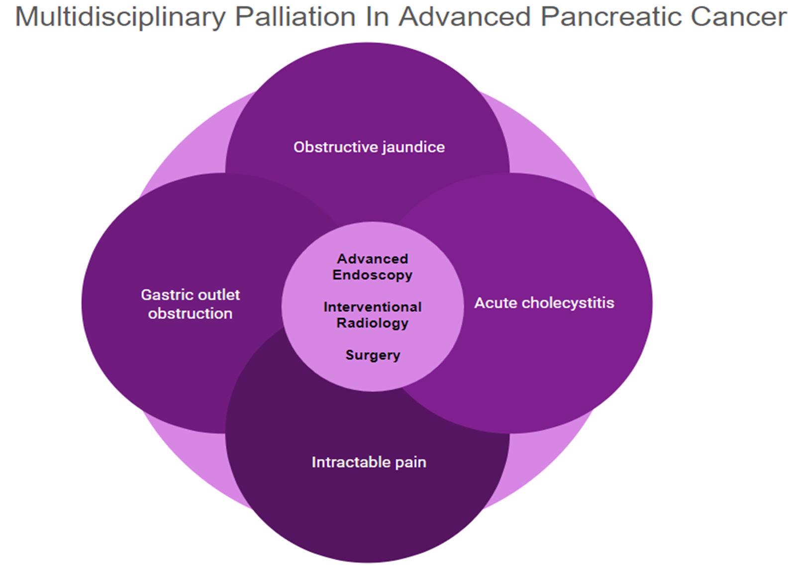
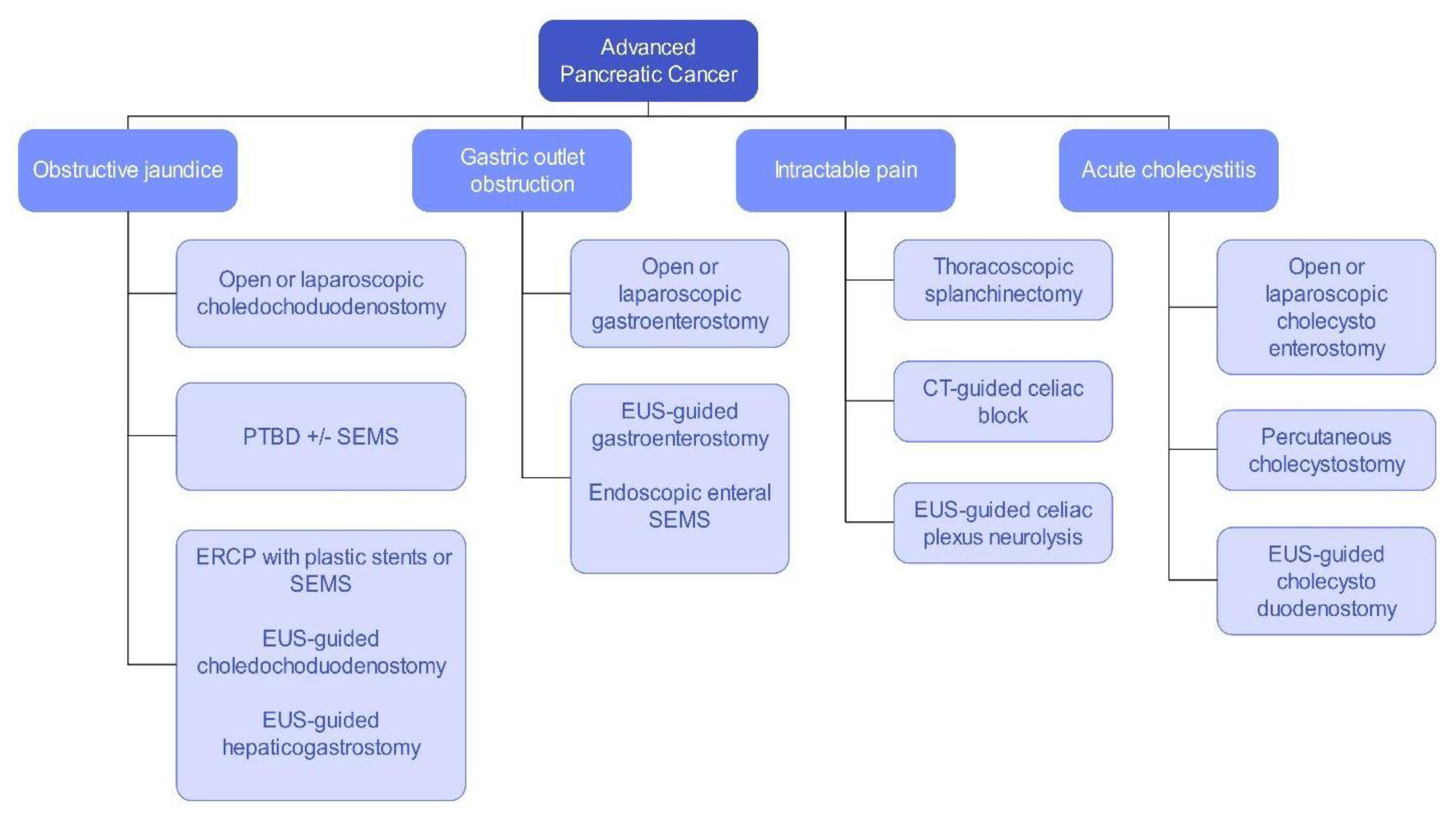
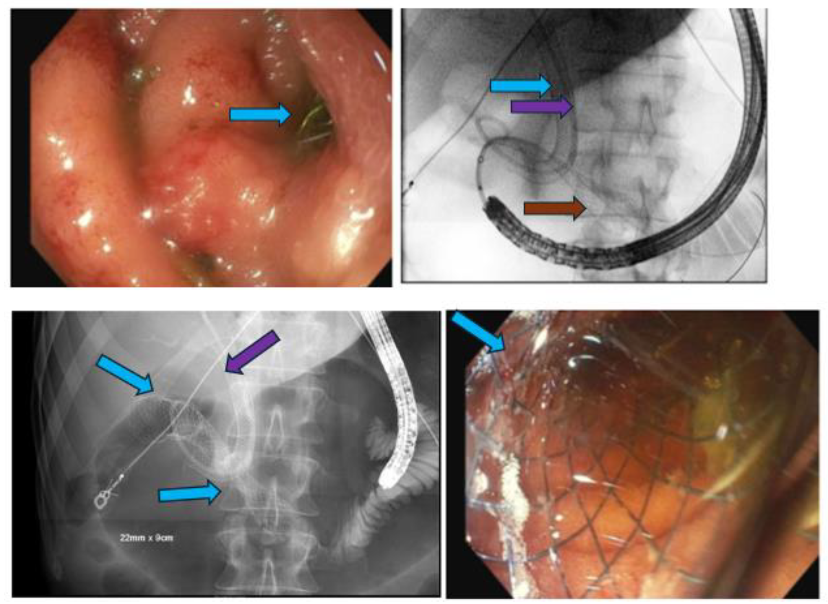
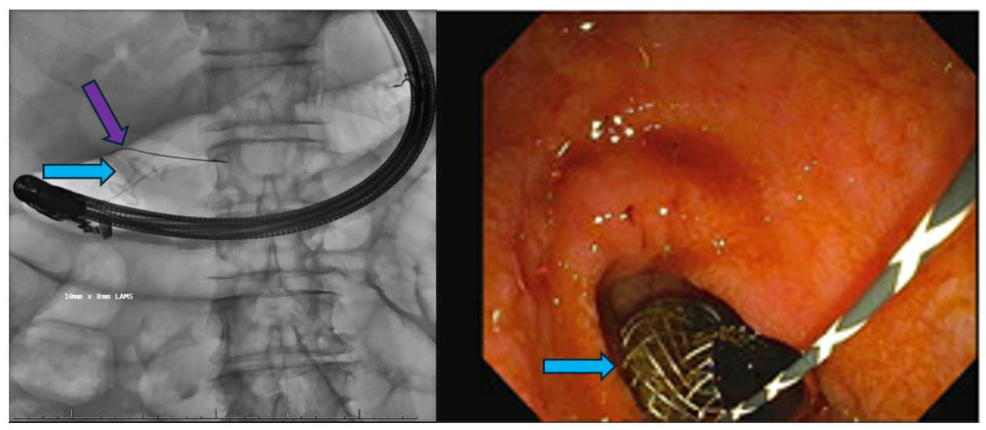
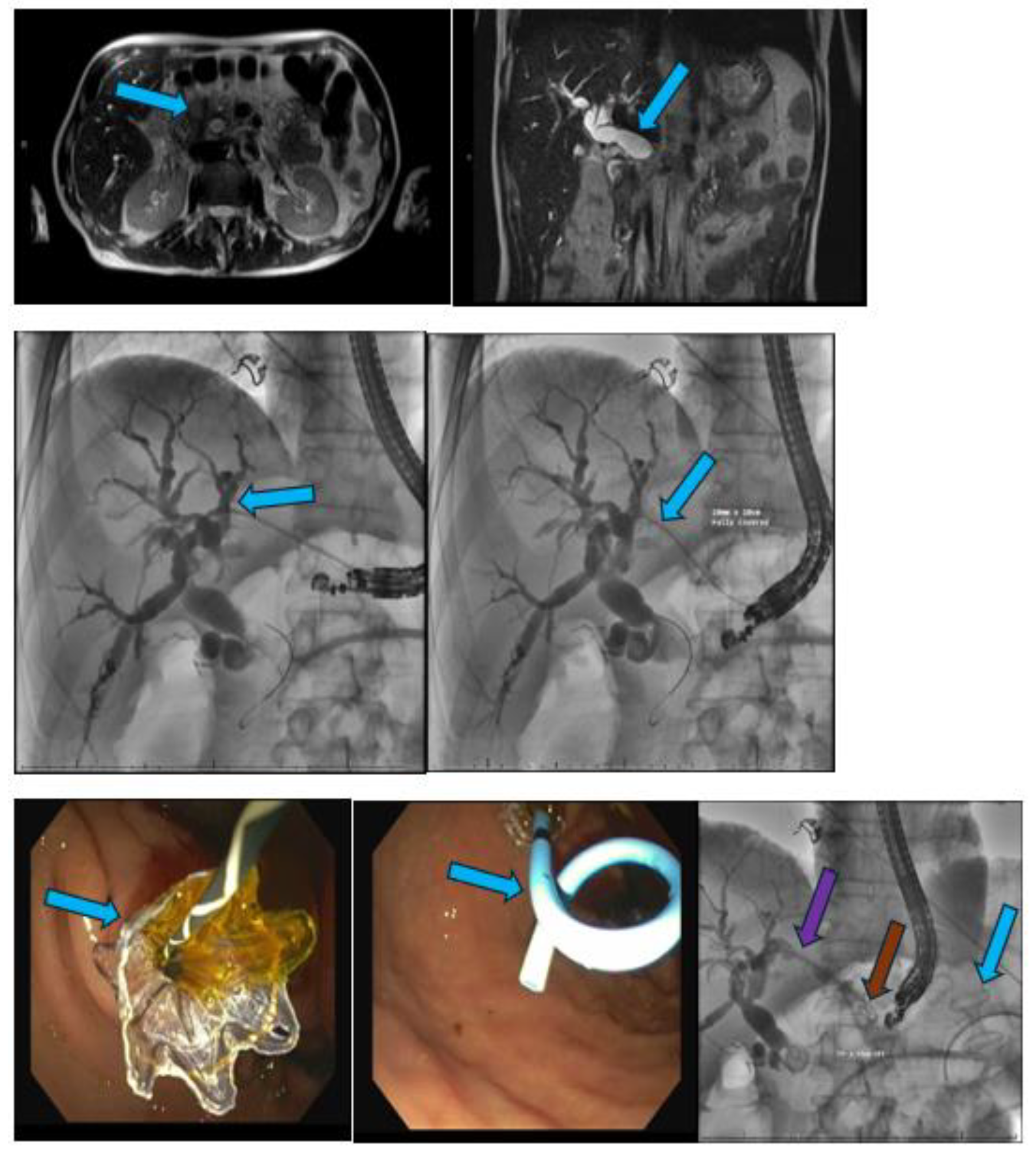
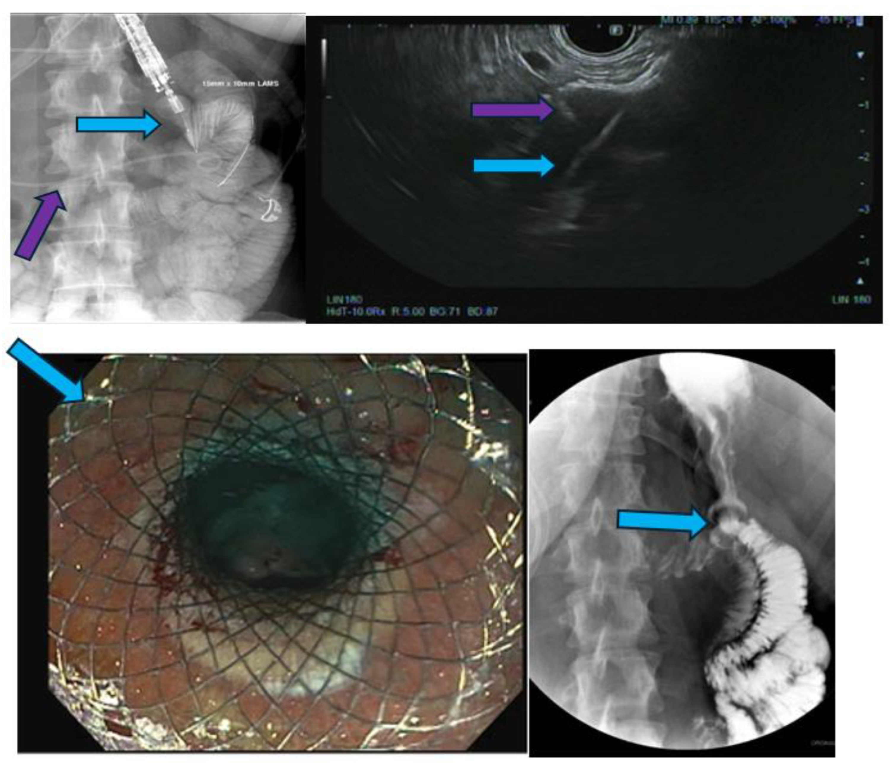
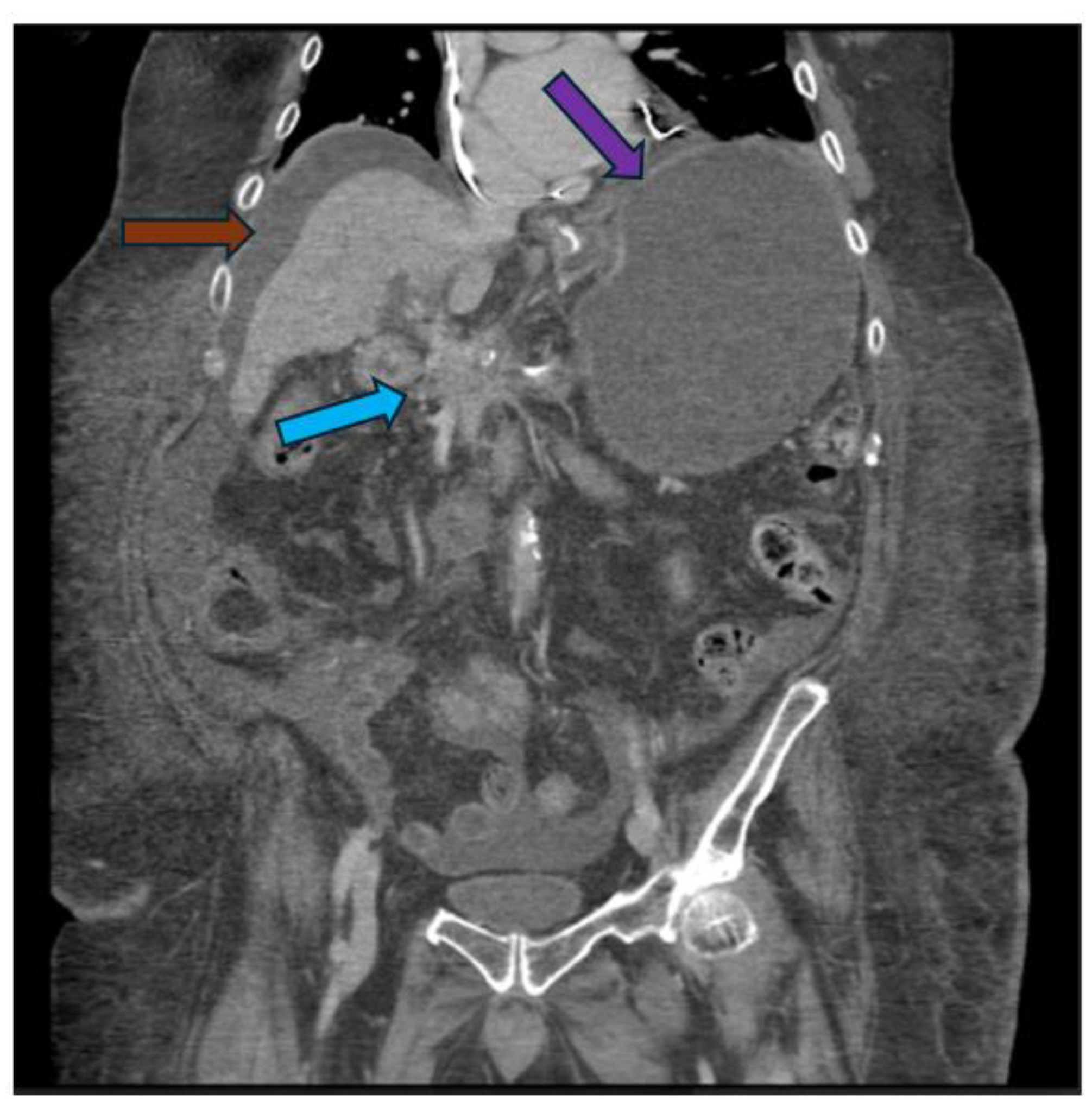
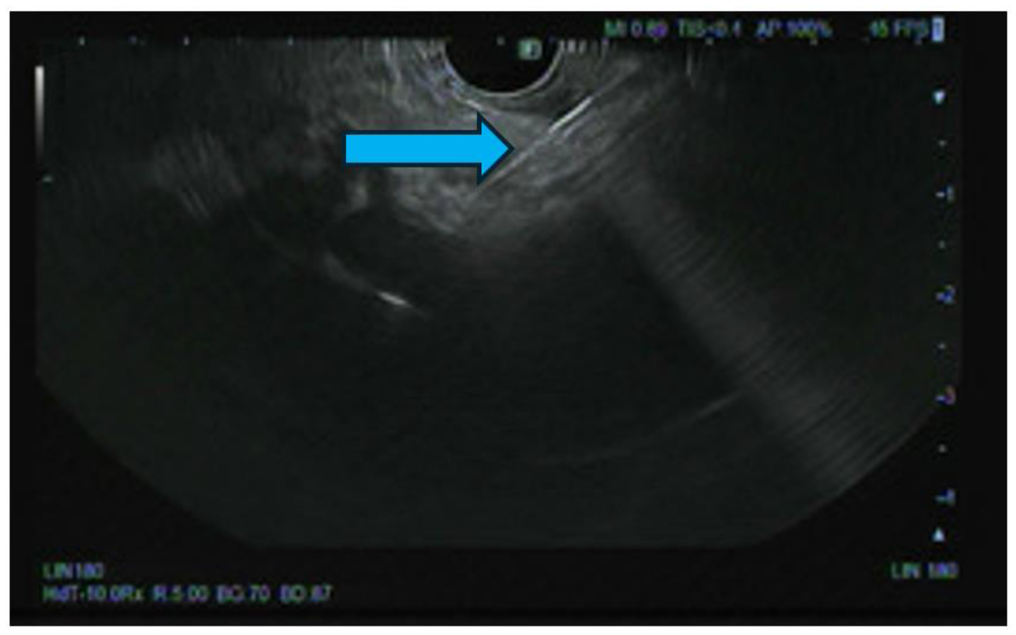
Pancreatic cancer is an aggressive malignancy, and the current 5-year survival rate in the United States, according to the Surveillance, Epidemiology, and End Results Program data, approximates 12%. Although the current standard for resectable pancreatic cancer most commonly includes neoadjuvant chemotherapy prior to a curative resection, surgery in the majority of patients has historically been palliative. The latter interventions include open or laparoscopic bypass of the bile duct or stomach in cases of obstructive jaundice or gastric outlet obstruction, respectively. Non-surgical interventional therapies started with percutaneous transhepatic biliary drainage (PTBD), both as a palliative maneuver in unresectable patients with obstructive jaundice and to improve liver functions in patients in whom surgery was delayed. Likewise, interventional radiologic techniques included placement of plastic and ultimately self-expandable metal stents (SEMS) through PTBD tracts in patients unresectable for cure as well as percutaneous cholecystostomy in patients who developed cholecystitis in the context of malignant obstructive jaundice. Endoscopic retrograde cholangiopancreatography (ERCP) and stent placement (plastic/SEMS) was subsequently used both preoperatively and palliatively, and this was followed by, or undertaken in conjunction with, endoscopic gastro-duodenal SEMS placement for gastric outlet obstruction. Although endoscopic ultrasound (EUS) was initially used to cytologically diagnose and stage pancreatic cancer, early palliation included celiac block or ablation for intractable pain. However, it took the development of lumen-apposing metal stents (LAMS) to facilitate a myriad of palliative procedures: Cholecystoduodenal, choledochoduodenal, gastrohepatic and gastroenteric anastomoses for cholecystitis, obstructive jaundice, and gastric outlet obstruction, respectively. In this review, we synopse these procedures which have variably supplanted surgery for the palliation of pancreatic cancer in this rapidly evolving field.

Keywords: 
;  ;  ;  ;  ;  

1. Introduction

Pancreatic cancer is the 3rd leading cause of cancer-related death in the US after lung cancer and colon cancer. It is projected to become the 2nd leading cause of cancer-related death by 2030. Most cases of pancreatic cancer are diagnosed at stage IV. The five-year survival rate for pancreatic cancer has increased from 7% to 13% over the past 10 years (Source: American Cancer Society. Cancer Facts & Figures 2024. Atlanta: American Cancer Society; 2024.). The majority of pancreatic cancers are located in the head of the pancreas and can result in biliary and gastric outlet obstruction. Approximately 15-20% of pancreatic tumors are resectable at diagnosis (Source: American Cancer Society. Cancer Facts & Figures 2024. Atlanta: American Cancer Society; 2024.). The current standard for resectable pancreatic cancer most commonly includes neoadjuvant chemotherapy prior to a curative resection [1,2]. Advanced pancreatic cancer may lead to several sequelae including obstructive jaundice, gastric outlet obstruction (GOO), intractable pain and acute cholecystitis, which can significantly affect the quality of life in patients. In this review, we describe the evolution of palliation in advanced pancreatic cancer from surgery to minimally invasive modalities involving interventional radiology and advanced endoscopy.
Figure 1. A schematic representation of the multimodal management of advanced pancreatic cancer sequelae.
Preprints 140136 g001

Surgical Palliation

Surgery in most patients with advanced pancreatic cancer has historically been palliative in nature. The latter interventions include open or laparoscopic bypass of the bile duct or stomach in cases of obstructive jaundice or GOO, respectively. A study of 42 patients, who underwent either palliative gastrojejunostomy and hepaticojejunostomy, reported severe postoperative morbidity and mortality rates and emphasized patient selection for acceptable postsurgical outcomes [3]. Espat et al. noted that the practice of routine prophylactic bypass procedures was not supported and should only be performed in patients with obstructive jaundice who fail endoscopic stent placement or in patients with GJ and confirmed GOO [4].
Lyons et al. demonstrated that prophylactic duodenal, biliary and double bypasses in patients with unresectable pancreatic cancer at index laparotomy was not associated with fewer invasive procedures and did not reduce the number of hospital days [5]. In the study, patients who underwent biliary bypass rarely required additional interventions for biliary obstruction and similarly, patients who underwent duodenal bypass rarely required interventions for GOO. The double bypass group was associated with just as many postoperative interventions and accrued hospital days as the duodenal bypass and biliary bypass groups [5]. In contrast, a prospective randomized controlled trial of 88 patients demonstrated that prophylactic GJ decreased the incidence of late GOO and that a retrocolic GJ should be performed routinely for surgical palliation of unresectable periampullary carcinoma [6]. In yet another study of 65 patients who were found to be unresectable at exploration and underwent either a single bypass (HJ) or a double-bypass (HJ and retrocolic GJ), prophylactic GJ decreased the incidence of GOO without increasing the rate of complications [7]. There were no significant differences in the quality of life between the single and double bypass groups. The study recommended double bypass rather than a single bypass [7].
When comparing open GJ to laparoscopic GJ for the treatment of malignant GOO, several studies have favored laparoscopic GJ. A randomized controlled trial of 24 patients demonstrated a shorter time to oral intake and a lower rate of delayed gastric emptying with laparoscopic GJ [8]. In a retrospective review of 20 patients who underwent a palliative GJ for malignant GOO, no significant differences in surgical outcomes were noted between the open and laparoscopic GJ groups although the study had a limited sample size [9].

Interventional Radiology Palliation

Non-surgical interventional therapies started with percutaneous transhepatic biliary drainage (PTBD), both as a palliative maneuver in unresectable patients with obstructive jaundice and to improve liver functions in patients in whom surgery was delayed (Figure 2). PTBD, and insertion of plastic or self-expandable metal stents (SEMS) through the PTBD tract in some cases, was previously considered as the standard method for biliary drainage in patients with non-resectable malignant obstructive jaundice before it was supplanted by ERCP and biliary stent placement (Figure 3). PTBD remains a useful tool especially in patients with a large disease burden or who are poor surgical candidates or those who have had unsuccessful surgical or endoscopic drainage [10]. The technical success of PTBD has been reported to be close to 100% whereas the clinical success rates may vary between 76.5% to 98% [11,12]. Approximately 20-25% of patients can develop complications i.e. cholangitis, bleeding, catheter occlusion or misplacement [11]. In a retrospective study of 16,822 patients who underwent PTBD for pancreaticobiliary malignancies, the 30-day mortality was 23.1% especially in older men with increased comorbidities [13]. In a large retrospective study of 14,808 patients using the Surveillance, Epidemiology, and End Results–Medicare database comparing overall survival with ERCP vs. PTBD in patients with pancreatic cancer, ERCP was associated with reduced mortality (adjusted hazard ratio [aHR] .67; 95% confidence interval [CI], .60-.75) [14].
Percutaneous cholecystostomy (PC) has been shown to be an effective option for acute cholecystitis especially in nonsurgical candidates [15,16]. PC has also been performed in patients who developed cholecystitis in the setting of malignant obstructive jaundice. In a case-control study of 206 patients with malignancy, PC was associated with a higher rate of acute cholecystitis resolution compared to antibiotics with abdominal malignancy increasing the odds of resolution [17]. A study of 283 patients with acute cholecystitis during neoadjuvant therapy for localized pancreatic cancer revealed that acute cholecystitis occurred in 6% of patients and PC did not delay the completion of neoadjuvant therapy and surgery [18]. Additional studies are warranted to explore the role of PC in patients with unresectable pancreatic cancer.

Advanced Endoscopy Palliation

Endoscopic Retrograde Cholangiopancreatography

An endoscopic approach has been the mainstay of palliation in patients with unresectable cancer since the 1990s. Endoscopic retrograde cholangiopancreatography (ERCP) and placement of plastic stent or self-expandable metal stents (SEMS) has been used both preoperatively and palliatively. ERCP was first performed as a diagnostic modality in 1968 by Dr. William S. McCune, an obstetrician. The first biliary sphincterotomy was performed in Japan and Germany in 1974 and the first plastic prosthesis inserted into the biliary tree endoscopically was subsequently reported by Soehendra et al [19]. Since then, ERCP has become a diagnostic and therapeutic tool for a variety of hepatoopancreaticobiliary pathologies.
While surgical and endoscopic biliary drainage have similar rates of technical success and efficacy, endoscopic biliary drainage is associated with fewer complications, improved quality of life, shorter hospital stay and a lower cost. Endoscopic transpapillary stenting has become the preferred therapy of choice for patients with obstructive jaundice in the setting of unresectable pancreatic cancer and high rates of improvement in jaundice and pruritus have been reported with transpapillary drainage.
Plastic stents were initially used for biliary drainage although they tend to occlude over several weeks or months after placement. In a study of 49 patients with resectable or locally advanced pancreatic adenocarcinoma and biliary obstruction, approximately 27 out of 49 patients required repeat ERCP for stent exchange with a median of 82.5 days after original stent placement [20]. SEMS were introduced in 1989 and have a larger diameter (10 Fr or 3.3 mm) compared to plastic stents [21,22]. Multiple studies have compared plastic stents vs. SEMS in patients with malignant biliary obstruction. A meta-analysis of 13 studies revealed lower stent dysfunction (21.6% vs 46.8%, P < 0.00001) and a lower rate of reintervention with SEMS (21.6% vs 56.6%, P < 0.00001) compared to plastic stents with no difference in complications [23]. Additionally, in the SEMS group, the mean survival rate was higher, the stent patency period was longer and there was a lower cumulative cost per patient [23]. Although SEMS, uncovered, partially covered or completely covered, are initially more expensive than plastic stents, the total costs after 1 year are not significantly different between the two groups [24]. In a randomized controlled trial of 119 patients with pancreatic cancer on neoadjuvant therapy, covered and uncovered SEMS were associated with similar rates of biliary drainage. Stent complications depended on stent type, stent length and the presence of a gallbladder [25]. Plastic stents remain commonly used as they are relatively easy to place. Covered SEMS have been associated with a higher rate of stent dysfunction from sludge, stent migration and tumor overgrowth compared to uncovered or partially covered SEMS which have been associated with higher rates of tumor ingrowth.
The complication rate for ERCP in the setting of malignant obstructive jaundice has been reported as 13% [26]. The most common adverse events of endoscopic transpapillary biliary drainage include pancreatitis, cholangitis, bleeding, perforation, cholecystitis and liver abscess. The risk of bleeding has been demonstrated to be increased with sphincterotomy. In a retrospective study of 73 patients with pancreatic cancer, palliative biliary drainage did not have an effect on median progression-free survival and overall survival [27]. In a study of 292 patients, among which 196 had pancreatic cancer and 96 patients had nonpancreatic cancer, who underwent stent placement for malignant GOO, the median survival post-stent placement was similar despite better overall survival in patients with nonpancreatic cancer [28]. Overall survival was reduced in patients with pancreatic cancer (13.7 vs. 17.1 months, p = 0.004) [28]. GOO was noted to be a marker for poor survival regardless of the type of malignancy. Factors which were associated with better post-stent survival in both groups included chemotherapy and the absence of distant metastasis.

Enteral Self-Expandable Metal Stents

Additionally, endoscopic gastro-duodenal SEMS have been utilized for GOO (Panel A). In a prospective, multicenter study of 39 patients with malignant GOO, duodenal stenting had a technical success rate of 100% and clinical success rate of 92.3% of patients [29]. Stent dysfunction occurred in 7.7% of patients [29]. In a study comparing enteral stenting to surgical gastroenterostomy, there were higher rates of persistent nausea and vomiting and increased length of stay in the surgical GE group (p = 0.0102). In a RCT of 18 patients, endoscopic stenting was more effective with respect to operative time, restoration of oral intake and median hospitalization although no statistically significant differences between the enteral stenting or gastrojejunostomy groups were noted with respect to morbidity, mortality, delayed gastric emptying and clinical outcomes at 3-month follow-up [30]. Complications occur in 2% to 12% and include occlusion (most common) by tumor ingrowth or food bolus, migration, hemorrhage, perforation, aspiration pneumonia.
Panel A. Endoscopic image reveals the biliary SEMS (top left, blue arrow) compressed by a malignant obstruction of the duodenum. Fluoroscopy image (top right) demonstrates a 7-French by 15 cm plastic stent (purple arrow) in a previously placed biliary SEMS (blue arrow). Note marked superior displacement of the biliary stents. The brown arrow demonstrates the placement of a duodenal SEMS in this patient with gastric outlet obstruction from advanced pancreatic cancer. Fluoroscopy image (bottom left) reveals a fully expanded, duodenal SEMS (blue arrows). The purple arrow demonstrates previously placed biliary stents. Endoscopic image (bottom right) of a fully expanded, duodenal SEMS (blue arrow). Abbreviation: SEMS = self-expandable metal stent.
Preprints 140136 sch001

Endoscopic Ultrasound-Guided Therapies

Endoscopic ultrasound (EUS) was initially used to cytologically diagnose and stage pancreatic cancer. In a comparative study, EUS was noted to be more accurate than CT for staging pancreatic malignancies, including vascular invasion and local resectability, and was significantly better than CT for T1, T2 and T3 tumors [31]. Several additional studies have revealed a high sensitivity, high specificity and accuracy of EUS compared to CT for pancreatic malignancy [32].
The role of EUS has recently evolved from a purely diagnostic technique to a complex, interventional modality. ERCP is often the first modality for biliary drainage in the setting of obstructive jaundice. Biliary cannulation is dependent upon several factors including patient anatomy and endoscopist expertise. In patients with pancreatic malignancy, biliary cannulation may be challenging due to the distortion of the ampulla, a malignant biliary stricture which is difficult to traverse, or biliary or duodenal obstruction. Advanced cannulation techniques (i.e. needle knife pre-cut, double guidewire and pancreatic septotomy) may be required, but confer an increased risk of adverse events. EUS-guided biliary drainage (EUS-BD) has increasingly become a feasible and efficacious choice for obstructive jaundice. A meta-analysis of 6 randomized controlled trials of 577 patients comparing EUS-BD and ERCP-biliary drainage (ERCP-BD) demonstrated similar efficacy and safety. EUS-BD, however, was associated with a significantly lower risk of reintervention, post-procedure pancreatitis, tumor ingrowth/overgrowth and reduced hospital stay [33].

Lumen-Apposing Metal Stents

EUS-guided placement of lumen-apposing metal stents (LAMS) was first described in 2012 by Binmoeller and Shah who successfully created a gastroenterostomy in a pig model [34]. Although LAMS were initially developed for the use of peripancreatic fluid collections, they have been utilized in multiple gastroenterologic applications [35,36]. LAMS have allowed a myriad of palliative procedures to include cholecystoduodenal, choledochoduodenal, gastrohepatic and gastroenteric anastomoses for cholecystitis, obstructive jaundice, and gastric outlet obstruction, respectively (Panel B, C and D, Figure 4) [37]. Electrocautery-enhanced delivery systems allow direct access to the target lumen and have simplified the multi-step procedure of EUS-guided drainage. A meta-analysis of 14 studies involving 620 patients who underwent EC-LAMS placement after a failed ERCP for malignant biliary obstruction (MDBO), the pooled rate of technical success was 96.7%, the pooled rate of clinical success was 91% and the rate of adverse events were noted to be 17.5% [38]. The overall re-intervention rate in the meta-analysis was 7.3% [38].
EUS-guided choledochoduodenostomy (EUS-CDS) has also been demonstrated to be a promising technique for the management of MDBO. A multicenter, randomized controlled trial of 144 patients with MDBO secondary to borderline resectable, locally advanced, or unresectable periampullary cancers, EUS-CDS was noted to be an efficient and safe alternative to ERCP with metal stent placement, although EUS-CDS was not superior with regard to stent function [39].
A network meta-analysis of 6 randomized controlled trials and 583 patients compared the effectiveness of EUS-CDS with LAMS, EUS-CDS with SEMS, EUS-hepaticogastrostomy, ERCP and PTBD performed upfront for the management of MDBO [40]. EUS-CDS with LAMS was associated with the highest rate of technical and clinical success and was noted to be significantly superior to ERCP as an upfront modality. Additionally, PTBD was associated with an increased risk of adverse events [40].
Panel B. A fluoroscopic image (left) reveals an endoscopic ultrasound-guided choledochoduodenostomy using a 10 mm x 8 mm lumen-apposing metal stent (blue arrow) and a guidewire (purple arrow) in an air-filled, dilated common bile duct in a patient with a malignant distal biliary obstruction from advanced pancreatic cancer. Endoscopic image (right) reveals the expanded, lumen-apposing metal stent with the guidewire in place (arrow).
Preprints 140136 sch002
While surgical gastroenterostomy and enteral stenting were the gold standard for the management of GOO, surgical GE is limited by its invasive nature and high morbidity rates whereas enteral stenting is limited by stent patency duration and higher rates of re-intervention. EUS-GE, initially described in 2002 by Fritscher-Ravens in a porcine animal model [34,41,42]. EUS-GE was subsequently used in clinical practice following the development of LAMS. The pooled technical success rate and the pooled clinical success rate for EUS-GE have been reported to be 92% and 90%, respectively [43]. Several studies have demonstrated that while EUS-GE is comparable to SGJ, EUS-GE is associated with fewer associated adverse events, earlier resumption of diet, and a shorter hospital stay. EUS-GE has been noted to have a lower risk of obstruction and a decreased symptom recurrence compared to ES. EUS-GE has also been associated with fewer reinterventions, improved stent patency and earlier oral intake compared to ES [44]. In a meta-analysis of 61 studies comparing enteral stenting to endoscopic or surgical GJ for malignant GOO, the clinical efficacy between the 3 groups was similar although duodenal SEMS were associated with a lower procedure-related bleeding and a higher rate of reintervention [45]. The European Society of Gastrointestinal Endoscopy has recommended EUS-GE for malignant gastric obstruction as an alternative to enteral stenting or surgery, in an expert setting [46].
Panel C. An axial view of a magnetic resonance cholangiopancreatography (top left image) reveals a 2.1 cm pancreatic head mass (blue arrow) and marked intrahepatic and extrahepatic biliary dilation (top right image) with the common bile duct approximately 1.98 cm in diameter (blue arrow). Fluoroscopy image (middle left) reveals puncture from the stomach to the left intrahepatic duct using a 19-gauge needle, a guidewire in place and an intraoperative cholangiogram with significant intrahepatic and extrahepatic biliary dilation. Fluoroscopy image (middle right, arrow) and endoscopy image (bottom left, arrow) reveals a 10 mm x 10 cm fully covered, self-expandable metal stent which was inserted into the biliary tree to create an endoscopic ultrasound-guided hepaticogastrostomy. Subsequently, a 7-French x 15 mm double-pigtail stent was inserted in the self-expandable metal stent (bottom middle image, arrow) for biliary drainage. The bottom right image reveals the self-expandable biliary stent (purple arrow) and the double pigtail stent (brown arrow). Additionally, a lumen-apposing metal stent was used to perform a gastroenterostomy due to gastric outlet obstruction (blue arrow). Image Courtesy: Amar Vedamurthy, Division of Gastroenterology and Hepatology, Virginia Mason Franciscan Health, Seattle, Washington, USA.
Preprints 140136 sch003
Panel D. Fluoroscopy image (A) reveals a distal stent delivery of a15 mm x 10 mm lumen-apposing metal stent (blue arrow) from the stomach to the small bowel. A nasobiliary drain (purple arrow) was used to instill contrast and methylene blue to localize and distend the small bowel. Endoscopic ultrasound image (B) reveals needle puncture (blue arrow) from the gastric wall to the small bowel. Note the delivery of the distal flange of the LAMS (purple arrow). Endoscopic image (C) of a fully deployed LAMS (blue arrow). Upper gastrointestinal series (D) with oral contrast demonstrates passage of oral contrast into the small bowel which demonstrates stent patency (blue arrow).
Preprints 140136 sch004
Figure 4. A coronal view of a computed tomography scan of the abdomen and pelvis an infiltrative pancreatic head mass (blue arrow) and a dilated, obstructed stomach (purple arrow). Ascitic fluid is demonstrated around the liver (brown arrow). Significant ascites usually precludes endoscopic ultrasound-guided gastroenterostomy.
Preprints 140136 g004
Several studies have revealed that EUS-gallbladder drainage using LAMS is a safe and effective technique in patients with acute cholecystitis who are non-surgical candidates [47,48,49,50]. However, many studies were not restricted solely to patients with unresectable pancreatic cancer.
In a retrospective study, EUS-gallbladder drainage using LAMS in patients with acute cholecystitis, a technical success rate of 94.8% and a clinical success rate of 100% was noted [48].

EUS-Guided Celiac Plexus Neurolysis

EUS has been utilized to perform a celiac block or ablation for palliation of unresectable pancreatic malignancy. EUS-guided celiac plexus neurolysis (EUS-CPN) involves the injection of alcohol and local anesthesia into the celiac plexus for ablation of the nerve tissue. The transgastric approach has been reported to be safer and more accessible than the percutaneous approach. EUS-CPN typically provides improvement in pain for 4-8 weeks. The efficacy of EUS-CPN varies from 50-94% in various studies [51].
Figure 5. Endoscopic ultrasound image reveals needle puncture (arrow) from the gastric wall to the celiac plexus. Alcohol and local anesthesia were injected to provide neurolysis in a patient with advanced pancreatic cancer.
Preprints 140136 g005

Novel Endoscopic Ultrasound-Guided Therapies

EUS-guided RFA (EUS-RFA) is an emerging modality and has been reported to be a safe and efficacious technique in patients with advanced pancreatic cancer [52]. EUS-RFA may reduce tumor burden and improve the efficacy of chemotherapy [53,54].
EUS has also been employed in the placement of fiducials, which are radiographic markers used to define the borders of the pancreatic malignancy, and can improve the accuracy of target delineation in stereotactic body radiation therapy [55,56,57].
Finally, EUS-guided brachytherapy with radioactive iodine seeds has been reported to be efficacious in several studies [58]. In a study of 15 patients with unresectable pancreatic cancer, 30% of patients had a favorable response [59]. In a study of 8 patients with T4 pancreatic adenocarcinoma with a median follow-up period of 8.3 months, EUS-brachytherapy was noted to be favorable, mostly due to decreased pain, in 4 out of 8 patients [60]. No local complications were reported [60]. Treatment with iodine-125 seed in patients with unresectable pancreatic cancer has also been demonstrated to prolong survival, biliary stent patency and time to GOO as well as improve patient quality of life by reduction in pain [61].

Conclusions

Palliation of unresectable pancreatic cancer has largely evolved from open or laparoscopic bypass of the bile duct or stomach in cases of obstructive jaundice or GOO, respectively, to minimally invasive therapies involving interventional radiology and advanced endoscopy. PTBD was initially utilized for patients with malignant obstructive jaundice and included placement of plastic stents and, ultimately SEMS, through PTBD tracts. Percutaneous cholecystostomy has also been used for the management of acute cholecystitis in the setting of malignant obstructive jaundice. Subsequently, ERCP with plastic stent or SEMS, both preoperatively and palliatively, became the mainstay for obstructive jaundice whereas endoscopic gastroduodenal SEMS were utilized for GOO. The adoption of EUS and LAMS allowed a multitude of palliative procedures to include cholecystoduodenal, choledochoduodenal, gastrohepatic and gastroenteric anastomoses for cholecystitis, obstructive jaundice, and gastric outlet obstruction, respectively. EUS-CPN has been demonstrated to be efficacious with regard to intractable pain in the setting of unresectable pancreatic cancer. Additional EUS-guided therapies, including hepaticogastrostomy followed by SEMS placement for biliary decompression, radiofrequency ablation, fiducial placement and radioiodine pellets, all serve as novel tools for palliation. Multidisciplinary collaboration, between surgeons, interventional radiologists, advanced gastroenterologists and primary care providers, is paramount to a successful outcome in the palliation of advanced pancreatic cancer.

References

  1. Lambert A, Schwarz L, Ducreux M, Conroy T. Neoadjuvant Treatment Strategies in Resectable Pancreatic Cancer. Cancers (Basel). 2021;13(18). [CrossRef]
  2. Su YY, Chao YJ, Wang CJ, Liao TK, Su PJ, Huang CJ, et al. The experience of neoadjuvant chemotherapy versus upfront surgery in resectable pancreatic cancer: a cross sectional study. Int J Surg. 2023;109(9):2614-23. [CrossRef]
  3. Pencovich N, Orbach L, Lessing Y, Elazar A, Barnes S, Berman P, et al. Palliative bypass surgery for patients with advanced pancreatic adenocarcinoma: experience from a tertiary center. World Journal of Surgical Oncology. 2020;18(1):63. [CrossRef]
  4. Espat NJ, Brennan MF, Conlon KC. Patients with laparoscopically staged unresectable pancreatic adenocarcinoma do not require subsequent surgical biliary or gastric bypass. J Am Coll Surg. 1999;188(6):649-55; discussion 55-7. [CrossRef]
  5. Lyons JM, Karkar A, Correa-Gallego CC, D'Angelica MI, DeMatteo RP, Fong Y, et al. Operative procedures for unresectable pancreatic cancer: does operative bypass decrease requirements for postoperative procedures and in-hospital days? HPB (Oxford). 2012;14(7):469-75.
  6. Lillemoe KD, Cameron JL, Hardacre JM, Sohn TA, Sauter PK, Coleman J, et al. Is Prophylactic Gastrojejunostomy Indicated for Unresectable Periampullary Cancer?: A Prospective Randomized Trial. Annals of Surgery. 1999;230(3):322.
  7. Van Heek NT, De Castro SMM, van Eijck CH, van Geenen RCI, Hesselink EJ, Breslau PJ, et al. The Need for a Prophylactic Gastrojejunostomy for Unresectable Periampullary Cancer: A Prospective Randomized Multicenter Trial With Special Focus on Assessment of Quality of Life. Annals of Surgery. 2003;238(6):894-905. [CrossRef]
  8. Navarra G, Musolino C, Venneri A, De Marco ML, Bartolotta M. Palliative antecolic isoperistaltic gastrojejunostomy: a randomized controlled trial comparing open and laparoscopic approaches. Surg Endosc. 2006;20(12):1831-4. [CrossRef]
  9. Guzman EA, Dagis A, Bening L, Pigazzi A. Laparoscopic gastrojejunostomy in patients with obstruction of the gastric outlet secondary to advanced malignancies. Am Surg. 2009;75(2):129-32. [CrossRef]
  10. McGrath PC, McNeill PM, Neifeld JP, Bear HD, Parker GA, Turner MA, et al. Management of biliary obstruction in patients with unresectable carcinoma of the pancreas. Ann Surg. 1989;209(3):284-8. [CrossRef]
  11. Ahmed O, Lee JH. Preoperative biliary drainage for pancreatic cancer. Int J Gastrointest Interv. 2018;7(2):67-73. [CrossRef]
  12. Zhang GY, Li WT, Peng WJ, Li GD, He XH, Xu LC. Clinical outcomes and prediction of survival following percutaneous biliary drainage for malignant obstructive jaundice. Oncol Lett. 2014;7(4):1185-90. [CrossRef]
  13. Rees J, Mytton J, Evison F, Mangat KS, Patel P, Trudgill N. The outcomes of biliary drainage by percutaneous transhepatic cholangiography for the palliation of malignant biliary obstruction in England between 2001 and 2014: a retrospective cohort study. BMJ Open. 2020;10(1):e033576. [CrossRef]
  14. Tavakkoli A, Elmunzer BJ, Waljee AK, Murphy CC, Pruitt SL, Zhu H, et al. Survival analysis among unresectable pancreatic adenocarcinoma patients undergoing endoscopic or percutaneous interventions. Gastrointest Endosc. 2021;93(1):154-62.e5. [CrossRef]
  15. Aroori S, Mangan C, Reza L, Gafoor N. Percutaneous Cholecystostomy for Severe Acute Cholecystitis: A Useful Procedure in High-Risk Patients for Surgery. Scandinavian Journal of Surgery. 2019;108(2):124-9. [CrossRef]
  16. Teoh WM, Cade RJ, Banting SW, Mackay S, Hassen AS. Percutaneous cholecystostomy in the management of acute cholecystitis. ANZ J Surg. 2005;75(6):396-8. [CrossRef]
  17. David S, Celia Robinson L, Angela L, Heather G, Brian B. Use of non-operative treatment and interval cholecystectomy for cholecystitis in patients with cancer. Trauma Surgery & Acute Care Open. 2020;5(1):e000439.
  18. Jariwalla NR, Khan AH, Dua K, Christians KK, Clarke CN, Aldakkak M, et al. Management of Acute Cholecystitis during Neoadjuvant Therapy in Patients with Pancreatic Adenocarcinoma. Ann Surg Oncol. 2019;26(13):4515-21. [CrossRef]
  19. Soehendra N, Reynders-Frederix V. Palliative bile duct drainage - a new endoscopic method of introducing a transpapillary drain. Endoscopy. 1980;12(1):8-11. [CrossRef]
  20. Boulay BR, Gardner TB, Gordon SR. Occlusion rate and complications of plastic biliary stent placement in patients undergoing neoadjuvant chemoradiotherapy for pancreatic cancer with malignant biliary obstruction. J Clin Gastroenterol. 2010;44(6):452-5. [CrossRef]
  21. Ballard DD, Rahman S, Ginnebaugh B, Khan A, Dua KS. Safety and efficacy of self-expanding metal stents for biliary drainage in patients receiving neoadjuvant therapy for pancreatic cancer. Endosc Int Open. 2018;6(6):E714-e21. [CrossRef]
  22. Jeong S. Basic Knowledge about Metal Stent Development. Clin Endosc. 2016;49(2):108-12. [CrossRef]
  23. Zorrón Pu L, de Moura EG, Bernardo WM, Baracat FI, Mendonça EQ, Kondo A, et al. Endoscopic stenting for inoperable malignant biliary obstruction: A systematic review and meta-analysis. World J Gastroenterol. 2015;21(47):13374-85.
  24. Walter D, van Boeckel PG, Groenen MJ, Weusten BL, Witteman BJ, Tan G, et al. Cost Efficacy of Metal Stents for Palliation of Extrahepatic Bile Duct Obstruction in a Randomized Controlled Trial. Gastroenterology. 2015;149(1):130-8. [CrossRef]
  25. Seo DW, Sherman S, Dua KS, Slivka A, Roy A, Costamagna G, et al. Covered and uncovered biliary metal stents provide similar relief of biliary obstruction during neoadjuvant therapy in pancreatic cancer: a randomized trial. Gastrointest Endosc. 2019;90(4):602-12.e4. [CrossRef]
  26. Tarar ZI, Farooq U, Gandhi M, Saleem S, Daglilar E. Safety of endoscopic retrograde cholangiopancreatography (ERCP) in cirrhosis compared to non-cirrhosis and effect of Child-Pugh score on post-ERCP complications: a systematic review and meta-analysis. Clin Endosc. 2023;56(5):578-89. [CrossRef]
  27. Erdoğan AP, Ekinci F, Yıldırım S, Özveren A, Göksel G. Palliative Biliary Drainage Has No Effect on Survival in Pancreatic Cancer: Medical Oncology Perspective. J Gastrointest Cancer. 2022;53(1):52-6. [CrossRef]
  28. Oh SY, Edwards A, Mandelson M, Ross A, Irani S, Larsen M, et al. Survival and clinical outcome after endoscopic duodenal stent placement for malignant gastric outlet obstruction: comparison of pancreatic cancer and nonpancreatic cancer. Gastrointest Endosc. 2015;82(3):460-8.e2. [CrossRef]
  29. Sasaki R, Sakai Y, Tsuyuguchi T, Nishikawa T, Fujimoto T, Mikami S, et al. Endoscopic management of unresectable malignant gastroduodenal obstruction with a nitinol uncovered metal stent: A prospective Japanese multicenter study. World J Gastroenterol. 2016;22(14):3837-44. [CrossRef]
  30. Fiori E, Lamazza A, Volpino P, Burza A, Paparelli C, Cavallaro G, et al. Palliative management of malignant antro-pyloric strictures. Gastroenterostomy vs. endoscopic stenting. A randomized prospective trial. Anticancer Res. 2004;24(1):269-71.
  31. Gress FG, Hawes RH, Savides TJ, Ikenberry SO, Cummings O, Kopecky K, et al. Role of EUS in the preoperative staging of pancreatic cancer: a large single-center experience. Gastrointest Endosc. 1999;50(6):786-91. [CrossRef]
  32. Yousaf MN, Chaudhary FS, Ehsan A, Suarez AL, Muniraj T, Jamidar P, et al. Endoscopic ultrasound (EUS) and the management of pancreatic cancer. BMJ Open Gastroenterol. 2020;7(1). [CrossRef]
  33. Barbosa EC, Santo PAdE, Baraldo S, Nau AL, Meine GC. EUS- versus ERCP-guided biliary drainage for malignant biliary obstruction: a systematic review and meta-analysis of randomized controlled trials. Gastrointestinal Endoscopy. 2024;100(3):395-405.e8. [CrossRef]
  34. Binmoeller KF, Shah JN. Endoscopic ultrasound-guided gastroenterostomy using novel tools designed for transluminal therapy: a porcine study. Endoscopy. 2012;44(5):499-503. [CrossRef]
  35. Anderloni A, Troncone E, Fugazza A, Cappello A, Del Vecchio Blanco G, Monteleone G, Repici A. Lumen-apposing metal stents for malignant biliary obstruction: Is this the ultimate horizon of our experience? World J Gastroenterol. 2019;25(29):3857-69.
  36. Goldman I, Ji K, Scheinfeld MH, Hajifathalian K, Morgan M, Yang J. A stent of strength: use of lumen-apposing metal stents (LAMS) for biliary pathologies and other novel applications. Abdominal Radiology. 2024. [CrossRef]
  37. Rimbaș M, Lau KW, Tripodi G, Rizzatti G, Larghi A. The Role of Luminal Apposing Metal Stents on the Treatment of Malignant and Benign Gastric Outlet Obstruction. Diagnostics (Basel). 2023;13(21). [CrossRef]
  38. Peng ZX, Chen FF, Tang W, Zeng X, Du HJ, Pi RX, et al. Endoscopic-ultrasound-guided biliary drainage with placement of electrocautery-enhanced lumen-apposing metal stent for palliation of malignant biliary obstruction: Updated meta-analysis. World J Gastrointest Surg. 2024;16(3):907-20. [CrossRef]
  39. Chen Y-I, Sahai A, Donatelli G, Lam E, Forbes N, Mosko J, et al. Endoscopic Ultrasound-Guided Biliary Drainage of First Intent With a Lumen-Apposing Metal Stent vs Endoscopic Retrograde Cholangiopancreatography in Malignant Distal Biliary Obstruction: A Multicenter Randomized Controlled Study (ELEMENT Trial). Gastroenterology. 2023;165(5):1249-61.e5. [CrossRef]
  40. Lauri G, Archibugi L, Arcidiacono PG, Repici A, Hassan C, Capurso G, Facciorusso A. Primary drainage of distal malignant biliary obstruction: A comparative network meta-analysis. Digestive and Liver Disease. 2024. [CrossRef]
  41. Fritscher-Ravens A, Mosse CA, Mills TN, Mukherjee D, Park PO, Swain P. A through-the-scope device for suturing and tissue approximation under EUS control. Gastrointest Endosc. 2002;56(5):737-42.
  42. Fritscher-Ravens A, Mosse CA, Mukherjee D, Mills T, Park PO, Swain CP. Transluminal endosurgery: single lumen access anastomotic device for flexible endoscopy. Gastrointest Endosc. 2003;58(4):585-91.
  43. Iqbal U, Khara HS, Hu Y, Kumar V, Tufail K, Confer B, Diehl DL. EUS-guided gastroenterostomy for the management of gastric outlet obstruction: A systematic review and meta-analysis. Endosc Ultrasound. 2020;9(1):16-23. [CrossRef]
  44. Teoh AYB, Lakhtakia S, Tarantino I, Perez-Miranda M, Kunda R, Maluf-Filho F, et al. Endoscopic ultrasonography-guided gastroenterostomy versus uncovered duodenal metal stenting for unresectable malignant gastric outlet obstruction (DRA-GOO): a multicentre randomised controlled trial. Lancet Gastroenterol Hepatol. 2024;9(2):124-32. [CrossRef]
  45. Krishnamoorthi R, Bomman S, Benias P, Kozarek RA, Peetermans JA, McMullen E, et al. Efficacy and safety of endoscopic duodenal stent versus endoscopic or surgical gastrojejunostomy to treat malignant gastric outlet obstruction: systematic review and meta-analysis. Endosc Int Open. 2022;10(6):E874-e97. [CrossRef]
  46. van der Merwe SW, van Wanrooij RLJ, Bronswijk M, Everett S, Lakhtakia S, Rimbas M, et al. Therapeutic endoscopic ultrasound: European Society of Gastrointestinal Endoscopy (ESGE) Guideline. Endoscopy. 2022;54(2):185-205. [CrossRef]
  47. Irani SS, Sharma NR, Storm AC, Shah RJ, Chahal P, Willingham FF, et al. Endoscopic Ultrasound-guided Transluminal Gallbladder Drainage in Patients With Acute Cholecystitis: A Prospective Multicenter Trial. Ann Surg. 2023;278(3):e556-e62.
  48. Chon HK, Lee YC, Kim TH, Lee SO, Kim S-H. Revolutionizing outcomes: endoscopic ultrasound-guided gallbladder drainage using innovative electrocautery enhanced-lumen apposing metal stents for high-risk surgical patients. Scientific Reports. 2024;14(1):12893. [CrossRef]
  49. Kozakai F, Kanno Y, Ito K, Koshita S, Ogawa T, Kusunose H, et al. Endoscopic Ultrasonography-Guided Gallbladder Drainage as a Treatment Option for Acute Cholecystitis after Metal Stent Placement in Malignant Biliary Strictures. Clin Endosc. 2019;52(3):262-8. [CrossRef]
  50. Binda C, Anderloni A, Forti E, Fusaroli P, Macchiarelli R, Manno M, et al. EUS-Guided Gallbladder Drainage Using a Lumen-Apposing Metal Stent for Acute Cholecystitis: Results of a Nationwide Study with Long-Term Follow-Up. Diagnostics. 2024;14(4):413. [CrossRef]
  51. Pérez-Aguado G, de la Mata DM, Valenciano CM, Sainz IF. Endoscopic ultrasonography-guided celiac plexus neurolysis in patients with unresectable pancreatic cancer: An update. World J Gastrointest Endosc. 2021;13(10):460-72. [CrossRef]
  52. Thosani N, Cen P, Rowe J, Guha S, Bailey-Lundberg JM, Bhakta D, et al. Endoscopic ultrasound-guided radiofrequency ablation (EUS-RFA) for advanced pancreatic and periampullary adenocarcinoma. Scientific Reports. 2022;12(1):16516. [CrossRef]
  53. Gollapudi LA, Tyberg A. EUS-RFA of the pancreas: where are we and future directions. Transl Gastroenterol Hepatol. 2022;7:18.
  54. Karaisz FG, Elkelany OO, Davies B, Lozanski G, Krishna SG. A Review on Endoscopic Ultrasound-Guided Radiofrequency Ablation (EUS-RFA) of Pancreatic Lesions. Diagnostics. 2023;13(3):536. [CrossRef]
  55. Coronel E, Singh BS, Cazacu IM, Moningi S, Romero L, Taniguchi C, et al. EUS-guided placement of fiducial markers for the treatment of pancreatic cancer. VideoGIE. 2019;4(9):403-6. [CrossRef]
  56. Cazacu IM, Singh BS, Martin-Paulpeter RM, Beddar S, Chun S, Holliday EB, et al. Endoscopic Ultrasound-Guided Fiducial Placement for Stereotactic Body Radiation Therapy in Patients with Pancreatic Cancer. Cancers (Basel). 2023;15(22). [CrossRef]
  57. Carrara S, Rimbas M, Larghi A, Di Leo M, Comito T, Jaoude JA, et al. EUS-guided placement of fiducial markers for image-guided radiotherapy in gastrointestinal tumors: A critical appraisal. Endoscopic Ultrasound. 2021;10(6):414-23. [CrossRef]
  58. Kerdsirichairat T, Shin EJ. Endoscopic ultrasound guided interventions in the management of pancreatic cancer. World J Gastrointest Endosc. 2022;14(4):191-204. [CrossRef]
  59. Sun S, Xu H, Xin J, Liu J, Guo Q, Li S. Endoscopic ultrasound-guided interstitial brachytherapy of unresectable pancreatic cancer: results of a pilot trial. Endoscopy. 2006;38(4):399-403. [CrossRef]
  60. Sun S, Ge N, Wang S, Liu X, Wang G, Guo J. Pilot trial of endoscopic ultrasound-guided interstitial chemoradiation of UICC-T4 pancreatic cancer. Endosc Ultrasound. 2012;1(1):41-7. [CrossRef]
  61. Li W, Wang X, Wang Z, Zhang T, Cai F, Tang P, et al. The role of seed implantation in patients with unresectable pancreatic carcinoma after relief of obstructive jaundice using ERCP. Brachytherapy. 2020;19(1):97-103. [CrossRef]
Figure 2. Fluoroscopy image reveals a percutaneous transhepatic biliary drain placed to bypass distal common bile duct obstruction from an unresectable pancreatic cancer (arrow).
Preprints 140136 g002
Figure 3. Fluoroscopy image reveals an uncovered self-expandable metal stent (blue arrow) inserted through a left-sided percutaneous transhepatic biliary drain tract (purple arrow). The percutaneous transhepatic biliary drain is typically capped and removed after confirming adequate biliary drainage through the self-expandable metal stent.
Preprints 140136 g003
Disclaimer/Publisher’s Note: The statements, opinions and data contained in all publications are solely those of the individual author(s) and contributor(s) and not of MDPI and/or the editor(s). MDPI and/or the editor(s) disclaim responsibility for any injury to people or property resulting from any ideas, methods, instructions or products referred to in the content.
Copyright: This open access article is published under a Creative Commons CC BY 4.0 license, which permit the free download, distribution, and reuse, provided that the author and preprint are cited in any reuse.
Prerpints.org logo

Preprints.org is a free preprint server supported by MDPI in Basel, Switzerland.

Subscribe

Disclaimer

Terms of Use

Privacy Policy

Privacy Settings

© 2025 MDPI (Basel, Switzerland) unless otherwise stated