
Submitted:
16 November 2024
Posted:
18 November 2024
You are already at the latest version
Abstract
Hydrophobization could improve the moisture resistance of biopolymer materials, depending on the methods and materials used, providing benefits for packaging applications. The aim of this study was to compare the effect of increasing concentrations (0-2%) of candelilla wax (CW) and oleic acid (OA) on the microstructural and physicochemical properties, including water affinity, of glycerol-plasticized pea protein isolate (PPI) films. OA acidified the film-forming solution and increased its viscosity more effectively than CW. At the highest concentration, OA prevented cohesive film formation. OA caused less yellowing, matting, and a smaller reduction in UV/VIS light transmittance compared to CW. Both lipids in most cases slightly reduced the films' water content. Phase separation (creaming) of CW enhanced surface hydrophobicity, resulting in a greater reduction in water vapor permeability than OA (~37-63% vs. 2-18%). The addition of lipids did not reduce film solubility or water absorption, and OA even increased these parameters. Increasing lipid content decreased the mechanical strength and stretchability of the films by 28–37% and 18–43%, respectively. At higher lipid levels, these properties were similar. The control film exhibited low heat-sealing strength (0.069 N/mm), which improved by 42% and 52% with the addition of CW and OA at optimal levels.
Keywords:
pea protein
; candelilla wax
; oleic acid
; emulsion
; edible films
; microstructure
; transparency
; WVP
; mechanical properties
; heat sealability
1. Introduction
Bioplastics are becoming a key element in the pursuit of a fully sustainable, circular bioeconomy. This is why many countries, including those in the European Union (EU), are actively supporting research and development in this area. Bioplastics represent a diverse group of materials that vary in origin, properties, and disposal methods after use. On an industrial scale, they are produced from both renewable and petrochemical resources. Depending on their origin and biodegradability, they can be classified into three categories:
- -
- bioplastics derived from renewable resources but not biodegradable, e.g., bio-polyamide, bio-polyethylene;
- -
- bioplastics derived from fossil (non-renewable) resources that are biodegradable, e.g., polycaprolactone, poly(butylene adipate-co-terephthalate);
- -
- bioplastics derived from renewable resources that are biodegradable, e.g., starch, cellulose, collagen, polylactic acid.
Given the need to reduce dependency on depleting fossil resources and address the increasing issue of non-biodegradable waste disposal, the last group of bioplastics is potentially the most eco-friendly [1]. In line with eco-design principles, used bio-packaging should be recyclable, including both aerobic (composting) and anaerobic (biomethanization) organic recycling [2]. It should be noted that if food-grade components such as proteins, polysaccharides, lipids of food additives are used in production, and if manufacturing methods are suitable, the resulting packaging could be consumable along with the packaged product. Edible packaging offers a niche alternative in eco-packaging and can be used wherever typical plastic packaging is restricted. Well-known examples include edible collagen casings for sausages, gelatin, pullulan, starch, or hydroxypropyl methylcellulose capsules, and certain food additives with coating properties applied to fresh fruits (e.g., glycerol esters of wood rosins, sucrose esters of fatty acids, beeswax, candelilla and carnauba wax, shellac) [3,4].
Approximately 80% of Earth's living biomass consists of plants [5]. Cultivating plants consumes less energy than animal farming, and plants absorb CO2 during growth, contributing to carbon footprint reduction. For example, a meta-analysis showed that, per kilogram of product, fruits and vegetables have a carbon footprint more than 100 times lower than that of ruminant meat. Plant-based biopolymers are therefore potentially more environmentally friendly than animal-based biopolymers [6,7]. Plants can be sustainably cultivated on a very large scale, ensuring a steady supply of raw materials.
Proteins, with their complex structures and versatile functions, stand out as an excellent choice for developing bio-based packaging materials. Unlike polysaccharides, proteins offer a unique combination of properties that can be fine-tuned based on their origin and amino acid profile. When selecting plant-based proteins for packaging production, it is important to consider their key properties—such as the ability to form effective barriers, mechanical durability, optical properties, processability, and compatibility with various production methods—as well as economic and legal factors like availability, cost, and regulatory compliance [8].
Pea protein isolate (PPI) is regarded as a high-quality, functional ingredient in the food industry because of its low allergenic potential, high protein content, widespread production ability, affordability, and origin from a sustainable and non-genetically modified crop. Additionally, pea protein exhibits relatively good functional properties, including solubility, water and oil retention, emulsifying ability, gel formation, and viscosity. These characteristics make pea protein a highly promising ingredient in the food industry [9], including production edible packaging [10].
Some protein-based films demonstrate excellent barriers to oxygen, aroma, and lipids at low relative humidity (RH). However, due to their hydrophilic nature, these films are less effective as moisture barriers compared to synthetic alternatives. Furthermore, in order to reduce brittleness and prevent cracks in the matrix, hydrophilic plasticizers are often incorporated into protein films, mainly glycerol or sorbitol, which enhance the hydrophilicity of the resulting material [11,12]. An effective strategy to improve moisture resistance could involve exploiting the amphipathic properties of proteins. Lipids, with their ability to disperse within the protein matrix, can form either homogeneous emulgel films or bilayer films due to phenomena such as creaming or sedimentation of the lipid phase [13]. Unfortunately, the addition of lipids typically introduces microdomains within the protein film, which can disrupt protein-protein interactions, thereby weakening the material's cohesive structural integrity. The properties of lipid-supplemented protein-based films are influenced by factors such as the polarity and degree of saturation of the added lipids, their concentration and placement within the polymer matrix, and lipid-protein interactions [14].
Candelilla wax (CW) is derived from the stems of the Euphorbia antisyphilitica shrub, native to desert regions in northern Mexico and the southwestern United States. It is a complex mixture containing wax hydrocarbons, resin esters, lactones, and free wax resin alcohols and acids. CW is yellow in color, brittle in texture, and melts between 56.84–79°C. Widely used in cosmetics, food coatings, and pharmaceuticals, it provides a moisture barrier, firm texture, and glossy finish. In the EU, CW is authorized as a glazing agent under the E 902 code [15,16]. CW exhibits very low water vapor permeability (WVP). According to some studies [17], its WVP is 7.4, 8.2, and ~74 times lower than that of beeswax, carnauba wax, and milk fraction, respectively. Interestingly, CW also has significantly lower WVP than films made from PVDC, LDPE, polyester, and PVC (its permeability is 1.6, 2.6, 14, and 51 times lower, respectively). This suggests that CW could act as an effective moisture barrier [18]. However, the type of biopolymer and the processing conditions appear to be the most critical factors influencing the performance of the films. For example, bees wax was more effective than CW and carnauba wax in increasing the hydrophobicity of starch/gelatin edible films made by extrusion blowing [19]. In contrast, other studies have shown that CW and carnauba wax similarly reduced the WVP of sodium caseinate film [20].
Oleic acid (OA) is a monounsaturated omega-9 fatty acid commonly found in various plant and animal fats, especially olive oil, which contains 55-80% OA. As a result, hydrolysis of olive oil is a common way to obtain OA. With a melting point of around 13-16°C, OA remains liquid at room temperature. Since OA contains only one carbon-carbon double bond, it exhibits stronger hydrophobicity and greater stability compared to polyunsaturated fatty acids [21,22]. Many studies have shown that the addition of OA can significantly reduce moisture uptake and/or transmission in biopolymer-based films [23,24]. However, its hydrophobic effect is generally weaker compared to natural waxes [18,25] or saturated fatty acids [26]. Nevertheless, OA, as a key component of the Mediterranean diet, is considered one of the healthier fat sources and is often recommended as a substitute for animal fats high in saturated fats. Numerous studies have shown that high consumption of extra virgin olive oil helps prevent or slow the progression of cardiovascular diseases and cancer through mechanisms such as hypotensive effects, reduced glucose and insulin levels, improved cholesterol/HDL cholesterol ratios, and increased HDL cholesterol levels [27,28] Given the properties outlined above, OA could be a useful functional ingredient in edible films.
The objective of the present study was to investigate how the concentration of CW and OA affects the microstructure, optical, water affinity, mechanical, and heat sealing properties of PPI films plasticized with glycerol. We hypothesized that incorporating an appropriate amount of lipid into the pea protein matrix could result in films with reduced WVP and/or improved water resistance, while simultaneously having a minimal negative impact on other properties, such as transparency and mechanical strength.
2. Results and Discussion
2.1. pH, Microstructure, and Viscosity of Film-Forming Solutions (FFSs)
Originally, before neutralization, the pH of the control FFS (mixture containing PPI, water, and glycerol) was 6.28 (Figure 1A). After heating and cooling, the pH of neutralized control FFS slightly decreased (to 6.84), consistent with our earlier observations[8]. As previously explained, this drop was likely due to the exposure of hidden carboxyl and amine groups, which may have altered the ionic balance and, consequently, the pH. The addition of CW had no impact on the pH of the FFS. Apparently, due to its highly nonpolar nature, components of CW does not dissociate to release H⁺ ions. In turn, a significant reduction in pH (to 6.43-6.60), was observed after the addition of higher amounts of OA (≥1%, Fig. 1A) likely due to the dissociation of OA’s carboxyl groups, leading to an increase in H⁺ concentration. Therefore, re-adjusting the pH of the FFSs was warranted to avoid values closer to the isoelectric point, which would be unfavorable for the film formation.
Since the pH of all cast FFSs was neutral (Figure 1A), the PPI did not fully dissolve and appeared as spherical particles with diameters reaching up to ~70 μm (Figure 2A), which is consistent with previous observations [18]. CW was observed in the emulsions as typically irregularly shaped particles, ranging in size from a few micrometers to ~70 μm. No OA droplets were detected in the emulsions, even after it was stained (Figure 2A), suggesting that OA was likely absorbed by the PPI. Thus, it can be concluded that OA exhibited a higher affinity for PPI than CW, which could favor the formation of OA–protein complexes. Studies of functional properties of proteins conducted by Webb et al. [29] showed that PPI can absorb sunflower oil in amounts ranging from approximately 0.5 to 1 g/g, depending on the manufacturing origin. So, the amount of PPI in the FFS was sufficient to ensure full absorption of OA. The comparison of microscopic images of FFSs containing CW and OA suggests that the latter were more translucent to light (Figure 2A), which is due to the fact that OA is a clear and colorless substance. Storage studies of FFSs obtained from dyed lipids revealed PPI sedimentation (Figure 2B). CW underwent significant creaming. Such a phenomenon was not observed in OA-containing emulsions, again indicating that the PPI particles may have absorbed the liquid lipid.
Generally, a higher lipid addition resulted in greater resistance to flow in the FFS, as can be observed in Figure 1B. OA more efficiently thickened the FFS compared to the CW (9.23-10.45 vs. 7.74-9.58 mPa·s), which is consistent with previous study [18]. This can be explained in part by the fact that OA is a relatively viscous substance (29.31 mPa·s at 25˚C) [30]. It should also be noted that, although the particle size increase is less pronounced (Figure 2A and Figure S1) than in previous work [18], the larger size of OA-swelled PPI could lead to a greater increase in viscosity compared to CW, which existed as crystals in the aqueous phase and contributed less to viscosity.
2.2. Microstructure and pH of the Films
The obtained films comprised clusters of PPI particles likely bonded by the dissolved portion of the protein (Figure 3), which aligns with previous observations [8,18,31]. The porous surface structure observed in some microimages of the air side of films containing CW is most likely the separated lipid fraction, possibly including wax-protein complexes. Regions with varying degrees of this layer’s presence were detected. Numerous pores may result from water evaporation from the emulsion during film formation. However, these pores may also be markers of air microbubbles that diffused to the surface. It should be noted that a comparison of the microtopography of the film's bottom side (Figure S1) suggests that wax was also present in the lower layer, as indicated by the irregular surface structure, including the distorted shape of PPI particles, likely due to coating by the wax. The addition of 2% OA caused discontinuities in the PPI film (Figure 3), and for this reason, no further testing was conducted on this film. The observed voids were not typical fractures with a classic “rock candy” appearance, but rather gaps formed during film formation as a result of localized reductions in adhesion between OA-soaked PPI particles. The negative effect of excessive OA addition on the cohesiveness of biopolymer-based films, including those made from PPI, has been previously reported [32,33]. It should be noted that in the case of PPI films containing less OA, edge cracking was observed (data not shown), which, however, did not prevent the samples from being suitable for further testing.
The pH of the films was lower (Table 1) than that of the solutions from which they were cast (Figure 1A). This can be explained by the fact that as water evaporates, the amount of solvent decreases, leading to an increase in the concentration of dissolved substances, including ions responsible for acidity (H⁺). It was observed that only the pH of films with higher OA content (1-1.5%) differed significantly (p < 0.05) from the control film (6.58-6.62 vs. 6.76). As previously mentioned in Section 2.1, this was likely attributed to the dissociation of carboxyl groups in OA. Interestingly, the films containing CW and OA did not differ significantly in pH (Table 1).
2.3. Optical Properties of the Films
The addition of lipids, in most cases, caused darkening (a decrease in L* and whiteness index), yellowing, and a reduction in the intensity of the red color in the films. Despite these changes, the color difference (ΔE*) values for all obtained emulsion films were below 3.5 (Table 1), which means that a clear difference in color, compared to the control film, was not noticeable [34]. Films containing CW were more yellow than those with OA, which was most likely due to the yellow color of the wax. Increasing concentrations of CW and OA did not have a significant effect on the color parameters of the films (Table 1), which is consistent with previous results on sorbitol-plasticized PPI films [18].
Three basic angles of incidence, 60°, 20°, and 85°, are used for specular gloss measurement of material surfaces. According to EN ISO 2813 [35], the angle for gloss measurement should be selected based on the anticipated gloss range. Since the gloss of the air-exposed surface of all films at 60° was <10 GU (Figure S2), meaning they were matte, the 85° geometry was more suitable for differentiating between low-gloss films. At this angle, all emulsion films had a lower gloss surface compared to the control (Figure 4A). Increasing lipid content resulted in even more matte surfaces, presumably because the lipids disrupted uniform light reflection. Films containing CW were significantly less reflective than those with OA. Similarly, Fabra et al. [36]observed that OA reduced the gloss of sodium caseinate-based films to a lesser extent than beeswax or saturated fatty acids. The CW layer (Figure 3), which resulted in a high number of microdefects and irregularities on the film surface, likely caused intense light scattering, leading to a lower gloss appearance. It has been proved that an increase in surface roughness leads to a decrease in instrumentally measured surface gloss. It is worth noting, however, that this rule does not apply to visual perception of gloss [37]. As can be seen from the comparison of Figure S2 and Figure 4A, the angle of illumination also significantly affects the gloss data.
All the films completely blocked UVC light (<280 nm) and demonstrated excellent UVB light (280-315 nm) barrier properties, with light transmission (LT) ranging from 2% to 10% (Figure 4B), consistent with previous studies [8,18,38]. As it is known, the distinctive UV light absorption properties of proteins are attributed to chromophores in the side chains of aromatic amino acids: tryptophan, tyrosine, and phenylalanine (absorption maximum around 280, 275, and 257 nm, respectively), as well as disulfide bonds (which exhibit weak absorption in the 250–320 nm range) and peptide bonds (showing strong absorption around 190 nm and weaker absorption between 210 and 220 nm) [8,39]. The incorporation of CW decreased LT and, consequently, transparency in a concentration-dependent manner (Figure 4B and C). This result was due to the absorption and/or scattering of light by solidified wax particles dispersed throughout the films (Figure 3) [18,40]. OA did not reduce the transparency of the films up to a concentration of 1% (Figure 4C). Above this concentration, a significant decrease in LT was observed, consistent with previous study [41]. Some researchers suggest that this effect may result from light scattering at the interfaces of oil droplets embedded in the film matrix [42,43]. However, in our study, observations of both the FFS and the films did not reveal the presence of such droplets (Figure 2 and Figure 3), indicating that OA was effectively absorbed by the protein.
2.4. Water Affinities of the Films
In most cases, both CW and OA, regardless of concentration, equally reduced the moisture content (MC) of the PPI film (Table 2). Soaking tests showed that lipids were ineffective in limiting water molecules’ access to the protein matrix. After 1 hour of shaking, both control and lipid-containing films disintegrated into several mushy pieces, making it difficult to conduct a precise analysis and requiring delicate handling of the water-soaked samples. Generally, in contrast to CW, the addition of OA significantly increased the film’s swelling (Sw) (Table 2). This suggests that OA might have acted as a more effective "disruptor" of protein-protein bonding, which likely led to increased porosity and/or a more loosely packed matrix structure, holding more water. A partial proof of this hypothesis is the idiopathically damaged structure of the film obtained from the FFS containing 2% OA (Figure 3). Contrary to expectations, the addition of lipids also did not decrease the solubility (So) of the films (Table 2). Quite the opposite, both OA and CW, at the highest concentrations, increased the films' susceptibility to disintegration (p<0.5) (Table 2). This indicates that the lipids disrupted the protein network, probably reducing hydrogen bonds and hydrophobic interactions between protein chains, which likely made the film structure less compact and more prone to water interaction. This finding is consistent with Fakhouri et al. [44], who observed that higher levels of palmitic and caprylic fatty acids increased the So of gelatin/gluten blend films by a few percent. It is worth mentioning that the authors also demonstrated that other saturated fatty acids did not affect the films' So. Also Gontard et al. [45]observed that beyond a certain lipid content, the So of emulsion-based gluten films sharply increased. Some authors, however, have observed improved water resistance (e.g., decreased solubility) of various protein-based films after the incorporation of lipids, including OA [46], attributing this phenomenon to the introduction of a hydrophobic phase, which is not accessible to water [47]. It should also be mentioned that part of the differing effects of lipids on So results may be due to different methodological approaches. Authors often use for testing heat-dried (~100°C, 24 h) films (residue after oven drying for MC determination) instead of acclimated samples, and they express So as the amount of undissolved dry mass after 24 hours. As shown, the dehydro-thermo-treatment of films develops tensional and compressional stresses in the material, which improves its integrity throughout the soaking procedure [48]. Therefore, the method used in this study accurately reflects the behavior of PPI films after coming into 1-h contact with water.
Except for the lowest level of OA addition (0.5%), lipid incorporation significantly improved the water vapor barrier properties, generally in a concentration-dependent manner (Table 2). The CW, which floated to the surface (Figure 3), provided a greater reduction in WVP (approximately 37–63%, depending on the concentration) compared to the OA, which was absorbed by PPI and reduced WVP by only 2–18%. It is well known that bilayer films, including those formed due to destabilization-induced lipid concentration gradients, are more effective in preventing moisture transfer than emulsions, where moisture can penetrate between the dispersed lipid phase [47]. Furthermore, as mentioned in the introduction section, among various natural and synthetic materials, CW has the lowest WVP [17], likely due to its high hydrocarbon content [49]. As a result, the largely separated CW, in contrast to OA, increased the contact angle (CA), i.e., the hydrophobicity of the PPI film surface, in a concentration-dependent manner (Table 2).
2.5. Mechanical Properties of the Films
Unlike OA, even a small addition of CW (0.5%) reduced both tensile strength (σmax) and elastic modulus (EM) of the PPI film (Table 3), indicating that the crystals (Figure 3) weakened the structural integrity of the proteinaceous matrix. Although a small addition of OA did not reduce the parameters mentioned above, the significant decrease in elongation at break (εb) (Table 3) also suggests a reduction in adhesion between pea protein particles. In general, a higher lipid addition led to a greater deterioration in the mechanical parameters, though this was not always the case. Overall, the addition of lipids decreased σmax values by approximately 28–37% and EM by about 18–43% (Table 3). At higher incorporation levels, the presence of lipids resulted in a statistically similar weakening of the film's strength and stretchability.
2.6. Hot Seal Strength (HSS) of the Films
Heat sealability is a key property of packaging materials, as it allows for the formation of hermetic seals that protect the product from moisture, air, and contaminants, extending shelf life and ensuring safety. It was observed that too high sealing jaw temperatures (>90 ± 10 °C, with a sealing time of 2 s) resulted in excessively fused seams, suggesting possible thermal degradation of the film components. Additionally, in the CW-containing films, excessive melting reduced the visual quality of the film around the seal (data not shown). As reported in the literature [50], higher temperatures also resulted in bubble formation within the sealing area (data not shown). The heat sealability of PPI-based film was improved by adding CW and OA, but only at levels of 1% and 0.5%, respectively. With these additions, HSS increased by 42% and 52%, respectively (Table 3). In the CW-added films, the enhanced bonding appeared to result from interlayer wax melting. In turn, OA-enhanced adhesion was likely due to its plasticizing effect, allowing for better material moulding and distribution under pressure. The plasticizing effect of OA is well-documented [51], and some sources suggesting that plasticizers are essential to impart heat-sealing ability. For example, this situation is evident in materials like starch [52], which lacks thermoplastic properties due to its semi-crystalline structure and strong intermolecular bonds, which resist heat-induced transformation. It should be noted that CW and OA, in amounts greater than optimal, did not significantly impact HSS, suggesting that the weakened mechanical structure of the films (Table 3) does not favor improvement in heat sealability of the PPI film.
Research on the heat-sealability of various biopolymer-based films, including those made from proteins, is growing rapidly. Comparing results is challenging, as seal quality depends not only on temperature and sealing time but also on pressure, as well as the amount and type of plasticizer [52,53]. The HSS observed in this study (0.069-0.105 N/mm, Table 3) is similar to that reported for glycerol-plasticized soy protein isolate (SPI)-based film (0.076 N/mm) [54]. In Lu et al. study [55], the HSS of glycerol-plasticized SPI-based films enriched with diatomite/thymol complex reached a peak of 0.16 N/mm at 140 °C (within the 110–150 °C range), which, according to the authors, was primarily due to heat-induced disulfide cross-linking. In turn, Tai et al. [56] showed that glycerol-plasticized SPI-based films could be effectively sealed at temperatures between 180-230 °C, as seal strength improved with increasing temperature. Notably, these authors demonstrated that increasing glycerol content (from 1% to 3%) reduced HSS (from ~0.4 to ~0.2 N/mm), as higher plasticizer levels seemed to diminish cohesion between protein chains, thereby weakening the seal. Kim and Ustunol [57] reported a maximum HSS of approximately 0.3 N/mm at 110 and 130 °C for glycerol- and sorbitol-plasticized whey protein isolate (WPI)-based films, respectively, which aligned with the films' onset temperatures. Seal formation in these films was driven mainly by hydrogen and covalent bonding, such as C–O–H and N–C bonds. The authors also noted mixed effects of CW on HSS, with variations depending on sealing conditions and the type of plasticizer used. Janjarasskul et al. [58] observed HSS of up to ~0.4 N/mm in WPI films, especially those derived from thermally denatured proteins. Gelatin films are known for their relatively good heat-sealing properties. Fish gelatin films can be sealed at temperatures near or above their melting transition (150 °C) with a sealing time 1-1.5 s [59]. Control fish gelatin film showed higher HSS compared to emulsion films containing oils or surfactants (~0.57–0.78 vs. ~0.03-0.50 N/mm, depending on heating time). According to the authors, this difference was just attributed to the higher σmax and EM values of the control films. In another study [60], the maximum HSS of gelatin films approached 1 N/mm. The addition of ZnO nanorods significantly increased HSS, likely due to the formation of hydrogen and other bonds, but excessive ZnO reduced HSS, probably due to decreased moisture content and, thus, reduced flexibility.
In summary, the PPI-based films obtained in this study offered relatively low HSS compared to WPI- and gelatin-based films and were similar only to those reported in one study on soy protein-based films.
3. Materials and Methods
3.1. Materials
Propulse PPI (MC: 6% ± 1%; chemical composition on a dry basis: protein content: 82% ± 2%; total carbohydrates: 10.5% ± 1.5%; total fat: 2.5% ± 0.5%; ash: 3.5% ± 0.5%) was donated by Nutri-Pea (Portage la Prairie, Manitoba, Canada). Glycerol (min. 99.5%), CW, and OA (90%) were obtained from Sigma Chemical Co. (St. Louis, MO, USA).
3.2. Methods
3.2.1. Preparation and Conditioning of Films
The films were prepared by evaporation of 10% (w/w) PPI solutions containing 4% (w/w) glycerol and varying concentrations of CW or OA (0%, 0.5%, 1%, 1.5%, and 2% w/w). Firstly, the mixture containing PPI, glycerol, and distilled water was neutralized with concentrated NaOH solution to pH 7.0 ± 0.05 and heated in a water bath at 90 °C for 20 min with constant stirring. Before the end of heating, the lipid was added, and the hot solution was mixed with a homogenizer (H-500, Pol-Eko Aparatura, Wodzisław Śląski, Poland) at 20,000 rpm for 5 min. The FFSs were cooled to 25 °C with continuous stirring, then the their pH was corrected to 7.0. Subsequently, the FFSs were re-homogenized at 20,000 rpm for 1 min, degassed, and cast onto polystyrene Petri dishes with an area of 145 cm2 (Nunc, Roskilde, Denmark). To maintain a consistent film thickness of 100 ± 10 μm, 1.5 g of total solids (PPI + glycerol + lipid) was cast over the leveled Petri dish. This corresponded to approximately 9.4-10.7 g of FFS, depending on the lipid concentration. The FFSs were dried at ~25 °C and 50% ± 5% RH for about 24 h. The resulting films were cut into samples, and their thickness was measured using a Mitutoyo No. 7327 micrometer (Mitutoyo, Tokyo, Japan), then were conditioned at 25 °C and 50% RH for 48 h in a test chamber MLR-350H (Sanyo Electric Biomedical Co. Ltd., Oizumi-Machi, Japan).
3.2.2. Characterization of the FFSs
A glass electrode (Elmetron ERH-11S, Zabrze, Poland) connected to a pH meter (Elmetron CPC 401, Zabrze, Poland) was used to measure the pH of the FFSs at 25 ± 1 °C, at different stages of their preparation. The FFSs, including were examined using a Leica 5500B microscope (Leica Microsystems GmbH, Wetzlar, Germany) equipped with a differential interference contrast optical system. Furthermore, the FFSs prepared with lipids stained using Sudan Red III were also observed under the microscope and stored for 72 h at room temperature to monitor their behavior (i.e., morphological instability). The dynamic viscosity of the FFSs was determined using a rotational viscometer ROTAVISC lo-vi (IKA, Staufen, Germany) under the following operating conditions: VOLS-1 adapter with spindle VOL-SP-6.7, 200 rpm, 23 °C and 6.7 mL of the sample. All analyses were performed in triplicate.
3.2.3. Microstructure of the Films
The microtopography of the air side of the films (the surface exposed to air during drying) was analyzed using a scanning electron microscope (1430VP, LEO Electron Microscopy Ltd., Cambridge, UK). Before observation the samples were re-dried under vacuum and coated with gold.
3.2.4. pH of the Films
A flat surface electrode (Elmetron EPX-3, Zabrze, Poland) connected to a pH meter (Elmetron CPC 401, Zabrze, Poland) was used to measure the pH of the films after surface hydration with 20 μL of deionized water.
3.2.5. Optical Properties
The color values (CIE L*a*b*) of the film samples (1 × 4 cm) were measured with a colorimeter X-RiteColor 8200 (X-Rite Inc., Grand Rapids, Michigan, USA) on the black background (L* = 25.63, a* = −0.12, b* = −0.47). Whiteness index (WI) was calculated by Equation (1)
The total color difference (ΔE*) was calculated using Equation (2)
where Δ is the difference between the color parameters of the film without lipid (control) and those with the lipids.
The gloss of the air side of the film was measured at 20°, 60°, and 85° angles using a NHG268 gloss meter (3nh, Guangzhou, China) on a black background.
The light-barrier properties of the film samples (1 × 4 cm) were measured using a spectrophotometer (Lambda 40, Perkin–Elmer, Shelton, Connecticut, USA) at selected wavelengths between 200 and 800 nm. The spectrophotometer was calibrated using air. The film sample was then placed in a special slit of the spectrophotometer, where the cuvette was typically positioned, and a UV/Vis spectrum scan was initiated. The opacity of the films was calculated using Equation (3):
where A600 is the absorbance of the film sample at 600 nm and t is the film sample thickness (mm).
The optical analyses were conducted at least five times.
3.2.6. Water Affinities
The specimens (2 × 2 cm) were dried in an oven at 105 °C for 24 h. The MC was determined by calculating the percentage of water removed from the samples. The specimens (2 × 2 cm) weighed to the nearest 0.001g were shaken with 30 mL of distilled water in 50 mL Falcon test tubes in an ES-60 incubator (MIULAB, Hangzhou, China) at 25 ± 1 °C and 70 rpm for 1 h. Then the films in swollen state were gently rubbed with a tissue and re-weighed. The swelling (Sw) degree was expressed as the percentage mass gain of the film. What remained of the sample after the test was placed in a weighed dish and conditioned at 25 °C and 50% RH for 48 h. Solubility (So) was expressed as the percentage of the film solubilized in water. Both the MC, Sw, and So analyses were conducted in quadruplicate.
WVP (g mm m−2 day−1 kPa−1) was calculated as follows:
where: WVTR is the water vapor transmission rate (g m−2 day−1) measured gravimetrically based on the ISO 2528 method [61], t is the mean film thickness (mm), and Δp is the difference in the water vapor pressure (kPa) between two sides of the film.
Briefly, poly(methyl methacrylate) permeation cell cups with an internal diameter of 7.98 cm (exposed film area = 50 cm²) and an internal depth of 2 cm were filled with 10 g of anhydrous calcium chloride (0% RH). The 10 cm diameter film samples were then placed over the circular openings and secured with O-ring rubber gaskets and screw tops. The cups were placed in a test chamber at 25 °C and 50% RH. Weight gain was monitored over 12 h, with weights recorded at 2-h intervals. The slopes of the steady-state (linear) portions of the weight gain versus time curves were used to calculate the WVTR. The WVP analyses were performed in triplicate.
A droplet of distilled water was carefully deposited on the air side of the film using a microsyringe, while simultaneously, the droplet image was captured using an Olympus SZX10 microscope (Olympus, Hamburg, Germany) equipped with a camera. The average (left and right) contact angle (CA) was determined using Ossila Contact Angle software 4.2.1 (Ossila Ltd., Sheffield, UK). The CA analysis was performed in quadruplicate.
3.2.7. Mechanical Properties
Tensile strength (σmax), elongation at break (εb), and elastic modulus (EM) were determined in at least eight repetitions using a TA-XT2i texture analyzer equipped with a 50 kg load cell (Stable Micro Systems, Godalming, UK) following the procedure outlined in PN-EN ISO 527-1, 2, 3:1998, with some modifications [62]. The dumbbell-shaped film samples were mounted on the analyzer with an initial grip separation of 80 mm and stretched at a speed of 1 mm s−1. The σmax, εb,, and EM were calculated using equations (5)–(7), respectively:
where Fmax is the maximum load for breaking the film (N), and A is the initial cross-sectional area (thickness × width, mm2) of the specimen,
where ΔL is the difference in the length at the moment of fracture, and Li is the initial gage length (mm),
where ε1 is a strain of 0.0025 (0.25%), ε2 is a strain of 0.01 (1%), σ1 (MPa) is the stress at ε1 and σ2 (MPa) is the stress at ε2.
3.2.8. Heat Sealing Properties
Heat sealing was carried out using an adjustable PFS-200 impulse sealer (Yongkang Golden Sky Imp. & Exp. Co., Ltd., Zhejiang, China). The air sides of conditioned film strips (2 x 5 cm) were aligned parallel, sealed along the shorter side at program 4 (temperature: 90 ± 10°C, time: 2 s) approximately 1 cm from the edge, and immediately removed from the sealer jaws. The temperature between the jaws was monitored using a K-type TP-01 thermocouple connected to a TM-902C thermometer (Shenzhen VSEC Electronic Co., Ltd., Guangzhou, China). The device's design ensured sealing under constant pressure. The sealed films were then reconditioned at 25 °C and 50% RH for 48 h. Next, the film samples were mounted on the tensile grips of a TA-XT2i texture analyzer with an initial separation of 30 mm (so that the seal was at the midpoint between the grips) and stretched at a speed of 1.5 mm s−1. The maximum force required to peel the seal apart was recorded and divided by the seal length (20 mm) to obtain the hot seal strength (HSS) in N/mm. The measurements were performed in six repetitions.
3.2.9. Statistical Analysis
Differences among the mean values of the data were tested for statistical significance at the p < 0.05 level using analysis of variance (STATISTICA 13.3, StatSoft Inc., Tulsa, OK, USA) and Fisher’s test.
4. Conclusions
This study demonstrates that both CW and OA effectively reduced the WVP of glycerol-plasticized PPI films, making them more promising candidates for packaging moisture-sensitive foods than the control film. CW increased surface hydrophobicity, leading to a greater reduction in WVP (37-63%), while OA did not affect hydrophobicity and caused only a 2-18% reduction. Despite having a smaller impact on optical properties compared to the wax, the high concentrations of OA inhibited cohesive film formation and increased solubility and water absorption. Although adding lipids reduced the films' mechanical strength and stretchability, both CW and OA significantly improved heat sealing efficiency at optimal concentrations, making the films more effective for thermal welding packaging applications. The obtained results highlight the importance of selecting and adjusting the concentration of hydrophobizing agents like CW and OA to optimize the functional properties of protein-based films. The PPI film with the high CW levels (1.5-2% in FFS) provides superior performance for packaging moisture-sensitive foods compared to the OA-added counterparts.
Supplementary Materials
The following supporting information can be downloaded at: Preprints.Org, Figure S1: Microtopography of the bottom side of glycerol-plasticized pea protein isolate-based films, obtained from film-forming solutions containing 2% candelilla wax (A) and oleic acid (B), visualized by scanning electron microscopy at 2500× magnification. Figure S2: Effect of candelilla wax (CW) and oleic acid (OA) concentrations on the gloss of glycerol-plasticized pea protein isolate films at 20° (A) and 60° (B).
Author Contributions
Conceptualization. D.K.; methodology. D.K., W.K., E.Z, M.L.; formal analysis. D.K. W.K., E.Z, M.L; investigation. D.K., W.K., E.Z, M.L.; data curation. D.K.; writing—original draft preparation. D.K.; writing—review and editing. D.K, W.K., E.Z, M.L; visualization. D.K., W.K., E.Z, M.L.; project administration. D.K.; funding acquisition. D.K. All authors have read and agreed to the published version of the manuscript.
Funding
This research was funded by the Ministry of Science and Higher Education (Poland), grant number NN 312 1722 33.
Institutional Review Board Statement
Not applicable.
Informed Consent Statement
Not applicable.
Data Availability Statement
The raw data supporting the conclusions of this article will be made available by the authors on request.
Acknowledgments
Thank you for the invaluable support of professor Barbara Baraniak, Former Chair of the Department of Biochemistry and Food Chemistry, which directed my research interests and led to the creation of this work, among others.
Conflicts of Interest
The authors declare no conflicts of interest.
References
- Dirpan, A.; Ainani, A.F.; Djalal, M. A Review on Biopolymer-Based Biodegradable Film for Food Packaging: Trends over the Last Decade and Future Research. Polymers (Basel) 2023, 15, 2781. [CrossRef]
- Zaborowska, M.; Bernat, K. The Development of Recycling Methods for Bio-Based Materials – A Challenge in the Implementation of a Circular Economy: A Review. Waste Management and Research 2023, 41, 68–80. [CrossRef]
- Nair, S.S.; Trafiałek, J.; Kolanowski, W. Edible Packaging: A Technological Update for the Sustainable Future of the Food Industry. Applied Sciences 2023, 13, 8234. [CrossRef]
- Regulation (EC) No 1333/2008 of the European Parliament and of the Council of 16 December 2008 on food additives (Text with EEA Relevance). OJ L 354, 31.12.2008. Available online: https://eur-lex.europa.eu/legal-content/EN/TXT/PDF/?uri=CELEX:02008R1333-20241028.
- Bar-On, Y.M.; Phillips, R.; Milo, R. The Biomass Distribution on Earth. Proc Natl Acad Sci USA 2018, 115, 6506–6511. [CrossRef]
- Żyłowski T. Ślad Węglowy Głównych Roślin Uprawnych Polsce. Studia i Raporty IUNG-PIB 2022, 67(21), 25-35. Available online: https://www.iung.pl/sir/zeszyt67_2.pdf. [CrossRef]
- Xu, X.; Lan, Y. A Comparative Study on Carbon Footprints between Plant- and Animal-Based Foods in China. J Clean Prod 2016, 112, 2581–2592. [CrossRef]
- Kowalczyk, D.; Kazimierczak, W. Impact of Calcium Chloride Addition on the Microstructural and Physicochemical Properties of Pea Protein Isolate-Based Films Plasticized with Glycerol and Sorbitol. Coatings 2024, 14, 1116. [CrossRef]
- Shanthakumar, P.; Klepacka, J.; Bains, A.; Chawla, P.; Dhull, S.B.; Najda, A. Molecules The Current Situation of Pea Protein and Its Application in the Food Industry. Molecules 2022, 27(16), 5354. [CrossRef]
- Linares-Castañeda, A.; Sánchez-Chino, X.M.; Yolanda de las Mercedes Gómez y Gómez; Jiménez-Martínez, C.; Martínez Herrera, J.; Cid-Gallegos, M.S.; Corzo-Ríos, L.J. Cereal and Legume Protein Edible Films: A Sustainable Alternative to Conventional Food Packaging. Int J Food Prop 2023, 26, 3197–3213.
- Chen, H.; Wang, J.; Cheng, Y.; Wang, C.; Liu, H.; Bian, H.; Pan, Y.; Sun, J.; Han, W. Application of Protein-Based Films and Coatings for Food Packaging: A Review. Polymers (Basel) 2019, 11, 2039. [CrossRef]
- Shah, Y.A.; Bhatia, S.; Al-Harrasi, A.; Tarahi, M.; Almasi, H.; Chawla, R.; Ali, A.M.M. Insights into Recent Innovations in Barrier Resistance of Edible Films for Food Packaging Applications. Int J Biol Macromol 2024, 271, 132354. [CrossRef]
- Devi, L.S.; Jaiswal, A.K.; Jaiswal, S. Lipid Incorporated Biopolymer Based Edible Films and Coatings in Food Packaging: A Review. Curr Res Food Sci 2024, 8, 100720. [CrossRef]
- Pérez-Gago, M. B.; Krochta J.M. 22 - Emulsion and bi-layer edible films, In Food Science and Technology, Innovations in Food Packaging, Editor(s): Jung H. Han, Academic Press, 2005, pp. 384-402. [CrossRef]
- Scientific Opinion on the Re-Evaluation of Candelilla Wax (E 902) as a Food Additive. EFSA Journal 2012, 10. [CrossRef]
- Aranda-Ledesma, N.E.; Bautista-Hernández, I.; Rojas, R.; Aguilar-Zárate, P.; Medina-Herrera, N. del P.; Castro-López, C.; Guadalupe Martínez-Ávila, G.C. Candelilla Wax: Prospective Suitable Applications within the Food Field. LWT 2022, 159, 113170. [CrossRef]
- Shellhammer, T.H.; Krochta, J.M. Whey Protein Emulsion Film Performance as Affected by Lipid Type and Amount. J Food Sci 1997, 62, 390–394. [CrossRef]
- Kowalczyk, D.; Gustaw, W.; Zieba, E.; Lisiecki, S.; Stadnik, J.; Baraniak, B. Microstructure and Functional Properties of Sorbitol-Plasticized Pea Protein Isolate Emulsion Films: Effect of Lipid Type and Concentration. Food Hydrocoll 2016, 60, 353–363. [CrossRef]
- Cheng, Y.; Zhai, X.; Wu, Y.; Li, C.; Zhang, R.; Sun, C.; Wang, W.; Hou, H. Effects of Natural Wax Types on the Physicochemical Properties of Starch/Gelatin Edible Films Fabricated by Extrusion Blowing. Food Chem 2023, 401, 134081. [CrossRef]
- Galus, S.; Gaouditz, M.; Kowalska, H.; Debeaufort, F. Effects of Candelilla and Carnauba Wax Incorporation on the Functional Properties of Edible Sodium Caseinate Films. Int J Mol Sci 2020, 21, 9349. [CrossRef]
- Knothe, G.; Dunn, R.O. A Comprehensive Evaluation of the Melting Points of Fatty Acids and Esters Determined by Differential Scanning Calorimetry. J Am Oil Chem Soc 2009, 86, 843–856. [CrossRef]
- Kazaz, S.; Miray, R.; Lepiniec, L.; Baud, S. Plant Monounsaturated Fatty Acids: Diversity, Biosynthesis, Functions and Uses. Prog Lipid Res 2022, 85, 101138. [CrossRef]
- Ma, Q.; Hu, D.; Wang, H.; Wang, L. Tara Gum Edible Film Incorporated with Oleic Acid. Food Hydrocoll 2016, 56, 127–133. [CrossRef]
- Ghasemlou, M.; Khodaiyan, F.; Oromiehie, A.; Yarmand, M.S. Characterization of Edible Emulsified Films with Low Affinity to Water Based on Kefiran and Oleic Acid. Int J Biol Macromol 2011, 49, 378–384. [CrossRef]
- Monedero, F.M.; Fabra, M.J.; Talens, P.; Chiralt, A. Effect of Oleic Acid–Beeswax Mixtures on Mechanical, Optical and Water Barrier Properties of Soy Protein Isolate Based Films. J Food Eng 2009, 91, 509–515. [CrossRef]
- Fernández, L.; De Apodaca, E.D.; Cebrián, M.; Villarán, M.C.; Maté, J.I. Effect of the Unsaturation Degree and Concentration of Fatty Acids on the Properties of WPI-Based Edible Films. European Food Research and Technology 2007, 224, 415–420. [CrossRef]
- Choi, S.-G.; Won, S.-R.; Rhee, H.-I. Oleic Acid and Inhibition of Glucosyltransferase. In Olives and Olive Oil in Health and Disease Prevention; Elsevier, 2010; pp. 1375–1383.
- Arsic, A. Oleic Acid and Implications for the Mediterranean Diet. In The Mediterranean Diet; Elsevier, 2020; pp. 267–274.
- Webb, D.; Dogan, H.; Li, Y.; Alavi, S. Physico-Chemical Properties and Texturization of Pea, Wheat and Soy Proteins Using Extrusion and Their Application in Plant-Based Meat. Foods 2023, 12, 1586. [CrossRef]
- González F. O. C.; González, M. M. P.; Gancedo, J.C.B; Suárez, R. A. Estudio de La Densidad y de La Viscosidad de Algunos Ácidos Grasos Puros. Grasas y Aceites 1999, 50(5), 359-368. Available online: https://digital.csic.es/bitstream/10261/22004/1/691.pdf.
- Kowalczyk, D.; Baraniak, B. Effects of Plasticizers, PH and Heating of Film-Forming Solution on the Properties of Pea Protein Isolate Films. J Food Eng 2011, 105, 295–305. [CrossRef]
- Kowalczyk, D.; Gustaw, W.; Zieba, E.; Lisiecki, S.; Stadnik, J.; Baraniak, B. Microstructure and Functional Properties of Sorbitol-Plasticized Pea Protein Isolate Emulsion Films: Effect of Lipid Type and Concentration. Food Hydrocoll 2016, 60. [CrossRef]
- Chen, G.; Zhang, B.; Zhao, J. Dispersion Process and Effect of Oleic Acid on Properties of Cellulose Sulfate- Oleic Acid Composite Film. Materials 2015, 8, 2346–2360. [CrossRef]
- Mokrzycki,W.S.; Tatol, M. Color Difference ΔE: A Survey. Machine Graphics and Vision 2011, 20(4), 383–411.
- ISO 2813:2014 Paints and Varnishes — Determination of Gloss Value at 20°, 60° and 85°.
- Fabra, M.J.; Jiménez, A.; Atarés, L.; Talens, P.; Chiralt, A. Effect of Fatty Acids and Beeswax Addition on Properties of Sodium Caseinate Dispersions and Films. Biomacromolecules 2009, 10, 1500–1507. [CrossRef]
- Gorji Kandi, S.; Panahi, B.; Zoghi, N. Impact of Surface Texture from Fine to Coarse on Perceptual and Instrumental Gloss. Prog Org Coat 2022, 171, 107028. [CrossRef]
- Kowalczyk, D.; Baraniak, B. Effects of Plasticizers, PH and Heating of Film-Forming Solution on the Properties of Pea Protein Isolate Films. J Food Eng 2011, 105. [CrossRef]
- Prasad, S.; Mandal, I.; Singh, S.; Paul, A.; Mandal, B.; Venkatramani, R.; Swaminathan, R. Near UV-Visible Electronic Absorption Originating from Charged Amino Acids in a Monomeric Protein. Chem Sci 2017, 8, 5416–5433. [CrossRef]
- Kowalczyk, D.; Baraniak, B. Effect of Candelilla Wax on Functional Properties of Biopolymer Emulsion Films - A Comparative Study. Food Hydrocoll 2014, 41, 195–209. [CrossRef]
- Kashiri, M.; Maghsoudlou, Y.; Moayedi, A. Fabrication of Active Whey Protein Isolate/Oleic Acid Emulsion Based Film as a Promising Bio-Material for Cheese Packaging. Food Science and Technology International 2023, 29, 395–405. [CrossRef]
- Norfarahin, A. H.; Sanny, M.; Sulaiman, R.; Nur Hanani, Z.A. Fish Gelatin Films Incorporated with Different Oils: Effect of Thickness on Physical and Mechanical Properties. International Food Research Journal 2018, 25(3), 1036-1043. Available online: http://ifrj.upm.edu.my/25%20(03)%202018/(21).pdf.
- Fabra, M.J.; Jiménez, A.; Atarés, L.; Talens, P.; Chiralt, A. Effect of Fatty Acids and Beeswax Addition on Properties of Sodium Caseinate Dispersions and Films. Biomacromolecules 2009, 10, 1500–1507. [CrossRef]
- Fakhouri, F.M.; Martelli, S.M.; Caon, T.; Velasco, J.I.; Buontempo, R.C.; Bilck, A.P.; Innocentini Mei, L.H. The Effect of Fatty Acids on the Physicochemical Properties of Edible Films Composed of Gelatin and Gluten Proteins. LWT 2018, 87, 293–300. [CrossRef]
- Gontard, N.; Duchez, C.; Cuq, J.; Guilbert, S. Edible Composite Films of Wheat Gluten and Lipids: Water Vapour Permeability and Other Physical Properties. Int J Food Sci Technol 1994, 29, 39–50. [CrossRef]
- Taqi, A.; Askar, K.A.; Nagy, K.; Mutihac, L.; Stamatin, I. Effect of Different Concentrations of Olive Oil and Oleic Acid on the Mechanical Properties of Albumen (Egg White) Edible Films. Afr J Biotechnol 2011, 10, 12963–12972. [CrossRef]
- Devi, L.S.; Jaiswal, A.K.; Jaiswal, S. Lipid Incorporated Biopolymer Based Edible Films and Coatings in Food Packaging: A Review. Curr Res Food Sci 2024, 8, 100720. [CrossRef]
- Kowalczyk, D.; Kordowska-Wiater, M.; Nowak, J.; Baraniak, B. Characterization of Films Based on Chitosan Lactate and Its Blends with Oxidized Starch and Gelatin. Int J Biol Macromol 2015, 77, 350–359. [CrossRef]
- Donhowe, G.; Fennema, O. Water Vapor and Oxygen Permeability of Wax Films. J Am Oil Chem Soc 1993, 70, 867–873. [CrossRef]
- Lu, J.; Li, T.; Ma, L.; Li, S.; Jiang, W.; Qin, W.; Li, S.; Li, Q.; Zhang, Z.; Wu, H. Optimization of Heat-Sealing Properties for Antimicrobial Soybean Protein Isolate Film Incorporating Diatomite/Thymol Complex and Its Application on Blueberry Packaging. Food Packag Shelf Life 2021, 29, 100690. [CrossRef]
- Xu, H.; Chai, Y.; Zhang, G. Synergistic Effect of Oleic Acid and Glycerol on Zein Film Plasticization. J Agric Food Chem 2012, 60, 10075–10081. [CrossRef]
- Bamps, B.; Buntinx, M.; Peeters, R. Seal Materials in Flexible Plastic Food Packaging: A Review. Packaging Technology and Science 2023, 36, 507–532. [CrossRef]
- Lu, J.; Li, T.; Ma, L.; Li, S.; Jiang, W.; Qin, W.; Li, S.; Li, Q.; Zhang, Z.; Wu, H. Optimization of Heat-Sealing Properties for Antimicrobial Soybean Protein Isolate Film Incorporating Diatomite/Thymol Complex and Its Application on Blueberry Packaging. Food Packag Shelf Life 2021, 29, 100690. [CrossRef]
- Di Giorgio, L.; Salgado, P.R.; Mauri, A.N. Flavored Oven Bags for Cooking Meat Based on Proteins. LWT 2019, 101, 374–381. [CrossRef]
- Lu, J.; Li, T.; Ma, L.; Li, S.; Jiang, W.; Qin, W.; Li, S.; Li, Q.; Zhang, Z.; Wu, H. Optimization of Heat-Sealing Properties for Antimicrobial Soybean Protein Isolate Film Incorporating Diatomite/Thymol Complex and Its Application on Blueberry Packaging. Food Packag Shelf Life 2021, 29, 100690. [CrossRef]
- Tai, J.; Chen, K.; Yang, F.; Yang, R. Heat-sealing Properties of Soy Protein Isolate/Polyvinyl Alcohol Film Made Compatible by Glycerol. J Appl Polym Sci 2014, 131. [CrossRef]
- Kim, S.; Ustunol, Z. Thermal Properties, Heat Sealability and Seal Attributes of Whey Protein Isolate/ Lipid Emulsion Edible Films. J Food Sci 2001, 66, 985–990. [CrossRef]
- Janjarasskul, T.; Tananuwong, K.; Phupoksakul, T.; Thaiphanit, S. Fast Dissolving, Hermetically Sealable, Edible Whey Protein Isolate-Based Films for Instant Food and/or Dry Ingredient Pouches. LWT 2020, 134, 110102. [CrossRef]
- Tongnuanchan, P.; Benjakul, S.; Prodpran, T.; Pisuchpen, S.; Osako, K. Mechanical, Thermal and Heat Sealing Properties of Fish Skin Gelatin Film Containing Palm Oil and Basil Essential Oil with Different Surfactants. Food Hydrocoll 2016, 56, 93–107. [CrossRef]
- Nafchi, A.M.; Nassiri, R.; Sheibani, S.; Ariffin, F.; Karim, A.A. Preparation and Characterization of Bionanocomposite Films Filled with Nanorod-Rich Zinc Oxide. Carbohydr Polym 2013, 96, 233–239. [CrossRef]
- PN-ISO 2528:2000 - Wersja Polska PN-ISO 2528:2000; Sheet Materials—Determination of Water Vapour Transmission Rate—Gravimetric (Dish) Method. Polish Committee for Standardization: Warsaw, Poland, 2000.
- Kowalczyk, D.; Gustaw, W.; Świeca, M.; Baraniak, B. A Study on the Mechanical Properties of Pea Protein Isolate Films. J Food Process Preserv 2014, 38. [CrossRef]
Figure 1.
pH (A) and viscosity (B) of pea protein isolate-based film-forming solutions containing increasing concentrations of candelilla wax (CW) and oleic acid (OA). Values with different superscript letters (a-f) are significantly different (p < 0.05). The control refers to the lipid-free FFS.
Figure 1.
pH (A) and viscosity (B) of pea protein isolate-based film-forming solutions containing increasing concentrations of candelilla wax (CW) and oleic acid (OA). Values with different superscript letters (a-f) are significantly different (p < 0.05). The control refers to the lipid-free FFS.
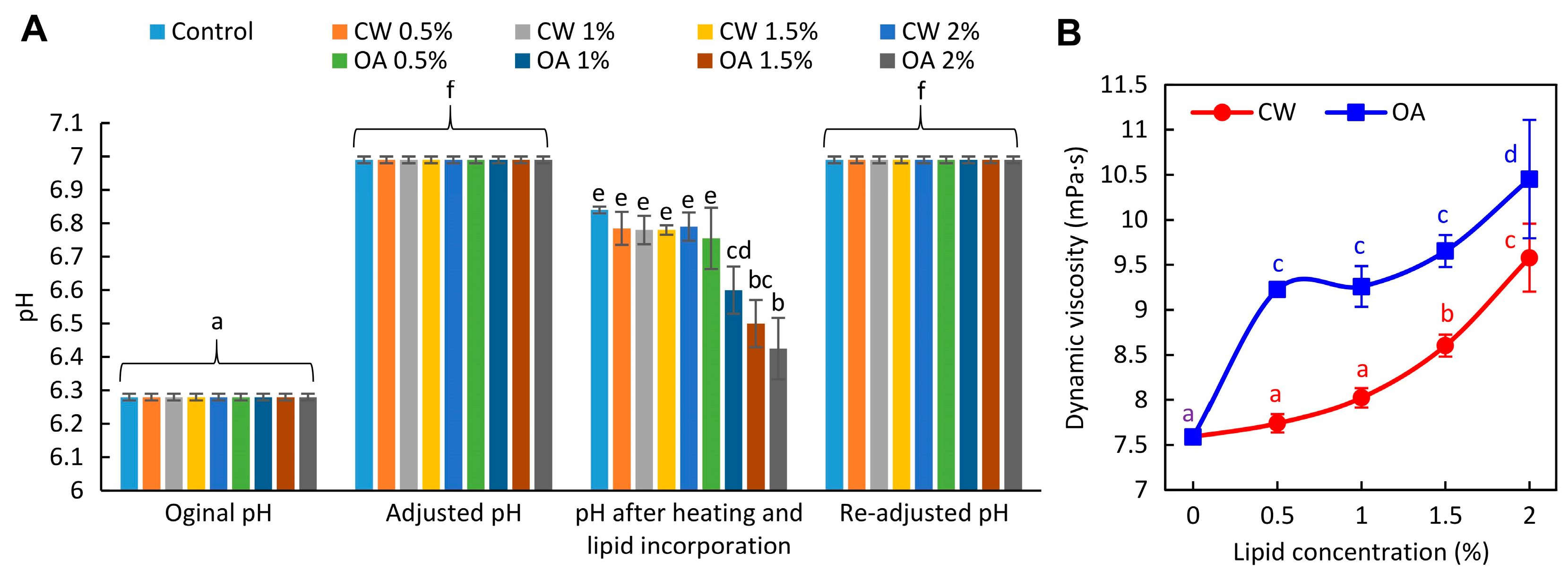
Figure 2.
Effect of increasing concentrations of candelilla wax (CW) and oleic acid (OA) on the microtopography of pea protein isolate-based film-forming solutions (FFSs), visualized by differential interference contrast microscopy at 100× magnification. Red frames show 200× magnifications of FFSs obtained from lipids stained with Sudan Red III (A). Appearance of FFSs after storage at 25°C (B). The control refers to the lipid-free FFS.
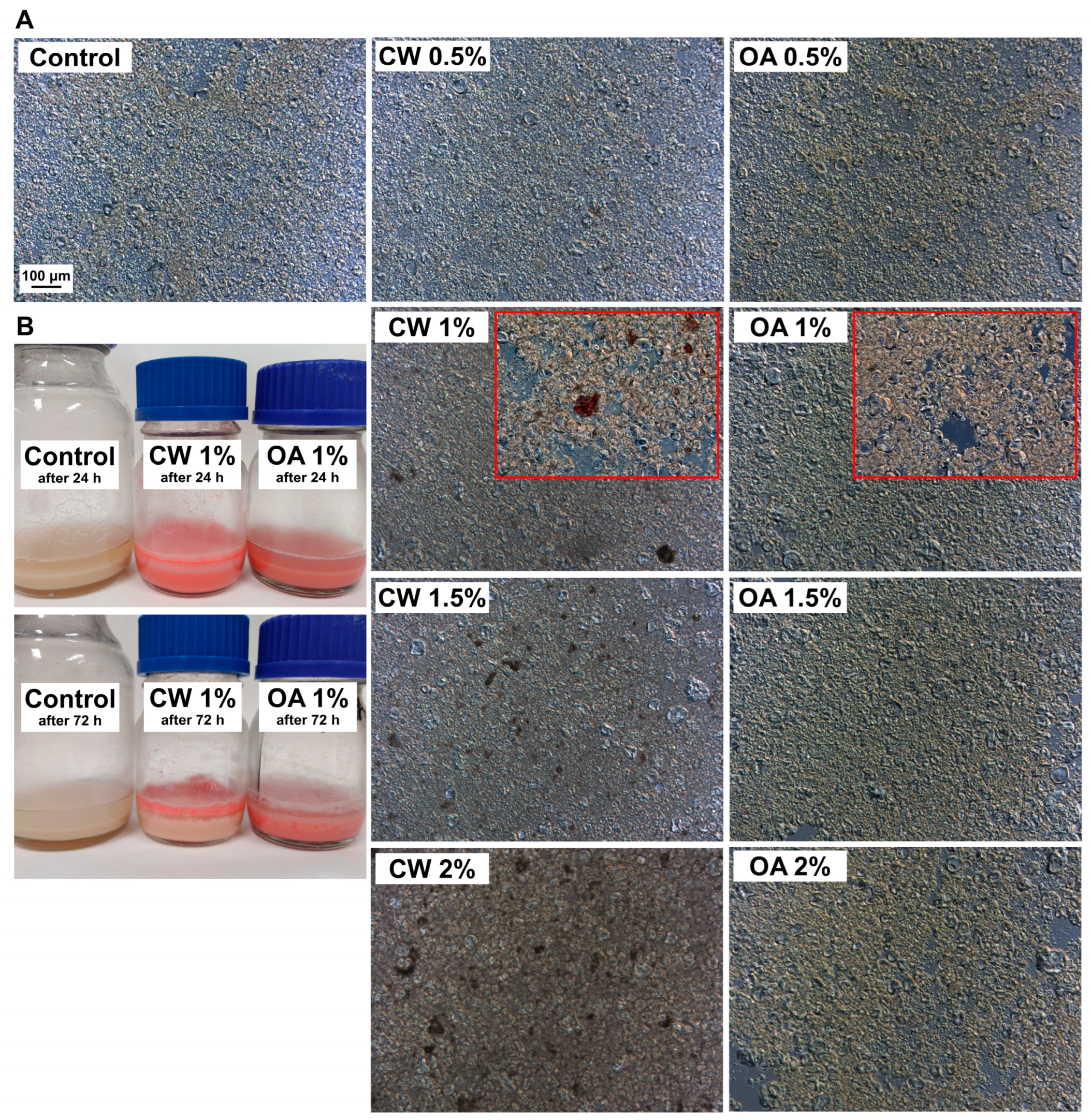
Figure 3.
Microtopography of the air side of glycerol-plasticized pea protein isolate-based films without (control) and with increasing additions of candelilla wax (CW) and oleic acid (OA), visualized by scanning electron microscopy (grayscale images) and differential interference contrast (DIC) microscopy (images in red frame) at magnifications of 1000× and 200×, respectively. Emulsion films for DIC microscopy were prepared using lipids stained with Sudan Red III.
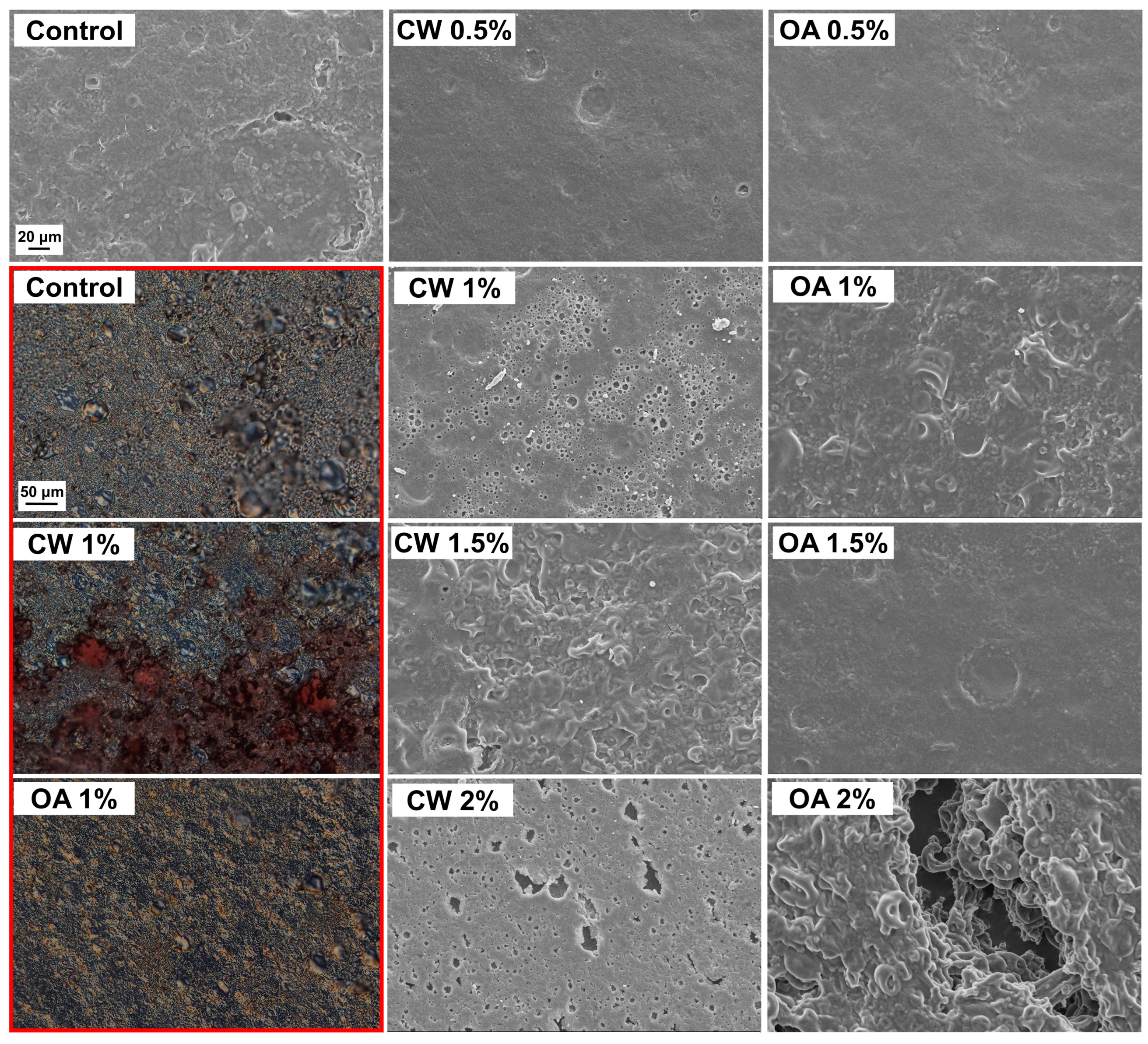
Figure 4.
Effect of candelilla wax (CW) and oleic acid (OA) concentrations on the gloss (A), light transmittance (B), and opacity (C) of glycerol-plasticized pea protein isolate films. The control refers to the lipid-free film.
Figure 4.
Effect of candelilla wax (CW) and oleic acid (OA) concentrations on the gloss (A), light transmittance (B), and opacity (C) of glycerol-plasticized pea protein isolate films. The control refers to the lipid-free film.
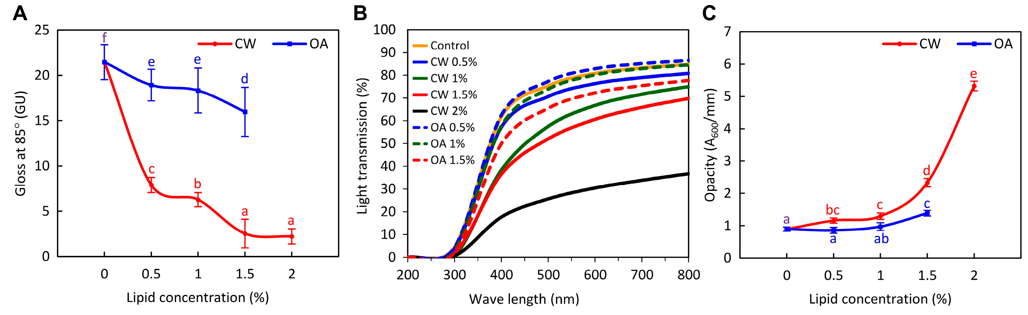

Table 1.
Effect of candelilla wax (CW) and oleic acid (OA) concentrations on the color parameters (L*, a*, b*), whiteness index (WI), and color difference (ΔE*) (between control (lipid-free) and lipid-added samples) of glycerol-plasticized pea protein isolate films.
Table 1.
Effect of candelilla wax (CW) and oleic acid (OA) concentrations on the color parameters (L*, a*, b*), whiteness index (WI), and color difference (ΔE*) (between control (lipid-free) and lipid-added samples) of glycerol-plasticized pea protein isolate films.
| Lipid type |
Lipid content (%) |
pH | L* | a* | b* | WI | ΔE* |
|---|---|---|---|---|---|---|---|
| Control | 0 | 6.76 ± 0.11 b | 41.39 ± 0.01 c | -0.21 ± 0.03 c | 2.13 ± 0.07 a | 41.35 ± 0.01 c | - |
| CW | 0.5 | 6.69 ± 0.02 ab | 42.05 ± 0.48 c | -076 ± 0.15 a | 5.21 ± 0.05 d | 41.81 ± 048 c | 3.22 ± 0.16 a |
| 1 | 6.66 ± 0.03 ab | 39.98 ± 1.71 b | -0.79 ± 0.03 a | 4.12 ± 037 c | 39.83 ± 1.70 b | 2.75 ± 1.08 a | |
| 1.5 | 6.65 ± 0.07 ab | 39.21 ± 0.30 ab | -0.88 ± 0.08a | 4.03 ± 0.33 c | 39.07 ± 0.39 ab | 2.96 ± 0.50 b | |
| 2 | 6.69 ± 0.08 ab | 39.51 ± 0.21 ab | -0.69 ± 0.02 ab | 4.94 ± 0.49 d | 39.45 ± 0.27 ab | 2.87 ± 0.49 a | |
| OA | 0.5 | 6.71 ± 0.06 ab | 39.94 ± 1.22 b | -0.46 ± 0.02 b | 2.69 ±0.46 b | 39.87 ± 1.21 b | 1.67 ± 1.12 a |
| 1 | 6.62 ± 0.02 a | 38.54 ± 0.86 a | -0.65 ± 0.17 ab | 2.68 ± 0.44 ab | 38.47 ± 0.84 a | 2.97 ± 0.79 b | |
| 1.5 | 6.58 ± 0.15 a | 39.62 ± 0.412 ab | -0.50 ± 0.05 b | 3.23 ± 0.18 b | 39.53 ± 0.16 ab | 2.11 ± 0.16 ab |
Values with different superscript letters (a-d) within a single column are significantly different (p < 0.05).
Table 2.
Effect of candelilla wax (CW) and oleic acid (OA) concentrations on the moisture content (MC), swelling (Sw), solubility (So), water vapor permeability (WVP), and contact angle (CA) of glycerol-plasticized pea protein isolate films. The control refers to the lipid-free film.
Table 2.
Effect of candelilla wax (CW) and oleic acid (OA) concentrations on the moisture content (MC), swelling (Sw), solubility (So), water vapor permeability (WVP), and contact angle (CA) of glycerol-plasticized pea protein isolate films. The control refers to the lipid-free film.
|
Lipid type |
Lipid content (%) |
MC (%) |
Sw (%) | So (%) | WVP (g mm m−2 day−1 kPa−1) | CA (O) |
| Control | 0 | 22.73 ± 1.20 b | 203.28 ± 19.35 a | 47.22 ± 2.53 a | 14.30 ± 0.68 f | 41.22 ± 6.66 a |
| CW | 0.5 | 22.80 ± 0.32 b | 222.32 ± 4.73 abc | 50.33 ± 3.68 ab | 8.96 ± 0.32 c | 74.71 ± 7.41 b |
| 1 | 19.99 ± 0.48 a | 238.36 ± 13.35 bcd | 51.73 ± 1.10 abc | 8.75 ± 0.51 c | 64.77 ± 4.01 b | |
| 1.5 | 20.62 ± 1.24 a | 223.33 ± 1.57 abc | 51.78 ± 3.43 abc | 6.89 ± 0.42 b | 97.60 ± 26.73 c | |
| 2 | 19.19 ± 0.73 a | 208.91 ± 15.40 ab | 57.40 ± 2.02 d | 5.36 ± 0.22 a | 100.13 ± 20.33 c | |
| OA | 0.5 | 19.86 ± 1.33 a | 247.20 ± 23.72 cd | 53.26 ± 0.67 bcd | 13.96 ± 1.02 ef | 36.25 ± 6.82 a |
| 1 | 20.06 ± 1.03 a | 245.17 ± 30.11 cd | 55.86 ± 4.77 cd | 12.90 ± 1.01 e | 28.95 ± 3.65 a | |
| 1.5 | 19.53 ± 0.95 a | 263.88 ± 31.14 d | 54.13 ± 4.24 bcd | 11.72 ± 0.68 d | 24.28 ± 2.08 a |
Values with different superscript letters (a-f) within a single column are significantly different (p < 0.05).
Table 3.
Effect of candelilla wax (CW) and oleic acid (OA) concentrations on the tensile strength (σmax), elongation at break (εb), elastic modulus (EM), and hot seal strength (HSS) of glycerol-plasticized pea protein isolate films. The control refers to the lipid-free film.
Table 3.
Effect of candelilla wax (CW) and oleic acid (OA) concentrations on the tensile strength (σmax), elongation at break (εb), elastic modulus (EM), and hot seal strength (HSS) of glycerol-plasticized pea protein isolate films. The control refers to the lipid-free film.
| Lipid type |
Lipid content (%) |
σmax (MPa) | εb (%) | EM (MPa) | HSS (N/mm) |
|---|---|---|---|---|---|
| Control | 0 | 3.56 ± 0.28 c | 81.23 ± 7.97 e | 99.11 ± 5.81 d | 0.069 ± 0.016 ab |
| CW | 0.5 | 2.70 ± 0.20 b | 50.38 ± 11.45 c | 71.14 ± 5.26 bc | 0.060 ± 0.030 a |
| 1 | 2.56 ± 0.25 ab | 55.44 ± 14.68 cd | 70.04 ± 3.60 b | 0.098 ± 0.015 cd | |
| 1.5 | 2.50 ± 0.27 ab | 39.88 ± 4.00 ab | 72.20 ± 9.32 bc | 0.063 ± 0.033 a | |
| 2 | 2.28 ± 0.13 a | 33.68 ± 5.01 a | 69.09 ± 6.12 b | 0.073 ± 0.006 abc | |
| OA | 0.5 | 3.61 ± 0.40 c | 64.19 ± 10.28 d | 99.63 ± 16.44 d | 0.105 ± 0.022 d |
| 1 | 2.57 ± 0.48 ab | 57.28 ± 7.60 cd | 81.47 ± 17.74 c | 0.091 ± 0.009 bcd | |
| 1.5 | 2.25 ± 0.36 a | 47.02 ± 5.44 bc | 56.76 ± 6.86 a | 0.088± 0.008 abcd |
Values with different superscript letters (a-e) within a single column are significantly different (p < 0.05).
Disclaimer/Publisher’s Note: The statements, opinions and data contained in all publications are solely those of the individual author(s) and contributor(s) and not of MDPI and/or the editor(s). MDPI and/or the editor(s) disclaim responsibility for any injury to people or property resulting from any ideas, methods, instructions or products referred to in the content. |
© 2024 by the authors. Licensee MDPI, Basel, Switzerland. This article is an open access article distributed under the terms and conditions of the Creative Commons Attribution (CC BY) license (http://creativecommons.org/licenses/by/4.0/).
Copyright: This open access article is published under a Creative Commons CC BY 4.0 license, which permit the free download, distribution, and reuse, provided that the author and preprint are cited in any reuse.


