Preprint
Review

This version is not peer-reviewed.

Potential Role of miR-455-3p in Liver Fibrosis Among Metabolic Dysfunction-Associated Steatotic Liver Disease Patients

Submitted:

14 November 2024

Posted:

17 November 2024

You are already at the latest version

Abstract

Liver fibrosis, a progressive condition often linked to metabolic dysfunction-associated steatotic liver disease (MASLD), lacks specific biomarkers to accurately gauge disease progression and therapeutic response. Emerging research suggests that microRNAs, particularly miR-455-3p, may play a crucial role in modulating fibrosis-related pathways. This narrative review explores the potential of miR-455-3p as a biomarker and therapeutic target in liver fibrosis among MASLD patients. The miR-455-3p’s effects on pathways, such as transforming growth factor-beta (TGF-β) signaling and extracellular matrix remodeling, both of which are pivotal in hepatic fibrosis. Additionally, the influence of miR-455-3p on hepatic stellate cell activation—a key process in fibrotic progression, should be considered. By challenging current perspectives, this review aims to elucidate the role of miR-455-3p in liver fibrosis, ultimately contributing to a deeper understanding of liver fibrosis mechanisms and highlighting the potential for innovative MASLD treatment strategies.

Keywords: 
;  ;  ;  ;  

Background

Metabolic dysfunction-associated steatotic liver disease (MASLD), formerly known as non-alcoholic fatty liver disease (NAFLD), has emerged as a significant global health concern, closely linked to the rising prevalence of obesity and metabolic syndrome[1]. As MASLD progresses, it can lead to liver fibrosis, a critical stage in the disease spectrum that significantly impacts patient outcomes[2]. The early detection and accurate staging of liver fibrosis in MASLD patients remain challenging, highlighting the need for novel, non-invasive diagnostic tools[3].
In recent years, microRNAs have gained attention as potential biomarkers for various liver diseases, including MASLD and associated fibrosis[4]. Among these, miR-455-3p has shown promise in regulating key pathways involved in hepatic stellate cells (HSCs) activation and fibrogenesis[5]. However, the specific role of miR-455-3p in the context of MASLD-related liver fibrosis remains to be fully elucidated, presenting an opportunity for further investigation into its diagnostic capabilities in this patient population.
This study review the role of miR-455-3p in the context of MASLD-related liver fibrosis.

Liver Fibrosis in MASLD

Liver fibrosis, a key feature of advanced MASLD, affects a subset of patients with the condition[3]. It is estimated that about 7.5% of individuals with MASLD will progress to develop advanced fibrosis[6,7]. The pooled prevalence rates of advanced liver fibrosis and cirrhosis in the general population were 3.3%[8]. The presence and severity of liver fibrosis are crucial determinants of long-term outcomes in MASLD patients, as advanced fibrosis is associated with increased risk of liver-related morbidity and mortality[3].
Epidemiological studies suggest that MASLD affects approximately 25-30% of the global adult population[9], with higher rates in Western countries and urban areas of developing nations[10]. The prevalence of MASLD increases with age and is more common in men than in women[11]. Among individuals with obesity or type 2 diabetes, the prevalence can reach up to 70-80%[12].
The disease burden of MASLD-related liver fibrosis is substantial. It is now one of the leading causes of liver cirrhosis, hepatocellular carcinoma, and liver transplantation in many countries[13]. The economic impact is also significant, with increased healthcare utilization and costs associated with managing the complications of advanced liver disease[14,15].

The Pathophysiology of Liver Fibrosis in MASLD

The pathophysiology of liver fibrosis in MASLD is complex and multifactorial, involving various cellular and molecular mechanisms. The progression from simple steatosis to fibrosis involves a series of events that lead to the activation of hepatic stellate cells (HSCs), the primary cell type responsible for extracellular matrix (ECM) production in the liver[2].
Lipotoxicity and oxidative stress play critical role in liver fibrosis progression during MASLD. The accumulation of lipids, particularly free fatty acids (FFAs), in hepatocytes is a hallmark of MASLD[16,17]. This lipid overload leads to lipotoxicity, which triggers several pathogenic processes. Moreover, mitochondrial dysfunction should be considered. Excess FFAs overwhelm the mitochondrial β-oxidation capacity, leading to the production of reactive oxygen species (ROS)[18-20]. This oxidative stress damages mitochondrial DNA and proteins, further impairing mitochondrial function. Furthermore, endoplasmic reticulum (ER) stress and lipid peroxidation could be responsible in this process. Lipid accumulation induces ER stress, activating the unfolded protein response (UPR)[17,21]. Prolonged UPR activation can lead to hepatocyte apoptosis. ROS interact with cellular lipids, forming lipid peroxidation products that can damage cellular structures and activate inflammatory pathways[22,23].
During inflammatory response, many cascades are engaged in liver fibrosis pathophysiology. Lipotoxicity and oxidative stress trigger an inflammatory response in the liver, characterized by activation of Kupffer cells, recruitment of inflammatory cells, the release of chemokine, NF-κB activation, and HSCs activation[24-26].
Kupffer cells as the liver-resident macrophages become activated and release pro-inflammatory cytokines such as TNF-α, IL-1β, and IL-6[27]. Besides, the release of chemokines, e.g., MCP-1, CXCL10 leads to the infiltration of inflammatory cells, including neutrophils and lymphocytes[28]. NF-κB is a key transcription factor and is activated in hepatocytes and immune cells, promoting the expression of pro-inflammatory genes[29].
The activation of HSCs is the central event in liver fibrogenesis. Several factors contribute to HSC activation[30]. TGF-β, the most potent profibrogenic cytokine, is released by Kupffer cells and activated HSCs[31]. It binds to TGF-β receptors on HSCs, activating SMAD2/3 signaling pathways that promote collagen synthesis[32,33]. Platelet-Derived Growth Factor (PDGF) is a potent mitogen for HSCs is upregulated in MASLD, promoting HSC proliferation and migration[34]. Additionally, ROS directly activate HSCs and stimulate the production of fibrogenic mediators[35]. The activation of NADPH oxidases in HSCs further contributes to ROS generation and fibrogenic signaling[36]. Likewise, Hedgehog signaling pathway is activated in MASLD and promotes HSC activation and survival[37].
Extracellular matrix (ECM) remodeling plays a crucial role in the progression of liver fibrosis in MASLD, primarily driven by the activation and transdifferentiation of HSCs into myofibroblast-like cells. These activated HSCs become the primary source of excessive ECM deposition, significantly altering the liver’s architecture and function[38]. The process is characterized by a marked increase in collagen production, particularly types I, III, and IV, along with other ECM proteins such as fibronectin and laminin[6]. Simultaneously, a complex interplay occurs between matrix metalloproteinases (MMPs) and their inhibitors, tissue inhibitors of metalloproteinases (TIMPs). While some MMPs are upregulated to degrade normal ECM, the overexpression of TIMPs inhibits this degradation, ultimately favoring ECM accumulation[39]. Adding to this fibrotic cascade is the upregulation of lysyl oxidase, an enzyme responsible for cross-linking collagen fibers, which contributes significantly to the increased stiffness observed in the fibrotic liver. This intricate balance of increased ECM production, altered degradation, and enhanced cross-linking collectively drives the progression of liver fibrosis in MASLD, fundamentally changing the liver’s structure and compromising its function[40,41].
Several interconnected molecular pathways contribute to the development and progression of liver fibrosis in MASLD, with the TGF-β/SMAD signaling pathway playing a central role. When TGF-β binds to its receptor, it activates SMAD2/3 through phosphorylation, leading to the formation of a complex with SMAD4 that translocates to the nucleus[32,33]. This complex interacts with various transcription factors to promote the expression of pro-fibrotic genes, including collagen and α-smooth muscle actin (α-SMA)[42]. Simultaneously, the PDGF signaling pathway is activated, binding to its receptor and triggering the Ras/MAPK and PI3K/Akt pathways, which promote HSC proliferation, migration, and survival, while also enhancing TGF-β effects on HSCs[43,44]. The Toll-Like Receptor (TLR) signaling pathway, particularly TLR4, is activated by gut-derived bacterial products, leading to NF-κB and JNK pathway activation, which further promotes inflammation and HSC activation[45,46].
Other significant pathways involved in liver fibrosis progression include the Wnt/β-catenin pathway, which promotes HSC activation and survival through β-catenin accumulation and nuclear translocation; the Notch signaling pathway, which interacts with TGF-β and Hedgehog pathways to amplify fibrogenic responses[47,48]; the JAK/STAT signaling pathway, activated by cytokines like IL-6, promoting hepatocyte survival and HSC activation through STAT3 activation[49,50]; and the Hippo/YAP pathway, whose dysregulation leads to increased YAP activity, promoting HSC activation and fibrogenesis through interaction with TEAD transcription factors[51-53]. These interconnected pathways collectively drive the complex process of liver fibrosis in MASLD, highlighting the multifaceted nature of the disease and the potential for targeted therapeutic interventions at various points in these signaling cascades.
Epigenetic mechanisms play a crucial role in the regulation of HSC activation and fibrogenesis in MASLD, influencing the progression of liver fibrosis through various molecular processes[54]. DNA methylation, particularly the hypermethylation of PPARγ, an anti-fibrotic transcription factor, contributes significantly to HSC activation. Histone modifications, including acetylation and methylation patterns, are altered in activated HSCs, affecting the expression of both pro-fibrotic and anti-fibrotic genes[55,56]. Additionally, microRNAs (miRNAs or miR) have emerged as important regulators of liver fibrosis in MASLD[4]. The dysregulation of these miRNAs contributes to the progression of liver fibrosis, highlighting the complex interplay between genetic and epigenetic factors in the pathogenesis of MASLD-related liver fibrosis. Understanding these epigenetic mechanisms provides potential targets for therapeutic interventions aimed at halting or reversing the fibrotic process[57,58].
The emerging concepts in MASLD-related liver fibrosis highlight the multifaceted nature of this condition and underscore the need for a holistic approach to its management. As research progresses, integrating these new insights into our understanding of MASLD pathophysiology will be essential for developing more targeted and effective treatments. Future studies should focus on elucidating the complex interactions between these various mechanisms and identifying key drivers of fibrosis progression in individual patients, paving the way for personalized therapeutic strategies in MASLD.

Liver Fibrosis Biomarkers

Early detection and accurate staging of liver fibrosis are crucial for effective management and improved patient outcomes. While liver biopsy remains the gold standard for fibrosis assessment, its invasive nature and potential complications have driven the search for non-invasive alternatives. Serum biomarkers have emerged as promising tools for the diagnosis, staging, and monitoring of liver fibrosis. This mini-review focuses on recent advances in serum biomarkers, with particular emphasis on galectins and miRNAs, as well as other emerging candidates[2,3,57].
Galectins have emerged as a promising family of biomarkers for liver fibrosis, owing to their involvement in various biological processes, including inflammation and fibrosis. These β-galactoside-binding proteins have demonstrated significant potential in assessing and monitoring liver fibrosis progression across different etiologies of chronic liver disease[2].
Galectin-3, in particular, has garnered substantial attention as a biomarker for liver fibrosis[2]. Numerous studies have reported elevated serum levels of galectin-3 that correlate strongly with the severity of fibrosis in various liver diseases[59]. The utility of galectin-3 extends beyond initial diagnosis, as it has also demonstrated potential in monitoring fibrosis progression and assessing treatment response, making it a versatile tool in the management of chronic liver diseases[2].
Galectin-9 is another member of the galectin family that has shown promise as a biomarker for liver fibrosis. Research has revealed increased expression of galectin-9 in cirrhotic livers, with levels correlating closely with the stage of fibrosis. What makes galectin-9 particularly interesting is its ability to reflect both inflammatory and fibrogenic processes in the liver, providing a more comprehensive picture of disease activity and progression[60].
Galectin-1 has also been identified as a potential biomarker for liver fibrosis. This protein is upregulated in activated HSCs, which are key players in the fibrogenic process, and is found in increased amounts in fibrotic liver tissue[61,62]. Galectin-1 are associated with fibrosis in chronic hepatitis, suggesting its utility in assessing fibrosis progression in viral hepatitis[63,64].
Beyond the galectin family, other emerging serum biomarkers have shown promise in the assessment of liver fibrosis. Cytokeratin-18 (CK-18) fragments, particularly the M30 fragment, have gained attention as markers of hepatocyte apoptosis. These fragments are elevated in metabolic dysfunction-associated steatohepatitis (MASH) and correlate with the degree of fibrosis. The M30 fragment, which represents caspase-cleaved CK-18, has demonstrated particular promise in differentiating MASH from simple steatosis, addressing a significant diagnostic challenge in the management of MASLD[65,66].
Procollagen III N-terminal propeptide (PIIINP) is another biomarker that has shown utility in assessing liver fibrosis. This marker reflects active fibrogenesis and has been found to correlate well with the stage of fibrosis[67,68].
The emergence of these novel biomarkers, including galectins, CK-18 fragments, and PIIINP, represents a significant advancement in the non-invasive assessment of liver fibrosis. Their ability to reflect various aspects of the fibrogenic process, from hepatocyte injury to active matrix deposition, offers a more nuanced understanding of disease progression. As research continues, these biomarkers may play an increasingly important role in the diagnosis, monitoring, and management of chronic liver diseases, potentially reducing the need for invasive liver biopsies and improving patient care.
YKL-40, also known as Chitinase-3-like protein 1, is a glycoprotein secreted by activated macrophages and HSCs[69]. This biomarker has gained attention in the field of hepatology due to its elevated levels in various liver diseases. Studies have shown that YKL-40 concentrations in serum correlate well with the severity of liver fibrosis, making it a promising candidate for non-invasive fibrosis assessment. Its ability to reflect both inflammatory and fibrogenic processes in the liver provides valuable insights into disease progression and may offer advantages over traditional markers[69-71].
Osteopontin is a multifunctional protein that plays crucial roles in inflammation and fibrosis[72]. In the context of liver diseases, osteopontin has emerged as a potential biomarker for fibrosis assessment. Research has demonstrated that serum levels of osteopontin are associated with fibrosis stage in both MASLD and viral hepatitis[73-75]. This correlation extends across different etiologies of chronic liver disease, suggesting that osteopontin may serve as a versatile biomarker for fibrosis progression. Its involvement in multiple pathways related to liver injury and repair makes it an intriguing target for both diagnostic and therapeutic purposes[76,77].
Mac-2 binding protein glycosylation isomer (M2BPGi) has garnered significant attention as a novel biomarker for liver fibrosis. This glycoprotein undergoes alterations during the progression of liver fibrosis, making it a sensitive indicator of fibrotic changes. Numerous studies have demonstrated M2BPGi to be a reliable marker for fibrosis staging across various liver diseases. Its utility is particularly noteworthy in assessing fibrosis in chronic hepatitis C and NAFLD, two of the most common causes of chronic liver disease worldwide. The ability of M2BPGi to accurately reflect fibrosis stage without the need for invasive procedures has positioned it as a valuable tool in the clinical management of liver diseases[78].
Autotaxin is an enzyme involved in the production of lysophosphatidic acid, a potent mediator of various cellular processes including inflammation and fibrosis. In recent years, autotaxin has emerged as a promising serum biomarker for liver fibrosis[79]. Studies have shown that serum levels of autotaxin correlate strongly with fibrosis stage and liver stiffness measurements obtained through elastography[80]. One of the most intriguing aspects of autotaxin as a biomarker is its potential to differentiate cirrhosis from earlier stages of fibrosis[81]. This capability addresses a critical need in hepatology, as the transition to cirrhosis marks a significant turning point in disease progression and patient management. The ability to non-invasively identify this transition could have substantial implications for treatment decisions and prognostic assessments in patients with chronic liver diseases.
Furthermore, composite biomarker panels have emerged as a powerful approach to improving the diagnostic accuracy of liver fibrosis assessment[82]. By combining multiple biomarkers, these panels often outperform single markers in their ability to detect and stage fibrosis. PIIINP’s value is further enhanced by its inclusion in several composite biomarker panels, such as the Enhanced Liver Fibrosis (ELF) score, which combines multiple markers to improve diagnostic accuracy. The ELF score, which combines procollagen III N-terminal propeptide (PIIINP), hyaluronic acid, and tissue inhibitor of metalloproteinase-1 (TIMP-1)[83]. The ELF score has been extensively validated for fibrosis assessment across various liver diseases. This recognition underscores its clinical utility and reliability in non-invasive fibrosis evaluation.
Another well-established composite panel is the FibroTest (FibroSure), which incorporates α2-macroglobulin, haptoglobin, apolipoprotein A1, γ-glutamyltransferase, and total bilirubin[82,84]. This panel has been widely validated for fibrosis staging, particularly in chronic hepatitis B and C, demonstrating its versatility across different etiologies of liver disease[85,86]. The NAFLD Fibrosis Score represents a targeted approach for non-alcoholic fatty liver disease, utilizing readily available clinical and laboratory parameters such as age, BMI, diabetes status, AST/ALT ratio, platelet count, and albumin. This score helps identify NAFLD patients at higher risk of advanced fibrosis, aiding in risk stratification and management decisions[87]. The Fibrosis-4 (FIB-4) index stands out for its simplicity and wide availability, combining age, AST, ALT, and platelet count to provide a quick assessment of fibrosis risk. Its ease of use and accessibility make it a valuable tool in various clinical settings[1,88].
The field of serum biomarkers for liver fibrosis continues to evolve rapidly, with several promising directions on the horizon. Multi-omics approaches, integrating data from proteomics, metabolomics, and transcriptomics, hold the potential to yield more accurate and comprehensive biomarker panels[89,90]. These integrative strategies may provide a more nuanced understanding of fibrosis progression and liver disease mechanisms. Liquid biopsy techniques, including the analysis of circulating tumor DNA, exosomes, and cell-free DNA, are emerging as powerful tools that may offer additional insights into fibrosis progression and liver disease etiology[91,92]. These minimally invasive approaches could potentially capture the dynamic nature of liver fibrosis more effectively than traditional static markers.
The application of artificial intelligence and machine learning to biomarker data represents another frontier in liver fibrosis assessment. Advanced algorithms have the potential to improve diagnostic accuracy and risk stratification by identifying complex patterns and relationships within biomarker data that may not be apparent through conventional analysis[93,94]. However, the development and implementation of these innovative approaches face several challenges. Standardization of assays and establishment of consistent cut-off values across different populations and disease etiologies remain crucial for widespread adoption. Extensive validation in large, diverse cohorts is necessary to ensure the generalizability of biomarker panels and AI-driven models[95]. The successful integration of these advanced biomarker strategies into clinical practice will require the development of clear guidelines for their use, as well as education and training for healthcare providers to effectively interpret and act upon the results. Addressing these challenges will be key to realizing the full potential of serum biomarkers in the non-invasive assessment and management of liver fibrosis.
Serum biomarkers represent a promising approach for non-invasive assessment of liver fibrosis. Galectins and miRNAs, along with other emerging biomarkers, offer potential advantages in terms of specificity, sensitivity, and ease of measurement. Composite panels that combine multiple biomarkers often provide improved diagnostic accuracy. While these biomarkers show great promise, further research is needed to fully validate their clinical utility across diverse patient populations and liver disease etiologies. As our understanding of liver fibrosis pathogenesis deepens and analytical technologies advance, we can expect the development of even more accurate and tailored biomarker strategies. The ultimate goal is to provide clinicians with reliable, non-invasive tools for early detection, accurate staging, and monitoring of liver fibrosis, thereby improving patient care and outcomes in chronic liver diseases.

The miRNAs as Liver Fibrosis Biomarkers

MiRNAs are small non-coding RNAs that regulate gene expression post-transcriptionally. Their stability in circulation and tissue-specific expression patterns make them attractive biomarker candidates[96]. The miRNAs have emerged as promising biomarkers for liver fibrosis due to their stability in circulation, ease of detection, and involvement in various stages of fibrogenesis[97]. These small non-coding RNAs play crucial roles in regulating gene expression and cellular processes associated with liver fibrosis, making them valuable indicators of disease progression and potential therapeutic targets[98].
Several miRNAs have been identified as potential biomarkers for liver fibrosis, with varying degrees of specificity and sensitivity[99]. Among the most extensively studied are miR-122, miR-21, miR-29 family members, miR-34a, and miR-155. Each of these miRNAs has demonstrated unique expression patterns and functional roles in the context of liver fibrosis[100,101].
miR-122 is the most abundant miRNA in the liver, accounting for approximately 70% of the total hepatic miRNA content[102]. Its expression is significantly altered in various liver diseases, including viral hepatitis, alcoholic liver disease, and MASLD. miR-122 is a liver-specific miRNA, decreased serum levels in advanced fibrosis. It is a potential marker for hepatocyte injury and fibrosis progression. In the context of liver fibrosis, serum levels of miR-122 have been found to correlate inversely with the stage of fibrosis. As liver damage progresses and hepatocytes are lost, circulating miR-122 levels decrease, making it a potential marker for advanced fibrosis and cirrhosis. However, the specificity of miR-122 as a fibrosis biomarker is limited by its elevation in acute liver injury, necessitating careful interpretation in the context of other clinical parameters[57,102,103].
miR-21 has gained attention as a pro-fibrotic miRNA that is upregulated in activated HSCs, the primary cell type responsible for extracellular matrix deposition in liver fibrosis[104]. Elevated serum levels of miR-21 have been associated with the progression of liver fibrosis in various etiologies, including chronic hepatitis B and C, as well as MASLD[57,105]. miR-21 is upregulated in activated HSCs and fibrotic liver tissue. This miR serum levels correlate with fibrosis stage in various liver diseases. Studies have shown that miR-21 levels correlate positively with fibrosis stage and can distinguish between mild and significant fibrosis with reasonable accuracy. The pro-fibrotic effects of miR-21 are mediated through its targeting of SMAD7, a negative regulator of TGF-β signaling, thus promoting HSC activation and fibrogenesis[106,107].
The miR-29 family, consisting of miR-29a, miR-29b, and miR-29c, has been extensively studied in the context of liver fibrosis[108,109]. These miRNAs are known to target multiple genes involved in extracellular matrix production, including various collagen isoforms. In liver fibrosis, the expression of miR-29 family members is typically downregulated, leading to increased collagen synthesis and fibrosis progression[109,110]. miR-29 family downregulated in fibrotic livers, correlating with increased ECM production. Serum levels may reflect fibrosis severity and treatment response[111]. Circulating levels of miR-29 have been explored as potential biomarkers, with some studies showing that decreased serum miR-29 levels correlate with advancing fibrosis stages[109,112]. The restoration of miR-29 expression has been proposed as a potential therapeutic strategy to attenuate fibrosis, highlighting the dual role of these miRNAs as both biomarkers and therapeutic targets[110,113].
miR-34a has emerged as a key player in liver fibrosis, particularly in the context of MASLD and MASH[4]. miR-34a is increased expression in MASLD/MASH and correlation with fibrosis severity[114]. This miRNA is involved in regulating cell cycle arrest, apoptosis, and senescence, processes that contribute to liver injury and fibrosis progression[115]. Elevated serum levels of miR-34a have been associated with the severity of liver fibrosis in MASLD patients, with some studies suggesting its potential as a biomarker for distinguishing MASH from simple steatosis[116]. The pro-fibrotic effects of miR-34a are mediated, in part, through its targeting of SIRT1, a key regulator of cellular metabolism and stress responses[4,117,118].
miR-155 is another miRNA that has shown promise as a biomarker for liver fibrosis. This miRNA plays a complex role in liver pathology, influencing inflammation, immune responses, and fibrogenesis [100,119,120]. Its inhibition can attenuate the liver fibrosis via STAT3 signaling[121]. Altered serum levels of miR-155 have been observed in patients with chronic liver diseases. The pro-fibrotic effects of miR-155 are attributed to its ability to promote the activation of Kupffer cells and HSCs, thereby liver fibrosis[122].
miR-200b, a member of the miR-200 family, has emerged as a significant player in liver fibrosis, particularly due to its involvement in the regulation of epithelial-to-mesenchymal transition (EMT)[123,124]. Moreover, in the context of liver fibrosis, miR-200a is typically downregulated, which contributes to the progression of fibrogenesis[125]. These miRNAs target key transcription factors involved in EMT, such as ZEB1 and ZEB2, which are critical for maintaining epithelial cell identity[126,127]. As liver fibrosis advances, the reduction in miR-200b levels allows for increased expression of these EMT-promoting factors, facilitating the transition of hepatocytes and cholangiocytes towards a more mesenchymal phenotype[124,128].

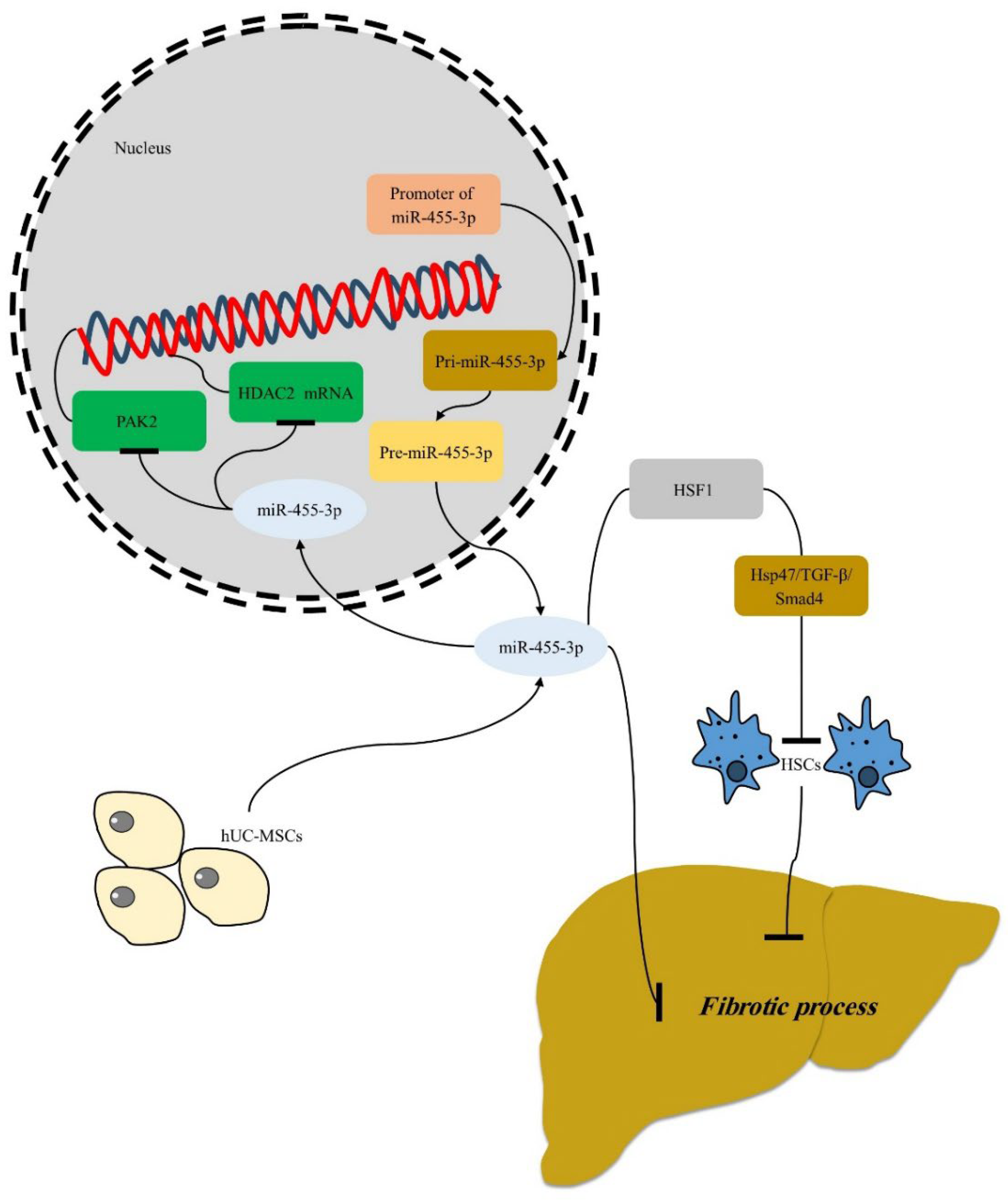
The Role of miR-455-3p in Liver Fibrosis

The role of miR-455-3p in liver fibrosis has emerged as a fascinating area of research, with potential implications for both diagnosis and treatment of this debilitating condition[129]. Recent studies have shed light on the complex interplay between miR-455-3p and various cellular mechanisms involved in the progression and reversal of liver fibrosis, particularly in the context of MASLD[5].
One of the most intriguing findings is the downregulation of miR-455-3p in activated HSCs and various hepatic fibrosis models. Wei et al. [5] demonstrated that miR-455-3p inhibits HSC activation by targeting heat shock factor 1 (HSF1), which is involved in the Hsp47/TGF-β/Smad4 signaling pathway. This discovery suggests that miR-455-3p acts as a natural brake on the fibrotic process, and its depletion may contribute to the progression of liver fibrosis in MASLD patients (Figure 1).
Further supporting this notion, You’s research [130] revealed that miR-455-3p targets histone deacetylase 2 (HDAC2), another key player in liver fibrosis. The study found that miR-455-3p was downregulated in liver tissues of cirrhosis patients and fibrotic mouse models, while being upregulated during the reversal stage of hepatic fibrosis. This dynamic expression pattern hints at the potential use of miR-455-3p as a biomarker for both the progression and regression of liver fibrosis in MASLD patients (Figure 1).
Interestingly, the regulation of miR-455-3p itself appears to be under epigenetic control. You’s [130] study demonstrated that DNA methyltransferases, particularly DNMT3b and DNMT1, mediate the hypermethylation of miR-455-3p promoter CpG islands in activated HSCs. This finding adds another layer of complexity to the role of miR-455-3p in liver fibrosis and suggests that epigenetic modulation could be a potential therapeutic approach for MASLD-related liver fibrosis.
The therapeutic potential of miR-455-3p is further underscored by studies exploring its delivery via exosomes. Shao’s [131] research showed that exosomes derived from human umbilical cord mesenchymal stem cells (hUC-MSCs) enriched with miR-455-3p could attenuate inflammatory liver injury. This approach not only highlights the anti-inflammatory properties of miR-455-3p but also presents a novel delivery method that could be exploited for treating MASLD-related liver fibrosis.
Building on this, Zhou’s [132] study demonstrated that hUC-MSCs could ameliorate hepatic fibrosis by upregulating miR-455-3p, which in turn suppresses p21-activated kinase-2 (PAK2). This finding expands our understanding of the downstream targets of miR-455-3p and reinforces its potential as a therapeutic target in liver fibrosis treatment (Figure 1).
However, despite these promising findings, several challenges remain in translating this knowledge into clinical applications for MASLD patients. The complex nature of miRNA regulation and the potential off-target effects of miRNA-based therapies need to be carefully considered. Moreover, the specific role of miR-455-3p in MASLD-related liver fibrosis, as opposed to other etiologies, requires further investigation to determine its diagnostic and therapeutic value in this particular patient population.
Future studies could explore the possibility of harnessing miR-455-3p in combination with other antifibrotic agents to achieve a synergistic effect that tackles fibrosis on multiple fronts. Longitudinal studies are essential to determine whether miR-455-3p expression changes correlate with clinical outcomes, potentially allowing miR-455-3p to serve as a biomarker not only for fibrosis but for broader MASLD-related disease stages.
Although miR-455-3p appears promising, miRNA therapies carry inherent risks due to their pleiotropic effects. Thus, assessing the safety and ethical implications of manipulating a miRNA like miR-455-3p is crucial before moving toward clinical trials. As with any novel therapy, miR-455-3p delivery systems must be scrutinized for safety, as inadvertent off-target effects could lead to unforeseen adverse outcomes in MASLD patients. The complex regulatory environment surrounding miRNA therapeutics necessitates thorough evaluation to ensure that any miR-455-3p-based treatment is both safe and effective for diverse patient populations.

Conclusion

In conclusion, the emerging role of miR-455-3p in liver fibrosis presents an exciting frontier in hepatology research, with potential implications in the pathogenesis of MASLD-related liver fibrosis and then for its diagnosis and treatment. As our understanding of this miRNA’s function and regulation continues to grow, it may pave the way for novel diagnostic tools and targeted therapies that could significantly improve outcomes for patients with this challenging condition.

Author Contributions

MS: Reviewing the literature, Methodology, Investigation, Conceptualization, Data curation, Writing – the original draft, review & and editing.

Funding

None.

Acknowledgments

Special thanks to Dr. Seyed-Mohamad-Sadegh Mirahmadi and Dr. Reza Azarbad.

Conflict of Interest

The author declare that they have no conflict of interest.

References

  1. Mokhtare, M.; Abdi, A.; Sadeghian, A.M.; Sotoudeheian, M.; Namazi, A.; Sikaroudi, M.K. Investigation about the correlation between the severity of metabolic-associated fatty liver disease and adherence to the Mediterranean diet. Clin. Nutr. ESPEN 2023, 58, 221–227. [CrossRef]
  2. Sotoudeheian, M. Galectin-3 and Severity of Liver Fibrosis in Metabolic Dysfunction-Associated Fatty Liver Disease. Protein Pept. Lett. 2024, 31, 290–304. [CrossRef]
  3. Sotoudeheian, M. Agile 3+ and Metabolic Dysfunction-Associated Fatty Liver Disease: Detecting Advanced Fibrosis based on Reported Liver Stiffness Measurement in FibroScan and Laboratory Findings. Int. J. Gastroenterol. Hepatol. Dis. 2024, 03, 1–12. [CrossRef]
  4. Ma, N.; Tan, J.; Chen, Y.; Yang, L.; Li, M.; He, Y. MicroRNAs in metabolic dysfunction-associated diseases: Pathogenesis and therapeutic opportunities. FASEB J. 2024, 38, e70038. [CrossRef]
  5. Wei, S.; Wang, Q.; Zhou, H.; Qiu, J.; Li, C.; Shi, C.; Zhou, S.; Liu, R.; Lu, L. miR-455-3p Alleviates Hepatic Stellate Cell Activation and Liver Fibrosis by Suppressing HSF1 Expression. Mol. Ther. - Nucleic Acids 2019, 16, 758–769. [CrossRef]
  6. Fan W, Bradford TM, Török NJ. Metabolic dysfunction− associated liver disease and diabetes: Matrix remodeling, fibrosis, and therapeutic implications. Annals of the New York Academy of Sciences. 2024;1538:21-33.
  7. Ciardullo, S.; Perseghin, G. Prevalence of NAFLD, MAFLD and associated advanced fibrosis in the contemporary United States population. Liver Int. 2021, 41, 1290–1293. [CrossRef]
  8. Zamani, M.; Alizadeh-Tabari, S.; Ajmera, V.; Singh, S.; Murad, M.H.; Loomba, R. Global Prevalence of Advanced Liver Fibrosis and Cirrhosis in the General Population: A Systematic Review and Meta-analysis. Clin. Gastroenterol. Hepatol. 2024. [CrossRef]
  9. Singh, A.; Buckholz, A.; Kumar, S.; Newberry, C. Implications of Protein and Sarcopenia in the Prognosis, Treatment, and Management of Metabolic Dysfunction-Associated Steatotic Liver Disease (MASLD). Nutrients 2024, 16, 658. [CrossRef]
  10. Younossi, Z.; Tacke, F.; Arrese, M.; Sharma, B.C.; Mostafa, I.; Bugianesi, E.; Wong, V.W.-S.; Yilmaz, Y.; George, J.; Fan, J.; et al. Global Perspectives on Nonalcoholic Fatty Liver Disease and Nonalcoholic Steatohepatitis. Hepatology 2019, 69, 2672–2682. [CrossRef]
  11. Schneider CV, Schneider KM, Raptis A, Huang H, Trautwein C, Loomba R. Prevalence of at-risk MASH, M et ALD and alcohol-associated steatotic liver disease in the general population. Alimentary Pharmacology & Therapeutics. 2024;59:1271-81.
  12. Younossi, Z.M.; Kalligeros, M.; Henry, L. Epidemiology of Metabolic Dysfunction Associated Steatotic Liver Disease. Clin. Mol. Hepatol. 2024. [CrossRef]
  13. Parente, A.; Milana, F.; Hajibandeh, S.; Menon, K.V.; Kim, K.-H.; Shapiro, A.M.J.; Schlegel, A. Liver transplant for hepatocellular carcinoma in metabolic dysfunction-associated steatotic liver disease versus other etiologies: A meta-analysis. Dig. Liver Dis. 2024. [CrossRef]
  14. Miao, L.; Targher, G.; Byrne, C.D.; Cao, Y.-Y.; Zheng, M.-H. Current status and future trends of the global burden of MASLD. Trends Endocrinol. Metab. 2024, 35, 697–707. [CrossRef]
  15. Torre, E.; Di Matteo, S.; Martinotti, C.; Bruno, G.M.; Goglia, U.; Testino, G.; Rebora, A.; Bottaro, L.C.; Colombo, G.L. Economic Impact of Metabolic Dysfunction-Associated Steatotic Liver Disease (MASLD) in Italy. Analysis and Perspectives. Clin. Outcomes Res. 2024, ume 16, 773–784. [CrossRef]
  16. Theys C. The new epigenetic driver role of PPARα and mitochondria in metabolic dysfunction associated liver disease (MASLD), paving the way towards new therapeutics and diagnostic biomarkers: University of Antwerp; 2024.
  17. Venkatesan N, Doskey LC, Malhi H. The Role of Endoplasmic Reticulum in Lipotoxicity during MASLD Pathogenesis. The American Journal of Pathology. 2023.
  18. Arumugam, M.K.; Gopal, T.; Kandy, R.R.K.; Boopathy, L.K.; Perumal, S.K.; Ganesan, M.; Rasineni, K.; Donohue, T.M.; Osna, N.A.; Kharbanda, K.K. Mitochondrial Dysfunction-Associated Mechanisms in the Development of Chronic Liver Diseases. Biology 2023, 12, 1311. [CrossRef]
  19. Karkucinska-Wieckowska, A.; Simoes, I.C.M.; Kalinowski, P.; Lebiedzinska-Arciszewska, M.; Zieniewicz, K.; Milkiewicz, P.; Górska-Ponikowska, M.; Pinton, P.; Malik, A.N.; Krawczyk, M.; et al. Mitochondria, oxidative stress and nonalcoholic fatty liver disease: A complex relationship. Eur. J. Clin. Investig. 2021, 52, e13622. [CrossRef]
  20. Svegliati-Baroni, G.; Pierantonelli, I.; Torquato, P.; Marinelli, R.; Ferreri, C.; Chatgilialoglu, C.; Bartolini, D.; Galli, F. Lipidomic biomarkers and mechanisms of lipotoxicity in non-alcoholic fatty liver disease. 2019, 144, 293–309. [CrossRef]
  21. Białek W, Hryniewicz-Jankowska A, Czechowicz P, Sławski J, Collawn JF, Czogalla A, et al. The lipid side of unfolded protein response. Biochimica et Biophysica Acta (BBA)-Molecular and Cell Biology of Lipids. 2024:159515.
  22. Zhang, J.; Guo, J.; Yang, N.; Huang, Y.; Hu, T.; Rao, C. Endoplasmic reticulum stress-mediated cell death in liver injury. Cell Death Dis. 2022, 13, 1–13. [CrossRef]
  23. Ashraf, N.U.; Sheikh, T.A. Endoplasmic reticulum stress and Oxidative stress in the pathogenesis of Non-alcoholic fatty liver disease. Free. Radic. Res. 2015, 49, 1405–1418. [CrossRef]
  24. Duarte, N.; Coelho, I.C.; Patarrão, R.S.; Almeida, J.I.; Penha-Gonçalves, C.; Macedo, M.P. How Inflammation Impinges on NAFLD: A Role for Kupffer Cells. BioMed Res. Int. 2015, 2015, 1–11. [CrossRef]
  25. Ma, Y.; Lee, G.; Heo, S.-Y.; Roh, Y.-S. Oxidative Stress Is a Key Modulator in the Development of Nonalcoholic Fatty Liver Disease. Antioxidants 2021, 11, 91. [CrossRef]
  26. Kumar, S.; Duan, Q.; Wu, R.; Harris, E.N.; Su, Q. Pathophysiological communication between hepatocytes and non-parenchymal cells in liver injury from NAFLD to liver fibrosis. Adv. Drug Deliv. Rev. 2021, 176, 113869. [CrossRef]
  27. Park, S.-J.; Diaz, J.G.; Um, E.; Hahn, Y.S. Major roles of kupffer cells and macrophages in NAFLD development. Front. Endocrinol. 2023, 14. [CrossRef]
  28. Dai, S.; Liu, F.; Qin, Z.; Zhang, J.; Chen, J.; Ding, W.-X.; Feng, D.; Ji, Y.; Qin, X. Kupffer cells promote T-cell hepatitis by producing CXCL10 and limiting liver sinusoidal endothelial cell permeability. Theranostics 2020, 10, 7163–7177. [CrossRef]
  29. Plaza-Diaz, J.; Gomez-Llorente, C.; Fontana, L.; Gil, A. Modulation of immunity and inflammatory gene expression in the gut, in inflammatory diseases of the gut and in the liver by probiotics. World J. Gastroenterol. 2014, 20, 15632–49. [CrossRef]
  30. Tsuchida, T.; Friedman, S.L. Mechanisms of hepatic stellate cell activation. Nat. Rev. Gastroenterol. Hepatol. 2017, 14, 397–411. [CrossRef]
  31. Dewidar, B.; Meyer, C.; Dooley, S.; Meindl-Beinker, N. TGF-β in Hepatic Stellate Cell Activation and Liver Fibrogenesis—Updated 2019. Cells 2019, 8, 1419. [CrossRef]
  32. Ni X-X, Li X-Y, Wang Q, Hua J. Regulation of peroxisome proliferator-activated receptor-gamma activity affects the hepatic stellate cell activation and the progression of NASH via TGF-β1/Smad signaling pathway. Journal of physiology and biochemistry. 2021;77:35-45.
  33. Breitkopf K, Weng H, Dooley S. TGF-β/Smad-signaling in liver cells: Target genes and inhibitors of two parallel pathways. Signal Transduction. 2006;6:329-37.
  34. Kikuchi AT. Emerging Roles of Platelet-Derived Growth Factor Receptor Alpha in Chronic Liver Injury: Potential Therapeutic Target in Hepatic Fibrosis: University of Pittsburgh; 2017.
  35. Bocca, C.; Protopapa, F.; Foglia, B.; Maggiora, M.; Cannito, S.; Parola, M.; Novo, E. Hepatic Myofibroblasts: A Heterogeneous and Redox-Modulated Cell Population in Liver Fibrogenesis. Antioxidants 2022, 11, 1278. [CrossRef]
  36. Qiao, H.; Zhou, Y.; Qin, X.; Cheng, J.; He, Y.; Jiang, Y. NADPH Oxidase Signaling Pathway Mediates Mesenchymal Stem Cell-Induced Inhibition of Hepatic Stellate Cell Activation. Stem Cells Int. 2018, 2018, 1–13. [CrossRef]
  37. Dutta, R.K.; Jun, J.; Du, K.; Diehl, A.M. Hedgehog Signaling: Implications in Liver Pathophysiology. Semin. Liver Dis. 2023, 43, 418–428. [CrossRef]
  38. Kim, J.-W.; Kim, Y.J. The evidence-based multifaceted roles of hepatic stellate cells in liver diseases: A concise review. Life Sci. 2024, 344, 122547. [CrossRef]
  39. Shan, L.; Wang, F.; Zhai, D.; Meng, X.; Liu, J.; Lv, X. Matrix metalloproteinases induce extracellular matrix degradation through various pathways to alleviate hepatic fibrosis. Biomed. Pharmacother. 2023, 161, 114472. [CrossRef]
  40. Zhang, N.; Yang, A.; Zhang, W.; Li, H.; Xu, A.; Yan, X.; Han, Q.; Wang, B.; You, H.; Chen, W. Crosstalk of lysyl oxidase-like 1 and lysyl oxidase prolongs their half-lives and regulates liver fibrosis through Notch signal. Hepatol. Commun. 2024, 8. [CrossRef]
  41. Khurana, A.; Sayed, N.; Allawadhi, P.; Weiskirchen, R. It’s all about the spaces between cells: role of extracellular matrix in liver fibrosis. Ann. Transl. Med. 2021, 9, 728–728. [CrossRef]
  42. Breitkopf, K.; Godoy, P.; Ciuclan, L.; Singer, M.V.; Dooley, S. TGF-β/Smad Signaling in the Injured Liver. Z. Fur Gastroenterol. 2006, 44, 57–66. [CrossRef]
  43. Ying, H.-Z.; Chen, Q.; Zhang, W.-Y.; Zhang, H.-H.; Ma, Y.; Zhang, S.-Z.; Fang, J.; Yu, C.-H. PDGF signaling pathway in hepatic fibrosis pathogenesis and therapeutics. Mol. Med. Rep. 2017, 16, 7879–7889. [CrossRef]
  44. Bourebaba, N.; Marycz, K. Hepatic stellate cells role in the course of metabolic disorders development – A molecular overview. Pharmacol. Res. 2021, 170, 105739. [CrossRef]
  45. Guo, J.; Friedman, S.L. Toll-like receptor 4 signaling in liver injury and hepatic fibrogenesis. Fibrogenesis Tissue Repair 2010, 3, 21–21. [CrossRef]
  46. Paik, Y.-H.; Schwabe, R.F.; Bataller, R.; Russo, M.P.; Jobin, C.; Brenner, D.A. Toll–Like Receptor 4 Mediates Inflammatory Signaling by Bacterial Lipopolysaccharide in Human Hepatic Stellate Cells. Hepatology 2003, 37, 1043–1055. [CrossRef]
  47. Akkız, H.; Gieseler, R.K.; Canbay, A. Liver Fibrosis: From Basic Science towards Clinical Progress, Focusing on the Central Role of Hepatic Stellate Cells. Int. J. Mol. Sci. 2024, 25, 7873. [CrossRef]
  48. Duspara, K.; Bojanic, K.; Pejic, J.I.; Kuna, L.; Kolaric, T.O.; Nincevic, V.; Smolic, R.; Vcev, A.; Glasnovic, M.; Curcic, I.B.; et al. Targeting the Wnt Signaling Pathway in Liver Fibrosis for Drug Options: An Update. J. Clin. Transl. Hepatol. 2021, 000, 000–000. [CrossRef]
  49. Zhao, J.; Qi, Y.-F.; Yu, Y.-R. STAT3: A key regulator in liver fibrosis. 2020, 21, 100224. [CrossRef]
  50. Xiang D-M, Sun W, Ning B-F, Zhou T-F, Li X-F, Zhong W, et al. The HLF/IL-6/STAT3 feedforward circuit drives hepatic stellate cell activation to promote liver fibrosis. Gut. 2018;67:1704-15.
  51. Mia, M.M.; Singh, M.K. New Insights into Hippo/YAP Signaling in Fibrotic Diseases. Cells 2022, 11, 2065. [CrossRef]
  52. Driskill, J.H.; Pan, D. The Hippo Pathway in Liver Homeostasis and Pathophysiology. Annu. Rev. Pathol. Mech. Dis. 2020, 16, 299–322. [CrossRef]
  53. Schiavoni, G.; Messina, B.; Scalera, S.; Memeo, L.; Colarossi, C.; Mare, M.; Blandino, G.; Ciliberto, G.; Bon, G.; Maugeri-Saccà, M. Role of Hippo pathway dysregulation from gastrointestinal premalignant lesions to cancer. J. Transl. Med. 2024, 22, 1–16. [CrossRef]
  54. Wang, X.; Zhang, L.; Dong, B. Molecular mechanisms in MASLD/MASH-related HCC. Hepatology 2024. [CrossRef]
  55. Dong, Q.-Q.; Yang, Y.; Tao, H.; Lu, C.; Yang, J.-J. m6A epitranscriptomic and epigenetic crosstalk in liver fibrosis: Special emphasis on DNA methylation and non-coding RNAs. Cell. Signal. 2024, 122, 111302. [CrossRef]
  56. Xue, T.; Qiu, X.; Liu, H.; Gan, C.; Tan, Z.; Xie, Y.; Wang, Y.; Ye, T. Epigenetic regulation in fibrosis progress. Pharmacol. Res. 2021, 173, 105910. [CrossRef]
  57. Tobaruela-Resola, A.L.; Milagro, F.I.; Elorz, M.; Benito-Boillos, A.; Herrero, J.I.; Mogna-Peláez, P.; Tur, J.A.; Martínez, J.A.; Abete, I.; Zulet, M.. Circulating miR-122-5p, miR-151a-3p, miR-126-5p and miR-21-5p as potential predictive biomarkers for Metabolic Dysfunction-Associated Steatotic Liver Disease assessment. J. Physiol. Biochem. 2024, 1–14. [CrossRef]
  58. Su, Q.; Kumar, V.; Sud, N.; Mahato, R.I. MicroRNAs in the pathogenesis and treatment of progressive liver injury in NAFLD and liver fibrosis. Adv. Drug Deliv. Rev. 2018, 129, 54–63. [CrossRef]
  59. Iacobini, C.; Menini, S.; Ricci, C.; Fantauzzi, C.B.; Scipioni, A.; Salvi, L.; Cordone, S.; Delucchi, F.; Serino, M.; Federici, M.; et al. Galectin-3 ablation protects mice from diet-induced NASH: A major scavenging role for galectin-3 in liver. J. Hepatol. 2010, 54, 975–983. [CrossRef]
  60. Fujita, K.; Niki, T.; Nomura, T.; Oura, K.; Tadokoro, T.; Sakamoto, T.; Tani, J.; Yoneyama, H.; Morishita, A.; Kuroda, N.; et al. Correlation between serum galectin-9 levels and liver fibrosis. J. Gastroenterol. Hepatol. 2017, 33, 492–499. [CrossRef]
  61. An, Y.; Xu, S.; Liu, Y.; Xu, X.; Philips, C.A.; Chen, J.; Méndez-Sánchez, N.; Guo, X.; Qi, X. Role of Galectins in the Liver Diseases: A Systematic Review and Meta-Analysis. Front. Med. 2021, 8. [CrossRef]
  62. Jiang, Z.; Shen, Q.; Chen, H.; Yang, Z.; Shuai, M.; Zheng, S. Galectin-1 gene silencing inhibits the activation and proliferation but induces the apoptosis of hepatic stellate cells from mice with liver fibrosis. Int. J. Mol. Med. 2018, 43, 103–116. [CrossRef]
  63. Wu M-H, Chen Y-L, Lee K-H, Chang C-C, Cheng T-M, Wu S-Y, et al. Glycosylation-dependent galectin-1/neuropilin-1 interactions promote liver fibrosis through activation of TGF-β-and PDGF-like signals in hepatic stellate cells. Scientific reports. 2017;7:11006.
  64. Potikha, T.; Pappo, O.; Mizrahi, L.; Olam, D.; Maller, S.M.; Rabinovich, G.A.; Galun, E.; Goldenberg, D.S. Lack of galectin-1 exacerbates chronic hepatitis, liver fibrosis, and carcinogenesis in murine hepatocellular carcinoma model. FASEB J. 2019, 33, 7995–8007. [CrossRef]
  65. Kawanaka, M.; Kamada, Y.; Takahashi, H.; Iwaki, M.; Nishino, K.; Zhao, W.; Seko, Y.; Yoneda, M.; Kubotsu, Y.; Fujii, H.; et al. Serum Cytokeratin 18 Fragment Is an Indicator for Treating Metabolic Dysfunction-Associated Steatotic Liver Disease. Gastro Hep Adv. 2024, 3, 1120–1128. [CrossRef]
  66. Zhang, H.; Rios, R.S.; Boursier, J.; Anty, R.; Chan, W.-K.; George, J.; Yilmaz, Y.; Wong, V.W.-S.; Fan, J.; Dufour, J.-F.; et al. Hepatocyte apoptosis fragment product cytokeratin-18 M30 level and non-alcoholic steatohepatitis risk diagnosis: an international registry study. Chin. Med J. 2023, 136, 341–350. [CrossRef]
  67. Meurer, S.K.; Karsdal, M.A.; Weiskirchen, R. Advances in the clinical use of collagen as biomarker of liver fibrosis. Expert Rev. Mol. Diagn. 2020, 20, 947–969. [CrossRef]
  68. Nielsen, M.J.; Veidal, S.S.; Karsdal, M.A.; Ørsnes-Leeming, D.J.; Vainer, B.; Gardner, S.D.; Hamatake, R.; Goodman, Z.D.; Schuppan, D.; Patel, K. Plasma Pro-C3 (N-terminal type III collagen propeptide) predicts fibrosis progression in patients with chronic hepatitis C. Liver Int. 2014, 35, 429–437. [CrossRef]
  69. Del Turco, S.; De Simone, P.; Ghinolfi, D.; Gaggini, M.; Basta, G. Comparison between galectin-3 and YKL-40 levels for the assessment of liver fibrosis in cirrhotic patients. Arab. J. Gastroenterol. 2021, 22, 187–192. [CrossRef]
  70. JABEEN R, JAMI A, SAEED A, NAEEM ST. Significance of YKL-40 in comparison to Fibroscan Elastography in Staging liver Fibrosis in Chronic Hepatitis C.
  71. Mehta, P.; Ploutz-Snyder, R.; Nandi, J.; Rawlins, S.R.; Sanderson, S.O.; Levine, R.A. Diagnostic Accuracy of Serum Hyaluronic Acid, FIBROSpect II, and YKL-40 for Discriminating Fibrosis Stages in Chronic Hepatitis C. Am. J. Gastroenterol. 2008, 103, 928–936. [CrossRef]
  72. Zhu, X.; Ji, J.; Han, X. Osteopontin: an essential regulatory protein in idiopathic pulmonary fibrosis. Histochem. J. 2023, 55, 1–13. [CrossRef]
  73. Xie, H.; Song, J.; Du, R.; Liu, K.; Wang, J.; Tang, H.; Bai, F.; Liang, J.; Lin, T.; Liu, J.; et al. Prognostic significance of osteopontin in hepatitis B virus-related hepatocellular carcinoma. Dig. Liver Dis. 2007, 39, 167–172. [CrossRef]
  74. Choi, S.S.; Claridge, L.C.; Jhaveri, R.; Swiderska-Syn, M.; Clark, P.; Suzuki, A.; Pereira, T.A.; Mi, Z.; Kuo, P.C.; Guy, C.D.; et al. Osteopontin is up-regulated in chronic hepatitis C and is associated with cellular permissiveness for hepatitis C virus replication. Clin. Sci. 2014, 126, 845–855. [CrossRef]
  75. Soysouvanh, F.; Rousseau, D.; Bonnafous, S.; Bourinet, M.; Strazzulla, A.; Patouraux, S.; Machowiak, J.; Farrugia, M.A.; Iannelli, A.; Tran, A.; et al. Osteopontin-driven T-cell accumulation and function in adipose tissue and liver promoted insulin resistance and MAFLD. Obesity 2023, 31, 2568–2582. [CrossRef]
  76. Coombes, J.D.; Manka, P.P.; Swiderska-Syn, M.; Vannan, D.T.; Riva, A.; Claridge, L.C.; Moylan, C.; Suzuki, A.; Briones-Orta, M.A.; Younis, R.; et al. Osteopontin Promotes Cholangiocyte Secretion of Chemokines to Support Macrophage Recruitment and Fibrosis in MASH. Liver Int. 2024. [CrossRef]
  77. Remmerie, A.; Martens, L.; Thoné, T.; Castoldi, A.; Seurinck, R.; Pavie, B.; Roels, J.; Vanneste, B.; De Prijck, S.; Vanhockerhout, M.; et al. Osteopontin Expression Identifies a Subset of Recruited Macrophages Distinct from Kupffer Cells in the Fatty Liver. Immunity 2020, 53, 641–657.e14. [CrossRef]
  78. Sotoudeheian, M. Value of Mac-2 Binding Protein Glycosylation Isomer (M2BPGi) in Assessing Liver Fibrosis in Metabolic Dysfunction-Associated Liver Disease: A Comprehensive Review of its Serum Biomarker Role. Curr. Protein Pept. Sci. 2024, 25, 1–16. [CrossRef]
  79. Magkrioti, C.; Galaris, A.; Kanellopoulou, P.; Stylianaki, E.-A.; Kaffe, E.; Aidinis, V. Autotaxin and chronic inflammatory diseases. J. Autoimmun. 2019, 104, 102327. [CrossRef]
  80. Nakagawa, H.; Ikeda, H.; Nakamura, K.; Ohkawa, R.; Masuzaki, R.; Tateishi, R.; Yoshida, H.; Watanabe, N.; Tejima, K.; Kume, Y.; et al. Autotaxin as a novel serum marker of liver fibrosis. Clin. Chim. Acta 2011, 412, 1201–1206. [CrossRef]
  81. Kaffe, E.; Katsifa, A.; Xylourgidis, N.; Ninou, I.; Zannikou, M.; Harokopos, V.; Foka, P.; Dimitriadis, A.; Evangelou, K.; Moulas, A.N.; et al. Hepatocyte autotaxin expression promotes liver fibrosis and cancer. Hepatology 2017, 65, 1369–1383. [CrossRef]
  82. Shiha G, Mousa N. Noninvasive Biomarkers for Liver Fibrosis. Liver Diseases: A Multidisciplinary Textbook. 2020:427-41.
  83. A Lidbury, J.; Hoffmann, A.R.; Fry, J.K.; Suchodolski, J.S.; Steiner, J.M. Evaluation of hyaluronic acid, procollagen type III N-terminal peptide, and tissue inhibitor of matrix metalloproteinase-1 as serum markers of canine hepatic fibrosis.. 2016, 80, 302–308.
  84. Baranova, A.; Lal, P.; Birerdinc, A.; Younossi, Z.M. Non-Invasive markers for hepatic fibrosis. BMC Gastroenterol. 2011, 11, 91–91. [CrossRef]
  85. Salkic, N.N.; Jovanovic, P.; Hauser, G.; Brcic, M. FibroTest/Fibrosure for Significant Liver Fibrosis and Cirrhosis in Chronic Hepatitis B: A Meta-Analysis. Am. J. Gastroenterol. 2014, 109, 796–809. [CrossRef]
  86. Poynard T, Vergniol J, Ngo Y, Foucher J, Thibault V, Munteanu M, et al. Staging chronic hepatitis B into seven categories, defining inactive carriers and assessing treatment impact using a fibrosis biomarker (FibroTest®) and elastography (FibroScan®). Journal of Hepatology. 2014;61:994-1003.
  87. Treeprasertsuk S, Björnsson E, Enders F, Suwanwalaikorn S, Lindor KD. NAFLD fibrosis score: a prognostic predictor for mortality and liver complications among NAFLD patients. World journal of gastroenterology: WJG. 2013;19:1219.
  88. Ciardullo S, Perseghin G. Advances in fibrosis biomarkers in nonalcoholic fatty liver disease. Advances in Clinical Chemistry: Elsevier; 2022. p. 33-65.
  89. Martinou, E.; Pericleous, M.; Stefanova, I.; Kaur, V.; Angelidi, A.M. Diagnostic Modalities of Non-Alcoholic Fatty Liver Disease: From Biochemical Biomarkers to Multi-Omics Non-Invasive Approaches. Diagnostics 2022, 12, 407. [CrossRef]
  90. Tsai, M.-T.; Chen, Y.-J.; Chen, C.-Y.; Tsai, M.-H.; Han, C.-L.; Mersmann, H.J.; Ding, S.-T. Identification of Potential Plasma Biomarkers for Nonalcoholic Fatty Liver Disease by Integrating Transcriptomics and Proteomics in Laying Hens. J. Nutr. 2017, 147, 293–303. [CrossRef]
  91. Zhang W, Xia W, Lv Z, Ni C, Xin Y, Yang L. Liquid biopsy for cancer: circulating tumor cells, circulating free DNA or exosomes? Cellular Physiology and Biochemistry. 2017;41:755-68.
  92. Temraz, S.; Nasr, R.; Mukherji, D.; Kreidieh, F.; Shamseddine, A. Liquid biopsy derived circulating tumor cells and circulating tumor DNA as novel biomarkers in hepatocellular carcinoma. Expert Rev. Mol. Diagn. 2022, 22, 507–518. [CrossRef]
  93. Gerussi, A.; Scaravaglio, M.; Cristoferi, L.; Verda, D.; Milani, C.; De Bernardi, E.; Ippolito, D.; Asselta, R.; Invernizzi, P.; Kather, J.N.; et al. Artificial intelligence for precision medicine in autoimmune liver disease. Front. Immunol. 2022, 13, 966329. [CrossRef]
  94. Baciu, C.; Xu, C.; Alim, M.; Prayitno, K.; Bhat, M. Artificial intelligence applied to omics data in liver diseases: Enhancing clinical predictions. Front. Artif. Intell. 2022, 5, 1050439. [CrossRef]
  95. Dana, J.; Venkatasamy, A.; Saviano, A.; Lupberger, J.; Hoshida, Y.; Vilgrain, V.; Nahon, P.; Reinhold, C.; Gallix, B.; Baumert, T.F. Conventional and artificial intelligence-based imaging for biomarker discovery in chronic liver disease. Hepatol. Int. 2022, 16, 509–522. [CrossRef]
  96. Nappi, F. Non-Coding RNA-Targeted Therapy: A State-of-the-Art Review. Int. J. Mol. Sci. 2024, 25, 3630. [CrossRef]
  97. Arrese, M.; Eguchi, A.; Feldstein, A.E. Circulating microRNAs: Emerging Biomarkers of Liver Disease. Semin. Liver Dis. 2015, 35, 043–054. [CrossRef]
  98. Ganguly, N.; Chakrabarti, S. Role of long non-coding RNAs and related epigenetic mechanisms in liver fibrosis (Review). Int. J. Mol. Med. 2021, 47, 1–1. [CrossRef]
  99. Tan, Y.; Ge, G.; Pan, T.; Wen, D.; Gan, J. A Pilot Study of Serum MicroRNAs Panel as Potential Biomarkers for Diagnosis of Nonalcoholic Fatty Liver Disease. PLOS ONE 2014, 9, e105192. [CrossRef]
  100. Wang, X.; He, Y.; Mackowiak, B.; Gao, B. MicroRNAs as regulators, biomarkers and therapeutic targets in liver diseases. Gut 2020, 70, 784–795. [CrossRef]
  101. Noetel, A.; Kwiecinski, M.; Elfimova, N.; Huang, J.; Odenthal, M. microRNA are Central Players in Anti- and Profibrotic Gene Regulation during Liver Fibrosis. Front. Physiol. 2012, 3, 49. [CrossRef]
  102. Bandiera, S.; Pfeffer, S.; Baumert, T.F.; Zeisel, M.B. miR-122 – A key factor and therapeutic target in liver disease. J. Hepatol. 2015, 62, 448–457. [CrossRef]
  103. Long J-K, Dai W, Zheng Y-W, Zhao S-P. miR-122 promotes hepatic lipogenesis via inhibiting the LKB1/AMPK pathway by targeting Sirt1 in non-alcoholic fatty liver disease. Molecular Medicine. 2019;25:1-13.
  104. Xue, X.; Li, Y.; Yao, Y.; Zhang, S.; Peng, C.; Li, Y. A comprehensive review of miR-21 in liver disease: Big impact of little things. Int. Immunopharmacol. 2024, 134, 112116. [CrossRef]
  105. Wu, H.-C.; Huang, C.-L.; Wang, H.-W.; Hsu, W.-F.; Tsai, T.-Y.; Chen, S.-H.; Peng, C.-Y. Serum miR-21 correlates with the histological stage of chronic hepatitis B-associated liver fibrosis. 2019, 12, 3819–+.
  106. Li W, Yu X, Chen X, Wang Z, Yin M, Zhao Z, et al. HBV induces liver fibrosis via the TGF-β1/miR-21-5p pathway. Experimental and therapeutic medicine. 2021;21:1-.
  107. Yang F, Luo L, Zhu Z-D, Zhou X, Wang Y, Xue J, et al. Chlorogenic acid inhibits liver fibrosis by blocking the miR-21-regulated TGF-β1/Smad7 signaling pathway in vitro and in vivo. Frontiers in Pharmacology. 2017;8:929.
  108. Dalgaard, L.T.; Sørensen, A.E.; Hardikar, A.A.; Joglekar, M.V. The microRNA-29 family: role in metabolism and metabolic disease. Am. J. Physiol. Physiol. 2022, 323, C367–C377. [CrossRef]
  109. Roderburg, C.; Urban, G.-W.; Bettermann, K.; Vucur, M.; Zimmermann, H.W.; Schmidt, S.; Janssen, J.; Koppe, C.; Knolle, P.; Castoldi, M.; et al. Micro-RNA profiling reveals a role for miR-29 in human and murine liver fibrosis. Hepatology 2011, 53, 209–218. [CrossRef]
  110. Huang Y-H, Yang Y-L, Wang F-S. The role of miR-29a in the regulation, function, and signaling of liver fibrosis. International journal of molecular sciences. 2018;19:1889.
  111. Kumar V, Kumar V, Luo J, Mahato RI. Therapeutic potential of OMe-PS-miR-29b1 for treating liver fibrosis. Molecular Therapy. 2018;26:2798-811.
  112. Bao, S.; Zheng, J.; Li, N.; Huang, C.; Chen, M.; Cheng, Q.; Yu, K.; Chen, S.; Zhu, M.; Shi, G. Serum MicroRNA Levels as a Noninvasive Diagnostic Biomarker for the Early Diagnosis of Hepatitis B Virus-Related Liver Fibrosis. Gut Liver 2017, 11, 860–869. [CrossRef]
  113. Wang, M.; Huo, Z.; He, X.; Liu, F.; Liang, J.; Wu, L.; Yang, D. The Role of MiR-29 in the Mechanism of Fibrosis. Mini-Reviews Med. Chem. 2023, 23, 1846–1858. [CrossRef]
  114. Ma, Y.; Wang, J.; Xiao, W.; Fan, X.; Ma, \. A review of MASLD-related hepatocellular carcinoma: progress in pathogenesis, early detection, and therapeutic interventions. Front. Med. 2024, 11, 1410668. [CrossRef]
  115. Wan, Y.; McDaniel, K.; Wu, N.; Ramos-Lorenzo, S.; Glaser, T.; Venter, J.; Francis, H.; Kennedy, L.; Sato, K.; Zhou, T.; et al. Regulation of Cellular Senescence by miR-34a in Alcoholic Liver Injury. Am. J. Pathol. 2017, 187, 2788–2798. [CrossRef]
  116. Soluyanova, P.; Quintás, G.; Pérez-Rubio, .; Rienda, I.; Moro, E.; van Herwijnen, M.; Verheijen, M.; Caiment, F.; Pérez-Rojas, J.; Trullenque-Juan, R.; et al. The Development of a Non-Invasive Screening Method Based on Serum microRNAs to Quantify the Percentage of Liver Steatosis. Biomolecules 2024, 14, 1423. [CrossRef]
  117. Shan W, Gao L, Zeng W, Hu Y, Wang G, Li M, et al. Activation of the SIRT1/p66shc antiapoptosis pathway via carnosic acid-induced inhibition of miR-34a protects rats against nonalcoholic fatty liver disease. Cell death & disease. 2015;6:e1833-e.
  118. Liang, M.; Xiao, X.; Chen, M.; Guo, Y.; Han, W.; Min, Y.; Jiang, X.; Yu, W. Artemisia capillaris Thunb. Water extract alleviates metabolic dysfunction-associated Steatotic liver disease Disease by inhibiting miR-34a-5p to activate Sirt1-mediated hepatic lipid metabolism. J. Ethnopharmacol. 2024, 338, 119030. [CrossRef]
  119. Csak, T.; Bala, S.; Lippai, D.; Kodys, K.; Catalano, D.; Iracheta-Vellve, A.; Szabo, G. MicroRNA-155 Deficiency Attenuates Liver Steatosis and Fibrosis without Reducing Inflammation in a Mouse Model of Steatohepatitis. PLOS ONE 2015, 10, e0129251. [CrossRef]
  120. Bala, S.; Csak, T.; Saha, B.; Zatsiorsky, J.; Kodys, K.; Catalano, D.; Satishchandran, A.; Szabo, G. The pro-inflammatory effects of miR-155 promote liver fibrosis and alcohol-induced steatohepatitis. J. Hepatol. 2016, 64, 1378–1387. [CrossRef]
  121. Bala, S.; Zhuang, Y.; Nagesh, P.T.; Catalano, D.; Zivny, A.; Wang, Y.; Xie, J.; Gao, G.; Szabo, G. Therapeutic inhibition of miR-155 attenuates liver fibrosis via STAT3 signaling. Mol. Ther. - Nucleic Acids 2023, 33, 413–427. [CrossRef]
  122. Wang L, Zhang N, Wang Z, Ai D-m, Cao Z-y, Pan H-p. Decreased MiR-155 level in the peripheral blood of non-alcoholic fatty liver disease patients may serve as a biomarker and may influence LXR activity. Cellular Physiology and Biochemistry. 2016;39:2239-48.
  123. Mao, Y.; Chen, W.; Wu, H.; Liu, C.; Zhang, J.; Chen, S. Mechanisms and Functions of MiR-200 Family in Hepatocellular Carcinoma. OncoTargets Ther. 2021, ume 13, 13479–13490. [CrossRef]
  124. Xiao, Y.; Zhou, Y.; Chen, Y.; Zhou, K.; Wen, J.; Wang, Y.; Wang, J.; Cai, W. The expression of epithelial–mesenchymal transition–related proteins in biliary epithelial cells is associated with liver fibrosis in biliary atresia. Pediatr. Res. 2014, 77, 310–315. [CrossRef]
  125. Yang J-J, Tao H, Liu L-P, Hu W, Deng Z-Y, Li J. miR-200a controls hepatic stellate cell activation and fibrosis via SIRT1/Notch1 signal pathway. Inflammation Research. 2017;66:341-52.
  126. Cong N, Du P, Zhang A, Shen F, Su J, Pu P, et al. Downregulated microRNA-200a promotes EMT and tumor growth through the wnt/β-catenin pathway by targeting the E-cadherin repressors ZEB1/ZEB2 in gastric adenocarcinoma. Oncology reports. 2013;29:1579-87.
  127. Mongroo PS, Rustgi AK. The role of the miR-200 family in epithelial-mesenchymal transition. Cancer biology & therapy. 2010;10:219-22.
  128. Wu, N.; Meng, F.; Zhou, T.; Han, Y.; Kennedy, L.; Venter, J.; Francis, H.; DeMorrow, S.; Onori, P.; Invernizzi, P.; et al. Prolonged darkness reduces liver fibrosis in a mouse model of primary sclerosing cholangitis by miR-200b down-regulation. FASEB J. 2017, 31, 4305–4324. [CrossRef]
  129. Lee, H.M.; Wong, W.K.K.; Fan, B.; Lau, E.S.; Hou, Y.; O, C.K.; Luk, A.O.Y.; Chow, E.Y.K.; Ma, R.C.W.; Chan, J.C.N.; et al. Detection of increased serum miR-122-5p and miR-455-3p levels before the clinical diagnosis of liver cancer in people with type 2 diabetes. Sci. Rep. 2021, 11, 1–11. [CrossRef]
  130. You, H.; Wang, L.; Bu, F.; Meng, H.; Pan, X.; Li, J.; Zhang, Y.; Wang, A.; Yin, N.; Huang, C.; et al. The miR-455-3p/HDAC2 axis plays a pivotal role in the progression and reversal of liver fibrosis and is regulated by epigenetics. FASEB J. 2021, 35, e21700. [CrossRef]
  131. Shao, M.; Xu, Q.; Wu, Z.; Chen, Y.; Shu, Y.; Cao, X.; Chen, M.; Zhang, B.; Zhou, Y.; Yao, R.; et al. Exosomes derived from human umbilical cord mesenchymal stem cells ameliorate IL-6-induced acute liver injury through miR-455-3p. Stem Cell Res. Ther. 2020, 11, 1–13. [CrossRef]
  132. Zhou, Q.; Gu, T.; Zhang, Y.; Li, H.; Zhuansun, X.; Xu, S.; Kuang, Y. Human Umbilical Cord Mesenchymal Stem Cells Ameliorate Hepatic Stellate Cell Activation and Liver Fibrosis by Upregulating MicroRNA-455-3p through Suppression of p21-Activated Kinase-2. BioMed Res. Int. 2021, 2021, 1–13. [CrossRef]
Figure 1. The schematic miRNA-455-3p interactions in liver fibrosis process.
Preprints 139672 g001
Disclaimer/Publisher’s Note: The statements, opinions and data contained in all publications are solely those of the individual author(s) and contributor(s) and not of MDPI and/or the editor(s). MDPI and/or the editor(s) disclaim responsibility for any injury to people or property resulting from any ideas, methods, instructions or products referred to in the content.
Copyright: This open access article is published under a Creative Commons CC BY 4.0 license, which permit the free download, distribution, and reuse, provided that the author and preprint are cited in any reuse.
Prerpints.org logo

Preprints.org is a free preprint server supported by MDPI in Basel, Switzerland.

Subscribe

Disclaimer

Terms of Use

Privacy Policy

Privacy Settings

© 2025 MDPI (Basel, Switzerland) unless otherwise stated