Submitted:
14 November 2024
Posted:
15 November 2024
Read the latest preprint version here
Abstract
Oxidative stress (OS), triggered by overproduction of reactive oxygen and nitrogen species (RONS), is the main responsible for several human diseases. With inflammation, OS is pivotal to the onset and the development of the clinical signs and the pathological hallmarks of Alzheimer disease (AD). AD is a multifactorial chronic neurodegenerative disorder characterized by a form of progressive dementia associated with the elderly. While one-target drugs only soften its symp-toms while generating drug resistance, multi-target polyphenols from fruits and vegetables, such as ellagitannins (ETs), ellagic acid (EA), and urolithins (UROs), having potent antioxidants and radical scavenging effects capable to counteract OS, could be new green options to treat human degenerative diseases, thus representing hopeful alternatives and/or adjuvants to one-target drugs to ameliorate AD. Unfortunately, in vivo ETs are rather not absorbed, while providing mainly ellagic acid (EA), which due to its trivial water-solubility, first pass effect, metabolism in the in-testine to give UROs, or irreversible binding to cellular DNA and proteins is in turn very low bioavailable, thus failing as therapeutic in vivo. Up-to-day, only UROs have confirmed the bene-ficial effect demonstrated in vitro, by reaching tissues to the extent necessary for having therapeutic outcomes. Unfortunately, upon administration of food rich in ETs or ETs and EA, UROs formation is affected by extreme interindividual variability that renders them unreliable as novel clinically usable drugs. Large attention has been therefore paid specifically to multitarget EA, which is in-cessantly investigated as such or nanotechnologically manipulated to be a potential “lead com-pound” with protective action towards AD. A brief overview of the multi-factorial and mul-ti-target aspects that characterize AD, and polyphenols activity respectively, as well as of the traditional and/or innovative clinical treatments available to treat AD constitutes the opening of this work. Upon focus on the pathophysiology of OS, and on EA chemical features and mechanisms leading to its antioxidant activity, an all-round updated analysis on the current EA-rich foods and EA involvement in the field of AD has been provided. The possible clinical usage of EA to treat AD has been shown reporting results by its applications in vivo and clinical trials. A critical view about the need for a more extensive use of the most rapid diagnostic methods to detect AD from its early symptoms has also been included in this work.
Keywords:
Alzheimer disease (AD)
; one-target vs. multi-target drugs
; oxidative stress (OS)
; reactive oxygen and nitrogen species (RONS)
; antioxidant effects
; radical scavenging activity
; ellagitannins (ETs)
; ellagic acid (EA)
; urolithins (UROs)
; in vitro and in vivo EA applications
; AD diagnosis
1. Introduction
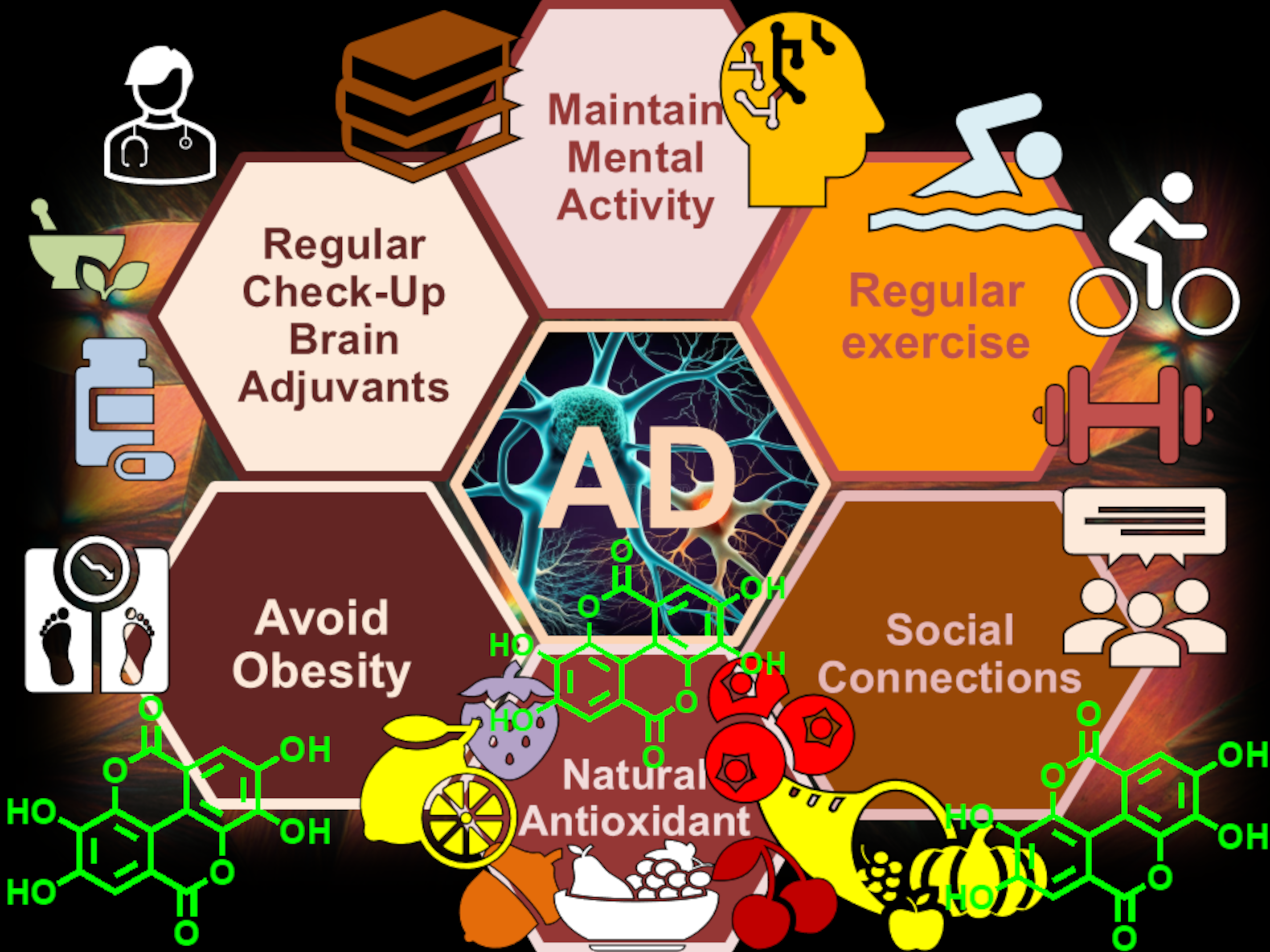
Alzheimer-Perusini disease, mainly known as Alzheimer’s disease (AD), presenile dementia of the Alzheimer type, primary degenerative dementia of the Alzheimer type, and for simplicity Alzheimer, is the most common form of progressively disabling degenerative dementia, with onset mainly in presenile age, specifically over 65 years[1]. It is estimated that approximately 50-70% of cases of dementia are due to the AD condition, while 10-20% are due to vascular dementia[2]. Some data from the World Alzheimer Report 2023 produced by Alzheimer’s Disease International established that in the next 25 years, the number of people living with dementia worldwide could increase from 55 million to 139 million. Furthermore, the costs associated with the disease could jump from 1.3 trillion dollars in 2019 to over 2.8 in 2030. The most frequent early symptom is represented by a difficulty in remembering recent events, followed by other symptoms which may appear by ageing, including aphasia, disorientation, sudden changes in mood, depression, inability to take care of oneself, and behavioural problems. Also, confusion, irritability and aggressiveness, mood swings, difficulty speaking, both short- and long-term memory loss and progressive sensory dysfunction further aggravate the already detrimental condition of patients suffering by AD [3,4].The subject tends to isolate himself from society and family and gradually, basic mental abilities are lost. It seems that, about 70% of the AD developmental would be genetic with several genes usually involved. But the exact cause and progression of AD are not still well understood. It is well established that AD is a well-unshakable neuronal disfunction, whose primary causes could be associated with toxin insults, heredity, metabolism, or even attack by infectious pathogens[5]. Several research indicates that AD is closely correlated with amyloid plaques and neurofibrillary tangles found in the brain, but the root cause of this degeneration is unknown[6]. Other well-explored factors, contributing to cognitive neurodegeneration driving to AD comprise excessive acetylcholine esterase enzymes (AChE), β amyloid (βA) precursor protein-cleaving enzyme 1 (BACE-1), glycogen synthase kinase 3 β (GSK-3 β), monoamine oxidases (MAOs), metal ions in the brain, N-methyl-D-aspartate (NMDA) receptor, and phosphodiesterases (PDE). It is anyway extensively recognized that OS, as well as the formation of free radical and not radical RONS, are strongly involved in the progression of brain aging and, in the onset, and evolution of AD. In addition, impaired bioenergetics, mitochondrial abnormalities, and neuroinflammatory processes are implicated too. Collectively, one-hundred years after the AD discovery, the scientific community is quite firm that, although the pathogenesis of AD is not yet fully understood, it is surely a multifactorial disease caused by both genetic, environmental, and endogenous factors (Figure 1), like other neurodegenerative disorders [7]. The excessive incorrect folding and aggregation of proteins often related to the ubiquitin-proteasomal system (UPS) are also accountable to AD.
Particularly, the increasing of RONS causes mitochondria and DNA damaging, with increased production of toxic Aβ causing in turn severe DNA repair dysfunctions. Currently, approved therapeutic treatments used to treat AD provide only little and temporarily benefits to symptoms and can partially slow the progression of the disease. Increasing insights, coupled with further ongoing discoveries about AD multi-factorial pathogenesis, have provided the rationale for the search for new therapies, which directly could target AD molecular causes [7]. New drug candidates with promising potential to modify the disease are now in the pipeline and have reached testing in clinical trials [9]. On the index date of January 1, 2023, there were 187 trials assessing 141 unique treatments for AD. Phase 3 included 36 agents in 55 trials; 87 agents were in 99 Phase 2 trials; and Phase 1 had 31 agents in 33 trials. The most common drugs, comprising 79% of drugs in trials, were those proposed as disease-modifying therapies, and 28% of candidate therapies were those using repurposed agents. Collectively, current Phase 1, 2, and 3 trials will require 57,465 participants [9]. Unfortunately, although nowadays over 500 clinical trials have been conducted to identify a possible effective treatment for AD, no treatment has yet been identified, capable to halt or reverse the disease[10]. The widespread and increasing diffusion of AD in the population, and the limited and non-resolving efficacy of the available therapies, as well as the enormous resources necessary for its diagnosis, management in terms of social, emotional, organizational and economic, make AD one of the diseases with the most serious social impact in the world [11]. This lack of pathogenesis-targeting therapies is principally due to the limiting effects of the blood–brain barrier (BBB), which keeps out of the brain about 99% of all “foreign substances”. Later their discovery, nanoparticles (NPs) have been successfully used for targeted delivery into many organs, including the brain[12]. In this context, new nano dimensional agents and/or formulations of existing drugs could be promising options for the possible diagnosis and treatment of various neurological disorders, including AD. Furthermore, it has been reported that drugs hitting a single molecular target are not effective for the treatment of diseases like the complex neurodegenerative syndromes, like diabetes, cardiovascular diseases, and cancer, which involve multiple pathogenic factors [13]. On the contrary, drugs that could cover up different pharmacological approaches could offer more possible ways of overcoming the problems that could arise from the use of single-target drugs, often well-functioning in vitro but not in vivo experiments. On this worrying scenario concerning the poor available arsenal of drugs and/or nano-drugs to treat AD, the several multitarget health effects of many fruits and vegetables could represent an appealing alternative treatment option. In fact, it has been demonstrated that foods including muscadine grape, berries as pomegranate, strawberry, raspberry, blackberry, nuts such as chestnuts, walnuts, almonds, pecans, pistachios, herbs such as Camellia sinensis, seeds including berries seeds, and their derived foods and/or beverages possess recognized healthy and/or preventive effects against several complex human diseases, thus evidencing their multitarget behaviour [14]. Such effects have been associated mostly with their high content in antioxidant molecules, mainly of polyphenol type [14,15,16], such ellagitannins (ETs) as well as gallic acid (GA) and ellagic acid (EA), which are produced by their hydrolysis in vivo (Figure 2) [17]. By limiting the hyperproduction of RONS they counteract OS, recognized as the foremost prompting factor of several human discomforts.
Particularly, the stringent correlation existing between the consumption of ETs-rich foods and the deriving ameliorating effect vs. several human degenerative diseases is extensively reported[17,18]. As examples, documented findings assert an association between the consumption of ET-rich foods and greater cardiovascular health [19,20], or among the consumption of fruits and vegetables and minor incidence of coronary heart disease [21]. Much empirical data guided to the hypothesis that ETs might be exploited to prevent chronic and degenerative diseases such as cancer, diabetes, cardiovascular diseases, and central nervous system (CNS) disorders, including AD [22]. Nonetheless, in Europe, EFSA has not been still approved for them any kind of health claims [14]. As abovementioned, ETs are capable to provide EA by hydrolysis, which is rationally considered the bioactive fragment of ETs possessing one of the strongest antioxidant power capable to counteract OS [17], confirmed already 10 years ago in a study by Kilic[23]. In vitro radical scavenging and antioxidant capacity of EA were clarified using different analytical methodologies such as total antioxidant activity determination by ferric thiocyanate, hydrogen peroxide scavenging, 1,1-diphenyl-2-picryl-hydrazyl free radical (DPPH) scavenging, 2,2′-azino-bis(3-ethylbenzthiazoline-6-sulfonic acid) (ABTS) radical scavenging activity and superoxide anion radical scavenging, ferrous ions (Fe2+) chelating activity and ferric ions (Fe3+) reducing ability[23]. . Being endowed with this relevant capability to combat OS, nowadays considered the key cause of all diseases, and therefore being gifted with the capacity to ameliorate human degenerative diseases, food chemists consider both ETs and EA as nutraceuticals (NTs). NTs are defined compounds which possess both the canonical nutritional values and several additional health benefits, so that the dietary intake of foods containing these components often translates in relevant beneficial biological effects. This review aims at more largely driving the researchers’ attention towards EA as actual possible multi-target treatment option for AD. A brief overview on the multi-factorial and multi-target aspects that characterize AD, and polyphenols such as EA respectively, open this work. Upon a focus on the pathophysiology of OS, on EA chemical features and on the mechanisms of its antioxidant activity, an all-round updated analysis concerning the EA-rich foods and EA involvement in the field of AD has been provided. The possible clinical usage of EA to treat AD has been shown reporting results by its applications in vivo and clinical trials. A critical view about the need for a more extensive use of the most rapid diagnostic methods to detect AD from its early symptoms has also been included in this work.
2. Multifactorial Nature of Neurodegenerative Diseases: Alzheimer Disease (AD)
Neurodegenerative diseases (NDs) have long been viewed as among the most mysterious and challenging issues in biomedicine [12]. While moving from descriptive phenomenology to mechanistic analysis, researchers have become progressively aware that the major processes involved in their onset are complex and multifactorial, including both genetic, environmental, and endogenous factors[24,25]. Such NDs comprehend, among others, Alzheimer’s disease (AD), Parkinson’s disease (PD), and Huntington’s disease (HD), as well as amyotrophic lateral sclerosis (ALS). As in other neurodegenerative conditions, the pathogenic cascade driving to AD includes protein incorrect folding and aggregation, OS and RONS formation, metal dyshomeostasis, mitochondrial dysfunction, and phosphorylation impairment, all occurring concurrently. Figure 3 summarizes the concomitant multiple factors conducting to the onset of the AD conditions, while Figure 4 evidence how some of these factors can damage directly the neurons causing their death or can trigger a detrimental cascade of events anyway leading to the death of neurons.
Protein wrong folding followed by self-association and subsequent deposition of aggregated, anyway supported by OS, RONS uncontrolled increasing and metal dyshomeostasis, has been observed in the brain tissues of patients affected by AD [26]. Findings suggest that protein assemblies produced by different amyloidogenic proteins share common structural and histological morphologies and might trigger similar neurotoxic mechanisms. The biophysical behaviour of these proteins, leading to their incorrect folding, aggregation, and deposition, has prompted scientists to group these kinds of neurological disorders under the common name of “conformational diseases” [27]. It is worth noting that amyloid oligomers such as amyloid-precursor protein (A) and R-synuclein have been widely reported to permeabilize both cell and mitochondrial membranes, thus impairing their functions [28]. They are therefore probably responsible for the subsequent calcium dysregulation, membrane depolarization, and deficiency of mitochondrial functions, which have been identified as a common feature of AD [29].
2.1. More in Deep in The Multifactorial Causes of AD: Reactive Oxygen and Nitrogen Species (RONS)
The role of RONS in many NDs was deemed to be as essential as the role of microorganisms in infectious diseases. In normal conditions, RONS generation is kept under control by the antioxidant defences and repair systems of cells[30]. On the contrary, when overproduced, the detoxification systems of cells fail to maintain RONS physiological levels. They accumulate, thus causing the onset of OS and inflammation. Irremediable damage to DNA, lipids, and proteins happens, thus promoting aging, age-related diseases, and several degenerative human disorders[30]. To respond to the answer “Is OS a cause or a consequence of the neurodegenerative cascade in AD?” has been and remain a daily challenge for experts in the field, which would need urgently a solution. At present, scientists agree almost unanimously to affirm that imbalance of intracellular oxidation state is an early event in the neurodegeneration and is therefore likely to be one of the major factors of neurodegenerative disorders. Neuronal tissue is particularly sensitive to OS, and the possible imbalance in pro-oxidant vs antioxidant homeostasis in central nervous system (CNS), can result in the production of several potentially toxic RONS, including both radical and nonradical species that participate in the initiation and/or propagation of radical chain reactions injuring neurons. Table 1 reports the possible sources of RONS, which can be endogenous, both enzymatic and non-enzymatic, as well as exogenous.
MPO = myeloperoxidase; NOX = NADPH oxidase; NAD = nicotinamide adenine dinucleotide; Fp = flavoprotein enzymes; EPFRs = environmental persistent free radicals present in particulate matter; BC-PFRs = biochar-related persistent free radicals.
The following Figure 5 schematizes specifically the main endogenous processes by which ROS can be created in cells and the detrimental effects they can have on health [30], including DNA damage, lipids, and protein peroxidation, telomere reduction, aging, and death.
In AD, OS has been found in every family of biological molecules within neurons, spanning from lipids to DNA and proteins. Anyway, several clinical studies have revealed that the simple administration of one or a few one-target antioxidants had modest success in the treatment of neurodegeneration. It has been reported that in AD, it exists also a direct cause and effect relationship between metal abnormalities and increased oxidative damage. Transition metals such as iron, copper, or other redox active metals are essential in many biological reactions, but the alterations in their homeostasis may result in increased free radical production. Moreover, while all the disease-specific proteins bear metal-binding motifs, metal ions favour the fibril generation, and the protein deposition found in AD (Section 2, Figure 4). Furthermore, in addition to be a cause of OS in neurons, metal-mediated OS is linked also to mitochondrial dysfunction, where ROS can be generated, as well. Morphological, biochemical, and molecular abnormalities in mitochondria in various tissues affected by AD have been signalled. Although the chronological hierarchy of events and underlying causes in AD about mitochondrial dysfunction and OS are not yet fully clarified, there is unequivocable evidence that both participates to the evolution of the others, setting in motion a self-sustaining, amplifying cycle that can ultimately activate the initiation of neuronal death processes as shown in the following Figure 6.
Also, the endoplasmic reticulum (ER) is an important apoptotic checkpoint. It has been shown that, in AD, apoptosis induced by badly bent proteins involves ER impairment. Another common mechanism shared by NDs concerns the alteration of the phosphorylation state of some key proteins participating in the pathogenic cascades. Besides the well-recognized hyperphosphorylated state of τ protein in the neurofibrillary tangles observed in AD brain, other specific altered patterns of kinase and phosphatase activities are associated with alteration in the phosphorylation state of disease-specific proteins, which are different for PD, ALS, and HD. Extensive molecular evidence demonstrated the cell-type specificity in neuronal disorders and the selective neuron degeneration in AD. However, none of these general mechanisms alone are sufficient to explain the high number of biochemical and pathological abnormalities of AD, which encompass a multitude of cross-related cellular and biochemical changes that cannot be adequately addressed by following treatments based on one-molecule, one-target paradigm. In our opinion, there should be a growing interest and an urgent need for the development of multi target directed ligands (MTDLs) to provide real disease-modifying drug candidates for such ND.
3. One-Target Drugs vs. Multi-Target Therapies in the Treatment of Degenerative Diseases
The scientific knowledge about the pathogenesis of several human diseases has advanced enormously in recent decades. Therefore, the sector of drug discovery has gradually shifted from seeking an entirely human phenotype-based approach to a more reductionist approach based on single molecular targets. This change has led to a type of drug research, still extensively followed, aimed mainly at the discovery of small molecules able to modulate the biological function of a single given target, believed to be fully responsible for a certain disease. Efforts in this sense have been devoted to achieving drug molecules selective for a certain protein, and many ligands endowed with outstanding in vitro selectivity and efficacy are today available. Although such one-molecule, one-target paradigm has led to the discovery of many successful drugs, and it will probably remain a milestone for years to come, it should be noted that a highly selective ligand for a given target in vitro, does not always result in a clinically efficacious drug in vivo (Table 2).
The low correspondence between results in vitro and those in vivo in the case of NDs, is mainly due to the multifactorial nature of human degenerative diseases. In these cases, the cells can often find ways to compensate for a single protein, whose activity is affected by the one-target drug administered, by taking advantage of the redundancy of the system, including the existence of parallel pathways [31]. Drugs hitting a single target may be inadequate for the treatment of diseases like neurodegenerative syndromes such as AD, diabetes, cardiovascular diseases, and cancer, which involve multiple pathogenic factors [32]. Different pharmacological approaches are necessary to overcoming the problems that arise from the use of single-target drugs (Table 2, column 1). When a single target medicine is not sufficient to effectively treat a disease, alternative approaches aiming at hitting more than one impaired process correlated to the disorder should be considered. Figure 7 shows some alternative medical approaches.
The three most adopted approaches (MMT, MCM and MTDLs) reported in Figure 7 have charted in Table 3 with related advantages and disadvantages.
The multiple-medication therapy (MMT) (Figure 7), also known as combination therapy, may be used as alternative option to one-target therapy. It is usually composed of two or three different drugs singularly administrated, thus combining different therapeutic mechanisms[36]. A second approach might be the use of a multiple-compound medication (MCM), also referred to as a “single-pill drug combination”, which implies the incorporation of different drugs into the same formulation. Finally, a very appealing strategy is now appearing assumes that a single compound may be able by per se to hit multiple targets, because comprehend in the same molecule more than one pharmacophore. Clearly, therapy with a single drug that has multiple biological properties would have inherent advantages over MMT or MCM as reported in Table 3. There is, therefore, a strong indication that the development of single compounds able to hit multiple targets might disclose new avenues for the treatment of major NDs, such as AD, for which new effective cures are an urgent need and an unmet goal. In the past, Morphy and Rankovic pleasingly discussed this approach in three articles, which anyway were mostly concerned with non-NDs [37,38,39]. In this context, we are convinced that the definition “multi-target-directed ligands” (MTDLs) more completely describes these compounds. Effectively, MTDLs should succeed in treating complex diseases, because of their ability to interact with the multiple targets thought to be responsible for the disease pathogenesis. The excellent perspective by Morphy and Rankovic [37] covered several aspects of the design strategy leading to MTDLs for different areas such as inflammation, dopaminergic D2-receptors, histaminergic H1-receptors, serotoninergic receptors, angiotensin system, peroxisome proliferators activated receptors, kinases, and nitric oxide releasing conjugates. Although more attention to the achievements of MTDLs also for NDs is increasing, there is still a paucity of review literature dealing with complex diseases associated with neurodegeneration, which we hope to compensate for, by our present work.
3.1. Alzheimer’s Disease (AD) and Currently Available Medicines and/or Treatments in Development
Among the NDs above reported, AD stands out as the fourth leading cause of death in the Western countries and the most common cause of acquired dementia in the elderly population. As shown in Figure 8, two main forms of AD are recognized, both characterized by neuronal death.
In line with an increase in average life expectancy of humans, the number of affected persons is expected to triple by 2050, with immense economic and personal tolls [35]. In parallel with this increase, the speed of drug research has accelerated noticeably in recent decades, but not enough. However, the number of therapeutic options on the market remains strongly restricted. Worryingly, the currently registered drugs for AD, i.e. acetylcholinesterase inhibitors (AChEIs) are not able to alter or prevent disease progression. They are instead palliative in alleviating disease symptomatology[40]. On this scenario, being AD a multifactorial disease whose insights and discoveries about its pathogenesis are progressively ongoing, the rationale exists for the discovery and study of multi-target drugs directly targeting different AD molecular causes at once.
3.1.1. Current AD Therapies
Although the path of the events leading to the AD onset is far to be completely clarified, the cholinergic hypothesis was the oldest one and had the strongest influence on the development of clinical treatment strategies for AD. Acetylcholine (Ach) is released in the synaptic cleft where it activates both postsynaptic and presynaptic cholinergic receptors [nicotinic (N) and muscarinic (M)], leading to an increase of cholinergic transmission, which results in cognition improvement. Anyway, ACh is removed from the synapse by the action of the enzyme acetylcholinesterase (AChE), which therefore has been become the target for the development and approval of acetylcholinesterase inhibitors (AChEIs) for AD treatment as visualized in Chart 1 and reported in Table 4.
The acetylcholinesterase inhibitor (AChEI) tacrine (Chart 1) was the first drug to be approved for the treatment of AD, now rarely used because of its hepatotoxicity. Later, three other AChEIs, donepezil, rivastigmine, and galantamine reached the market, becoming the standard for AD therapy, only later complemented by memantine, a noncompetitive NMDA antagonist (Chart 1). Table 4 include the advantages and disadvantage connected to the use of such therapeutics.
Although the diffused clinical practice, the debate on the effective activity of AChEIs medications endures. So, the search for novel AChEIs, such as inhibitors of the “non-classical function” of AChE have rehabilitated interest in expanding their potential as real disease-modifying agents. Current AD drug development programs focus primarily on agents with anti-amyloid disease-modifying properties, and several studies have been carried out on molecules capable to reduce amyloid pathology (Table 5). Classes of therapeutic modalities currently in the advanced stage of clinical trial testing comprise forms of immunotherapy which uses several drugs (Table 5), including also medicaments with anti-amyloid properties. Nontraditional dementia therapies, such as those using the HMG-CoA reductase inhibitors, including mainly statins[42], such as atorvastatin, simvastatin, fluvastatin, pravastatin, rosuvastatin and lovastatin are now being evaluated also for their clinical benefits in AD as disease-modifying treatments [42].
3.1.2. Versus Disease-Modifying Therapies in Alzheimer’s Disease [123]
The long-expected era of disease-modifying therapy (DMT) for AD has finally arrived and will substantially influence how the disease is perceived and managed. Unfortunately, the new treatments closest to extensive clinical implementation (Figure 9), will pose challenges for rightful access. No national health-care system is ready to deliver these drugs to more than a fraction of patients who might be eligible.
These active principles (APs) include lecanemab and donanemab, which are intravenous monoclonal antibodies capable to remove βA plaques from the brain, thus slowing cognitive and functional decline. Paradoxically, lecanemab and donanemab have revealed side-effects, mainly amyloid-related imaging abnormalities (ARIA) in about 21% and 39% of patients, respectively[124]. While usually asymptomatic and transient, ARIA requires close monitoring. Symptoms and signs of ARIA can be non-specific, including blurred vision, headaches, and unsteadiness, or can include focal deficits such as dysphasia. However, many patients with ARIA can be re-dosed safely after a period off treatment[124].
3.1.3. Multi-Target Therapy (MTT) for AD
However, the adoption of MMT, MCM and MDTLs (or MTSM) might result in more effective treatment strategies for AD, due to the multifactorial nature of this disorder. MMT has already proven successful in the treatment of other complex diseases such as cancer, HIV, and hypertension. Due to the possibility of attacking several targets simultaneously, exploiting synergy, and minimizing the individual toxicity of the administered single drugs, maximum efficacy has been achieved. With similar outcomes and advantages, MCMs, were fought, to ameliorate the compliance of patients with AD. From 2006, the number of patented MCM, where new compounds, which revealed potentialities to ameliorate AD were administered in combination with old therapeutics (AChEIs or NDMA receptors antagonists, as well as NSAID or a combination of two) has overtaken that of single-drug entities for the potential treatment of AD[125] (Table 6).
In clinic, the MMT of memantine plus an AChEI appears to produce an additional effect resulting in a well-tolerated, effective treatment strategy [137]. Considering the well-accepted clinical use of MMT only as a starting point, the MTDL design strategy might represent its natural evolution, and MTDLs emerge as valuable tools for better hitting the multiple targets implicated in AD aetiology [138]. Several MTDLs have been developed by academia and industry in recent years. These have been the subject of some interesting review articles, which readers particularly interested could examine at the related references [139,140,141,142]. The main design strategy usually applied to build up a possible new MTDL involve detecting the active portions of different drugs and combining them in a single structure to afford hybrid molecules[8]. In principle, each pharmacophore of these new drugs should retain the ability to interact with its specific site(s) on the target and, consequently, to produce specific pharmacological responses that, taken together, should slow or block the neurodegenerative process of AD. Specifically, it is in use to modify the molecular structure of an AChEI by inserting opportune pharmacophores (indicated as PG groups in Figure 10) already present inside other drugs, which demonstrated beneficial effects in neurodegenerative diseases, to provide the traditional drug with additional ameliorative effects, while reducing side effects of separate single drugs and enhancing the compliance of patients [8].
4. Ellagitannins (ETs) and EA as Multi-Target Compounds: Strengths and Weaknesses
Both ETs and EA have proven, at least in vitro, to prevent and/or ameliorate chronic diseases such as cancer, diabetes, cardiovascular [143] and lately also neurodegenerative diseases[144,145]. It seems that these positive effects are due to their multi-target action accounting for anti-angiogenic, anti-atherogenic, anti-carcinogenic, anti-obesity, anti-inflammatory, antioxidant and anti-thrombotic properties, together with anti-neurodegenerative capability. All these gains seem to derive from their antioxidant power and therefore their capability to contain OS, the key cause of all human disorders[14,17]. Since neurodegenerative disorders including AD are multifactorial diseases, the application of the usual and extensively approached one-molecule, one-target paradigm, providing drugs able to hit only a single target, could have limited effects, mainly in vivo, and may also translate in the emergence of resistance. On the contrary, a compound capable to interfere with different targets involved in the cascade of the pathological events leading to a given disease could be highly effective for treating multifactorial diseases, as AD [13]. The synthetic design of such drugs may not be easy, because the obtained drugs could bind in vivo targets that are not involved with the disease of interest and could be not necessarily responsible for side effects. On the contrary, natural polyphenols such as ETs and EA, per se possessing the multifaceted health activity above reported as demonstrated by the outcomes deriving by the assumption of food containing them, are provided ready by nature and could be promising options to ameliorate/treat AD. However, they could serve at least as template molecules to be used as starting platforms to design new multi-target drugs.
4.1. Bioavailability Drawbacks Associated with ETs and EA
According to a review reported in 2020, except for an insignificant amount (e.g. 0.7–4.7 mg/100 g of berries, wet weight), the free form of EA is produced mostly in vivo, after the consumption of ETs-rich food, due to the physiological massive hydrolysis of ETs in the gastrointestinal tract (GIT) [17]. Anyway, even if according to some other authors, free EA makes up only a small part of the total EA pool in plants, others suggest that its portion can reach and even exceed 50% of the total content, depending on the plant species [146]. Interestingly, in the fruits of Terminalia ferdinandiana Exell, a native Australian plant known as the Kakadu plum, EA was found to be mostly free form, with a percentage reaching 70.6% of the total EA pool [147]. By contrast, the percentage of free EA in strawberries, as shown by the same study, reaches only 7.4% of its total content[147]. Despite early studies did not show the presence of EA in plants of the Fabaceae family, there is now evidence of relatively high levels of this phytochemical in several sprouted legumes, such as sprouted adzuki bean (Vigna angularis), some varieties of bean (Phaseolus vulgaris L.), cowpea (Vigna unguiculata (L.) Walp.), pea (Pisum sativum L.), and soybean (Glycine max (L.) Merr.)[148]. Sprouted soybeans have been found to have a considerably higher EA content than other sprouted legumes (45.6–48.9 mg/100 g vs. 8.96–18.3 mg/100 g dry weight) [148]. Although, the ratio between free and bound forms of EA in plants may vary considerably depending on the plant species, the proportion of unbound EA may also depend on the method chosen for determination, the type of storage, and processing practice [149]. Freezing fruits, as well as processing them to produce beverages and jams, may have different effects on the content of EA[146]. However, after the intake of ETs-rich foods, ETs are only slightly absorbed and reach the small intestine, where they are hydrolysed to EA by the gut microbiota action [17]. Once produced, EA is practically not absorbed, due to its trivial water solubility, unfavourable physicochemical characteristics and low bioavailability (Table 7) and reaches the large intestine untouched. A justification for EA poor bioavailability and its low concentrations in plasma and tissues is based on the EA capacity to bind permanently to cellular DNA and proteins, or to form weakly soluble complexes with calcium and magnesium ions, which greatly reduce transcellular absorption[150]. Also, still active metabolites of EA, such as methyl and dimethyl ethers or glucuronic acid conjugates, sparkly detected in plasma and urine at 1 and 5 h after ingestion, corresponded to very low concentrations as well, not sufficient to produce significant beneficial effects[17]. In large intestine, EA is metabolized to the more hydrophilic urolithins (UROs), secondary polyphenol metabolites derived from the gut microbial action [151], and/or converted to its dimethyl, as well as dimethyl glucuronate and sulphate derivatives which are excreted.
A representative structure of an ET (casuarictin), that of EA, and those of URO A, B, C, D, iso-A and iso-B have been shown in Scheme 1, which shown the path of EA formation after the intake of ETs-rich foods and its subsequent metabolism to UROs and dimethyl ether derivatives[17]. A more recent article has introduced also URO-M5 and M6 among the URO-type metabolites of EA[151]. Precisely, in this new route EA is transformed in URO-M5 which is in turn converted in URO-D, while URO-M5 is converted in URO-M6 which then provides URO-C as URO-D [151].
In the year 2022, a study reported the existence and structure of up to 13 UROs [156]. Collectively, being ETs poorly adsorbed in GIT, they cannot reach blood and tissues where they could exert their beneficial effects but provide the bioactive EA upon hydrolysis. Nonetheless, instead to be absorbed and reaching blood and tissues where acting, due to its very low water-solubility [155], also EA undergoes a massive metabolism. Specifically, it is transformed in UROs, and in other metabolites excretable with urine and the amount of EA detected in blood and tissues observed after ETs-rich foods intake results insignificant to improve the conditions associated to chronic human diseases [146]. Due to this process, the findings obtained with ETs and EA in vivo studies against several human pathologies did not coincide with the promising ones observed in vitro, as generally happens for dietary polyphenols[14,157]. As observable in Scheme 1, UROs are dibenzopyran-6-one derivatives with different hydroxyl substitutions. UROs are more lipophilic than EA, and this has been suggested as a factor responsible for the greater urolithins absorption rate as compared to EA, thus being the only active phenolic molecules sufficiently absorbed and detectable in the circle and cells after ETs-rich foods intake[151]. URO-A and URO-B serve as the major metabolites of EA found in the gut, being URO-A as the most biologically active as compared to the rest of the EA metabolites[151]. In enterocytes and hepatocytes, UROs undergo biotransformation to UROs metabolites[146]. The main metabolites of urolithins found in plasma and urine are their glucuronyl and sulfate conjugates, including URO-A and URO-B glucuronide and sulfate, while the minor metabolites are URO-C and iso-URO-A glucuronide[146].
4.2. Ellagic Acid or Urolithins?
Both in vitro and in vivo studies have shown evidence that also UROs own anti-inflammatory, anti-carcinogenic, anti-glycative, antioxidant (lower than ETs and EA), antimicrobial properties, as well as preventive effects on gut and systemic inflammation. Furthermore, UROs seem also to play the role of hormone analogues [158]. Table 8 reports the most relevant studies concerning the in vivo effects of UROs assessed in animal models.
Due to the confirmations both in vitro and in vivo about the pharmacological properties of UROs, currently there is an extensive tendency to think that UROs, rather than EA, could be the actual bioactive molecules accountable for benefits coming from the consumption of ETs and EA rich foods[14,67]. This proposition is supported by the awareness that, although in vitro findings have shown that EA and UROs are almost equally active, studies in vivo provided trustworthy verification about this fact, only regarding UROs. Only UROs have been found in fluids, cells, and tissues and were measured, finding concentrations capable to exert the ameliorative effects already evidenced in vitro. On the other hand, the interest in knowing more about the possible EA activity in vivo has led scientists to increasingly and incessantly focus on preparing water soluble and absorbable EA formulations, able to defend EA and to lower or annul EA metabolism to UROs, so that it could reach cells and tissue in its pristine form [152]. The formulation of drug delivery systems, capable of transporting and releasing EA to the target site, represents a valid approach for bypassing the bad biopharmaceutical features of this polyphenol, thus allowing a better evaluation of its potential application as radical scavenger antioxidant therapeutic. In this context, from the year 2019, we studied some micro- and nanosized solutions which revealed interesting performance[192,193,194].
4.4. Drawbacks Associated to UROs Hamper Their Clinical Development Thus Quenching the Researcher Interest
Even if gifted with healthy properties like those of EA, UROs are not suitable for safer therapeutic purposes, due to their double faceted behaviour. They can be beneficial but, depending on their structure, environmental conditions, the type of target cells under study, age, and health state of the individuals, they could result also harmful[17]. The amount and typology of UROs produced in the gut of individuals depends also on the type of vegetables which has been introduced, the individual microbiota metabolic activity, that is typified by a highly inter-individual heterogeneity, depending on several factors and humans metabotype (0, A, B)[17]. Moreover, this highly interindividual and intra-individual process is not completely elucidated yet [34,35]. Let’s imagine that even living species which do not produce UROs exist. Table 9 reports the UROs mainly found in different mammalian species after the consumption of different vegetables.
UROs absorption, blood and tissue concentrations, and inter-subject variability in the comebacks to UROs exposure, are arbitrary variables, which drive to various responses that, ironically, could promote adverse effects. In addition, human microbiota activity is difficult to be reproduced in animal models and cannot be easily studied and/or controlled [17].
5. EA as Template Antioxidant Molecule for the Development of New Therapeutics for AD
EA attracts the interest of researchers as promising molecule to provide benefits in neurodegenerative disorders including AD, mainly due to its anti-inflammatory and the antioxidant properties. Defining which pharmacophore/pharmacophores in EA is the actual responsible/s for its health benefits, but also for its possible collateral effects is crucial for in silico screening investigations and to design new multi-target EA-type CNS drugs. The mechanisms at the basis of the EA multifaceted bioactivity are based mainly on its antioxidant, radical scavenger and anti-ageing effects, capable to contrast OS. Collectively, EA is to counteract the detrimental RONS, which are a byproduct of physiologic aerobic metabolism. For a more precise distinction, OS refers to a torrent of destructive proceedings that frequently triggers and accompanies the molecular/cellular pathogenic events, responsible for several human disorders, including AD [144,198]. Differently, inflammation, being both the cause and the effect of RONS accumulation, is considered a pathological characteristic of the most part of human diseases including those developing in the CNS including AD.
5.1. EA Antioxidant Effects: Proposed Mechanisms of Action
Natural antioxidants are fundamentally present in vegetable food, and polyphenols, such as EA, are supposed to be more than 8000 molecules, all characterized by possessing at least a phenol moiety. EA hydroxyl groups and the lactone systems give the molecules the capacity of forming hydrogen bonds, while can also act as electron acceptors and/or hydrogen donors. Consequently, EA is endowed with the capacity to take electrons from different substrates thus promoting antioxidant redox reactions and functioning as a very efficient free radicals (FRs) scavenger[199]. The EA anion is proposed as the key species for its protective effects against OS[199]. It is predicted to be efficiently and continuously regenerated after scavenging two free radicals per cycle[199]. Chemical species able to prevent oxidation can be classified in primary antioxidants (Type I, or chain breaking) and secondary antioxidants (Type II, or preventive). EA can behaviour as both Type I and Type II antioxidant, thus exerting a multiple-function antioxidant activity (Table 10)[200].
5.1.2. Type I Scavenging Reactions
Type I scavenging reactions, which can occur between EA and FRs, follow second order kinetics and scavenging capacity, as well as its velocity, depend both on the concentration of EA and FRs. Factors which could modify their chemical structures, such as the pH, polarity, the reaction conditions, and mainly the medium could also affect EA scavenging capacity. In general, the antioxidant capacity of EA reduces strongly in solvents able to form hydrogen bonds with EA and improve in solvents favouring EA ionization to anion phenoxide[201]. The alcohols may act as acceptors of hydrogen bonds, thus decreasing EA antiradical effects by hydrogen atom transfer (HAT) reactions. On the other hand, they can favour the ionization of the EA to anion phenoxides, which can react rapidly with the peroxyl radicals, through an electron transfer, thus improving EA radical scavenging activity by SET reactions[201]. In general, the antiradical properties of different natural and synthetic Type I antioxidants possessing OH groups, derives mainly from their capacity to transfer hydrogen atoms to FRs. This process can occur by mechanisms reported in Table 11. These mechanisms generate non-radical species or new radicals, more stable and less reactive than the previous ones, thus restricting the development of OS. Table 11 reports also the chemical equations associated to these proposed mechanisms.
EA can exercise antioxidant effects mainly through three of the above-mentioned reaction mechanisms, such as SET, HAT and SPLHAT reactions. Although the result is always the inactivation of FRs to neutral, cationic, or anionic species, the kinetics and secondary reactions involved in the processes are different (Figure 11).
When EA reacts, for example, with the radical specie ROO•, a hydrogen cation coming from its hydroxyls into other radical species, is transferred forming a transition state of an H-O bond with one electron. On the other hand, the hydroxyl groups can interact with the π-electrons of the benzene ring providing molecules endowed with the ability to generate free long-living radicals stabilized by delocalization, able to interfere and modify radical-mediated oxidation processes, by SET reactions.
5.1.3. Type II scavenging reactions
EA is also a Type II antioxidant, thus providing its protective effects against FRs by inhibiting the endogenous production of oxidants and radical hydroxyl (•OH) molecule, which is the most reactive and electrophilic specie of the oxygen-based radicals [30]. • OH is the main responsible of tissues and DNA damage and therefore, its inhibition is of prime significance for reducing OS generated from the metal-catalysed Fenton reaction and the Haber Weiss recombination (HWR), according to Equations (1)–(4), involving the reduced forms of Fe and Cu.
Fe (II) + H2O2 → Fe (III) + OH− + •OH (1)
Cu (I) + H2O2 → Cu (II) + H− + •OH (2)
Fe (III) + O2 •− → Fe (II) + O2 (3)
Cu (II) + O2 •− → Cu (I) + O2 (Fenton) (4)
In this context, EA is an excellent antioxidant due to its capability to chelating and subtracting metal as Fe2+, Fe3+, and copper ions involved in the production of FRs, thus preventing the oxidation of low-density lipoproteins (LDL)[199,200,202]. EA can also interact with enzymes involved in radical generation, such as various cytochrome P450 isoforms, lipoxygenases, cyclooxygenase, and xanthine oxidase, thus inhibiting RONS over production. This capability derives from the presence of the hydrophobic benzenoid rings and from the skill of the phenolic hydroxyl groups to form hydrogen-bonding interactions [203]. Moreover, EA can act synergistically with other endogenous and exogenous antioxidants, such as ascorbic acid, β-carotene, and β-tocopherol, thus increasing their effectiveness and regulating intracellular glutathione levels[203]. Unfortunately, some of hydroxyl groups of EA, in conditions of high dosage, high concentrations of transition metal ions, alkali pH, and/or the presence of oxygen molecules, can act unexpectedly also as pro-oxidants moieties[204]. These groups may sometimes induce significant DNA damage in the presence of Cu (II) or may create ROS through the reduction of Cu (II)→Cu. The pro-oxidant activity is peculiar of small polyphenols, as EA, while is limited in large molecular-weight phenols, such as ETs. On the other hand, this apparent issue can trigger apoptosis in cancer cells[205,206].
6. EA-rich foods, EA Food Supplements, and EA Involvement in the Treatment of AD
As above-mentioned, the polyphenolic lactone by formula C14H6O8, known as EA, as well as the intake of EA food supplements, foods rich in ETs and/or EA can translate in altering profuse signaling inside cells thus preventing and/or pauperizing the progression of diverse neurodegenerative abnormalities, including AD [207]. Its neuroprotective effectiveness is attributable mainly to its ROS scavenging, iron chelating properties, positive regulation of energetics of mitochondrial respiratory complex, and abundant modulation of neuronal molecular signaling pathways[208].
6.1. Most Relevant In Vitro and In Vivo Studies Using ETs and EA-rich Plants
Table 12 summarizes the beneficial properties demonstrated in vitro and/or in vivo studies using different experimental models, or even in clinical settings, observed upon the assumption of ETs and EA-rich plants.
From information reported in Table 12, it appears unequivocable that the clinical interest in the possible beneficial properties of EA-rich plants is very limited. Particularly, among the studies here considered (56), the clinical ones represent only 5%, and in vivo ones are largely under half percent (25%) of the in vitro ones (70%) (Figure 12).
Collectively, practically all studies, regardless they were conducted in vitro, in vivo, or in clinical setting, revealed mainly antioxidants and anti-inflammatory effects. Although among the considered studies, a neuroprotective action was mentioned in only one case [241], as already extensively claimed in this review, inflammation and OS evidenced in all other studies are detrimental processes pivotal in the onset and development of AD, thus confirming the high potentialities of EA and EA-rich plants to at least prevent AD arrival. Anyway, other in vitro studies exist reporting on the neuroprotective effects of Punica granatum [260] and Cochlospermum. angolensis bark extracts[245]. The administration of P. granatum reduced Aβ deposition by a specific non-competitive inhibition of BACE1 activity[260]. Bark extracts exerted potent radical scavenging activity thus limiting OS, reduced cholinesterase activities, while potentiating monoaminergic functions by reducing MAO activity and preserving biogenic amine[245]. Moreover, the in vivo administration of pomegranate extracts (POMx) 6.25 mL/L in the drinking water for 3 months [261] to C57BL/6 APPswe/PS1dE9 transgenic mice (male) reduced microgliosis, AD progression, while improved spatial learning, motor functions, memory performance and behavioural performance by decreasing the concentration of TNF-α, NFAT and cytokine, reducing Aβ production and IkB degradation, while inhibiting production of NF-kB. Similarly, the administration of pomegranate juice (PJ) 6.25 mL/L in the drinking water for 6 to 12.5 months of age to C57BL/6 APPsw/Tg2576 trans-genic mice (male) reduced the amyloid deposition in the hippocampus, while improved learning and memory abilities, motor functions and behavioural performance by dipping Aβ42 concentrations[262]. Table 13 reports results of quantitative analyses of the ETs and EA content in various fruits, nuts, and beverages. It is important knowing that among ETs-rich food as an in vivo source of EA, punicalagin, found predominantly in pomegranate, sanguiin H-6 in strawberry and raspberry, vescalagin in oak-aged wines and spirits, and pedunculagin in walnuts, are the ETs providing the highest amounts of EA.
Despite its very low bioavailability, more interest was demonstrated in the evaluation of effects of isolated EA both on stressors associated to the AD and on AD symptoms. Table 14 reports some relevant in vitro studies which revealed the effects of isolated EA against several stressors found in AD and/or recognized as engaged in the onset and development of AD.
In addition, the administration in vitro of commercial EA, was able to decrease the oxidative DNA damage and free radicals’ concentration [270,277] by limiting dopamine oxidation, as well as the concentrations of neurotoxins, oxygen superoxide and H2O2, and exerting potent radical scavenging activity. Additionally, a reduction in AChE activity detrimental in AD was observed [270]. Another study reported that EA administration reduced the production and toxicity of Aβ oligomers, by decreasing Aβ oligomerization, the soluble Aβ42 levels and the Aβ42 toxicity in SH-SY5Y neuroblastoma cells used as in vitro model[269]. Also, EA in vitro administration was able to improve the monoaminergic functions by reducing the MAO-A activity [245].
Table 15 and 16 summarize the in vivo assessment of the neuroprotective effects of EA in various AD animal models and animal models of pathologic conditions present in the AD developmental. Specifically, in Table 15, the biomarkers which were evaluated and the positive variations observed in the pathology processes were included, while the involved mechanisms of action of EA were inserted in Table 16.
It is universally recognized that inflammation and OS are pivotal to the onset and the development of the clinical signs and the pathological hallmarks that typify AD [14]. Increased levels of pro-inflammatory cytokines such as TNF-α, IL-1β, IL-6 and interferon g (IFN-g) reduce the Aβ phagocytosis in AD affected brain, interfering with the physiological mechanisms of plaque removal and then worsening astrogliosis and neural death that support the progression of the disease [14,17]. On the other hand, over accumulation of RONS and developmental of OS, caused by metal ions imbalance, contribute to the development and progression of AD. Specifically, they promote amyloid-β (Aβ) overproduction, cause τ hyperphosphorylation, disrupt organelles, causing endoplasmic reticulum (ER) stress; mitochondrial and autophagic dysfunctions, which impair synaptic functions, thus leading to a chronic neurodegeneration and cognitive deficits, such as seen in AD patients[306]. Other abnormalities observable in CNS, including malondialdehyde and 4-hydroxy-2-nonenal altered levels, increase lipid peroxidation and pervasive protein oxidation, determine high levels of nitro-tyrosine and increase amounts of 8-hydroxy-2-deoxyguanosine link OS to AD[307]. Even if adjusting metal balance by supplementing chelators of the metal ions may be potential in ameliorating AD pathologies, the possible therapeutic benefits of dietary multifaceted molecules such as EA capable to both contrast inflammation and OS in AD have been and are currently under intense investigation. It has been reported that in vitro, EA from Punica granatum inhibits the activity of the b-secretase (BACE1), a cleaving enzyme involved in the production of Aβ from amyloid precursor protein (APP), with a relative specificity[308]. Accordingly, in vivo administration of pomegranate juice (which is particularly enriched in EA and punicalagin, source of EA) to APP/PS1 transgenic mice, an animal model of AD, elicited a significant amelioration in spatial learning and motor functions and a marked reduction of the endogenous level of Aβ peptide (Aβ42), TNF-a, NFAT and microgliosis in the hippocampus[309,310]. Although apparently in contrast with such results, also Feng and colleagues concluded that EA could be neuroprotective in patients suffering from AD because of its ability to promote endogenous mechanisms of protection aimed at reducing the bioavailability of the soluble form of Aβ protein in the bio-phase [269]. Kiasalari and co-workers confirmed that in vivo, EA ameliorates behavioural skills and neuronal defects, provoked by microinjection of Aβ peptide in the CNS[303]. The anti-inflammatory and antioxidant properties of EA were further confirmed in a Streptozotocin (STZ) intra-cerebral injected animal model of AD (SAD rats), which developed detrimental hallmarks that mimic those observed in the sporadic form of AD[270]. The in vivo EA treatment in these animals revealed a marked reduction in AChE activity paralleled by the restoration of the synaptic pool of ACh. EA also caused a significant reduction of the Aβ deposition, a reduced OS and neural apoptosis. Summing up, although further studies are needed to confirm the hypothesis of the neuroprotective action of EA in AD, the results from both in vitro and in vivo experiments assert rational justifications for looking to EA as a compound of great interest for potential applications as a memory restorative agent in the treatment of dementia and AD[270]. Finally, in a relatively recent study by our colleagues, it has been demonstrated that the oral administration of a new oral EA micro-dispersion (EAm), with increased EA solubility, although did not modify animal weight and behavioral skills, significantly recovered changes in “ex-vivo, in vitro” parameters in old animals, when compared to young ones[193]. Moreover, EAm treatment significantly reduced CD45 signal in both young and old cortical lysates and it diminished GFAP immunopositivity in young mice. Finally, EAm treatment significantly reduced IL1β expression in old mice. These results suggest that EAm is beneficial to aging and represents a nutraceutical ingredient for elders[193].
8. Conclusions, Perspective for the Future and Authors Opinions
Currently available dementia services worldwide are inadequately resourced and staffed, mainly community based and strongly fragmented. On the contrary, multidisciplinary teams and facilities will be needed to administer correctly and safely all new therapies which are arising for AD, and their correct delivery will require an accurate molecular diagnosis of AD. In the UK, only about 60% of people potentially with dementia receive even a clinical diagnosis of dementia. Despite the guidance from the National Institute for Heath and Care Excellence recommends structural imaging, there is wide variation in imaging use between centres.
8.1. Imaging Analyses Available to Confirm the Presence of AD
There is wide variation in the proportion of patients receiving a scan. More worryingly, among people which have a scan, the majority had only a computed tomography (CT) scanning of head, which combines special x-ray equipment with sophisticated computers to produce multiple images or pictures of the brain to look for and rule out other causes of dementia, such as a brain tumor, subdural hematoma or stroke, with only 26% having an MRI. Specifically, the magnetic resonance imaging (MRI) uses a powerful magnetic field, radio frequency pulses and a computer to produce detailed pictures that can detect brain abnormalities associated with mild cognitive impairment (MCI) and can be used to predict which patients with MCI may eventually develop AD. Although in the early stages of AD, an MRI scan of the brain may be normal, in later stages, MRI may show a decrease in the size of different areas of the brain (mainly affecting the temporal and parietal lobes). Anyway, less than 2% of patients have molecular confirmation of their disease using CSF biomarkers, as included in NICE guidance, or an amyloid positron emission tomography (PET) scan analysis, which is a diagnostic examination that uses small amounts of radioactive material (called a radiotracer) to diagnose and determine the severity of a variety of diseases. A combined PET/CT exam fuses images from a PET and CT scan together to provide detail on both the anatomy (from the CT scan) and function (from the PET scan) of brain. A PET/CT scan can help differentiate Alzheimer’s disease from other types of dementia. Another nuclear medicine test called a single-photon emission computed tomography (SPECT) scan could be also used for this purpose. Additionally, using PET scanning and a new radiotracer called C-11 PIB, scientists have recently imaged the build-up of beta-amyloid plaques in the living brain. Radiotracers similar to C-11 PIB are currently being developed for use in the clinical setting.
8.2. An Opportunity to Change
Although NICE guidelines are not available for the investigation and management of people with mild cognitive impairment, the advent of new therapies provides an opportunity for change. The recent availability of disease-modifying drugs for AD might bring an influx of people into clinical services including both those with AD, those with other dementias, and individuals concerned about their risk of developing dementia and/or AD. Clear referral criteria and equitable pathways from primary care to specialist services will be required. Access must not be limited to those living near specialist centres, and health systems must also ensure access for minorities and individuals living alone. “Time is brain” should be adopted. Diagnostic delays for AD might adversely affect outcomes of the new disease-modifying therapies. If disease progression can be slowed, then initiating treatment as early as possible could result in maximal benefit. The clinical implementation of these new drugs will, at least initially, likely resemble the methodology used in clinical trials. Greater access to diagnostic tests will be required, and demand for MRI could be a major bottleneck. It is likely that more scanners will be needed, and also a more efficient use of existing scanners, including the development of shorter, focussed protocols; and neuroradiological expertise for scan interpretation, and for the detection of amyloid-related imaging abnormalities (ARIA).
Author Contributions
The authors participated equally in this review article and have read, as well as agreed to the published version of the manuscript.
Funding
This research received no external funding.
Institutional Review Board Statement
Not applicable.
Informed Consent Statement
Not applicable.
Data Availability Statement
Not necessary.
Conflicts of Interest
The authors declare no conflicts of interest.
Appendix A
Usually: abbreviations included in the main text, Figures and Tables should be already specified at their first mention or in the captions, as well as in footnotes of Figures and Tables, respectively. Anyway, in this Appendix A, we have provided the full list of all possible abbreviations meetable in the manuscript with their significance.
Aβ, β-amyloid.
AChE, acetyl cholinesterase.
ACR, acrylamide.
AD, Alzheimer disease.
AGE, advanced glycation end-product.
ASD, amorphous solid dispersion.
ATRA, all-trans retinoic acid.
BBB, blood–brain barrier.
BDNF, brain-derived neurotrophic factor.
BP, blood pressure.
BuChE, butyrylcholinesterase.
Cmax, maximum concentration in plasma.
CA, cornus ammonis.
CAAdP, cellulose acetate adipate propionate.
Ca2+-EA-ALG NP, ellagic acid encapsulated in calcium-alginate nanoparticles.
CAT, catalase.
Ch/β-GP, chitosan/β-glycerophosphate.
CMCAB, carboxymethyl cellulose acetate butyrate.
CNS, central nervous system.
COX, cyclooxygenase.
Cup, cuprizone.
cyt C, cytochrome c.
DG, dentate gyrus.
d-gal, d-galactose.
DOX, doxorubicin.
EA, ellagic acid.
EA-NP, ellagic acid nanoparticle.
EEG, electroencephalographic.
eNOS, endothelial nitric oxide synthase.
EPM, elevated plus-maze.
Erβ, estrogen receptor β.
ET, ellagitannin.
FST, forced swimming test.
GABA, γ-aminobutyric acid type.
GFAP, glial fibrillary acidic protein.
GPx, glutathione peroxidase.
GSH, reduced glutathione.
HPMCAS, hydroxy-propyl-methyl cellulose acetate succinate.
HPC, hippocampus/hippocampal.
HO-1, heme oxygenase-1.
iNOS, nitric oxide synthase.
LDH, lactate dehydrogenase.
LPO, lipid peroxidation.
LTP, long-term potentiation.
MAO, monoamine oxidase.
MAPK, mitogen-activated protein kinase.
MDA, malondialdehyde.
MFB, medial forebrain bundle.
Nrf2, nuclear factor erythroid 2-related factor-2.
OLG, oligodendrocyte.
PCL, poly(ε-caprolactone).
PCO, protein carbonylation.
PCPA, p-chlorophenylalanine.
PD, Parkinson disease.
PDI, protein disulfide isomerase.
PI3K, phosphoinositide 3-kinase.
PON-1, paraoxonase.
PTZ, pentylenetetrazol.
PVP, polyvinylpyrrolidone.
RAGE, receptor of advanced glycation end-products.
ROS, reactive oxygen species.
SA, sodium arsenite.
SAD, sporadic Alzheimer disease.
SNc, substantia nigra pars compacta.
SNO, S-nitrosylation.
SNO-PDI, S-nitrosylation of protein disulfide isomerase.
SOD, superoxide dismutase.
SSB, single-strand break.
STZ, streptozotocin.
TAC, total antioxidant capacity.
TBI, traumatic brain injury.
TCDD, 2,3,7,8-tetrachlorodibenzo-p-dioxin.
ThT, thioflavin T.
TOS, total oxidant status.
TST, tail suspension test.
β-gal, β-galactosidase.
5-HT, 5-hydroxytryptamine.
6-OHDA, 6-hydroxydopamine.
References
- Brookmeyer, R.; Gray, S.; Kawas, C. Projections of Alzheimer’s Disease in the United States and the Public Health Impact of Delaying Disease Onset. Am J Public Health 1998, 88, 1337–1342. [Google Scholar] [CrossRef] [PubMed]
- Mok, V.C.T.; Cai, Y.; Markus, H.S. Vascular Cognitive Impairment and Dementia: Mechanisms, Treatment, and Future Directions. International Journal of Stroke 2024, 19, 838–856. [Google Scholar] [CrossRef] [PubMed]
- Tabert, M.H.; Liu, X.; Doty, R.L.; Serby, M.; Zamora, D.; Pelton, G.H.; Marder, K.; Albers, M.W.; Stern, Y.; Devanand, D.P. A 10-item Smell Identification Scale Related to Risk for Alzheimer’s Disease. Ann Neurol 2005, 58, 155–160. [Google Scholar] [CrossRef] [PubMed]
- Waldemar, G.; Dubois, B.; Emre, M.; Georges, J.; McKeith, I.G.; Rossor, M.; Scheltens, P.; Tariska, P.; Winblad, B. Recommendations for the Diagnosis and Management of Alzheimer’s Disease and Other Disorders Associated with Dementia: EFNS Guideline. Eur J Neurol 2007, 14. [Google Scholar] [CrossRef] [PubMed]
- Javaid, N.; Shah, M.A.; Rasul, A.; Chauhdary, Z.; Saleem, U.; Khan, H.; Ahmed, N.; Uddin, Md.S.; Mathew, B.; Behl, T.; et al. Neuroprotective Effects of Ellagic Acid in Alzheimer’s Disease: Focus on Underlying Molecular Mechanisms of Therapeutic Potential. Curr Pharm Des 2021, 27, 3591–3601. [Google Scholar] [CrossRef]
- Tiraboschi, P.; Hansen, L.A.; Thal, L.J.; Corey-Bloom, J. The Importance of Neuritic Plaques and Tangles to the Development and Evolution of AD. Neurology 2004, 62, 1984–1989. [Google Scholar] [CrossRef]
- Cavalli, A.; Bolognesi, M.L.; Minarini, A.; Rosini, M.; Tumiatti, V.; Recanatini, M.; Melchiorre, C. Multi-Target-Directed Ligands To Combat Neurodegenerative Diseases. J Med Chem 2008, 51, 347–372. [Google Scholar] [CrossRef]
- Oset-Gasque, M.J.; Marco-Contelles, J. Alzheimer’s Disease, the “One-Molecule, One-Target” Paradigm, and the Multitarget Directed Ligand Approach. ACS Chem Neurosci 2018, 9, 401–403. [Google Scholar] [CrossRef]
- Cummings, J.; Zhou, Y.; Lee, G.; Zhong, K.; Fonseca, J.; Cheng, F. Alzheimer’s Disease Drug Development Pipeline: 2023. Alzheimer’s & Dementia: Translational Research & Clinical Interventions 2023, 9. [Google Scholar] [CrossRef]
- Zhang, J.; Zhang, Y.; Wang, J.; Xia, Y.; Zhang, J.; Chen, L. Recent Advances in Alzheimer’s Disease: Mechanisms, Clinical Trials and New Drug Development Strategies. Signal Transduct Target Ther 2024, 9, 211. [Google Scholar] [CrossRef]
- Zhu, C.W.; Sano, M. Economic Considerations in the Management of Alzheimer’s Disease. Clin Interv Aging 2006, 1, 143–154. [Google Scholar] [CrossRef] [PubMed]
- Lamptey, R.N.L.; Chaulagain, B.; Trivedi, R.; Gothwal, A.; Layek, B.; Singh, J. A Review of the Common Neurodegenerative Disorders: Current Therapeutic Approaches and the Potential Role of Nanotherapeutics. Int J Mol Sci 2022, 23, 1851. [Google Scholar] [CrossRef] [PubMed]
- Patil, V.M.; Masand, N.; Gautam, V.; Kaushik, S.; Wu, D. Multi-Target-Directed Ligand Approach in Anti-Alzheimer’s Drug Discovery. In Deciphering Drug Targets for Alzheimer’s Disease; Springer Nature Singapore: Singapore, 2023; pp. 285–319. [Google Scholar]
- Alfei, S.; Turrini, F.; Catena, S.; Zunin, P.; Grilli, M.; Pittaluga, A.M.; Boggia, R. Ellagic Acid a Multi-Target Bioactive Compound for Drug Discovery in CNS? A Narrative Review. Eur J Med Chem 2019, 183, 111724. [Google Scholar] [CrossRef] [PubMed]
- Gil, M.I.; Tomás-Barberán, F.A.; Hess-Pierce, B.; Holcroft, D.M.; Kader, A.A. Antioxidant Activity of Pomegranate Juice and Its Relationship with Phenolic Composition and Processing. J Agric Food Chem 2000, 48, 4581–4589. [Google Scholar] [CrossRef]
- Gil, M.I.; Tomás-Barberán, F.A.; Hess-Pierce, B.; Holcroft, D.M.; Kader, A.A. Antioxidant Activity of Pomegranate Juice and Its Relationship with Phenolic Composition and Processing. J Agric Food Chem 2000, 48, 4581–4589. [Google Scholar] [CrossRef]
- Alfei, S.; Marengo, B.; Zuccari, G. Oxidative Stress, Antioxidant Capabilities, and Bioavailability: Ellagic Acid or Urolithins? Antioxidants 2020, 9, 707. [Google Scholar] [CrossRef]
- Beretta, G.; Rossoni, G.; Santagati, N.; Facino, R. Anti-Ischemic Activity and Endothelium-Dependent Vasorelaxant Effect of Hydrolysable Tannins from the Leaves of Rhus Coriaria (Sumac) in Isolated Rabbit Heart and Thoracic Aorta. Planta Med 2009, 75, 1482–1488. [Google Scholar] [CrossRef]
- Larrosa, M.; García-Conesa, M.T.; Espín, J.C.; Tomás-Barberán, F.A. Ellagitannins, Ellagic Acid and Vascular Health. Mol Aspects Med 2010, 31, 513–539. [Google Scholar] [CrossRef]
- Larrosa, M.; González-Sarrías, A.; Yáñez-Gascón, M.J.; Selma, M. V.; Azorín-Ortuño, M.; Toti, S.; Tomás-Barberán, F.; Dolara, P.; Espín, J.C. Anti-Inflammatory Properties of a Pomegranate Extract and Its Metabolite Urolithin-A in a Colitis Rat Model and the Effect of Colon Inflammation on Phenolic Metabolism☆. J Nutr Biochem 2010, 21, 717–725. [Google Scholar] [CrossRef]
- Mente, A.; de Koning, L.; Shannon, H.S.; Anand, S.S. A Systematic Review of the Evidence Supporting a Causal Link Between Dietary Factors and Coronary Heart Disease. Arch Intern Med 2009, 169, 659. [Google Scholar] [CrossRef]
- Fukushima, Y.; Ohie, T.; Yonekawa, Y.; Yonemoto, K.; Aizawa, H.; Mori, Y.; Watanabe, M.; Takeuchi, M.; Hasegawa, M.; Taguchi, C.; et al. Coffee and Green Tea As a Large Source of Antioxidant Polyphenols in the Japanese Population. J Agric Food Chem 2009, 57, 1253–1259. [Google Scholar] [CrossRef] [PubMed]
- Kilic, I.; Yeşiloğlu, Y.; Bayrak, Y. Spectroscopic Studies on the Antioxidant Activity of Ellagic Acid. Spectrochim Acta A Mol Biomol Spectrosc 2014, 130, 447–452. [Google Scholar] [CrossRef] [PubMed]
- Castellani, R.J.; Plascencia-Villa, G.; Perry, G. Pathogenesis of Alzheimer’s Disease. In Handbook of Neurotoxicity; Springer International Publishing: Cham, 2021; pp. 1–20. [Google Scholar]
- DeTure, M.A.; Dickson, D.W. The Neuropathological Diagnosis of Alzheimer’s Disease. Mol Neurodegener 2019, 14, 32. [Google Scholar] [CrossRef] [PubMed]
- Khan, A.N.; Khan, R.H. Protein Misfolding and Related Human Diseases: A Comprehensive Review of Toxicity, Proteins Involved, and Current Therapeutic Strategies. Int J Biol Macromol 2022, 223, 143–160. [Google Scholar] [CrossRef]
- Majid, N.; Khan, R.H. Protein Aggregation: Consequences, Mechanism, Characterization and Inhibitory Strategies. Int J Biol Macromol 2023, 242, 125123. [Google Scholar] [CrossRef]
- Nguyen, P.H.; Ramamoorthy, A.; Sahoo, B.R.; Zheng, J.; Faller, P.; Straub, J.E.; Dominguez, L.; Shea, J.-E.; Dokholyan, N. V.; De Simone, A.; et al. Amyloid Oligomers: A Joint Experimental/Computational Perspective on Alzheimer’s Disease, Parkinson’s Disease, Type II Diabetes, and Amyotrophic Lateral Sclerosis. Chem Rev 2021, 121, 2545–2647. [Google Scholar] [CrossRef]
- Schrank, S.; Barrington, N.; Stutzmann, G.E. Calcium-Handling Defects and Neurodegenerative Disease. Cold Spring Harb Perspect Biol 2020, 12, a035212. [Google Scholar] [CrossRef]
- Alfei, S.; Schito, G.C.; Schito, A.M.; Zuccari, G. ..Reactive Oxygen Species (ROS)-Mediated Antibacterial Oxidative Therapies: Available Methods to Generate ROS and a Novel Option Proposal. IJMS, 0240. [Google Scholar]
- Prati, F.; Bottegoni, G.; Bolognesi, M.L.; Cavalli, A. BACE-1 Inhibitors: From Recent Single-Target Molecules to Multitarget Compounds for Alzheimer’s Disease. J Med Chem 2018, 61, 619–637. [Google Scholar] [CrossRef]
- Makhoba, X.H.; Viegas Jr., C.; Mosa, R.A.; Viegas, F.P.; Pooe, O.J. <p>Potential Impact of the Multi-Target Drug Approach in the Treatment of Some Complex Diseases</P>. Drug Des Devel Ther 2020, Volume 14, 3235–3249. [Google Scholar] [CrossRef]
- Morató, X.; Pytel, V.; Jofresa, S.; Ruiz, A.; Boada, M. Symptomatic and Disease-Modifying Therapy Pipeline for Alzheimer’s Disease: Towards a Personalized Polypharmacology Patient-Centered Approach. Int J Mol Sci 2022, 23, 9305. [Google Scholar] [CrossRef]
- Löscher, W.; Klein, P. New Approaches for Developing Multi-Targeted Drug Combinations for Disease Modification of Complex Brain Disorders. Does Epilepsy Prevention Become a Realistic Goal? Pharmacol Ther 2022, 229, 107934. [Google Scholar] [CrossRef] [PubMed]
- Abatematteo, F.S.; Niso, M.; Contino, M.; Leopoldo, M.; Abate, C. Multi-Target Directed Ligands (MTDLs) Binding the Σ1 Receptor as Promising Therapeutics: State of the Art and Perspectives. Int J Mol Sci 2021, 22, 6359. [Google Scholar] [CrossRef] [PubMed]
- Alfei, S.; Giannoni, P.; Signorello, M.G.; Torazza, C.; Zuccari, G.; Athanassopoulos, C.M.; Domenicotti, C.; Marengo, B. The Remarkable and Selective In Vitro Cytotoxicity of Synthesized Bola-Amphiphilic Nanovesicles on Etoposide-Sensitive and -Resistant Neuroblastoma Cells. Nanomaterials 2024, 14, 1505. [Google Scholar] [CrossRef] [PubMed]
- Morphy, R.; Rankovic, Z. Designed Multiple Ligands. An Emerging Drug Discovery Paradigm. J Med Chem 2005, 48, 6523–6543. [Google Scholar] [CrossRef]
- Morphy, R.; Rankovic, Z. Fragments, Network Biology and Designing Multiple Ligands. Drug Discov Today 2007, 12, 156–160. [Google Scholar] [CrossRef]
- Morphy, R.; Kay, C.; Rankovic, Z. From Magic Bullets to Designed Multiple Ligands. Drug Discov Today 2004, 9, 641–651. [Google Scholar] [CrossRef]
- Bortolami, M.; Rocco, D.; Messore, A.; Di Santo, R.; Costi, R.; Madia, V.N.; Scipione, L.; Pandolfi, F. Acetylcholinesterase Inhibitors for the Treatment of Alzheimer’s Disease – a Patent Review (2016–Present). Expert Opin Ther Pat 2021, 31, 399–420. [Google Scholar] [CrossRef]
- Galimberti, D.; Scarpini, E. Old and New Acetylcholinesterase Inhibitors for Alzheimer’s Disease. Expert Opin Investig Drugs 2016, 25, 1181–1187. [Google Scholar] [CrossRef]
- Padala, K.P.; Padala, P.R.; McNeilly, D.P.; Geske, J.A.; Sullivan, D.H.; Potter, J.F. The Effect of HMG-CoA Reductase Inhibitors on Cognition in Patients With Alzheimer’s Dementia: A Prospective Withdrawal and Rechallenge Pilot Study. Am J Geriatr Pharmacother 2012, 10, 296–302. [Google Scholar] [CrossRef]
- Egan, M.F.; Kost, J.; Tariot, P.N.; Aisen, P.S.; Cummings, J.L.; Vellas, B.; Sur, C.; Mukai, Y.; Voss, T.; Furtek, C.; et al. Randomized Trial of Verubecestat for Mild-to-Moderate Alzheimer’s Disease. New England Journal of Medicine 2018, 378, 1691–1703. [Google Scholar] [CrossRef]
- Egan, M.F.; Kost, J.; Voss, T.; Mukai, Y.; Aisen, P.S.; Cummings, J.L.; Tariot, P.N.; Vellas, B.; van Dyck, C.H.; Boada, M.; et al. Randomized Trial of Verubecestat for Prodromal Alzheimer’s Disease. New England Journal of Medicine 2019, 380, 1408–1420. [Google Scholar] [CrossRef] [PubMed]
- Henley, D.; Raghavan, N.; Sperling, R.; Aisen, P.; Raman, R.; Romano, G. Preliminary Results of a Trial of Atabecestat in Preclinical Alzheimer’s Disease. New England Journal of Medicine 2019, 380, 1483–1485. [Google Scholar] [CrossRef] [PubMed]
- Wessels, A.M.; Tariot, P.N.; Zimmer, J.A.; Selzler, K.J.; Bragg, S.M.; Andersen, S.W.; Landry, J.; Krull, J.H.; Downing, A.M.; Willis, B.A.; et al. Efficacy and Safety of Lanabecestat for Treatment of Early and Mild Alzheimer Disease. JAMA Neurol 2020, 77, 199. [Google Scholar] [CrossRef] [PubMed]
- Lo, A.C.; Evans, C.D.; Mancini, M.; Wang, H.; Shcherbinin, S.; Lu, M.; Natanegara, F.; Willis, B.A. Phase II (NAVIGATE-AD Study) Results of LY3202626 Effects on Patients with Mild Alzheimer’s Disease Dementia. J Alzheimers Dis Rep 2021, 5, 321–336. [Google Scholar] [CrossRef]
- ClinicalTrials.Gov. A Study of CNP520 Versus Placebo in Participants at Risk for the Onset of Clinical Symptoms of Alzheimer’s Disease.
- Iraji, A.; Khoshneviszadeh, M.; Firuzi, O.; Khoshneviszadeh, M.; Edraki, N. Novel Small Molecule Therapeutic Agents for Alzheimer Disease: Focusing on BACE1 and Multi-Target Directed Ligands. Bioorg Chem 2020, 97, 103649. [Google Scholar] [CrossRef]
- Imbimbo, B.P.; Watling, M. Investigational BACE Inhibitors for the Treatment of Alzheimer’s Disease. Expert Opin Investig Drugs 2019, 28, 967–975. [Google Scholar] [CrossRef]
- Doody, R.S.; Raman, R.; Farlow, M.; Iwatsubo, T.; Vellas, B.; Joffe, S.; Kieburtz, K.; He, F.; Sun, X.; Thomas, R.G.; et al. A Phase 3 Trial of Semagacestat for Treatment of Alzheimer’s Disease. New England Journal of Medicine 2013, 369, 341–350. [Google Scholar] [CrossRef]
- Coric, V.; Salloway, S.; van Dyck, C.H.; Dubois, B.; Andreasen, N.; Brody, M.; Curtis, C.; Soininen, H.; Thein, S.; Shiovitz, T.; et al. Targeting Prodromal Alzheimer Disease With Avagacestat. JAMA Neurol 2015, 72, 1324. [Google Scholar] [CrossRef]
- Green, R.C. Effect of Tarenflurbil on Cognitive Decline and Activities of Daily Living in Patients With Mild Alzheimer Disease<Subtitle>A Randomized Controlled Trial/Subtitle. ; JAMA 2009, 302, 2557. [Google Scholar] [CrossRef]
- Ritchie, C.W.; Bush, A.I.; Mackinnon, A.; Macfarlane, S.; Mastwyk, M.; MacGregor, L.; Kiers, L.; Cherny, R.; Li, Q.-X.; Tammer, A.; et al. Metal-Protein Attenuation With Iodochlorhydroxyquin (Clioquinol) Targeting Aβ Amyloid Deposition and Toxicity in Alzheimer Disease. Arch Neurol 2003, 60, 1685. [Google Scholar] [CrossRef]
- Lannfelt, L.; Blennow, K.; Zetterberg, H.; Batsman, S.; Ames, D.; Harrison, J.; Masters, C.L.; Targum, S.; Bush, A.I.; Murdoch, R.; et al. Safety, Efficacy, and Biomarker Findings of PBT2 in Targeting Aβ as a Modifying Therapy for Alzheimer’s Disease: A Phase IIa, Double-Blind, Randomised, Placebo-Controlled Trial. Lancet Neurol 2008, 7, 779–786. [Google Scholar] [CrossRef] [PubMed]
- Villemagne, V.L.; Rowe, C.C.; Barnham, K.J.; Cherny, R.; Woodward, M.; Bozinosvski, S.; Salvado, O.; Bourgeat, P.; Perez, K.; Fowler, C.; et al. A Randomized, Exploratory Molecular Imaging Study Targeting Amyloid β with a Novel 8-OH Quinoline in Alzheimer’s Disease: The PBT2-204 IMAGINE Study. Alzheimer’s & Dementia: Translational Research & Clinical Interventions 2017, 3, 622–635. [Google Scholar] [CrossRef]
- Mantile, F.; Prisco, A. Vaccination against β-Amyloid as a Strategy for the Prevention of Alzheimer’s Disease. Biology (Basel) 2020, 9, 425. [Google Scholar] [CrossRef] [PubMed]
- Tolar, M.; Abushakra, S.; Hey, J.A.; Porsteinsson, A.; Sabbagh, M. Aducanumab, Gantenerumab, BAN2401, and ALZ-801—the First Wave of Amyloid-Targeting Drugs for Alzheimer’s Disease with Potential for near Term Approval. Alzheimers Res Ther 2020, 12, 95. [Google Scholar] [CrossRef]
- Ostrowitzki, S.; Lasser, R.A.; Dorflinger, E.; Scheltens, P.; Barkhof, F.; Nikolcheva, T.; Ashford, E.; Retout, S.; Hofmann, C.; Delmar, P.; et al. A Phase III Randomized Trial of Gantenerumab in Prodromal Alzheimer’s Disease. Alzheimers Res Ther 2017, 9, 95. [Google Scholar] [CrossRef]
- Howard, R.; Liu, K.Y. Questions EMERGE as Biogen Claims Aducanumab Turnaround. Nat Rev Neurol 2020, 16, 63–64. [Google Scholar] [CrossRef]
- Alexander, G.C.; Emerson, S.; Kesselheim, A.S. Evaluation of Aducanumab for Alzheimer Disease. JAMA 2021, 325, 1717. [Google Scholar] [CrossRef]
- Malpas, C.B.; Vivash, L.; Genc, S.; Saling, M.M.; Desmond, P.; Steward, C.; Hicks, R.J.; Callahan, J.; Brodtmann, A.; Collins, S.; et al. A Phase IIa Randomized Control Trial of VEL015 (Sodium Selenate) in Mild-Moderate Alzheimer’s Disease. Journal of Alzheimer’s Disease 2016, 54, 223–232. [Google Scholar] [CrossRef]
- Cardoso, B.R.; Roberts, B.R.; Malpas, C.B.; Vivash, L.; Genc, S.; Saling, M.M.; Desmond, P.; Steward, C.; Hicks, R.J.; Callahan, J.; et al. Supranutritional Sodium Selenate Supplementation Delivers Selenium to the Central Nervous System: Results from a Randomized Controlled Pilot Trial in Alzheimer’s Disease. Neurotherapeutics 2019, 16, 192–202. [Google Scholar] [CrossRef]
- Khalil, H.S.; Mitev, V.; Vlaykova, T.; Cavicchi, L.; Zhelev, N. Discovery and Development of Seliciclib. How Systems Biology Approaches Can Lead to Better Drug Performance. J Biotechnol 2015, 202, 40–49. [Google Scholar] [CrossRef]
- Leggio, G.M.; Catania, M.V.; Puzzo, D.; Spatuzza, M.; Pellitteri, R.; Gulisano, W.; Torrisi, S.A.; Giurdanella, G.; Piazza, C.; Impellizzeri, A.R.; et al. The Antineoplastic Drug Flavopiridol Reverses Memory Impairment Induced by Amyloid-ß 1-42 Oligomers in Mice. Pharmacol Res 2016, 106, 10–20. [Google Scholar] [CrossRef] [PubMed]
- Lovestone, S.; Boada, M.; Dubois, B.; Hüll, M.; Rinne, J.O.; Huppertz, H.-J.; Calero, M.; Andrés, M. V.; Gómez-Carrillo, B.; León, T.; et al. A Phase II Trial of Tideglusib in Alzheimer’s Disease. Journal of Alzheimer’s Disease 2015, 45, 75–88. [Google Scholar] [CrossRef] [PubMed]
- Andrade Nunes, M.; Araujo Viel, T.; Sousa Buck, H. Microdose Lithium Treatment Stabilized Cognitive Impairment in Patients with Alzheimer’s Disease. Curr Alzheimer Res 2013, 10, 104–107. [Google Scholar] [CrossRef]
- Matsunaga, S.; Kishi, T.; Annas, P.; Basun, H.; Hampel, H.; Iwata, N. Lithium as a Treatment for Alzheimer’s Disease: A Systematic Review and Meta-Analysis. Journal of Alzheimer’s Disease 2015, 48, 403–410. [Google Scholar] [CrossRef]
- Matsunaga, S.; Kishi, T.; Annas, P.; Basun, H.; Hampel, H.; Iwata, N. Lithium as a Treatment for Alzheimer’s Disease: A Systematic Review and Meta-Analysis. Journal of Alzheimer’s Disease 2015, 48, 403–410. [Google Scholar] [CrossRef]
- Wischik, C.M.; Staff, R.T.; Wischik, D.J.; Bentham, P.; Murray, A.D.; Storey, J.M.D.; Kook, K.A.; Harrington, C.R. Tau Aggregation Inhibitor Therapy: An Exploratory Phase 2 Study in Mild or Moderate Alzheimer’s Disease. Journal of Alzheimer’s Disease 2015, 44, 705–720. [Google Scholar] [CrossRef]
- Gauthier, S.; Feldman, H.H.; Schneider, L.S.; Wilcock, G.K.; Frisoni, G.B.; Hardlund, J.H.; Moebius, H.J.; Bentham, P.; Kook, K.A.; Wischik, D.J.; et al. Efficacy and Safety of Tau-Aggregation Inhibitor Therapy in Patients with Mild or Moderate Alzheimer’s Disease: A Randomised, Controlled, Double-Blind, Parallel-Arm, Phase 3 Trial. The Lancet 2016, 388, 2873–2884. [Google Scholar] [CrossRef]
- Cox, K.H.; Pipingas, A.; Scholey, A.B. Investigation of the Effects of Solid Lipid Curcumin on Cognition and Mood in a Healthy Older Population. Journal of Psychopharmacology 2015, 29, 642–651. [Google Scholar] [CrossRef]
- ClinicalTrials.gov. Study to Evaluate the Safety, Tolerability and the Effect of BMS-241027 on Cerebrospinal Fluid Biomarkers in Subjects with Mild Alzheimer’s Disease.
- Morimoto, B.H.; Schmechel, D.; Hirman, J.; Blackwell, A.; Keith, J.; Gold, M. A Double-Blind, Placebo-Controlled, Ascending-Dose, Randomized Study to Evaluate the Safety, Tolerability and Effects on Cognition of AL-108 after 12 Weeks of Intranasal Administration in Subjects with Mild Cognitive Impairment. Dement Geriatr Cogn Disord 2013, 35, 325–339. [Google Scholar] [CrossRef]
- Gozes, I.; Stewart, A.; Morimoto, B.; Fox, A.; Sutherland, K.; Schmechel, D. Addressing Alzheimers Disease Tangles: From NAP to AL-108. Curr Alzheimer Res 2009, 6, 455–460. [Google Scholar] [CrossRef]
- Tsai, R.M.; Miller, Z.; Koestler, M.; Rojas, J.C.; Ljubenkov, P.A.; Rosen, H.J.; Rabinovici, G.D.; Fagan, A.M.; Cobigo, Y.; Brown, J.A.; et al. Reactions to Multiple Ascending Doses of the Microtubule Stabilizer TPI-287 in Patients With Alzheimer Disease, Progressive Supranuclear Palsy, and Corticobasal Syndrome. JAMA Neurol 2020, 77, 215. [Google Scholar] [CrossRef] [PubMed]
- ClinicalTrials.gov. 24 Months Safety and Efficacy Study of AADvac1 in Patients with Mild Alzheimer’s Disease.
- Congdon, E.E.; Sigurdsson, E.M. Tau-Targeting Therapies for Alzheimer Disease. Nat Rev Neurol 2018, 14, 399–415. [Google Scholar] [CrossRef] [PubMed]
- Wang, J.-C.; Zhu, K.; Zhang, H.-Y.; Wang, G.-Q.; Liu, H.-Y.; Cao, Y.-P. Early Active Immunization with Aβ 3–10 -KLH Vaccine Reduces Tau Phosphorylation in the Hippocampus and Protects Cognition of Mice. Neural Regen Res 2020, 15, 519. [Google Scholar] [CrossRef] [PubMed]
- ClinicalTrials.gov. Phase 2 Study of BIIB092 in Participants with Early Alzheimer’s Disease.
- ClinicalTrials.gov. A Study to Evaluate the Efficacy and Safety of ABBV-8E12 in Subjects with Early Alzheimer’s Disease.
- ClinicalTrials.gov. An Extension Study of ABBV-8E12 in Early Alzheimer’s Disease (AD).
- ClinicalTrials.Gov. A Study to Evaluate the Efficacy and Safety of Semorinemab in Patients with Prodromal to Mild Alzheimer’s Disease.
- ClinicalTrials.gov. A Study of Semorinemab in Patients with Moderate Alzheimer’s Disease.
- ClinicalTrials.gov. Single-Ascending-Dose Study of BIIB076 in Healthy Volunteers and Participants with Alzheimer’s Disease.
- ClinicalTrials.gov. A Study of LY3303560 in Participants with Early Symptomatic Alzheimer’s Disease.
- ClinicalTrials.gov. A Study of JNJ-63733657 in Participants with Early Alzheimer’s Disease.
- ClinicalTrials.gov. A Study to Test the Safety and Tolerability and Pharmacokinetics of Single Doses of UCB0107 in Healthy Japanese Subjects.
- Courade, J.-P.; Angers, R.; Mairet-Coello, G.; Pacico, N.; Tyson, K.; Lightwood, D.; Munro, R.; McMillan, D.; Griffin, R.; Baker, T.; et al. Epitope Determines Efficacy of Therapeutic Anti-Tau Antibodies in a Functional Assay with Human Alzheimer Tau. Acta Neuropathol 2018, 136, 729–745. [Google Scholar] [CrossRef]
- Zhou, Y.; Chen, Y.; Xu, C.; Zhang, H.; Lin, C. TLR4 Targeting as a Promising Therapeutic Strategy for Alzheimer Disease Treatment. Front Neurosci 2020, 14. [Google Scholar] [CrossRef]
- Olmos-Alonso, A.; Schetters, S.T.T.; Sri, S.; Askew, K.; Mancuso, R.; Vargas-Caballero, M.; Holscher, C.; Perry, V.H.; Gomez-Nicola, D. Pharmacological Targeting of CSF1R Inhibits Microglial Proliferation and Prevents the Progression of Alzheimer’s-like Pathology. Brain 2016, 139, 891–907. [Google Scholar] [CrossRef]
- Mancuso, R.; Fryatt, G.; Cleal, M.; Obst, J.; Pipi, E.; Monzón-Sandoval, J.; Ribe, E.; Winchester, L.; Webber, C.; Nevado, A.; et al. CSF1R Inhibitor JNJ-40346527 Attenuates Microglial Proliferation and Neurodegeneration in P301S Mice. Brain 2019, 142, 3243–3264. [Google Scholar] [CrossRef]
- Sosna, J.; Philipp, S.; Albay, R.; Reyes-Ruiz, J.M.; Baglietto-Vargas, D.; LaFerla, F.M.; Glabe, C.G. Early Long-Term Administration of the CSF1R Inhibitor PLX3397 Ablates Microglia and Reduces Accumulation of Intraneuronal Amyloid, Neuritic Plaque Deposition and Pre-Fibrillar Oligomers in 5XFAD Mouse Model of Alzheimer’s Disease. Mol Neurodegener 2018, 13, 11. [Google Scholar] [CrossRef]
- Choi, M.; Kim, H.; Yang, E.-J.; Kim, H.-S. Inhibition of STAT3 Phosphorylation Attenuates Impairments in Learning and Memory in 5XFAD Mice, an Animal Model of Alzheimer’s Disease. J Pharmacol Sci 2020, 143, 290–299. [Google Scholar] [CrossRef]
- ClinicalTrials.gov. A Pilot Open Labeled Study of Tacrolimus in Alzheimer’s Disease.
- Kheiri, G.; Dolatshahi, M.; Rahmani, F.; Rezaei, N. Role of P38/MAPKs in Alzheimer’s Disease: Implications for Amyloid Beta Toxicity Targeted Therapy. Rev Neurosci 2018, 30, 9–30. [Google Scholar] [CrossRef]
- Maphis, N.; Jiang, S.; Xu, G.; Kokiko-Cochran, O.N.; Roy, S.M.; Van Eldik, L.J.; Watterson, D.M.; Lamb, B.T.; Bhaskar, K. Selective Suppression of the α Isoform of P38 MAPK Rescues Late-Stage Tau Pathology. Alzheimers Res Ther 2016, 8, 54. [Google Scholar] [CrossRef] [PubMed]
- Gee, M.S.; Son, S.H.; Jeon, S.H.; Do, J.; Kim, N.; Ju, Y.-J.; Lee, S.J.; Chung, E.K.; Inn, K.-S.; Kim, N.-J.; et al. A Selective P38α/β MAPK Inhibitor Alleviates Neuropathology and Cognitive Impairment, and Modulates Microglia Function in 5XFAD Mouse. Alzheimers Res Ther 2020, 12, 45. [Google Scholar] [CrossRef] [PubMed]
- Reichenbach, N.; Delekate, A.; Breithausen, B.; Keppler, K.; Poll, S.; Schulte, T.; Peter, J.; Plescher, M.; Hansen, J.N.; Blank, N.; et al. P2Y1 Receptor Blockade Normalizes Network Dysfunction and Cognition in an Alzheimer’s Disease Model. Journal of Experimental Medicine 2018, 215, 1649–1663. [Google Scholar] [CrossRef] [PubMed]
- Craft, S.; Claxton, A.; Baker, L.D.; Hanson, A.J.; Cholerton, B.; Trittschuh, E.H.; Dahl, D.; Caulder, E.; Neth, B.; Montine, T.J.; et al. Effects of Regular and Long-Acting Insulin on Cognition and Alzheimer’s Disease Biomarkers: A Pilot Clinical Trial. Journal of Alzheimer’s Disease 2017, 57, 1325–1334. [Google Scholar] [CrossRef]
- Avgerinos, K.I.; Kalaitzidis, G.; Malli, A.; Kalaitzoglou, D.; Myserlis, P.Gr.; Lioutas, V.-A. Intranasal Insulin in Alzheimer’s Dementia or Mild Cognitive Impairment: A Systematic Review. J Neurol 2018, 265, 1497–1510. [Google Scholar] [CrossRef]
- Craft, S.; Raman, R.; Chow, T.W.; Rafii, M.S.; Sun, C.-K.; Rissman, R.A.; Donohue, M.C.; Brewer, J.B.; Jenkins, C.; Harless, K.; et al. Safety, Efficacy, and Feasibility of Intranasal Insulin for the Treatment of Mild Cognitive Impairment and Alzheimer Disease Dementia. JAMA Neurol 2020, 77, 1099. [Google Scholar] [CrossRef]
- Gejl, M.; Gjedde, A.; Egefjord, L.; Møller, A.; Hansen, S.B.; Vang, K.; Rodell, A.; Brændgaard, H.; Gottrup, H.; Schacht, A.; et al. In Alzheimer’s Disease, 6-Month Treatment with GLP-1 Analog Prevents Decline of Brain Glucose Metabolism: Randomized, Placebo-Controlled, Double-Blind Clinical Trial. Front Aging Neurosci 2016, 8. [Google Scholar] [CrossRef]
- Luchsinger, J.A.; Perez, T.; Chang, H.; Mehta, P.; Steffener, J.; Pradabhan, G.; Ichise, M.; Manly, J.; Devanand, D.P.; Bagiella, E. Metformin in Amnestic Mild Cognitive Impairment: Results of a Pilot Randomized Placebo Controlled Clinical Trial. Journal of Alzheimer’s Disease 2016, 51, 501–514. [Google Scholar] [CrossRef]
- Koenig, A.M.; Mechanic-Hamilton, D.; Xie, S.X.; Combs, M.F.; Cappola, A.R.; Xie, L.; Detre, J.A.; Wolk, D.A.; Arnold, S.E. Effects of the Insulin Sensitizer Metformin in Alzheimer Disease. Alzheimer Dis Assoc Disord 2017, 31, 107–113. [Google Scholar] [CrossRef]
- Wójtowicz, S.; Strosznajder, A.K.; Jeżyna, M.; Strosznajder, J.B. The Novel Role of PPAR Alpha in the Brain: Promising Target in Therapy of Alzheimer’s Disease and Other Neurodegenerative Disorders. Neurochem Res 2020, 45, 972–988. [Google Scholar] [CrossRef]
- Sato, T.; Hanyu, H.; Hirao, K.; Kanetaka, H.; Sakurai, H.; Iwamoto, T. Efficacy of PPAR-γ Agonist Pioglitazone in Mild Alzheimer Disease. Neurobiol Aging 2011, 32, 1626–1633. [Google Scholar] [CrossRef] [PubMed]
- ClinicalTrials.gov. AD-4833/TOMM40_303 Extension Study of the Safety and Efficacy of Pioglitazone to Slow Cognitive Decline in Participants with Mild Cognitive Impairment Due to Alzheimer Disease.
- ClinicalTrials.gov. Biomarker Qualification for Risk of Mild Cognitive Impairment (MCI) Due to Alzheimer’s Disease (AD) and Safety and Efficacy Evaluation of Pioglitazone in Delaying Its Onset.
- Therapeutic Advantages of Dual Targeting of PPAR-δ and PPAR-γ in an Experimental Model of Sporadic Alzheimer’s Disease. J Parkinsons Dis Alzheimers Dis 2018, 5, 01–08. [CrossRef] [PubMed]
- Wang, T.; Kuang, W.; Chen, W.; Xu, W.; Zhang, L.; Li, Y.; Li, H.; Peng, Y.; Chen, Y.; Wang, B.; et al. A Phase II Randomized Trial of Sodium Oligomannate in Alzheimer’s Dementia. Alzheimers Res Ther 2020, 12, 110. [Google Scholar] [CrossRef] [PubMed]
- Xiao, S.; Chan, P.; Wang, T.; Hong, Z.; Wang, S.; Kuang, W.; He, J.; Pan, X.; Zhou, Y.; Ji, Y.; et al. A 36-Week Multicenter, Randomized, Double-Blind, Placebo-Controlled, Parallel-Group, Phase 3 Clinical Trial of Sodium Oligomannate for Mild-to-Moderate Alzheimer’s Dementia. Alzheimers Res Ther 2021, 13, 62. [Google Scholar] [CrossRef] [PubMed]
- ClinicalTrials.gov. Study of AGB101 in Mild Cognitive Impairment Due to Alzheimer’s Disease.
- ClinicalTrials.gov. Nighttime Agitation and Restless Legs Syndrome in People with Alzheimer’s Disease.
- Lin, C.-H.; Chen, P.-K.; Chang, Y.-C.; Chuo, L.-J.; Chen, Y.-S.; Tsai, G.E.; Lane, H.-Y. Benzoate, a D-Amino Acid Oxidase Inhibitor, for the Treatment of Early-Phase Alzheimer Disease: A Randomized, Double-Blind, Placebo-Controlled Trial. Biol Psychiatry 2014, 75, 678–685. [Google Scholar] [CrossRef]
- Lane, H.-Y.; Tu, C.-H.; Lin, W.-C.; Lin, C.-H. Brain Activity of Benzoate, a D-Amino Acid Oxidase Inhibitor, in Patients With Mild Cognitive Impairment in a Randomized, Double-Blind, Placebo Controlled Clinical Trial. International Journal of Neuropsychopharmacology 2021, 24, 392–399. [Google Scholar] [CrossRef]
- Lin, C.-H.; Chen, P.-K.; Wang, S.-H.; Lane, H.-Y. Effect of Sodium Benzoate on Cognitive Function Among Patients With Behavioral and Psychological Symptoms of Dementia. JAMA Netw Open 2021, 4, e216156. [Google Scholar] [CrossRef]
- ClinicalTrials.gov. Riluzole in Mild Alzheimer’s Disease.
- ClinicalTrials.gov. Study of BHV-4157 in Alzheimer’s Disease.
- Quinn, J.F.; Raman, R.; Thomas, R.G.; Yurko-Mauro, K.; Nelson, E.B.; Van Dyck, C.; Galvin, J.E.; Emond, J.; Jack, C.R.; Weiner, M.; et al. Docosahexaenoic Acid Supplementation and Cognitive Decline in Alzheimer Disease. JAMA 2010, 304, 1903. [Google Scholar] [CrossRef]
- ClinicalTrials.gov. DHA Brain Delivery Trial (PreventE4).
- Bhatt, D.L.; Hull, M.A.; Song, M.; Van Hulle, C.; Carlsson, C.; Chapman, M.J.; Toth, P.P. Beyond Cardiovascular Medicine: Potential Future Uses of Icosapent Ethyl. European Heart Journal Supplements 2020, 22, J54–J64. [Google Scholar] [CrossRef]
- Belder, C.R.S.; Schott, J.M.; Fox, N.C. Preparing for Disease-Modifying Therapies in Alzheimer’s Disease. Lancet Neurol 2023, 22, 782–783. [Google Scholar] [CrossRef]
- Honig, L.S.; Barakos, J.; Dhadda, S.; Kanekiyo, M.; Reyderman, L.; Irizarry, M.; Kramer, L.D.; Swanson, C.J.; Sabbagh, M. ARIA in Patients Treated with Lecanemab (BAN2401) in a Phase 2 Study in Early Alzheimer’s Disease. Alzheimer’s & Dementia: Translational Research & Clinical Interventions 2023, 9. [Google Scholar] [CrossRef]
- Tumiatti, V.; Bolognesi, M.L.; Minarini, A.; Rosini, M.; Milelli, A.; Matera, R.; Melchiorre, C. Progress in Acetylcholinesterase Inhibitors for Alzheimer’s Disease: An Update. Expert Opin Ther Pat 2008, 18, 387–401. [Google Scholar] [CrossRef]
- Myriad Genetics. Pharmaceutical Methods, Dosing Regimes and Dosage for the Treatment of Alzheimer’s Disease. 2005.
- Mayo Foundation for Medical Education and Research Method of Reducing Abeta42 and Treating Diseases. . 2006.
- Dainippon Sumitomo Pharma Co., Ltd. Medicine Comprising Combination of Acetylcholine Esterase Inhibitor and 5-Substituted 2-Oxadiazolyl-1,6-Naphthyridin-2(1H)-One Derivative. 2005.
- Gallagher, M. Method for Improving Cognitive Function by Co-Administration of a GABAB Receptor Antagonist and an Acetylcholinesterase Inhibitor. 2005.
- Prevention and Treatment of Cognitive Impairment. 2006.
- Huang Y, L.G.S.A. Substituted Amide Beta Secretase Inhibitors. 2006.
- Stamford AW, H.Y.L.G.S.C.V.J. Macrocyclic Beta-Secretase Inhibitors 2006.
- Gregory CW, S.P. Leuprolide Acetate and Acetylcholinesterase Inhibitors/NMDA Receptor Antagonists for the Treatment of Alzheimer’s Disease.
- Rabinoff M Novel Medication Treatment and Delivery Strategies for Alzheimer’s Disease, Other Disorders with Memory Impairment, and Possible Treatment Strategies for Memory Improvement. 2006.
- Compositions and Methods for Treating CNS Disorders. 2007.
- Method for the Treatment of Cognitive Dysfunction. 2007.
- Parsons, C.G.; Danysz, W.; Dekundy, A.; Pulte, I. Memantine and Cholinesterase Inhibitors: Complementary Mechanisms in the Treatment of Alzheimer’s Disease. Neurotox Res 2013, 24, 358–369. [Google Scholar] [CrossRef] [PubMed]
- Lazarus, A.A. Multimodal Therapy in Clinical Psychology. In International Encyclopedia of the Social & Behavioral Sciences; Elsevier, 2001; pp. 10193–10197.
- Mesiti, F.; Chavarria, D.; Gaspar, A.; Alcaro, S.; Borges, F. The Chemistry Toolbox of Multitarget-Directed Ligands for Alzheimer’s Disease. Eur J Med Chem 2019, 181, 111572. [Google Scholar] [CrossRef] [PubMed]
- Lin, H.; Li, Q.; Gu, K.; Zhu, J.; Jiang, X.; Chen, Y.; Sun, H. Therapeutic Agents in Alzheimer’s Disease Through a Multi-Targetdirected Ligands Strategy: Recent Progress Based on Tacrine Core. Curr Top Med Chem 2017, 17. [Google Scholar] [CrossRef]
- Kumar, N.; Jangid, K.; Kumar, V.; Devi, B.; Arora, T.; Mishra, J.; Kumar, V.; Dwivedi, A.R.; Parkash, J.; Bhatti, J.S.; et al. Mannich Reaction Mediated Derivatization of Chromones and Their Biological Evaluations as Putative Multipotent Ligands for the Treatment of Alzheimer’s Disease. RSC Med Chem 2024. [CrossRef]
- de Freitas Silva, M.; Dias, K.S.T.; Gontijo, V.S.; Ortiz, C.J.C.; Viegas Jr., C. Multi-Target Directed Drugs as a Modern Approach for Drug Design Towards Alzheimer’s Disease: An Update. Curr Med Chem 2018, 25, 3491–3525. [Google Scholar] [CrossRef]
- Fukushima, Y.; Ohie, T.; Yonekawa, Y.; Yonemoto, K.; Aizawa, H.; Mori, Y.; Watanabe, M.; Takeuchi, M.; Hasegawa, M.; Taguchi, C.; et al. Coffee and Green Tea As a Large Source of Antioxidant Polyphenols in the Japanese Population. J Agric Food Chem 2009, 57, 1253–1259. [Google Scholar] [CrossRef]
- Ahmed, T.; N. Setzer, W.; Fazel Nabavi, S.; Erdogan Orhan, I.; Braidy, N.; Sobarzo-Sanchez, E.; Mohammad Nabavi, S. Insights Into Effects of Ellagic Acid on the Nervous System: A Mini Review. Curr Pharm Des 2016, 22, 1350–1360. [Google Scholar] [CrossRef]
- Heber, D.; Schulman, R.N.; Seeram, N.P. Pomegranates: Ancient Roots to Modern Medicine ; 1st ed.; CRC Press: Boca Raton, 2006. [Google Scholar]
- Sharifi-Rad, J.; Quispe, C.; Castillo, C.M.S.; Caroca, R.; Lazo-Vélez, M.A.; Antonyak, H.; Polishchuk, A.; Lysiuk, R.; Oliinyk, P.; De Masi, L.; et al. Ellagic Acid: A Review on Its Natural Sources, Chemical Stability, and Therapeutic Potential. Oxid Med Cell Longev 2022, 2022, 1–24. [Google Scholar] [CrossRef] [PubMed]
- Williams, D.J.; Edwards, D.; Pun, S.; Chaliha, M.; Sultanbawa, Y. Profiling Ellagic Acid Content: The Importance of Form and Ascorbic Acid Levels. Food Research International 2014, 66, 100–106. [Google Scholar] [CrossRef]
- Gan, R.-Y.; Chan, C.-L.; Yang, Q.-Q.; Li, H.-B.; Zhang, D.; Ge, Y.-Y.; Gunaratne, A.; Ge, J.; Corke, H. Bioactive Compounds and Beneficial Functions of Sprouted Grains. In Sprouted Grains; Elsevier, 2019; pp. 191–246.
- Zafrilla, P.; Ferreres, F.; Tomás-Barberán, F.A. Effect of Processing and Storage on the Antioxidant Ellagic Acid Derivatives and Flavonoids of Red Raspberry ( Rubus Idaeus ) Jams. J Agric Food Chem 2001, 49, 3651–3655. [Google Scholar] [CrossRef] [PubMed]
- González-Sarrías, A.; García-Villalba, R.; Núñez-Sánchez, M.Á.; Tomé-Carneiro, J.; Zafrilla, P.; Mulero, J.; Tomás-Barberán, F.A.; Espín, J.C. Identifying the Limits for Ellagic Acid Bioavailability: A Crossover Pharmacokinetic Study in Healthy Volunteers after Consumption of Pomegranate Extracts. J Funct Foods 2015, 19, 225–235. [Google Scholar] [CrossRef]
- Al-Harbi, S.A.; Abdulrahman, A.O.; Zamzami, M.A.; Khan, M.I. Urolithins: The Gut Based Polyphenol Metabolites of Ellagitannins in Cancer Prevention, a Review. Front Nutr 2021, 8. [Google Scholar] [CrossRef]
- Zuccari, G.; Baldassari, S.; Ailuno, G.; Turrini, F.; Alfei, S.; Caviglioli, G. Formulation Strategies to Improve Oral Bioavailability of Ellagic Acid. Applied Sciences 2020, 10, 3353. [Google Scholar] [CrossRef]
- PubChem. Available online: https://pubchem.ncbi.nlm.nih.gov (accessed on 30 September 2024).
- Landete, J.M. Ellagitannins, Ellagic Acid and Their Derived Metabolites: A Review about Source, Metabolism, Functions and Health. Food Research International 2011, 44, 1150–1160. [Google Scholar] [CrossRef]
- Ríos, J.-L.; Giner, R.; Marín, M.; Recio, M. A Pharmacological Update of Ellagic Acid. Planta Med 2018, 84, 1068–1093. [Google Scholar] [CrossRef]
- García-Villalba, R.; Giménez-Bastida, J.A.; Cortés-Martín, A.; Ávila-Gálvez, M.Á.; Tomás-Barberán, F.A.; Selma, M.V.; Espín, J.C.; González-Sarrías, A. Urolithins: A Comprehensive Update on Their Metabolism, Bioactivity, and Associated Gut Microbiota. Mol Nutr Food Res 2022, 66. [Google Scholar] [CrossRef]
- Zhang, H.; Tsao, R. Dietary Polyphenols, Oxidative Stress and Antioxidant and Anti-Inflammatory Effects. Curr Opin Food Sci 2016, 8, 33–42. [Google Scholar] [CrossRef]
- García-Villalba, R.; Giménez-Bastida, J.A.; Cortés-Martín, A.; Ávila-Gálvez, M.Á.; Tomás-Barberán, F.A.; Selma, M.V.; Espín, J.C.; González-Sarrías, A. Urolithins: A Comprehensive Update on Their Metabolism, Bioactivity, and Associated Gut Microbiota. Mol Nutr Food Res 2022, 66, e2101019. [Google Scholar] [CrossRef] [PubMed]
- Ishimoto, H.; Shibata, M.; Myojin, Y.; Ito, H.; Sugimoto, Y.; Tai, A.; Hatano, T. In Vivo Anti-Inflammatory and Antioxidant Properties of Ellagitannin Metabolite Urolithin A. Bioorg Med Chem Lett 2011, 21, 5901–5904. [Google Scholar] [CrossRef] [PubMed]
- Savi, M.; Bocchi, L.; Mena, P.; Dall’Asta, M.; Crozier, A.; Brighenti, F.; Stilli, D.; Del Rio, D. In Vivo Administration of Urolithin A and B Prevents the Occurrence of Cardiac Dysfunction in Streptozotocin-Induced Diabetic Rats. Cardiovasc Diabetol 2017, 16, 80. [Google Scholar] [CrossRef] [PubMed]
- Guada, M.; Ganugula, R.; Vadhanam, M.; Ravi Kumar, M.N.V. Urolithin A Mitigates Cisplatin-Induced Nephrotoxicity by Inhibiting Renal Inflammation and Apoptosis in an Experimental Rat Model. Journal of Pharmacology and Experimental Therapeutics 2017, 363, 58–65. [Google Scholar] [CrossRef]
- Singh, R.; Chandrashekharappa, S.; Bodduluri, S.R.; Baby, B. V.; Hegde, B.; Kotla, N.G.; Hiwale, A.A.; Saiyed, T.; Patel, P.; Vijay-Kumar, M.; et al. Enhancement of the Gut Barrier Integrity by a Microbial Metabolite through the Nrf2 Pathway. Nat Commun 2019, 10, 89. [Google Scholar] [CrossRef]
- Zou, D.; Ganugula, R.; Arora, M.; Nabity, M.B.; Sheikh-Hamad, D.; Kumar, M.N.V.R. Oral Delivery of Nanoparticle Urolithin A Normalizes Cellular Stress and Improves Survival in Mouse Model of Cisplatin-Induced AKI. American Journal of Physiology-Renal Physiology 2019, 317, F1255–F1264. [Google Scholar] [CrossRef]
- Zheng, D.; Liu, Z.; Zhou, Y.; Hou, N.; Yan, W.; Qin, Y.; Ye, Q.; Cheng, X.; Xiao, Q.; Bao, Y.; et al. Urolithin B, a Gut Microbiota Metabolite, Protects against Myocardial Ischemia/Reperfusion Injury via P62/Keap1/Nrf2 Signaling Pathway. Pharmacol Res 2020, 153, 104655. [Google Scholar] [CrossRef]
- Chen, P.; Chen, F.; Lei, J.; Li, Q.; Zhou, B. Activation of the MiR-34a-Mediated SIRT1/MTOR Signaling Pathway by Urolithin A Attenuates d-Galactose-Induced Brain Aging in Mice. Neurotherapeutics 2019, 16, 1269–1282. [Google Scholar] [CrossRef]
- Jing, T.; Liao, J.; Shen, K.; Chen, X.; Xu, Z.; Tian, W.; Wang, Y.; Jin, B.; Pan, H. Protective Effect of Urolithin a on Cisplatin-Induced Nephrotoxicity in Mice via Modulation of Inflammation and Oxidative Stress. Food and Chemical Toxicology 2019, 129, 108–114. [Google Scholar] [CrossRef]
- Yang, J.; Guo, Y.; Henning, S.M.; Chan, B.; Long, J.; Zhong, J.; Acin-Perez, R.; Petcherski, A.; Shirihai, O.; Heber, D.; et al. Ellagic Acid and Its Microbial Metabolite Urolithin A Alleviate Diet-Induced Insulin Resistance in Mice. Mol Nutr Food Res 2020, 64. [Google Scholar] [CrossRef]
- Yuan, T.; Ma, H.; Liu, W.; Niesen, D.B.; Shah, N.; Crews, R.; Rose, K.N.; Vattem, D.A.; Seeram, N.P. Pomegranate’s Neuroprotective Effects against Alzheimer’s Disease Are Mediated by Urolithins, Its Ellagitannin-Gut Microbial Derived Metabolites. ACS Chem Neurosci 2016, 7, 26–33. [Google Scholar] [CrossRef] [PubMed]
- Gong, Z.; Huang, J.; Xu, B.; Ou, Z.; Zhang, L.; Lin, X.; Ye, X.; Kong, X.; Long, D.; Sun, X.; et al. Urolithin A Attenuates Memory Impairment and Neuroinflammation in APP/PS1 Mice. J Neuroinflammation 2019, 16, 62. [Google Scholar] [CrossRef] [PubMed]
- Fu, X.; Gong, L.-F.; Wu, Y.-F.; Lin, Z.; Jiang, B.-J.; Wu, L.; Yu, K.-H. Urolithin A Targets the PI3K/Akt/NF-ΚB Pathways and Prevents IL-1β-Induced Inflammatory Response in Human Osteoarthritis: In Vitro and in Vivo Studies. Food Funct 2019, 10, 6135–6146. [Google Scholar] [CrossRef] [PubMed]
- Ahsan, A.; Zheng, Y.; Wu, X.; Tang, W.; Liu, M.; Ma, S.; Jiang, L.; Hu, W.; Zhang, X.; Chen, Z. Urolithin A-activated Autophagy but Not Mitophagy Protects against Ischemic Neuronal Injury by Inhibiting ER Stress in Vitro and in Vivo. CNS Neurosci Ther 2019, 25, 976–986. [Google Scholar] [CrossRef]
- Ghosh, N.; Das, A.; Biswas, N.; Gnyawali, S.; Singh, K.; Gorain, M.; Polcyn, C.; Khanna, S.; Roy, S.; Sen, C.K. Urolithin A Augments Angiogenic Pathways in Skeletal Muscle by Bolstering NAD+ and SIRT1. Sci Rep 2020, 10, 20184. [Google Scholar] [CrossRef]
- Lee, H.J.; Jung, Y.H.; Choi, G.E.; Kim, J.S.; Chae, C.W.; Lim, J.R.; Kim, S.Y.; Yoon, J.H.; Cho, J.H.; Lee, S.-J.; et al. Urolithin A Suppresses High Glucose-Induced Neuronal Amyloidogenesis by Modulating TGM2-Dependent ER-Mitochondria Contacts and Calcium Homeostasis. Cell Death Differ 2021, 28, 184–202. [Google Scholar] [CrossRef]
- Tang, L.; Mo, Y.; Li, Y.; Zhong, Y.; He, S.; Zhang, Y.; Tang, Y.; Fu, S.; Wang, X.; Chen, A. Urolithin A Alleviates Myocardial Ischemia/Reperfusion Injury via PI3K/Akt Pathway. Biochem Biophys Res Commun 2017, 486, 774–780. [Google Scholar] [CrossRef]
- Cui, G.-H.; Chen, W.-Q.; Shen, Z.-Y. Urolithin A Shows Anti-Atherosclerotic Activity via Activation of Class B Scavenger Receptor and Activation of Nef2 Signaling Pathway. Pharmacological Reports 2018, 70, 519–524. [Google Scholar] [CrossRef]
- Zhao, W.; Wang, L.; Haller, V.; Ritsch, A. A Novel Candidate for Prevention and Treatment of Atherosclerosis: Urolithin B Decreases Lipid Plaque Deposition in ApoE −/− Mice and Increases Early Stages of Reverse Cholesterol Transport in Ox-LDL Treated Macrophages Cells. Mol Nutr Food Res 2019, 63. [Google Scholar] [CrossRef]
- Toney, A.M.; Fan, R.; Xian, Y.; Chaidez, V.; Ramer-Tait, A.E.; Chung, S. Urolithin A, a Gut Metabolite, Improves Insulin Sensitivity Through Augmentation of Mitochondrial Function and Biogenesis. Obesity 2019, 27, 612–620. [Google Scholar] [CrossRef]
- Mousavi, S.; Weschka, D.; Bereswill, S.; Heimesaat, M.M. Immune-Modulatory Effects upon Oral Application of Cumin-Essential-Oil to Mice Suffering from Acute Campylobacteriosis. Pathogens 2021, 10, 818. [Google Scholar] [CrossRef] [PubMed]
- Abdulrahman, A.O.; Kuerban, A.; Alshehri, Z.A.; Abdulaal, W.H.; Khan, J.A.; Khan, M.I. <p>Urolithins Attenuate Multiple Symptoms of Obesity in Rats Fed on a High-Fat Diet</P>. Diabetes Metab Syndr Obes 2020, Volume 13, 3337–3348. [Google Scholar] [CrossRef]
- Abdulrahman, A.O.; Alzubaidi, M.Y.; Nadeem, M.S.; Khan, J.A.; Rather, I.A.; Khan, M.I. Effects of Urolithins on Obesity-Associated Gut Dysbiosis in Rats Fed on a High-Fat Diet. Int J Food Sci Nutr 2021, 72, 923–934. [Google Scholar] [CrossRef] [PubMed]
- Adachi, S.; Sasaki, K.; Kondo, S.; Komatsu, W.; Yoshizawa, F.; Isoda, H.; Yagasaki, K. Antihyperuricemic Effect of Urolithin A in Cultured Hepatocytes and Model Mice. Molecules 2020, 25, 5136. [Google Scholar] [CrossRef] [PubMed]
- Lin, X.-H.; Ye, X.-J.; Li, Q.-F.; Gong, Z.; Cao, X.; Li, J.-H.; Zhao, S.-T.; Sun, X.-D.; He, X.-S.; Xuan, A.-G. Urolithin A Prevents Focal Cerebral Ischemic Injury via Attenuating Apoptosis and Neuroinflammation in Mice. Neuroscience 2020, 448, 94–106. [Google Scholar] [CrossRef]
- Shen, P.-X.; Li, X.; Deng, S.-Y.; Zhao, L.; Zhang, Y.-Y.; Deng, X.; Han, B.; Yu, J.; Li, Y.; Wang, Z.-Z.; et al. Urolithin A Ameliorates Experimental Autoimmune Encephalomyelitis by Targeting Aryl Hydrocarbon Receptor. EBioMedicine 2021, 64, 103227. [Google Scholar] [CrossRef]
- Ryu, D.; Mouchiroud, L.; Andreux, P.A.; Katsyuba, E.; Moullan, N.; Nicolet-dit-Félix, A.A.; Williams, E.G.; Jha, P.; Lo Sasso, G.; Huzard, D.; et al. Urolithin A Induces Mitophagy and Prolongs Lifespan in C. Elegans and Increases Muscle Function in Rodents. Nat Med 2016, 22, 879–888. [Google Scholar] [CrossRef]
- Liu, H.; Kang, H.; Song, C.; Lei, Z.; Li, L.; Guo, J.; Xu, Y.; Guan, H.; Fang, Z.; Li, F. Urolithin A Inhibits the Catabolic Effect of TNFα on Nucleus Pulposus Cell and Alleviates Intervertebral Disc Degeneration in Vivo. Front Pharmacol 2018, 9. [Google Scholar] [CrossRef]
- Dahiya, N.R.; Chandrasekaran, B.; Kolluru, V.; Ankem, M.; Damodaran, C.; Vadhanam, M. V. A Natural Molecule, Urolithin A, Downregulates Androgen Receptor Activation and Suppresses Growth of Prostate Cancer. Mol Carcinog 2018, 57, 1332–1341. [Google Scholar] [CrossRef]
- Lv, M.; Shi, C.; Pan, F.; Shao, J.; Feng, L.; Chen, G.; Ou, C.; Zhang, J.; Fu, W. Urolithin B Suppresses Tumor Growth in Hepatocellular Carcinoma through Inducing the Inactivation of Wnt/Β-catenin Signaling. J Cell Biochem 2019, 120, 17273–17282. [Google Scholar] [CrossRef]
- Xia, B.; Shi, X.C.; Xie, B.C.; Zhu, M.Q.; Chen, Y.; Chu, X.Y.; Cai, G.H.; Liu, M.; Yang, S.Z.; Mitchell, G.A.; et al. Urolithin A Exerts Antiobesity Effects through Enhancing Adipose Tissue Thermogenesis in Mice. PLoS Biol 2020, 18, e3000688. [Google Scholar] [CrossRef] [PubMed]
- Tuohetaerbaike, B.; Zhang, Y.; Tian, Y.; Zhang, N. nan; Kang, J.; Mao, X.; Zhang, Y.; Li, X. Pancreas Protective Effects of Urolithin A on Type 2 Diabetic Mice Induced by High Fat and Streptozotocin via Regulating Autophagy and AKT/MTOR Signaling Pathway. J Ethnopharmacol 2020, 250, 112479. [Google Scholar] [CrossRef] [PubMed]
- Wang, Y.; Jasper, H.; Toan, S.; Muid, D.; Chang, X.; Zhou, H. Mitophagy Coordinates the Mitochondrial Unfolded Protein Response to Attenuate Inflammation-Mediated Myocardial Injury. Redox Biol 2021, 45, 102049. [Google Scholar] [CrossRef] [PubMed]
- Luan, P.; D’Amico, D.; Andreux, P.A.; Laurila, P.-P.; Wohlwend, M.; Li, H.; Imamura de Lima, T.; Place, N.; Rinsch, C.; Zanou, N.; et al. Urolithin A Improves Muscle Function by Inducing Mitophagy in Muscular Dystrophy. Sci Transl Med 2021, 13. [Google Scholar] [CrossRef] [PubMed]
- Boggia, R.; Turrini, F.; Villa, C.; Lacapra, C.; Zunin, P.; Parodi, B. Green Extraction from Pomegranate Marcs for the Production of Functional Foods and Cosmetics. Pharmaceuticals 2016, 9, 63. [Google Scholar] [CrossRef]
- Boggia, R.; Turrini, F.; Roggeri, A.; Olivero, G.; Cisani, F.; Bonfiglio, T.; Summa, M.; Grilli, M.; Caviglioli, G.; Alfei, S.; et al. Neuroinflammation in Aged Brain: Impact of the Oral Administration of Ellagic Acid Microdispersion. Int J Mol Sci 2020, 21, 3631. [Google Scholar] [CrossRef]
- Alfei, S.; Turrini, F.; Catena, S.; Zunin, P.; Parodi, B.; Zuccari, G.; Pittaluga, A.M.; Boggia, R. Preparation of Ellagic Acid Micro and Nano Formulations with Amazingly Increased Water Solubility by Its Entrapment in Pectin or Non-PAMAM Dendrimers Suitable for Clinical Applications. New Journal of Chemistry 2019, 43, 2438–2448. [Google Scholar] [CrossRef]
- Smeriglio, A.; Barreca, D.; Bellocco, E.; Trombetta, D. Proanthocyanidins and Hydrolysable Tannins: Occurrence, Dietary Intake and Pharmacological Effects. Br J Pharmacol 2017, 174, 1244–1262. [Google Scholar] [CrossRef]
- González-Barrio, R.; Truchado, P.; Ito, H.; Espín, J.C.; Tomás-Barberán, F.A. UV and MS Identification of Urolithins and Nasutins, the Bioavailable Metabolites of Ellagitannins and Ellagic Acid in Different Mammals. J Agric Food Chem 2011, 59, 1152–1162. [Google Scholar] [CrossRef]
- Tomás-Barberán, F.A.; García-Villalba, R.; González-Sarrías, A.; Selma, M. V.; Espín, J.C. Ellagic Acid Metabolism by Human Gut Microbiota: Consistent Observation of Three Urolithin Phenotypes in Intervention Trials, Independent of Food Source, Age, and Health Status. J Agric Food Chem 2014, 62, 6535–6538. [Google Scholar] [CrossRef]
- Kaneto, H.; Katakami, N.; Matsuhisa, M.; Matsuoka, T. Role of Reactive Oxygen Species in the Progression of Type 2 Diabetes and Atherosclerosis. Mediators Inflamm 2010, 2010, 1–11. [Google Scholar] [CrossRef] [PubMed]
- Galano, A.; Francisco Marquez, M.; Pérez-González, A. Ellagic Acid: An Unusually Versatile Protector against Oxidative Stress. Chem Res Toxicol 2014, 27, 904–918. [Google Scholar] [CrossRef] [PubMed]
- Galano, A.; Mazzone, G.; Alvarez-Diduk, R.; Marino, T.; Alvarez-Idaboy, J.R.; Russo, N. Food Antioxidants: Chemical Insights at the Molecular Level. Annu Rev Food Sci Technol 2016, 7, 335–352. [Google Scholar] [CrossRef] [PubMed]
- Francenia Santos-Sánchez, N.; Salas-Coronado, R.; Villanueva-Cañongo, C.; Hernández-Carlos, B. Antioxidant Compounds and Their Antioxidant Mechanism. In Antioxidants; IntechOpen, 2019.
- Álvarez-Diduk, R.; Galano, A.; Tan, D.X.; Reiter, R.J. N -Acetylserotonin and 6-Hydroxymelatonin against Oxidative Stress: Implications for the Overall Protection Exerted by Melatonin. J Phys Chem B 2015, 119, 8535–8543. [Google Scholar] [CrossRef]
- Pereira, D.M.; Valentão, P.; Pereira, J.A.; Andrade, P.B. Phenolics: From Chemistry to Biology. Molecules 2009, 14, 2202–2211. [Google Scholar] [CrossRef]
- Eghbaliferiz, S.; Iranshahi, M. Prooxidant Activity of Polyphenols, Flavonoids, Anthocyanins and Carotenoids: Updated Review of Mechanisms and Catalyzing Metals. Phytotherapy Research 2016, 30, 1379–1391. [Google Scholar] [CrossRef]
- Alfei, S.; Marengo, B.; Domenicotti, C. Polyester-Based Dendrimer Nanoparticles Combined with Etoposide Have an Improved Cytotoxic and Pro-Oxidant Effect on Human Neuroblastoma Cells. Antioxidants 2020, 9, 50. [Google Scholar] [CrossRef]
- Olas, B. Berry Phenolic Antioxidants – Implications for Human Health? Front Pharmacol 2018, 9. [Google Scholar] [CrossRef]
- Seeram, N.P. Berry Fruits for Cancer Prevention: Current Status and Future Prospects. J Agric Food Chem 2008, 56, 630–635. [Google Scholar] [CrossRef]
- Gupta, A.; Singh, A.K.; Kumar, R.; Jamieson, S.; Pandey, A.K.; Bishayee, A. Neuroprotective Potential of Ellagic Acid: A Critical Review. Advances in Nutrition 2021, 12, 1211–1238. [Google Scholar] [CrossRef]
- Srivastava, A.; Jagan Mohan Rao, L.; Shivanandappa, T. Isolation of Ellagic Acid from the Aqueous Extract of the Roots of Decalepis Hamiltonii: Antioxidant Activity and Cytoprotective Effect. Food Chem 2007, 103, 224–233. [Google Scholar] [CrossRef]
- da Silva, A.O.; Damaceno Alves, A.; Almeida, D.A.T. de; Balogun, S.O.; de Oliveira, R.G.; Aires Aguiar, A.; Soares, I.M.; Marson-Ascêncio, P.G.; Ascêncio, S.D.; de Oliveira Martins, D.T. Evaluation of Anti-Inflammatory and Mechanism of Action of Extract of Macrosiphonia Longiflora (Desf.) Müll. Arg. J Ethnopharmacol 2014, 154, 319–329. [Google Scholar] [CrossRef] [PubMed]
- Jia, X.; Luo, H.; Xu, M.; Zhai, M.; Guo, Z.; Qiao, Y.; Wang, L. Dynamic Changes in Phenolics and Antioxidant Capacity during Pecan (Carya Illinoinensis) Kernel Ripening and Its Phenolics Profiles. Molecules 2018, 23, 435. [Google Scholar] [CrossRef] [PubMed]
- Vu, D.C.; Vo, P.H.; Coggeshall, M. V.; Lin, C.-H. Identification and Characterization of Phenolic Compounds in Black Walnut Kernels. J Agric Food Chem 2018, 66, 4503–4511. [Google Scholar] [CrossRef]
- Subramoniam, A.; Ambrose, Ss.; Solairaj, P. Hepatoprotective Activity of Active Fractions of Thespesia Lampas Dalz and Gibs (Malvaceae). J Pharmacol Pharmacother 2012, 3, 326. [Google Scholar] [CrossRef]
- de Britto Policarpi, P.; Turcatto, L.; Demoliner, F.; Ferrari, R.A.; Bascuñan, V.L.A.F.; Ramos, J.C.; Jachmanián, I.; Vitali, L.; Micke, G.A.; Block, J.M. Nutritional Potential, Chemical Profile and Antioxidant Activity of Chichá (Sterculia Striata) Nuts and Its by-Products. Food Research International 2018, 106, 736–744. [Google Scholar] [CrossRef]
- Tseng, H.-C.; Wu, W.-T.; Huang, H.-S.; Wu, M.-C. Antimicrobial Activities of Various Fractions of Longan ( Dimocarpus Longan Lour. Fen Ke) Seed Extract. Int J Food Sci Nutr 2014, 65, 589–593. [Google Scholar] [CrossRef]
- Hernández, C.; Ascacio-Valdés, J.; De la Garza, H.; Wong-Paz, J.; Aguilar, C.N.; Martínez-Ávila, G.C.; Castro-López, C.; Aguilera-Carbó, A. Polyphenolic Content, in Vitro Antioxidant Activity and Chemical Composition of Extract from Nephelium Lappaceum L. (Mexican Rambutan) Husk. Asian Pac J Trop Med 2017, 10, 1201–1205. [Google Scholar] [CrossRef]
- Owczarek, A.; Gudej, J. Investigation into Biologically Active Constituents of Geum Rivale L. Acta Pol Pharm 2013, 70, 111–114. [Google Scholar]
- Gao, J.; Sun, C.; Yang, J.; Shi, J.; Du, Y.; Zhang, Y.; Li, J.; Wan, H. Evaluation of the Hepatoprotective and Antioxidant Activities of Rubus Parvifolius L. J Zhejiang Univ Sci B 2011, 12, 135–142. [Google Scholar] [CrossRef]
- Im, S.H.; Wang, Z.; Lim, S.S.; Lee, O.-H.; Kang, I.-J. Bioactivity-Guided Isolation and Identification of Anti-Adipogenic Compounds from Sanguisorba Officinalis. Pharm Biol 2017, 55, 2057–2064. [Google Scholar] [CrossRef] [PubMed]
- Kapoor, M.P.; Suzuki, K.; Derek, T.; Ozeki, M.; Okubo, T. Clinical Evaluation of Emblica Officinalis Gatertn (Amla) in Healthy Human Subjects: Health Benefits and Safety Results from a Randomized, Double-Blind, Crossover Placebo-Controlled Study. Contemp Clin Trials Commun 2020, 17, 100499. [Google Scholar] [CrossRef] [PubMed]
- Navarro, M.; Moreira, I.; Arnaez, E.; Quesada, S.; Azofeifa, G.; Vargas, F.; Alvarado, D.; Chen, P. Flavonoids and Ellagitannins Characterization, Antioxidant and Cytotoxic Activities of Phyllanthus Acuminatus Vahl. Plants 2017, 6, 62. [Google Scholar] [CrossRef] [PubMed]
- Fracassetti, D.; Costa, C.; Moulay, L.; Tomás-Barberán, F.A. Ellagic Acid Derivatives, Ellagitannins, Proanthocyanidins and Other Phenolics, Vitamin C and Antioxidant Capacity of Two Powder Products from Camu-Camu Fruit (Myrciaria Dubia). Food Chem 2013, 139, 578–588. [Google Scholar] [CrossRef]
- Cuadrado-Silva, C.; Pozo-Bayón, M.; Osorio, C. Targeted Metabolomic Analysis of Polyphenols with Antioxidant Activity in Sour Guava (Psidium Friedrichsthalianum Nied. ) Fruit. Molecules 2016, 22, 11. [Google Scholar] [CrossRef]
- Sathyanarayanan, S.; Chandran, R.; Thankarajan, S.; Abrahamse, H.; Thangaraj, P. Phytochemical Composition, Antioxidant and Anti-Bacterial Activity of Syzygium Calophyllifolium Walp. Fruit. J Food Sci Technol 2018, 55, 341–350. [Google Scholar] [CrossRef]
- Gajera, H.P.; Gevariya, S.N.; Hirpara, D.G.; Patel, S. V.; Golakiya, B.A. Antidiabetic and Antioxidant Functionality Associated with Phenolic Constituents from Fruit Parts of Indigenous Black Jamun (Syzygium Cumini L.) Landraces. J Food Sci Technol 2017, 54, 3180–3191. [Google Scholar] [CrossRef]
- de Oliveira, L.M.; Porte, A.; de Oliveira Godoy, R.L.; da Costa Souza, M.; Pacheco, S.; de Araujo Santiago, M.C.P.; Gouvêa, A.C.M.S.; da Silva de Mattos do Nascimento, L.; Borguini, R.G. Chemical Characterization of Myrciaria Floribunda (H. West Ex Willd) Fruit. Food Chem 2018, 248, 247–252. [Google Scholar] [CrossRef]
- Falcão, T.R.; de Araújo, A.A.; Soares, L.A.L.; de Moraes Ramos, R.T.; Bezerra, I.C.F.; Ferreira, M.R.A.; de Souza Neto, M.A.; Melo, M.C.N.; de Araújo, R.F.; de Aguiar Guerra, A.C.V.; et al. Crude Extract and Fractions from Eugenia Uniflora Linn Leaves Showed Anti-Inflammatory, Antioxidant, and Antibacterial Activities. BMC Complement Altern Med 2018, 18, 84. [Google Scholar] [CrossRef]
- Díaz-de-Cerio, E.; Arráez-Román, D.; Segura-Carretero, A.; Ferranti, P.; Nicoletti, R.; Perrotta, G.M.; Gómez-Caravaca, A.M. Establishment of Pressurized-Liquid Extraction by Response Surface Methodology Approach Coupled to HPLC-DAD-TOF-MS for the Determination of Phenolic Compounds of Myrtle Leaves. Anal Bioanal Chem 2018, 410, 3547–3557. [Google Scholar] [CrossRef]
- Campos, J.F.; Espindola, P.P. de T. ; Torquato, H.F. V.; Vital, W.D.; Justo, G.Z.; Silva, D.B.; Carollo, C.A.; de Picoli Souza, K.; Paredes-Gamero, E.J.; dos Santos, E.L. Leaf and Root Extracts from Campomanesia Adamantium (Myrtaceae) Promote Apoptotic Death of Leukemic Cells via Activation of Intracellular Calcium and Caspase-3. Front Pharmacol 2017, 8. [Google Scholar] [CrossRef]
- Puig, C.G.; Reigosa, M.J.; Valentão, P.; Andrade, P.B.; Pedrol, N. Unravelling the Bioherbicide Potential of Eucalyptus Globulus Labill: Biochemistry and Effects of Its Aqueous Extract. PLoS One 2018, 13, e0192872. [Google Scholar] [CrossRef] [PubMed]
- Phan, A.D.T.; Chaliha, M.; Sultanbawa, Y.; Netzel, M.E. Nutritional Characteristics and Antimicrobial Activity of Australian Grown Feijoa (Acca Sellowiana). Foods 2019, 8, 376. [Google Scholar] [CrossRef] [PubMed]
- Garcia-Alvarez, M.-C.; Moussa, I.; Njomnang Soh, P.; Nongonierma, R.; Abdoulaye, A.; Nicolau-Travers, M.-L.; Fabre, A.; Wdzieczak-Bakala, J.; Ahond, A.; Poupat, C.; et al. Both Plants Sebastiania Chamaelea from Niger and Chrozophora Senegalensis from Senegal Used in African Traditional Medicine in Malaria Treatment Share a Same Active Principle. J Ethnopharmacol 2013, 149, 676–684. [Google Scholar] [CrossRef]
- Siraj, Md.A.; Shilpi, J.A.; Hossain, Md.G.; Uddin, S.J.; Islam, Md.K.; Jahan, I.A.; Hossain, H. Anti-Inflammatory and Antioxidant Activity of Acalypha Hispida Leaf and Analysis of Its Major Bioactive Polyphenols by HPLC. Adv Pharm Bull 2016, 6, 275–283. [Google Scholar] [CrossRef]
- Ochoa-Pacheco, A.; Escalona Arranz, J.; Beaven, M.; Peres-Roses, R.; Gámez, Y.; Camacho-Pozo, M.; Maury, G.; de Macedo, M.; Cos, P.; Tavares, J.; et al. Bioassay-Guided In Vitro Study of the Antimicrobial and Cytotoxic Properties of the Leaves from Excoecaria Lucida Sw. Pharmacognosy Res 2017, 9, 396. [Google Scholar] [CrossRef]
- Lee, I.-S.; Jung, S.-H.; Kim, J. Polyphenols from Euphorbia Pekinensis Inhibit AGEs Formation In Vitro and Vessel Dilation in Larval Zebrafish In Vivo. Planta Med 2018, 84, 176–181. [Google Scholar] [CrossRef]
- Nugroho, A.; Rhim, T.-J.; Choi, M.-Y.; Choi, J.S.; Kim, Y.-C.; Kim, M.-S.; Park, H.-J. Simultaneous Analysis and Peroxynitrite-Scavenging Activity of Galloylated Flavonoid Glycosides and Ellagic Acid in Euphorbia Supina. Arch Pharm Res 2014, 37, 890–898. [Google Scholar] [CrossRef]
- Wang, S.-H.; Kao, M.-Y.; Wu, S.-C.; Lo, D.-Y.; Wu, J.-Y.; Chang, J.-C.; Chiou, R.Y.-Y. Oral Administration of Trapa Taiwanensis Nakai Fruit Skin Extracts Conferring Hepatoprotection from CCl 4 -Caused Injury. J Agric Food Chem 2011, 59, 3686–3692. [Google Scholar] [CrossRef]
- Syed, Y.; Khan, M. Chromatographic Profiling of Ellagic Acid in Woodfordia Fruticosa Flowers and Their Gastroprotective Potential in Ethanol-Induced Ulcers in Rats. Pharmacognosy Res 2016, 8, 5. [Google Scholar] [CrossRef]
- Pereira, L.O.M.; Vilegas, W.; Tangerina, M.M.P.; Arunachalam, K.; Balogun, S.O.; Orlandi-Mattos, P.E.; Colodel, E.M.; Martins, D.T. de O. Lafoensia Pacari A. St.-Hil.: Wound Healing Activity and Mechanism of Action of Standardized Hydroethanolic Leaves Extract. J Ethnopharmacol 2018, 219, 337–350. [Google Scholar] [CrossRef] [PubMed]
- Park, S.W.; Kwon, M.J.; Yoo, J.Y.; Choi, H.-J.; Ahn, Y.-J. Antiviral Activity and Possible Mode of Action of Ellagic Acid Identified in Lagerstroemia Speciosa Leaves toward Human Rhinoviruses. BMC Complement Altern Med 2014, 14, 171. [Google Scholar] [CrossRef] [PubMed]
- Shen, Y.-C.; Juan, C.-W.J.; Juan, C.-W.J.; Lin, C.-S.; Lin, C.-S.; Chen, C.-C.; Chen, C.-C.; Chang, C.-L.; Chang, C.-L. NEUROPROTECTIVE EFFECT OF TERMINALIA CHEBULA EXTRACTS AND ELLAGIC ACID IN PC12 CELLS. African Journal of Traditional, Complementary and Alternative Medicines 2017, 14, 22–30. [Google Scholar] [CrossRef]
- Gupta, A.; Kumar, R.; Pandey, A.K. Antioxidant and Antidiabetic Activities of Terminalia Bellirica Fruit in Alloxan Induced Diabetic Rats. South African Journal of Botany 2020, 130, 308–315. [Google Scholar] [CrossRef]
- Mahmoudi, H.; Aouadhi, C.; Kaddour, R.; Gruber, M.; Zargouni, H.; Zaouali, W.; Hamida, N. Ben; Nasri, M. Ben; Ouerghi, Z.; Hosni, K. Comparison of Antioxidant and Antimicrobial Activities of Two Cultivated Cistus Species from Tunisia. Bioscience Journal 2016, 32, 226–237. [Google Scholar] [CrossRef]
- Kong, K.W.; Mat-Junit, S.; Ismail, A.; Aminudin, N.; Abdul-Aziz, A. Polyphenols in Barringtonia Racemosa and Their Protection against Oxidation of LDL, Serum and Haemoglobin. Food Chem 2014, 146, 85–93. [Google Scholar] [CrossRef]
- Ferreres, F.; Grosso, C.; Gil-Izquierdo, A.; Valentão, P.; Andrade, P.B. Ellagic Acid and Derivatives from Cochlospermum Angolensis Welw. Extracts: HPLC–DAD–ESI/MS n Profiling, Quantification and In Vitro Anti-depressant, Anti-cholinesterase and Anti-oxidant Activities. Phytochemical Analysis 2013, 24, 534–540. [Google Scholar] [CrossRef]
- Krishnappa, P.; Venkatarangaiah, K.; Venkatesh; Shivamogga Rajanna, S. K.; Kashi Prakash Gupta, R. Antioxidant and Prophylactic Effects of Delonix Elata L., Stem Bark Extracts, and Flavonoid Isolated Quercetin against Carbon Tetrachloride-Induced Hepatotoxicity in Rats. Biomed Res Int 2014, 2014, 1–14. [Google Scholar] [CrossRef]
- Sumi, S.A.; Siraj, Md.A.; Hossain, A.; Mia, Md.S.; Afrin, S.; Rahman, Md.M. Investigation of the Key Pharmacological Activities of Ficus Racemosa and Analysis of Its Major Bioactive Polyphenols by HPLC-DAD. Evidence-Based Complementary and Alternative Medicine 2016, 2016. [Google Scholar] [CrossRef]
- Ko, H.-J.; Chen, J.-H.; Ng, L.T. Hepatoprotection of Gentiana Scabra Extract and Polyphenols in Liver of Carbon Tetrachloride-Intoxicated Mice. Journal of Environmental Pathology, Toxicology and Oncology 2011, 30, 179–187. [Google Scholar] [CrossRef]
- Wu, Q.-Y.; Zhou, Y.; Jin, X.; Guan, Y.; Xu, M.; Liu, L.-F. Chromatographic Fingerprint and the Simultaneous Determination of Five Bioactive Components of Geranium Carolinianum L. Water Extract by High Performance Liquid Chromatography. Int J Mol Sci 2011, 12, 8740–8749. [Google Scholar] [CrossRef] [PubMed]
- Sun, J.; Chen, P. Ultra High-Performance Liquid Chromatography with High-Resolution Mass Spectrometry Analysis of African Mango ( Irvingia Gabonensis ) Seeds, Extract, and Related Dietary Supplements. J Agric Food Chem 2012, 60, 8703–8709. [Google Scholar] [CrossRef] [PubMed]
- Alañón, M.E.; Palomo, I.; Rodríguez, L.; Fuentes, E.; Arráez-Román, D.; Segura-Carretero, A. Antiplatelet Activity of Natural Bioactive Extracts from Mango (Mangifera Indica L.) and Its By-Products. Antioxidants 2019, 8, 517. [Google Scholar] [CrossRef] [PubMed]
- Baldisserotto, A.; Buso, P.; Radice, M.; Dissette, V.; Lampronti, I.; Gambari, R.; Manfredini, S.; Vertuani, S. Moringa Oleifera Leaf Extracts as Multifunctional Ingredients for “Natural and Organic” Sunscreens and Photoprotective Preparations. Molecules 2018, 23, 664. [Google Scholar] [CrossRef]
- RK Abbas; AA Al-Mushhin; FS Elsharbasy; KO Ashiry Reduce the Risk of Oxidation and Pathogenic Bacteria Activity by Moringa Oleifera Different Leaf Extract Grown in Sudan. . J Microb Biochem Technol 2020, 12, 427. [CrossRef]
- Tran, T.T.; Kim, M.; Jang, Y.; Lee, H.W.; Nguyen, H.T.; Nguyen, T.N.; Park, H.W.; Le Dang, Q.; Kim, J.-C. Characterization and Mechanisms of Anti-Influenza Virus Metabolites Isolated from the Vietnamese Medicinal Plant Polygonum Chinense. BMC Complement Altern Med 2017, 17, 162. [Google Scholar] [CrossRef]
- Lee, J.-H.; Talcott, S.T. Fruit Maturity and Juice Extraction Influences Ellagic Acid Derivatives and Other Antioxidant Polyphenolics in Muscadine Grapes. J Agric Food Chem 2004, 52, 361–366. [Google Scholar] [CrossRef]
- Talcott, S.T.; Lee, J.-H. Ellagic Acid and Flavonoid Antioxidant Content of Muscadine Wine and Juice. J Agric Food Chem 2002, 50, 3186–3192. [Google Scholar] [CrossRef]
- Mahfoudhi, A.; Prencipe, F.P.; Mighri, Z.; Pellati, F. Metabolite Profiling of Polyphenols in the Tunisian Plant Tamarix Aphylla (L. ) Karst. J Pharm Biomed Anal 2014, 99, 97–105. [Google Scholar] [CrossRef]
- Utomo, B.; Daningtia, N.R.; Yuliani, G.A.; Yuniarti, W.M. Effects of a Standardized 40% Ellagic Acid Pomegranate (Punica Granatum L.) Extract on Seminiferous Tubule Histopathology, Diameter, and Epithelium Thickness in Albino Wistar Rats after Heat Exposure. Vet World 2019, 12, 1261–1265. [Google Scholar] [CrossRef]
- Wang, D.; Özen, C.; Abu-Reidah, I.M.; Chigurupati, S.; Patra, J.K.; Horbanczuk, J.O.; Jóźwik, A.; Tzvetkov, N.T.; Uhrin, P.; Atanasov, A.G. Vasculoprotective Effects of Pomegranate (Punica Granatum L. ). Front Pharmacol 2018, 9. [Google Scholar] [CrossRef] [PubMed]
- Kwak, H.-M.; Jeon, S.-Y.; Sohng, B.-H.; Kim, J.-G.; Lee, J.-M.; Lee, K.-B.; Jeong, H.-H.; Hur, J.-M.; Kang, Y.-H.; Song, K.-S. β-Secretase(BACE1) Inhibitors from Pomegranate (Punica Granatum) Husk. Arch Pharm Res 2005, 28, 1328–1332. [Google Scholar] [CrossRef] [PubMed]
- Rojanathammanee, L.; Puig, K.L.; Combs, C.K. Pomegranate Polyphenols and Extract Inhibit Nuclear Factor of Activated T-Cell Activity and Microglial Activation In Vitro and in a Transgenic Mouse Model of Alzheimer Disease. J Nutr 2013, 143, 597–605. [Google Scholar] [CrossRef] [PubMed]
- Hartman, R.E.; Shah, A.; Fagan, A.M.; Schwetye, K.E.; Parsadanian, M.; Schulman, R.N.; Finn, M.B.; Holtzman, D.M. Pomegranate Juice Decreases Amyloid Load and Improves Behavior in a Mouse Model of Alzheimer’s Disease. Neurobiol Dis 2006, 24, 506–515. [Google Scholar] [CrossRef]
- Food B.
- Williams, D.J.; Edwards, D.; Pun, S.; Chaliha, M.; Burren, B.; Tinggi, U.; Sultanbawa, Y. Organic Acids in Kakadu Plum ( Terminalia Ferdinandiana ): The Good (Ellagic), the Bad (Oxalic) and the Uncertain (Ascorbic). Food Research International 2016, 89, 237–244. [Google Scholar] [CrossRef]
- Masci, A.; Coccia, A.; Lendaro, E.; Mosca, L.; Paolicelli, P.; Cesa, S. Evaluation of Different Extraction Methods from Pomegranate Whole Fruit or Peels and the Antioxidant and Antiproliferative Activity of the Polyphenolic Fraction. Food Chem 2016, 202, 59–69. [Google Scholar] [CrossRef]
- Määttä-Riihinen, K.R.; Kamal-Eldin, A.; Törrönen, A.R. Identification and Quantification of Phenolic Compounds in Berries of Fragaria and Rubus Species (Family Rosaceae). J Agric Food Chem 2004, 52, 6178–6187. [Google Scholar] [CrossRef]
- Ghorbani, A.; Esmaeilizadeh, M. Pharmacological Properties of Salvia Officinalis and Its Components. J Tradit Complement Med 2017, 7, 433–440. [Google Scholar] [CrossRef]
- Rojanathammanee, L.; Puig, K.L.; Combs, C.K. Pomegranate Polyphenols and Extract Inhibit Nuclear Factor of Activated T-Cell Activity and Microglial Activation In Vitro and in a Transgenic Mouse Model of Alzheimer Disease. J Nutr 2013, 143, 597–605. [Google Scholar] [CrossRef]
- Feng, Y.; Yang, S.; Du, X.; Zhang, X.; Sun, X.; Zhao, M.; Sun, G.; Liu, R. Ellagic Acid Promotes Aβ42 Fibrillization and Inhibits Aβ42-Induced Neurotoxicity. Biochem Biophys Res Commun 2009, 390, 1250–1254. [Google Scholar] [CrossRef]
- Jha, A.B.; Panchal, S.S.; Shah, A. Ellagic Acid: Insights into Its Neuroprotective and Cognitive Enhancement Effects in Sporadic Alzheimer’s Disease. Pharmacol Biochem Behav 2018, 175, 33–46. [Google Scholar] [CrossRef] [PubMed]
- Rahimi, V.B.; Askari, V.R.; Mousavi, S.H. Ellagic Acid Reveals Promising Anti-Aging Effects against d-Galactose-Induced Aging on Human Neuroblastoma Cell Line, SH-SY5Y: A Mechanistic Study. Biomedicine & Pharmacotherapy 2018, 108, 1712–1724. [Google Scholar] [CrossRef]
- Fjaeraa, C.; Nånberg, E. Effect of Ellagic Acid on Proliferation, Cell Adhesion and Apoptosis in SH-SY5Y Human Neuroblastoma Cells. Biomedicine & Pharmacotherapy 2009, 63, 254–261. [Google Scholar] [CrossRef]
- Alfredsson, C.F.; Rendel, F.; Liang, Q.-L.; Sundström, B.E.; Nånberg, E. Altered Sensitivity to Ellagic Acid in Neuroblastoma Cells Undergoing Differentiation with 12-O-Tetradecanoylphorbol-13-Acetate and All-Trans Retinoic Acid. Biomedicine & Pharmacotherapy 2015, 76, 39–45. [Google Scholar] [CrossRef]
- Yang, C.; Tzou, B.; Liu, Y.; Tsai, M.; Shyue, S.; Tzeng, S. Inhibition of Cadmium-induced Oxidative Injury in Rat Primary Astrocytes by the Addition of Antioxidants and the Reduction of Intracellular Calcium. J Cell Biochem 2008, 103, 825–834. [Google Scholar] [CrossRef]
- Kabiraj, P.; Marin, J.E.; Varela-Ramirez, A.; Zubia, E.; Narayan, M. Ellagic Acid Mitigates SNO-PDI Induced Aggregation of Parkinsonian Biomarkers. ACS Chem Neurosci 2014, 5, 1209–1220. [Google Scholar] [CrossRef]
- Liu, Q.; Deng, R.; Li, S.; Li, X.; Li, K.; Kebaituli, G.; Li, X.; Liu, R. Ellagic Acid Protects against Neuron Damage in Ischemic Stroke through Regulating the Ratio of Bcl-2/Bax Expression. Applied Physiology, Nutrition, and Metabolism 2017, 42, 855–860. [Google Scholar] [CrossRef]
- Spencer, W.A.; Jeyabalan, J.; Kichambre, S.; Gupta, R.C. Oxidatively Generated DNA Damage after Cu(II) Catalysis of Dopamine and Related Catecholamine Neurotransmitters and Neurotoxins: Role of Reactive Oxygen Species. Free Radic Biol Med 2011, 50, 139–147. [Google Scholar] [CrossRef]
- Rizk, H.A.; Masoud, M.A.; Maher, O.W. Prophylactic Effects of Ellagic Acid and Rosmarinic Acid on Doxorubicin-induced Neurotoxicity in Rats. J Biochem Mol Toxicol 2017, 31. [Google Scholar] [CrossRef]
- Goudarzi, M.; Amiri, S.; Nesari, A.; Hosseinzadeh, A.; Mansouri, E.; Mehrzadi, S. The Possible Neuroprotective Effect of Ellagic Acid on Sodium Arsenate-Induced Neurotoxicity in Rats. Life Sci 2018, 198, 38–45. [Google Scholar] [CrossRef]
- Firdaus, F.; Zafeer, M.F.; Anis, E.; Ahmad, M.; Afzal, M. Ellagic Acid Attenuates Arsenic Induced Neuro-Inflammation and Mitochondrial Dysfunction Associated Apoptosis. Toxicol Rep 2018, 5, 411–417. [Google Scholar] [CrossRef] [PubMed]
- Goudarzi, M.; Mombeini, M.A.; Fatemi, I.; Aminzadeh, A.; Kalantari, H.; Nesari, A.; Najafzadehvarzi, H.; Mehrzadi, S. Neuroprotective Effects of Ellagic Acid against Acrylamide-Induced Neurotoxicity in Rats. Neurol Res 2019, 41, 419–428. [Google Scholar] [CrossRef] [PubMed]
- Sanadgol, N.; Golab, F.; Tashakkor, Z.; Taki, N.; Moradi Kouchi, S.; Mostafaie, A.; Mehdizadeh, M.; Abdollahi, M.; Taghizadeh, G.; Sharifzadeh, M. RETRACTED ARTICLE: Neuroprotective Effects of Ellagic Acid on Cuprizone-Induced Acute Demyelination through Limitation of Microgliosis, Adjustment of CXCL12/IL-17/IL-11 Axis and Restriction of Mature Oligodendrocytes Apoptosis. Pharm Biol 2017, 55, 1679–1687. [Google Scholar] [CrossRef] [PubMed]
- Hassoun, E.A.; Vodhanel, J.; Abushaban, A. The Modulatory Effects of Ellagic Acid and Vitamin E Succinate on TCDD-induced Oxidative Stress in Different Brain Regions of Rats after Subchronic Exposure. J Biochem Mol Toxicol 2004, 18, 196–203. [Google Scholar] [CrossRef]
- Hassoun, E.A.; Vodhanel, J.; Holden, B.; Abushaban, A. The Effects of Ellagic Acid and Vitamin E Succinate on Antioxidant Enzymes Activities and Glutathione Levels in Different Brain Regions of Rats After Subchronic Exposure to TCDD. J Toxicol Environ Health A 2006, 69, 381–393. [Google Scholar] [CrossRef]
- Aslan, A.; Gok, O.; Beyaz, S.; Arslan, E.; Erman, O.; Ağca, C.A. The Preventive Effect of Ellagic Acid on Brain Damage in Rats via Regulating of Nrf-2, NF-kB and Apoptotic Pathway. J Food Biochem 2020, 44. [Google Scholar] [CrossRef]
- Mansouri, M.T.; Farbood, Y.; Naghizadeh, B.; Shabani, S.; Mirshekar, M.A.; Sarkaki, A. Beneficial Effects of Ellagic Acid against Animal Models of Scopolamine- and Diazepam-Induced Cognitive Impairments. Pharm Biol 2016, 54, 1947–1953. [Google Scholar] [CrossRef]
- Farbood, Y.; Sarkaki, A.; Dolatshahi, M.; Taqhi Mansouri, S.M.; Khodadadi, A. Ellagic Acid Protects the Brain Against 6-Hydroxydopamine Induced Neuroinflammation in a Rat Model of Parkinson’s Disease. Basic Clin Neurosci 2015, 6, 83–89. [Google Scholar]
- Sarkaki, A.; Farbood, Y.; Dolatshahi, M.; Mansouri, S.M.T.; Khodadadi, A. Neuroprotective Effects of Ellagic Acid in a Rat Model of Parkinson’s Disease. Acta Med Iran 2016, 54, 494–502. [Google Scholar]
- Dolatshahi, M.; Farbood, Y.; Sarkaki, A.; Mansouri, S.M.T.; Khodadadi, A. Ellagic Acid Improves Hyperalgesia and Cognitive Deficiency in 6-Hydroxidopamine Induced Rat Model of Parkinson’s Disease. Iran J Basic Med Sci 2015, 18, 38–46. [Google Scholar]
- Baluchnejadmojarad, T.; Rabiee, N.; Zabihnejad, S.; Roghani, M. Ellagic Acid Exerts Protective Effect in Intrastriatal 6-Hydroxydopamine Rat Model of Parkinson’s Disease: Possible Involvement of ERβ/Nrf2/HO-1 Signaling. Brain Res 2017, 1662, 23–30. [Google Scholar] [CrossRef] [PubMed]
- Kakeyama, M.; Sone, H.; Miyabara, Y.; Tohyama, C. Perinatal Exposure to 2,3,7,8-Tetrachlorodibenzo-p-Dioxin Alters Activity-Dependent Expression of BDNF MRNA in the Neocortex and Male Rat Sexual Behavior in Adulthood. Neurotoxicology 2003, 24, 207–217. [Google Scholar] [CrossRef] [PubMed]
- El-Missiry, M.A.; Othman, A.I.; Amer, M.A.; Sedki, M.; Ali, S.M.; El-Sherbiny, I.M. Nanoformulated Ellagic Acid Ameliorates Pentylenetetrazol-Induced Experimental Epileptic Seizures by Modulating Oxidative Stress, Inflammatory Cytokines and Apoptosis in the Brains of Male Mice. Metab Brain Dis 2020, 35, 385–399. [Google Scholar] [CrossRef] [PubMed]
- Chen, P.; Chen, F.; Zhou, B. Antioxidative, Anti-Inflammatory and Anti-Apoptotic Effects of Ellagic Acid in Liver and Brain of Rats Treated by D-Galactose. Sci Rep 2018, 8, 1465. [Google Scholar] [CrossRef]
- Uzar, E.; Alp, H.; Cevik, M.U.; Fırat, U.; Evliyaoglu, O.; Tufek, A.; Altun, Y. Ellagic Acid Attenuates Oxidative Stress on Brain and Sciatic Nerve and Improves Histopathology of Brain in Streptozotocin-Induced Diabetic Rats. Neurological Sciences 2012, 33, 567–574. [Google Scholar] [CrossRef]
- Kumar, M.; Bansal, N. Ellagic Acid Prevents Dementia through Modulation of PI3-Kinase-Endothelial Nitric Oxide Synthase Signalling in Streptozotocin-Treated Rats. Naunyn Schmiedebergs Arch Pharmacol 2018, 391, 987–1001. [Google Scholar] [CrossRef]
- Nejad, K.H.; Dianat, M.; Sarkaki, A.; Naseri, M.K.G.; Badavi, M.; Farbood, Y. Ellagic Acid Improves Electrocardiogram Waves and Blood Pressure against Global Cerebral Ischemia Rat Experimental Models. Electron Physician 2015, 7, 1153–1162. [Google Scholar] [CrossRef]
- Wang, Y.; Wu, Y.; Liang, C.; Tan, R.; Tan, L.; Tan, R. Pharmacodynamic Effect of Ellagic Acid on Ameliorating Cerebral Ischemia/Reperfusion Injury. Pharmacology 2019, 104, 320–331. [Google Scholar] [CrossRef]
- Farbood, Y.; Sarkaki, A.; Dianat, M.; Khodadadi, A.; Haddad, M.K.; Mashhadizadeh, S. Ellagic Acid Prevents Cognitive and Hippocampal Long-Term Potentiation Deficits and Brain Inflammation in Rat with Traumatic Brain Injury. Life Sci 2015, 124, 120–127. [Google Scholar] [CrossRef]
- Mashhadizadeh S; Farbood Y; Dianat M; Khodadadi A; Sarkaki A Therapeutic Effects of Ellagic Acid on Memory, Hippocampus Electrophysiology Deficits, and Elevated TNF-α Level in Brain Due to Experimental Traumatic Brain Injury. Iran J Basic Med Sci. 2017, 20, 399–407. [CrossRef]
- Girish, C.; Raj, V.; Arya, J.; Balakrishnan, S. Evidence for the Involvement of the Monoaminergic System, but Not the Opioid System in the Antidepressant-like Activity of Ellagic Acid in Mice. Eur J Pharmacol 2012, 682, 118–125. [Google Scholar] [CrossRef] [PubMed]
- Bedel, H.A.; Kencebay Manas, C.; Özbey, G.; Usta, C. The Antidepressant-like Activity of Ellagic Acid and Its Effect on Hippocampal Brain Derived Neurotrophic Factor Levels in Mouse Depression Models. Nat Prod Res 2018, 32, 2932–2935. [Google Scholar] [CrossRef] [PubMed]
- Girish, C.; Raj, V.; Arya, J.; Balakrishnan, S. Involvement of the GABAergic System in the Anxiolytic-like Effect of the Flavonoid Ellagic Acid in Mice. Eur J Pharmacol 2013, 710, 49–58. [Google Scholar] [CrossRef] [PubMed]
- Kiasalari, Z.; Heydarifard, R.; Khalili, M.; Afshin-Majd, S.; Baluchnejadmojarad, T.; Zahedi, E.; Sanaierad, A.; Roghani, M. Ellagic Acid Ameliorates Learning and Memory Deficits in a Rat Model of Alzheimer’s Disease: An Exploration of Underlying Mechanisms. Psychopharmacology (Berl) 2017, 234, 1841–1852. [Google Scholar] [CrossRef]
- Jha, A.B.; Chaube, U.J.; Jha, A.B. Ellagic Acid Improves the Symptoms of Early-Onset Alzheimer’s Disease: Behavioral and Physiological Correlates. Heliyon 2024, 10. [Google Scholar] [CrossRef]
- Dhingra, D.; Chhillar, R. Antidepressant-like Activity of Ellagic Acid in Unstressed and Acute Immobilization-Induced Stressed Mice. Pharmacological Reports 2012, 64, 796–807. [Google Scholar] [CrossRef]
- Wang, L.; Yin, Y.-L.; Liu, X.-Z.; Shen, P.; Zheng, Y.-G.; Lan, X.-R.; Lu, C.-B.; Wang, J.-Z. Current Understanding of Metal Ions in the Pathogenesis of Alzheimer’s Disease. Transl Neurodegener 2020, 9, 10. [Google Scholar] [CrossRef]
- Ayala, A.; Muñoz, M.F.; Argüelles, S. Lipid Peroxidation: Production, Metabolism, and Signaling Mechanisms of Malondialdehyde and 4-Hydroxy-2-Nonenal. Oxid Med Cell Longev 2014, 2014, 1–31. [Google Scholar] [CrossRef]
- Kwak, H.-M.; Jeon, S.-Y.; Sohng, B.-H.; Kim, J.-G.; Lee, J.-M.; Lee, K.-B.; Jeong, H.-H.; Hur, J.-M.; Kang, Y.-H.; Song, K.-S. β-Secretase(BACE1) Inhibitors from Pomegranate (Punica Granatum) Husk. Arch Pharm Res 2005, 28, 1328–1332. [Google Scholar] [CrossRef]
- Hartman, R.E.; Shah, A.; Fagan, A.M.; Schwetye, K.E.; Parsadanian, M.; Schulman, R.N.; Finn, M.B.; Holtzman, D.M. Pomegranate Juice Decreases Amyloid Load and Improves Behavior in a Mouse Model of Alzheimer’s Disease. Neurobiol Dis 2006, 24, 506–515. [Google Scholar] [CrossRef]
- Rojanathammanee, L.; Puig, K.L.; Combs, C.K. Pomegranate Polyphenols and Extract Inhibit Nuclear Factor of Activated T-Cell Activity and Microglial Activation In Vitro and in a Transgenic Mouse Model of Alzheimer Disease. J Nutr 2013, 143, 597–605. [Google Scholar] [CrossRef]
Figure 1.
Some of the endogenous factors and possible biological targets involved in AD pathology. Readapted with PERMISSION/LICENSE GRANTED AT NO CHARGE by ACS Chemical Neuroscience (American Chemical Society) from [8].
Figure 1.
Some of the endogenous factors and possible biological targets involved in AD pathology. Readapted with PERMISSION/LICENSE GRANTED AT NO CHARGE by ACS Chemical Neuroscience (American Chemical Society) from [8].
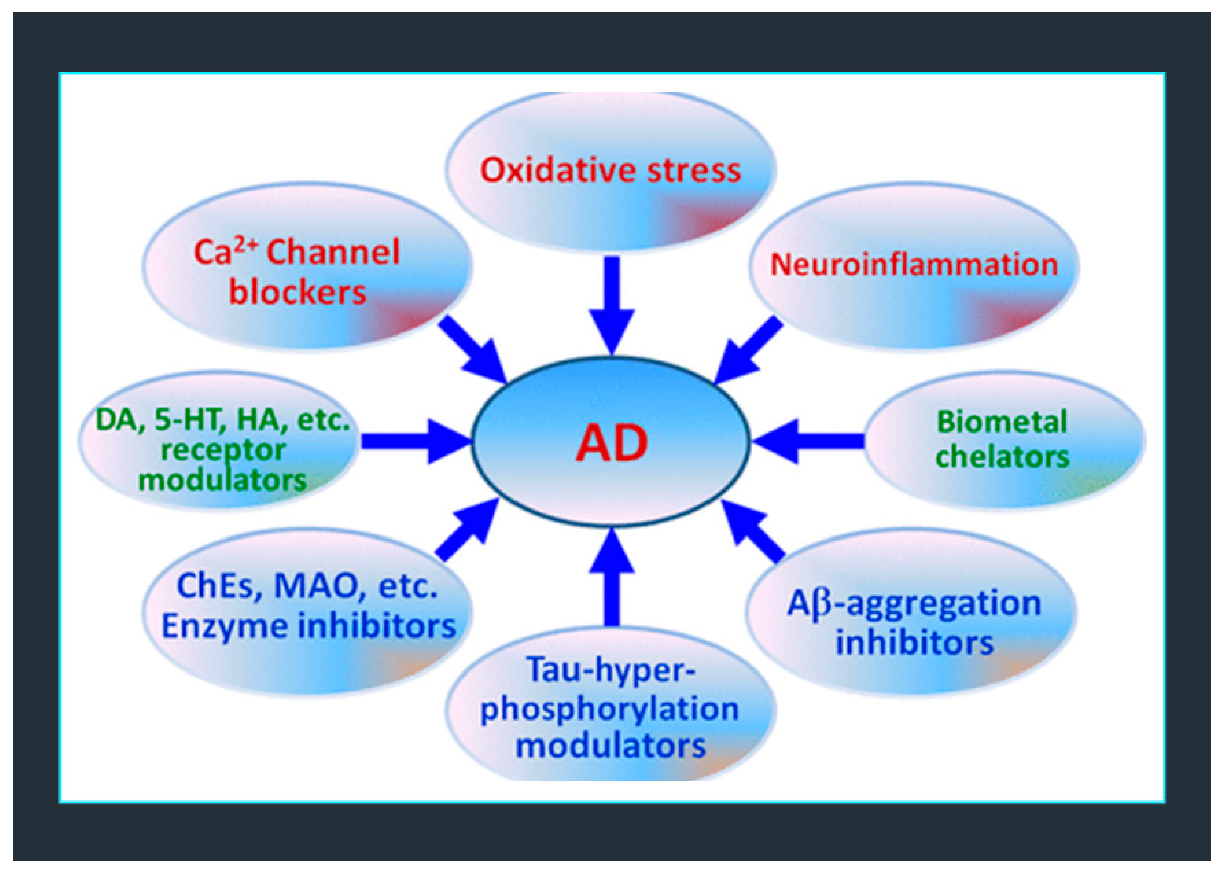
Figure 2.
Chemical structure of ellagic acid (EA).
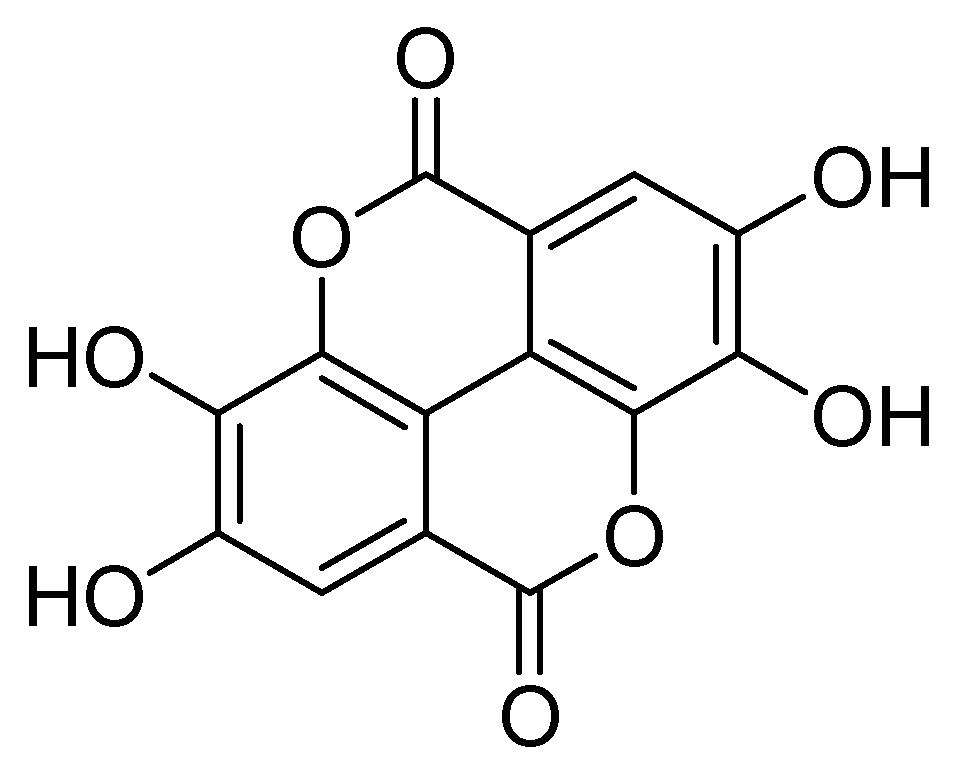
Figure 3.
Multifactorial pathogenic cascade leading to the onset and development of AD.
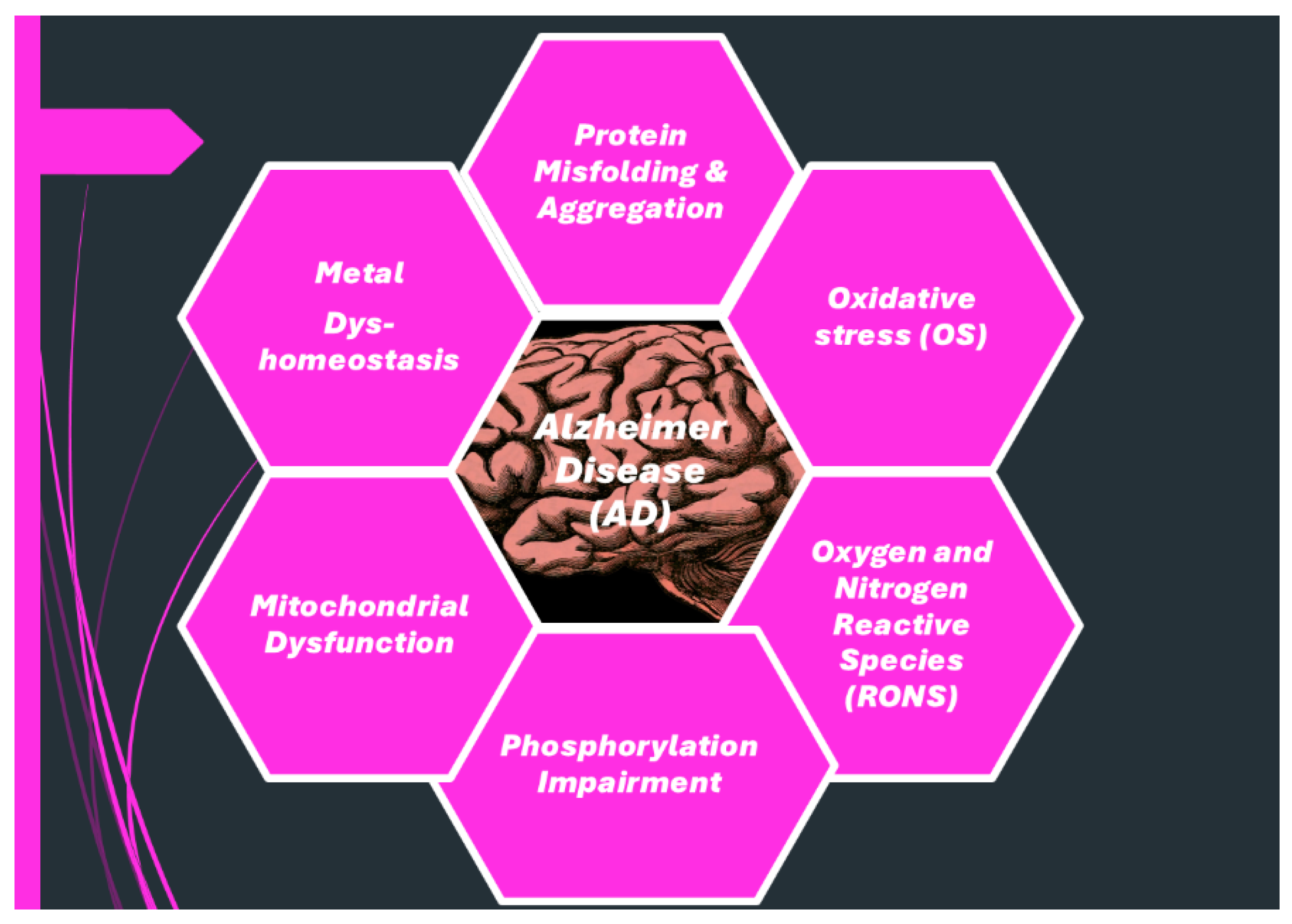
Figure 4.
Schematic pathways of the multifactorial events leading to neuronal death. General mechanisms, such as protein misfolding and aggregation, oxidative stress (OS), metal (M) dyshomeostasis, mitochondrial dysfunction, and altered protein phosphorylation, have been identified in several neuronal disorders. Readapted with PERMISSION/LICENSE GRANTED AT NO CHARGE by ACS Chemical Neuroscience (American Chemical Society) from [7].
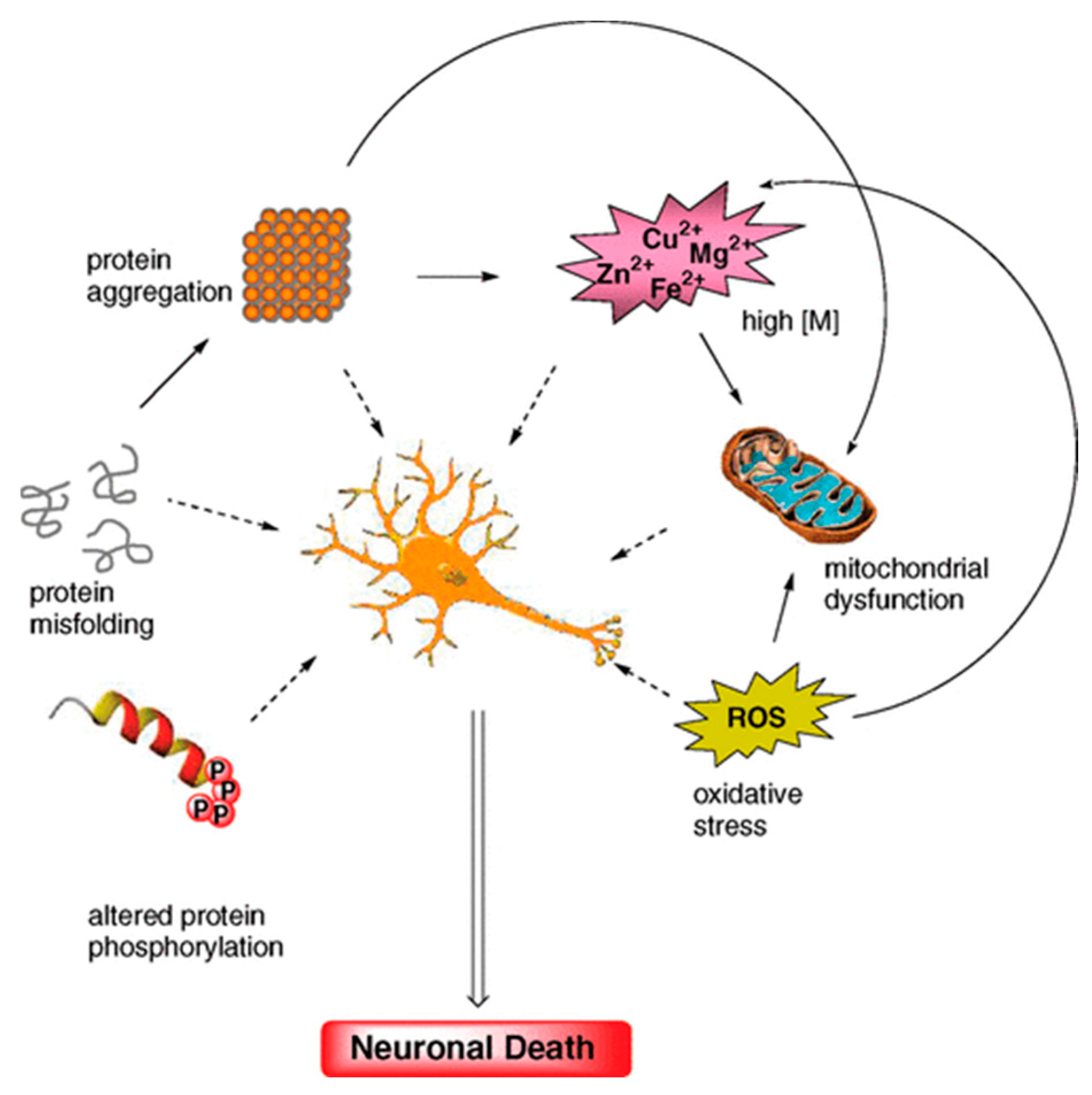
Figure 5.
Schematic pathways of ROS production and their main effects on biological systems. Nrf2 = erythroid nuclear transcription factor-2; NF-kB = transcription factor involved in cellular responses to stimuli such as stress, cytokines, free radicals, heavy metals, ultraviolet irradiation, oxidized low-density lipoproteins (LDL), etc. Reproduced from our article [30].
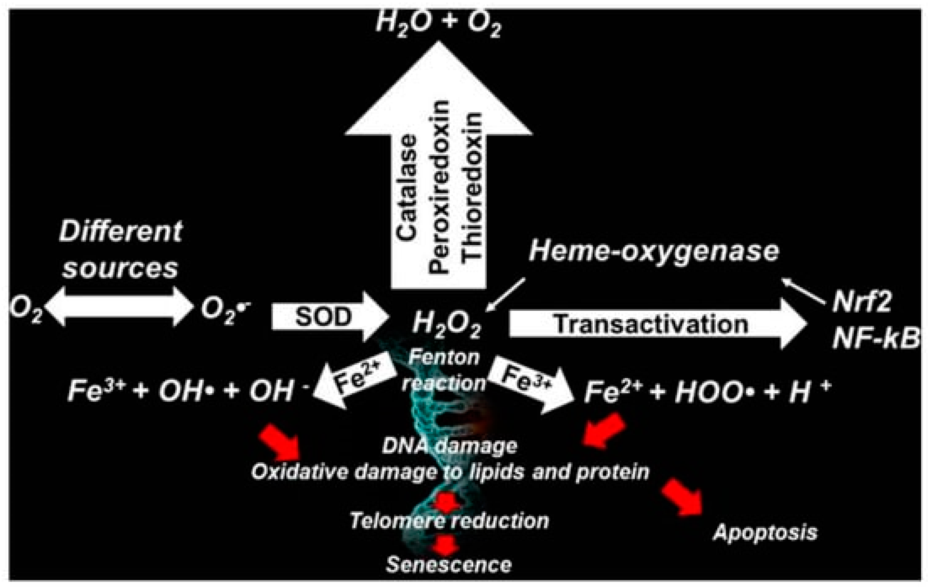
Figure 6.
Possible molecular causes of neuronal death and protective cyclic mechanisms in AD. The central event in AD pathogenesis is an imbalance between Aβ production and clearance. The enhanced activity of β- and γ-secretases leads to increased release of amyloidogenic Aβ42, which forms oligomers and then extracellular deposits (senile plaques). In this regard, one way to confront AD pathogenesis may be to combat the oligomerization processes by means of small molecules. A role for metal ions and ROS in the Aβ oligomerization has also been advanced. Therefore, also metal chelation and antioxidants are two general mechanisms to be considered in the search for disease-modifying anti-AD drug candidates. Also, β- and γ-secretase inhibitors may be promising lead compounds because they tackle an early event in AD pathogenesis. Mitochondrial dysfunction plays a fundamental role in the neuronal death associated with AD, as it is likely that intracellular Aβ could compromise the function of this organelle. τ hyperphosphorylation leading to tangle formation is regarded as a downstream event but could contribute to reinforcing neuronal dysfunction and cognitive impairment. Readapted with PERMISSION/LICENSE GRANTED AT NO CHARGE by ACS Chemical Neuroscience (American Chemical Society) from [7].
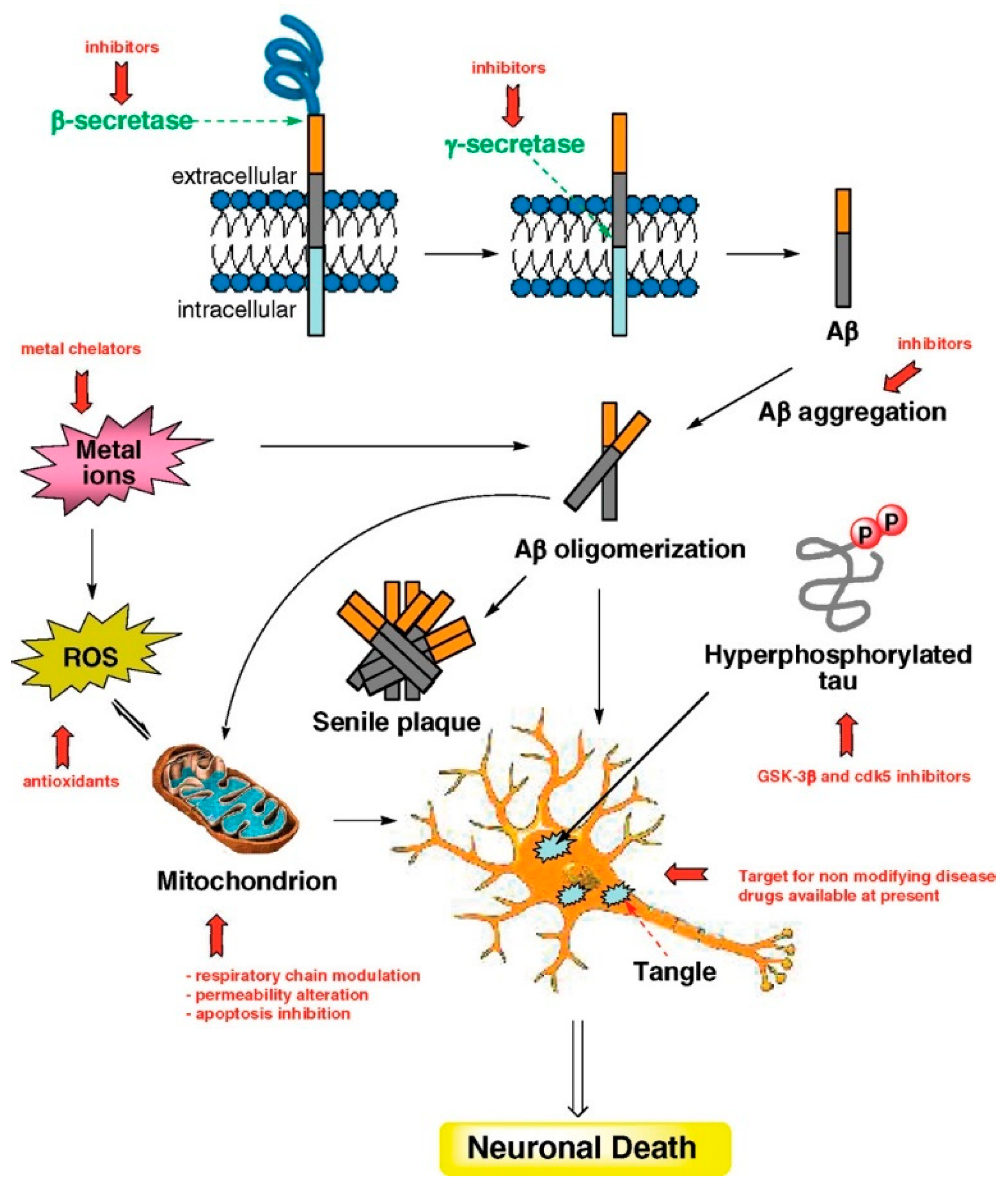
Figure 7.
Alternative approaches to the one-molecule/one-target drug one. MMT= multimodal therapy; MCM = multiple-compound medication. (Single compounds hitting multiple targets can be abbreviated also as MTDLs).
Figure 7.
Alternative approaches to the one-molecule/one-target drug one. MMT= multimodal therapy; MCM = multiple-compound medication. (Single compounds hitting multiple targets can be abbreviated also as MTDLs).
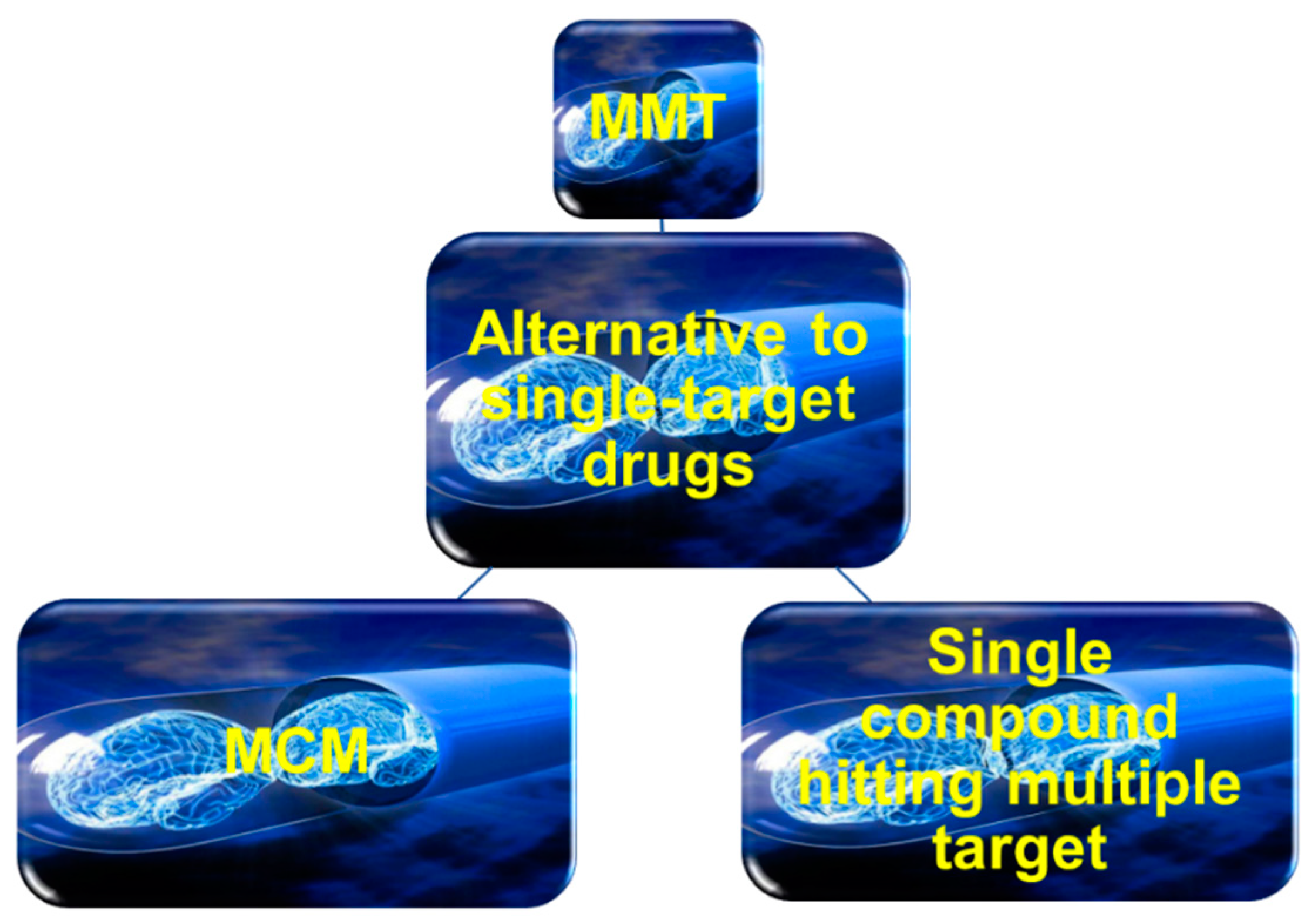
Figure 8.
The main possible forms of AD.
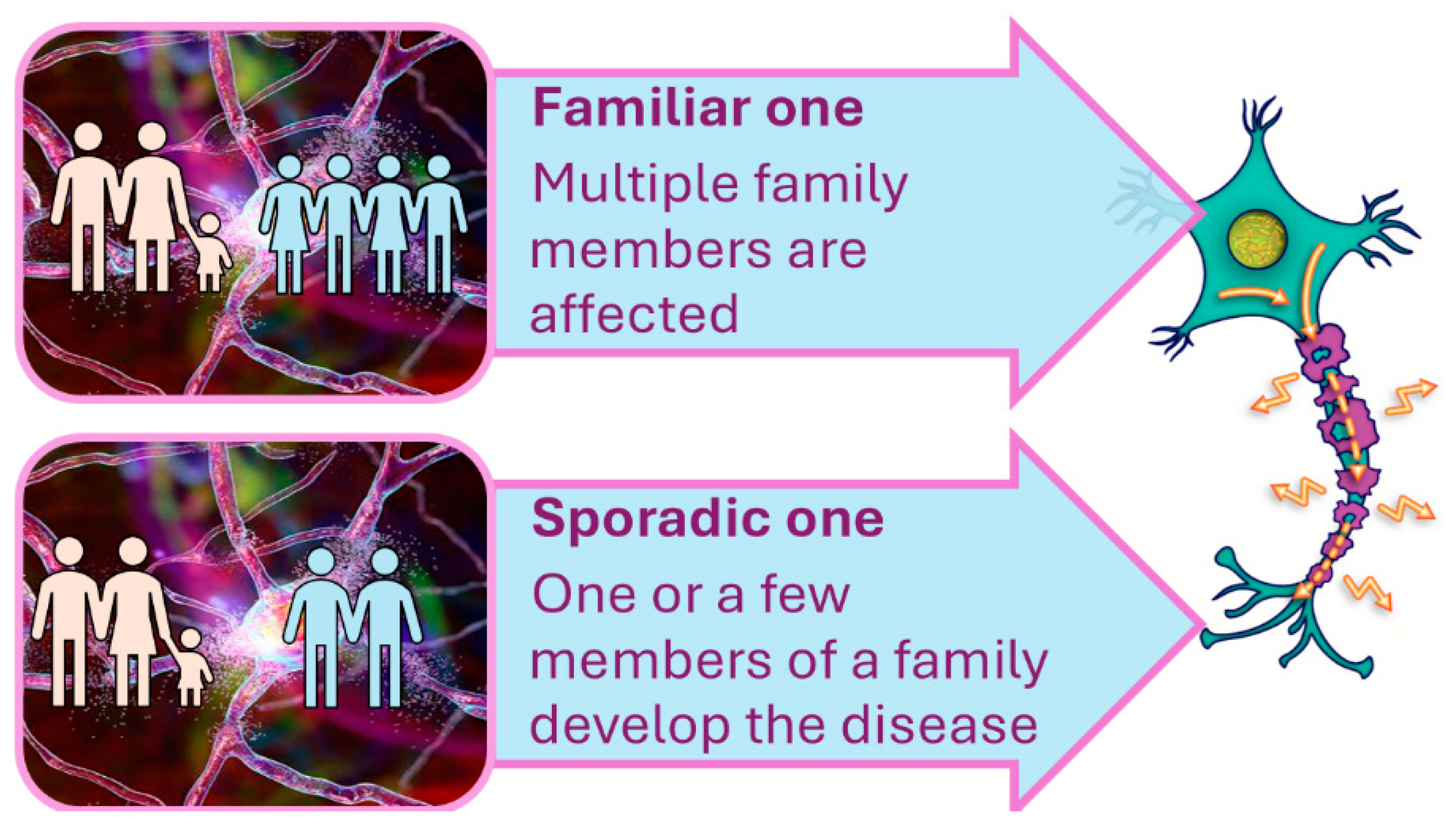
Chart 1.
Structure of traditional AChEIs.
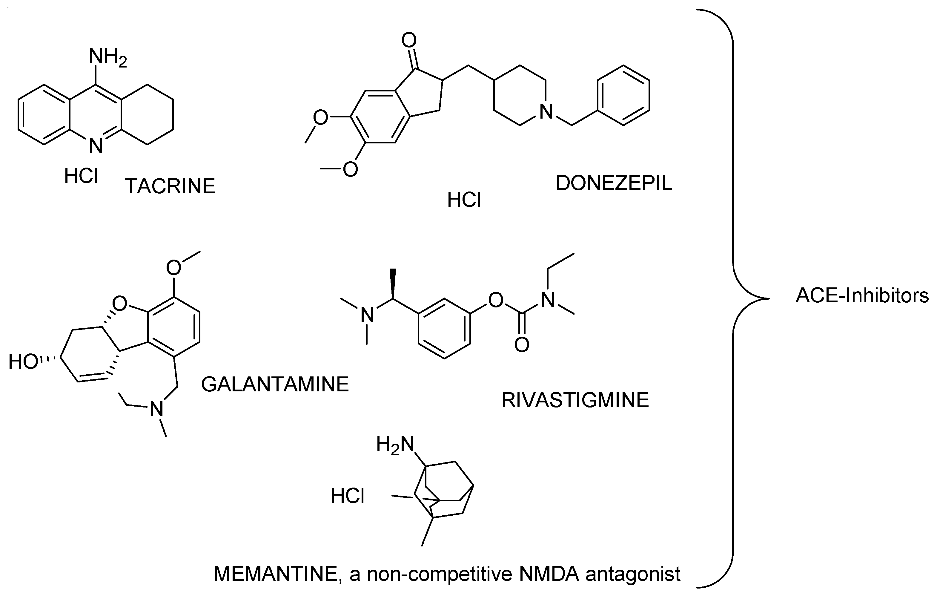
Figure 9.
Developmental root to the clinical implementation of new active principles (PA) for MDTs. FDA = food and drug administration; EMA = European medical agency; MHPRA = Medicine and Healthcare Products Regulatory Agency; * refers to donanemab.
Figure 9.
Developmental root to the clinical implementation of new active principles (PA) for MDTs. FDA = food and drug administration; EMA = European medical agency; MHPRA = Medicine and Healthcare Products Regulatory Agency; * refers to donanemab.
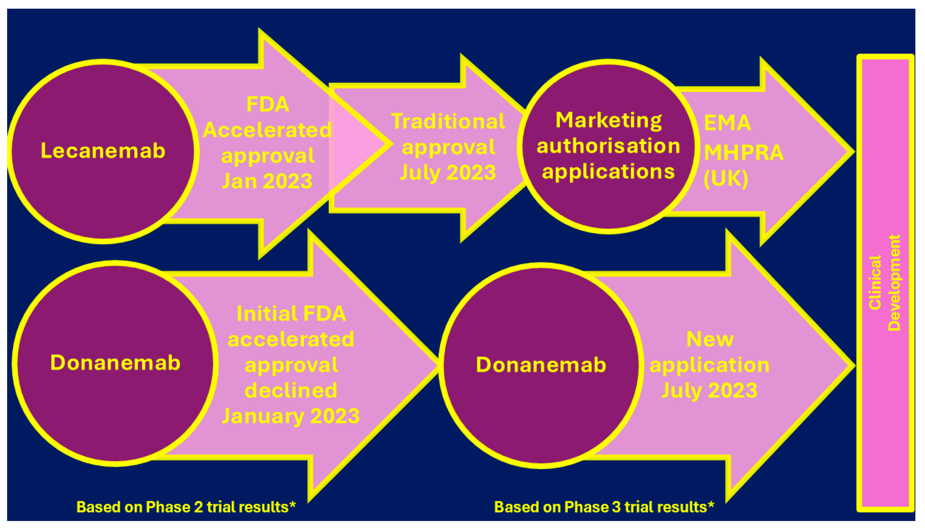
Figure 10.
Ideal and efficient MTSM (equal to say MTDLs) for AD therapy, showing their corresponding pharmacophoric groups (PG). Readapted with PERMISSION/LICENSE GRANTED AT NO CHARGE by ACS Chemical Neuroscience (American Chemical Society) from [8].
Figure 10.
Ideal and efficient MTSM (equal to say MTDLs) for AD therapy, showing their corresponding pharmacophoric groups (PG). Readapted with PERMISSION/LICENSE GRANTED AT NO CHARGE by ACS Chemical Neuroscience (American Chemical Society) from [8].
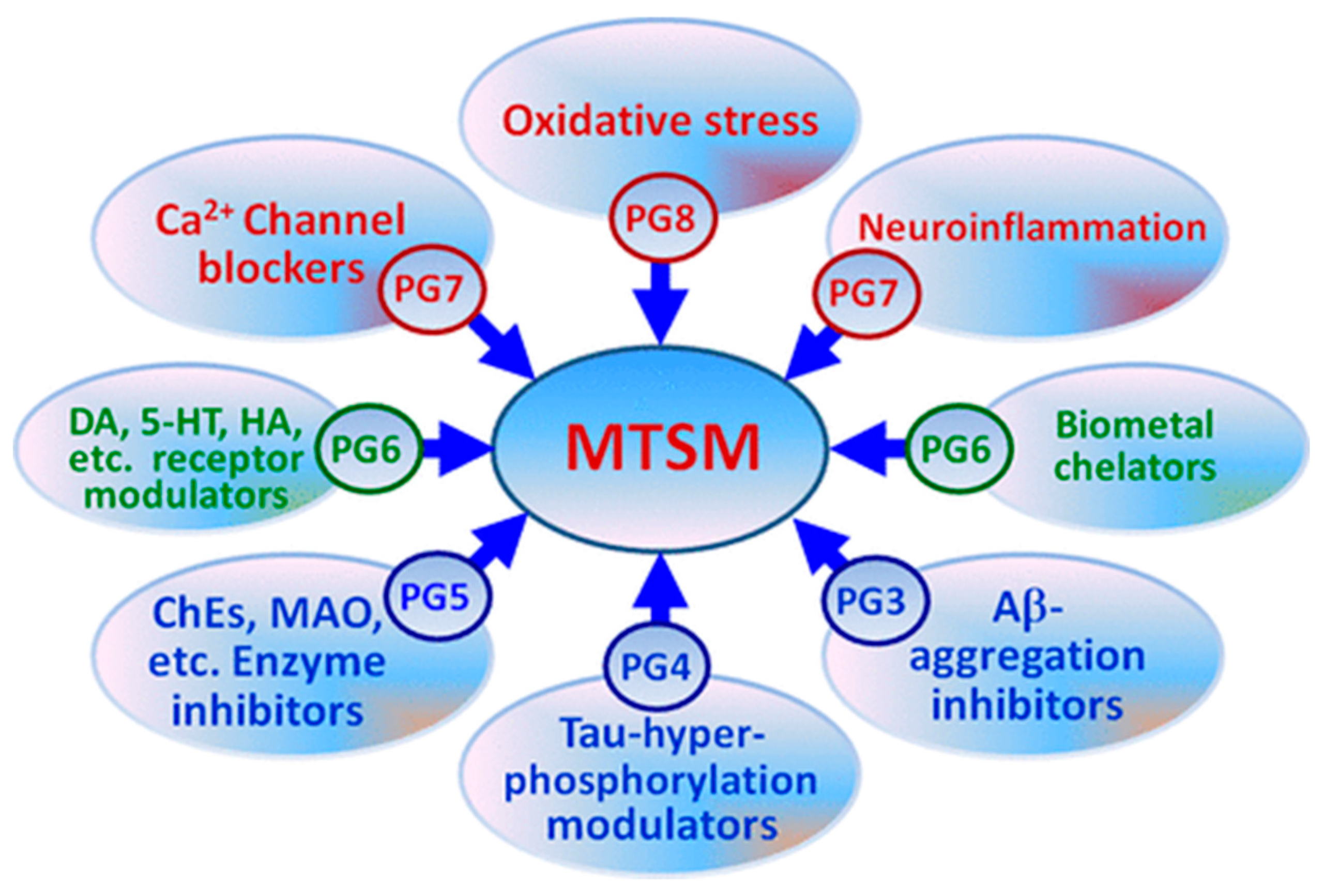
Scheme 1.
Chemical structures of casuarictin, EA and the most known UROs.
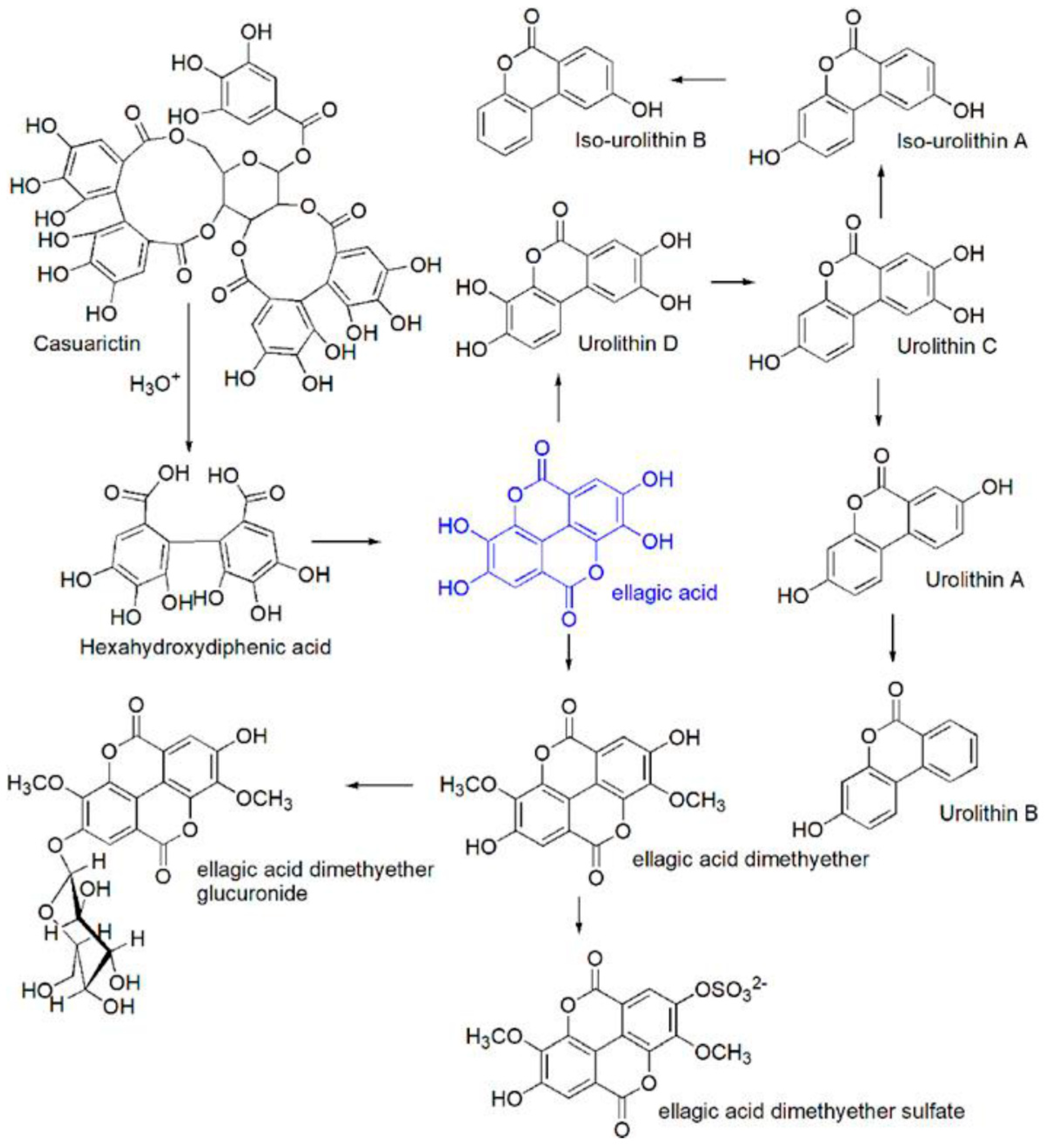
Figure 11.
Antioxidant mechanism of EA.
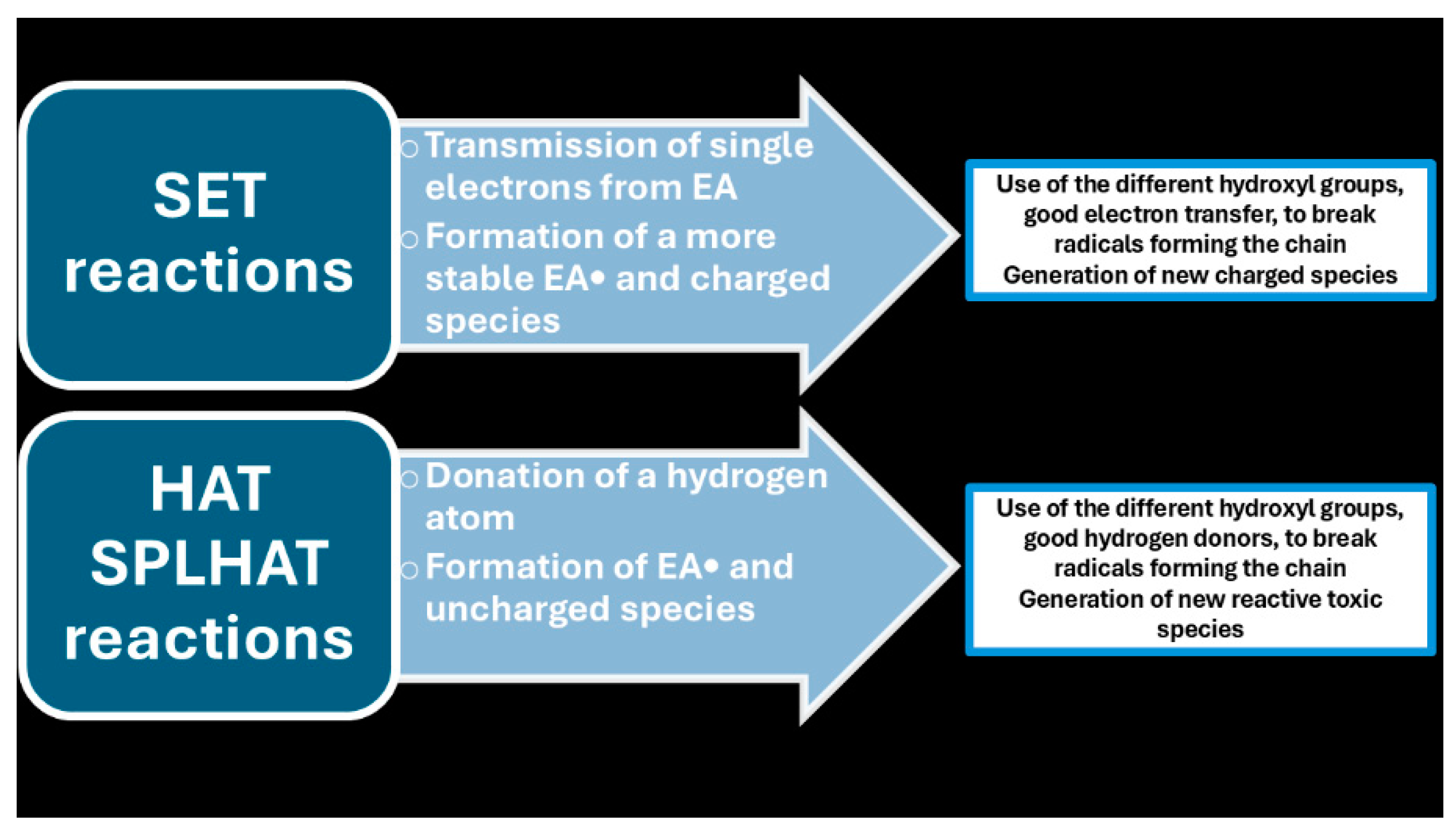
Figure 12.
Percentages of in vitro, in vivo and clinical reports on the pharmacological activity of EA containing plants among 56 studies considered.
Figure 12.
Percentages of in vitro, in vivo and clinical reports on the pharmacological activity of EA containing plants among 56 studies considered.
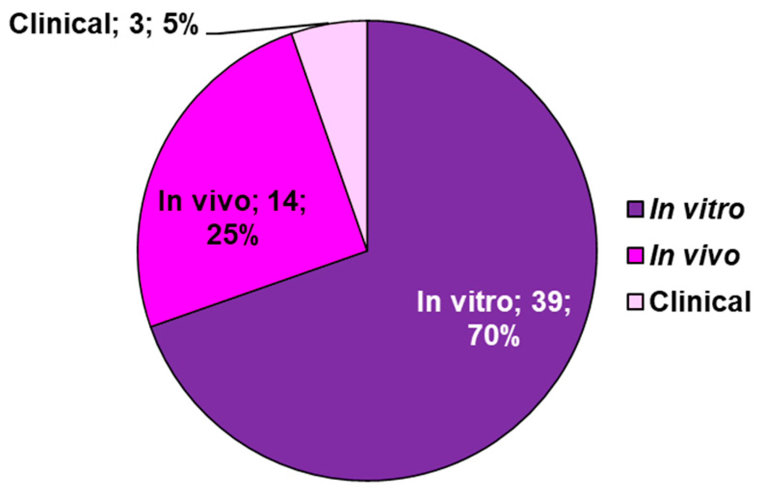

Table 1.
Endogenous and exogenous sources of ROS and the main reactive species of RONS, which can be in turn produced.
Table 1.
Endogenous and exogenous sources of ROS and the main reactive species of RONS, which can be in turn produced.
| Endogenous Sources | Exogenous Sources | Reactive Species | |
| Enzymatic | Non-Enzymatic | ||
| NOX MPO Cytochrome P450 Lipoxygenase Angiotensin II Xanthene oxidase Cyclooxygenase FpH• |
Mitochondria Respiratory chain Glucose auto-oxidation NAD• Semiquinone radicals Radical pyridinium Hemoproteins |
Air Water pollution Tobacco Alcohol Heavy/transition metals Drugs Industrial solvents Cooking Radiation EPFRs BC-PFRs |
O2•− H2O2 •OH •OOH ONOO• NO2• NO• ONOOCO2− NO2+ ONOOH N2O3 ONOO− ONOOCO2− CO3•− |
Table 2.
In vitro and in vivo outcomes of the one-molecules/one target paradigm approach.
| In vitro | In vivo |
| High Selectivity Strong efficacy Tendency to develop resistance |
Not recognizing the target by the ligand in vivo |
| Not reaching of the site of action by the ligand | |
| One target interaction is not enough to have a sufficient impact on the complex diseased system |
Table 3.
Alternative multi-target approaches.
| Approaches | Advantages | Disadvantages | Ref. |
|---|---|---|---|
| MMT | Attack the multifaceted discomfort from multiple mechanisms | Compliance problems by patients | [33] |
| Undesired in vivo drug-drug interactions | |||
| In vivo unbeneficial side effects | |||
| Different bioavailability, pharmacokinetics metabolism of the single drugs | |||
| MCM | Attack the multifaceted discomfort from multiple mechanisms | Undesired in vitro and in vivo drug-drug incompatibility hampering single formulation |
[34] |
| Simpler dosing regimens | Different bioavailability, pharmacokinetics, metabolism of the single drugs in the cocktail | ||
| ↑ Patient compliance | Unbeneficial side effects in vivo Undesired in vivo drug-drug interactions |
||
| MTDLs | Unique bioavailability pharmacokinetics, metabolism (ADMET profile) |
Complex ADMET Complex pharmacokinetic |
[35] |
| Simpler pharmacokinetic and ADMET optimization | |||
| ↓ Risk of possible drug-drug interactions | |||
| Simplified therapeutic regimen in relation to MMT |
MMT= multimodal therapy; MCM = multiple-compound medication; MTDLs = multi-target direct ligands; ADMET = absorption, distribution, metabolism, excretion and toxicity.
Table 4.
Current old and more recent one-target therapeutics approved for AD.
| Family | Subfamily | Drugs | Advantages | Disadvantages | Ref. |
|---|---|---|---|---|---|
| Old AChEI | Tacrine °,* | ↑ Cognitive, behavioural, functional impairments | Hepatotoxic | [41] | |
| Denezepil * Rivastigmine * Galantamine * |
Unable to address the molecular mechanisms that underlie the pathogenic processes Not able to resolve the causes |
||||
| Non-competitive NMDA antagonist | Memantine * |
IChEI = acetylcholinesterase inhibitors; ↑ = improved, higher, ameliorated; * approved standards of AD therapy; ° nowadays rarely used.
Table 5.
Summary of recent pharmacological interventions against AD.
| Class of Drugs | Compounds | Mechanism | Subjects | Trial Phase | Summary | [Ref] |
| Anti-amyloid therapy | ||||||
| Secretase inh. | Verubecestat | BACE1 inh. | PTM AD | II/III | ↓Efficacy | [43,44] |
| Atabecestat | P AD | ↓ Cognition Psychiatric disorder |
[45] | |||
| Lanabecestat | MCI to mild AD | III | ↓ Cognition ↓ Weight loss Psychiatric disorder |
[46] | ||
| LY3202626 | Mild AD | ↓ Efficacy | [47] | |||
| Umibecestat | Cognitively healthy APOE4 carriers |
II/III | Completed Failed analysis due to ↓ number of events |
[48] | ||
| Elenbecestat | MCI to moderate AD | III | ↓ Efficacy Nightmare |
[49,50] | ||
| Semagacestat | γ-secretase inh. | Mild to moderate AD | ↓ Efficacy Skin cancer, ↓ weight Hematologic disorder Infection |
[51] | ||
| Avagacestat | MCI | II | ↓ Efficacy Non-melanoma cancer, GIT symptoms |
[52] | ||
| Tarenflurbil | γ-secretase modulator |
Mild AD | ↓ Efficacy Anaemia infection |
[53] | ||
| Aβ aggregation inhibitor |
PBT1 | MPAC | MCI to moderate AD | Rescue of cognitive decline in severely affected patients (ADAS-cog ≥25) Visual impairment | [54] | |
| PBT2 | MPAC | MTM AD | ↓ Efficacy ↑ Individual variance |
[55,56] | ||
| Aβ immunotherapy | ACI-24 | Aβ vaccine | Adults with Down syndrome | ↓ Immunogenicity | [57] | |
| CAD106 | Mild AD | ↓ Efficacy | [57] | |||
| UB-311 | No published data | [57] | ||||
| ABVac40 | MCI to mild AD | Ongoing | [57] | |||
| BAN2401 | Monoclonal antibody |
III | ↓ Efficacy among APOE4 carriers | [58] | ||
| Gantenerumab | PTM AD | ↓ Efficacy | [59] | |||
| Aducanumab | Monoclonal antibody | Termination ↓ Change in efficacy FDA approval for now |
[60,61] | |||
| Anti-τ therapy | ||||||
| Phosphatase modifier | Selenate | PP2A activator | MTM AD | II | ↓ Efficacy | [62,63] |
| Kinase inhibitor | Roscovitine | CDK5 inh. | 5XFAD mice | In vivo | Prevention of τ phosphorylation |
[64,65] |
| Flavopiridol | CD1 mice | Rescue of cognitive decline |
[64,65] | |||
| Tideglusib | GSK3β inh. | MTM AD | II | ↓ Efficacy transaminase increase |
[66] | |
| Lithium | MCI | Rescue of cognitive decline |
[67,68,69] | |||
| τ aggregation inh. | MB | Disrupts polymerization | MTM AD | ↑ Cognition | [70] | |
| LMTX | III | ↓ Efficacy | [71] | |||
| Curcumin | ↓ β-sheet in τ | CHE | II | ↑ Working memory (short-term course) |
[72] | |
| Microtubule stabilizer |
EpoD | ↑ Microtubule bundling | Mild AD | I | Discontinuation Frequent adverse effects No published data |
[73] |
| NAP | Protects microtubules from katanin disruption | MCI | II | ↑ Cognition and functionalities |
[74,75] | |
| TPI-287 | Stabilizes microtubules | MTM AD | I | Rescue of cognitive Decline Anaphylactoid reactions |
[76] | |
| τ immunotherapy | AADvac1 | τ Vaccine | Mild AD | II | Completed No published data |
[77] |
| ACI-35 | MTM AD | I | Safe and tolerated | [78] | ||
| Aβ 3–10-KLH | 3 × Tg-AD mice | In vivo | ↑ Cognition | [79] | ||
| BIIB092 | Monoclonal antibody | Early AD | II | Ongoing | [80] | |
| ABBV-8E12 | [81,82] | |||||
| RO7105705 | PTM AD | [82,83] | ||||
| BIIB076 | Healthy volunteers, MCI | I | Safe and tolerated | [84] | ||
| LY3303560 | Early AD | II | Completed No available data |
[85] | ||
| JNJ-63733657 | II | Ongoing | [86] | |||
| UCB0107 | Healthy volunteers | I | [87,88] | |||
| Anti-neuroinflammatory therapy | ||||||
| Microglia modulator |
Thymoquinone | TLR4 inh. | AD mice induced by AlCl3 | In vivo | Rescue of cognitive impairment |
[89] |
| Ethyl pyruvate | [89] | |||||
| TAK-242 | APP/PS1 mice | ↑ Cognition | [90] | |||
| GW2580 | CSF1R inh. | Recovery of short-term memory and behavioural deficit | [91] | |||
| JN-J527 | P301S mice | ↑ Functionalities | [92] | |||
| PLX3397 | 5XFAD mice | Recovery of spatial and emotional memory deficit | [93] | |||
| Astrocyte modulator |
Stattic | STAT3 inh. | 5XFAD mice | Rescue of learning and memory impairment | [94] | |
| FK506 | Calcineurin/NFAT inh. | MCI to AD | II | Not yet recruiting | [95] | |
| SB202190 | P38 MAPK inh. | Wip1-deficient mice | In vivo | Rescue of learning and memory impairment | [96] | |
| PD169316 | Aβ-injected mice | Rescue of spatial memory and learning impairment | [96] | |||
| MW108 | H τ mice | Rescue of cognitive impairment |
[97] | |||
| NJK14047 | 5XFAD mice | ↑ Cognition | [98] | |||
| MRS2179 | P2Y1R inh. | APPPS1 mice | ↑ Spatial learning | [99] | ||
| BPTU | [99] | |||||
| Insulin resistance management |
Intranasal insulin therapy | Intranasal supplement | MCI to moderate AD | II | ↑ Cognition ↑ Modulation by APOE4 genotype |
[100,101] |
| MCI to AD | II/III | ↓ Efficacy | [102] | |||
| Liraglutide | Incretin receptor agonist |
Mild AD | II | Delay of cognitive impairment |
[103] | |
| Metformin | Biguanide | MCI | ↓ Recall memory decline | [104] | ||
| MCI to early AD | ↑ Executive functionalities | [105] | ||||
| Gemfibrozil | PPAR-α agonist | MCI | I | Completed No published data |
[106] | |
| Pioglitazone | PPAR-γ agonist | Mild AD | II | ↑ Cognition | [107] | |
| MCI | III | ↓ Efficacy | [108,109] | |||
| T3D-959 | PPAR-δ/γ agonist | STZ-induced AD | In vivo | ↓ Neuroinflammation | [110] | |
| Microbiome therapy | Sodium oligomannate |
Dysbiosis of gut microbiota |
MTM AD | III | ↑ Cognition | [111,112] |
| Neuroprotective agents | ||||||
| Antiepileptics | Levetiracetam | SV2A receptor | MCI | III | Ongoing | [113] |
| Gabapentin | VGCCs inh. | MTS AD | IV | [114] | ||
| NMDAR modification |
Sodium benzoate |
DAAO inh. | MCI to mild AD | II | ↑ Cognition | [115] |
| MCI | ↑ Cognition and functionalities |
[116] | ||||
| MTS AD with BPSD | ↑ Cognition in female | [117] | ||||
| Riluzole | Glutamate modulator | Mild AD | Completed No published data |
[118] | ||
| Troriruzole | MTM AD | Ongoing | [119] | |||
| Omega 3 FA supplements | DHA | Anti-oxidative effect | III | ↓ Efficacy | [120] | |
| CHE | II | Ongoing | [121] | |||
| Icosapent ethyl | III | [122] | ||||
Inh. = inhibitor; PTM = prodromal to mild; FA = fatty acids; ↓ = slow, reduce, decreased, lower, lack of; ↑ = higher, improved, enhanced; BACE1—β-secretase1, APOE4—apolipoprotein E type 4, PBT1—clioquinol, PBT2—second-generation clioquinol, MPAC—metal protein attenuating compound, ADAS-cog—Alzheimer’s Disease Assessment Scale–Cognitive Subscale, MB—methylene blue, EpoD—Epothilone D, NAP—davunetide, TPI-287—abeotaxane, DHA—docosahexaenoic acid; MTM = mild to moderate; MTS = moderate to severe; CHE = cognitively healthy elderly; DAAO = D-amino acids oxidase; MTS = magnetic transcranial stimulation; MCI = mild cognitive impairments; BPSD = behavioural and psychological symptoms of dementia.
Table 6.
Some patented MCM.
| Patented by | Combination Ingredients | Advantages/Finalization | Mechanism of the additional ingredient | Ref. |
|---|---|---|---|---|
| Myriad Genetics * |
AChEI + (R)-flurbiprofen ** | Therapeutic or prophylactic treatment of AD due to the capability of NSAIDs to reduce the incidence of AD | ↓ The level of Aβ associated with plaque formation inhibit cyclooxygenase enzymes |
[126] |
| Mayo Foundation |
AChEI+Aβ-lowering agent | ↓the concentration of Aβ ↓agents acting on the same level |
[127] | |
| N.R. | 5-substituted-3-oxadiazolyl-1,6-naphthyridin-2(1H)-one + reported AChEIs | Stimulation of cerebral functions and amelioration of AD to the anti-dysmnesics effects of the additional ingredient | Negative allosteric modulators of GABAA | [128] |
| Johns Hopkins University | ABPA + reported AChEIs | ↑ Cognition properties by ABPA ↑ Memory performances ↑ Therapeutic effects for AD treatment ↓Doses of the two compounds Retained therapeutic efficacy ↓Side effects Cost saving |
Specific GABAB antagonist and GABAC agonist | [129] |
| MS-153 + reported AChEIs | ↓Ischemia-induced neuron damage by MS-153 ↑Oral bioavailability ↑Enhanced cognitive performance in aged rats in Morris Water Maze tests of spatial memory |
↓Glutamate release ↑ Glutamate uptake No blocking NMDA or AMPA receptors |
[130] | |
| Schering Corporation |
Macrocyclic lactones+ AChEIs and/or an NSAID | Ameliorate Neurodegenerative diseases such as AD | ↓β-secretase ↓BACE-1 enzyme (IC50 value of 4-186 nM) |
[131,132] |
| Voyager Pharmaceutical Corporation | AChEI + NMDA RA + leuprolide acetate (G-R HA) | ↓AD development | ↓Biosynthesis and secretion of gonadotropins | [133] |
| Rabinoff | CPC+5-CDPC | ↑ Memory For AD therapy and prevention |
Neurotrophic factors | [134] |
| Epix Pharmaceuticals | 5-HT4 AGO + Galantamine | ↑ Memory | Modification of ACh release | [135] |
| Wyeth | 5-HT6 ANTA + Donazepil 5-HT6 ANTA + Galantamine 5-HT6 ANTA + Donazepil |
↑ Memory ↓Dose of the AChEI ↓Typical side effects of AChEIs ↓Cardiovascular effect of 5-HT6 antagonist |
Modulation of multiple neurotransmitter systems | [136] |
↓ = slow, reduce, decreased, lower; ↑ = higher, improved, enhanced; * the same applicant published related patents, which focused on the combination of flurbiprofen derivatives, specifically with donezepil, rivastigmine and galantamine ; ** non-steroidal anti-inflammatory drug (NSAID)[130,131,132]; N.R. = not reported; MS-153 = (R)-(-)-5-methyl-1-nicotinoyl-2-pyrazoline; RA = receptor antagonist; G-R HA = gonadotropin-releasing hormone analogues suppress the pituitary gland’s secretion of LH; CPC = glycerylphosphorylcholine; 5-CDPC = 5′-cytidine diphosphocholine; 5-HT = receptors members coupled to a G protein contributing to dopamine secretion and regulating learning and long-term memory by modification of ACh release. ANTA =antagonist; AGO = agonist; ABPA = 3-aminopropyl-(n-butyl)-phosphinic acid.
| Physicochemical Identifiers | Descriptive Data |
|---|---|
| Chemical Name 1 | Ellagic Acid |
| CAS number | 476-66-4 |
| Molecular formula | C14H6O8 |
| Molecular weight | 302.194 g/mol |
| Hydrogen bond donor count | 4 |
| Hydrogen bond acceptor count | 8 |
| Covalently bonded unit count | 1 |
| Form/colour | Cream coloured needles from pyridine Yellow powder |
| Melting point | >360 °C |
| Density | 1.667 at 18 °C |
| Dissociation constants | pKa1 = 6.69 (phenol) pKa2 = 7.45 (phenol) pKa3 = 9.61 (phenol) pKa4 = 11.50 (phenol) |
| Solubility 2 | Slightly soluble in alcohol [154] Poorly soluble in water [155] Insoluble in ether Soluble in alkalis and pyridine[152] |
| Vapor pressure | 2.81×10⁻¹⁵ mm Hg at 25 °C |
| Spectral properties | UV max (ethanol): 366, 255 nm |
1 traditional IUPAC name; 2 EA water solubility = 9.3–9.7 µg/mL at pH 7.4 and 21 ◦C [155].
Table 8.
Biological activities of UROs in different animal models.
| Animal model | Assay conditions | Main outcomes | Ref. |
|---|---|---|---|
| Anti-inflammatory activity | |||
| F344 rat | Uro-A (15 mg kg−1 d−1 p.o.; HED: ∼150 mg 70 kg−1 person) for 25 days prior to DSS-induced colon inflammation (UC colitis model) | Preservation of colonic architecture ↓iNOS, COX-2 and PTGES protein expression ↓Pro-inflammatory IL-1β and IL-4 gene expression |
[20] |
| ICR mice | Uro-A (300 mg kg−1 d−1 p.o.; HED: ∼1.5 g 70 kg−1 person) for 1 or 6 h prior to inducing inflammation (carrageenan-induced paw edema model) | ↓Volume of paw edema ↑ORAC antioxidant activity in plasma |
[159] |
| Wistar rats | Uro-A or Uro-B (2.5 mg kg−1 d−1 i.p.) for 3 weeks in a streptozotocin-induced type-1 diabetes model | ↓Fractalkine, Prevention of cardiac dysfunction ↑Maximal rate of ventricular pressure rise and parallel ↓ in the isovolumic contraction time Recovery of cardiomyocyte contractility and Ca+2 dynamics and ↑velocity of shortening (only for Uro-B) |
[160] |
| Sprague-Dawley rats | Uro-A (50 mg kg−1 d−1 p.o.) for 5 days in a cisplatin-induced nephrotoxicity model | ↓Cisplatin-induced inflammatory cascade and inhibition of the proapoptotic pathway Prevention of renal dysfunction and histopathological damage |
[161] |
| C57BL/6J or Nrf2−/− mice | Uro-A (20 mg kg−1 d−1 p.o.) at 0, 6, 12, 18, and 24 h before LPS-induced peritonitis in C57BL/6J mice Uro-A (20 mg kg−1 d−1 p.o.) (4 or 20 mg kg−1 d−1 p.o.) after 12 h of TNBS-induced colitis (C57BL/6 or Nrf2−/− mice) and every 12 h thereafter up to 72 h Uro-A (20 mg kg−1 d−1 p.o.) on the 4th and 6th day of DSS-induced colitis C57BL/6 model |
↓LPS-induced increase in serum IL-6 and TNF-α levels.; Protection of TNBS-induced tissue damage (body weight loss, reduction of DAI score, intestinal permeability, colon shortening and weight to length ratio) and inflammation scores (reduction of neutrophil infiltration, MPO activity, and serum inflammatory markers such as IL-6, TNF-α, CXCL1, and IL-1β); Protection of DSS-induced acute colitis (↓DAI scores, colon shortening, gut permeability and increase of colon weight/length ratio); ↓inflammation (serum IL-6, IL-1β, TNF-α and colonic tissue MPO levels) | [162] |
| C57BL/6J mice | Uro-A (nanoparticle encapsulated) (50 mg kg−1 d−1 p.o.) for 19 days in cisplatin-induced acute kidney injury model | Attenuation of the histopathological hallmarks of cisplatin-induced acute kidney injury; ↓mortality by lower renal oxidative and apoptotic stress (Nrf2/antioxidant response element and P53 pathways) | [163] |
| C57BL/6 mice | Uro-A (20 mg kg−1 d−1 i.g.) for 8 weeks in surgically osteoarthritis model | Protective effect in osteoarthritis development by ↓OARSI score, ↓PI3K/AKT pathway activation and the nuclear p65 expression in chondrocytes | [164] |
| C57BL/6 mice | Uro-A (50 mg kg−1 d−1 p.o.) for 3 days and 30 min before surgery in a model of ischemia reperfusion injury | ↓TNFα, IL1β, MIP1α and MIP2 mRNA expression; ↑autophagy; attenuation of associated kidney injury; protection against ischemia reperfusion injury | [165] |
| C57BL/6 mice | Uro-A (100 mg kg−1 d−1 i.p.) for 5 days in a cisplatin-induced ischemic neuronal injury model | ↓Histological damage in proximal tubular cells; ↓cisplatin-induced pro-inflammatory cytokines/chemokines (TNF-α, IL-23, IL-18 and MIP2) and attenuation of renal oxidative/nitrative stress | [166] |
| IL-10−/− C57BL/6j mice | Uro-A (0.114 mg kg−1 d−1 p.o.) for 2 days in Campylobacter jejuni infected, microbiota-depleted IL-10−/− mice as preclinical inflammation model | Improve clinical outcome and less pronounced macroscopic (less colonic shrinkage) and microscopic (less colonic histopathology and apoptosis) inflammatory sequelae of infection; ↓intestinal pro-inflammatory immune responses (IFN-γ, TNF-α, MCP-1 and NO) and systemic markers (IFN-γ, MCP-1 and IL-6); ↓abundance of macrophages, monocytes and T lymphocytes in the mucosa and lamina propria | [167] |
| FUNDC1f/f mice and cardiomyocyte-specific FUNDC1 knockout (FUNDC1CKO) mice | Uro-A (30.0 mg kg−1 i.p.) prior to LPS treatment (48 h) to induce septic cardiomyopathy | Attenuate inflammation-mediated myocardial injury levels and normalization of cardiac function, including LVEF, LVDd, and FS in FUNDC1f/f mice, but not in FUNDC1CKO mice | N.R. |
| Neuroprotective effect and(or) improvement of cognitive function | |||
| Transgenic (express human amyloid β 1–42 in the muscle tissue after a heat shock) Caenorhabditis elegans (CL4176) | Exposure to Uro-A (43.8 µM), Uro-B (47.2 µM), methyl-Uro-A (41.3 µM), methyl-Uro-B (44.2 µM) | Only methyl-Uro-B has a protective effect against Aβ1–42 induced neurotoxicity and worm paralysis | [168] |
| Alzheimer’s disease APP/PS1 transgenic mice model | Uro-A (300 mg kg−1 d−1 p.o.) for 14 days | ↑of learning, ↑of memory deficits Prevention of neuronal apoptosis ↑Neurogenesis; ↓plaque Aβ deposition ↓Peri-plaque microgliosis and astro cytosis in the cortex and HPC Anti-(neuro)-inflammatory activity ↓Pro-inflammatory cytokine levels ↓Activation of NF-κB p65 subunit ↓p38 (MAPK) |
[169] |
| ICR mice | Uro-A (150, 100 or 50 mg kg−1 d−1 p.o.) for 8 weeks in a D-gal-induced brain aging model | ↓D-gal-induced cognitive impairment ↓Brain aging by suppression of miR-34a induced upregulation ↓Apoptosis induction, ↑autophagy by upregulating the SIRT1 signalling pathway and downregulating the mTOR signalling pathway |
[170] |
| C57BL/6 mice | Uro-A (2.5 or 5.0 mg kg−1 d−1 i.p.) for 24 h and 1 h before surgery in an ischemic neuronal injury model | ↓Infarction volume; reinforcement of ischemia-induced autophagy by ↑LC3-II and ↓p62 level; ↓ER stress by autophagy activation | [171,172] |
| ICR mice | Uro-A (1.5 or 2.0 mg kg−1 d−1 i.p.) at 1 and 24 h prior to surgery, and 1 h after surgery in an ischemic neuronal injury model (transient middle cerebral artery occlusion) | Ameliorate infarction, neurological deficit scores, and spatial memory deficits after cerebral ischemia; ↓neuron loss and ↑neurogenesis after ischemic stroke; Attenuate apoptosis by regulating apoptotic-related proteins; ↓glial activation via affecting inflammatory signaling pathways (↑AMPK and IκBα activation, and ↓Akt, NFκB p65, ERK, JNK, and p38) | [172]*** |
| ICR mice | Uro-A (2.5 mg kg−1 d−1 i.p.) for 8 weeks in an STZ-induced diabetic mouse model | Alleviate APP and BACE1 expressions, Tau phosphorylation, Aβ deposition, and cognitive impairment; ameliorate the high glucose-induced TGM2 expression | [173] |
| Cardioprotective activity | |||
| C57BL/6J mice | Uro-A (1 mg kg−1 d−1 i.p.) at 24 and 1 h before ischemia induction in a myocardial ischemia reperfusion injury model | Improvement of cardiac function by ↓myocardial infarct size, prevention of cardiomyocyte apoptosis and ↑serum CK and LDH activities after ischemia | [174,175] |
| Wistar rat | Uro-A (3 mg kg−1 d−1 p.o.) combined with a high cholesterol diet supplemented with Vit. D3 for 3 days prior to the balloon injury of the aorta and 12 weeks of treatment | Improvement of aortic atherosclerotic lesions; ↓plasma lipid (total cholesterol, TGs, and LDL) and angiotensin II levels in aortic tissue | [175] |
| ApoE−/− mice | Uro-B (10 mg kg−1 d−1 p.o.; equal to 1.11 mg kg−1 to human) for 14 days | ↓Lipid plaque deposition and oxidized-LDL uptake | [176] |
| C57BL/6 mice | Uro-A (20 µg d−1 i.p.) accompanied with a high-fat diet for 12 weeks | Anti-obesity activity by ↑systemic insulin sensitivity, ↓total and LDL cholesterol levels. In liver: ↓TGs accumulation, inflammation and elevation of mitochondrial biogenesis. In adipose tissue: ↓adipocyte hypertrophy and macrophage infiltration | [177] |
| Sprague Dawley rats | Uro-B (0.7 mg kg−1 d−1 i.p.) at 24 and 48 h before ischemia induction in a myocardial ischemia reperfusion injury model | ↓Myocardial infarct size; ↓cardiac dysfunction after ischemia reperfusion; protection against myocardial ischemia/reperfusion injury via p62/Keap1/Nrf2 signalling pathway | [178]*** |
| Wistar rats | Uro-A or Uro-B (2.5 mg kg−1 d−1 i.p.) four times a week for 4 weeks, in rats fed on a high-fat diet | Anti-obesity effect by ↓body weight and visceral adipose tissue mass; restore hepatic antioxidant capacity, serum lipid profile; ↓lipid accumulation; ↑faecal fat excretion. ↓LXRα and SREBP1c (lipogenesis) level; ↓PERK and IRE1α (hepatic endoplasmic reticulum stress) level; ↑PPARα expression (fatty acid oxidation) | [179,180] |
| C57BL/6 mice and ob/ob mice | Uro-A (30 mg kg−1 d−1 i.g.) for 10 weeks, in mice fed on a high-fat diet | ↓HFD-induced and genetic obesity; ↑in energy expenditure via ↑thermogenesis in brown adipose tissue and ↑browning of white adipose tissue | [181,182] |
| DBA2J mice | Uro-A or Uro-A and ellagic acid (0.1 % p.o.) for 8 weeks, in mice fed on a high fat/high sucrose diet (starting 8 weeks before to induce insulin resistance) | ↓Diet-induced insulin resistance via ↓fasting glucose, serum free fatty acids and TGs levels and ↑adiponectin fasting. Differential expression of genes related to mitochondrial function in liver and skeletal muscle | [182] |
| C57BL/6 mice | Uro-A (50 mg kg−1 d−1 i.p.) alone or in combination with chloroquine for 8 weeks in an induced by high fat and STZ-induced type 2 diabetic model | Improvement of diabetic symptoms:↓high water intake and urine volumes, ↓fasting blood glucose, glycated hemoglobin levels, plasma C-peptide, MDA and IL-1β level; ↑reduced glutathione, IL-10 content, glucose tolerance, and pancreatic function indexes such as HOMA-β; ↓mitochondrial swelling and myelin-like cytoplasmic inclusions; ↑upregulate the LC3-II and beclin1; ↓sequestosome 1 (p62) accompanied by ↓apoptotic protein cleaved caspase3 in pancreas via regulating autophagy and AKT/mTOR signalling pathway | [183] |
| Other biological activities | |||
| F344 rat | Uro-A (15 mg kg−1 d−1 p.o.; HED: ∼150 mg/70 kg person) for 25 days before inducing DSS-induced colon inflammation (UC model) | Gut microbiota modulation: ↑bifidobacteria and lactobacilli | [20] |
| C57BL/6J mice and Caenorhabditis elegans | 1) Uro-A (25 or 50 mg kg−1 d−1 p.o.) for 6 weeks and 8 months, respectively, in age-related muscle decline mice model 2) Exposure to Uro-A, Uro-B, Uro-C or Uro-D (50 µM) in C. elegans for 50 days |
1) Improvement of exercise capacity via ↑muscle function manifested by greater grip strength and level of spontaneous exercise. 2) Uro-A, Uro-B, Uro-C or Uro-D extended lifespan by 45.4, 36.6, 36.0 and 19.0%, respectively |
[184] |
| Sprague–Dawley rats | Uro-A (25 mg kg−1 d−1 p.o.) for one day after surgery, and for 4 weeks of treatment in intervertebral disc degeneration (needle-punctured tail) model | Amelioration of intervertebral disc degeneration mediated by ↓loss and destruction of disc height, and osteophyte formation | [185] |
| BALB/c athymic mice (nu/nu) | Uro-A (50 mg kg−1, 5 days per week p.o.) for 4–5 weeks in xenograft with PC-3 and C4-2B cells model | Anticancer activity: ↓tumour growth and Ki-67 expression in both PC-3 and C4-2B xenografts; ↓AR/pAKT signalling in C4-2B tumours | [186] |
| Nude mice | Uro-B (40 mg kg−1 i.p. and s.c.) every 2 days for 30 days in a subcutaneous xenograft with HEG2 cells model | Anticancer activity: ↓average tumor volume, weight, and Ki-67 levels | [187] |
| C57BL/6 mice (wild type, Nrf2−/− and AhR−/−) | Uro-A (20 mg kg−1 d−1 p. o.) for 7 days | Improvement of gut barrier function: activation of AhR-Nrf2-dependent pathways to upregulate epithelial tight junction proteins (Cldn4, NQO1, Ocln, ZO1, and TJP3). Cyp1A1 activity induction in colon and liver of wild type but not in AhR−/− mice | [162] |
| C57BL/6 mice | Uro-A (10 mg kg−1 d−1 i. g.) for 12–16 weeks | Angiogenic effect: ↑angiogenic pathways and markers such as VEGFA and CDH5, which were blunted in skeletal muscles; ↑skeletal muscle vascularization via silent information regulator 1 and PGC-1α pathway; ↑ATP and NAD+ levels in skeletal muscle | [188] |
| ICR mice | Uro-A (80 or 240 mg kg−1 d−1 p. o.) for 1 or 3 days in a purine bodies-induced hyperuricemia model | Anti hyperuricemia effect: Inhibit the increase in plasma uric acid levels and hepatic xanthine oxidase activity; ↓expression of genes associated with hepatic purine metabolism | [189,190] |
| C57BL/6 mice | Uro-A (10, 25, or 50 mg kg−1 d−1 p.o.) at 0, 11 and 17 days after immunization in an EAE model |
Effect against autoimmune diseases: Suppression of disease progression at prevention, induction, and effector phases of preclinical EAE at the highest dose; ↓number of inflammatory cells and demyelination; lower numbers of M1-type microglia and activate dendritic cells; ↓infiltrating Th1/Th17 cells in the CNS | [190] |
| mdx and mdx/Utr −/− (DKO) mice, and Caenorhabditis elegans dys-1; hlh-1 strain | Uro-A (mg kg−1 d−1 p.o.) for 10 weeks in DMD mice models Exposure to Uro-A (25 µM) for 4 days in C. elegans dys-1; hlh-1 model (lacking the human DMD gene) |
Improvement of muscle function by ↑mitophagy in muscular dystrophy: ↑skeletal muscle respiratory capacity, and improved MuSCs’ regenerative ability, resulting in the recovery of muscle function and ↑survival in DMD mouse models ↑Expression of pink-1 and pdr-1 mitophagy genes, with no impact on the expression of autophagy genes. Improvement in the mitochondrial network, mitochondrial respiration, citrate synthase activity, and the mitochondrial DNA over nuclear DNA (mtDNA/nDNA) ratio. Positive impact on muscle function and motility of the dystrophic worms |
[191] |
| Wistar rats | Uro-A or Uro-B (2.5 mg kg−1 d−1 i.p.) four times a week for 4 weeks, in rats fed on a high-fat diet | Gut microbiota modulation: modulate gut microbes related to body weight, dysfunctional lipid metabolism and inflammation | [180] |
N.R. = not reported; ↑ = improvement, improved, higher; ↓ = lowered, decreased, lower; αKGDH, alpha-ketoglutarate dehydrogenase; AhR, aryl hydrocarbon receptor; AMP, adenosine monophosphate; AMPK, AMP activated protein kinase; APP, amyloid precursor protein; AR, androgen receptor; ATP, adenosine triphosphate; BACE1, β-secretase-1; CDH5, cadherin 5; CK, creatine kinase; Cldn4, claudin 4; CNS, central nervous system; COX, cyclooxygenase; CXCL1, chemokine ligand 1; CYP, cytochrome P450; DAI, disease activity index; DHT, 5α-dihydrotestosterone; DMBA, dimethylbenz[a]anthracene; DMD; Duchenne muscular dystrophy; DSS, dextran sulphate sodium; EAE, experimental autoimmune encephalomyelitis; ER, endoplasmic reticulum; ERα, estrogen receptor alpha; ERK, extracellular signal-regulated kinase; FRAP, ferric-reducing antioxidant power; FS, fractional shortening; GDX, gonadectomized; GnRH, gonadotropin releasing hormone; GR, glutathione reductase; GSH, reduced glutathione; GSSG, oxidized glutathione; HED, human equivalent dose; HOMA, homeostasis model assessment; ICR, Institute of Cancer Research; IFN, intereferon; IκBα, nuclear factor of kappa light polypeptide gene enhancer in B-cells inhibitor alpha; IL, interleukin; iNOS, nitric oxide synthase; IRE1α, inositol-requiring transmembrane kinase/endoribonuclease 1α; JNK, c-Jun N-terminal kinase; Keap1, Kelch like ECH associated protein 1; LC3-II, protein levels of microtubule-associated protein 1 light chain 3-II; LDL, low-density lipoprotein; LDH, lactate dehydrogenase; LH, luteinizing hormone; LPS, lipopolysaccharide; LVDd, left ventricular diastolic; LVEF, left ventricular ejection fraction; LXRα Liver X receptor α; MAPK, mitogen-activated protein kinase; MCP-1, monocyte chemoattractant protein 1; MDA, malondialdehyde; MIP, macrophage inflammatory protein; miR, microRNA; MPO, myeloperoxidase; mTOR, mammalian target of rapamycin; NAD, nicotinamide adenine dinucleotide; NF-κB, nuclear factor kappa-light-chain-enhancer of activated B cells; NO, nitric oxide; NQO1, NAD(P)H dehydrogenase [quinone] 1; Nrf2, nuclear factor erythroid 2-related factor 2; OARSI, Osteoarthritis Research Society International; Ocln, occludin; ODMA, O-desmethylangolensin; ORAC, oxygen radical absorbance capacity; OVX, ovariectomy; PDH, pyruvate dehydrogenase; PERK, protein kinase R-like endoplasmic reticulum kinase; PGC-1α, peroxisome proliferator-activated receptor-gamma coactivator-1-alpha; PI3K, phosphoinositide 3-kinase; PPARα, peroxisome proliferator-activated receptor α; PRL, prolactin; PTGES, prostaglandin E synthase; SIRT, sirtuin 1; SOD, superoxide dismutase; SREBP1, sterol regulatory element binding protein 1; STZ, streptozotocin; TBARS, Thio barbituric acid reactive substances; TC, total cholesterol; TERP, truncated estrogen receptor product; TG, triglycerides; TGM2, transglutaminase type 2; TJP3, tight junction protein 3; TNBS, 2,4,6-Trinitrobenzenesulfonic acid; TNF-α, tumour necrosis factor alpha; TP, testosterone propionate; UC, ulcerative colitis; Uro, urolithin; VEGFA, vascular endothelial growth factor A; ZO1, zonula occludens-1.
Table 9.
Production of UROs in different mammalian species.
| Mammalian | Source | URO Type | Refs |
| Rat (Rattus norvegicus) | Pomegranate husk | A, B, C | [195] |
| Ellagic acid | A | ||
| Oak-flavored milk | A, B, C | ||
| Pomegranate extract | A, M-6, M-7 | ||
| Geraniin (Geranium thunbergii) | M-5 | ||
| Mouse (Mus musculos) | Pomegranate extract | A | |
| Pomegranate husk | A | ||
| Baver (Castor canadensis) | Wood | A, B | |
| Complex toothed squirrel (Trogopterus xanthipes) | Unknown | A | |
| Sheep (Ovis Aries) | Trifoleum Subterraneum | A, B | |
| Sheep (Ovis Aries) | Quebracho | A | |
| Cattle (Bos primigenius) | Young oak leaves | A, Iso A, B | |
| Pig (Sus scrofa domesticus) | Acorns | A, C, D, B | |
| Humans (Homo Sapiens) | Pomegranate juice | A, C, Iso A, B | |
| Pomegranate extract | A, B, C | ||
| Walnuts | A, B, C | ||
| Strawberry | A, C, Iso A, B | ||
| Raspberry | A, C, Iso A, B | ||
| Humans (Homo Sapiens) | Blackberry | A, C | [196] |
| Humans (Homo Sapiens) | Cloudberry | A | [197] |
| Humans (Homo Sapiens) | Oak-aged red wine | [195] | |
| Tea | A | ||
| Nuts | A, Iso A, B |
Table 10.
Classification of antioxidants.
| Antioxidant Type | Action Type | Modalities | Ref. |
|---|---|---|---|
| Type I | Free-radical scavengers Break the chain leading to FRs formation |
HAT PCET SET SET-PT SPLET RAF SPLHAT |
[200] |
| Type II | Preventive molecules Retard the oxidation process |
Metal chelation Hydroperoxides decomposition to non-radical species Repairing of primary antioxidants with hydrogen or electron donation Deactivating of singlet oxygen Impounding of triplet oxygen Absorbing UV radiation |
[200] |
HAT = hydrogen atom transfer; PCET = proton coupled electron transfer; SET = single electron transfer; SET-PT = single electron transfer followed by proton transfer; SPLET = sequential proton loss electron transfer; RAF = radical adduct formation; SPLHAT = sequential proton loss hydrogen atom transfer.
Table 11.
Possible action mechanisms of the Type I antioxidants and related equations.
| Action Mechanism | Chemical Equation | Features |
Natural Compounds [200] |
| HAT | HnAntiox + •R → Hn−1Antiox• + HR | A key reaction mechanism | Polyphenols EA |
| PCET | HnAntiox + •R → Hn−1Antiox• + H+ + • → HR | Exactly the same products as HAT | Flavonoids Quinone-hydroquinone |
| RAF | HnAntiox + •R → [HnAntiox-R]• | Presence of multiple bonds peculiar of electrophilic radicals | Carotenoids Gentisic acid Rebamipide Hydroxybenzyl alcohols |
| SET | HnAntiox + •R → HnAntiox+• + R− | Primary pathway | EA Curcumin Carotenoids Catechins Edaravone Resveratrol |
| HnAntiox + •R → HnAntiox+ −• + R+ | Secondary pathway | Xanthones Carotenoids Trolox Caffeic acid Genistein |
|
| SPLET | HnAntiox → Hn−1Antiox− + H+ Hn−1Antiox− + •R → Hn−1Antiox• + R− |
Crucial mechanism in the scavenging activity in polar environments | Trolox Curcumin Vitamin E Quercetin Epicatechin Piceatannol Resveratrol Kaempferol Esculetin Fraxetin Morin Hydroxybenzoic Dihydroxybenzoic Flavonoids Isoflavonoids Xanthones Procyanidins Edaravone GA Erodiol |
| SEPT | (1) HnAntiox + •R → Hn−1Antiox•+ + R− (2) Hn−1Antiox•+ → Hn−1Antiox• + H+ |
A two-step mechanism involving electron transfer and deprotonation as in SPLET but in a different order |
Vitamin E Galvinoxyl α-tocopherol Baicalein Astaxanthin Quercetin |
| SPLHAT | (1) HnAntiox → Hn−1Antiox− + H+ (2) Hn−1Antiox− + •R → Hn−2Antiox•− + HR |
Deprotonation of the antioxidant and an H transfer reaction | EA Anthocyanidins GA Esculetin α-Mangostin Propyl gallate |
Table 12.
List of plants reported for the presence of ETs and EA and the medicinal properties observed upon their assumption.
Table 12.
List of plants reported for the presence of ETs and EA and the medicinal properties observed upon their assumption.
| Family | Plant | Plant part | Model | Medicinal properties | Refs. |
|---|---|---|---|---|---|
| Apocynaceae | Decalepis hamiltonii | Roots | In vivo | Anticancer | [209] |
| Macrosiphonia longiflora | Xylopodium | Clinical | Anti-inflammatory | [210] | |
| Juglandaceae | Carya illinoinensis | Kernels and shells | In vivo | Toxicological effect Antioxidant |
[211] |
| Juglans regia | Kernels | N.D. | N.D. | [212] | |
| Malvaceae | Thespesia lampas | Roots |
In vitro In vivo |
Antioxidant Hepatoprotective |
[213] |
| Sterculia striata | Nut | In vitro | Antioxidant | [214] | |
| Sapindaceae | Dimocarpus longan | Seeds | Antioxidant Antimicrobial |
[215] | |
| Nephelium lappaceum | Husk | Antioxidant | [216] | ||
| Rosaceae | Geum rivale | Aerial | N.D. | N.D. | [217] |
| Rubus parvifolius | Whole plant | In vivo | Hepatoprotective Antioxidant |
[218] | |
| Sanguisorba officinalis | In vitro | Antiadipogenic | [219] | ||
| Phyllanthaceae | Emblica officinalis | Fruits |
In vitro In vivo Clinical |
Antioxidant Antihepatotoxic Anti-inflammatory Antidiabetic |
[220] |
| Phyllanthus acuminatus | Leaves | In vitro | Antioxidant Cytotoxic |
[221] | |
| Myrtaceae | Myrciaria dubia | Fruit | In vitro | Antioxidant | [222] |
| Psidium friedrichsthalianum | Antioxidant Metabolomic |
[223] | |||
| Syzygium calophyllifolium | Antioxidant Antibacterial |
[224] | |||
| Syzygium cumini | Antidiabetic Antioxidant |
[225] | |||
| Myrciaria floribunda | Antioxidant | [226] | |||
| Eugenia uniflora | Leaves |
In vitro In vivo |
Anti-inflammatory Antioxidant Antibacterial |
[227] | |
| Myrtus communis | N.D. | N.D. | [228] | ||
| Campomanesia adamantium | Leaves and root | In vitro | Apoptotic death of leukemic cells |
[229] | |
| Eucalyptus globulus | Bark, stem, leaves Fruit | In vitro | Antioxidant Bioherbicide |
[230] | |
| Acca sellowiana | Fruits, pulp, peel | Antimicrobial | [231] | ||
| Euphorbiaceae | Chrozophora senegalensis | Leaves and stem |
In vitro In vivo |
Cytotoxicity Antimalarial |
[232] |
| Acalypha hispida | Anti-inflammatory Antioxidant |
[233] | |||
| Gymnanthes lucida | Leaves | In vitro | Antimicrobial Cytotoxic |
[234] | |
| Euphorbia pekinensis | Root |
In vitro In vivo |
Antidiabetic | [235] | |
| Euphorbia supina | Herb | In vitro | Antioxidant | [236] | |
| Sebastiania chamaelea | Whole plant |
In vitro In vivo |
Cytotoxicity Antimalarial |
[232] | |
| Lythraceae | Trapa taiwanensis | Fruit | Antioxidant Hepatoprotective |
[237] | |
| Woodfordia fruticose | Flower | In vivo | Antiulcer | [238] | |
| Lafoensia pacari | Leaves |
In vitro In vivo |
Cytotoxicity Wound healing |
[239] | |
| Lagerstroemia speciosa | Leaves and stem | In vitro | Antiviral | [240] | |
| Combretaceae | Terminalia chebula | Fruit | Antioxidant Antibacterial Neuroprotective |
[241] | |
| Terminalia bellirica | Fruit | Antioxidant Hepatoprotective Antidiabetic |
[242] | ||
| Cistaceae | Cistus laurifolius | Leaves | Antioxidant Prostaglandin inh. Antimicrobial |
[243] | |
| Lecythidaceae | Barringtonia racemosa | Leaves and stems | Antioxidant | [244] | |
| Bixaceae | Cochlospermum angolensis | Bark | Antioxidant Antidepressant |
[245] | |
| Fabaceae | Delonix elata | Stem and bark | Antioxidant Hepatoprotective |
[246] | |
| Moraceae | Ficus glomerata | Fruit and leaf | Antioxidant Gastroprotective |
[247] | |
| Gentianaceae | Gentiana scabra | Rhizome | Antioxidant Hepatoprotective |
[248] | |
| Geraniaceae | Geranium carolinianum | Aerial | Anti-hepatitis B virus | [249] | |
| Irvingiaceae | Irvingia gabonensis | Seed | N.D. | N.D. | [250] |
| Anacardiaceae | Mangifera indica | Flower and fruit | In vitro | Antioxidant Antiplatelet aggregation |
[251] |
| Moringaceae | Moringa oleifera | Leaves |
In vitro Clinical |
Antioxidant Antimicrobial Photoprotective |
[252,253] |
| Polygonaceae | Polygonum chinense | Whole plant | In vitro | Antiviral | [254] |
| Vitaceae | Vitis rotundifolia | Fruit | Antioxidant | [255,256] | |
| Tamaricaceae | Tamarix aphylla | Leaves and stem | N.D. | N.D. | [257] |
| Punicaceae | Punica granatum | Husk, fruit, and seeds |
In vitro In vivo |
Antioxidant Anti-inflammatory Vasculo-protective |
[258,259] |
N.D. = Not determined; inh. = inhibitor.
Table 13.
Content of the main ET (most represented) and the mean content of ETs, expressed as mg/100 g of fresh weight (FW) for foods or mg/100 mL for beverages[14]. The mean content of EA, expressed as mg/100 g (FW), with the exceptions mentioned in the footnotes. Free or total EA values depending on the food source are reported usually without any specifications.
| Food sources | ET | ETs * | EA * | Refs. | |
|---|---|---|---|---|---|
| Alcoholic beverages | Cognac | Vescalagin | 4.3 mg/100 mL | 1.13 mg/100 mL | [14] |
| Oak-age red wine | 2.97 mg/100 mL | 0.94 mg/100 mL | [154] | ||
| Rum | 0.21 mg/100 mL | [14] | |||
| Walnut liquor | 1.22 mg/100 mL | ||||
| Whisky | 0.15 mg/100 mL | 0.82 mg/100 mL | |||
| 0.12 mg/100 mL | [154] | ||||
| Fruits and fruit products |
Apple | DNQ | [154] | ||
| Arctic blackberry | Casuarictin | 195 mg/100 g | 17.15 mg/100 g | [14] | |
| Arctic bramble | 390 mg/100 g | [154] | |||
| Bilberry | DNQ | ||||
| Blackberry | Sanguiin H-6 Lambertianin C Sanguiin H-2 Lambertianin A Lambertianin D |
175 mg/100 g | 43.67 mg/100 g | [14] | |
| Blackcurrant | DNQ | [154] | |||
| Bog-whortleberry | DNQ | ||||
| Boysenberry | 70 mg/100 g Seeds: 30 mg/g |
||||
| Cherry | DNQ | ||||
| Chokeberry | DNQ | ||||
| Cloudberry | Sanguiin H-6 | 262 mg/100 g | 15.30 mg/100 g | [14] | |
| Lambertianin C | 644 mg/100 g | ||||
| Cloves | DNQ | [154] | |||
| Cranberry | DNQ | ||||
| Evergreen blackberry | 60 mg/100 g Seeds: 21 mg/g |
||||
| Gooseberry | DNQ | ||||
| Guava | DNQ | [263] | |||
| Highbush blueberry | 1.40 mg/100 g | ||||
| Java plum | DNQ | ||||
| Kakadu plum | Whole fruit 826 mg/100 g DW (F) 1470 mg/100 g DW (T) Puree 615 mg/100 g DW (F) 1331 mg/100 g DW (T) |
[264] | |||
| Kiwi | DNQ | [154] | |||
| Mango | Seeds 1.2 mg/g |
||||
| Marionberry | 73 mg/100 g | ||||
| Muscadine grape | Sanguiin H-5 | 4.6 mg/100 mL (juice) | Whole fruit 0.92 mg/100 g Juice Black grape 0.90 mg/100 mL Green grape 0.93 mg/100 mL |
[263] [14] |
|
| Pomegranate | Punicalagin Punicalin Pedunculagin Casuarin Castalagin Vescalagin Granatin B Pomegraniins A Pomegraniins B |
Whole fruit 55 mg/100 g Juice 202 mg/100 mL |
861 mg/100 g | [265] | |
| Whole fruit 9.67 mg/100 g |
[263] | ||||
| Juice from concentrate 17.28 mg/100 mL Pure juice 2.06 mg/100 mL |
[14] | ||||
| External peels 2853 mg/100 g DW |
[265] | ||||
| Internal marcs 85 mg/100 g |
[192] | ||||
| Raspberry | Sanguiin H-6 Lambertianin C Sanguiin H-10 Sanguiin H-2 |
244 mg/100 g 76 mg/100 g (jam) |
719 mg/100 g | [154] | |
| Black 38.00 mg/100 g | [14] | ||||
| Red 2.12 mg/100 g | |||||
| Yellow 190 mg/100 g | [266] | ||||
| Wild 270 mg/100 g | |||||
| Juice: 0.84 mg/100 mL | [14] | ||||
| Jam: 1.14 mg/100 g | |||||
| Seeds Black 6.7 mg/g |
[154] | ||||
| Red 8.7 mg/g | |||||
| Strawberry | Agriimonin Sanguiin H-6 Pedunculagin Lambertianin C Sanguiin H-10 Casuarictin |
53 mg/100 g 24 mg/100 g (jam) |
1.24 mg/100 g | [14] | |
| 75 mg/100 g cv. Honeoye 77.6 mg/100 g | [154] | ||||
| cv. Jonsok 79.9 mg/100 g | |||||
| cv. Polka 68.3 mg/100 g | |||||
| Strawberry guava | DNQ | [267] | |||
| Herbs and Spices | Common sage | DNQ | [267] | ||
| Evening primrose | DNQ | [267] | |||
| Wild turnip top | 1.32 mg/100 g | [14] | |||
| Nuts | Brazil nut | Castalagin | 1.33 mg/100 g | DNQ | [154] |
| Cashews | |||||
| Chestnut | 735.44 mg/100 g | [14] | |||
| Japanese walnut | 15.67 mg/100 g | ||||
| Peanut | DNQ | [154] | |||
| Pecan | Pedunculagin | 5358 mg/100 g | 33 mg/100 g | [154] | |
| Walnut | Pedunculagin | 1604 mg/100 g | 28.5 mg/100 g | [14] | |
| Dehulled 5.90 mg/100 g |
[14] | ||||
| 59 mg/100 g | [154] | ||||
DNQ = Detected but not quantified; DW = dry weight; * mean content.
Table 14.
In vitro neuroprotective role of EA by its effects against various types of stressors observed in AD.
Table 14.
In vitro neuroprotective role of EA by its effects against various types of stressors observed in AD.
| Stressor | Experimental model | EA concentration | Observations | Refs. |
|---|---|---|---|---|
| Aβ | Primary murine cortical microglia | 10 μM/L | Inhibited microglial activation via attenuation of TNF-α, and NFAT activity | [268] |
| SH-SY5Y cells | 2 mg/mL | Prevented Aβ neurotoxicity by promoting Aβ aggregation into fibrils with significant oligomer loss | [269] | |
| 0.1–0.4 mM | Suppressed proinflammatory and disease aggravation markers | [270] | ||
| D-gal | SH-SY5Y cells | 0.01–10μM | Increased cell proliferation and GSH concentration, while decreasing concentrations of ROS, MDA, TNF-α, β-GAL, and AGEs | [271] |
| ATRA and TPA | SH-SY5Y cells | 30–100 μM | EA induced cell detachment, decreased cell viability, and induced apoptosis | [272] |
| 50 μM | EA decreased cell detachment, loss of viability, and activation of apoptosis | [273] | ||
| Cadmium | Rat primary astrocytes | 30 μM | Decreased ROS production and astrocyte cell death | [274] |
| Rotenone | PC12 pheochromocytoma | 10 μM | Decreased ROS and RNS production, PARP1, HSP70, and α-synuclein aggregation | [275] |
| OGD/R | Primary culture of rat cortical neurons | 10 and 30μg/mL | Decreased the number of apoptotic/necrotic cells, and remedied the decrease in the ratio of Bcl-2/Bax expression | [276] |
| Tumor | Human glioblastoma and rat glioma cell line | 5.5 mg or 10 mg | Chitosan-EA composite films induced the accumulation of the tumor suppressor protein p53 and increased caspase-3 activation, which preceded induction of apoptosis | [253] |
| 5.5 mg or 10 mg | EA induced apoptosis in cancer cells as well as suppressing angiogenesis in dose-dependent manner | [251] | ||
| Antidepressant | AChE, BuChE, and MAO-A | EA exhibited appreciable MAO-A inhibition activity compared with cholinesterase inhibitors | [245] |
Aβ = β-amyloid; AChE = acetylcholinesterase; AGE = advanced glycation end-product; ATRA = all-trans retinoic acid; BuChE = butyrylcholinesterase; D-gal = d-galactose; EA = ellagic acid; GSH = reduced glutathione; HSP70 = heat shock protein 70; MAO-A = monoamine oxidase A; MDA = malondialdehyde; NFAT = nuclear factor of activated T-cells; OGD/R = oxygen-glucose deprivation and reoxygenation; PARP = poly(ADP-ribose) polymerase; RNS = reactive nitrogen species; ROS = reactive oxygen species; TPA = 12-O-tetradecanoylphorbol-13-acetate; β-GAL = β-galactose.
Table 15.
In vivo neuroprotective effects of EA in various AD animal models.
|
Neurotoxin/Cause* Concomitant Pathology + |
Animals | Time | EA (mg/kg) | Administration | Biomarkers | Observations | Refs |
| DOX * | Male Sprague Dawley rats |
14 d | 10 | Oral | Brain MDA, TNF-α, iNOS, caspase-3, COX, cholinesterase GSH, monoamines | ↓MDA, ↓TNF-α, ↓iNOS, ↓caspase-3 ↓COX, ↓cholinesterase ↑GSH, ↑monoamines |
[278] |
| SA * | Male Wistar rats | 21 d | 10 and 30 | Oral | MDA, NO, PCO, TNF-α, IL-1β TAC, GSH, GPx | ↓MDA, ↓NO, ↓TNF-α, ↓IL-1β ↓PCO↑TAC, ↑GSH, ↑GPx | [279] |
| As induced Neuroinflammation * |
Wistar rats | 11 d | 20 and 40 | Oral | Total ROS, DNA fragmentation BAX, IL-1β, TNF-α, IFN-γ, MMP |
↓Total ROS, ↓TNF-α, ↓IFN-γ ↓DNA fragmentation, ↓BAX, ↓Bcl-2 ↓IL-1β, ↑MMP |
[280] |
| ACR * | Male Wistar rats | 30 d | 30 | Oral | MDA, NO, IL-1β, TNF-α SOD, GPx, CAT |
↓MDA, ↓NO, ↓TNF-α, ↓IL-1β ↑Glutathione, ↑SOD, ↑GPx, ↑CAT |
[281] |
| Cup * | C57BL/6J mice | 4 wk | 40 and 80 | Oral | Oligodendrocyte apoptosis IL-11, IL-17, SDF-1a, Cxcl12 |
↓Apoptosis, ↓macrophage activity ↓IL-17, ↑IL-11 ↑Mature oligodendrocyte population |
[282] |
| TCDD * | Sprague Dawley female rats | 13 wk | 1 | Oral | Superoxide anion, LPO DNA single-strand breaks |
↓Superoxide anion, ↓LPO ↓DNA single-strand breaks |
[283] |
| Male Wistar rats | 10 d | 50 | Antioxidant enzyme activities Glutathione concentrations | ↑SOD, ↑CAT, ↑GSH, ↑GPx | [284] | ||
| CCl4-induced brain injury * | Male Wistar rats | 8 wk | 10 | Intraperitoneal | TNF-α, NF-κB, Nrf2, caspase-3 VEGF, Bcl-2 protein expression MDA, CAT, GSH concentrations | ↓VEGF, ↓NF-κB, ↓TNF-α, ↓Bcl-2 ↓MDA, ↑Caspase-3, ↑Nrf2 ↑CAT, ↑GSH |
[285] |
| Scopolamine+diazepam * | Male Wistar rats and mice | 10 d | 10, 30, and 100 | Oral | Elevated plus maze and passive avoidance | ↓Amnesia and restored memory dysfunction | [286] |
| 6-OHDA * | Wistar rats | 10 d | 50 | Stride length and cylinder tests TNF-α, IL-1β concentrations | ↓Contralateral rotation, ↓TNF-α ↓IL-1β, ↑Stride-length |
[287] | |
| Male Wistar rats | 14 d | 50 | MDA, SOD, GPx, stride-length, Bar decent latency Frequency bands’ power of pallidal EEG |
↓MDA, ↓stride-length ↓Bar decent latency ↓Frequency bands’ power of pallidal EEG ↑SOD, ↑GPx |
[288] | ||
| 10 d | Tail-flick and hot-plate tests Morris water maze test |
↓OS | [289] | ||||
| 1 wk | 50 | Rotational test Elevated narrow beam test OS, MAO-B, S100, Nrf2 DNA damage, HO-1 assessment |
↓MDA, ↓ROS, ↑Nrf2, ↑HO-1 ↓DNA fragmentation, ↑MAO-B |
[290] | |||
| PTZ * | Swiss male albino mice | 14 d | 20 and 40 | Onset of convulsions Brain GABA concentration |
↑Onset of convulsions ↑Brain GABA concentration |
[291] | |
| Swiss male albino mice | 33 d | 50 | Homocysteine, Aβ1–42, GABA, Glutamate, 4HNE, GSH, GR, GPx, TNF-α, IL-6, cyt C | ↑GABA, ↑GSH, ↑GR, ↑GPx ↓Glutamate, ↓homocysteine ↓4HNE, ↓cyt C, ↓p53, ↓Bax, ↓Bcl-2 ↓Caspase-3, ↓caspase-9 ↓DNA damage |
[292] | ||
| D-gal-induced Aging * |
Male Sprague Dawley rats | 8 wk | 50 | Oral | Antioxidative Anti-inflammatory Anti-apoptotic potential |
↑SOD, ↑CAT, ↑GPx, ↑TAC ↓MDA, ↓TNF-α, ↓IL-6, ↓IL-1β |
[293] |
| Diabetic neuropathy * | Female Wistar rats | 28 d | 50 | CAT, PON-1, TAS, TOS, OSI, MDA, NO | ↓MDA, ↓TOS, ↓OSI, ↓NO ↑CAT, ↑PON-1, ↑TAS |
[294] | |
| Wistar rats | 4 wk | 35 | ↑Brain oxidative stress markers Nitrite, LDH, TNF-α, AChE, eNOS |
↓Brain OS, ↓nitrite, ↓TNF-α ↓AChE, ↓LDH |
[295] | ||
| Sporadic Alzheimer disease * | Wistar rats | 5 wk | 50 | OS, AchE pool, Aβ plaque Inflammatory response ↑Synaptic plasticity ↑Mitochondrial energetics |
↓OS, ↓proinflammatory markers ↑Synaptophysin | [270] | |
| Ischemic stroke/reperfusion/hypoperfusion * | Male Sprague Dawley rats | 2 d | 10 and 30 | Photothrombotic nerve injury Neurological function score |
↓Volume of cerebrum infarction ↓Neurological deficit scores ↑Neuronal viability ↑Cell nuclear viability |
[276] | |
| Male Wistar rats | 10 d | 100 | ↑Blood pressure, heart rate, MDA EEG determination | ↓MDA, restored the heart rate ↓Blood pressure |
[296] | ||
| Ischemic stroke/reperfusion/hypoperfusion | 14 d | 50 | Oral | MDA and thiol (-SH) group | ↓MDA, ↓thiol (-SH) | [297] | |
| TBI * | Male Wistar rats | 7 d | 100 | Passive avoidance memory HPC LTP, IL-1β, IL-6 BBB permeability |
↓Memory, ↓IL-1β, ↓IL-6 ↓HPC LTP impairments ↓BBB permeability |
[298] | |
| 4 d | Intraperitoneal | PAT, HPC LTP BBB permeability, TNF-α |
↓Neurologic severity score ↓BBB permeability ↓Cognition ↓HPC LTP abnormalities, ↓TNF-α |
[299] | |||
| Depression + | Female albino mice | 14 d | 25, 50, and 100 | Oral | Forced swimming test Tail suspension test |
Antidepressant-like effects ↑ Serotonergic and noradrenergic systems functionalities |
[300] |
| Mice | 1, 2.5, and 5 | EA (2.5 mg/kg) ↓Immobility time ↑HPC BDNF concentration |
[301] | ||||
| Male albino mice | 25, 50, and 100 | ↑Plus-maze test GABAergic and serotonergic systems in antianxiety activity |
↑Percentage of time spent ↑Entry into the open arms |
[302] |
MMP = mitochondrial membrane potential; OS = oxidative stress; for other abbreviations see Appendix A.
Table 16.
Results obtained by in vivo administration of EA to differently induced AD animal models or to animal models with induced pathologies concomitant to AD as depression, and brain inflammation.
Table 16.
Results obtained by in vivo administration of EA to differently induced AD animal models or to animal models with induced pathologies concomitant to AD as depression, and brain inflammation.
| Dosage/Route of Administration | Animals (sex) | Animal model | In vivo effects | Molecular/cellular mechanism | Refs. |
|---|---|---|---|---|---|
| 100 mg/kg/day by gavage 14 days after TBI |
Wistar rats (male) | Traumatic brain injury (TBI) | ↓Neuroinflammation | ↓IL-1β | [298] |
| ↓Cognition defects | ↓IL-6 | ||||
| ↓Motor deficiencies | ↓ BBB permeability | ||||
| ↑Memory, ↑HPC LTP | ↓TNF-α protein | ||||
| 100 mg/kg/day i.p. for 7 days |
Adult Wistar rats (male) | Bilateral intra-HPC microinjection of Aβ25–35 |
↑Learning and memory abilities ↑Motor functions ↑Behavioural performance ↑Learning and recognition memory ↑Neuronal protection ↑Spatial memory, ↓OS ↓Lipid peroxidation |
Modulation of NF-κB/Nrf2/TLR4 signalling pathway ↓AChE activity ↓[NF-κB] ↓[Nrf2] ↓[TLR4] ↓[MDA] ↑CAT ↑GSH activity |
[303] |
| 50 mg/kg/day per os For 30 days |
Adult Wistar rats (either sex) | Streptozotocin induced sporadic AD |
↓Biochemical abnormalities ↓Mitochondrial dysfunction, ↓OS ↓Aβ plaque, ↑Neuroprotection ↓Irregular locomotor behaviour |
↓[GFAP] ↓[CRP] ↓[Aβ] ↓AchE levels ↑synaptophysin expression ↓[MDA] ↑GSH activity ↑[BMA] |
[304] |
| 17.5–35.0 mg/kg per os + fluoxetine 20 mg/kg/i.p |
Swiss adult male albino mice | Immobilization-stressed animals * | ↓Antidepressant-like activity ↓Immobility periods No effect on locomotor activity ↓Plasma nitrite levels |
Modulation of the adrenergic/serotonergic central system ↓NOS activity |
[305] |
| 25, 50, 100 mg/kg p er os acute and chronic 14 days administration |
Adult female albino mice | ↓Depressive-like symptoms ↓Immobility periods No effect on locomotor activity |
Modulation of the serotonergic/noradrenergic central system (5-HT1, 5-HT2, 5-HT3), (α-1, α-2) |
[300] | |
| 1–5 mg/kg Acute administration |
Mice | ↓Immobility time ↓Depressant-like symptoms |
↑HPC BDNF level | [301] |
BBB = Blood–brain barrier; * to induce depression as an AD concomitant pathologic status. Abbreviations are specified in Appendix A.
Disclaimer/Publisher’s Note: The statements, opinions and data contained in all publications are solely those of the individual author(s) and contributor(s) and not of MDPI and/or the editor(s). MDPI and/or the editor(s) disclaim responsibility for any injury to people or property resulting from any ideas, methods, instructions or products referred to in the content. |
© 2024 by the authors. Licensee MDPI, Basel, Switzerland. This article is an open access article distributed under the terms and conditions of the Creative Commons Attribution (CC BY) license (https://creativecommons.org/licenses/by/4.0/).
Copyright: This open access article is published under a Creative Commons CC BY 4.0 license, which permit the free download, distribution, and reuse, provided that the author and preprint are cited in any reuse.


