
Submitted:
27 October 2024
Posted:
28 October 2024
You are already at the latest version
Abstract
Cellulose nanocrystals are of major research interest because of their use in biodegradable bioplastcs and food packaging, biomaterials, medical and pharmaceutical applications, cosmetics, electronics, and construction materials. This study introduces an approach combining microwave-assisted extraction (MAE) of cellulose from Aloe Vera plant residue and subsequent isolation of cellulose nanocrystals (CNCs) utilizing sulfuric acid hydrolysis. The MAE process, characterized by its rapid heating and penetrating ability, was optimized to maximize cellulose yield from raw biomass sources under various conditions (microwave power, solvent ratio, and time). Following extraction, the purified cellulose was subjected to sulfuric acid hydrolysis under controlled conditions to yield CNCs, with the aim of preserving the intrinsic crystalline structure while enhancing the material's physicochemical properties. The search for biocompatible and biodegradable materials suitable for biomedical applications has led to significant interest in hydrogels composed of natural polymers. Among these, hydrogels synthesized from cellulose and polyvinyl alcohol (PVA) have emerged as promising candidates due to their unique physical properties and compatibility with biological tissues. This study presents the formulation, characterization, and potential biomedical applications of a cellulose/PVA hydrogel. The hydrogel was synthesized to enable the formation of a semi-crystalline network that imparts the material with enhanced mechanical properties.
Keywords:
Cellulose
; Microwave-assisted extraction
; Cellulose nanocrystals
; Sulfuric acid hydrolysis
; Renewable biomass
; Polyvinyl Alcohol (PVA)
; Borax
; Hydrogel
1. Introduction
In recent years, the studies of new non-traditional renewable sources for the production of biodegradable nanomaterials have decreased. Most of the natural resources are rich in cellulose, which have yet to be fully utilized to produce cellulose derivatives like cellulose microfibers (CMF), cellulose nanofibrils (CNF), and cellulose nanocrystals (CNC). These valuable cellulose-rich materials represent promising avenues for innovation and development in the field of sustainable materials. These non-conventional sources present a significant alternative to traditional sources like wood and cotton [1]. Nanocellulose can be derived from various sources like plants, microorganisms, and aquatic animals such as tunicates, all of which are rich in cellulose. Banana leaves, corn cobs, cotton, ramie, rice husks, wood, sugarcane bagasse, sisal leaves, wheat straw, aloe vera leaves and coconut husks are all promising candidates for extracting nanocellulose from plant sources [3].
Nanocellulose is a biopolymer consisting of a crystalline fiber structure embedded in an amorphous matrix of lignin, pectin, and hemicellulose. It can be categorized primarily into two groups: cellulose nanocrystals (CNCs) and cellulose nanofibers (CNFs) [4]. The categorization of nanocellulose is determined by various factors including nanocellulose’s inherent properties, dimensions, extraction techniques, and intended applications. The properties of these nanomaterials are significantly influenced by the source of cellulose and the methods employed during processing. While the chemical properties of acid-hydrolyzed cellulose nanocrystals (CNCs) and mechanically extracted cellulose nanofibers (CNFs) may exhibit similarities, their physical attributes differ [5]. Several techniques, including mechanical and/or chemical procedures, have been investigated throughout the years to extract CNFs from lignocellulosic biomass [6]. CNCs, also known as cellulose substances, are categorized as sustainable bio-based nanomaterials generated from the degradation of the lignocellulosic biomass. Nanoscale dimensions with numerous intrinsic properties targeting various applications, ranging from packaging to biomedical, are obtained [7]. Additionally, inherent properties including low density, renewability, biodegradability, adjustable surface chemistry, non-toxicity, and increased thermal stability are significant. Each of these properties plays a crucial role in various fields, particularly in the context of material science, environmental sustainability and health. By processing these properties, materials can be engineered to meet specific performance requirements while minimizing negative impacts on human health and the environment [7].
Innovative microwave-assisted extraction (MAE) techniques have been developed, offering advantages that surpass traditional methods. These include enhanced reproducibility and minimal sample manipulation, as well as reduced solvent volume, exposure time, and energy consumption during the extraction process [8]. Microwave-assisted extraction (MAE) processes were recently developed, demonstrating advantages that overcome possible issues of the traditional approaches. The aim of the present study is the development of a green technique for the extraction of CNCs from Aloe Vera leaves by MAE by minimizing the cost food-related applications and overcoming the major drawbacks of traditional extraction procedures [9]. The MAE set up and parameters are provided in section 2.2, while cellulose, CNCs samples and CNC/PVA hydrogel are thoroughly analyzed using Fourier-Transform Infrared Spectroscopy (FTIR), X-Ray Diffraction (XRD), Scanning Electron Microscopy (SEM) and Dynamic Mechanical Analysis (DMA) [10].
Hydrogels are polymeric materials that have a 3D structure and the ability to absorb and reversibly release large amounts of water and biological fluids [11]. The hydrogels can also respond to various condition changes such as pH, temperature, ions as well as magnetic or electric fields [12]. According to the origin of the polymers, hydrogels can be divided into natural or synthetic. Those based on natural polymers derive from hydrophilic polymers, such as cellulose, gelatine, chitosan, hyaluronic acid, as well as some of their derivatives. In addition to natural polymers, there are also hydrogels based on synthetic polymers, which typically include polymers such as polyethylene glycol (PEG), polyacrylic acid (PAA), polyacrylamide (PAAm), polyvinyl alcohol (PVA), as well as their copolymers [13].
PVA, a synthetic polymer, is both safe and eco-friendly, designed to degrade naturally. Its versatility has led to its adoption across diverse industries such as medicine, packaging, food production, and papermaking [14]. Moreover, there’s potential for PVA in biomedical uses like tissue engineering, cell culture, and crafting implants for vascular systems. It has been combined with a range of fibers and micro-sized cellulose-based materials, such as cellulose nanocrystals (CNCs), to improve its properties. Given that CNCs are hydrophilic, PVA-CNC hydrogels present are an environmentally friendly option for biomaterials development. These hydrogels can be customized to be lightweight and biodegradable [15]. PVA and CNC interact via secondary interactions, which enhances the mechanical properties of the hydrogel [16].
Cellulose-based hydrogels present characteristics that make them suitable for use in biomedical applications. Cellulose is non-toxic, which is essential for any material intended for use in biomedical applications. Cellulose-based hydrogels typically exhibit high water absorption capacity due to the hydrophilic nature of cellulose [17], which enables them to retain significant amount of water, making them suitable for applications such as wound dressings, where moisture management is crucial for healing promotion. They can also be engineered to have a wide range of mechanical properties, porosity, and degradation rates, depending on the application. Overall, the properties such as biodegradability, biocompatibility, non-toxicity, hydrophilicity, and structural versatility makes them possible candidates for numerous biomedical applications, offering potential benefits in terms of safety, efficacy, and performance [18]. When incorporated into hydrogels, cellulose acts as a reinforcing material, enhancing the mechanical strength of the hydrogel matrix. The presence of cellulose fibers or nanoparticles within the hydrogel network helps to distribute mechanical stresses more effectively, resulting in improved elasticity and structural integrity. Furthermore, cellulose’s ability to form hydrogen bonds with water molecules contributes to the hydrogel’s stability and swelling behavior. The hydrophilic nature of cellulose facilitates water absorption, which is beneficial for applications such as tissue engineering and drug delivery [19].
By incorporating therapeutic agents, such as growth factors or anti-inflammatory drugs, into the hydrogel matrix, controlled release can be achieved, aiding in tissue regeneration, and reducing inflammation in the affected area. Moreover, they can be combined with other biomaterials, such as ceramics or polymers, to create composite materials with enhanced properties suitable for specific bone repair applications. These composite materials can offer a synergistic approach to address complex bone defects or injuries [20].
An additional feature is that “smart” cellulose hydrogels can be created that are capable of being used as drug carriers, where proteins and peptides will be protected from the host environment but will be capable of allowing the acceleration as well as the retardation of their release, as the polymer chains will expand or contract [21]. Smart” cellulose hydrogels can be engineered to function as advanced drug carriers with controlled release capabilities. These hydrogels possess the ability to respond to various stimuli, such as pH, temperature, or specific biochemical signals, allowing for precise modulation of drug release kinetics. The expansion and contraction of polymer chains within the hydrogel matrix plays a crucial role in achieving controlled release profiles [22]. This results in the ability to remotely control drug delivery from a control switch, thus presenting a significant advantage in many biomedical applications. Finally, cellulose nanocrystals, which are isolated through acid hydrolysis of native cellulose, can be used to reinforce the polymeric hydrogel due to its mechanical properties [23].
2. Results and Discussion
2.1. FT-IR Spectrum
In Figure 1a the spectrum of aloe vera samples show bands of ether R-OR group at 1017 cm−1 and secondary alcohol R-OH group at 1238 cm−1. An ether ROOR group is seen at 1230 cm−1 and an aromatic group is seen at 1421 cm−1. A band of nitro NO2 group is also seen at 1600 cm−1. These bands show the probable chromophoric groups likely to be present in the gel of aloe vera. The strong absorption band at 1600 cm−1 is due to C = C stretching which indicates the presence of vinyl ether and aloin compound [25].
Also in Figure 1a the observed peaks in the wave number range of 3660–2900 cm−1 are characteristic for stretching vibration of O-H and C-H bonds in polysaccharides. The broad peak at 3331 cm−1 is characteristic for stretching vibration of the hydroxyl group in polysaccharides. This peak also includes inter- and intra-molecular hydrogen bond vibrations in cellulose. Typical bands assigned to cellulose were observed in the region of 1630–900 cm−1. The peaks located at 1641 cm−1 correspond to vibration of water molecules absorbed in cellulose. The absorption bands at 1402, 1308, 1026 cm−1 and 876 cm−1 belong to stretching and bending vibrations of -CH2 and -CH, -OH and C-O bonds in cellulose [26]. Figure 1b shows the FTIR spectra of PVA/CNC hydrogel. A band around 3443 cm−1 is observed in all spectra and is assigned to the free O-H stretching vibration of the OH groups. O-H stretching vibration from the intramolecular hydrogen bonds within the PVA and intermolecular hydrogen bonding between hydroxyl groups of PVA and the CNC are responsible for this band. The addition of CNC causes a shift toward higher wavenumber. The peak for C-H stretching vibrations from alkyl groups is present at 2945 cm−1 in all formulations. The peak around 1732 cm−1 is attributed to the C-O stretching from the residual acetate groups in the PVA matrix. However, the intensity of that peak was reduced with the addition of CNC. The band at 1261 cm−1 is attributed to the C-O stretching vibration. The reduction in peak intensity is due to the formation of hydrogen bonds between PVA and CNC. The peak at 1438 cm−1 ascribed to C-H deformation vibration The peak at 1121 cm−1 could be attributed to the C-H bending vibration of CH2 group [27].
2.2. XRD Analysis
X-ray diffraction (XRD) analysis of aloe vera leaves typically reveals diffraction peaks corresponding to the crystalline structure of the materials present in the leaves. Aloe vera leaves are composed of various compounds, including cellulose, hemicellulose, lignin, and other polysaccharides.
The XRD pattern may show peaks associated with the crystalline components, such as cellulose, Figure 2a, which usually appear around 2θ values of 14.8°, 16.5°, and 23.6°. These peaks can provide information about the crystallinity and structural properties of the cellulose and other crystalline components in the aloe vera leaves, Figure 2a [24]. X-ray diffraction (XRD) analysis of cellulose typically shows diffraction peaks indicative of its crystalline structure. The XRD pattern usually displays sharp peaks at specific 2θ values, typically around 18.8°, 22°, and 29.7°. The intensity and position of these peaks can provide information about the crystallinity, crystal size, and orientation of the cellulose molecules within the sample [25]. The PVA/CNC hydrogel exhibits a diffraction peak at 2θ=21o, with decreased density and a shoulder peak at 2θ=28.2o, with increased density, which suggests the physical interaction of PVA and CNC, Figure 2b. This indicates that the incorporation of CNCs and PVA does not affect the crystalline structure of PVA matrix. This means that the PVA were well dispersed in the CNCs suspension to form the PVA/CNC hydrogel [26].
2.3. Scanning Electron Microscopy (SEM)
Figure 3a shows the amorphous form and surface morphology that were demonstrated in the SEM. The micrograph shows the presence of surface holes that correspond to the pores present in the material. These images revealed a porous and irregular surface (different heterogeneous surface structures) with deep pores such as granules, nanofibers, nanotubes, nanospheres, microspheres, and flakes and aggregation of irregularly shaped grains, well interconnected that appear brighter, and flakes like a flat surface revealed.
Bleaching with H2O2 after the hydrothermal method helps to eliminate the rest of lignin and further disintegration leading to the development of cellulose micro fibrils. This is clearly observed from Figure 3b. The bleached pulp exhibits smoother and uniform fibril surface, confirming the removal of non-cellulosic components. Morphological examination of the CNCs is essential because the source of cellulose and hydrolysis technique has profound influence on the dimension and properties of nanocellulose. SEM image, Figure 3c, of CNCs shows highly agglomerated and rough surface. It can be clearly observed that there is considerable reduction in size after acid hydrolysis due to the successful removal of amorphous phase.
To study the interior structure of the hydrogel, it was freeze-dried to prevent shrinking and examined using SEM imaging. Figure 4 shows that the hydrogel produced an open, interconnected, continuous, macroporous structure. Figure 4a shows tiny extensions reaching outwards from the matrix cell walls. It is possible that these projections are CNC whiskers. There were no such projections visible in Figure 4b, instead, lengthy filaments extending across many cells may be detected on occasion [27].
2.4. Dynamic Mechanical Analysis (DMA)
Frequency sweeps were performed on the CNC/PVA hydrogel. Storage modulus and loss modulus obtained from dynamic frequency sweep measurements can give further information about the microstructure of CNC/PVA hydrogel, as shown in Figure 5a. It is shown that for a fixed temperature (35 °C), the storage modulus rises as the frequency increased. The increase in storage modulus as function of frequency indicates that the hydrogel retains a strong network structure while becoming more elastic and producing stable structures. Increasing the crosslinking density enhances the network’s mechanical strength, leading to a higher storage modulus. Additionally, a high crosslinking density restricts chain mobility, resulting in reduced energy dissipation and thus a lower loss modulus. The restriction of the movement of polymer chains and the reduction of their ability to dissipate energy result the decrease of tan delta, as it is shown in Figure 5b [28].
3. Materials and Methods
3.1. Materials
Sigma–Aldrich (St. Louis, MO, USA) was the supplier of Sodium Hydroxide, Hydrogen Peroxide, Polyvinyl alcohol, and Borax. Aloe-Vera (av) waste leaves were supplied by the Greek Industrial Company, Hellenic Aloe, Ethnikis Antistaseos 21, Heraklion, Crete.
3.2. Cellulose Extraction
AV waste leaves were cut into small pieces and dried in an oven at 60 °C for 4 hours under vacuum. Then the dried AV leaves were blended in a blender until powder was collected. The collected powder (2g) and 50ml distilled water were immersed in a 100ml Teflon vessel. The vessel was placed in the microwave at 200 °C for 15 min. 25 bar pressure was reached at 1080 Watt. After the microwave treatment, the sample was filtered with filter paper. Then a beaker containing 50 ml of 12% NaOH solution was heated at 80 °C for 2 hours. This procedure was repeated three times, the mixture was filtered, and the solid sample was dried at 60 °C for 4 h. The obtained sample was bleached with 30% H2O2 for 4 h at 60 °C. Then the bleached product was washed multiple times with distilled water and placed in an oven to dry, Figure 6.
3.3. Cellulose Nanocrystals Isolation
CNCs were extracted by subjecting the extracted CMF to sulfuric acid hydrolysis. For that, CMF was added to a preheated sulfuric acid solution (64 wt%) at 50 °C under mechanical stirring. Then, the mixture was diluted with cold distilled water to stop the reaction. The obtained mixture was centrifugated at 12,000 rpm for 15 min for several. Then, the obtained mixture was dialyzed against distilled water until it reached a neutral ph. Afterwards, the obtained CNC aqueous suspension was homogenized using a probe-type ultrasonic homogenizer for 5 min, Figure 7 [29].
3.4. PVA/CNC Hydrogel Preparation
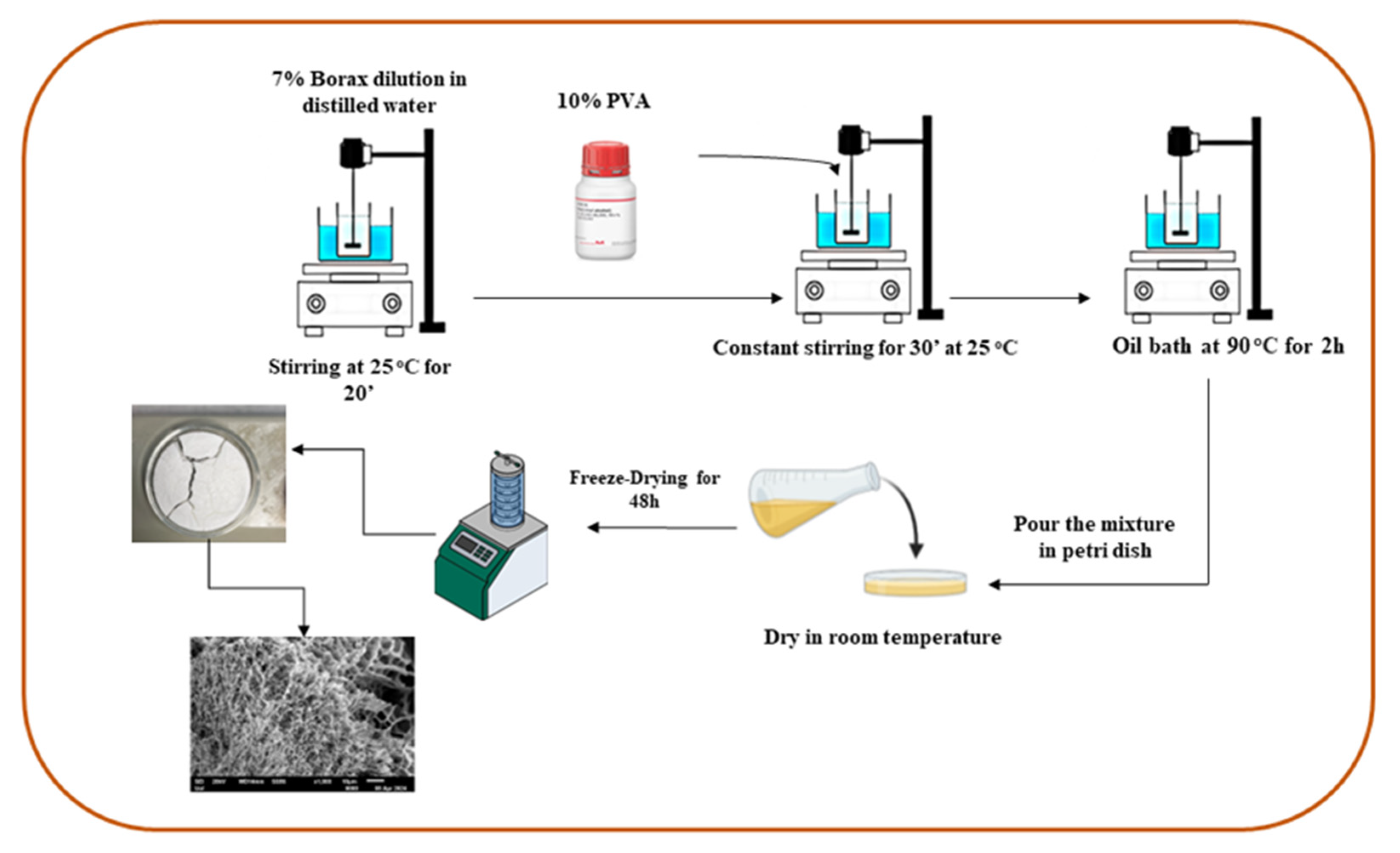
For the preparation of the PVA-CNC hydrogels, in a beaker containing CNC suspension, 7% Borax was diluted under continuous stirring at 25 °C for 20 minutes. Then 10% of PVA was added to the same beaker stirring for another 30 min. After the fully dilution of PVA, the beaker was placed in oil bath at 90 °C for 2 hours under continuous stirring. The mixture was poured) in a petri dish and was let to dry at room temperature. The sample was stored at -20 °C and then it was lyophilized to receive the porous hydrogel, Figure 8.
4. Conclusions
The prepared cellulose nanocrystal-based hydrogels were synthesized through a solution casting-evaporation method and characterized through all the analytical measurements. Cellulose was extracted from aloe vera leaves residue. Cellulose nanocrystals were then isolated using sulfuric acid. For the hydrogel preparation PVA was used because of its hydrogel forming properties and Borax also was added as cross-linker. Thus, we are willing to do more characterizations to prove that the prepared materials can be used as sustainable packaging materials or/and as drug delivery systems.
Author Contributions
Conceptualization, E.T., C.E.S. and N.E.Z.; methodology, E.T., C.E.S. M.A.K..; software, E.T., D.M., A.K-M.; validation, A.A., C.E.S. and N.E.Z.; formal analysis, D.M., K.C.V. C.E.S..; investigation, E.T., A.K-M., D.M. C.K., K.C.V.; resources, A.A., A.E.G., C.E.S., N.E.Z.; data curation, E.T., D.M., C.K. C.E.S.; writing—original draft preparation, E.T., A.M-K., D.M., C.E.S., N.E.Z.; writing—review and editing, E.T., A.M-K., D.M., C.E.S., N.E.Z.; visualization, M.A.K., C.E.S., N.E.Z.; supervision, C.E.S., N.E.Z.; project administration, C.E.S., N.E.Z.; funding acquisition, A.E.G., M.A.K., A.A., C.E.S., N.E.Z.; All authors have read and agreed to the published version of the manuscript.
Funding
This research received no external funding.
Conflicts of Interest
The authors declare no conflicts of interest.
References
- Szymańska-Chargot, M., et al., Isolation and Characterization of Cellulose from Different Fruit and Vegetable Pomaces. Polymers (Basel), 2017. 9(10). [CrossRef]
- Abraham, E., et al., Environmental friendly method for the extraction of coir fibre and isolation of nanofibre. Carbohydrate Polymers, 2013. 92(2): p. 1477-1483. [CrossRef]
- Hernandez, C. and D. Rosa, Extraction of cellulose nanowhiskers: natural fibers source, methodology and application. 2016. p. 232-242.
- Brinchi, L., et al., Production of nanocrystalline cellulose from lignocellulosic biomass: Technology and applications. Carbohydrate Polymers, 2013. 94(1): p. 154-169. [CrossRef]
- Nagarajan, K.J., A.N. Balaji, and N.R. Ramanujam, Extraction of cellulose nanofibers from cocos nucifera var aurantiaca peduncle by ball milling combined with chemical treatment. Carbohydr Polym, 2019. 212: p. 312-322. [CrossRef]
- Chang, C., et al., Structure and Properties of Cellulose Nanocrystals. 2019. p. 21-52.
- Habibi, Y., L.A. Lucia, and O.J. Rojas, Cellulose Nanocrystals: Chemistry, Self-Assembly, and Applications. Chemical Reviews, 2010. 110(6): p. 3479-3500. [CrossRef]
- Gil-Martín, E., et al., Influence of the extraction method on the recovery of bioactive phenolic compounds from food industry by-products. Food Chem, 2022. 378: p. 131918.
- Chowdhury, Z. and S.B. Abd Hamid, Preparation and Characterization of Nanocrystalline Cellulose using Ultrasonication Combined with a Microwave-assisted Pretreatment Process. BioResources, 2016. 11: p. 3397-3415. [CrossRef]
- Valdés, A., et al., Microwave-assisted extraction of cellulose nanocrystals from almond (Prunus amygdalus) shell waste. Front Nutr, 2022. 9: p. 1071754.
- Kabir, S.M.F., et al., Cellulose-based hydrogel materials: chemistry, properties and their prospective applications. Prog Biomater, 2018. 7(3): p. 153-174. [CrossRef]
- Zhao, Z., et al., Bioinspired Nanocomposite Hydrogels with Highly Ordered Structures. Advanced Materials, 2017. 29(45): p. 1703045.
- Ge, G., et al., Stretchable, Transparent, and Self-Patterned Hydrogel-Based Pressure Sensor for Human Motions Detection. Advanced Functional Materials, 2018. 28(32): p. 1802576.
- Chiellini, E., et al., Biodegradation of poly (vinyl alcohol) based materials. Progress in Polymer Science, 2003. 28(6): p. 963-1014. [CrossRef]
- Tsuchiya, Y. and K. Sumi, Thermal decomposition products of poly(vinyl alcohol). Journal of Polymer Science Part A-1: Polymer Chemistry, 1969. 7(11): p. 3151-3158.
- Moud, A.A., et al., Viscoelastic properties of poly (vinyl alcohol) hydrogels with cellulose nanocrystals fabricated through sodium chloride addition: Rheological evidence of double network formation. Colloids and Surfaces A: Physicochemical and Engineering Aspects, 2021. 609: p. 125577.
- Shen, X., et al., Hydrogels based on cellulose and chitin: fabrication, properties, and applications. Green Chemistry, 2016. 18(1): p. 53-75.
- Alvarez-Lorenzo, C., et al., Crosslinked ionic polysaccharides for stimuli-sensitive drug delivery. Advanced Drug Delivery Reviews, 2013. 65(9): p. 1148-1171.
- Yang, J., et al., Cellulose Nanocrystals Mechanical Reinforcement in Composite Hydrogels with Multiple Cross-Links: Correlations between Dissipation Properties and Deformation Mechanisms. Macromolecules, 2014. 47: p. 4077–4086.
- You, J., et al., Improved Mechanical Properties and Sustained Release Behavior of Cationic Cellulose Nanocrystals Reinforeced Cationic Cellulose Injectable Hydrogels. Biomacromolecules, 2016. 17(9): p. 2839-2848.
- Berglund, L., et al., Self-Assembly of Nanocellulose Hydrogels Mimicking Bacterial Cellulose for Wound Dressing Applications. Biomacromolecules, 2023. 24(5): p. 2264-2277.
- Hebeish, A., et al., Development of cellulose nanowhisker-polyacrylamide copolymer as a highly functional precursor in the synthesis of nanometal particles for conductive textiles. Cellulose, 2014. 21(4): p. 3055-3071. [CrossRef]
- Loh, E.Y.X., et al., Development of a bacterial cellulose-based hydrogel cell carrier containing keratinocytes and fibroblasts for full-thickness wound healing. Scientific Reports, 2018. 8(1): p. 2875. [CrossRef]
- Lusiana, S., A. Srihardyastutie, and M. Masruri, Cellulose nanocrystal (CNC) produced from the sulphuric acid hydrolysis of the pine cone flower waste (Pinus merkusii Jungh Et De Vriese). Journal of Physics: Conference Series, 2019. 1374: p. 012023.
- Fardsadegh, B. and H. Jafarizadeh, Aloe vera leaf extract mediated green synthesis of selenium nanoparticles and assessment of their In vitro antimicrobial activity against spoilage fungi and pathogenic bacteria strains. Green Processing and Synthesis, 2019. 8: p. 399-407.
- El-Sakhawy, M., et al., Preparation and infrared study of cellulose based amphiphilic materials. 2018.
- Jahan, Z., M.B.K. Niazi, and Ø.W. Gregersen, Mechanical, thermal and swelling properties of cellulose nanocrystals/PVA nanocomposites membranes. Journal of Industrial and Engineering Chemistry, 2018. 57: p. 113-124.
- Putri, L.Z. and Ratnawulan, Analysis of Aloe vera Nano Powder (Aloe vera L.) using X-Ray Diffraction (XRD). Journal of Physics: Conference Series, 2023. 2582(1): p. 012029.
- Gong, J., et al., Research on cellulose nanocrystals produced from cellulose sources with various polymorphs. RSC Adv., 2017. 7: p. 33486-33493.
- Jayaramudu, T., et al., Electroactive Hydrogels Made with Polyvinyl Alcohol/Cellulose Nanocrystals. Materials, 2018. 11: p. 1615. [CrossRef]
- Tummala, G.K., Hydrogels of Poly (vinyl alcohol) and Nanocellulose for ophthalmic applications: synthesis, characterization, biocompatibility and drug delivery studies. 2018, Acta Universitatis Upsaliensis.
- Veloso, S.R.S., et al., Cellulose Nanocrystal (CNC) Gels: A Review. Gels, 2023. 9(7). [CrossRef]
Figure 1.
FTIR spectra of a) pure aloe vera leaves and extracted cellulose and b) CNC/PVA Hydrogel.
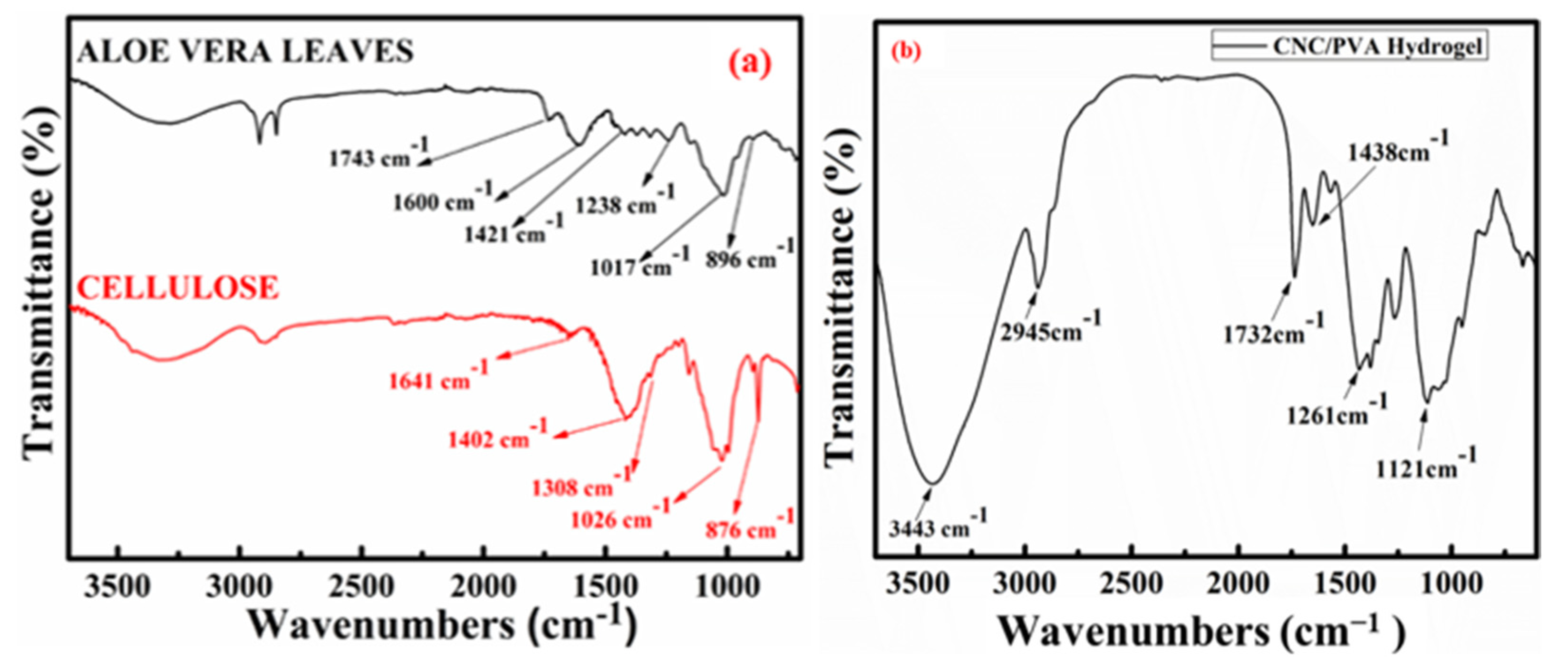
Figure 2.
XRD diffractograms of a) pure aloe vera leaves and extracted cellulose and b) CNC/PVA hydrogel.
Figure 2.
XRD diffractograms of a) pure aloe vera leaves and extracted cellulose and b) CNC/PVA hydrogel.
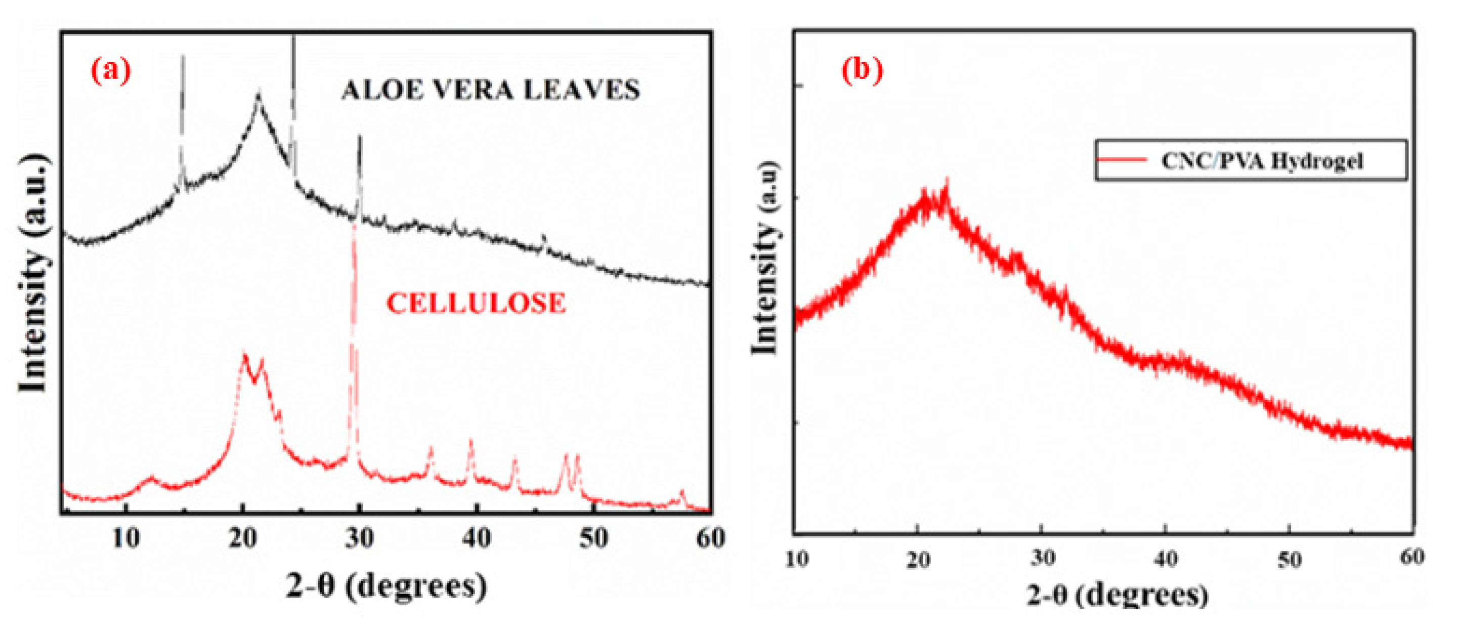
Figure 3.
SEM images of a) pure aloe vera leaves, b) extracted cellulose and c) isolated CNCs.
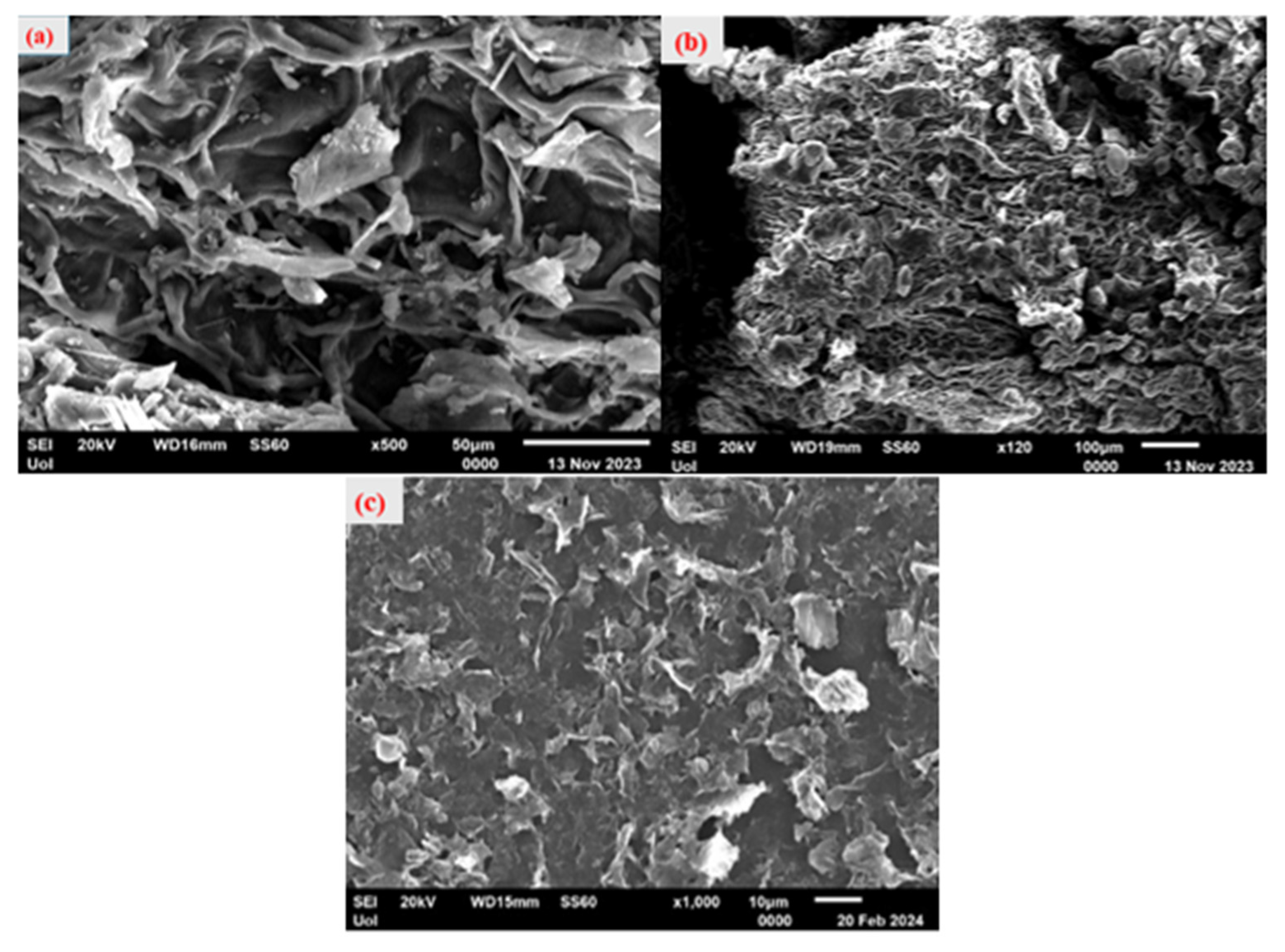
Figure 4.
SEM images for CNC/PVA hydrogel.
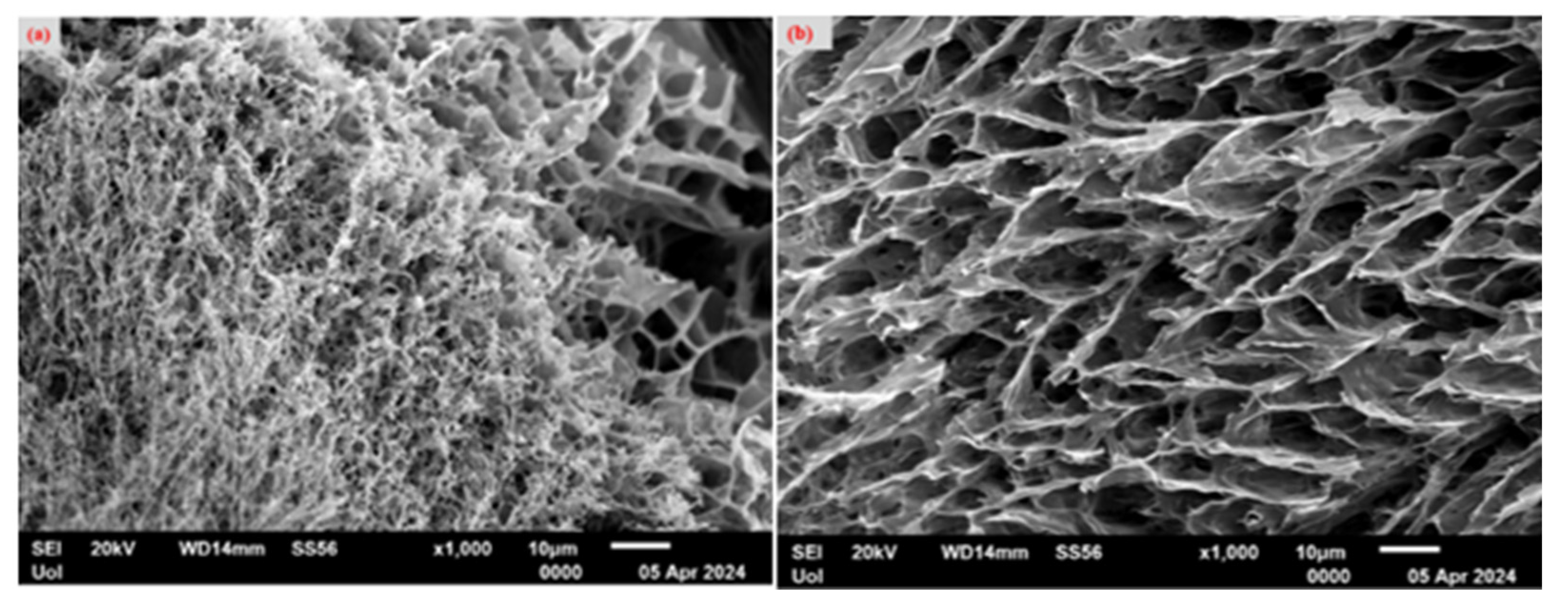
Figure 5.
Dynamic Mechanical Analysis of CNC/PVA hydrogel.

Figure 6.
Schematic illustration of cellulose extraction.
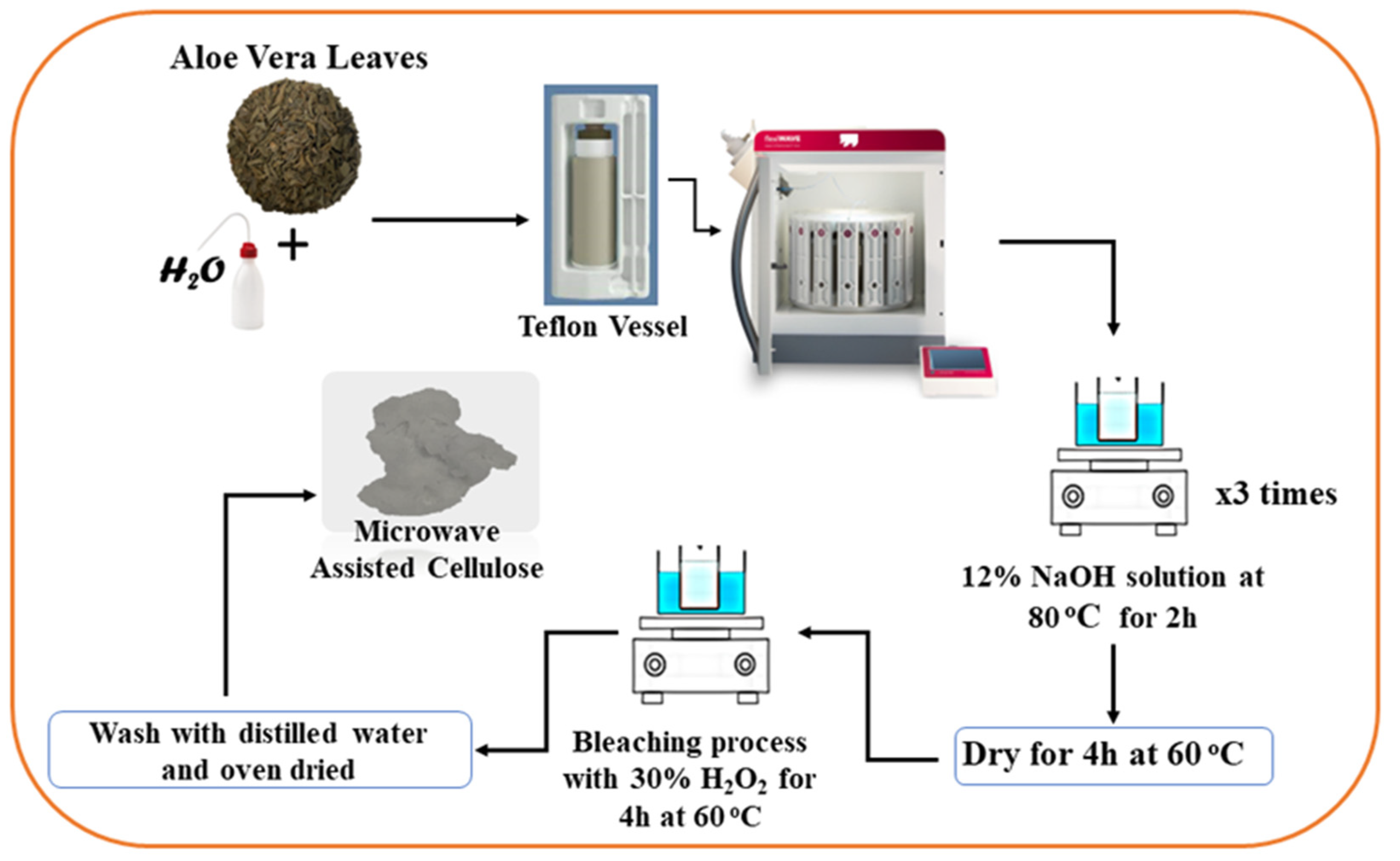
Figure 7.
Schematic illustration of cellulose nanocrystals isolation.
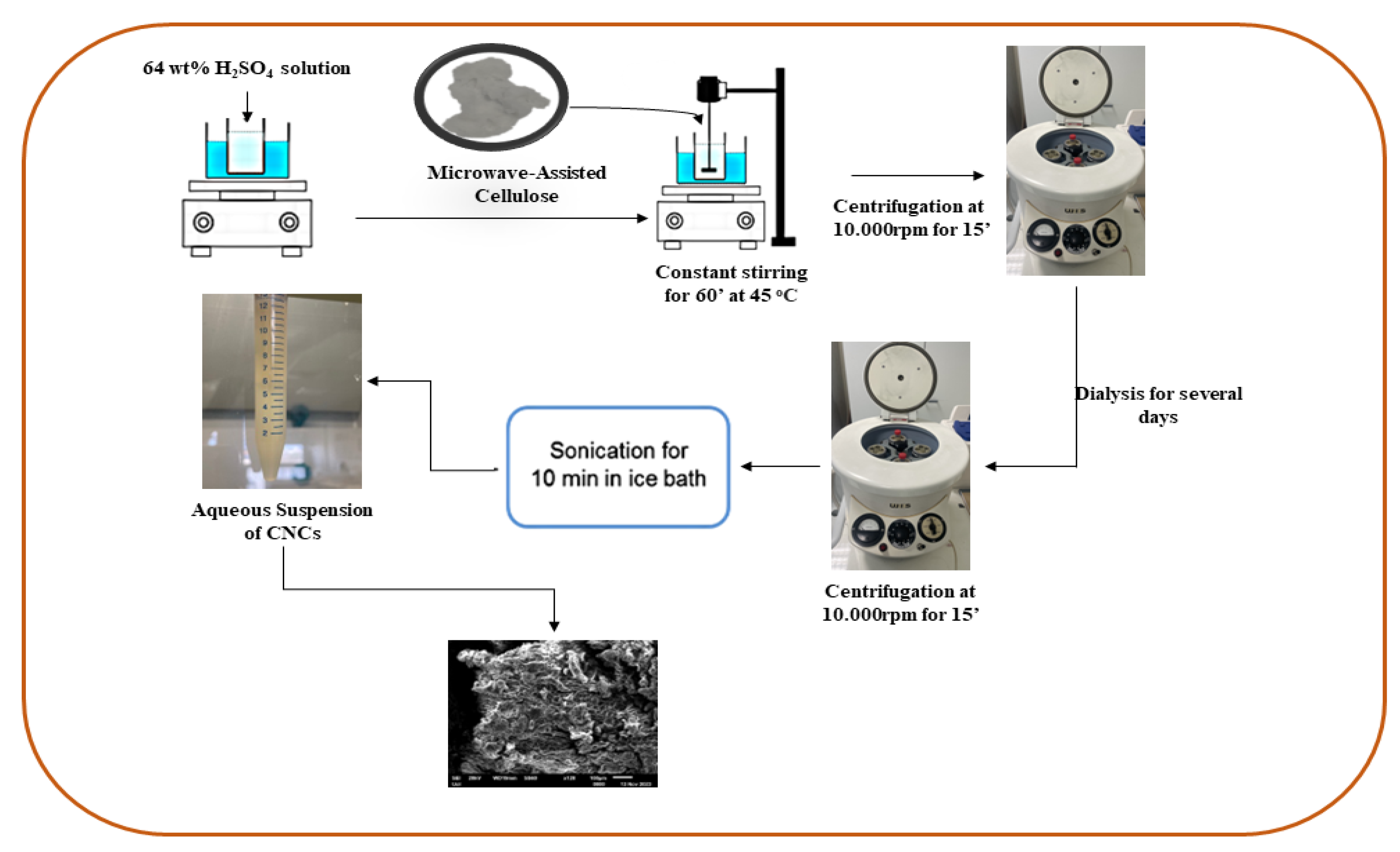
Figure 8.
Schematic illustration of CNCs/PVA hydrogel preparation.
Disclaimer/Publisher’s Note: The statements, opinions and data contained in all publications are solely those of the individual author(s) and contributor(s) and not of MDPI and/or the editor(s). MDPI and/or the editor(s) disclaim responsibility for any injury to people or property resulting from any ideas, methods, instructions or products referred to in the content. |
© 2024 by the authors. Licensee MDPI, Basel, Switzerland. This article is an open access article distributed under the terms and conditions of the Creative Commons Attribution (CC BY) license (http://creativecommons.org/licenses/by/4.0/).
Copyright: This open access article is published under a Creative Commons CC BY 4.0 license, which permit the free download, distribution, and reuse, provided that the author and preprint are cited in any reuse.


