Submitted:
23 October 2024
Posted:
24 October 2024
You are already at the latest version
Abstract
Gold nanoparticles (NPs) have demonstrated significance in several important fields, including drug delivery and anticancer research, due to their unique properties. Gold NPs possess significant optical characteristics that enhance their application in biosensor development for diagnosis, photothermal and photodynamic therapies for anticancer treatment, as well as in targeted drug delivery and bioimaging. The broad surface modification possibilities of gold NPs have been utilized in the delivery of various molecules, including nucleic acids, drugs, and proteins. Moreover, gold NPs possess strong localized surface plasmon resonance (LSPR) properties, facilitating their use in surface-enhanced Raman scattering for precise and efficient biomolecule detection. These optical properties are extensively utilized in anticancer research. Both photothermal and photodynamic therapies show significant results in anticancer treatments using gold NPs. Additionally, the properties of gold NPs demonstrate potential in other biological areas, particularly in antimicrobial activity. In addition to delivering antigens, peptides, and antibiotics to enhance antimicrobial activity, gold NPs can penetrate cell membranes and induce apoptosis through various intracellular mechanisms. Among other types of metal NPs, gold NPs show more tolerable toxicity capacity, supporting their application in wide-ranging areas. Gold NPs hold a special position in nanomaterial research, offering limited toxicity and unique properties. This review aims to address recently highlighted applications, current status of gold NP research and discuss their future in nanomedicine.
Keywords:
gold nanoparticle
; drug delivery
; bioimaging
; biosensor
; antibacterial
; photothermal therapy
; photodynamic therapy
; anticancer
; LSPR
; SERS
1. Introduction
Nanomaterials have become one of the most significant materials utilized in wide-ranging applications. Nanoparticles in this case, have proven their significance in nanotechnology with unique characteristic features. They possess significant reactivity through their large surface area, tunable size and modifiable surface for wide-ranging applications, and shape that can alter physicochemical properties [1].
Gold NPs possess unique optical properties that are easily modified with alterations of physicochemical properties [2]. They are found in various shapes, such as nanocubes, nanorods, triangular, nanostars and nanospheres, and different sizes that affect their color, which is utilized in many applications [3]. In addition, their surface chemistry can be easily altered and conjugated with various molecules, which is expanding their application to many areas, such as drug delivery and photothermal therapies (PTT) (Figure 1) [4].
Gold NPs’ unique physicochemical properties, including strong LSPR, ease of functionalization with a wide range of biomolecules, antimicrobial characteristics, anticancer and antioxidant activity, have been frequently exploited in the current literature. Besides, attributes that originate from their nature, such as biocompatibility, inertness and low toxicity, creates an advantageous state for the future uses where gold NPs are employed. These unique properties are highly expressed in wide-ranging applications, especially in imaging, sensor development, and in cancer radiotherapy as an efficient radiosensitizer (Figure 1) [6]. Thanks to their unique physicochemical properties and significant X-ray absorption, they have been used as a radiosensitizer in many in vitro and in vivo studies [7].
When the total number of published articles in the last five years is observed, there is notable consistency in research numbers during the first four years (Figure 2). Even though there is a slight decline in the last two years, it is evident that the field remains active with approximately 10,000 publications annually. This can be explained by the number of published patents and the current challenges, which are evaluated in later sections.
According to the given pie chart, drug delivery and biosensor application of gold NPs lead the majority of research compared to other applications that have been discussed in this review. Moreover, antimicrobial activity, in total of antibacterial, antifungal and antiviral studies, manages to closely follow these areas. This distribution is heavily influenced by the unique optical properties and efficient carrier characteristics of gold NPs.
Due to their unique optical characteristics, distribution of their application shows great difference compared to other types of metals NPs. For instance, silver NPs, one of the most studied types of metal NPs in the literature, are known with significant antimicrobial activity [9]. Based on this, while silver NPs are predominantly utilized in antibacterial and wound healing studies [10], gold NPs seems to be tested in delivery, imaging and anticancer studies, including photodynamic therapy (PDT) and PTT, as the distribution is clearly visible in the recent clinical trials [11]. That is the reason why review articles, focusing on recent applications and advances of gold NPs, are very important. The distribution analysis of the currently published research works, pointing out the challenges, and discussion of future perspectives are important to guarantee further development in gold NP applications considering the highly active status of this field. In particular, we have addressed the currently growing application areas and the ones that need attention, suggesting research gaps that need addressing. These are highly critical in allowing wider clinical and industrial applications. This review provides a comprehensive overview of well-explored areas, especially drug delivery and biosensors, and overlooked applications like bioimaging and anti-inflammatory uses, therefore offering valuable insights into potential research directions that could drive innovation in the future of NP research.
2. Applications of Gold Nanoparticles
Thanks to the unique and significant characteristics of gold NPs, they have been widely used in nanomedicine with a great impact. With tunable surface chemistry, significant physicochemical-dependent optical properties, especially LSPR, and higher tolerability with good biocompatibility, gold NPs are utilized in a wide range of biomedical applications. As shown in Figure 2, gold NPs are predominantly applied in drug delivery, biosensors, and PDT/PTT-based therapies, primarily focusing on anticancer research. In this section, we have highlighted and provided detailed examples of efficient localized heat generation upon light irradiation, the potential of surface modifications in antimicrobial treatment and drug delivery, the unique LSPR property for sensor development, and areas requiring further research, such as wound healing and bioimaging applications.
2.1. Delivery systems
Gold NPs are capable of delivering different types of biomolecules, including recombinant proteins, wide-ranging drugs, and nucleotides [12]. Since they possess a high capacity to be functionalized and the possibility to be altered, both in size and shape, allow their utilization in the delivery of not only small but also large biomolecules [13]. This is why, various structures of Gold NPs, such as nanorods, spherical, and composites, are utilized in drug delivery applications [14]. In addition, they possess susceptibility to penetrate tumor cells more than the blood vessels of vital tissues, such as the heart and lung [7]. Therefore, gold NPs are currently used in various delivery systems in several areas (Figure 3).

Table 1.
Recent Drug Delivery Applications of Gold NPs.
| Application | Synthesis Method | Properties | Results | Reference |
|---|---|---|---|---|
| In vitro cervical cancer treatment with curcumin conjugation | Chemical synthesis | Average size of 7 nm ± 2.29 nm Spherical morphology SPR peaks at 525 nm |
-Enhanced bioavailability of curcumin against HeLa cells. -Insignificant toxicity in the zebrafish embryo model. -Significant radiosensitizing activity. -Enhanced intracellular reactive oxygen species (ROS) generation, apoptotic signals and DNA damage. |
[16] |
| Co-delivery with miRNA-33a to MCF-7 breast cancer cells | Purchased from Nanosany with >95% purity. . |
Size ranges from 50 to 100 nm Spherical morphology (properties of modified gold NPs) |
-Enhanced gene expression in co-delivery. -Enhanced inhibitory activity against breast cancer cells. -Potential synergistic effect through modulation of signaling pathways -Increased apoptosis rate. |
[17] |
| Enhanced delivery of bleomycin in electrochemotherapy | Chemical synthesis | 13 nm size Spherical morphology LSPR peak at 521 nm |
-Enhanced cell permeabilization by 40%. -The electric field of 0.9 kV/cm and 1–100 kHz protocols demonstrated significant cytotoxicity by gold NP involvement. |
[18] |
| Delivery of chlorpromazine | Chemical synthesis | Average size of 15 nm and 55 nm Quasi-spherical morphology |
-Enhanced activity of chlorpromazine in cytotoxicity assays against human (COLO 679) and murine (B16-F0) melanoma cells. -Inhibition of mitochondrial activity and disruption of the cell membrane. |
[19] |
| Delivery of colistin | Chemical synthesis | Average size of 44.34 ± 1.02 Absorbance peaks at 300 ± 0.2 and 515 ± 0.3 nm (values from chitosan capped particles) |
-Average drug loading efficacy by 76.4%. -A consistent drug release profile. -A developed metered-dose inhaler efficiently destroys bacteria over a 12 hour period. |
[20] |
| Delivery of phosphazene Delivery of yeast RNA |
Chemical synthesis | Spherical Morphology | -Efficient, controlled and long-term release profiles for both drug and RNA delivery. -Antibacterial and antifungal activity. |
[21] |
| Nucleic acid DNA RNA | ||||
| Delivery of anti Glut1 SiRNA | Chemical synthesis | Approximate size of 14 nm Uniform Morphology LSPR peak at 520 nm (red-shifted to 528 nm) (values from SiRNA containing particles) |
-Significant reduction in Glut1 expression. -Promotion of apoptosis through glucose starvation and ROS cascade signaling. -Inhibition of cancer cell proliferation and tumor growth. (in vivo) -Induction of apoptosis. |
[22] |
| Delivery of Fluc mRNA | Chemical synthesis | Size between 11.52 nm - 12.97 nm Spherical Morphology Absorption peak at 520 nm |
-Significant expression of the luciferase gene compared to naked mRNA delivery. | [23] |
| SiRNA delivery | Commercially purchased | Size ranging between 20 - 30 nm Spherical Morphology SPR peak at 520 nm |
-At pH 5.5, green fluorescent protein knockdown levels decreased to 65% in HeLa cells. | [24] |
| Delivery of Fluc-zetagreen reporter genes Delivery of plasmid DNA and synthetic mRNA of SARS-CoV-2 S protein |
Chemical synthesis | Mean size of 53 nm Nanostar Morphology Absorbance peak at 630 nm |
-Enhanced transfection efficiency of Fluc gene delivery by increased gold NP-included nanocomposite concentrations in several cell lines. -Significant transfection efficiency of mRNA (at 1000 ng) and DNA (at 250 ng) of S protein by gold NPs. |
[25] |
| Protein | ||||
| Delivery of SARS-CoV-2 spike protein | Chemical synthesis | Size of 50 nm Spherical Morphology SPR peak at 529 nm |
-Induced IgG levels. -Induced neutralizing antibody levels. -Significant levels of IFN-γ. (All levels observed in mice) |
[26] |
| Delivery of atrial natriuretic peptide | Chemical synthesis | Size of 22.34 ± 0.54 | -Significant reduction of tumor formation capacity of retinoblastoma cells from >75% to <50% and <25%. -Significant reduction of tumor formation capacity in mice when the particles are topically administered. |
[27] |
| Antimicrobial peptide delivery | Chemical synthesis | Size of 10 nm SPR band at 518.5 nm |
-Significant reduction in MIC and MBC values by average of 200-fold. -20-fold faster killing capability in bacterial killing kinetics. -17-fold lower minimum biofilm eradication concentration (MBEC). |
[28] |
| Antibiotic | ||||
| Delivery of ciprofloxacin | Chemical synthesis | Approximate size of 13 nm Spherical Morphology Absorption peak at 520 nm |
-Significant reduction in MIC values when the ciprofloxacin administered with gold NPs (2 to 4 time reduction) -Increased zone of inhibition by approximately 4 mm. -Significant biofilm inhibition (from 36.30 nm to 12 nm in analysis on nebulizer masks) and antioxidant capacity (84.66%). |
[29] |
| Conjugation of amikacin for contact lens preservation | Chemical synthesis | Average size of 21 nm Spherical Morphology Absorption peak at 520 nm |
-Significant reduction of MIC with conjugation on NPs (2 to 4 time reduction). -More than 50% increase in inhibitory zones. -Efficient prevention of biofilm formation in contact lenses (approximate 2-4 time reduction). -Antioxidant capacity by 87.40 at the highest concentration, 100 µg/mL. |
[30] |
| Enhanced antimicrobial activity of berberine | Chemical synthesis | Average size of 49.38 nm Spherical Morphology Absorption peak at 520 nm |
-72% maximum release at 72 hours. -Nearly 50% decrease in MIC values. -Enhanced bactericidal activity through ROS-mediated membrane disruption and DNA damage. -Approximately 2-fold higher antibiofilm activity by 73.35%. -Significant enhancement of wound healing percentage in mice, with only 2.7 % bacteria survival rate. |
[31] |
2.1.1 Delivery for Cancer Treatment
The drug delivery capacity of gold NPs is commonly used to deliver anticancer drugs. They are included in different types of anticancer drug delivery systems, including light-responsive, pH-based, and glutathione responsive with attachment of different therapeutic molecules in various shapes and sizes [32]. In addition to delivering the drug at the target site with an efficient drug release profile, gold NP can indirectly enhance the activity of the drug. Since the targeting and internalization of the drug will be enhanced, the anticancer drug’s overall activity will be increased. For example, gum karaya stabilized gold NPs were used to deliver an anticancer drug, gemcitabine hydrochloride, which was loaded into the surface of the NP [33]. The surface coating of the drug exhibited 19.2% efficiency. At three various doses (0.1, 0.5, and 1 μg/mL) A549 human lung cancer cells were treated with the anticancer drug with and without the NP. The cell viability of the cancer cells decreased by nearly 10% percent in all concentrations when administered with the gold NP (46.8% to 35.1% at the highest concentration). A significant reduction in colony formation was also observed in the colony formation inhibition assay. To test the efficiency of the drug, the determination of intracellular ROS levels through fluorescence imaging was performed. The anticancer drug showed higher fluorescence intensity when treated with gold NP than the sole administration.
The size and structure of the NP influence the efficiency of the drug delivery of the gold NPs. It has been discussed that the efficiency and internalization of the gold NPs can be highly affected by the size, surface chemistry, and shape of the NP [34]. As an example, the shape-dependent cytotoxicity of the gold NPs was demonstrated in a research [35]. Three types of gold NPs, nanospheres, nanorods, and nanostars, were tested against multiple types of pancreatic and bone cancer cells. The results demonstrated that nanostar-shaped gold NPs exhibited the highest cytotoxicity, meanwhile, nanospheres showed the least potential as an anticancer agent.
A similar dependency is also observed in the size of the NP. For instance, a pH-sensitive drug delivery system for methotrexate was developed for breast cancer treatment, involving two different structures of gelation-coated gold NPs (spherical 50 and 100 nm/nanorod 20, 50, and 100 nm) [36]. In vitro drug release revealed that small-sized gold NPs, independent from the structure, exhibited a higher release rate at 5.4 pH. There was an observable change between the two structures of gold NPs in the cell viability test on MCF-7 cells. The spherical gold NPs demonstrated similar cytotoxicity when compared to the control group. In the drug-loaded form, a significant decrease in the cell viability, higher than in the free drug group, was observed. Additionally, the highest results were obtained in 50 nm size. Conversely, nanorod-shaped gold NPs were highly toxic to cancer cells compared to spherical-shaped ones, meanwhile, gelatin coating decreased the cytotoxicity. Similarly, drug-loaded nanorods efficiently destroyed breast cancer cells compared to the free drug group. The overall results concluded that nanorod-shaped gold NPs were more efficient as drug carriers than spherical-shaped ones.
Gold NPs, especially nanorods, and nanoshells, are used in PTT-based cancer studies (due to their absorption properties) as anticancer drug carriers [37]. The PTT-based anticancer and tumor applications of gold NPs are detailly evaluated in the further sections. Still, its drug delivery perspective will be briefly given in this section. The PTT with gold NPs can alter the environment, and increase the cellular permeability and uptake of both the encapsulated drug and NP itself, which leads to enhanced cytotoxic activity [38].
Niikura et al. demonstrated efficient drug release from gold NP vesicles through light irradiation [39]. Doxorubicin carrier gold NP vesicles were tested in vitro on HeLa cells with and without laser irradiation. The irradiation group induced cell death up to 50%, meanwhile normal treatment did not manage to alter cell viability. Significant morphological changes in HeLa cells were observed in bright-field images, however, this was not the case for other groups (unloaded and not irritated). A similar approach, a combination of anticancer drug delivery with near-infrared (NIR) irradiation, was demonstrated by Kadkhoda et al. [40]. Paclitaxel-loaded aptamer-conjugated gold NPs were modified with poly (ethylene glycol) (PEG) and demonstrated an efficient NIR and pH-dependent drug release. The efficiency of drug encapsulation was determined by 86%. To determine the influence of pH and irradiation on drug release ratio, three different pH levels (5.5, 6.5, 7.4) with and without exposure to irradiation were observed. The highest drug release % was observed in the lowest pH levels with irradiation. Additionally, all drug release % in each pH level was higher with NIR by approximately 10%. The most efficient power of NIR was found to be 160 mW/cm2. The in vitro cytotoxicity analysis showed a significant decrease in cell viability with NIR compared to non-irradiated NPs. After 3 hours, 96% of the NP internalized into the MUC-1 positive cells, higher than the MUC-1 negative cells (by nearly 2-fold) indicating the targeted activity of the NP. The modification of the gold NP caused a significant increase in the apoptosis ratio from 12.8% to 41.49%. Gene expression levels were also demonstrated that aptamer conjugation enhanced the apoptosis induction of gold NPs.
2.1.2. Nucleic acid delivery
Since usage of NPs as a carrier provides enhanced solubility and biodistribution, they are also highlighted with their potential in gene therapies by carrying nucleic acids [41]. Gold NPs draw a notable interest in gene therapies due to their presence in the area. Their low toxicity capacity, high uptake efficiency, and capacity enhance their potential in gene silencing and transfection [42]. Different gold NPs, such as positively charged, nanorod, or small-sized, have been coated and utilized for efficient DNA and RNA delivery [43]. This is why nucleic acid delivery of gold NPs is applied in various approaches like other gold NP-based delivery systems. Stimuli-responsive delivery of nucleic acids, such as GSH-mediated release or light-irradiation, are promising alternatives in this manner [44]. Similar to the previous section, along with the future section that discusses anticancer activity of the gold NPs, gold NPs are also used in cancer treatment through their efficient nucleic acid delivery. Their capability to be modified with wide-ranging probes makes them preferred to deliver RNA silencers to tumors [45]. Therefore, gold NPs are utilized to carry vectors and induce desired intracellular activity following cellular uptake for multiple purposes.
DNA
DNA is a promising target in gold NP-based nucleic acid delivery. Most of the time, the DNA is bound to the surface of the NP with various types of interactions. Attachment of the DNA to gold NPs can be mediated with sulfur (S) (more preferable) and nitrogen (N) bond, or with the charge interactions through the surface charge of the particle [46]. In addition, high amounts of DNA can bind into gold NPs with great bond strength [47]. This feature is commonly highlighted in modification of the gold NPs with DNA, which is highly used in different type of applications, including drug delivery [48]. Still, there are notable amounts of research efforts that utilize this feature in nucleic acid (DNA) delivery applications.
As an example, PEG-functionalized positively charged gold NPs were used to deliver nonviral vectors (three types that range between 4 to 40 kpb) to various cell lines (HeLa and Hek293t cells) [49]. Based on the cell viability results, doses that below 50 μg/mL for HeLa, below 25 μg/mL for Hek293t cells were used. The evaluation of the transfection efficiency of the plasmid DNA was observed in fluorescence microscopy and compared to the control group. As a result, up to 20% increase in the fluorescent intensity was observed. Later on, the potential of the gene therapy was evaluated with expression of herpes virus thymidine kinase through treatment of prodrug ganciclovir. Compared to commercial formulations, the cell viability of the HeLa cells was significantly lower due to the efficient gene delivery by the gold NPs. Another method was deployed to co-deliver DNA and siRNA with multi-layered degradable polymer coatings onto gold NPs [50]. High doses of DNA (200 to 2400 ng) and siRNA (160 to 240) was used in layered gold NPs. Both siRNA-mediated knockdown and DNA-mediated expressions were determined during the experiments. Up to 25% knockdown was achieved in siRNA delivery, as the highest rate was obtained on days 6-7 in 240 ng loading. The fluorescence images revealed the DNA expressions of both including one and two nucleic acid DNA layered formulations. Two layered gold NPs, without siRNAs, showed the highest transfection ratio of 28%.
One particular study used gold NPs to deliver donor DNA and clustered regularly interspaced short palindromic repeats (CRISPR)–associated protein 9 (Cas9) ribonucleoprotein (RNP) to induce DNA repair in vivo [51]. The induction of homology-directed repair by the complex was observed in human embryonic kidney cells. Before the test, the induction of the DNA repair, encapsulation efficiency (61.5%), and enzymatic activity (preserved with encapsulation) were observed. In in vitro conditions, the DNA repair of 11.3% of the total cells was induced by the treatment of CRISPR-Gold NPs. Additionally, the rate of the DNA repair reached to maximum at a concentration of 8 μg/ml, while exceeding this value caused cellular cytotoxicity by the CRISPR-Gold NPs. The in vitro study extended to dendritic and primary myoblast cells, along with multiple types of stem cells. The CRISPR-Gold NP managed to target all the used types of cells and exhibited a DNA repair ratio between 3-4% through simultaneous delivery of the Cas9 protein, the donor DNA, and guide RNA. The gene editing significance of the system was also demonstrated in a reporter mouse model. At last, CRISPR-Gold NPs are intramuscularly injected into mice for the possibility of correcting dystrophin mutation. The results showed that the particles successfully corrected the dystrophin gene (5.4%) and restored the dystrophin protein expression. The treatment without the gold NP showed an 18-fold lower (0.3%) correction rate. The treatment improved the strength and muscle function of the mice with extremely minimal off-target genomic damage (between 0.005 - 0.2%). These promising results not only extend the application of the CRISPR-Cas9 system but also show that gold NPs can also be applied to complex systems as an efficient carrier.
RNA
Usually, RNA molecules bound to the surface of gold NPs with thiol or electrostatic interactions [52]. However, various types of RNAs (especially siRNAs) can be bound on gold NP surfaces with additional interactions: electrostatic interactions (layer-by-layer approach is included), DNA hybridization, and crosslinking [53]. Most of the time, the siRNAs are bound with gold NPs to enhance their stability and delivery efficiency. This approach has been used for various purposes.
For instance, an in vitro study was conducted to improve the delivery of the siRNAs to inhibit dengue virus infection [54]. Against four serotypes of dengue virus, antiviral siRNAs were tested in Vero cells. Except for serotype 1, the antiviral activity of the siRNAs was demonstrated against the dengue virus. Later on, these siRNAs were modified with gold NPs by layer-by-layer (electrostatic) approach and delivered to Vero cells. Transmission electron microscopy (TEM) images confirmed that gold NPs were successful in the delivery of siRNAs into the cell. Finally, the complex was tested for potential inhibition of dengue virus infection at both pre- and post-infection phases. Both viral propagation and virus replication were inhibited by the gold NP-siRNA complex. The complex exhibited efficient inhibition by demonstrating up to 15-fold higher inhibition than the control group. Additionally, significant protection of siRNAs by gold NPs was shown, along with their protected activity after treatment of RNases.
Gene silencing is commonly preferred in RNA delivery with gold NPs. Conde et al. demonstrated in vitro and in vivo gene silencing through the delivery of siRNA with gold NPs for RNA interference (RNAi) application [55]. A primary gene, c-myc protooncogene, was chosen as a target for RNAi, and targeted in HeLa cells as in vitro, in freshwater polyps and mouse models as in vivo for gene silencing. The attachment of siRNAs to gold NPs was mediated with ionic (with the negative charge of siRNA) and covalent (thiol-gold bond) approaches. TEM images showed the accumulation of gold NP conjugates in the cytoplasm of the cells. To detect the efficiency of the gene silencing, the luciferase gene was used as a reporter. As expected, there was a significant reduction in luciferase activity by the siRNA activity from 110-100% (control groups) to 50-25% (depending on the attachment approach). The same results were not observed when the gold NPs were bound with non-related siRNAs, indicating the selectivity of the gene silencing. In vivo models were not different in terms of gene silencing efficiency. Both approaches that mediated the siRNA bonding significantly reduced the Hydrac-myc expression levels to less than 50% (the covalent approach was superior compared to ionic). Similar results were also obtained in a mouse model, as the covalent approach showed significant inhibition of mouse-myc expression levels (to nearly 30%) compared to the ionic approach (nearly 60%). These findings not only represent the potential of gold NPs in therapeutic applications but also highlight the importance of the chosen approach in gold NP-based RNA delivery complexes.
The approach of releasing the RNAs is as important as the approach for binding the RNAs on the NP surface. Stimuli-responsive delivery is a common alternative that is used in gold NP-based RNA delivery. The light-irradiation method was used on gold nanorods in the treatment of pancreatic tumors through co-delivery of siRNA (for K-Ras gene silencing) and doxorubicin [56]. Several types of gold NP samples were used during the experiment, including separately coated doxorubicin and siRNAs. Fluorescent images of Panc-1 cells confirmed the efficient cellular uptake and the transfection efficiency was found to be higher than 83%. Each sample (the ones that include doxorubicin and siRNA) managed to decrease the mRNA and protein levels and effectively inhibited K-Ras expression. This, later on, was explained by the blocked proliferation of the Panc-1 cells through S-cell cycle arrest by gold nanorod-mediated delivery. Furthermore, the release profiles were observed under the 665 nm light irradiation. A burst release of doxorubicin (90%) was observed under 20 minutes with light irradiation, meanwhile, the drug release reached 80% after exceeding 16 hours. A similar profile was also observed for siRNA release, with more than 60% of the RNAs released in under 20 minutes. Without light irradiation, the same released amounts reached after the 6th hour. Light-irradiated formulations showed the highest tumor suppression in vivo as well. Another stimuli-responsive RNA delivery was mediated with capped gold nanorods but to deliver small hairpin RNAs, not siRNAs [57]. A significant tumor gene silencing was observed with the gold nanorod-RNA complex. Both in vivo and in vitro experiments showed the enhanced RNA release by the glutathione triggering.
Gold NPs possess great potential in nucleic acid delivery by their favorable surface modification. Depending on their shape and other characteristics, methods to mediate delivery systems show diversity, indicating the presence of wide-ranging approaches. With slight differences, both gene expression and silencing can be induced with the gold NP models. Based on the discussed studies and current literature, gold NPs are also utilized in more complex systems and different applications.
2.1.3. Protein Delivery
Similar to nucleic acids, many types of proteins are attached to the gold NP surface for cell-targeted delivery purposes. Protein-coated gold NPs possess enhanced internalization with directed targeting and modulated functions [58]. Most commonly, protein/peptide-based functionalization of gold NPs are utilized for cancer cell targeting or to increase the efficiency of the drug delivery capability of the particle [59]. Nevertheless, many studies used gold NPs as a nanocarrier to delivery therapeutic enzyme and peptides for various applications, including but not limited to anticancer, biosensing, and neurodegenerative diseases [60]. Possibly gold NPs can be utilized in many major application areas with their enhanced delivery characteristics. In this manner, we have evaluated a few studies that conjugate various types of proteins onto gold NPs. Additional recent studies are given in Table 1.
Ghosh et al. demonstrated the intracellular delivery of enzyme (β-galactosidase), which is incapable of passing through cellular membrane, with peptide-coated gold NPs as a protein transporter [61]. The modified gold NP-protein complex was treated with HeLa cells, then administrated X-gal to observe a color change upon enzymatic hydrolysis. Compared to the control group (sole treatment of the enzyme), a significant color change was observed in the gold NP-protein complex group, approximately 98% at the highest protein concentration. Another study used DNA aptamer conjugated gold NPs for in vivo delivery of functional proteins [62]. The researchers loaded His-tagged proteins on gold NPs and performed both in vitro and in vivo experiments. A significant internalization of the protein through gold NP carrier was demonstrated in the in vitro study. In vivo animal models demonstrated significant antitumor activity by the carrier protein (BIM protein) by tumor-targeted delivery.
One study included gold NPs in a nanocomposite, including manganese ferrite NPs, for efficient delivery of milk protein bovine lactoferrin (bLf) for antifungal therapeutic applications [63]. To test the antifungal activity, bLf-loaded nanocomposite was tested on Saccharomyces Cerevisiae along with solo bLf and unloaded nanocomposite treatments. The cell viability results indicated that both solo bLf and loaded nanocomposite significantly inhibited fungal cells, showing the efficient release of the protein from the NPs. Lactoferrin exhibits significant antimicrobial activity, including antifungal activity [64,65]. A novel, site-directed nanosystem that can mediate the transport of this protein might possess new therapeutic applications for both Lf and other milk proteins. As an example, in addition to Lf, both β-Lactoglobulin and α-Lactoglobulin [66,67] were conjugated with gold NPs, and highlighted with their potential in targeted drug delivery.
Concerning peptide delivery, an in vivo study was conducted to deliver peptide (ovalbumin) vaccine to induce anti-tumor response with gold NPs [68]. Mice injected with gold NP-peptide complex show a significant increase in interferon-gamma producing splenocytes, meanwhile, control groups or sole peptide treated mice barely show any increase. Later on, these groups’ tumor sizes and areas (mm2) were investigated. At day 20, both control and only peptide-treated groups exhibited the highest tumor size, while gold NP-delivered peptide groups did not show any increase in tumor size. Tumor areas were also boosted between days 6 and 8 for control and sole peptide-treated groups. The survival rate of the mice was also similar, which was 100% in the gold NP-delivered group. As a result, gold NP-mediated peptide delivery managed to inhibit tumor growth in mice models, and preserved the survival rate at the highest levels. Similar to other molecules that have been discussed, peptides are conjugated onto gold NPs for delivery of other molecules, such as DNA. Niu et al. demonstrated the utilization of peptide-conjugated cationic gold NPs for gene delivery to reverse the progression of melanoma [69]. The results indicate a significant internalization, high transfection and efficient skin penetration for possible gold NP-based topical gene therapy.
When the gold NP-based protein delivery approaches are considered, certain types of proteins and protein-based structures are highlighted, including but not limited to enzymes, peptides, and structural proteins. Since gold NPs can be conjugated with various types of proteins, their potential in protein delivery should not be overlooked.
2.1.4. Antibiotic Delivery
Antibiotic delivery is another promising application of gold NPs, especially in dealing with antimicrobial resistance (AMR). Recently, gold NPs have been highlighted as a potential drug delivery agent to combat AMR, with much recent research existing in the current literature [70]. There are certain approaches that gold NPs are used for antibacterial applications, such as photothermal therapies, various types of ligand conjugations, or pristine gold NPs [71].
A study showed the delivery of amino-glycosidic antibiotics (4 types) with protein capped (bovine serum albumin), stabilized gold NPs [72]. Each type of the antibiotic (streptomycin, neomycin, gentamicin, and kanamycin) was tested on three types of bacteria for their antibacterial activity, with and without gold NP carrier. Each antibiotic-gold NP conjugate demonstrated enhanced antibacterial activity from 4.7% to 33.3% for E. coli, from 21.0% to 50.0% for P. aeruginosa, and from 18.1% to 66.66% for S. aureus. Similar research was conducted on multi-drug resistant (MDR) bacteria [73]. Gold NP-mediated and solo administration of the antibiotics were compared on MDR bacteria (K. pneumoniae, S. aureus, and E. coli). Solo treatment of gold NPs and antibiotics did not show any zone of inhibition. Meanwhile, all antibiotics showed a zone of inhibition between 7.2 to 10.7 mm when delivered with gold NPs. Minimum inhibitory concentration (MIC) and minimum bactericidal concentration (MBC) values were significantly reduced with gold NP delivery, along with the bacterial survival rate.
In addition to conjugation, Meeker et al. showed the biofilm inhibition by photothermal and antibiotic co-treatment through the incorporation of the antibiotics onto polydopamine-coated gold nanocages [74]. The major finding during the experiment is the reveal of the insufficient antibacterial activity of the PTT gold nanocages without antibiotic loading. The nanocages successfully reduced the bacterial cell numbers, yet after 24 hours the viable cells started to reach back to the control levels. However, this was not the case when the PTT was applied to antibiotic-loaded gold nanocages, which indicated significant cell-targeted antibacterial activity by the complex. Another study showed the impact of antibiotic-loaded gold NPs on MIC values against various bacteria isolates [75]. According to the size of the gold NPs, the smallest size particles (35 nm) exhibited the highest loading efficiency. The antibacterial tests showed that using gold NPs as an antibiotic carrier decreased the MIC values up to 3- and 4-fold.
The findings demonstrate that both conjugation and loading of antibiotics to types of gold NPs possess significant potential in MDR treatment. The antibiotic-based approach represents one example of the gold NP-based antimicrobial activity. Many studies highlight the antibacterial activity of various types of gold NPs with MDR bacteria strains. Even without antibiotic conjugation, gold NPs are still effective against MDR bacteria through modification with other biomolecules. For instance, a study showed the antibacterial activity of gold NPs against MDR bacteria with carbohydrate coating [76]. The study also demonstrated the in vivo activity of the particles, and indicated their potential in wound healing through MDR-based antibacterial activity. We have briefly discussed similar approaches to both non-resistant and MDR bacteria in the antibacterial section.
2.2. Anticancer
We have discussed the applications of anticancer drug delivery with gold NPs. However, many studies indicate the direct anticancer activity of the gold NPs. It was suggested that gold NPs can initiate anticancer activity through ROS formation, which can lead to DNA and mitochondrial damage, along with caspase activation (apoptosis) [77]. Similar to many inorganic metal NPs, the concentration of the gold NPs in treatment possesses a primary factor. In the anticancer activity of the gold NPs, this factor is also extremely valid. As an example, an in vitro study showed the anticancer activity of green-gold NPs against lung and liver cancer cells (Hep-G2 and A549) [78]. The anticancer test was performed depending on the used NP concentrations (1 μg, 10 μg, 25 μg, 50 μg and 100 μg). After the gold NP concentration exceeded 25 μg, observable cell viability changes were seen in both cell lines. Subsequently, the highest concentration showed the most significant and respectable changes in the cell viability %.
As a result, green-synthesis of the NPs, especially metal NPs since they possess wide-ranging activity and applications with a great risk of toxicity, are currently the most highlighted synthesis method in the literature [79]. In the anticancer activity of gold NPs, green synthesis is also highly considered to reduce any potential cytotoxicity against normal cells [77].
Table 2.
Recent Anticancer Applications of Gold NPs.
| Application | Synthesis Method | Properties | Results | Reference |
|---|---|---|---|---|
| Anticancer Activity Against Osteosarcoma | Green-synthesis using Phormidesmis communis Strain AB_11_10 | Average size of 9.6 ± 4.3 nm Size between 4 – 20 nm (chemically synthesized) Quasi-spherical and Triangular morphology SPR peak at 524.5 nm |
-Significant cytotoxicity against MG-63 and SAOS-2 cell lines with 50% inhibition at concentration 297.5 and 15.5 µg/mL, respectively. -Chemically synthesized gold NPs inhibited 50% of cells at concentrations of 72 and 62 µg/mL, respectively. -Green-synthesized particles showed significant specificity against SAOS-2 cells. |
[80] |
| Anticancer Activity Against Pancreatic Cell Lines | Chemical Synthesis | Mean sizes of 83 ± 20 nm (coated with hyaluronic and oleic acids) 49 ± 12 nm (coated with bombesin peptides) Spherical Morphology |
-Significant anticancer activity against BxPC-3 tumor cells with combined treatment of radiation therapy. | [81] |
| Determination of anticancer and antioxidant properties of green-synthesized NPs. | Green synthesis from Coleus scutellarioides (L.) Benth leaves | Average size of 40.10 nm Spherical Morphology SPR band at 532 nm |
-At maximum concentration (120 µg/ml), DPPH scavenging activity is determined by 38.07 %. -Significant cytotoxicity against MDA-MB-231 cell line (IC50 at 36.10 µg/ml). |
[82] |
| Anticancer Effect on Hepatic Carcinoma Through Immunoregulation | Green synthesis from polygahatous polysaccharides | Average sizes of 10-14 nm (green-NP) and 30 - 34 nm (NP) Spherical Morphology |
-Induction of TNF-α, and IL-12p70 levels (in vitro) -Increased body weights of mice (decreased when NPs administered with adriamycin). -Increased serum TNF-α levels and CD4+/CD8+ lymphocyte ratios and decreased serum IL-10 levels (in vivo) -Increased tumor inhibition rate and decreased tumor growth. |
[83] |
| Determination of Anticancer Property | Green-synthesis using the seed extracts of Momordica cymbalaria. | Average size of 38 nm Spherical Morphology |
-Decreased cell viability of lung cancer lines to nearly 20% levels, through ROS synthesis and apoptosis induction. -Up to 58% inhibition of ROS synthesis. -Antibacterial activity against Klebsiella pneumoniae. -Capability to protect proteins and DNA from oxidative damage. |
[84] |
| Anticancer And Anti-plasmodial Activity | Green-synthesis from multiple types of leaf extracts | Size between 13.8 - 25.1 nm (depending on the extract) Polydisperse and spherical morphology |
-Significant inhibition of cancer growth up to 96% at the highest concentration, 50 μg/mL (IC 50 at 8.253 μg/mL). -Effective antiplasmodial activity with high selectivity. |
[85] |
| Determination of Anticancer Property | Green-synthesis from Chrysothemis pulchella leaf extracts | Average size of 14.7 nm Spherical morphology Absorption band at 527 nm |
-Significant cytotoxicity against HEK 293 and HeLa cells with IC50 values of 34.5 and 54.05 µg, respectively. -Strong antimicrobial activity. |
[86] |
| Anticarcinogenic Activity | Chemical Synthesis | Average size of 14 nm Spherical morphology Absorbance peak at 520 nm |
-Significant reduction in cell viabilities of MCF7 and A549 cancer cell lines to 31.25% and 28.13% at the highest concentration 100 µM, respectively. -Significant increase in TNF-α levels and apoptosis levels. |
[87] |
| Anticancer Activity Against Lymphoma Cells | Green synthesis from Moringa Oleifera leaf Extract | Size ranging from 6 - 18 nm Spherical, trigonal and hexagonal morphologies |
-Significant reduction in cell viability of Dalton’s lymphoma to approximately 30% at the highest concentration 150 μg/mL (IC50 at 75 ± 2.31 μg/mL). -Induction of apoptosis through nuclear fragmentation, diffuse chromatin condensation and increased apoptosis protein expression. -Increased ROS levels by 68.41% at the highest concentration. -Loss of mitochondrial membrane potential by 50.21%. - Cell cycle arrest at G2/M phase by increase of 35.66%. |
[88] |
Another primary mechanism behind the anticancer activity of the gold NP is its influence on the apoptotic pathway. Research showed that green-synthesized gold NPs inhibited anticancer cell proliferation through the apoptotic pathway [89]. Starting from 2.5 μg to 25 μg, various doses of gold NPs were used in the anticancer test. Similar to previously discussed results, increased concentration significantly decreased cell viability compared to lower doses. To determine the effect of gold NPs on apoptotic proteins, levels of caspase 3, caspase 8, and caspase 9 were observed. The colorimetric assay revealed that the increase in the concentration and levels of these proteins were directly proportional. Change of the caspase proteins indicates the induced apoptosis of the cells by the gold NPs.
One final gold NP-based approach that is used in anticancer applications is cancer immunotherapy. Along with the PTT and drug delivery, these discussed approaches in anticancer activity are widely used to trigger tumor-specific immune responses, including wide-ranging immunotherapeutic agents/genes on many models [90]. Ong et al. demonstrated the combined treatment of cancer immunotherapy and PTT with gold NP doped silica NPs [91]. The nanocomposite was internalized in bone-marrow-derived dendritic cells and their activation was investigated in terms of immunotherapy. The activated cells demonstrated a significant increase in CD11c and CD86 percentages, along with increased TNF-α and IL-12 secretion. As a result, the desired inhibition of tumor growth and survival rate of mice were observed. Another study also showed the significant induction of TNF-α, IL-6, and granulocyte-colony stimulating factor expression by gold NP-based immunotherapy application [92]. However, one important feature that needs to be highlighted in this study is the impact of the particle size. The peak induction was only observed in the small-sized (15 nm) NPs, meanwhile larger particles (30 nm and 80 nm) could not manage to.
Overall, without involving the PTT, gold NP can prevent tumor growth and inhibit cancer cell growth with several approaches, either directly or indirectly. When looking into the anticancer application of gold NPs, excluding the anticancer-related drug and peptide delivery studies, most types of experiments are predominantly found in vitro. Since there is not a certain optimum concentration for treatments and various factors that influence the toxicity capacity of the gold NPs, especially size, the application of in vivo models might be hindered. Still, considering the current research, the study of gold NPs in the anticancer application should preserve its importance for further findings with their great potential.
2.3. Photothermal Therapy Applications
PTT is a powerful method that generates heat energy under receiving light energy (such as NIR), which is a commonly used approach for killing tumor cells [93]. Since gold NPs possess unique optical characteristics, such as absorbing and scattering light in the visible regions, they are used in PTT to enhance the anticancer application [94]. LSPR of gold NPs is one of the main factors that significantly alters the PTT application. Depending on the size and the temperature, the LSPR property of the gold NPs can be greatly influenced, possibly affecting the efficiency of PTT [95]. In addition to size, the shape of the gold NPs changes the light absorption at various wavelengths, altering their application depending on the area [96]. Moreover, the current literature widely combines gold NP-based PTT with other types of therapies for precise and efficient anticancer application.
Table 3.
Recent PTT-based Gold NP Applications.
| Application | Synthesis Method | Properties | Results | Reference |
|---|---|---|---|---|
| Selective Destruction of Cancer Cells | Chemical synthesis | Average size of 204 nm Nanostar morphology |
-Selective, site-specific destruction of HeLa, HEK-293, and SAOS-2 cell lines through laser irradiation. | [97] |
| Surface-Enhanced Raman Scattering (SERS) Image-guided Tumor PTT | Chemical synthesis | Average size of 35 nm Spherical morphology Peak at 521 nm (red-shifted to 548 nm with coating) |
-Significant PTT activity with 808-nm laser irradiation in both in vitro and in vivo. -Enhanced therapeutic efficacy, achieving complete cure in mice by day 30. |
[98] |
| Synergistic Ionidamine Release With PTT for Anticancer Activity | Chemical synthesis | Size of 5–30 nm Spherical morphology |
-Enhanced cytotoxicity through the release of lonidamine and 808 nm laser irradiation. Nanoparticle aggregation at tumor sites in mice, with enhanced PTT effects. |
[99] |
| PTT Against Drug-Resistant Cancer Cells | Green synthesis by fabrication with histidine and carboxylated chitosan | Approximate size of 6.37 nm SPR peak at 535 nm |
-27.8% photothermal efficiency under 660 nm laser irradiation. -Significant reduction in cell viability through PTT, down to 10%, at concentrations above 0.5 mg/mL. -90% reduction in tumor volume in mice treated with PTT. |
[100] |
| PTT for Cancer Treatment With Nucleic Acid Functionalization | Chemical synthesis | Approximate size of 13.7 nm Spherical morphology Absorption peak at 520 nm |
-PTT induction with 808 nm NIR laser irradiation in the presence of intracellular mRNAs. -Significant reduction in tumor growth in animal models. |
[101] |
| PTT for Cancer Treatment with 2D Self-Assembled Amphiphilic Peptide Modification | Chemical synthesis | Average size of 12.71 nm Ellipsoid-like morphology SPR peak at 520 nm (slight red-shift to 530 nm) |
-Enhanced photothermal conversion efficiency from 19.79% to 27.42%. -High biocompatibility and low toxicity. -79% death rate of MCF-7 cells under 808 nm laser irradiation. |
[102] |
| Plasmonic PTT Through Synergistic Drug Release With PLGA NPs | Chemical synthesis | Spherical and nanostar morphology | -Concentration and dose dependent cytotoxicity against neuroblastoma cells. -Off/On based triggered drug release for targeted delivery. -Enhanced cytotoxicity with 808 nm NIR laser irradiation. -Significant induction of ROS. |
[103] |
| PTT-mediated Multi-wavelength Photomagnetic Imaging (PMI) | Chemical synthesis | Size of 10 nm Nanorod morphology Peak absorption at 850 nm |
-Potential new model to make precise determination of the concentration of gold NPs in tumors. -Determination of PTT parameters, such as illumination power, duration and wavelength can be possible with PMI. |
[104] |
| Combination of PTT and Radiotherapy For Breast Cancer Treatment | Green-synthesis by using dopamine (DA)-conjugated alginate as a reducing and stabilizing agent | Mean size of 8.7 ± 1.3 nm Spherical and monodisperse morphology SPR peak at 540 nm |
-High biocompatible. -Simultaneous treatment of PTT (NIR) and radiotherapy (X-ray) enhanced cell viability reduction up to 35%. -Lowest rate of colony formation was observed in combined therapy (~ 0.37). -Increased ROS levels. -Significant reduction in tumor growth and volumes in vivo. |
[105] |
| Combined Antibacterial Activity In Dental resin Delivery With PTT | Purchased | Approximate size of 20 nm Spherical norphology (Shell) Peak absorbance at 660 nm |
-MIC of gold NPs against S.mutants was determined as 100 μg/mL. -Significant reduction in OD values of S.mutants with light irradiation. |
[106] |
| PTT With Methotrexate Delivery Through Dual-targeted NPs For Colorectal Cancer | Chemical synthesis | Size of 51.33 ± 5.70 nm Spherical Morphology (hollow) SPR peaks at 690 nm and between 800–820 nm |
-Negligible cytotoxicity. -Stabile drug release profile. -Significant reduction in tumor growths in both PTT (most significant) and non-PTT mice groups. -Larger necrotic region in tumor tissue of PTT-treated mice group. |
[107] |
Gold NP-based PTT for Anticancer Application
PTT treatment is quite common in Gold NP-based anticancer studies. Gold NP-mediated PTT treatment can induce several anticancer mechanisms, including the induction of necrosis (through structural changes of protein and lipids) and apoptosis (increased gene expression) of cancer cells [108]. Most recently, this approach is combined with various treatments to increase the efficiency and overcome sensitivities during the treatments. However, some findings indicate the efficient anticancer application of solo PTT treatment. Most importantly, these studies are crucial to determine the impact of the physical properties of gold NPs, such as size and shape, in PTT-based anticancer applications, since they greatly affect cell penetration and heat generation.
Yang et al. showed the influence of the shape on gold NP-based photothermal cancer therapy [109]. Three types of gold NP shapes were tested during the experiment; nanospheres, nanorods, and nanostars. Under NIR light irradiation, gold nanostars showed the most efficient photothermal conversion (46.2%) compared to the other two types (21.6% and 20.4%). All types of NPs were tested with and without NIR light irradiation in a toxicity test. Even at the highest concentration (200 μg/mL), only gold nanorods managed to show lower cell toxicity, which was approximately 80%. On the other hand, all types of gold NPs demonstrated significant cytotoxicity, which was extended to nearly 30% by the gold nanostars at the highest concentrations. The in vitro cytotoxicity test indicated the enhanced anticancer activity of gold NPs with PTT, with a great dependence on their shapes. Fluorescence images of the cells significantly visualized the decreased cell numbers by NIR light irradiation, especially in gold nanostars. Thanks to gold NPs’ unique LSPR effect, all cells are exposed to local hyperthermia by light irradiation, resulting in a promising potential for PTT in cancer therapy.
Depciuch et al. demonstrated the influence of the size in photothermal conversion efficiency using spherical gold NPs [110]. Between 8 and 16 nm, various size of gold NPs were tested with two different irradiation, 650 and 808 nm. The researchers showed that the smallest gold NPs exhibited the most efficient photothermal conversion, up to 70%. Most visible morphological and chemical alterations in in vitro experiments was observed by the smallest particle as well. Even though the small-sized gold NPs showed the most reduction in cell viability test, the difference between the particles was not significant. The authors highlight that this might be because of the difference between the size of the particles, which are quite small.
As highlighted in the previous sections, the physical properties of the gold NPs, especially LSPR, and size, impact the utility of gold NP-based PTT. Solo treatment of gold NP-based PTT is quite effective in anticancer applications (Figure 4). Since the PTT-based anticancer activity of gold NPs is widely discussed in the current literature, we have evaluated gold NP-based combined therapies using PTT for anticancer applications.
Gold NP-based PTT with CRISPR-Cas9 System
Photothermal therapy of gold NPs is utilized in wide-ranging applications. As highlighted previously, anticancer and tumor-targeting applications heavily use the PTT with gold NPs. Recently, gold-NP mediated PTT has been under discussion for application with CRISPR-Cas9 system in tumor therapy [112]. It was highlighted that photothermal effect of the gold NPs can be used for efficient DNA release by excitation with laser irradiation, and combined with the CRISPR-Cas9 system as a photothermal release agent. In addition, since PTT can lead to increased tumor death through NIR-responsive nanomaterials, a gold NP-included CRISPR-Cas9 system is also an alternative for anticancer research [113].
To give an example, a gold nanocomposite, with various plasmon resonances, was used to deliver a CRISPR-Cas9 system for synergistic gene-photothermal therapy [114]. A multi-branched gold nano octopus was loaded with CRISPR-Cas9 RNP, and coated with PEG-folic acid. The synthesized nanostructure demonstrated 99.7% internalization into tumor cells. The gold NP-CRISPR system under irradiation showed the most significant reduction in cell viability, demonstrating lower than 20% with strong antitumor activity. The accumulation of the system was similar in the in vivo model, showing high fluorescence intensity. The normal and single PTT-treated groups exhibited limited antitumor activity. However, most importantly, synergistic PTT treatment with NIR irradiation showed the most significant antitumor activity by inhibiting tumor growth. The synergistic PTT group also demonstrated 26.7% gene disruption, indicating the desired gene editing performance of the system.
A similar synergistic application of the CRISPR-Cas9 system was mediated with gold nanorods (derived from cancer cell membrane) to target cancer cells [115]. With a great targeting and cellular uptake ratio, both solo treatments, PTT and gene therapy, and their combination significantly decreased cell viability and high apoptosis levels in vitro. Combined treatment demonstrated approximately %40 higher apoptosis rate compared to solo therapy of both groups. Gene editing efficiency was also estimated as 33%, while it was 23% when PTT was not applied. The antitumor activity and tumor inhibition were the greatest in the combined therapy group (with NIR irradiation) in the in vivo model. The most impactful results were observed in the decreased relative tumor volume.
For the past few years, nanotechnology-based CRISPR-Cas9 delivery systems have been extremely highlighted. Many types of NPs, including gold NPs (especially nanorods), have been tested with both plasmid-based, mRNA-based, and RNP-based CRISPR-Cas9 [116]. Gold NPs possess multiple characteristics related to their increased potential as an alternative for PTT applications [117]. Taking into consideration these, gold NP-based CRISPR-Cas9 systems may create a significant influence on the PTT applications.
Gold NP-based PTT Combined With Immunotherapy
Similar to gene therapy, PTT is also combined with immunotherapy for anticancer treatments. The tumor cells can attempt to avoid the immune system by increasing the secretion of certain immunosuppressive cytokines (such as Interleukin-10 and tumor growth factor-beta), upregulation of programmed death ligand 1 (PD-L1), and downregulating MHC class I molecules [118]. It is thought that adding immunotherapy to combined therapy applications can reverse these strategies by including tumor-specific T cells and checkpoint inhibitors (such as PD-L1, which is exemplified below) in the therapy [38]. As highlighted in this section, this approach proved the superior efficiency of combined therapy compared to solo treatment of each therapy.
A study demonstrated the combined therapy of PTT and immunotherapy against tumor cells with dendric cell-derived gold NPs [119]. Both in vivo and in vitro experiments demonstrated significant antitumor activity of gold NPs through laser irradiation (non-NIR treatment did not manage to decrease cell viability or tumor growth in mice). Gold NP treatment with NIR almost completely inhibited the tumor growth by showing a 96.7% suppression rate. A significant increase in the numbers of T cells and cytokine were observed with gold NP treatment. The increase in the numbers was almost doubled when particles were treated with NIR. This combination was also highlighted in recently published research as well. For example, T cell carrier gold NPs demonstrated enhanced antitumor activity when compared to monotherapy of both approaches [120].
Another study highlighted the enhanced antitumor response of gold nanostars when PTT was combined with dendritic-cell-based immunotherapy and anti-PD-L1 immune checkpoint blockade therapy in a mouse model [121]. The gold nanostars-mediated PTT treatment with solo antibody administration negatively affects the antitumor response by increasing PD-L1 expression and tumor-to-muscle ratio in mice. The separate treatment of both therapies limitedly affected the tumor growth, and antibody treatment demonstrated anti-tumor activity with lesser efficiency. Yet, combined therapy with antibody treatment significantly extended the survival rate of mice to 60%, along with the least tumor volume values, while other groups did not survive to day 30. The PD-L1 expression was approximately reduced by 90%, and reversed the non-sensitive effect of PTT therapy, indicating the significance of the combined therapy.
Such research demonstrates that gold NP-based PTT treatments can be inefficient in anticancer applications under certain conditions. Even if it demonstrates sufficient activity, the enhanced results from combined therapy are nonexpendable. To achieve the most efficient and reliable therapy approach for gold-NP-based therapies in anticancer studies, these combinations should be further investigated.
2.4. Photodynamic Therapy Applications
PDT is a method used to initiate photon energy transfer through NIR that is mediated by the photosensitizers [122]. Upon exposure to NIR light, photosensitizer initiates the electron transfer, thus generating ROS that creates a cytotoxic environment for cells [123]. This mechanism is extremely beneficial in the applications that aim to initiate the destruction of targeted cells, including bacteria. PDT can initiate the destruction of cancer cells through the activation of photosynthesizers that are specifically internalized into tumors [124]. Cellular uptake is crucial in the determination of the efficiency of PDT due to the main mechanism of the photosynthesizer in light exposure. This is why NPs with strong optical properties that are capable of carrying these agents hold significance in PDT treatments, especially in cancer treatment [125].
The delivery of photosensitizer with NPs can be performed through various methods such as surface-binding and encapsulation, along with the unbound co-application and only NP application as the photosensitizer itself [126]. Considering the high utility of gold NPs in anticancer research, gold NP-based PDT applications can increase the photosensitizers inside the cancer cells (Figure 4). This is why, the usage of photosensitizer-conjugated gold NPs is extremely prestigious in PDT-based anticancer applications [111]. A similar approach is also used in antimicrobial applications. Many metal NPs can be used in PDT to initiate bacterial cell death through the NIR of the PDT agent [127]. Thanks to the strong LSPR property, different types of gold NPs can be used in PDT to initiate cellular death of the bacteria [128].
Table 4.
Recent Gold NP-based PDT Applications.
| Application | Synthesis Methods | Properties | Results | Reference |
|---|---|---|---|---|
| Photo-Eradication of Methicillin-Resistant Staphylococcus aureus Biofilm | Green synthesis using the cell-free filtrate obtained from Trichoderma koningii | Two size averagely 15 ± 3 nm and 20 ± 3 nm Spherical morphology |
-Enhanced photodestruction efficiency against biofilms. -Increased ROS production. -Approximately 100% destruction of biofilms. |
[129] |
| PDT-based Anticancer Therapy | Chemical synthesis | Size of 120 nm Star-like morphology |
-Enhanced anticancer activity with light treatment. -Increased ROS synthesis under 660 nm light irradiation. |
[130] |
| PDT against Staphylococcus aureus | Chemical synthesis | Size of length 53.2 nm±1.8 nm and width 23.6 nm±1.3 nm Nanorod morphology Transversal and longitudinal peaks at 520 nm and 660 nm |
-Significant bactericidal activity through 525 (superior) and 660 nm light irradiation (near to 100% reduction) -Agglomeration of NPs in bacteria surface. |
[131] |
| PDT for Hypoxic Tumor | Chemical synthesis | Mean size of 3 nm Nanocluster morphology Absorption peak at 385 nm |
-Strong photosensitizing property. -Modified NPs selectively target cancer cells. -Significant cytotoxicity (decreased up to 40%) through ROS generation with 532 nm light irradiation. |
[132] |
| SERS Imaging Integrated PTT/PDT | Chemical synthesis | Size of 40 nm and 17 nm in width Nanorod morphology |
-52.38% photothermal conversion efficiency -Significant ROS generation under 808 nm laser irradiation. -Successful targeting and imaging of 4T1 cells both in vitro and in vivo. -Combined therapy reduced cell viability to less than 15% and demonstrated 86.2% apoptosis rate. |
[133] |
| PDT against resistant bacteria | Chemical synthesis | Average size of 11.38 ± 4.38 nm Spherical morphology (properties of bismuth-gold NP hybrid) |
-Significant bacterial reduction by PDT up to 46.57% (almost 2x higher than non-PDT treatment). | [134] |
| Combined Therapy with PTT Against Breast Cancer | Chemical synthesis | Size between 30 - 40 nm Spherical morphology SPR peak at 530 nm |
-Reduced cytotoxicity of MB by conjugation into NPs. -Significant cytotoxicity levels (to 10%) with combined therapy. -Strong cytotoxicity, including at low concentrations. |
[135] |
| PDT-based Anticancer Activity Through Nanocomplex Against Melanoma | Chemical synthesis | Size of 13.58 nm Spherical morphology Absorption peak at 535 nm |
-Reduction in cell survival rate down to less than 20%. -Increased levels of lactate dehydrogenase (LDH) and caspase-3 and decreased levels of ATP and mitochondrial membrane potential. |
[136] |
Gold Nanoparticles-based PDT in Antimicrobial Applications
Methylene blue is a common photosensitizer used in PDT in antibacterial and anticancer applications [137]. In studies that utilize gold NP-based PDT for antibacterial applications, methylene blue is usually used as the photosensitizer as well. Moreover, the addition of gold NPs in PDT, whereas methylene blue is the photosensitizer, can enhance the efficiency of the therapy in antibacterial application. The enhanced antibacterial application of PDT was demonstrated in research using biogenic gold NPs to decrease methylene blue photobleaching [138]. The reduction of the cell viability of bacteria (S. aureus and E. coli) with methylene blue treatment reached 61% and 45% at the highest concentration (250 mg/L), respectively. The addition of light irradiation to methylene blue and gold NP groups increased the cellular death up to 99.87%. At the highest light intensity, the results were more significant by showing 99.96% cellular death in the gold NP-methylene blue group. The kinetic profile of methylene blue revealed that the involvement of gold NPs significantly lowered the photobleaching, and slowed down the photo-fading process. The results showed that gold NP-based PDT is not only effective in antibacterial applications but also can enhance the overall efficiency of PDT when methylene blue is used as the photosensitizer. An in vitro study demonstrated the antibacterial and antibiofilm application of methylene blue conjugated gold NPs through PDT on Streptococcus mutans [139]. The enhanced antibacterial activity from PDT was observed in minimum inhibitory and bactericidal concentrations, which were the lowest. Even though it required a higher inhibitory concentration (250 µg/mL), gold NPs showed inhibition in sole treatment. Under 490 and 570 nm wavelengths, gold NP-based PDT significantly prevented biofilm formation compared to other groups.
In addition to bacteria, some studies aim to utilize gold NP-based PDT in some types of fungi and viruses. Based on this, a study combined the PDT and PTT, with methylene blue being the photosensitizer, to demonstrate the enhanced antifungal activity with gold NPs through the addition of P-123 copolymer [140]. In vitro susceptibility test on Candida albicans showed that gold NPs alone did not induce notable antifungal activity with solo treatment of PTT, which includes only green LED light. However, the addition of P-123 and methylene blue, the combination of both red and green LED light, demonstrated the reduction of the desired concentrations in fungal cells. Yet, it was highlighted that heat generation from gold NPs could negatively affect cellular death, potentially by the reflected radiation from methylene blue to gold NPs. Still, the researchers suggested that increased concentrations of gold NPs and light could still enhance the treatment. An additional in vitro test was performed on Escherichia coli and S. aureus bacteria, which was more effective compared to the fungal test, in terms of gold NP efficiency. Another thing that needs to be mentioned is the ineffectiveness of P-123 copolymer treatment against bacteria, with and without gold NPs, indicating the crucial contribution of PDT in the gold NP-based antimicrobial treatment.
Some studies involve gold NP-based PDT in fungal research, especially on C. Albicans [141,142]. Even though recent studies involve methylene blue-based PDT against C. albicans with other types of structures, such as micelles [143] and nanomaterials [144], the up to date researches are quite deficient in terms of gold NPs. The inadequacy of studies is more severe for antiviral applications.
Considering the efficiency of gold NP-based therapies, PDT-based applications can be an alternative in certain antimicrobial applications. The most important potential is enhanced PDT efficiency by adding gold NPs in the treatment. Hereupon, gold NPs might be a leading alternative in PDT-based applications, at least within the current applications of gold NPs. Even though antimicrobial activity is not the strongest part of the gold NPs application, there is a probability that this insufficiency can be improved with PDT and combined therapies.
Gold Nanoparticles-based PDT in Cancer Applications
The delivery of photosensitizers with gold NPs is commonly utilized in in vitro anticancer research. As previously discussed, the cellular uptake of photosensitizers is a critical factor in determining the efficacy of PDT-based cancer treatments. Given the drug delivery capabilities of gold NPs, photosensitizer-conjugated gold NPs are emerging as promising candidates for the future of PDT.
An in vitro example of PDT application of gold NPs for anticancer treatment was conducted on MCF-7 breast cancer cells [145]. Hypericin, a photosensitizer, was conjugated to gold NPs to improve its internalization for PDT. The intracellular localization of hypericin was significantly enhanced with gold NP conjugation, showing higher intensity and a two-fold increase in uptake, particularly in lysosomal and mitochondrial regions. The gold NP-treated group exhibited significant changes in cell morphology post-PDT, as highlighted by increased LDH levels, reduced ATP levels, and increased apoptosis (both early and late stages), indicating the effectiveness of PDT with gold NPs.
This strategy is extremely advantageous for improving the efficacy of PDT. However, in some cases, the treatment’s effectiveness may not meet the desired outcomes. To address this, one common approach in PDT-based anticancer applications using gold NPs is the combination with PTT. Given the effectiveness of PTT in targeting cancer cells, a combined PDT-PTT therapy holds considerable potential in anticancer applications and overcomes the limitations of either therapy alone. Since gold NPs exhibit unique optical properties and serve as transporters for delivery agents, systems involving these particles are highly significant in anticancer research. For example, one study highlighted the combination of PDT and PTT using surface-modified gold NPs conjugated with photosensitizers [146]. Dendrimer modified gold NPs demonstrated the highest fluorescence intensity, indicating significant levels of intracellular ROS. Combined treatment of PTT and PDT resulted in high internalization of the gold NPs into cancer cells. The involvement of both PDT and PTT agents showed a nearly 3-fold impact on cell viability compared to non-irradiated groups. The effectiveness of the combined therapy was further demonstrated in in vivo results, achieving up to 88% tumor suppression.
Another example of combined therapy was applied to osteosarcoma, where gold nanotriangles were used to enhance tumor targeting and deliver photosensitive drugs as nanoprobes [147]. The internalization of the gold nanoprobes was observed near to the mitochondria of U2OS cells. Detection of the singlet oxygen molecules generated from PDT was conducted using a laser confocal microscope, confirming the successful delivery of the photosensitive drug. The Nanoprobe group showed higher signal levels than other groups thanks to the efficient cellular uptake and protection of the drug during the delivery. Under light irradiation (808 nm), separate treatments of photosensitizer (for PDT) and gold nanotriangles (for PTT) managed to induce apoptosis in 31.7% and 30.7% of total cells, respectively. As expected, combined treatment nearly doubled the apoptosis induction by demonstrating 64.9% of total cellular death. Similar observations were seen in the in vivo experiment, where separate treatments demonstrated survival rates of 40% and 60%, while the nanoprobe group demonstrated a 100% survival rate with maximum tumor growth inhibition.
Similar designed studies indicate the enhancement of the anticancer activity in combined therapies. Gold NPs are highly effective in absorbing or scattering the irradiated light, along with the delivery of the photosensitizer for improved cytotoxicity. Both in vivo and in vitro results show significant improvements compared to solo applications of the therapies. As with drug delivery strategies, gold NPs could serve as the foundation for future synergistic systems involving PTT and PDT in anticancer research.
2.5. Bioimaging and Biosensor Applications
Gold NPs offer a significant advantage in sensing applications, as the oscillation of electrons under irradiation results in a detectable color change [148]. There are various types of gold NP-based biosensors, such as optical, electrochemical, and piezoelectric, each exhibiting significant sensitivity due to gold NP properties [149]. In addition to these advantages, gold NPs are predominantly used in bioimaging and biosensing applications due to their strong electrochemical properties, optimal conductivity, and highly resonant particle plasmons [150].
Table 5.
Recently Developed Gold NP-based LSPR and SERS Sensors.
| Application | Synthesis Methods | Properties | Results | Reference |
|---|---|---|---|---|
| Morphine Quantification | Chemical synthesis | Approximately 4.13 nm sized particles. LSPR peaks at 532 nm. Negative surface charge. |
-The linear range for detecting increased morphine concentration was determined to be between 0.01-1.0 µg/mL -The limit of detection was determined to be 0.006 µg/mL. -Recovery range between 96.4 - 101.6% in real samples. -High specificity. |
[151] |
| Development of highly sensitive label-free optical biosensor | Chemical synthesis | Average size of 10.1 ± 1.7 nm Absorbance peak at 524 nm |
-Enhancement in performance with involvement of thin glass substrates (1 mm). -The limit of detection for streptavidin was determined to be 3.2 × 10⁻¹⁰ M. -Limit of detection for dinitrophenyl antibodies determined as 5.8 × 10⁻¹¹ M. |
[152] |
| Detection of Interleukin-6 | Chemical synthesis | Size of 32.8 nm Spherical morphology (shell) Absorbance peak at 779 nm |
-Colorimetric detection of interleukin-6 with a limit of 5 ng/mL. -Photothermal quantitative detection of interleukin-6 with a limit of detection of 0.3 ng/mL (20-time lower than naked eye detection). -Rapid and specific detection. |
[153] |
| SERS | ||||
| Detection of Serum Dopamine | Chemical synthesis | Approximately 25 nm size Spherical morphology (nanoshell) (Size considered by the increased nm after coating) |
-Successful dopamine detection but non-specific in a label-free SERS system. -A direct and sensitive detection of dopamine was observed under the Azo reaction. -Significant linear range 10-3 – 10-12 mol/L and low limit of detection 10-12 mol/L in serum sample. |
[154] |
| Detection of Biothiols | Chemical synthesis | Approximate size of 25 ± 2.3 nm (Nanocomposite) Spherical morphology Extinction peak at 530 nm (redshifted peak at 545 nm) |
-Enhanced detection of biothiols with SERS-based dye-conjugated gold NPs. -The limit of detection ranges between 10-12 – 10-15. -Enhanced cellular imaging through discrimination of cancer cells based on biothiol concentration. |
[155] |
| Biosensor Development Through Freeze-Driven Synthesis | Chemical synthesis | Predominant sizes of 20, 40 and 80 nm Absorption peak at 520 nm (red-shifted to ∼650 nm) |
-The successful development of DNA hairpin-conjugated gold nanoparticles enabled dual-mode detection using novel methods. | [156] |
| Others | ||||
| Visualization of Tissue-specific Distribution Patterns of Functional Metabolites | Chemical synthesis | Approximately 27 nm size Spherical morphology 355 nm UV–VIS absorption (Synthesis based on cited references in the paper) |
-Wide-ranging detection of pesticides. -Visualization of primary and secondary metabolites and mechanical damages between healthy and infected citrus leaves. |
[157] |
| Detection of miRNA Levels in Raw Milk Samples | Chemical synthesis | Average size of 16 ± 1 nm Spherical morphology |
-Sensitive and rapid miRNA detection from four different milk samples through color change. | [158] |
| Detection of Sesame DNA in Food | Chemical synthesis | Average size of 13.6 ± 1.6 and 15.2 ± 1.2 nm (15 nm used) Spherical morphology Maximum absorbance ~527 nm (541 nm in non-sesame samples) |
-High specific, significant detection of sesame DNA in various food samples. -Easy determination by clear color changes. |
[159] |
| Detection of Hepatitis Virus | Purchased | 20 nm in size Spherical morphology Maximum absorbance at 520 nm (red-shifted to 550 nm) |
-Colorimetric response of gold NP-DNA Walkers in presence of hepatitis A virus target sequences. -Specific detection of target sequence. -Approximate limit of detection by 200 copies/mL. |
[160] |
| Detection of Candida albicans | Chemical synthesis | 40 nm in size |
-Colorimetric detection of Candida albicans β-1,3-D-glucans aptamers. -Significant stability and non-aggregative behavior of gold NPs. |
[161] |
Gold NP-included SERS Sensors
Gold NPs are highly emphasized in both in vitro and in vivo bioimaging studies due to their SERS, which allows for extreme sensitivity, precise determination of chemical bonds, and a wide range of surface modification possibilities [148]. The physical properties, especially the shape and arrangement of the particles, are key features that impact the application of gold NPs in SERS sensors. The efficiency of these sensors can be enhanced by optimizing the physical properties of the gold NPs.
Huang et al. compared the effects of a planar array and a bridge array of gold NPs on the DNA hybridization efficiency of SERS sensors [162]. There was an approximately 3-fold difference in the normalized fluorescence intensity between the planar and bridge arrays, with saturation times of 180 and 60 minutes, respectively. The normalized Raman ratio and fluorescence intensity were nearly 5-fold higher in the bridge array after 3 hours. Overall, the bridge array exhibited 15-fold higher efficiency compared to the planar array. The bridge array was further tested for microRNA-21 detection, where the gold NP-based DNA sensor successfully detected microRNA-21 concentrations in both HeLa and MCF-7 cells.
Certain shapes of gold NPs are specifically preferred due to their unique properties that enhance the activity of SERS sensors. Gold nanostars are known for their ability to increase electromagnetic field intensity, leading to a significant increase in light absorption [163]. Additionally, the structure of gold nanostars, particularly their sharp surface tips, further enhances SERS activity. This was highlighted in a study demonstrating the compatibility of gold nanostars for colloidal SERS detection of probe molecules [164]. The sharp branches of gold nanostars enhanced the SERS activity compared to those with rougher surfaces.
Gold NP-based SERS sensors have been tested in various fields for multiple purposes. It would be challenging to include and discuss all of these applications in detail. However, a few recent studies in the current literature will be evaluated to briefly demonstrate the latest applications of gold NP-based SERS sensors.
SERS-based sensors are heavily demanded and possess potential in the current agricultural applications for precise detection of chemicals to ensure food safety [165]. There are certain recent applications that gold NPs are used for that reason, especially for pesticide detection.
Several recent studies highlight the modified gold NPs’ efficient detection of pesticide residues with SERS-based approaches. For instance, Duan et al. developed a sensitive SERS-based detection of pesticide residues (chlorpyrifos) with buck paper-modified gold NPs [166]. The synthesized gold NP-buck paper SERS substrate demonstrated significant SERS activity and reproducibility. Moreover, they demonstrated high SERS intensity in the direct detection of chlorpyrifos, showing a 0.937 linear correlation coefficient. At last, the pesticide detection was confirmed on peach samples, showing a recovery rate between 94.2 - 115.5%. Another study designed a hydrophobic SERS sensor for pesticide residue detection through gold NPs that are decorated on silicon nanorod arrays [167]. The designed sensor demonstrated a significant LSPR effect that was induced by the gold NPs. The improvement led to an enhancement of the electric field on the nanorods’ surface. A highly low standard deviation between 4% to 6% was also a notable finding during the pesticide residue detection.
Gold NPs are also used for simultaneous pesticide detection, especially in juice samples for future of food safety research. Based on this, a research used gold-silver NPs as a SERS substrate for the detection of multiple pesticide residues (thiram and acetamiprid) in juice samples, simultaneously [168]. The SERS method showed the intensity of both residues in the juice samples with notable peaks in Raman shifts. Moreover, the addition of both residues at the same time was also detected by the SERS method, indicating the simultaneous detection capability of the system. The accuracy of the SERS method was observed at the recovery rate of thiram and acetamiprid, which was between 90.38 – 113.24% and 89.86 - 122.12 %, respectively.
Gold NPs are usually used as SERS tags through surface modifications with Raman reporters and furtherly coated with target ligands if necessary [169]. As a result, depending on the physical properties, gold NPs can enhance Raman scattering and brightness of SERS tags for strong SERS responses during detection.
You et al. demonstrated the SERS-based detection of E. coli with starch magnetic beads coated gold NPs with SERS tags [170]. A significant induction of local enhancement of the electromagnetic field was observed specifically when the SERS tags were able to interact with the target bacteria. The gold NP-based SERS system successfully detected the bacteria even at the lowest concentration (1 CFU/ml) with a high specificity.
Another example for gold NP conjugated tags was conducted for dopamine detection, where the particles are coupled with nanolaminate plasmonic crystals [171]. Gold NPs, conjugated with Raman tags, combined with plasmonic crystals for dual-recognition of the dopamine. Both the Raman tag in the gold NPs and N-hydroxysuccinimide ester functionalized plasmonic crystals will sense and bind into dopamines, constituting an “off” and “on” system in signal levels depending on the presence of dopamine. The Raman intensity of modified plasmonic crystals was compared with label-free SERS sensing, where there was a 1.3-fold difference on behalf of the crystals. Once again, the Raman intensity was strongly enhanced by the involvement of the gold NPs, showing the potential of the dual recognition of the system. Based on the “on” and “off” system, a quantitative determination of dopamine was also conducted. Digital SERS sensing results demonstrated enhanced dopamine specificity, high reproducibility (93.7% recovery rate), and accurate determination capability of the system.
Gold NP-based LSPR Sensors
The LSPR-based sensors take advantage of the shift of gold NP’s LSPR wavelengths since they are heavily influenced by the interparticle interaction, environment, and surface modifications, especially the size and shape of the NP [172]. Based on this, the LSPR-based sensors determine the change of the local index of refraction that occurs in the surface of the NPs, making a sensitive detection of the molecule [173].
Compared to SERS sensors, where gold nanostars are highlighted, gold nanorods possess unique optical features for LSPR sensors. They exhibit extreme sensitivity to longitudinal plasmon wavelengths, with a high extinction coefficient that leads to observable bright colors, and enable plasmon band shifts dependent on distance, allowing for additional assays in applications [174].
Certain studies use gold NPs in LSPR sensors for hormone detection. As an example, Gao et al. recently used gold NP-based composite fiber sensor, utilizing both SPR and LSPR, for dopamine detection [175]. The SPR performance measurement revealed that gold NP-included composite coupled both SPR and LSPR, indicating a significant enhancement of SPR signals, detection sensitivity, biomolecular absorption capacity, and surface electric field intensity. The authors indicate that, compared to other dopamine fiber optic sensors, they demonstrate the lowest detection limit and highest detection range for dopamine-mediated by fiber optic sensors. The sensor demonstrated a noteworthy linear response in desirable concentrations that give potential for dopamine detection in practical solutions. Another recent study combined the LSPR absorption of gold-silver NPs and fluorescence intensity of carbon dots as a rapid and sensitive sensor for dopamine detection [176]. The sensor showed significant specificity towards dopamine by generating notable signals in the existence of common substances from human serum. When tested on human serum, the sensor had a low standard deviation (lower than 5%) and a high recovery rate between 92.51 – 104.79%, indicating their potential in biomedical analysis. The gold NP-based LSPR sensors were also used for the detection of other hormones, such as serotonin [177] and anti-Müllerian hormone [178].
Another common application of LSPR-based gold NPs is for the detection of biomarkers for diagnosis purposes. An LSPR label-free biosensor was used for dengue diagnosis with gold nanospheres [179]. Experiments on the optical properties of gold NPs and refractive index-based sensings showed that the size of the NPs heavily influences the sensor’s efficiency. Based on the tested sizes (2.5, 30, and 50 nm), 30 nm gold NPs were used for dengue antigen detection with the most efficient LSPR properties. The gold NPs demonstrated molecular identification at very low concentrations (1.50 nM), highlighting the system’s strong sensing ability. Another LSPR-based biosensor was used for the detection of alpha-synuclein biomarkers using mercaptoundecanoic acid-capped gold nanorods [180]. The recovery rates and coefficient of variations of synuclein were determined between 97.30 – 118.65% and 1.114 – 11.903%, respectively. The lower limit of detection (11 pM) and high selectivity of the platform on alpha-synuclein oligomers indicate another potential of the gold NPs in LSPR-based sensors in diagnosis applications.
2.6. Other Biological Applications
Up to this point, we have covered most of the predominant applications in gold NP research. Their nanoparticle characteristics and unique optical properties generally form the basis of their applications. However, it is worth mentioning that gold NPs are employed in various biological applications for multiple purposes.
Antimicrobial activity
Various types of gold NPs, including nanostars, nanorods, and nanoclusters, have demonstrated significant antibacterial activity through several main mechanisms: ROS-mediated DNA and membrane damage, cell wall disruption, and structural disturbance, including PTT-based approaches (Figure 5) [181]. In addition to shape, surface modifications are commonly used in antibacterial applications [182], especially with other types of metals and organic compounds [183]. Many antibacterial applications of gold NPs utilize their surface chemistry, which is easily manipulated based on the intended target [184]. Synthesis methods are also under investigation. Gold NPs derived from various plant extracts have demonstrated strong antibacterial activity, showing considerable zones of inhibition [185].
When considering the anticancer studies in Table 2, it is evident that most studies aim to determine multiple properties of gold NPs (especially if the synthesis method is green), rather than directly investigate the particles’ antibacterial or anticancer activities. We have discussed various antimicrobial applications of gold NPs in this review. In many cases, the study of anticancer and antibacterial activities leverages characteristic properties like optical behavior and surface modification. Despite similarities with other metal NPs, gold NPs can, in specific cases, exhibit significant antimicrobial activity, which can vary based on the physicochemical properties of the particle.
Dheyab et al. demonstrated the potential antibacterial activity of gold NPs depending on the synthesis method [187]. In the comparative study, gold NPs were synthesized using both sonochemical and reduction methods. The synthesized particles exhibited spherical morphology, with average diameters of 18.5 and 20 nm, respectively. The zeta potential of NPs from the sonochemical method was determined as −48 mV, which was higher and indicated greater stability compared to the reduction method, at −21 mV. Later on, the antibacterial efficiency of these two gold NPs was tested on S. aureus using inhibition zones. Highly stable, sonochemical-synthesized gold NPs demonstrated an 11 mm inhibition zone at the lowest concentration (20 µg/mL), while gold NPs synthesized by the reduction method showed no inhibition zone. At the highest concentration (80 µg/mL), there was a 2 mm difference between the two methods, with the sonochemical gold NPs demonstrating the higher value. The potential mechanism of action behind the antibacterial activity was briefly discussed, involving the penetration of cell membranes, leading to various interactions with DNA and other intracellular components.
As highlighted, numerous studies have employed green methods to synthesize gold NPs and evaluate their antibacterial capacity, along with other properties. One study demonstrated the anticancer, antifungal, antibacterial and catalytic properties of spherical gold NPs synthesized from Pistacia vera hull extracts [188]. The antibacterial assays revealed that gold NPs exhibit significant potential against various bacterial strains, with gram-positive bacteria being more susceptible to the particles. The most significant MIC value was 0.5 µg/ml for E. faecalis, which was close to the value of Ciprofloxacin, 0.21 µg/ml. Additionally, gold NPs were effective against multi-drug resistant strains as well, showing a range of MIC values between 34.3 and 137.5 µg/ml. The antifungal tests also showed positive results, but only for two strains of Candida albicans (IFRC1873 and IFRC1874) with 550 and 137 µg/ml, respectively. The other three types of fungi were resistant to the gold NPs with no measurable MIC values. The catalytic and anticancer activity of the particles were also discussed.
Another study demonstrated various properties of green synthesized gold NPs from Trichoderma saturnisporum, along with silver NPs [189]. There was an expected difference in the MIC values of silver NPs and gold NPs, with values differing by 2- to 4-fold (120/250 to 500/1000 µg/mL). Although silver NPs required lower concentrations to demonstrate antibacterial activity, gold NPs exhibited higher antibiofilm activity, ranging from 59.4% to 93.6%, depending on the administered concentration (15.62 - 250 µg/mL). In contrast, silver NPs exhibited antibiofilm activity ranging from 39.36% to 57.5%. Moreover, it was shown that gold NPs demonstrated slightly higher antioxidant and anticancer activities compared to silver NPs, except at the highest concentration of 1000 µg/mL. Numerous other studies highlight the multiple properties of green-synthesized gold NPs, including their antibacterial activity [190].
Although fewer in number, some recent studies have also investigated the antifungal and antiviral activities of gold NPs. One study demonstrated the antifungal activity of green-synthesized gold NPs from Allium sativum against Candida species [191]. The significant toxicity from gold NPs were observed with increased levels of LDH and ROS, leading to cellular disruption-mediated death. Another study also demonstrated antifungal activity of the green-synthesized gold NPs from extracellular extract of the fungus Schizophyllum commune, along with anticancer and delivery capacity [192]. The gold NPs produced inhibition zones of 2.3 and 2.7 cm against Trichoderma sp. and Aspergillus flavus, respectively. Additionally, antibiotic conjugation increased these values to 2.5 cm and 2.9 cm, respectively. Moreover, scanning electron microscopy (SEM) images revealed damage on the spore walls caused by gold NPs. Increased ROS levels in lung carcinoma A549 cells were also evaluated. Similar examples are provided at Table 6.
Antiviral activity of gold NPs against influenza virus was demonstrated in an experiment, along with their anticancer activity [193]. A hemagglutination inhibition assay (HAI) was conducted to evaluate the antiviral efficiency of gold NPs. Three phases of infection were tested for antiviral activity: pre-incubation, post-incubation, and current-incubation. The results showed that gold NPs were most effective when introduced to the virus during pre-incubation. Current-incubation and post-incubation also demonstrated antiviral activity, though to a significantly lesser degree. The tissue culture infectious dose 50 (TCID50) and PCR values of the virus were lowest during the pre-incubation with gold NPs. Another in vitro study demonstrated antiviral activity of green-synthesized gold NPs from blue-green algal strain Spirulina platensis against the Herpes Simplex (HSV-1) virus [194]. During the study, the antiviral activity of gold NPs was compared to that of green-synthesized silver NPs.TThe reduction rate of gold NPs was 42.75%, while silver NPs exhibited a higher rate of 49.23%. A potential mechanism for the antiviral activity of gold NPs against HSV-1 was proposed [195]. The study revealed that pre-treatment with gold NPs reduced the cytopathic effect of HSV-1 and decreased viral load by up to 100-fold, depending on time, concentration, and size (with smaller particles being more effective). The authors hypothesized that absorption of gold NPs onto the virion may block viral attachment, thus preventing cellular entry. More recent experiments have primarily focused on the conjugation of gold NPs, as shown in Table 6.
Wound Healing
Considering the wide-ranging utilization of NPs in wound healing applications, gold NPs also stand as a potential agent in the area. Considering their more tolerable toxicity levels and accumulated researches on their green synthesis, they are regularly studied for their potential [196]. Their antibacterial activity and ROS scavenging capacity, including their significant research background with various therapies and modifications, can be enhanced in levels that required in wound management [197]. The general mechanisms behind the induction of wound healing by gold NPs, which are highlighted in various researches, are the inhibition of bacterial growth (most likely through modifications and/or combined therapies), adjusting the levels of immune response, supporting the formation of fibroblasts and regulating ROS levels [198,199].
Antibacterial activity is typically the primary characteristic desired in materials used for wound dressings. In addition to promoting wound healing and providing antioxidant activity, antibacterial activity is crucial to inhibit the growth of bacteria commonly found in the early stages of many wounds [200]. Research has shown the antibacterial effects of gold NPs (synthesized with Lavandula angustifolia essential oil) on multi-drug resistant wound-causing bacteria [201]. Treatment with only gold NPs or Lavandula angustifolia demonstrated MIC value of 256 and 128 µg/mL against Proteus mirabilis, respectively. However, conjugating the essential oil onto gold NPs reduced the MIC value to 8 µg/mL. The MBEC values matched the MIC values for both compounds, and the conjugated nanostructure reduced it to 16 µg/mL. Additionally, both the conjugated nanostructure and gold NPs showed visible wound healing efficiencies of 96.78% and 28.91%, and wound closure rates of 70.74% and approximately 40%, respectively. We have detailed that the modification and conjugation of gold NPs are among the most impactful factors tailoring their application. This example, considering the visible differences in results, further indicates the importance of these factors in gold NP applications.
Another study utilized biogenic gold NPs (derived from the sericin protein of Bombyx mori silk cocoon) to investigate wound healing activity, along with antioxidant and antibacterial potential [202]. Contrary to expectations, low concentrations of gold NPs (5 and 10 µg/mL) showed the highest wound closure efficiencies of 70.96% and 69.76%, respectively, compared to the 25 µg/mL concentration, which was approximately 60%. However, the cell viability tests showed values higher than 94% in all concentrations. The antibacterial activity was tested on four types of foodborne pathogens, with the highest concentration showing the most significant MIC (25 µg/mL) and MBC (50 µg/mL) values, except for E. coli, where the values were doubled.
Anti-inflammatory
There has been several mechanisms that potentially comprises the anti-inflammatory activity of gold NPs: reduction of oxidative stress, leukocyte adhesion, pro-inflammaotry cytokines and induction of macrophage polarization [203]. Various in vivo and in vitro studies in the past demonstrated the anti-inflammatory activity of gold NPs through reduction of cytokine levels [204].
For example, the green synthesis of gold NPs from citrus peel extract demonstrated enhanced anti-inflammatory activity when synthesized using ultrasound [205]. Gold NPs were tested in two forms: with ultrasound-assisted synthesis and without it. Both types of gold NPs were shown to decrease nitric oxide (NO) synthesis, with IC50 values of 82.91 and 157.71 μg/mL, respectively. The authors suggested that the enhanced anti-inflammatory activity was due to the reduced size of the gold NPs from ultrasound treatment. Additionally, dose-dependent inhibition of cyclooxygenase-2 and inducible nitric oxide synthase mRNA expression was demonstrated. Another study demonstrated the enhanced anti-inflammatory activity of green-synthesized gold NPs from Diospyros kaki fruit extracts against skin inflammatory diseases [206]. Significant inhibition of interleukin 6 and 8 (IL-6 and IL-8) secretion and activation-regulated chemokine was achieved with pre-treatment of gold NPs at concentrations of 100 and 200 µg/mL. Additionally, mRNA expressions of interleukins and chemokines were also observed. Moreover, gold NPs downregulated MAPKs/NF-κB signaling pathways. Further discussion on the skin moisturizing activity of gold NPs was also included.
Anti-diabetic Activity
In addition to the previously mentioned biological activities, there are some studies that highlight anti-diabetic potential of gold NPs. One frequently highlighted activity is the anti-diabetic effect of gold NPs. One study demonstrated enhanced anti-diabetic activity of gold NPs synthesized from Physalis minima, alongside the evaluation of their antioxidant and antimicrobial activities [207]. The antidiabetic potential of gold NPs was demonstrated through an alpha-amylase inhibition assay, where enzyme inhibition ranged from 90 to 93%. Another study used green-synthesized gold NPs from Moringa oleifera leaves, showing alpha-amylase inhibition with an IC50 value of 130 µg/mL [208]. Given the suggested potential of metal NPs as novel inhibitors of alpha-amylase enzymes [209], it is unsurprising that gold NPs demonstrate similar activity in some studies.
Table 6.
Recent Biological Applications of Gold NPs.
| Application | Synthesis Methods | Properties | Results | Reference |
|---|---|---|---|---|
| Antibacterial | ||||
| Metabolomic and Docking Study of Gold NP’s Antimicrobial Activity | Green-synthesis using using Arthrospira platensis extract | Mean size of 134.8 nm Rod shaped morphology |
-Antibacterial activity against Streptococcus pneumoniae was shown with 12 μg/mL MIC value. -The molecular docking study showed the docking score of A. platensis derived compound by −6.84 kcal/mol against many residues. |
[210] |
| Antibacterial Activity Against Bovine Locomotion Disorders | Commercially purchased, synthesized with physical methods | 5-40 nm size range Spherical morphology |
-6.25 mg/L concentration of gold NP treatment reduced bacterial cell viability between 40–50%. -Higher concentrations up to 50 mg/L increased the reduction to approximately 90%. |
[211] |
| Antibacterial activity against both Gram-positive and Gram-negative bacteria | Green-synthesis from Lannea discolor. | Size between 30 - 97 nm Flower-shaped SPR peak at 316 nm |
-Antibacterial activity against E. coli, S. aureus, K. pneumoniae, B. subtilis, P. aeruginosa, with MIC values down to 7.81 µg/mL for B. subtilis, K. pneumoniae, P. aeruginosa. | [212] |
| Antibacterial activity against Salmonella typhimurium (S. typhimurium), one of the most important food pathogens | Green-synthesis from Jatropha curcas | Average size of 17 nm Predominantly spherical SPR peak at 526 nm |
-Efficient bactericidal activity, in comparison to the plant extract alone, was observed against S. typhimurium -Reduction in bacterial growth was observed at 18 μg/mL of gold NPs. |
[213] |
| Antifungal | ||||
| Determination of Antifungal Activity | Green-synthesis from aqueous extract of Ricinus cummunis leaves | Size between 15 - 20 nm Predominantly spherical, and some triangular morphology SPR peak at 550 nm |
-Against Aspergillus fumigatus and Candida albicans IC50 values of gold NPs were determined as 88.90 and 58.31 μg/mL, respectively. -Antibacterial and dye degeneration studies were also conducted. |
[214] |
| Determination of Antifungal Activity | Green-synthesis from Callistemon viminalis extracts | 100 nm size Spherical morphology Absorption peak at 525 nm |
-The antifungal activity against multiple types of fungal strains with inhibition zones ranging from 8.5 - 10.5 mm. -Antibacterial activity was also evaluated. |
[215] |
| Antifungal activity against multidrug-resistant fungus | Tricyclic microwave-assisted chemical synthesis | Size ranging 9 - 55 nm Spherical morphology Absorption peak at 506 nm (from three variants) |
-Zone of inhibition determined between 1.2 and 1.7 cm. -MIC values ranged from 21-52, 24-46 and 44-102 µg/mL, depending on the gold NP formulation. |
[216] |
| Determination of Antifungal Activity of Functionalized Gold NPs | Chemical synthesis | Near size of 7 nm Spherical morphology SPR peak at 515 nm |
-Conjugation of vancomycin onto gold NPs reduced the MIC value from 37.5 to 4.68 µg/mL and 75.0 to 1.5 µg/mL (difference from fungal strains). -Germination inhibition of fungal strains by 93% and 35%. -Increased ROS levels. |
[217] |
| Antiviral | ||||
| Antiviral activity against human adenovirus serotype 5 (HAdV-5). | Green-synthesis using fodder yeast | Size ranging 10 - 23 nm Irregular and spherical SPR peak at 540 nm |
Virucidal activity against HAdV-5 was observed with a reduction of -2.25 log10 50% tissue culture infection dose (TCID50). | [218] |
| Antiviral activity against herpes simplex virus-2 (HSV-2) by the use of gold NPs coated with poly(styrene sulfonate | Brust-Schriffin method | Size - Spherical morphology |
-Antiviral activity against HSV-2 was observed with an IC50 of 89.91 ng/mL | [219] |
| Antiviral activity against White spot syndrome virus (WSSV) | Green-synthesis from Brevibacterium casei (SOSIST-06) | Size ranging 9.5 to 52.3 nm Spherical and triangular morphologies |
-Dose-dependent antiviral activity was demonstrated against WSSV. -At a concentration of 320 μg/mL, NPs reduced the expression of viral gene VP-28. |
[220] |
| Wound Healing | ||||
| Diabetes-induced Wound Healing Activity With Hydrogels | Chemical synthesis | Size of 14.15±1.02 nm Spherical morphology Absorbance peak at 520 nm |
-Significant antibacterial activity against wound infections with >15mm inhibition zone and colony reduction near to 0%.. -Significant reduction in wound area by day 6 compared to the control group, ranging from 80.5% - 50.7% and 92.8%, respectively (in vivo). -Enhanced wound healing through immunoregulation and induction of angiogenesis. (results demonstrate difference based on the loaded gold NP ratio, from 5% to 50%) |
[221] |
| Determination of Wound Healing Potential of Collagen-I Coated gold NPs | Chemical synthesis | Average size of ∼19 ± 0.2 nm Spherical morphology Absorption peak at 524 nm |
-Coated gold NPs induced the proliferation of human skin fibroblast (HSF) cells, ranging from 135.21 ± 6.69% to 154.74 ± 9.72%. -Uncoated particles did not induce visible toxicity or alterations in HSF proliferation. -Wound closure rates of ∼57.16 ± 1.31% and ∼67.67 ± 1.67% for uncoated and coated particles, respectively, while the control group showed only 22.12 ± 1.22% wound closure at 24 hours. -After 48 hours, the wound closure rates increased to 91.01 ± 2.71%, 95.19 ± 1.67%, and 59.96 ± 2.66% for coated particles, uncoated particles, and the control group, respectively. |
[222] |
| Determination of wound healing activity | Green-synthesis using Bulbine frutescens (L.) Willd | Various size between 51.82 ± 33.76 nm and 289.3 ± 88.68 Round, hexagonal and triangular morphologies Absorption peak at 550 nm (considered differences among 4 types of extracts) |
-Up to 31.40 ± 0.88% wound closure ratio compared to control group 9.63 ± 0.22% -Increased cell viability to 116.40 ± 9.35%. |
[223] |
| Anti-inflammatory | ||||
| Therapeutic effects by gold NPs on asthma treatment | Green-synthesis from Descurainia sophia extract | Size ranging 10 - 38 nm Spherical morphology Absorption peak at 537 nm |
-Gold NPs showed anti-inflammatory effects by reducing inflammatory markers IgE, PLA2 and total protein levels -Following treatment with gold NPs, improved lung pathology was observed in sensitized rats |
[224] |
| Determination of anti-inflammatory activity with ginsenoside compound K (CK) loading | Green synthesis using probiotic bacteria, Bifidobacterium animalis subsp. lactis. | Size ranging 10 - 25 nm Spherical morphology |
-Inhibition of ROS activation, reduction in the expression of iNOS, COX-2 and pro-inflammatory cytokines, IL-1β, IL-6, and TNF-α, were observed in RAW 264.7 cells. -Reduction in inflammation was observed in kidney, lung and liver tissues of mice, without toxicity, following administration of gold NPs. |
[225] |
| Antioxidant | ||||
| Determination of antioxidant activity | Green synthesis from Allium cepa L. peel aqueous extract | Size ranging 6.08 and 54.20 nm Spherical morphology SPR peak at 561.11 nm |
-The antioxidant activity was evaluated with 4 different assays, with IC50 values ranging between 165.3 ± 1.93 and 193.7 ± 0.36 µg/mL. -Antipathogenic and enzyme inhibition activities were also observed. |
[226] |
| Antioxidant activity with carrying extran-graft-polyacrylamide polymer | Chemical synthesis | Size of 5.5 ± 2.0 nm Spherical morphology |
-At the highest concentration (50 mg/L), 64.66% radical scavenging activity was observed. -Antimicrobial and antibiofilm activity were also demonstrated. |
[227] |
| Antidiabetic | ||||
| Determination of therapeutics effects of nature-friendly synthesized gold NPs | Green-synthesis using Nepeta bodeana Bunge leaf extract | Size ranging 20 - 30 nm Spherical morphology SPR peak at 547 nm |
Antidiabetic activity was demonstrated with 52% inhibition of α-amylase and 62% glucose uptake activity in yeast cells, at 300 µg/mL of gold NPs. | [228] |
| Demonstration of antidiabetic activity of gold NPs | Green-synthesis using seaweed extracts (Ulva linza, Ulva fasciata, Ulva intestinalis, Petalonia fascia, and Corallina officinalis | Average diameter of 9.02 ±1.7 nm Spherical morphology SPR peak at 540 nm |
-Gold NPs achieved maximum α-glucosidase inhibition of 90.6%, at a concentration of 100 mg/mL, with IC50 value of 0.078 ± 0.003 mg/mL. -In the same tested concentrations, NPs exhibited maximum inhibition of 87.4% against α-amylase, with IC50 of 0.312 ± 0.014 mg/mL. |
[229] |
4. Toxicity
Gold NPs are attracting attention because of their distinctive physicochemical and visual characteristics. They are being investigated in biomedical domains, specifically for localized thermal ablation of neoplastic cells following light irradiation.
Nonetheless, despite their significant potential applications in biological, environmental, and industrial fields, there is scant evidence regarding the short- and long-term health effects on organisms and the broader environment. Synthesized NPs can traverse the body without eliciting immune system rejection, attributable to their surface charges and diminutive size. The health concerns stem from their physicochemical features; nevertheless, there is insufficient evidence regarding their health impacts and an absence of regulatory safety requirements for their toxicities. The impact of these nanoparticles on future health outcomes continues to be cause for concern. Despite numerous studies indicating the low toxicity of gold NPs relative to other metal-based nanoparticles, their extensive application necessitates an assessment of their consequences in consumer products.
A variety of potential mechanisms, including genotoxicity, ROS production, mitochondrial damage, cell death pathways, toxic material leakage, the interaction of endocrine disruption with molecules, and changes in cell shape, are taken into consideration when evaluating the toxicity of gold NPs (Figure 6) [230]. Together with these potential mechanisms, there are several factors that affect the toxicity of gold NPs mechanisms. The dimensions of gold NPs, measuring fewer than 100 nm, are critical for their biological function and toxicity. These nanoparticles can traverse biological barriers via self-assembly mechanisms, accessing previously unreachable locations. Nonetheless, their diminutive dimensions may cause cellular harm, resulting in cytotoxicity, genotoxicity, and inflammatory reactions, presenting possible health hazards [231]. On the other hand, the morphology of gold NPs considerably influences their interaction with biological systems, as various forms such as rods, spheres, and cubes affect their cellular uptake and distribution. Rod-shaped nanoparticles have distinct biodistribution patterns, influencing their toxicity and usefulness in biomedical applications [232].
The physicochemical characteristics of gold NPs, which internalize them within cells and render them more poisonous than larger particles, are the main cause of their interactions with biological systems. Because of their high surface area to volume ratio, this emphasizes how crucial their size is in biomedical systems [233]. Nonetheless, a developing paradigm in green toxicology places a strong emphasis on creating gold NPs that are safer by design [234].
It is thought that gold nanoparticles accumulate in the spleen and liver, possibly leading to more harm. Gold nanorods were delivered subcutaneously into mice, resulting in oxidative damage to the tissue. To evaluate their dispersion and accumulation in organs, researchers have carried out many investigations. Lopez-Chaves et al. (2018) tested the distribution and impact of gold NPs on HT-29 and HepG2 cells as well as wistar rats. Traces were discovered in the kidney, spleen, liver, gut, urine, and feces. Particle size affected excretion and biodistribution pathways, with the smallest particles inflicting more harm [235]. It is conceivable because, according to Schmid et al., ultra-small gold NPs have distinct biodistribution and better circulation times than larger gold NPs [236].
According to Lipka et al., rats that were treated intratracheally showed a buildup of PEG-coated gold NPs in their liver and spleen [237]. Furthermore, the liver of mice has undergone acute inflammation and apoptosis due to PEG-coated gold NPs. Similarly, a size-dependent ROS-induced cytotoxicity was generated by the PEG-coated gold NPs. Additionally, accumulations of 42.5 and 61.2 nm in size are typically found in the liver and spleen. The authors discovered that gold NPs’ in vitro toxicity depends on their size and dosage, with smaller sizes and greater concentrations causing cytotoxicity and more cell damage via the generation of ROS. Size also affects biodistribution, which shows components of buildup and clearance [238].
Kadhim et al. (2021) investigated the toxicity of spherical gold NPs on embryonic fibroblast cells (REF) both in vitro and in vivo. REF cells were treated with gold NP doses of 1, 5, and 10 μg/mL for 48 hours, followed by intraperitoneal administration at 100 μg/Kg for 28 days. Toxicity was determined to be merely 10% at the maximum gold NP dosage, with even reduced values noted at concentrations of 1 and 5 μg/mL. The NPs did not influence the body weight of mice, hence affirming the safety of gold nanoparticle application at low quantities [239]. Lasagna-Reeves et al. (2010) investigated the efficacy of gold NPs in drug delivery and illness diagnostics using 12-week-old mice. Various amounts of gold NPs were delivered, with the maximum concentration detected in the liver. The research indicated that the uptake and absorption of gold NPs predominantly transpire in tissues, with the proportion of stored gold diminishing as the dosage increased, implying effective elimination from the organism. The research assessed the nephrotoxicity of gold NP in mice by analyzing levels of urea nitrogen, uric acid, and creatinine. The results indicated no significant variations in metabolites or inflammation, and no tissue damage was seen in any regions of the kidneys, liver, spleen, brain, or lungs [240].
A thorough toxicological analysis of gold NPs on mammalian cell lines was presented by Chuang et al. The results showed that gold NPs significantly alter the expression of 436 genes and protein functions linked to cell cycle progression and apoptosis [241]. Gold NPs disrupt the expression of 19 genes in human fetal lung fibroblasts, according to a similar result by Ng et al. [242].
The toxic characteristics of gold NPs requires an extensive safety evaluation. Through the integration of sophisticated analytical techniques, toxicological research, and concepts of green toxicology, researchers and policymakers may guarantee the responsible production and utilization of gold NPs, thereby fostering sustainable applications in various industries, safeguarding public health, and preserving the environment.
5. Future Trends
A review of papers titled “gold nanoparticles” published between 2019 and 2024 (Figure 2) reveals a slight decrease in the number of papers each year. However, when specific applications are searched in titles, 41% of the total published papers on gold nanoparticles are related to these applications. Given the predominance of property-based and synthesis-based gold NP research in recent years, the fact that nearly half of the published papers focus on these applications underscores the significance of gold NP applications in current trends. The total number of published papers in these areas shows either an increase or stability each year compared to past years, except for the most recent year. However, it is worth noting that this year’s total published articles may show a decrease compared to last year.
Antioxidant activity is predominantly included in studies investigating the biological activity of green-synthesized gold NPs. Considering the other biological activities tested alongside antioxidant activity, especially anticancer and antimicrobial activities, it can be assumed that most antioxidant studies in the last five years overlap with other types of studies, since they are often included in the same research. One specific reason for that is the majority of the recent research aims to determine the biological properties of novel green-synthesized gold NPs from various biosources. This is why antioxidant studies are shown as the fourth most studied application, with a 9% share among the discussed applications of gold NPs. The same cases apply to some other types of applications as well. For instance, some antimicrobial and anticancer studies are also involved in drug delivery applications. Considering the wide-ranging PDT and PTT-based anticancer research, we did not conduct a search based solely on the “anticancer” title. To draw a clear conclusion and provide precise data about the distribution of gold NP applications within the overall research, these factors should also be considered.
Still, applications derived from the most unique feature of gold NPs are not affected by this overlap. Biosensor, bioimaging, PDT, and PTT-based applications are all investigated solely based on the optical properties of gold NPs (a slight deviation can still be observed when combined PDT/PTT treatments and biosensor applications are considered). These four applications comprise approximately 40% of the studies in the sections discussed in this review. Considering the involvement of carrier capabilities, the majority of studies in the last five years have heavily utilized the optical properties of gold NPs. This factor clearly indicates the extraordinary potential of gold NPs in diagnostics and medicine applications. Considering the shift of green-synthesis methods and toxicity studies, overcoming the current limitations in gold NP research can enhance the clinical viability in the area.
Recently, gold NPs frequent utilization in a wide range of applications, from optical to antibacterial, positioned them as one of the most studied nanomaterials. Due to the information provided from Google Patents, approximately 700 patents including “gold nanoparticle” in their title have been registered in the last five years (Figure 7).
Although there has been a slight decrease in the number of published documents between 2019 and 2022 (Figure 2), the number of created patents remained at a constant level with nearly 150 patents per year. This implies a strong interest in advancing NP technologies, including the development of green synthesis approaches, drug delivery systems and cancer therapies, where gold NPs are regarded as the main focus. However, data indicates a decrease starting from 2023, which might be attributed to the growing focus on green-synthesis approaches. Lately, researchers are conducting experiments with a wide array of alternative materials for the production of gold NPs. In most of these studies, the main focus is the modification of input materials where core synthesis methods remain unchanged. This consequently leads to a situation where less new patent protections are required, as the fundamental process is adapted to different materials rather than being completely reinvented.
Even though there is a decrease in the number of registered patents, high demand towards gold NPs persists. There are many studies in the industrial area focusing on gold NPs utilization in development of biosensors and environmental remediation approaches. Hence, establishing production methods that are more cost-effective and scalable would be crucial to address the restoration of patent activity in this area.
6. Conclusion
Gold NPs have been highlighted in the current literature with their unique optical properties. They exhibit significant carrier capability, potential antimicrobial activity with alterations of surface chemistry, and magnificent anti-cancer activity through multiple approaches. Gold NPs offer a lot of potential in biological applications because of their distinct physicochemical characteristics, especially LSPR. Numerous functionalizing agents, including polymers, surfactants, ligands, dendrimers, medications, DNA, RNA, proteins, peptides, and oligonucleotides, can be coupled with these NPs, which are easily synthesized, modifiable, biocompatible, and non-toxic. Thanks to their surface Plasmon absorption, gold NPs also have exceptional optical qualities, which makes them valuable for sensing, imaging, and labeling. They show promise for cancer detection, imaging, diagnostics, and therapeutic applications in gene therapy and other disorders since they are biocompatible and readily conjugated with biomaterials. Applications for miniature devices, such as nano-barcodes, are also of interest to them.
Owing to the high capability of Gold NPs to be easily functionalized with numerous biomolecules, researchers have been developing novel approaches in cancer treatment, especially for PDT. In PDT, gold NPs’ utilization is based on the principle of surface functionalization with photosensitizers, which are then exposed to specific wavelengths of light to induce apoptosis. Gold NPs superior optical properties combined with their advantageous nature, including their high biocompatibility, inertness, resistance to chemical reactions and low toxicity, contribute to the overall outcomes of these applications.
In conclusion, gold NPs have enormous potential for use in medicinal and diagnostic applications, and the worldwide market is predicted to achieve new heights. The transition of lab bench technology to real-world applications remains a gap, despite the abundance of research articles on the use of gold NPs in biomedical domains. Enhancements should concentrate on their transferability and thorough evaluation of the gold NP-based field in order to advance the field. This will maximize toxicity without sacrificing the effectiveness of treatments or diagnostics.
Author Contributions
Conceptualization, S.K.; writing—original draft preparation, H.D., E.A. and F.E.; writing—review and editing, S.K., and M.B.; visualization, H.D., E.A. and F.E. All authors have read and agreed to the published version of the manuscript.
Funding
This research received no external funding.
Conflicts of Interest
The authors declare no conflicts of interest.
References
- Joudeh, N.; Linke, D. Nanoparticle Classification, Physicochemical Properties, Characterization, and Applications: A Comprehensive Review for Biologists. J Nanobiotechnol 2022, 20, 262. [Google Scholar] [CrossRef] [PubMed]
- Elahi, N.; Kamali, M.; Baghersad, M.H. Recent Biomedical Applications of Gold Nanoparticles: A Review. Talanta 2018, 184, 537–556. [Google Scholar] [CrossRef] [PubMed]
- Bansal, S.A.; Kumar, V.; Karimi, J.; Singh, A.P.; Kumar, S. Role of Gold Nanoparticles in Advanced Biomedical Applications. Nanoscale Adv. 2020, 2, 3764–3787. [Google Scholar] [CrossRef]
- Das, M.; Shim, K.H.; An, S.S.A.; Yi, D.K. Review on Gold Nanoparticles and Their Applications. Toxicol. Environ. Health Sci. 2011, 3, 193–205. [Google Scholar] [CrossRef]
- Hammami, I.; Alabdallah, N.M.; Jomaa, A.A.; Kamoun, M. Gold Nanoparticles: Synthesis Properties and Applications. J. King Saud Univ. - Sci. 2021, 33, 101560. [Google Scholar] [CrossRef]
- Her, S.; Jaffray, D.A.; Allen, C. Gold Nanoparticles for Applications in Cancer Radiotherapy: Mechanisms and Recent Advancements. Adv. Drug Deliv. Rev. 2017, 109, 84–101. [Google Scholar] [CrossRef]
- Chen, Y.; Yang, J.; Fu, S.; Wu, J. Gold Nanoparticles as Radiosensitizers in Cancer Radiotherapy. IJN 2020, Volume 15, 9407–9430. [Google Scholar] [CrossRef]
- Document Search - Web of Science Core Collection. Available online: Https://Www.Webofscience.Com/Wos/Woscc/Basic-Search (accessed on day month year).
- Duman, H.; Eker, F.; Akdaşçi, E.; Witkowska, A.M.; Bechelany, M.; Karav, S. Silver Nanoparticles: A Comprehensive Review of Synthesis Methods and Chemical and Physical Properties. Nanomaterials 2024, 14, 1527. [Google Scholar] [CrossRef]
- Eker, F.; Duman, H.; Akdaşçi, E.; Witkowska, A.M.; Bechelany, M.; Karav, S. Silver Nanoparticles in Therapeutics and Beyond: A Review of Mechanism Insights and Applications. Nanomaterials 2024, 14, 1618. [Google Scholar] [CrossRef]
- Burlec, A.F.; Corciova, A.; Boev, M.; Batir-Marin, D.; Mircea, C.; Cioanca, O.; Danila, G.; Danila, M.; Bucur, A.F.; Hancianu, M. Current Overview of Metal Nanoparticles’ Synthesis, Characterization, and Biomedical Applications, with a Focus on Silver and Gold Nanoparticles. Pharmaceuticals 2023, 16, 1410. [Google Scholar] [CrossRef]
- Kong, F.-Y.; Zhang, J.-W.; Li, R.-F.; Wang, Z.-X.; Wang, W.-J.; Wang, W. Unique Roles of Gold Nanoparticles in Drug Delivery, Targeting and Imaging Applications. Molecules 2017, 22, 1445. [Google Scholar] [CrossRef] [PubMed]
- Ghosh, P.; Han, G.; De, M.; Kim, C.; Rotello, V. Gold Nanoparticles in Delivery Applications☆. Adv. Drug Deliv. Rev. 2008, 60, 1307–1315. [Google Scholar] [CrossRef] [PubMed]
- Kumar, A.; Zhang, X.; Liang, X.-J. Gold Nanoparticles: Emerging Paradigm for Targeted Drug Delivery System. Biotechnol. Adv. 2013, 31, 593–606. [Google Scholar] [CrossRef] [PubMed]
- Amina, S.J.; Guo, B. A Review on the Synthesis and Functionalization of Gold Nanoparticles as a Drug Delivery Vehicle. IJN 2020, Volume 15, 9823–9857. [Google Scholar] [CrossRef]
- Yadav, P.; Bandyopadhyay, A.; Sarkar, K. Enhancement of Gold-Curcumin Nanoparticle Mediated Radiation Response for Improved Therapy in Cervical Cancer: A Computational Approach and Predictive Pathway Analysis. Discov. Nano 2024, 19, 153. [Google Scholar] [CrossRef]
- Ahmadi, S.M.; Seyedabadi, M.; Ebrahimnejad, P.; Abasi, M.; Nokhodchi, A. Efficient Delivery of Gold Nanoparticles and miRNA-33a Via Cationic PEGylated Niosomal Formulation to MCF-7 Breast Cancer Cells. AAPS PharmSciTech 2024, 25, 213. [Google Scholar] [CrossRef]
- Lekešytė, B.; Mickevičiūtė, E.; Malakauskaitė, P.; Szewczyk, A.; Radzevičiūtė-Valčiukė, E.; Malyško-Ptašinskė, V.; Želvys, A.; German, N.; Ramanavičienė, A.; Kulbacka, J.; et al. Application of Gold Nanoparticles for Improvement of Electroporation-Assisted Drug Delivery and Bleomycin Electrochemotherapy. Pharmaceutics 2024, 16, 1278. [Google Scholar] [CrossRef]
- Oćwieja, M.; Barbasz, A.; Kowalska, O.; Maciejewska-Prończuk, J.; Lada, A. The Adsorption of Chlorpromazine on the Surface of Gold Nanoparticles and Its Effect on the Toxicity to Selected Mammalian Cells. Materials 2024, 17, 4774. [Google Scholar] [CrossRef]
- Changsan, N.; Atipairin, A.; Muenraya, P.; Sritharadol, R.; Srichana, T.; Balekar, N.; Sawatdee, S. In Vitro Evaluation of Colistin Conjugated with Chitosan-Capped Gold Nanoparticles as a Possible Formulation Applied in a Metered-Dose Inhaler. Antibiotics 2024, 13, 630. [Google Scholar] [CrossRef]
- Ozsoy, F.; Ozay, O. Phosphazene-Based Nanostructures Modified with Gold Nanoparticles as Drug and Gene Carrier Materials with Antibacterial and Antifungal Properties. Int. J. Polym. Mater. Polym. Biomater. 2024, 73, 383–394. [Google Scholar] [CrossRef]
- Li, J.; Yu, J.; Fang, Q.; Du, Y.; Zhang, X. Gold Nanoparticle Delivery of Glut1 SiRNA Facilitates Glucose Starvation Therapy in Lung Cancer. ChemBioChem 2024, 25, e202400239. [Google Scholar] [CrossRef] [PubMed]
- Venkatas, J.; Singh, M. Chemical and Green Synthesis of Gold Nanoparticles for mRNA Delivery in Vitro. Adv. Nat. Sci: Nanosci. Nanotechnol. 2024, 15, 015009. [Google Scholar] [CrossRef]
- Martinez Júnior, A.M.; Aparecida De Oliveira Tiera, V.; José Tiera, M. Tuning the Surface Charge and Colloidal Stability of Hybrid Gold-Chitosan Derivative Nanoparticles for siRNA Delivery. J. Drug Deliv. Sci. Technol. 2024, 101, 106167. [Google Scholar] [CrossRef]
- Tabesh, F.; Haghverdi, G.; Devarakonda, K.P.; Massoud, T.F.; Paulmurugan, R. Synthesis, Characterization, and Application of a Biocompatible Gene Delivery Nanocarrier Constructed from Gold Nanostars and a Chitosan–Cyclodextrin–Poly(Ethylene Imine) Graft Polymer. Mater. Adv. 1039. [Google Scholar] [CrossRef]
- Salazar, V.A.; Comenge, J.; Suárez-López, R.; Burger, J.A.; Sanders, R.W.; Bastús, N.G.; Jaime, C.; Joseph-Munne, J.; Puntes, V. Gold Nanoparticle Virus-like Particles Presenting SARS-CoV-2 Spike Protein: Synthesis, Biophysical Properties and Immunogenicity in BALB/c Mice. Vaccines 2024, 12, 829. [Google Scholar] [CrossRef] [PubMed]
- Haase, A.; Miroschnikov, N.; Klein, S.; Doege, A.; Dünker, N.; Van Meenen, D.; Junker, A.; Göpferich, A.; Apaolaza, P.S.; Busch, M.A. New Retinoblastoma ( RB ) Drug Delivery Approaches: Anti-tumor Effect of Atrial Natriuretic Peptide ( ANP )-conjugated Hyaluronic-acid-coated Gold Nanoparticles for Intraocular Treatment of Chemoresistant RB. Mol. Oncol. 2024, 18, 832–849. [Google Scholar] [CrossRef]
- Rajchakit, U.; Lamba, S.; Wang, K.; Lyons, N.; Lu, J.; Swift, S.; Pletzer, D.; Sarojini, V. Size-Controlled Synthesis of Gold Nanoparticles Tethering Antimicrobial Peptides with Potent Broad-Spectrum Antimicrobial and Antibiofilm Activities. Mol. Pharm. 2024, 21, 596–608. [Google Scholar] [CrossRef]
- Hasoon, B.A.; Jawad, K.H.; Abdulsahib, S.S. Synthesis of Ciprofloxacin-Conjugated Gold Nanoparticles and Their Study Antibacterial Effects on Growth Biofilm Formation Through Nebulizer Mask Against Respiratory Infection. Plasmonics 2024, 19, 1875–1889. [Google Scholar] [CrossRef]
- Jawad, K.H.; Jamagh, F.K.; Sulaiman, G.M.; Hasoon, B.A.; Albukhaty, S.; Mohammed, H.A.; Abomughaid, M.M. Antibacterial and Antibiofilm Activities of Amikacin-Conjugated Gold Nanoparticles: A Promising Formulation for Contact Lens Preservation. Inorg. Chem. Commun. 2024, 162, 112286. [Google Scholar] [CrossRef]
- Sadeghi, S.; Agharazi, F.; Hosseinzadeh, S.A.; Mashayekhi, M.; Saffari, Z.; Shafiei, M.; Nader Shahrokhi; Ebrahimi-Rad, M. ; Sadeghi, M. Gold Nanoparticle Conjugation Enhances Berberine’s Antibacterial Activity against Methicillin-Resistant Staphylococcus Aureus (MRSA). Talanta 2024, 268, 125358. [Google Scholar] [CrossRef]
- Chandran, P.R.; Thomas, R.T. Gold Nanoparticles in Cancer Drug Delivery. In Nanotechnology Applications for Tissue Engineering; Elsevier, 2015; pp. 221–237 ISBN 978-0-323-32889-0.
- Pooja, D.; Panyaram, S.; Kulhari, H.; Reddy, B.; Rachamalla, S.S.; Sistla, R. Natural Polysaccharide Functionalized Gold Nanoparticles as Biocompatible Drug Delivery Carrier. Int. J. Biol. Macromol. 2015, 80, 48–56. [Google Scholar] [CrossRef]
- Ajnai, G.; Chiu, A.; Kan, T.; Cheng, C.-C.; Tsai, T.-H.; Chang, J. Trends of Gold Nanoparticle-Based Drug Delivery System in Cancer Therapy. J. Exp. Clin. Med. 2014, 6, 172–178. [Google Scholar] [CrossRef]
- Steckiewicz, K.P.; Barcinska, E.; Malankowska, A.; Zauszkiewicz–Pawlak, A.; Nowaczyk, G.; Zaleska-Medynska, A.; Inkielewicz-Stepniak, I. Impact of Gold Nanoparticles Shape on Their Cytotoxicity against Human Osteoblast and Osteosarcoma in in Vitro Model. Evaluation of the Safety of Use and Anti-Cancer Potential. J Mater Sci: Mater Med 2019, 30, 22. [Google Scholar] [CrossRef] [PubMed]
- Khodashenas, B.; Ardjmand, M.; Rad, A.S.; Esfahani, M.R. Gelatin-Coated Gold Nanoparticles as an Effective pH-Sensitive Methotrexate Drug Delivery System for Breast Cancer Treatment. Mater. Today Chem. 2021, 20, 100474. [Google Scholar] [CrossRef]
- Lim, D.-J.; Park, H. Near-Infrared Light for on-Demand Drug Delivery. J. Biomater. Sci. Polym. Ed. 2018, 29, 750–761. [Google Scholar] [CrossRef]
- Riley, R.S.; Day, E.S. Gold Nanoparticle-mediated Photothermal Therapy: Applications and Opportunities for Multimodal Cancer Treatment. WIREs Nanomed Nanobiotechnol 2017, 9, e1449. [Google Scholar] [CrossRef]
- Niikura, K.; Iyo, N.; Matsuo, Y.; Mitomo, H.; Ijiro, K. Sub-100 Nm Gold Nanoparticle Vesicles as a Drug Delivery Carrier Enabling Rapid Drug Release upon Light Irradiation. ACS Appl. Mater. Interfaces 2013, 5, 3900–3907. [Google Scholar] [CrossRef]
- Kadkhoda, J.; Aghanejad, A.; Safari, B.; Barar, J.; Rasta, S.H.; Davaran, S. Aptamer-Conjugated Gold Nanoparticles for Targeted Paclitaxel Delivery and Photothermal Therapy in Breast Cancer. J. Drug Deliv. Sci. Technol. 2022, 67, 102954. [Google Scholar] [CrossRef]
- Pissuwan, D.; Niidome, T.; Cortie, M.B. The Forthcoming Applications of Gold Nanoparticles in Drug and Gene Delivery Systems. J. Control. Release 2011, 149, 65–71. [Google Scholar] [CrossRef]
- Mendes, R.; Fernandes, A.; Baptista, P. Gold Nanoparticle Approach to the Selective Delivery of Gene Silencing in Cancer—The Case for Combined Delivery? Genes 2017, 8, 94. [Google Scholar] [CrossRef]
- Siddique, S.; Chow, J.C.L. Gold Nanoparticles for Drug Delivery and Cancer Therapy. Appl. Sci. 2020, 10, 3824. [Google Scholar] [CrossRef]
- Ding, Y.; Jiang, Z.; Saha, K.; Kim, C.S.; Kim, S.T.; Landis, R.F.; Rotello, V.M. Gold Nanoparticles for Nucleic Acid Delivery. Mol. Ther. 2014, 22, 1075–1083. [Google Scholar] [CrossRef] [PubMed]
- Ferreira, D.; Fontinha, D.; Martins, C.; Pires, D.; Fernandes, A.R.; Baptista, P.V. Gold Nanoparticles for Vectorization of Nucleic Acids for Cancer Therapeutics. Molecules 2020, 25, 3489. [Google Scholar] [CrossRef] [PubMed]
- Fratoddi, I.; Venditti, I.; Cametti, C.; Russo, M.V. Gold Nanoparticles and Gold Nanoparticle-Conjugates for Delivery of Therapeutic Molecules. Progress and Challenges. J. Mater. Chem. B 2014, 2, 4204–4220. [Google Scholar] [CrossRef] [PubMed]
- Wang, C.; Wu, S.; Li, H.; Chang, H. Biomedical Applications of DNA-Conjugated Gold Nanoparticles. ChemBioChem 2016, 17, 1052–1062. [Google Scholar] [CrossRef] [PubMed]
- Ma, X.; Li, X.; Luo, G.; Jiao, J. DNA-Functionalized Gold Nanoparticles: Modification, Characterization, and Biomedical Applications. Front. Chem. 2022, 10, 1095488. [Google Scholar] [CrossRef]
- Encabo-Berzosa, M.M.; Sancho-Albero, M.; Sebastian, V.; Irusta, S.; Arruebo, M.; Santamaria, J.; Martín Duque, P. Polymer Functionalized Gold Nanoparticles as Nonviral Gene Delivery Reagents. J Gene Med 2017, 19, e2964. [Google Scholar] [CrossRef]
- Bishop, C.J.; Tzeng, S.Y.; Green, J.J. Degradable Polymer-Coated Gold Nanoparticles for Co-Delivery of DNA and siRNA. Acta Biomater. 2015, 11, 393–403. [Google Scholar] [CrossRef]
- Lee, K.; Conboy, M.; Park, H.M.; Jiang, F.; Kim, H.J.; Dewitt, M.A.; Mackley, V.A.; Chang, K.; Rao, A.; Skinner, C.; et al. Nanoparticle Delivery of Cas9 Ribonucleoprotein and Donor DNA in Vivo Induces Homology-Directed DNA Repair. Nat Biomed Eng 2017, 1, 889–901. [Google Scholar] [CrossRef]
- Graczyk, A.; Pawlowska, R.; Jedrzejczyk, D.; Chworos, A. Gold Nanoparticles in Conjunction with Nucleic Acids as a Modern Molecular System for Cellular Delivery. Molecules 2020, 25, 204. [Google Scholar] [CrossRef]
- Artiga, Á.; Serrano-Sevilla, I.; De Matteis, L.; Mitchell, S.G.; De La Fuente, J.M. Current Status and Future Perspectives of Gold Nanoparticle Vectors for siRNA Delivery. J. Mater. Chem. B 2019, 7, 876–896. [Google Scholar] [CrossRef]
- Paul, A.M.; Shi, Y.; Acharya, D.; Douglas, J.R.; Cooley, A.; Anderson, J.F.; Huang, F.; Bai, F. Delivery of Antiviral Small Interfering RNA with Gold Nanoparticles Inhibits Dengue Virus Infection in Vitro. J. Gen. Virol. 2014, 95, 1712–1722. [Google Scholar] [CrossRef] [PubMed]
- Conde, J.; Ambrosone, A.; Sanz, V.; Hernandez, Y.; Marchesano, V.; Tian, F.; Child, H.; Berry, C.C.; Ibarra, M.R.; Baptista, P.V.; et al. Design of Multifunctional Gold Nanoparticles for In Vitro and In Vivo Gene Silencing. ACS Nano 2012, 6, 8316–8324. [Google Scholar] [CrossRef] [PubMed]
- Yin, F.; Yang, C.; Wang, Q.; Zeng, S.; Hu, R.; Lin, G.; Tian, J.; Hu, S.; Lan, R.F.; Yoon, H.S.; et al. A Light-Driven Therapy of Pancreatic Adenocarcinoma Using Gold Nanorods-Based Nanocarriers for Co-Delivery of Doxorubicin and siRNA. Theranostics 2015, 5, 818–833. [Google Scholar] [CrossRef] [PubMed]
- Wang, F.; Zhang, W.; Shen, Y.; Huang, Q.; Zhou, D.; Guo, S. Efficient RNA Delivery by Integrin-Targeted Glutathione Responsive Polyethyleneimine Capped Gold Nanorods. Acta Biomater. 2015, 23, 136–146. [Google Scholar] [CrossRef]
- Rana, S.; Bajaj, A.; Mout, R.; Rotello, V.M. Monolayer Coated Gold Nanoparticles for Delivery Applications. Adv. Drug Deliv. Rev. 2012, 64, 200–216. [Google Scholar] [CrossRef]
- Khandelia, R.; Jaiswal, A.; Ghosh, S.S.; Chattopadhyay, A. Gold Nanoparticle–Protein Agglomerates as Versatile Nanocarriers for Drug Delivery. Small 2013, 9, 3494–3505. [Google Scholar] [CrossRef]
- Verma, M.L.; Kumar, P.; Sharma, S.; Dhiman, K.; Sharma, D.; Verma, A. Gold Nanoparticle-Mediated Delivery of Therapeutic Enzymes for Biomedical Applications. In Nanoscience in Medicine Vol. 1; Daima, H.K., Pn, N., Ranjan, S., Dasgupta, N., Lichtfouse, E., Eds.; Environmental Chemistry for a Sustainable World; Springer International Publishing: Cham, 2020; ISBN 978-3-030-29206-5. [Google Scholar]
- Ghosh, P.; Yang, X.; Arvizo, R.; Zhu, Z.-J.; Agasti, S.S.; Mo, Z.; Rotello, V.M. Intracellular Delivery of a Membrane-Impermeable Enzyme in Active Form Using Functionalized Gold Nanoparticles. J. Am. Chem. Soc. 2010, 132, 2642–2645. [Google Scholar] [CrossRef]
- Ryou, S.-M.; Yeom, J.-H.; Kang, H.J.; Won, M.; Kim, J.-S.; Lee, B.; Seong, M.-J.; Ha, N.-C.; Bae, J.; Lee, K. Gold Nanoparticle–DNA Aptamer Composites as a Universal Carrier for in Vivo Delivery of Biologically Functional Proteins. J. Control. Release 2014, 196, 287–294. [Google Scholar] [CrossRef]
- Pereira, M.; Rodrigues, A.R.O.; Amaral, L.; Côrte-Real, M.; Santos-Pereira, C.; Castanheira, E.M.S. Bovine Lactoferrin-Loaded Plasmonic Magnetoliposomes for Antifungal Therapeutic Applications. Pharmaceutics 2023, 15, 2162. [Google Scholar] [CrossRef]
- Eker, F.; Akdaşçi, E.; Duman, H.; Yalçıntaş, Y.M.; Canbolat, A.A.; Kalkan, A.E.; Karav, S.; Šamec, D. Antimicrobial Properties of Colostrum and Milk. Antibiotics 2024, 13, 251. [Google Scholar] [CrossRef]
- Eker, F.; Bolat, E.; Pekdemir, B.; Duman, H.; Karav, S. Lactoferrin: Neuroprotection against Parkinson’s Disease and Secondary Molecule for Potential Treatment. Front. Aging Neurosci. 2023, 15, 1204149. [Google Scholar] [CrossRef] [PubMed]
- Waghmare, M.N.; Qureshi, T.S.; Krishna, C.M.; Pansare, K.; Gadewal, N.; Hole, A.; Dongre, P.M. β-Lactoglobulin-Gold Nanoparticles Interface and Its Interaction with Some Anticancer Drugs – an Approach for Targeted Drug Delivery. J. Biomol. Struct. Dyn. 2022, 40, 6193–6210. [Google Scholar] [CrossRef]
- Waghmare, M.N.; Qureshi, T.S.; Shaikh, A.N.; Khade, B.S.; Murali Krishna, C.; Dongre, P.M. Functionalized Alpha-lactalbumin Conjugated with Gold Nanoparticle for Targeted Drug Delivery. ChemistrySelect 2020, 5, 2035–2049. [Google Scholar] [CrossRef]
- Almeida, J.P.M.; Lin, A.Y.; Figueroa, E.R.; Foster, A.E.; Drezek, R.A. In Vivo Gold Nanoparticle Delivery of Peptide Vaccine Induces Anti-Tumor Immune Response in Prophylactic and Therapeutic Tumor Models. Small 2015, 11, 1453–1459. [Google Scholar] [CrossRef] [PubMed]
- Niu, J.; Chu, Y.; Huang, Y.-F.; Chong, Y.-S.; Jiang, Z.-H.; Mao, Z.-W.; Peng, L.-H.; Gao, J.-Q. Transdermal Gene Delivery by Functional Peptide-Conjugated Cationic Gold Nanoparticle Reverses the Progression and Metastasis of Cutaneous Melanoma. ACS Appl. Mater. Interfaces 2017, 9, 9388–9401. [Google Scholar] [CrossRef]
- Sarma, P.P.; Rai, A.; Baruah, P.K. Recent Advances in the Development of Antibiotics-Coated Gold Nanoparticles to Combat Antimicrobial Resistance. Antibiotics 2024, 13, 124. [Google Scholar] [CrossRef]
- Okkeh, M.; Bloise, N.; Restivo, E.; De Vita, L.; Pallavicini, P.; Visai, L. Gold Nanoparticles: Can They Be the Next Magic Bullet for Multidrug-Resistant Bacteria? Nanomaterials 2021, 11, 312. [Google Scholar] [CrossRef]
- Rastogi, L.; Kora, A.J.; J. , A. Highly Stable, Protein Capped Gold Nanoparticles as Effective Drug Delivery Vehicles for Amino-Glycosidic Antibiotics. Mater. Sci. Eng. : C 2012, 32, 1571–1577. [Google Scholar] [CrossRef]
- Pradeepa; Vidya, S. M.; Mutalik, S.; Udaya Bhat, K.; Huilgol, P.; Avadhani, K. Preparation of Gold Nanoparticles by Novel Bacterial Exopolysaccharide for Antibiotic Delivery. Life Sci. 2016, 153, 171–179. [Google Scholar] [CrossRef]
- Meeker, D.G.; Jenkins, S.V.; Miller, E.K.; Beenken, K.E.; Loughran, A.J.; Powless, A.; Muldoon, T.J.; Galanzha, E.I.; Zharov, V.P.; Smeltzer, M.S.; et al. Synergistic Photothermal and Antibiotic Killing of Biofilm-Associated Staphylococcus Aureus Using Targeted Antibiotic-Loaded Gold Nanoconstructs. ACS Infect. Dis. 2016, 2, 241–250. [Google Scholar] [CrossRef]
- Shaker, M.A.; Shaaban, M.I. Formulation of Carbapenems Loaded Gold Nanoparticles to Combat Multi-Antibiotic Bacterial Resistance: In Vitro Antibacterial Study. Int. J. Pharm. 2017, 525, 71–84. [Google Scholar] [CrossRef] [PubMed]
- Kumar, S.; Majhi, R.K.; Singh, A.; Mishra, M.; Tiwari, A.; Chawla, S.; Guha, P.; Satpati, B.; Mohapatra, H.; Goswami, L.; et al. Carbohydrate-Coated Gold–Silver Nanoparticles for Efficient Elimination of Multidrug Resistant Bacteria and in Vivo Wound Healing. ACS Appl. Mater. Interfaces 2019, 11, 42998–43017. [Google Scholar] [CrossRef] [PubMed]
- Patil, M.P.; Kim, G.-D. Eco-Friendly Approach for Nanoparticles Synthesis and Mechanism behind Antibacterial Activity of Silver and Anticancer Activity of Gold Nanoparticles. Appl Microbiol Biotechnol 2017, 101, 79–92. [Google Scholar] [CrossRef] [PubMed]
- Rajeshkumar, S. Anticancer Activity of Eco-Friendly Gold Nanoparticles against Lung and Liver Cancer Cells. J. Genet. Eng. Biotechnol. 2016, 14, 195–202. [Google Scholar] [CrossRef]
- Ying, S.; Guan, Z.; Ofoegbu, P.C.; Clubb, P.; Rico, C.; He, F.; Hong, J. Green Synthesis of Nanoparticles: Current Developments and Limitations. Environ. Technol. Innov. 2022, 26, 102336. [Google Scholar] [CrossRef]
- Hamida, R.S.; AlMotwaa, S.M.; Al-Otaibi, W.A.; Alqhtani, H.A.; Ali, M.A.; Bin-Meferij, M.M. Apoptotic Induction by Biosynthesized Gold Nanoparticles Using Phormidesmis Communis Strain AB_11_10 against Osteosarcoma Cancer. Biomedicines 2024, 12, 1570. [Google Scholar] [CrossRef]
- Martins, A.; Ferreira, B.C.; Gaspar, M.M.; Vieira, S.; Lopes, J.; Viana, A.S.; Paulo, A.; Mendes, F.; Campello, M.P.C.; Martins, R.; et al. Enhanced Cytotoxicity against a Pancreatic Cancer Cell Line Combining Radiation and Gold Nanoparticles. Pharmaceutics 2024, 16, 900. [Google Scholar] [CrossRef]
- Al-Mafarjy, S.S.; Suardi, N.; Ahmed, N.M.; Kernain, D.; Hisham Alkatib, H.; Dheyab, M.A. Green Synthesis of Gold Nanoparticles from Coleus Scutellarioides (L.) Benth Leaves and Assessment of Anticancer and Antioxidant Properties. Inorg. Chem. Commun. 2024, 161, 112052. [Google Scholar] [CrossRef]
- Leng, M.; Jiang, H.; Zhang, S.; Bao, Y. Green Synthesis of Gold Nanoparticles from Polygahatous Polysaccharides and Their Anticancer Effect on Hepatic Carcinoma through Immunoregulation. ACS Omega 2024, 9, 21144–21151. [Google Scholar] [CrossRef]
- Mathimaran, A.; Pandian, C.J.; Sappanimuthu, P.; Kirshnakumar, H.; Amala, M.; Veerapandiyan, M.; Kingsly, J.; Solomon, A.; Sonamuthu, J.; Jeyaraman, J. Synthesis of Multifunctional Silver Oxide, Zinc Oxide, Copper Oxide and Gold Nanoparticles for Enhanced Antibacterial Activity against ESKAPE Pathogens and Antioxidant, Anticancer Activities Using Momordica Cymbalaria Seed Extract. Mater. Today Commun. 2024, 39, 108838. [Google Scholar] [CrossRef]
- Jayaseelan, C.; Upadhyay, P.; Sahal, D.; Kamaraj, C.; Thirugnanasambandam, R.; Siva, D.; Saravanan, D.; Regina Mary, R. Biosynthesis of Gold Nanoparticles Mediated by Medicinal Phytometabolites: An Effective Tool against Plasmodium Falciparum and Human Breast Cancer Cells. J. Drug Deliv. Sci. Technol. 2024, 95, 105520. [Google Scholar] [CrossRef]
- Ogwuche, C.E.; Elemike, E.E.; Oju, D.; Onwudiwe, D.C.; Singh, M.; Akpeji, B.H. Synthesis, Characterization, Anticancer and Antimicrobial Potentials of Chrysothemis Pulchella Leaf Extract Mediated Gold Nanoparticles. J Inorg Organomet Polym 2024, 34, 944–951. [Google Scholar] [CrossRef]
- Yeşildağ, A.; Kızıloğlu, H.T.; Dirican, E.; Erbaş, E.; Gelen, V.; Kara, A. Anticarcinogenic Effects of Gold Nanoparticles and Metformin Against MCF-7 and A549 Cells. Biol Trace Elem Res 2024, 202, 4494–4507. [Google Scholar] [CrossRef] [PubMed]
- Kumar, S.; Shukla, A.; Singh, S.P.; Singh, R.K.; Patel, A.K.; Verma, P.K.; Kumar, S.; Kumar, N.; Singh, V.; Wasnik, K.; et al. Synthesized Gold Nanoparticles with Moringa Oleifera Leaf Extract Induce Mitotic Arrest (G2/M Phase) and Apoptosis in Dalton’s Lymphoma Cells. Cell Biochem Biophys 2024, 82, 1043–1059. [Google Scholar] [CrossRef]
- Sun, B.; Hu, N.; Han, L.; Pi, Y.; Gao, Y.; Chen, K. Anticancer Activity of Green Synthesised Gold Nanoparticles from Marsdenia Tenacissima Inhibits A549 Cell Proliferation through the Apoptotic Pathway. Artif. Cells Nanomed. Biotechnol. 2019, 47, 4012–4019. [Google Scholar] [CrossRef]
- Huang, H.; Liu, R.; Yang, J.; Dai, J.; Fan, S.; Pi, J.; Wei, Y.; Guo, X. Gold Nanoparticles: Construction for Drug Delivery and Application in Cancer Immunotherapy. Pharmaceutics 2023, 15, 1868. [Google Scholar] [CrossRef]
- Ong, C.; Cha, B.G.; Kim, J. Mesoporous Silica Nanoparticles Doped with Gold Nanoparticles for Combined Cancer Immunotherapy and Photothermal Therapy. ACS Appl. Bio Mater. 2019, 2, 3630–3638. [Google Scholar] [CrossRef]
- Lin, A.Y.; Mattos Almeida, J.P.; Bear, A.; Liu, N.; Luo, L.; Foster, A.E.; Drezek, R.A. Gold Nanoparticle Delivery of Modified CpG Stimulates Macrophages and Inhibits Tumor Growth for Enhanced Immunotherapy. PLoS ONE 2013, 8, e63550. [Google Scholar] [CrossRef]
- He, X.; Zhang, S.; Tian, Y.; Cheng, W.; Jing, H. Research Progress of Nanomedicine-Based Mild Photothermal Therapy in Tumor. IJN 2023, Volume 18, 1433–1468. [Google Scholar] [CrossRef]
- Dheyab, M.A.; Aziz, A.A.; Khaniabadi, P.M.; Jameel, M.S.; Oladzadabbasabadi, N.; Rahman, A.A.; Braim, F.S.; Mehrdel, B. Gold Nanoparticles-Based Photothermal Therapy for Breast Cancer. Photodiagnosis Photodyn. Ther. 2023, 42, 103312. [Google Scholar] [CrossRef]
- Bhatia, P.; Verma, S.S. Enhancement of LSPR Properties of Temperature-Dependent Gold Nanoparticles. Mater. Today: Proc. 2023, 78, 871–876. [Google Scholar] [CrossRef]
- Vines, J.B.; Yoon, J.-H.; Ryu, N.-E.; Lim, D.-J.; Park, H. Gold Nanoparticles for Photothermal Cancer Therapy. Front. Chem. 2019, 7, 167. [Google Scholar] [CrossRef] [PubMed]
- Mishra, A.; Inaam, R.; Okamoto, S.; Shibata, T.; Santra, T.S.; Nagai, M. Visible Pulsed Laser-Assisted Selective Killing of Cancer Cells with PVP-Capped Plasmonic Gold Nanostars. Micromachines 2023, 14, 1173. [Google Scholar] [CrossRef]
- Zhang, S.; Yu, S.; Sun, J.; Huang, T.; Lin, H.; Li, Z.; Xiao, Z.; Lu, W. Au@CuS Nanoshells for Surface-Enhanced Raman Scattering Image-Guided Tumor Photothermal Therapy with Accelerated Hepatobiliary Excretion. Pharmaceutics 2024, 16, 1089. [Google Scholar] [CrossRef]
- Yan, X.; Li, K.; Xie, T.; Jin, X.; Zhang, C.; Li, Q.; Feng, J.; Liu, C.; Zhang, X. Bioorthogonal “Click and Release” Reaction-Triggered Aggregation of Gold Nanoparticles Combined with Released Lonidamine for Enhanced Cancer Photothermal Therapy. Angew Chem Int Ed 2024, 63, e202318539. [Google Scholar] [CrossRef]
- Wang, R.; Xue, L.; Dong, X.; Yan, W.; Li, Y. Chitosan-Initiated Gold Nanoparticles with Enhanced Fluorescence for Unique Fe3+/PPi Sensing and Photothermal Therapy. Talanta 2024, 271, 125719. [Google Scholar] [CrossRef]
- Tang, H.; Zhang, X.; Bao, Y.; Shen, H.; Fan, M.; Wang, Y.; Xiang, S.; Ran, X. Nucleic Acid-Functionalized Gold Nanoparticles as Intelligent Photothermal Therapy Agents for Precise Cancer Treatment. Nanotechnology 2024, 35, 465101. [Google Scholar] [CrossRef]
- Xu, Y.; He, P.; Gu, G.; Zhu, D.; Luan, X.; Mu, R.; Wei, G. Gold Nanoparticles-Modified 2D Self-Assembled Amphiphilic Peptide Nanosheets with High Biocompatibility and Photothermal Therapy Efficiency. Macromol. Rapid Commun. 2024, 2400386. [Google Scholar] [CrossRef]
- Erdoğan, H.; Bacanlı, M.G.; Karayavuz, B.; Eşim, Ö.; Sarper, M.; Erdem, O.; Özkan, Y. Plasmonic Photothermal Therapy Based Synergistic Drug Release Application through Gold Nanoparticles Coated and Embedded PLGA Nanoparticles. J. Drug Deliv. Sci. Technol. 2024, 97, 105794. [Google Scholar] [CrossRef]
- Nouizi, F.; Algarawi, M.; Erkol, H.; Gulsen, G. Gold Nanoparticle-Mediated Photothermal Therapy Guidance with Multi-Wavelength Photomagnetic Imaging. Photodiagnosis Photodyn. Ther. 2024, 45, 103956. [Google Scholar] [CrossRef]
- Ghaffarlou, M.; Rashidzadeh, H.; Mohammadi, A.; Mousazadeh, N.; Barsbay, M.; Sharafi, A.; Gharbavi, M.; Danafar, H.; Javani, S. Photothermal and Radiotherapy with Alginate-Coated Gold Nanoparticles for Breast Cancer Treatment. Sci Rep 2024, 14, 13299. [Google Scholar] [CrossRef] [PubMed]
- Darvish, S.; Budala, D.-G.; Goriuc, A. Antibacterial Properties of an Experimental Dental Resin Loaded with Gold Nanoshells for Photothermal Therapy Applications. JFB 2024, 15, 100. [Google Scholar] [CrossRef] [PubMed]
- Hassibian, S.; Taghdisi, S.M.; Jamshidi, Z.; Samie, A.; Alinezhad Nameghi, M.; Shayan, M.; Farrokhi, N.; Alibolandi, M.; Ramezani, M.; Dehnavi, S.M.; et al. Surface Modification of Hollow Gold Nanoparticles Conducted by Incorporating Cancer Cell Membrane and AS1411 Aptamer, Aiming to Achieve a Dual-Targeted Therapy for Colorectal Cancer. Int. J. Pharm. 2024, 655, 124036. [Google Scholar] [CrossRef]
- Yang, W.; Liang, H.; Ma, S.; Wang, D.; Huang, J. Gold Nanoparticle Based Photothermal Therapy: Development and Application for Effective Cancer Treatment. Sustain. Mater. Technol. 2019, 22, e00109. [Google Scholar] [CrossRef]
- Yang, W.; Xia, B.; Wang, L.; Ma, S.; Liang, H.; Wang, D.; Huang, J. Shape Effects of Gold Nanoparticles in Photothermal Cancer Therapy. Mater. Today Sustain. 2021, 13, 100078. [Google Scholar] [CrossRef]
- Depciuch, J.; Stec, M.; Maximienko, A.; Baran, J.; Parlinska-Wojtan, M. Size-Dependent Theoretical and Experimental Photothermal Conversion Efficiency of Spherical Gold Nanoparticles. Photodiagnosis Photodyn. Ther. 2022, 39, 102979. [Google Scholar] [CrossRef]
- García Calavia, P.; Bruce, G.; Pérez-García, L.; Russell, D.A. Photosensitiser-Gold Nanoparticle Conjugates for Photodynamic Therapy of Cancer. Photochem Photobiol Sci 2018, 17, 1534–1552. [Google Scholar] [CrossRef]
- Wang, P.; Zhang, L.; Zheng, W.; Cong, L.; Guo, Z.; Xie, Y.; Wang, L.; Tang, R.; Feng, Q.; Hamada, Y.; et al. Thermo-triggered Release of CRISPR-Cas9 System by Lipid-Encapsulated Gold Nanoparticles for Tumor Therapy. Angew Chem Int Ed 2018, 57, 1491–1496. [Google Scholar] [CrossRef]
- Pu, Y.; Wu, W.; Xiang, H.; Chen, Y.; Xu, H. CRISPR/Cas9-Based Genome Editing for Multimodal Synergistic Cancer Nanotherapy. Nano Today 2023, 48, 101734. [Google Scholar] [CrossRef]
- Tao, W.; Cheng, X.; Sun, D.; Guo, Y.; Wang, N.; Ruan, J.; Hu, Y.; Zhao, M.; Zhao, T.; Feng, H.; et al. Synthesis of Multi-Branched Au Nanocomposites with Distinct Plasmon Resonance in NIR-II Window and Controlled CRISPR-Cas9 Delivery for Synergistic Gene-Photothermal Therapy. Biomaterials 2022, 287, 121621. [Google Scholar] [CrossRef]
- Huang, L.; Zhou, M.; Abbas, G.; Li, C.; Cui, M.; Zhang, X.; Wang, D. A Cancer Cell Membrane-Derived Biomimetic Nanocarrier for Synergistic Photothermal/Gene Therapy by Efficient Delivery of CRISPR/Cas9 and Gold Nanorods. Adv Healthc. Mater. 2022, 11, 2201038. [Google Scholar] [CrossRef] [PubMed]
- Zhou, S.; Li, Y.; Wu, Q.; Gong, C. Nanotechnology-based CRISPR/Cas9 Delivery System for Genome Editing in Cancer Treatment. MedComm – Biomaterials and Applications 2024, 3, e70. [Google Scholar] [CrossRef]
- Hwang, S.; Nam, J.; Jung, S.; Song, J.; Doh, H.; Kim, S. Gold Nanoparticle-Mediated Photothermal Therapy: Current Status and Future Perspective. Nanomed. (Lond.) 2014, 9, 2003–2022. [Google Scholar] [CrossRef] [PubMed]
- Kuol, N.; Stojanovska, L.; Nurgali, K.; Apostolopoulos, V. The Mechanisms Tumor Cells Utilize to Evade the Host’s Immune System. Maturitas 2017, 105, 8–15. [Google Scholar] [CrossRef]
- Zhang, D.; Wu, T.; Qin, X.; Qiao, Q.; Shang, L.; Song, Q.; Yang, C.; Zhang, Z. Intracellularly Generated Immunological Gold Nanoparticles for Combinatorial Photothermal Therapy and Immunotherapy against Tumor. Nano Lett. 2019, 19, 6635–6646. [Google Scholar] [CrossRef]
- Kim, H.; Baek, Y.; Ha, T.; Choi, D.; Lee, W.J.; Cho, Y.; Park, J.; Kim, S.; Doh, J. Gold Nanoparticle-Carrying T Cells for the Combined Immuno-Photothermal Therapy. Small 2023, 19, 2301377. [Google Scholar] [CrossRef]
- Hsieh, H.-H.; Chen, C.-L.; Chan, H.-W.; Chi, K.-H.; Wu, C.-Y. Enhanced Antitumour Response of Gold Nanostar-Mediated Photothermal Therapy in Combination with Immunotherapy in a Mouse Model of Colon Carcinoma. Br J Cancer 2024, 130, 406–416. [Google Scholar] [CrossRef]
- Zhi, D.; Yang, T.; O’Hagan, J.; Zhang, S.; Donnelly, R.F. Photothermal Therapy. J. Control. Release 2020, 325, 52–71. [Google Scholar] [CrossRef]
- Kadkhoda, J.; Tarighatnia, A.; Barar, J.; Aghanejad, A.; Davaran, S. Recent Advances and Trends in Nanoparticles Based Photothermal and Photodynamic Therapy. Photodiagnosis Photodyn. Ther. 2022, 37, 102697. [Google Scholar] [CrossRef]
- Didamson, O.C.; Chandran, R.; Abrahamse, H. A Gold Nanoparticle Bioconjugate Delivery System for Active Targeted Photodynamic Therapy of Cancer and Cancer Stem Cells. Cancers 2022, 14, 4558. [Google Scholar] [CrossRef]
- Chen, W.; Zhang, J. Using Nanoparticles to Enable Simultaneous Radiation and Photodynamic Therapies for Cancer Treatment. J. Nanosci. Nanotech. 2006, 6, 1159–1166. [Google Scholar] [CrossRef] [PubMed]
- Perni, S.; Prokopovich, P.; Pratten, J.; Parkin, I.P.; Wilson, M. Nanoparticles: Their Potential Use in Antibacterial Photodynamic Therapy. Photochem Photobiol Sci 2011, 10, 712–720. [Google Scholar] [CrossRef] [PubMed]
- Dediu, V.; Ghitman, J.; Gradisteanu Pircalabioru, G.; Chan, K.H.; Iliescu, F.S.; Iliescu, C. Trends in Photothermal Nanostructures for Antimicrobial Applications. IJMS 2023, 24, 9375. [Google Scholar] [CrossRef] [PubMed]
- Chen, Y.; Gao, Y.; Chen, Y.; Liu, L.; Mo, A.; Peng, Q. Nanomaterials-Based Photothermal Therapy and Its Potentials in Antibacterial Treatment. J. Control. Release 2020, 328, 251–262. [Google Scholar] [CrossRef]
- Maliszewska, I.; Zdubek, A. On the Photo-Eradication of Methicillin-Resistant Staphylococcus Aureus Biofilm Using Methylene Blue. IJMS 2023, 24, 791. [Google Scholar] [CrossRef]
- Yeshchenko, O.; Khort, P.; Fedotov, O.; Chumachenko, V.; Virych, P.; Warren, H.S.; Booth, B.W.; Bliznyuk, V.; Kutsevol, N. Third-Generation Anticancer Photodynamic Therapy Systems Based on Star-like Anionic Polyacrylamide Polymer, Gold Nanoparticles, and Temoporfin Photosensitizer. Molecules 2024, 29, 2224. [Google Scholar] [CrossRef]
- Ziental, D.; Błaszkiewicz, P.; Długaszewska, J.; Güzel, E.; Dudkowiak, A.; Sobotta, L. Modified Gold Nanoparticles Modulated Fluorescence and Singlet Oxygen Generation of Pheophorbide a as an Effective Platform for Photodynamic Therapy against Staphylococcus Aureus. Eur J Inorg Chem 2024, 27, e202300668. [Google Scholar] [CrossRef]
- A․N․, R.; S․, S.; C․R․, R.; Papasouli, E.; Kunnumpurathu, J.; Praveen, C.S.; Koukaras, E.N.; Rerat, M.; Karamanis, P.; Jayasree, R.S. Nanoarchitectonics of Fluorescent Gold Nanoclusters: A Platform for Image Guided Photodynamic Therapy of Hypoxic Tumor. Appl. Mater. Today 2024, 39, 102273. [Google Scholar] [CrossRef]
- Liu, H.; Gao, C.; Xu, P.; Li, Y.; Yan, X.; Guo, X.; Wen, C.; Shen, X. Biomimetic Gold Nanorods-Manganese Porphyrins with Surface-Enhanced Raman Scattering Effect for Photoacoustic Imaging-Guided Photothermal/Photodynamic Therapy. Small 2024, 2401117. [Google Scholar] [CrossRef]
- Nomani, A.; Nosrati, H.; Faraji, N.; Charmi, J.; Javani, S. Bismuth-Gold Nanohybrid Based Nano Photosensitizer to Combat Antimicrobial Resistance. Sci Rep 2024, 14, 22598. [Google Scholar] [CrossRef]
- Sangaletti, P.; Bergmann, E.V.; Vieira, G.N.; Horn Jr, A.; Malacarne, L.C.; Zanotto-Filho, A.; Gerola, A.P. N-(2-Hydroxyl)Propyl-3-Trimethyl Ammonium Chitosan-Coated Gold Nanoparticle-Based Platform for Photothermal/Chemo-Photodynamic Therapy. J. Mol. Liq. 2024, 399, 124358. [Google Scholar] [CrossRef]
- Nene, L.C.; Nkune, N.W.; Abrahamse, H. Anticancer Photodynamic Activities of Triphenylphosphine-Labelled Phthalocyanines and Their Bovine Serum Albumin-Gold Nanoparticles- Complexes on Melanoma A375 Cell Lines in Vitro. J. Inorg. Biochem. 2024, 256, 112570. [Google Scholar] [CrossRef] [PubMed]
- Lim, D.-J. Methylene Blue-Based Nano and Microparticles: Fabrication and Applications in Photodynamic Therapy. Polymers 2021, 13, 3955. [Google Scholar] [CrossRef] [PubMed]
- Maliszewska, I.; Wanarska, E.; Thompson, A.C.; Samuel, I.D.W.; Matczyszyn, K. Biogenic Gold Nanoparticles Decrease Methylene Blue Photobleaching and Enhance Antimicrobial Photodynamic Therapy. Molecules 2021, 26, 623. [Google Scholar] [CrossRef]
- Lavaee, F.; Motamedifar, M.; Rafiee, G. The Effect of Photodynamic Therapy by Gold Nanoparticles on Streptococcus Mutans and Biofilm Formation: An in Vitro Study. Lasers Med Sci 2021, 37, 1717–1725. [Google Scholar] [CrossRef]
- Braga, T.L.; Conrado, P.C.V.; Silva, L.G.Z.; Bergmann, E.V.; Da Silva, A.C.P.; De Souza Bonfim De Mendonça, P.; Mikcha, J.M.G.; Herculano, L.S.; Malacarne, L.C.; I Muniz, E.C.; et al. Pluronic® P-123 as a Reductant and Stabilizing Agent for Gold Nanoparticles (AuNPs) Combined with Methylene Blue for Photodynamic and Photothermal Therapy. J. Mol. Liq. 2023, 383, 122111. [Google Scholar] [CrossRef]
- Khan, A. ; Khan; Azam; Alam Gold Nanoparticles Enhance Methylene Blue– Induced Photodynamic Therapy: A Novel Therapeutic Approach to Inhibit Candida Albicans Biofilm. IJN 2012, 3245. [Google Scholar] [CrossRef]
- Maliszewska, I.; Lisiak, B.; Popko, K.; Matczyszyn, K. Enhancement of the Efficacy of Photodynamic Inactivation of Candida Albicans with the Use of Biogenic Gold Nanoparticles. Photochem Photobiol. 2017, 93, 1081–1090. [Google Scholar] [CrossRef]
- Soares, J.C.M.; Luiz, M.T.; Oshiro Junior, J.A.; Besegato, J.F.; De Melo, P.B.G.; Rastelli, A.N.D.S.; Chorilli, M. Antimicrobial Photodynamic Therapy Mediated by Methylene Blue-Loaded Polymeric Micelles against Streptococcus Mutans and Candida Albicans Biofilms. Photodiagnosis Photodyn. Ther. 2023, 41, 103285. [Google Scholar] [CrossRef]
- Rodrigues, C.H.; Silva, B.P.; Silva, M.L.R.; Gouveia, D.C.; Fontes, A.; Macêdo, D.P.C.; Santos, B.S. Methylene Blue@silver Nanoprisms Conjugates as a Strategy against Candida Albicans Isolated from Balanoposthitis Using Photodynamic Inactivation. Photodiagnosis Photodyn. Ther. 2024, 46, 104066. [Google Scholar] [CrossRef]
- Mokoena, D.; George, B.P.; Abrahamse, H. Conjugation of Hypericin to Gold Nanoparticles for Enhancement of Photodynamic Therapy in MCF-7 Breast Cancer Cells. Pharmaceutics 2022, 14, 2212. [Google Scholar] [CrossRef] [PubMed]
- Saw, W.S.; Anasamy, T.; Do, T.T.A.; Lee, H.B.; Chee, C.F.; Isci, U.; Misran, M.; Dumoulin, F.; Chong, W.Y.; Kiew, L.V.; et al. Nanoscaled PAMAM Dendrimer Spacer Improved the Photothermal‒Photodynamic Treatment Efficiency of Photosensitizer-Decorated Confeito-Like Gold Nanoparticles for Cancer Therapy. Macromol. Biosci. 2022, 22, 2200130. [Google Scholar] [CrossRef] [PubMed]
- Xiong, S.; Xiong, G.; Li, Z.; Jiang, Q.; Yin, J.; Yin, T.; Zheng, H. Gold Nanoparticle-Based Nanoprobes with Enhanced Tumor Targeting and Photothermal/Photodynamic Response for Therapy of Osteosarcoma. Nanotechnology 2021, 32, 155102. [Google Scholar] [CrossRef] [PubMed]
- Patil, T.; Gambhir, R.; Vibhute, A.; Tiwari, A.P. Gold Nanoparticles: Synthesis Methods, Functionalization and Biological Applications. J Clust Sci 2023, 34, 705–725. [Google Scholar] [CrossRef]
- Li, Y.; Schluesener, H.J.; Xu, S. Gold Nanoparticle-Based Biosensors. Gold Bull 2010, 43, 29–41. [Google Scholar] [CrossRef]
- Cao, X.; Ye, Y.; Liu, S. Gold Nanoparticle-Based Signal Amplification for Biosensing. Anal. Biochem. 2011, 417, 1–16. [Google Scholar] [CrossRef]
- Karimzadeh, Z.; Jouyban, A.; Khoubnasabjafari, M.; Jouyban-Gharamaleki, V.; Rahimpour, E. Quantification of Morphine in Exhaled Breath Condensate Using a Double Network Polymeric Hybrid Hydrogel Functionalized with AuNPs. BMC Chem. 2024, 18, 175. [Google Scholar] [CrossRef]
- Hsu, W.-T.; Lin, Y.-C.; Yang, H.-C.; Barshilia, D.; Chen, P.-L.; Huang, F.-C.; Chau, L.-K.; Hsieh, W.-H.; Chang, G.-E. Label-Free Biosensor Based on Particle Plasmon Resonance Coupled with Diffraction Grating Waveguide. Sensors 2024, 24, 5536. [Google Scholar] [CrossRef]
- Wen, C.; Dou, Y.; Liu, Y.; Jiang, X.; Tu, X.; Zhang, R. Au Nanoshell-Based Lateral Flow Immunoassay for Colorimetric and Photothermal Dual-Mode Detection of Interleukin-6. Molecules 2024, 29, 3683. [Google Scholar] [CrossRef]
- Peng, Y.; Wang, C.; Li, G.; Cui, J.; Jiang, Y.; Li, X.; Wang, Z.; Zhou, X. The Efficient and Sensitive Detection of Serum Dopamine Based on a MOF-199/Ag@Au Composite SERS Sensing Structure. Chemosensors 2024, 12, 187. [Google Scholar] [CrossRef]
- Kumar, P.P.P. A Multimode Detection Platform for Biothiols Using BODIPY Dye-Conjugated Gold Nanoparticles. Colorants 2024, 3, 214–228. [Google Scholar] [CrossRef]
- San Juan, A.M.; Jaitpal, S.; Ng, K.W.; Martinez, C.; Tripathy, S.; Phillips, C.; Coté, G.L.; Mabbott, S. Freeze-Driven Synthesis of DNA Hairpin-Conjugated Gold Nanoparticle Biosensors for Dual-Mode Detection. ACS Appl. Bio Mater. 2024, 7, 3005–3013. [Google Scholar] [CrossRef] [PubMed]
- Sun, Y.; Chen, D.; Chen, X.; Wu, X. Stress Response of Citrus Leaves under Mechanical Damage and Huanglongbing Disease Infection Using Plasmonic TiO2 Nanotube Substrate-Based Imprinting Mass Spectrometry Imaging. Agronomy 2024, 14, 1797. [Google Scholar] [CrossRef]
- Lopez-Benitez, K.; Alcazar-Gonzalez, P.; El Qassim, L.A.; Fernandez-Argüelles, M.T.; Vicente, F.; Royo, L.J.; Menendez-Miranda, M. Development of a Gold Nanoparticle-Based Sensor for Authentication of Organic Milk Based on Differential Levels of miRNA. Nanomaterials 2024, 14, 1364. [Google Scholar] [CrossRef]
- Llano-Suárez, P.; Sánchez-Visedo, A.; Ortiz-Gómez, I.; Fernández-Argüelles, M.T.; Prado, M.; Costa-Fernández, J.M.; Soldado, A. Sesame Detection in Food Using DNA-Functionalized Gold Nanoparticles: A Sensitive, Rapid, and Cost-Effective Colorimetric Approach. Biosensors 2024, 14, 377. [Google Scholar] [CrossRef]
- Sun, B.; Zheng, C.; Pan, D.; Shen, L.; Zhang, W.; Chen, X.; Wen, Y.; Shi, Y. Using AuNPs-DNA Walker with Fluorophores Detects the Hepatitis Virus Rapidly. Biosensors 2024, 14, 370. [Google Scholar] [CrossRef]
- Clack, K.; Sallam, M.; Matheson, C.; Muyldermans, S.; Nguyen, N.-T. Towards a Wearable Feminine Hygiene Platform for Detection of Invasive Fungal Pathogens via Gold Nanoparticle Aggregation. Micromachines 2024, 15, 899. [Google Scholar] [CrossRef]
- Huang, X.; Zhao, W.; Chen, X.; Li, J.; Ye, H.; Li, C.; Yin, X.; Zhou, X.; Qiao, X.; Xue, Z.; et al. Gold Nanoparticle-Bridge Array to Improve DNA Hybridization Efficiency of SERS Sensors. J. Am. Chem. Soc. 2022, 144, 17533–17539. [Google Scholar] [CrossRef]
- Mousavi, S.M.; Zarei, M.; Hashemi, S.A.; Ramakrishna, S.; Chiang, W.-H.; Lai, C.W.; Gholami, A. Gold Nanostars-Diagnosis, Bioimaging and Biomedical Applications. Drug Metab. Rev. 2020, 52, 299–318. [Google Scholar] [CrossRef]
- Meng, X.; Dyer, J.; Huo, Y.; Jiang, C. Greater SERS Activity of Ligand-Stabilized Gold Nanostars with Sharp Branches. Langmuir 2020, 36, 3558–3564. [Google Scholar] [CrossRef]
- Liu, C.; Xu, D.; Dong, X.; Huang, Q. A Review: Research Progress of SERS-Based Sensors for Agricultural Applications. Trends Food Sci. Technol. 2022, 128, 90–101. [Google Scholar] [CrossRef]
- Duan, L.; Liu, X.; Meng, X.; Qu, L. Highly Sensitive SERS Detection of Pesticide Residues Based on Multi-Hotspot Buckypaper Modified with Gold Nanoparticles. Spectrochim. Acta Part A: Mol. Biomol. Spectrosc. 2024, 308, 123665. [Google Scholar] [CrossRef] [PubMed]
- Chen, F.; Zhao, Y.; Zhang, S.; Wei, S.; Ming, A.; Mao, C. Hydrophobic Wafer-Scale High-Reproducibility SERS Sensor Based on Silicon Nanorods Arrays Decorated with Au Nanoparticles for Pesticide Residue Detection. Biosensors 2022, 12, 273. [Google Scholar] [CrossRef] [PubMed]
- Ma, L.; Han, E.; Yin, L.; Xu, Q.; Zou, C.; Bai, J.; Wu, W.; Cai, J. Simultaneous Detection of Mixed Pesticide Residues Based on Portable Raman Spectrometer and Au@Ag Nanoparticles SERS Substrate. Food Control 2023, 153, 109951. [Google Scholar] [CrossRef]
- Liu, H.; Gao, X.; Xu, C.; Liu, D. SERS Tags for Biomedical Detection and Bioimaging. Theranostics 2022, 12, 1870–1903. [Google Scholar] [CrossRef]
- You, S.-M.; Luo, K.; Jung, J.-Y.; Jeong, K.-B.; Lee, E.-S.; Oh, M.-H.; Kim, Y.-R. Gold Nanoparticle-Coated Starch Magnetic Beads for the Separation, Concentration, and SERS-Based Detection of E. Coli O157:H7. ACS Appl. Mater. Interfaces 2020, 12, 18292–18300. [Google Scholar] [CrossRef]
- Nam, W.; Kim, W.; Zhou, W.; You, E.-A. A Digital SERS Sensing Platform Using 3D Nanolaminate Plasmonic Crystals Coupled with Au Nanoparticles for Accurate Quantitative Detection of Dopamine. Nanoscale 2021, 13, 17340–17349. [Google Scholar] [CrossRef]
- Anh, N.H.; Doan, M.Q.; Dinh, N.X.; Huy, T.Q.; Tri, D.Q.; Ngoc Loan, L.T.; Van Hao, B.; Le, A.-T. Gold Nanoparticle-Based Optical Nanosensors for Food and Health Safety Monitoring: Recent Advances and Future Perspectives. RSC Adv. 2022, 12, 10950–10988. [Google Scholar] [CrossRef]
- Unser, S.; Bruzas, I.; He, J.; Sagle, L. Localized Surface Plasmon Resonance Biosensing: Current Challenges and Approaches. Sensors 2015, 15, 15684–15716. [Google Scholar] [CrossRef]
- Pellas, V.; Hu, D.; Mazouzi, Y.; Mimoun, Y.; Blanchard, J.; Guibert, C.; Salmain, M.; Boujday, S. Gold Nanorods for LSPR Biosensing: Synthesis, Coating by Silica, and Bioanalytical Applications. Biosensors 2020, 10, 146. [Google Scholar] [CrossRef]
- Gao, J.; Yang, W.; Liu, R.; Feng, J.; Li, Y.; Jiang, M.; Jiang, S. A Reliable Gold Nanoparticle/Cu-TCPP 2D MOF/Gold/D-Shaped Fiber Sensor Based on SPR and LSPR Coupling for Dopamine Detection. Appl. Surf. Sci. 2024, 655, 159523. [Google Scholar] [CrossRef]
- Li, J.; Lu, C.; Yang, S.; Xie, Q.; Danzeng, Q.; Liu, C.; Zhou, C.-H. Integrating Carbon Dots and Gold/Silver Core-Shell Nanoparticles to Achieve Sensitive Detection of Dopamine with Fluorometric/Colorimetric Dual Signal. Anal Bioanal Chem 2024, 416, 4951–4960. [Google Scholar] [CrossRef] [PubMed]
- Do, P.Q.T.; Huong, V.T.; Phuong, N.T.T.; Nguyen, T.-H.; Ta, H.K.T.; Ju, H.; Phan, T.B.; Phung, V.-D.; Trinh, K.T.L.; Tran, N.H.T. The Highly Sensitive Determination of Serotonin by Using Gold Nanoparticles (Au NPs) with a Localized Surface Plasmon Resonance (LSPR) Absorption Wavelength in the Visible Region. RSC Adv. 2020, 10, 30858–30869. [Google Scholar] [CrossRef] [PubMed]
- Mirshekari, H.; Dabirmanesh, B.; Daneshjou, S.; Khajeh, K. Fabrication and Evaluation of a Plasmonic Biosensor Based on Silica-Coated Gold Nanorods for Highly-Sensitive Detection of Anti-Müllerian Hormone. Colloid Interface Sci. Commun. 2024, 61, 100795. [Google Scholar] [CrossRef]
- Farooq, S.; Wali, F.; Zezell, D.M.; De Araujo, R.E.; Rativa, D. Optimizing and Quantifying Gold Nanospheres Based on LSPR Label-Free Biosensor for Dengue Diagnosis. Polymers 2022, 14, 1592. [Google Scholar] [CrossRef]
- Apaydın, B.B.; Çamoğlu, T.; Canbek Özdil, Z.C.; Gezen-Ak, D.; Ege, D.; Gülsoy, M. Chitosan-enhanced Sensitivity of Mercaptoundecanoic Acid (MUA)- Capped Gold Nanorod Based Localized Surface Plasmon Resonance (LSPR) Biosensor for Detection of Alpha-synuclein Oligomer Biomarker in Parkinson’s Disease. Biotech App Biochem, 2653. [Google Scholar] [CrossRef]
- Mutalik, C.; Saukani, M.; Khafid, M.; Krisnawati, D.I.; Widodo; Darmayanti, R. ; Puspitasari, B.; Cheng, T.-M.; Kuo, T.-R. Gold-Based Nanostructures for Antibacterial Application. IJMS 2023, 24, 10006. [Google Scholar] [CrossRef]
- Eker, F.; Duman, H.; Akdaşçi, E.; Bolat, E.; Sarıtaş, S.; Karav, S.; Witkowska, A.M. A Comprehensive Review of Nanoparticles: From Classification to Application and Toxicity. Molecules 2024, 29, 3482. [Google Scholar] [CrossRef]
- Gu, X.; Xu, Z.; Gu, L.; Xu, H.; Han, F.; Chen, B.; Pan, X. Preparation and Antibacterial Properties of Gold Nanoparticles: A Review. Env. Chem Lett 2021, 19, 167–187. [Google Scholar] [CrossRef]
- Ge, P.; Zhang, J.; Ding, T.; Xianyu, Y. Surface Chemistry of Gold Nanoparticles for Bacterial Detection and Antimicrobial Applications. ACS Mater. Lett. 2023, 5, 638–655. [Google Scholar] [CrossRef]
- Akintelu, S.A.; Olugbeko, S.C.; Folorunso, A.S. A Review on Synthesis, Optimization, Characterization and Antibacterial Application of Gold Nanoparticles Synthesized from Plants. Int Nano Lett 2020, 10, 237–248. [Google Scholar] [CrossRef]
- Tian, E.-K.; Wang, Y.; Ren, R.; Zheng, W.; Liao, W. Gold Nanoparticle: Recent Progress on Its Antibacterial Applications and Mechanisms. J. Nanomater. 2021, 2021, 1–18. [Google Scholar] [CrossRef]
- Dheyab, M.A.; Aziz, A.A.; Oladzadabbasabadi, N.; Alsaedi, A.; Braim, F.S.; Jameel, M.S.; Ramizy, A.; Alrosan, M.; Almajwal, A.M. Comparative Analysis of Stable Gold Nanoparticles Synthesized Using Sonochemical and Reduction Methods for Antibacterial Activity. Molecules 2023, 28, 3931. [Google Scholar] [CrossRef] [PubMed]
- Shirzadi-Ahodashti, M.; Mizwari, Z.M.; Mohammadi-Aghdam, S.; Ahmadi, S.; Ali Ebrahimzadeh, M.; Mortazavi-Derazkola, S. Optimization and Evaluation of Anticancer, Antifungal, Catalytic, and Antibacterial Activities: Biosynthesis of Spherical-Shaped Gold Nanoparticles Using Pistacia Vera Hull Extract (AuNPs@PV). Arab. J. Chem. 2023, 16, 104423. [Google Scholar] [CrossRef]
- Soliman, M.K.Y.; Salem, S.S.; Abu-Elghait, M.; Azab, M.S. Biosynthesis of Silver and Gold Nanoparticles and Their Efficacy Towards Antibacterial, Antibiofilm, Cytotoxicity, and Antioxidant Activities. Appl Biochem Biotechnol 2023, 195, 1158–1183. [Google Scholar] [CrossRef]
- Obaid, A.S.; Hassan, K.T.; Hassan, O.M.; Ali, H.H.; Ibraheem, I.J.; Salih, T.A.; Adil, B.H.; Almoneef, M.M. In-Vitro Antibacterial, Cytotoxicity, and Anti-Prostate Cancer Effects of Gold Nanoparticles Synthesized Using Extract of Desert Truffles (Tirmania Nivea). Mater. Chem. Phys. 2023, 301, 127673. [Google Scholar] [CrossRef]
- Umamaheswari, K.; Abirami, M. Assessment of Antifungal Action Mechanism of Green Synthesized Gold Nanoparticles (AuNPs) Using Allium Sativum on Candida Species. Mater. Lett. 2023, 333, 133616. [Google Scholar] [CrossRef]
- Alqurashi, Y.E.; Almalki, S.G.; Ibrahim, I.M.; Mohammed, A.O.; Abd El Hady, A.E.; Kamal, M.; Fatima, F.; Iqbal, D. Biological Synthesis, Characterization, and Therapeutic Potential of S. Commune-Mediated Gold Nanoparticles. Biomolecules 2023, 13, 1785. [Google Scholar] [CrossRef]
- Babaei, A.; Mousavi, S.M.; Ghasemi, M.; Pirbonyeh, N.; Soleimani, M.; Moattari, A. Gold Nanoparticles Show Potential in Vitro Antiviral and Anticancer Activity. Life Sci. 2021, 284, 119652. [Google Scholar] [CrossRef]
- El-Sheekh, M.M.; Shabaan, M.T.; Hassan, L.; Morsi, H.H. Antiviral Activity of Algae Biosynthesized Silver and Gold Nanoparticles against Herps Simplex (HSV-1) Virus in Vitro Using Cell-Line Culture Technique. Int. J. Environ. Health Res. 2022, 32, 616–627. [Google Scholar] [CrossRef]
- Paradowska, E.; Studzińska, M.; Jabłońska, A.; Lozovski, V.; Rusinchuk, N.; Mukha, I.; Vitiuk, N.; Leśnikowski, Z.J. Antiviral Effect of Nonfunctionalized Gold Nanoparticles against Herpes Simplex Virus Type-1 (HSV-1) and Possible Contribution of Near-Field Interaction Mechanism. Molecules 2021, 26, 5960. [Google Scholar] [CrossRef]
- Ehtesabi, H.; Fayaz, M.; Hosseini-Doabi, F.; Rezaei, P. The Application of Green Synthesis Nanoparticles in Wound Healing: A Review. Mater. Today Sustain. 2023, 21, 100272. [Google Scholar] [CrossRef]
- Gowda, B.H.J.; Mohanto, S.; Singh, A.; Bhunia, A.; Abdelgawad, M.A.; Ghosh, S.; Ansari, M.J.; Pramanik, S. Nanoparticle-Based Therapeutic Approaches for Wound Healing: A Review of the State-of-the-Art. Mater. Today Chem. 2023, 27, 101319. [Google Scholar] [CrossRef]
- Ovais, M.; Ahmad, I.; Khalil, A.T.; Mukherjee, S.; Javed, R.; Ayaz, M.; Raza, A.; Shinwari, Z.K. Wound Healing Applications of Biogenic Colloidal Silver and Gold Nanoparticles: Recent Trends and Future Prospects. Appl Microbiol Biotechnol 2018, 102, 4305–4318. [Google Scholar] [CrossRef] [PubMed]
- Subbulakshmi, A.; Durgadevi, S.; Anitha, S.; Govarthanan, M.; Biruntha, M.; Rameshthangam, P.; Kumar, P. Biogenic Gold Nanoparticles from Gelidiella Acerosa: Bactericidal and Photocatalytic Degradation of Two Commercial Dyes. Appl Nanosci 2023, 13, 4033–4042. [Google Scholar] [CrossRef]
- Moeini, A.; Pedram, P.; Makvandi, P.; Malinconico, M.; Gomez d’Ayala, G. Wound Healing and Antimicrobial Effect of Active Secondary Metabolites in Chitosan-Based Wound Dressings: A Review. Carbohydr. Polym. 2020, 233, 115839. [Google Scholar] [CrossRef]
- Fadel, B.A.; Elwakil, B.H.; Fawzy, E.E.; Shaaban, M.M.; Olama, Z.A. Nanoemulsion of Lavandula Angustifolia Essential Oil/Gold Nanoparticles: Antibacterial Effect against Multidrug-Resistant Wound-Causing Bacteria. Molecules 2023, 28, 6988. [Google Scholar] [CrossRef]
- Das, G.; Seo, S.; Yang, I.-J.; Nguyen, L.T.H.; Shin, H.-S.; Patra, J.K. Synthesis of Biogenic Gold Nanoparticles by Using Sericin Protein from Bombyx Mori Silk Cocoon and Investigation of Its Wound Healing, Antioxidant, and Antibacterial Potentials. IJN 2023, Volume 18, 17–34. [Google Scholar] [CrossRef]
- Aili, M.; Zhou, K.; Zhan, J.; Zheng, H.; Luo, F. Anti-Inflammatory Role of Gold Nanoparticles in the Prevention and Treatment of Alzheimer’s Disease. J. Mater. Chem. B 2023, 11, 8605–8621. [Google Scholar] [CrossRef]
- Hornos Carneiro, M.F.; Barbosa, F. Gold Nanoparticles: A Critical Review of Therapeutic Applications and Toxicological Aspects. J. Toxicol. Environ. Health Part B 2016, 19, 129–148. [Google Scholar] [CrossRef]
- Gao, L.; Mei, S.; Ma, H.; Chen, X. Ultrasound-Assisted Green Synthesis of Gold Nanoparticles Using Citrus Peel Extract and Their Enhanced Anti-Inflammatory Activity. Ultrason. Sonochemistry 2022, 83, 105940. [Google Scholar] [CrossRef]
- Dhandapani, S.; Wang, R.; Cheol Hwang, K.; Kim, H.; Kim, Y.-J. Enhanced Skin Anti-Inflammatory and Moisturizing Action of Gold Nanoparticles Produced Utilizing Diospyros Kaki Fruit Extracts. Arab. J. Chem. 2023, 16, 104551. [Google Scholar] [CrossRef]
- Sekar, V.; Al-Ansari, M.M.; Narenkumar, J.; Al-Humaid, L.; Arunkumar, P.; Santhanam, A. Synthesis of Gold Nanoparticles (AuNPs) with Improved Anti-Diabetic, Antioxidant and Anti-Microbial Activity from Physalis Minima. J. King Saud Univ. - Sci. 2022, 34, 102197. [Google Scholar] [CrossRef]
- Kiran, M.S.; Rajith Kumar, C.R.; Shwetha, U.R.; Onkarappa, H.S.; Betageri, V.S.; Latha, M.S. Green Synthesis and Characterization of Gold Nanoparticles from Moringa Oleifera Leaves and Assessment of Antioxidant, Antidiabetic and Anticancer Properties. Chem. Data Collect. 2021, 33, 100714. [Google Scholar] [CrossRef]
- Lin, Q.; Qiu, C.; Li, X.; Sang, S.; McClements, D.J.; Chen, L.; Long, J.; Jiao, A.; Tian, Y.; Jin, Z. The Inhibitory Mechanism of Amylase Inhibitors and Research Progress in Nanoparticle-Based Inhibitors. Crit. Rev. Food Sci. Nutr. 2023, 63, 12126–12135. [Google Scholar] [CrossRef]
- Azmy, L.; Al-Olayan, E.; Abdelhamid, M.A.A.; Zayed, A.; Gheda, S.F.; Youssif, K.A.; Abou-Zied, H.A.; Abdelmohsen, U.R.; Ibraheem, I.B.M.; Pack, S.P.; et al. Antimicrobial Activity of Arthrospira Platensis-Mediated Gold Nanoparticles against Streptococcus Pneumoniae: A Metabolomic and Docking Study. IJMS 2024, 25, 10090. [Google Scholar] [CrossRef]
- Kalińska, A.; Wawryło, C.; Tlatlik, W.; Gołębiewski, M.; Kot, M.; Lange, A.; Jaworski, S. Preliminary In Vitro Evaluation of Silver, Copper and Gold Nanoparticles as New Antimicrobials for Pathogens That Induce Bovine Locomotion Disorders. IJMS 2024, 25, 9494. [Google Scholar] [CrossRef]
- Rambau, U.; Masevhe, N.A.; Samie, A. Green Synthesis of Gold and Copper Nanoparticles by Lannea Discolor: Characterization and Antibacterial Activity. Inorganics 2024, 12, 36. [Google Scholar] [CrossRef]
- Gómez-Gómez, A.L.; Martínez-Ayala, A.L.; Corea-Ventura, P.; Stasiewicz, M.J.; De Mejia, E.G.; Dávila-Ortiz, G. Green Synthesis of Gold Nanoparticles by Curcin from Jatropha Curcas: Characterization and Antibacterial Activity. MRS Adv. 2024, 9, 254–258. [Google Scholar] [CrossRef]
- Parmar, M.; Sanyal, M. Biosynthesis of Gold Nanoparticles Using Aqueous Extract of Ricinus Cummunis Leaves to Augment Catalytic Degradation of Organic Dyes and Study of Its Antifungal and Antibacterial Activities. Particuology 2024, 87, 87–98. [Google Scholar] [CrossRef]
- Khan, S.; Rauf, A.; Aljohani, A.S.M.; Al-Awthan, Y.S.; Ahmad, Z.; Bahattab, O.S.; Khan, S.; Saadiq, M.; Khan, S.A.; Thiruvengadam, R.; et al. Green Synthesis of Silver and Gold Nanoparticles in Callistemon Viminalis Extracts and Their Antimicrobial Activities. Bioprocess Biosyst Eng 2024, 47, 1197–1211. [Google Scholar] [CrossRef]
- Al Azzam, S.; Ullah, Z.; Azmi, S.; Islam, M.; Ahmad, I.; Hussain, M.K. Tricyclic Microwave-Assisted Synthesis of Gold Nanoparticles for Biomedical Applications: Combatting Multidrug-Resistant Bacteria and Fungus. Beni-Suef Univ J Basic Appl Sci 2024, 13, 53. [Google Scholar] [CrossRef]
- Nikhil, A.; Tiwari, A.K.; Tilak, R.; Kumar, S.; Bharti, P.S.; Pandey, P.C.; Narayan, R.J.; Gupta, M.K. Vancomycin-Conjugated Polyethyleneimine-Stabilized Gold Nanoparticles Attenuate Germination and Show Potent Antifungal Activity against Aspergillus Spp. Appl. Sci. 2024, 14, 6926. [Google Scholar] [CrossRef]
- Khedr, W.E.; Shaheen, M.N.F.; Elmahdy, E.M.; El-Bendary, M.A.; Hamed, A.A.; Mohamedin, A.H. Silver and Gold Nanoparticles: Eco-Friendly Synthesis, Antibiofilm, Antiviral, and Anticancer Bioactivities. Prep. Biochem. Biotechnol. 2024, 54, 470–482. [Google Scholar] [CrossRef] [PubMed]
- Bhebhe, L.M.; Kim, J.; Jones, L.M.; Super, E.H.; Jones, S.T. Antiviral Mechanism Change of Poly(Styrene Sulfonate) through Gold Nanoparticle Coating. Polym. Chem. 2024, 15, 945–951. [Google Scholar] [CrossRef]
- Namitha, R.; Abirami, B.; Anoop, B.S.; Dominic, D.V.D.; Ameer, A.; Manigundan, K.; Radhakrishnan, M.; Santhiya, P.; Bhaskar, P.V.; Govindaraju, K.; et al. Synthesis and Characterization of Gold Nanoparticles Using Brevibacterium Casei (SOSIST-06) Isolated from Southern Ocean Water Samples and Their in Vitro and in Silico Anti-WSSV Activity. Aquaculture 2024, 579, 740205. [Google Scholar] [CrossRef]
- Meng, H.; Zhao, Y.; Cai, H.; You, D.; Wang, Y.; Wu, S.; Wang, Y.; Guo, W.; Qu, W. Hydrogels Containing Chitosan-Modified Gold Nanoparticles Show Significant Efficacy in Healing Diabetic Wounds Infected with Antibiotic-Resistant Bacteria. IJN 2024, Volume 19, 1539–1556. [Google Scholar] [CrossRef]
- Poomrattanangoon, S.; Pissuwan, D. Gold Nanoparticles Coated with Collagen-I and Their Wound Healing Activity in Human Skin Fibroblast Cells. Heliyon 2024, 10, e33302. [Google Scholar] [CrossRef]
- Cuyler, M.; Twilley, D.; Thipe, V.; Mandiwana, V.; Kalombo, M.; Ray, S.; Rikhotso-Mbungela, R.; Janse Van Vuuren, A.; Coetsee, W.; Katti, K.; et al. Antihistamine and Wound Healing Potential of Gold Nanoparticles Synthesized Using Bulbine Frutescens (L.) Willd. NSA, 17. [CrossRef]
- An, X.; Gao, Y.; Liu, X.; Yin, Q.; Meng, L.; Wei, L. Gold Nanoparticles Green-Mediated by Descurainia Sophia Extract for the Treatment of Ovalbumin-Induced Asthma in Rats. Inorg. Chem. Commun. 2024, 161, 112011. [Google Scholar] [CrossRef]
- Kim, S.; Wang, R.; Dhandapani, S.; Kang, K.; Cho, I.-H.; Kim, Y.-J. Novel Modified Probiotic Gold Nanoparticles Loaded with Ginsenoside CK Exerts an Anti-Inflammation Effect via NF-κB/MAPK Signaling Pathways. Arab. J. Chem. 2024, 17, 105650. [Google Scholar] [CrossRef]
- İpek, P.; Baran, M.F.; Baran, A.; Hatipoğlu, A.; Keskin, C.; Yildiztekin, M.; Küçükaydin, S.; Becerekli, H.; Kurt, K.; Eftekhari, A.; et al. Green Synthesis and Evaluation of Antipathogenic, Antioxidant, and Anticholinesterase Activities of Gold Nanoparticles (Au NPs) from Allium Cepa L. Peel Aqueous Extract. Biomass Conv. Bioref. 2024, 14, 10661–10670. [Google Scholar] [CrossRef]
- Tkachenko, A.; Özdemir, S.; Tollu, G.; Dizge, N.; Ocakoglu, K.; Prokopiuk, V.; Onishchenko, A.; Сhumachenko, V.; Virych, P.; Pavlenko, V.; et al. Antibacterial and Antioxidant Activity of Gold and Silver Nanoparticles in Dextran–Polyacrylamide Copolymers. Biometals 2024, 37, 115–130. [Google Scholar] [CrossRef] [PubMed]
- Sharifi-Rad, M.; Kishore Mohanta, Y.; Pohl, P.; Nayak, D.; Messaoudi, M. Facile Phytosynthesis of Gold Nanoparticles Using Nepeta Bodeana Bunge: Evaluation of Its Therapeutics and Potential Catalytic Activities. J. Photochem. Photobiol. A: Chem. 2024, 446, 115150. [Google Scholar] [CrossRef]
- Badr, H.A.; Abd ElMohsen E Nasr, S.; Obiedallah, M. Rapid Synthesis of Gold Nanoparticles from Some Egyptian Seaweed: Characterization and Antidiabetic Potential. Egypt. J. Phycol. 2024, 25, 46–56. [Google Scholar] [CrossRef]
- Sani, A.; Cao, C.; Cui, D. Toxicity of Gold Nanoparticles (AuNPs): A Review. Biochem. Biophys. Rep. 2021, 26, 100991. [Google Scholar] [CrossRef]
- Bailly, A.-L.; Correard, F.; Popov, A.; Tselikov, G.; Chaspoul, F.; Appay, R.; Al-Kattan, A.; Kabashin, A.V.; Braguer, D.; Esteve, M.-A. In Vivo Evaluation of Safety, Biodistribution and Pharmacokinetics of Laser-Synthesized Gold Nanoparticles. Sci Rep 2019, 9, 12890. [Google Scholar] [CrossRef]
- Van Pomeren, M.; Peijnenburg, W.J.G.M.; Vlieg, R.C.; Van Noort, S.J.T.; Vijver, M.G. The Biodistribution and Immuno-Responses of Differently Shaped Non-Modified Gold Particles in Zebrafish Embryos. Nanotoxicology 2019, 13, 558–571. [Google Scholar] [CrossRef]
- Donaldson, K.; Borm, P.J.; Castranova, V.; Gulumian, M. The Limits of Testing Particle-Mediated Oxidative Stress in Vitro in Predicting Diverse Pathologies; Relevance for Testing of Nanoparticles. Part Fibre Toxicol 2009, 6, 13. [Google Scholar] [CrossRef]
- Lee, K.X.; Shameli, K.; Yew, Y.P.; Teow, S.-Y.; Jahangirian, H.; Rafiee-Moghaddam, R.; Webster, T. Recent Developments in the Facile Bio-Synthesis of Gold Nanoparticles (AuNPs) and Their Biomedical Applications. IJN 2020, Volume 15, 275–300. [Google Scholar] [CrossRef]
- Lopez-Chaves, C.; Soto-Alvaredo, J.; Montes-Bayon, M.; Bettmer, J.; Llopis, J.; Sanchez-Gonzalez, C. Gold Nanoparticles: Distribution, Bioaccumulation and Toxicity. In Vitro and in Vivo Studies. Nanomed. : Nanotechnol. Biol. Med. 2018, 14, 1–12. [Google Scholar] [CrossRef]
- Schmid, G.; Kreyling, W.G.; Simon, U. Toxic Effects and Biodistribution of Ultrasmall Gold Nanoparticles. Arch Toxicol 2017, 91, 3011–3037. [Google Scholar] [CrossRef]
- Lipka, J.; Semmler-Behnke, M.; Sperling, R.A.; Wenk, A.; Takenaka, S.; Schleh, C.; Kissel, T.; Parak, W.J.; Kreyling, W.G. Biodistribution of PEG-Modified Gold Nanoparticles Following Intratracheal Instillation and Intravenous Injection. Biomaterials 2010, 31, 6574–6581. [Google Scholar] [CrossRef] [PubMed]
- Li, X.; Hu, Z.; Ma, J.; Wang, X.; Zhang, Y.; Wang, W.; Yuan, Z. The Systematic Evaluation of Size-Dependent Toxicity and Multi-Time Biodistribution of Gold Nanoparticles. Colloids Surf. B: Biointerfaces 2018, 167, 260–266. [Google Scholar] [CrossRef] [PubMed]
- Kadhim, R.J.; Karsh, E.H.; Taqi, Z.J.; Jabir, M.S. Biocompatibility of Gold Nanoparticles: In-Vitro and In-Vivo Study. Mater. Today: Proc. 2021, 42, 3041–3045. [Google Scholar] [CrossRef]
- Lasagna-Reeves, C.; Gonzalez-Romero, D.; Barria, M.A.; Olmedo, I.; Clos, A.; Sadagopa Ramanujam, V.M.; Urayama, A.; Vergara, L.; Kogan, M.J.; Soto, C. Bioaccumulation and Toxicity of Gold Nanoparticles after Repeated Administration in Mice. Biochem. Biophys. Res. Commun. 2010, 393, 649–655. [Google Scholar] [CrossRef]
- Chuang, S.-M.; Lee, Y.-H.; Liang, R.-Y.; Roam, G.-D.; Zeng, Z.-M.; Tu, H.-F.; Wang, S.-K.; Chueh, P.J. Extensive Evaluations of the Cytotoxic Effects of Gold Nanoparticles. Biochim. Et Biophys. Acta (BBA) - Gen. Subj. 2013, 1830, 4960–4973. [Google Scholar] [CrossRef]
- Ng, C.-T.; Li, J.J.; Gurung, R.L.; Hande, M.P.; Ong, C.-N.; Bay, B.-H.; Yung, L.-Y.L. Toxicological Profile of Small Airway Epithelial Cells Exposed to Gold Nanoparticles. Exp Biol Med (Maywood) 2013, 238, 1355–1361. [Google Scholar] [CrossRef]
- Jia, Y.-P.; Ma, B.-Y.; Wei, X.-W.; Qian, Z.-Y. The in Vitro and in Vivo Toxicity of Gold Nanoparticles. Chin. Chem. Lett. 2017, 28, 691–702. [Google Scholar] [CrossRef]
- Google Patents Available Online:. Available online: Https://Patents.Google.Com/ (accessed on day month year).
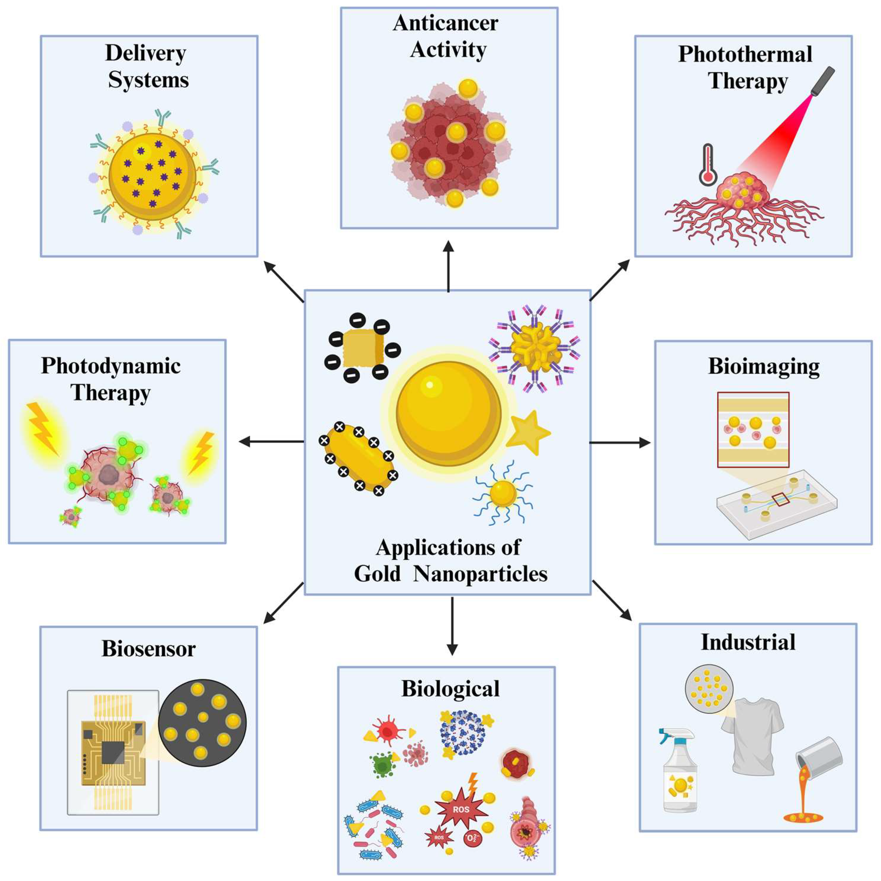
Figure 2.
Graph representing the published research papers that include “Gold Nanoparticles” in their title for the last 5 years, with a pie chart showing the distribution of applications based on the discussed sections [8].
Figure 2.
Graph representing the published research papers that include “Gold Nanoparticles” in their title for the last 5 years, with a pie chart showing the distribution of applications based on the discussed sections [8].
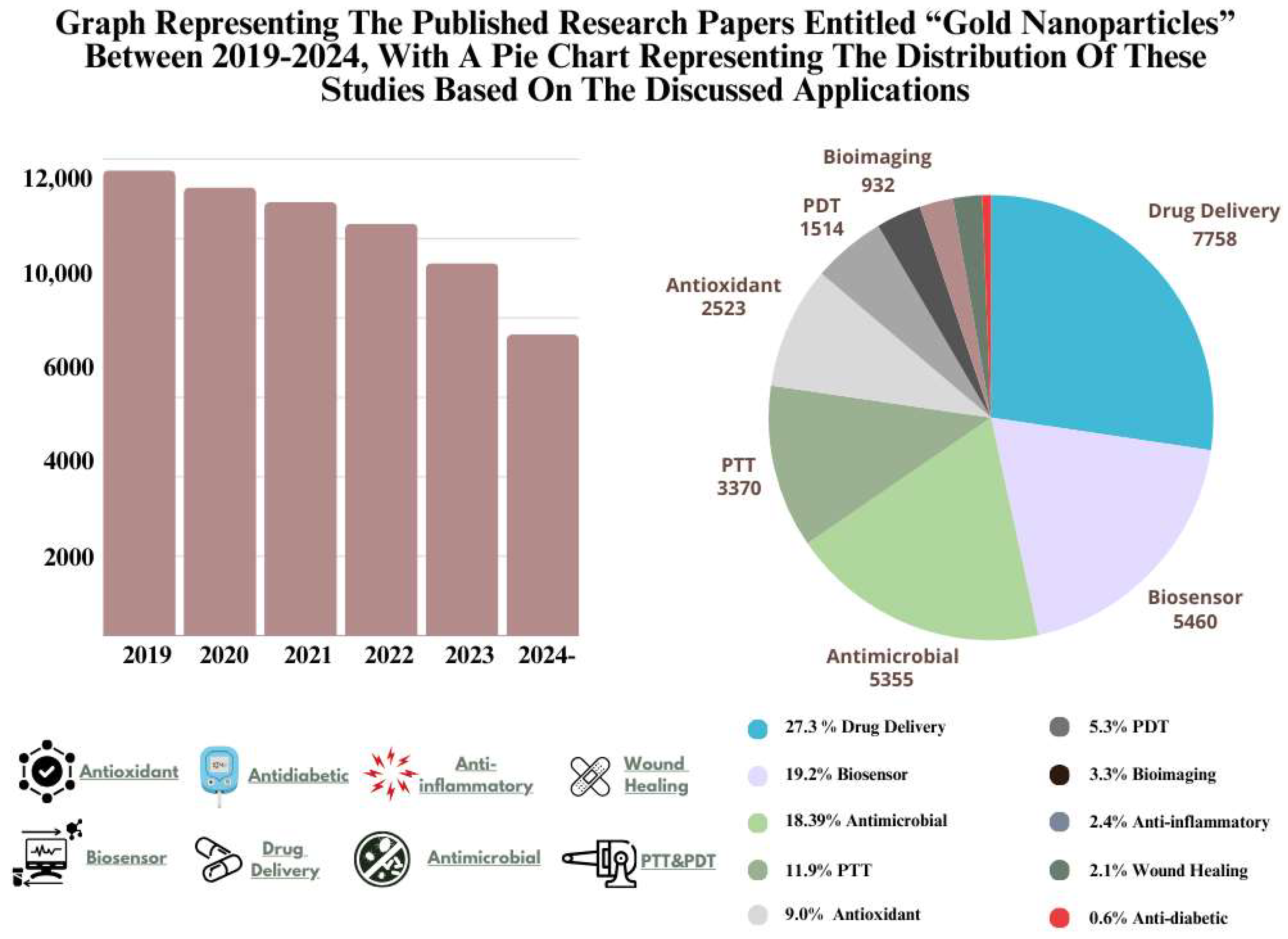
Figure 3.
Drug Delivery Application of Gold NPs [15].
Figure 3.
Drug Delivery Application of Gold NPs [15].
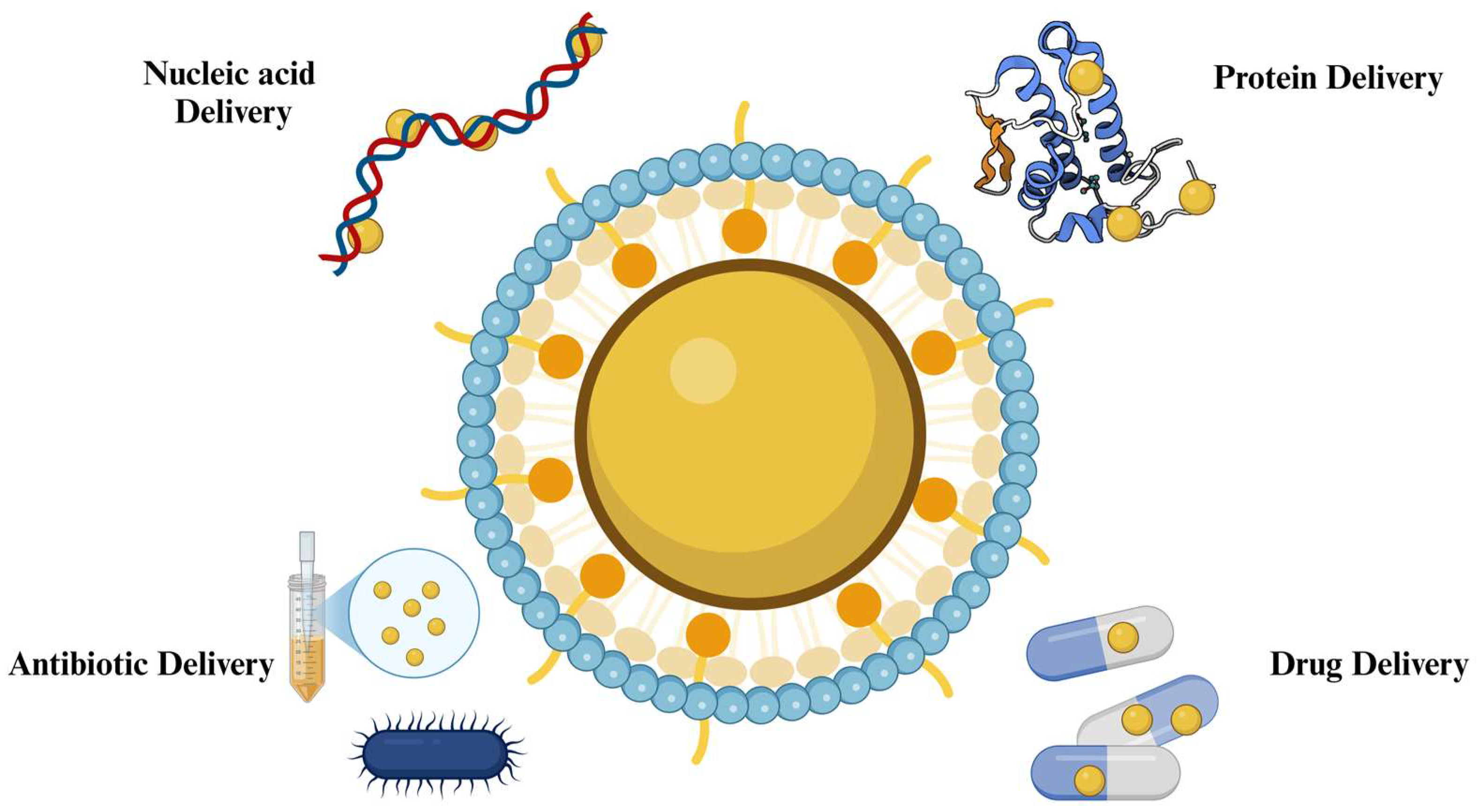
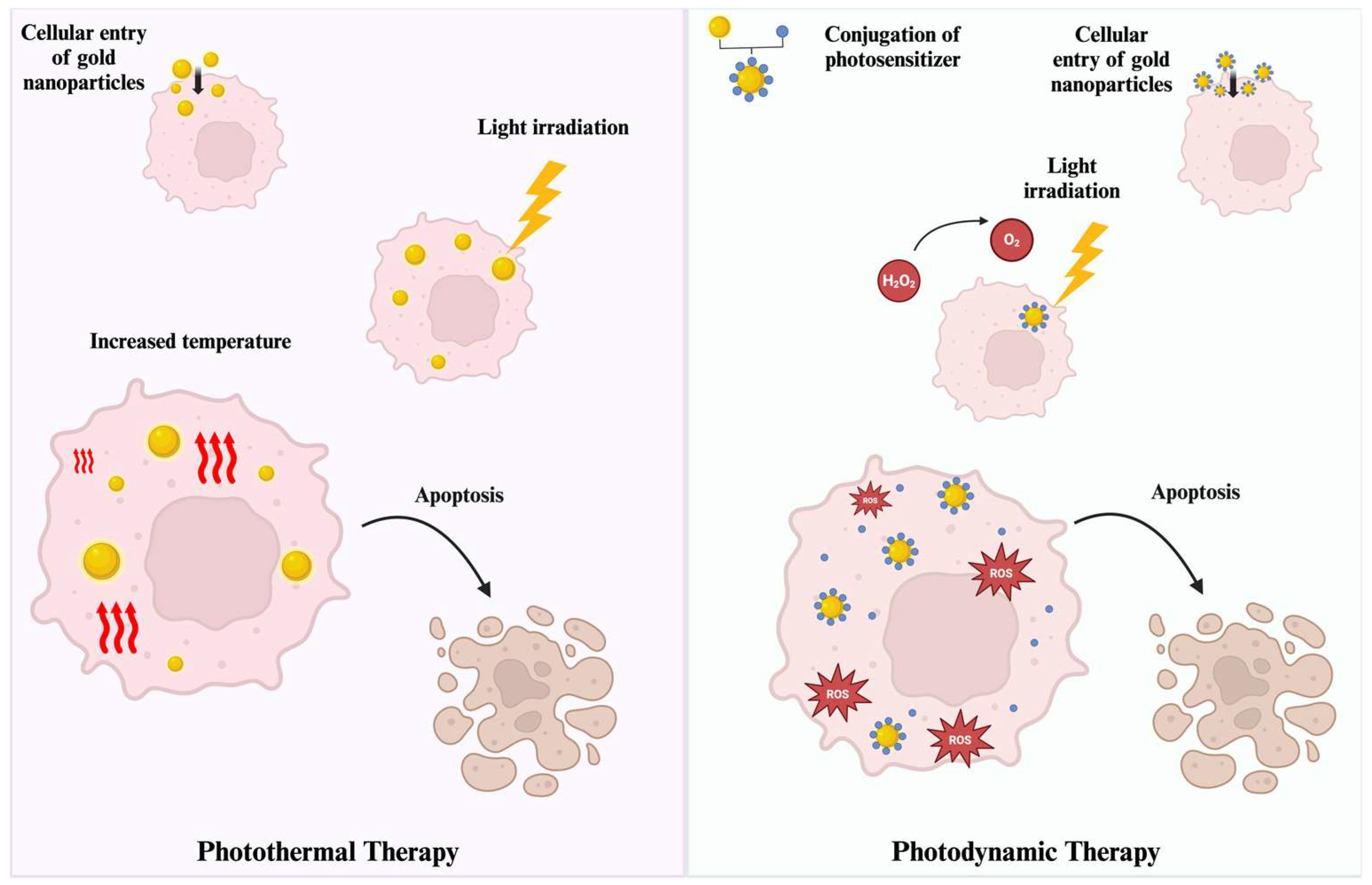
Figure 5.
Antibacterial activity of gold NPs by multiple mechanisms [186].
Figure 5.
Antibacterial activity of gold NPs by multiple mechanisms [186].
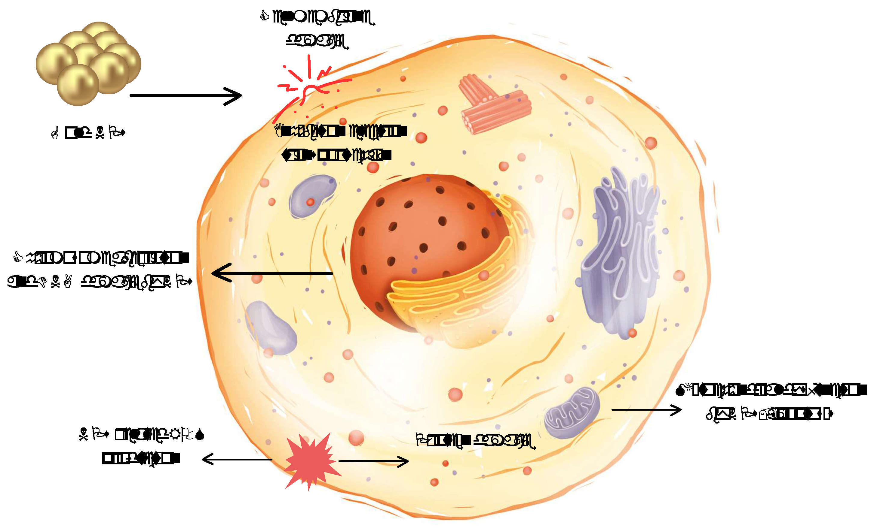
Figure 7.
Number of registered patents containing “Gold Nanoparticle” in their title in the last five years [244].
Figure 7.
Number of registered patents containing “Gold Nanoparticle” in their title in the last five years [244].
Disclaimer/Publisher’s Note: The statements, opinions and data contained in all publications are solely those of the individual author(s) and contributor(s) and not of MDPI and/or the editor(s). MDPI and/or the editor(s) disclaim responsibility for any injury to people or property resulting from any ideas, methods, instructions or products referred to in the content. |
© 2024 by the authors. Licensee MDPI, Basel, Switzerland. This article is an open access article distributed under the terms and conditions of the Creative Commons Attribution (CC BY) license (http://creativecommons.org/licenses/by/4.0/).
Copyright: This open access article is published under a Creative Commons CC BY 4.0 license, which permit the free download, distribution, and reuse, provided that the author and preprint are cited in any reuse.


