Preprint
Article

This version is not peer-reviewed.

Synthesis and Anti-Cancer Activity in Vitro of Synephrine Derivatives

A peer-reviewed article of this preprint also exists.

Submitted:

16 October 2024

Posted:

18 October 2024

You are already at the latest version

Abstract

Glucocorticoids (GC) are widely used in the treatment of hematological malignancies; however, the long-term treatment lead to atrophic and metabolic adverse effects. Selective glucocorticoid receptor agonists (SEGRA) with reduced side effects could be the better GC alternative. More than 30 SEGRA were described but none of them reached clinical trials as anticancer treatment. In the present work, we proposed the novel approach to broaden the list of potential SEGRA by synthesis of derivatives of synephrine, the molecule of natural origin. We synthesized 26 compounds from the class of synephrine derivatives, and studied their anticancer effect in vitro in leukemia and lymphoma cells with MTT assay as well as their potential affinity to glucocorticoid receptor (GR) in silico using molecular docking approach. Novel derivative 1-[4-(benzyloxy)phenyl]-2-(hexylamino)ethanol (10S-E2) with the highest GR affinity in silico revealed the micromolar range cytotoxicity in lymphoma and leukemia cells after 24 h of treatment. The other compound, 2-(hexylamino)-1-(4-nitrophenyl)ethanol (13S-G2) with high GR affinity demonstrated cytotoxicity in the range of 50-70 µM. Overall, our results provide the rationale for the development and further investigation of synephrine derivatives as SEGRA with anticancer activity.

Keywords: 
;  ;  ;  ;  

1. Introduction

Glucocorticoids (GCs) have been widely used in the therapy of autoimmune and inflammatory diseases (rheumatoid arthritis, psoriasis, atopic dermatitis, asthma) as well as cancer treatment for the last 75 years. Unfortunately, in most cases chronic GC application leads to the development of severe adverse effects: osteoporosis, steroid diabetes, water-salt imbalance, lipodystrophy and other metabolic complications [1,2]. Biological effects of GC are mediated via activation of glucocorticoid receptor (GR), well-known transcription factor regulating gene expression through two distinct mechanisms. Therapeutic effects of GC are realized by DNA-independent transrepression (TR), protein-protein interaction of GR and other transcription factors, leading to suppression of cancer cell survival. Side effects of GC is associated with transactivation (TA), requiring GR dimerization and dimer binding with glucocorticoid-responsive elements in the promoters of pro-inflammatory and anti-apoptotic genes [2,3,4].
The approaches to the increase of GC therapeutic activity and simultaneous decrease of the side effects include the development of selective glucocorticoid receptor agonists (SEGRAs) activating only GR TR [4,5,6]. In the last decades, more than 30 SEGRAs of natural and synthetic origin were described [4,7]. Specifically, we and others previously described that low molecular weight compound, 2-(4-acetoxyphenyl)-2-chloro-N-methylammonium chloride or CpdA, isolated from African shrub Salsola tuberculatiformis Botschantzev, acting as SEGRA: selectively induces the GR transrepression and reveals the anti-cancer effects in the models of blood and solid cancers in vitro and in vivo individually or in combination with anticancer drugs (Bortezomib, MLN4924 and some others) [4,8,9,10]. However, CpdA use in clinics is restricted by its chemical instability and decomposition to the intermediate metabolite aziridine, a well-known carcinogen [11,12]. Therefore, we extended SEGRA list by synthesis of 8 CpdA derivatives, and 4-(1-hydroxy-2-(piperidin-1-yl)ethyl)phenol or CpdA-03 demonstrated superior GR affinity and stability compared to CpdA as well as ant-lymphoma properties in vitro and in vivo [13].
In the presented work, we aimed to test another synthetic strategy for SEGRA development using as a template not CpdA but synephrine molecule. Synephrine is the final product of CpdA metabolism and it serves as precursor for CpdA synthesis as well [12,14,15,16]. Synephrine derivatives may have potential clinical benefits in comparison with the effects of GR ligands, selective glucocorticoid receptors agonist CpdA, which are structurally similar to synephrine. Firstly, we aimed to design the strategies of synthesis of novel synephrine derivatives as potential selective glucocorticoid receptor agonists (SEGRA), the derivatives of synephrine. The second aim of the study was to perform the screening of their biological activity in vitro and in silico with the selection of hit compounds for further investigation.

2. Materials and Methods

2.1. Chemistry

2.1.1. General Procedures and Reagents

All reagents were obtained from commercial sources (Merck KGaA, Darmstadt, Germany) and used without additional purification. Deuterated solvents were purchased from Cambridge Isotope Laboratories, Inc. (Tewkesbury, Massachusetts, USA). Silica gel 60 (Merck KGaA, Darmstadt, Germany) was used for column chromatography. Analytical TLC was performed on Sorbfil PTX-AF-A-UV silica gel plates (Russia).
Solvents were removed on a Buchi Rotavapor R-200 rotary vacuum evaporator using a vacuum water jet pump (Switzerland). 1Н and 13С NMR spectra were recorded on a Bruker DPX-300 instrument (300 and 75 MHz, respectively). Chemical shifts are in parts per million (ppm, δ) and are relative to the solvent CDCl3 (7.25 ppm), DMSO-d6 (2.49 ppm), CD3OD (4.78 ppm) for 1H NMR and CDCl3 (77.2 ppm), DMSO-d6 (39.5 ppm) and CD3OD (49.0 ppm) for 13C NMR. The data are presented as follows: chemical shifts, multiplicity (s – singlet, br.s – broad singlet, m – multiplet) and the relative value of integration. The determination of solvent peaks was carried out in accordance with the literature data [17]. The spectra could be find in Supplementary Figures S1-S52. High resolution mass spectra (HRMS) were recorded on Agilent 6224 using electron sputtering ionization (ESI). HPLC-MS measurements were performed on an Agilent InfinityLab LC/MSD iQ liquid chromatograph with a Agilent Single Quadrupole LC/MSD iQ mass spectrometric detector. The separation was performed on a Agilent Poroshell 300SB C18 column, 2.1×75 mm. Eluent: 0.1% acetonitrile in deionized water with 0.1% trifluoroacetic acid, consumption 0.5 ml per minute.

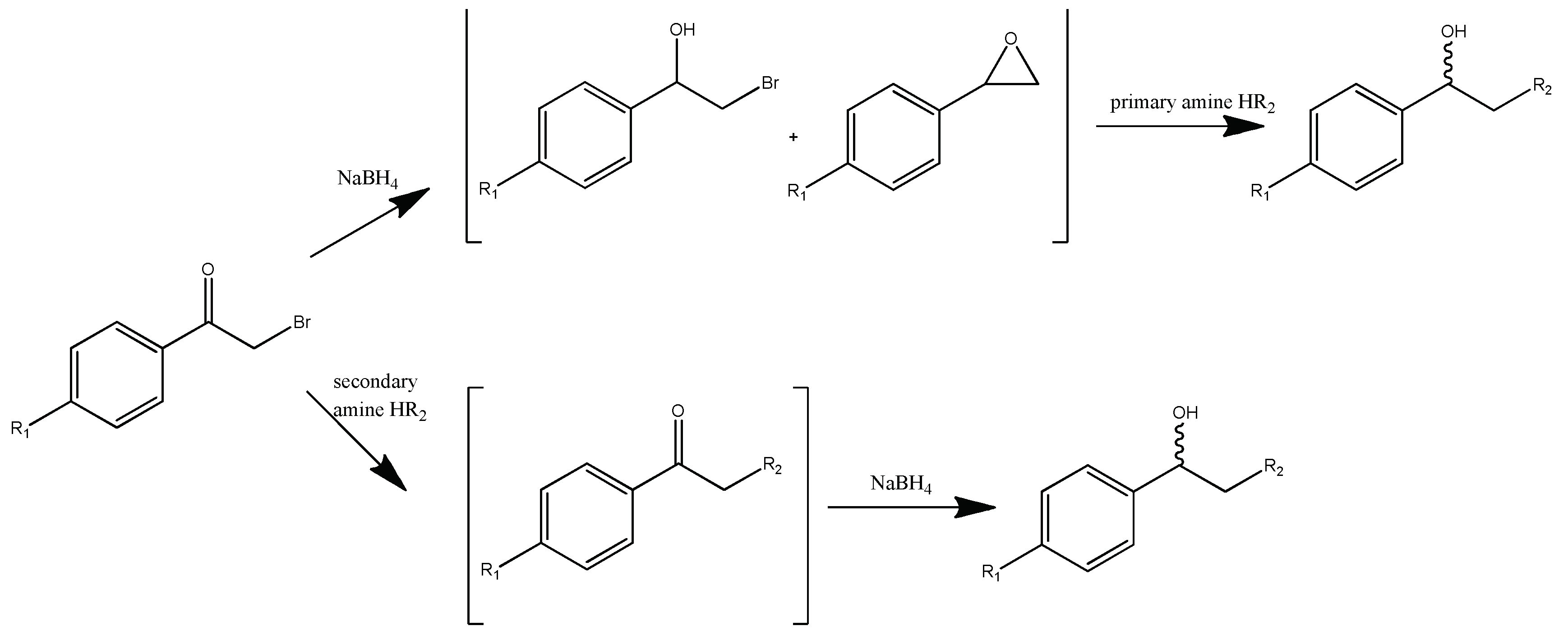
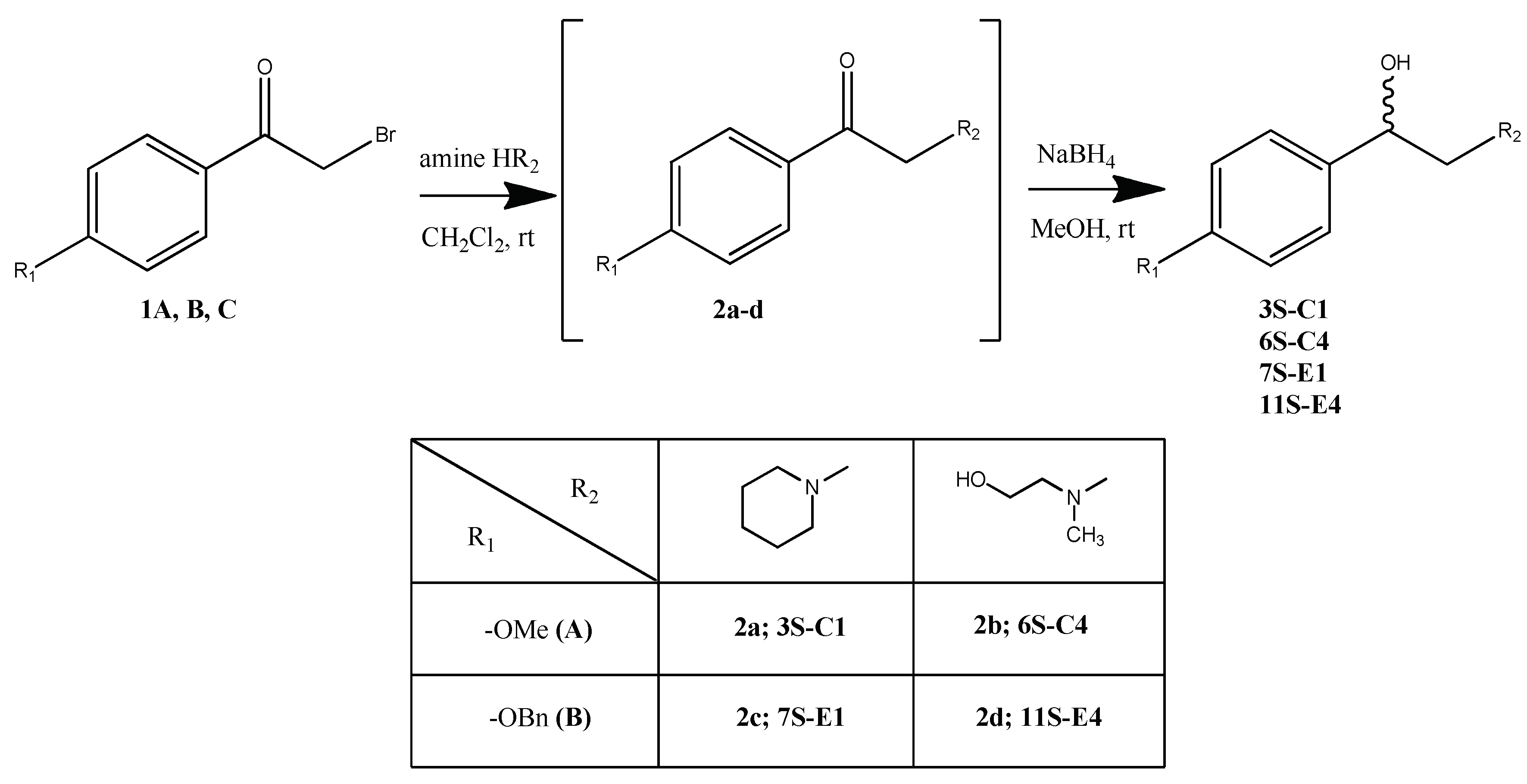
2.1.2. General Procedure for Compounds 3S-C1, 6S-C4, 7S-E1, 11S-E4 Synthesis

A solution of bromoacetophenone 1 in methylene chloride (1 ml of solvent per 100 mg 1) were added drop by drop to a solution of amine in methylene chloride (10 ml of solvent per 1 ml of amine). The reaction was controlled by the initial bromoacetophenone 1 conversion by TLC. After bromoacetophenone 1 conversion was complete, volatile components were evaporated. The residue was dissolved in 5 ml of water and extracted with methylene chloride (3 x 10 ml). The organic layers are combined and dried with anhydrous calcium chloride, filtered off and evaporated. The resulting oil was dissolved in 10 ml of methanol, after which NaBH4 was added in portions during cooling. After intermediate conversion (control by TLC), the reaction mixture was acidified with 10% aqueous HCl solution to pH 3, the volatile components was evaporated. The extraction and drying process was repeated. The products 3S-C1, 6S-C4, 7S-E1, 11S-E4 were isolated by column chromatography (methanol gradient from 0 to 15%).
1-(4-methoxyphenyl)-2-piperedine-1-yl ethanol (3S-C1)
From 0.63 ml (6.30 mmol) piperedine, 0.42 g (1.80 mmol) of 4-methoxybromoacetophenone , 0.07 g (1.80 mmol) NaBH4 the product 3S-C1 0.31 g (74%) was obtained. Rf = 0.45 (10% methanol in chloroform). The content of the target substance according to HPLC data is 97.5%. 1H NMR spectrum (CDCl3) δ: 7.31-7.28 (m, 2H, Ph); 6.83-6.80 (m, 2H, Ph); 5.33-5.30 (m, 1H, -CH); 4.60 (br. s, 1H, -OH); 3.76-3.69 (m, 2H,-CH2-OH); 3.74 (s, 1H, -O-CH3); 3.19-2.99 (m, 2H, piperedine -CH2-); 2.83-2.76 (m, 2H, piperedine -CH2-); 2.29-2.16 (m, 2H, piperedine -CH2-); 1.83-1.77 (m, 3H, piperedine 2×-CH2-); 1.47-1.37 (m, 1H, piperedine -CH2-). 13C NMR spectrum (CDCl3) δ: 159.42; 132.18; 127.15; 114.03; 67.43; 65.52; 55.51; 55.26; 54.04; 22.72; 22.67; 21.80. For C14H21NO2 [M+H]+ calculated: 236.1650, found: 236.1652.
2-(2-hydroxyethyl)(methyl) amino)-1-(4-methoxyphenyl)ethanol (6S-C4)
From 0.55 ml (7.00 mmol) N-methyl ethanolamine, 0.45 g (2.00 mmol) of 4-methoxybromoacetophenone , 0.08 g (2.00 mmol) NaBH4 the product 6S-C4 0.20 g (45%) was obtained. Rf = 0.75 (5% methanol in chloroform). The content of the target substance according to HPLC data is 99%. 1H NMR spectrum (CDCl3) δ: 7.28-7.25 (m, 2H, Ph); 6.87-6.84 (m, 2H, Ph); 4.53-4.49 (m, 1H, -CH-); 3.99-3.84 (m, 2H, -CH2-OH); 3.77 (s, 3H,-O-CH3); 2.88-2.72 (m, 2H, -N(CH3)-CH2-); 2.32 (s, 3H, -N-CH3); 2.28-2.06 (m, 2H, -CH(OH)-CH2-). 13C NMR-spectrum (CDCl3) δ: 159.11; 132.16; 127.34; 113.61; 77.48; 66.74; 61.86; 55.15; 54.46; 45.92. For C12H19NO3 [M+H]+ calculated: 226.1443, found: 226.1445.
1-(4-(benzyloxy) phenyl)-2-piperidine-1-yl-ethanol (7S-E1)
From 0.63 ml (6.35 mmol) piperidine, 0.39 g (1.27 mmol) 4-benzyloxybromoacetophenone and 0.05 g (1.27 mmol) NaBH4 the product 7S-E1 0.30 g (76%) was obtained. Rf = 0.45 (5% methanol in chloroform). The content of the target substance according to HPLC data is 98%. 1H NMR spectrum (CDCl3) δ: 7.43-7.27 (m, 7H, Ph); 6.96-6.93 (m, 2H, Ph); 5.04 (s, 2H, -CH2-O-); 4.91-4.86 (m, 1H, -CH-OH); 4.62 (br.s., 1H, -OH); 2.86 (br.s., 2H, -CH2-); 2.71-2.59 (m, 4H, piperidine 2×CH2); 1.78-1.74 (m, 4H, piperidine 2×CH2); 1.56-1.50 (m, 2H, piperidine CH2). 13C NMR spectrum (CDCl3) δ: 158.31; 136.89; 133.80; 128.54; 127.42; 127.13; 114.77; 69.96; 67.96; 66.57; 54.62; 24.94; 23.42. For C20H25NO2 [M+H]+ calculated: 312.1963, found: 312.1965.
1-(4-(benzyloxy)phenyl)-2-((2-hydroxyethyl)(methyl)amino)ethanol (11S-E4)
From 0.39 ml (4.9 mmol) N-methyl ethanolamine, 0.30 g (0.98 mmol) of 4-benzyloxybromoacetophenone and 0.04 g (0.98 mmol) NaBH4 the product 11S-E4 0.18 g (61%) was obtained. Rf = 0.55 (7% methanol in chloroform). The content of the target substance according to HPLC data is 98%.1H NMR spectrum (CDCl3) δ: 7.43-7.27 (m, 7H, Ph); 6.96-6.93 (m, 2H, Ph); 5.04 (s, 2H, -O-CH2-); 4.91-4.86 (m, 1H, -CH(OH)-); 4.63 (br.s., 1H, -OH); 2.87 (br.s., 2H, -CH(OH)-CH2-) 2.71-2.59 (m, 2H, -N(СН3)-CH2-); 1.76 (s, 3H, -N-CH3); 1.54-1.52 (m, 2H, -CH2-OH). 13C NMR spectrum (CDCl3) δ: 158.93; 136.88; 130.18; 128.60; 128.02; 127.44; 123.29; 114.94; 70.15; 68.97; 61.42; 58.63; 55.24; 37.62. For C18H23NO3 [M+H]+ calculated: 302.1756, found: 302.1760.

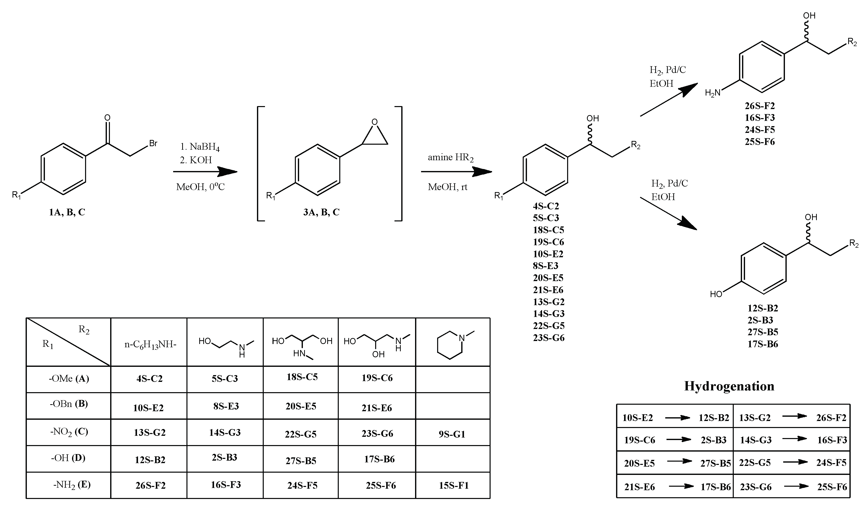
2.1.3. General procedure for compounds 4S-C2, 5S-C3, 18S-C5, 19S-C6, 10S-E2, 8S-E3, 20S-E5, 21S-E6, 13S-G2, 14S-G3, 22S-G5, 23S-G6, 9S-G1 synthesis

To a solution of bromoacetophenone 1 in methanol (for 4S-C2, 5S-C3, 18S-C5, 19S-C6, 10S-E2, 8S-E3, 20S-E5, 21S-E6) or 1,4-dioxane (for 13S-G2, 14S-G3, 22S-G5, 23S-G6, 9S-G1) (1 ml per 100 mg) while cooling in an ice bath (in the case of compounds 9S-G1 obtaining, the reaction was carried out at room temperature), 1 eq NaBH4 was added in portions. After the bromoacetophenone 1 was converted (control by TLC), a solution of 5 eq of primary amine and 1.2 eq КОН in methanol (1 ml per 1 ml amine) was added. After 12 h stirring, the reaction mixture was acidified with 10% aqueous solution of HCl to pH 3, the volatile components was removed on a vacuum rotary evaporator, the residue was suspended in 5 ml of water and extracted with methylene chloride (3 x 10 ml). The organic layers were combined and dried with anhydrous calcium chloride, filtered off and evaporated. The product was isolated by column chromatography on silica gel in a chloroform-methanol solvent system (methanol gradient from 0 to 20%).
2-(hexylamino)-1-(4-methoxyphenyl)ethanol (4S-C2f)
From 0.5 g (2.70 mmol) 4-methoxybromoacetophenone 1A, 0.1 g (2.70 mmol) NaBH4, 1.75 ml (13.5 mmol) amine and 0.19 g (3.30 mmol) KOH obtained 0.37 g (54%) of product 4S-C2. Rf (10% methanol in chloroform) = 0.45. The content of the target substance according to HPLC data is 98%. 1H NMR spectrum (CDCl3) δ: 7.30-7.27 (m, 2H, Ph); 6.87-6.84 (m, 2H, Ph); 4.86-4.82 (m, 1H, -CH); 3.82 (br.s, 1H, -OH); 3.78 (s, 3H, O-CH3); 2.94-2.67 (m, 4H, 2×CH2); 1.59-1.52 (m, 2H, -CH2-); 1.29-1.23 (m, 6H, 3×CH2); 0.88-0.84 (m, 3H, -CH3). 13C NMR spectrum (CDCl3) δ: 159.13; 133.70; 127.06; 113.80; 70.24; 56.30; 55.24; 49.11; 31.47; 28.39; 26.67; 22.51; 13.99. For C15H25NO2 [M+H]+ calculated: 252.1963, found: 252.1966.
2-((2-hydroxyethyl)amino)-1-(4-methoxyphenyl)ethanol (5S-C3)
From 0.5 g (2.70 mmol) 4-methoxybromoacetophenone 1A, 0.1 g (2.70 mmol) NaBH4, 0.82 ml (13.5 mmol) ethanolamine and 0.19 g (3.30 mmol) KOH obtained 0.33 g (58%) of product 5S-C3. Product Rf = 0.40 (20% methanol in chloroform). The content of the target substance according to HPLC data is 99%. 1H NMR spectrum (DMSO-d6) δ: 7.44-7.41 (m, 2H, Ph); 6.96-6.93 (m, 2H, Ph); 5.40 (br.s., 1H, -CH2-OH-); 5.02 (br.s., 1H, -CH-OH-); 4.11-4.07 (m, 1H, -CH-); 3.74 (s, 3H, -O-CH3); 3.71-3.32 (m, 4H, -CH2-CH2-OH); 2.75-2.56 (m, 2H, -CH2-NH-). 13C NMR spectrum (DMSO-d6) δ: 159.30; 129.63; 127.28; 113.96; 62.92; 57.21; 55.11; 47.79. For C11H17NO3 [M+H]+ calculated: 212.1286, found: 212.1290.
2-((2-hydroxy-2-(4-methoxyphenyl)ethyl)amino)propane-1,3-diol (18S-C5)
From 0.5 g (2.20 mmol) 4-methoxybromoacetophenone 1A, 0.08 g (2.20 mmol) NaBH4, 1.0 g (11.0 mmol) 2-aminopropane-1,3-diol and 0.15 g (2.60 mmol) KOH obtained 0.07 g (13%) of product 18S-C5. Product Rf = 0.55 (15% methanol in chloroform). The content of the target substance according to HPLC data is 96%. 1H NMR spectrum (DMSO-d6) δ: 7.26-7.21 (m, 2H, Ph); 6.87-6.82 (m, 2H, Ph); 4.72 (br.s., 1H, -CH-OH); 4.31 (br.s., 1H, -CH2-OH); 4.21 (br.s., 1H, -CH-OH); 3.78-2.74 (m, 1H, -CH-NH-); 3.71 (s, 3H, -O-CH3); 3.42-3.23 (m, 5H, -CH2-NH- and -CH-(CH2-OH)2); 2.36-2.31 (m, 1H, -CH2-NH-). 13C NMR spectrum (DMSO-d6) δ: 158.17; 134.47; 128.42; 113.43; 66.90; 61.89; 61.35; 57.69; 54.93. For C12H20NO4 [M+H]+ calculated: 242.1392, found: 242.1395.
3-((2-hydroxy-2-(4-methoxyphenyl)ethyl)amino)propane-1,2-diol (19S-C6)
From 0.5 g (2.20 mmol) 4-methoxybromoacetophenone 1A, 0.08 g (2.20 mmol) NaBH4, 0.85 ml (11.0 mmol) 3-aminopropane-1,2-diol and 0.15 g (2.60 mmol) KOH obtained 0.05 g (9%) of product 19S-C6. Product Rf = 0.46 (13% methanol in chloroform). The content of the target substance according to HPLC data is 98%. 1H NMR spectrum (CD3OD) δ: 7.27-7.22 (m, 2H, Ph); 6.90-6.86 (m, 2H, Ph); 3.76 (s, 3H, -O-CH3); 3.61-3.43 (m, 4H, -CH2-CH(OH)-CH2-OH); 3.30-3.28 (m, 1H, -CH(OH)-CH2-OH); 2.65-2.40 (m, 2H, -CH2-NH-). 13C NMR spectrum (CD3OD) δ: 160.78; 132.86; 132.76; 129.92; 129.89; 115.06; 115.04; 67.41; 67.27; 66.26; 66.13; 66.03; 65.07; 55.73; 51.53; 50.75. For C12H20NO4 [M+H]+ calculated: 242.1392, found: 242.1394.
1-(4-(benzyloxy)phenyl)-2(hexylamino)ethanol (10S-E2)
From 0.5 g (1.92 mmol) of 4-benzyloxybromoacetophenone , 0.07 g (1.92 mmol) NaBH4, 1.25 ml (9.6 mmol) hexylamine and 0.17 g (3.0 mmol) KOH obtained 0.17 g (27%) of product 10S-E2. Rf = 0.55 (20% methanol in chloroform). The content of the target substance according to HPLC data is 98%. 1H NMR spectrum (CDCl3) δ: 7.43-7.33 (m, 7H, Ph); 6.94-6.92 (m, 2H, Ph); 5.38 (br. s, 1H, -CH-OH-); 5.03 (s, 2H, -CH2-O-); 4.23 (br.s., 1H, -OH); 3.16-3.03 (m, 4H, -CH2-NH-CH2); 1.90 (br.s., 2H, -CH2-(CH2)3-CH3); 1.35-1.28 (m, 6H, 3×CH2); 0.89-0.85 (m, 3H, -CH3). 13C NMR spectrum (CDCl3) δ: 158.66; 136.76; 132.37; 128.56; 127.96; 127.41; 127.18; 69.97; 68.69; 55.11; 48.65; 31.13; 26.36; 25.82; 22.38; 13.92. For C21H29NO2 [M+H]+ calculated: 328.2277, found: 328.2281.
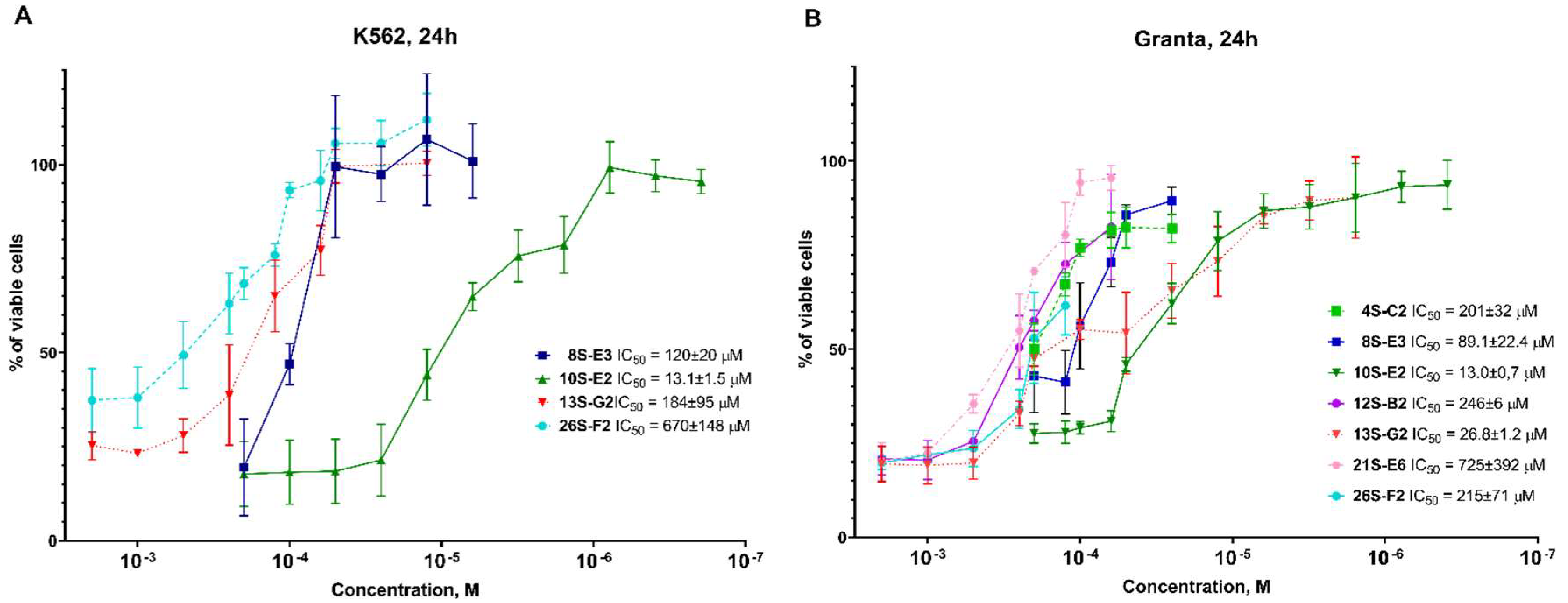
1-(4-(benzyloxy)phenyl)-2-((2-hydroxyethyl)amino)ethanol (8S-E3)
From 0.65 g (2.50 mmol) of 4-benzyloxybromoacetophenone , 0.09 g (2.50 mmol) NaBH4, 0.75 ml (12.5 mmol) ethanolamine, and 0.17 g (3.0 mmol) KOH obtained 0.16 g (22%) of product 8S-E3. Rf = 0.35 (20% methanol in chloroform). The content of the target substance according to HPLC data is 98%. 1H NMR spectrum (DMSO-d6) δ: 7.45-7.29 (m, 7H, Ph); 7.03-7.00 (m, 2H, Ph); 5.27 (br.s, 1H, -CH-OH); 5.09 (s, 2H, -O-CH2-); 4.89 (br.s, 1H, -CH2-OH); 4.04-4.00 (m, 1H, -CH-); 3.64-3.55 (m, 4H, -CH2-CH2-OH); 2.75-2.54 (m, 2H, -CH2-CH(OH)). 13C NMR spectrum (DMSO-d6) δ: 158.28; 135.95; 129.42; 128.37; 127.78; 127.59; 114.73; 88.84; 69.18; 69.46; 63.00; 57.65; 48.01. For C17H21NO3 [M+H]+ calculated: 288.1599, found: 288.1602.
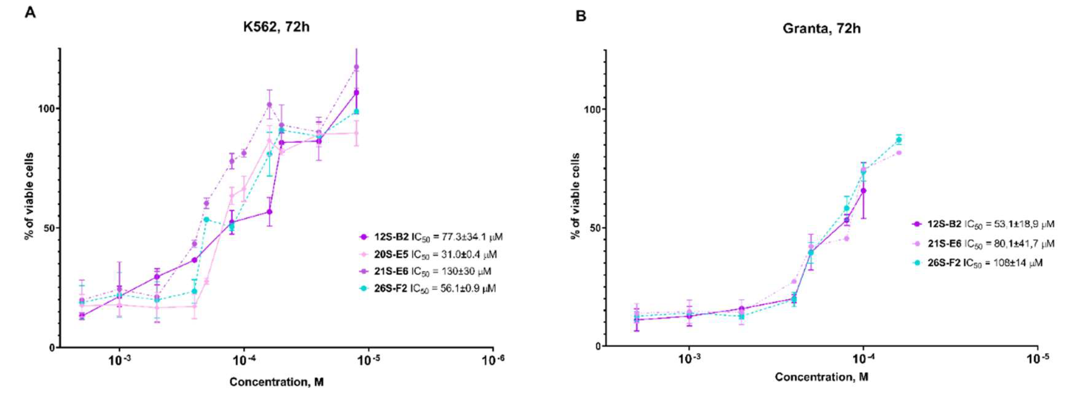
2-((2-(4-(benzyloxy)phenyl)-2-hydroxyethyl)amino)propane-1,3-diol (20S-E5)
From 1.0 g (3.30 mmol) of 4-benzyloxybromoacetophenone , 0.12 g (3.30 mmol) NaBH4, 1.5 g (16.5 mmol) 2-aminopropane-1,3-diol, and 0.22 g (4.0 mmol) KOH obtained 0.27 g (27%) of product 20S-E5. Rf = 0.67 (13% methanol in chloroform). The content of the target substance according to HPLC data is 98%. 1H NMR spectrum (DMSO-d6) δ: 7.45-7.35 (m, 5H, Ph); 7.25-7.22 (m, 2H, Ph); 6.96-6.91 (m, 2H, Ph); 5.18 (br.s., 1H, -CH-OH); 5.07 (s., 2H, -O-CH2-); 4.53-4.49 (m., 1H, - CH-OH); 4.44-4.26 (m, 2H, 2×-CH2-OH-); 3.44-3.24 (m, 5H, -CH-(-CH2-OH)2); 2.67-2.51 (m, 2H, -CH2-NH-). 13C NMR spectrum (DMSO-d6) δ: 157.25; 137.24; 136.87; 128.43; 127.77; 127.63; 127.07; 114.25; 71.61; 69.12; 61.36; 61.24; 61.08; 55.64. For C18H24NO4 [M+H]+ calculated: 318.1705, found: 318.1707.
3-((2-(4-(benzyloxy)phenyl)-2-hydroxyethyl)amino)propane-1,2-diol (21S-E6)
From 1.0 g (3.30 mmol) of 4-benzyloxybromoacetophenone , 0.12 g (3.30 mmol) NaBH4, 1.27 ml (16.5 mmol) 3-aminopropane-1,2-diol, and 0.22 g (4.0 mmol) KOH obtained 0.12 g (12%) of product 21S-E6. Rf = 0.75 (15% methanol in chloroform). The content of the target substance according to HPLC data is 98%. 1H NMR spectrum (DMSO-d6) δ: 7.45-7.29 (m, 5H, Ph); 7.25-7.20 (m, 2H, Ph); 6.95-6.92 (m, 2H, Ph); 5.05 (s, 2H, -O-CH2-); 4.58-4.56 (m, 1H, -CH-OH); 4.34 (br.s, 4H, -CH(OH)-CH2-OH and HO-CH-CH2-NH-); 3.58-3.18 (m, 5H, -CH2-CH(OH)-CH2-OH). 13C NMR spectrum (DMSO-d6) δ: 157.38; 157.33; 137.24; 137.21; 134.11; 134.06; 128.40; 128.36; 127.71; 127.58; 127.54; 127.01; 126.98; 114.39; 71.01; 70.15; 69.14; 66.75; 66.60; 64.76; 64.48; 63.89; 51.15; 50.35. For C18H24NO4 [M+H]+ calculated: 318.1705, found: 318.1708.
2-(hexylamino)-1-(4-nitrophenyl)ethanol (13S-G2)
From 0.85 g (3.50 mmol) of 4-nitrobromoacetophenone , 0.13 g (3.50 mmol) NaBH4, 2.30 ml (17.5 mmol) hexylamine, and 0.23 g (4.2 mmol) KOH obtained 0.32 g (34%) of product 13S-G2. Rf = 0.61 (15% methanol in chloroform). The content of the target substance according to HPLC data is 98%. 1H NMR spectrum (DMSO-d6) δ: 8.18-8.16 (m, 2H, Ph); 7.62-7.59 (m, 2H, Ph); 5.58 (br.s, 1H,-OH); 4.77-4.73 (m, 1H, -CH); 2.69-2.53 (m, 4H, -CH2-NH-CH2-); 1.40-1.35 (m, 2H, -NH-CH2-CH2-); 1.26-1.22 (m, 6H, -(CH2)3-CH3); 0.86-0.81 (m, 3H, -CH3).13C NMR spectrum (DMSO-d6) δ: 152.80; 146.37; 127.11; 123.08; 70.78; 57.20; 49.01; 31.25; 29.53; 26.48; 22.12; 13.94. For C14H23N2O3 [M+H]+ calculated: 267.1709, found: 267.1712.
2-((2-hydroxyethyl)amino)-1-(4-nitrophenyl)ethanol (14S-G3)
From 1.25 g (5.14 mmol) of 4-nitrobromoacetophenone , 0.19 g (5.14 mmol) NaBH4, 1.55 ml (25.7 mmol) ethanolamine, and 0.34 g (6.17 mmol) KOH obtained 0.41 g (35%) of product 14S-G3. Rf = 0.55 (13% methanol in chloroform). The content of the target substance according to HPLC data is 95%. 1H NMR spectrum (DMSO-d6) δ: 8.19-8.16 (m, 2H, Ph); 7.63-7.60 (m, 2H, Ph); 5.61 (br.s, 1H, -CH-OH); 4.78-4.74 (m, 1H, -CH-OH); 4.49 (br.s, 1H, -CH2-OH); 3.45-3.37 (m, 2H, -CH2- OH); 2.72-2.53 (m, 4H, -CH2-NH-CH2-). 13C NMR spectrum (DMSO-d6) δ: 152.76; 146.39; 127.12; 123.11; 70.96; 60.42; 57.15; 51.45. For C10H15N2O4 [M+H]+ calculated: 227.1032, found: 227.1034.
2-((2-hydroxy-2-(4-nitrophenyl)ethyl)amino)propane-1,3-diol (22S-G5)
From 1.7 g (7.00 mmol) of 4-nitrobromoacetophenone , 0.265 g (7.00 mmol) NaBH4, 3.18 g (35.0 mmol) 2-aminopropane-1,3-diol, and 0.47 g (8.4 mmol) KOH obtained 0.72 g (40%) of product 22S-G5. Rf = 0.58 (10% methanol in chloroform). The content of the target substance according to HPLC data is 98%. 1H NMR spectrum (DMSO-d6) δ: 8.18-8.15 (m, 2H, Ph); 7.64-7.61 (m, 2H, Ph); 5.51 (br.s., 1H, -CH-OH); 4.74-4.72 (m, 1H, -CH-OH); 4.31-4.27 (m, 2H, 2×-CH2-OH); 3.43-3.24 (m, 4H, 2×-CH2-OH-); 2.84-2.64 (m, 2H, -CH2-NH); 2.55-2.51 (m, 1H, -CH-NH-). 13C NMR spectrum (DMSO-d6) δ: 152.69; 146.39; 127.13; 123.13; 71.39; 61.39; 61.23; 60.98; 55.10. For C11H17N2O5 [M+H]+ calculated: 257.1137, found: 257.1140.
3-((2-hydroxy-2-(4-nitrophenyl)ethyl)amino)propane-1,2-diol (23S-G6)
From 1.7 g (7.00 mmol) of 4-nitrobromoacetophenone , 0.265 g (7.00 mmol) NaBH4, 2.7 ml (35.0 mmol) 3-aminopropane-1,2-diol, and 0.47 g (8.4 mmol) KOH obtained 0.52 g (29%) of product 23S-G6. Rf = 0.49 (10% methanol in chloroform). The content of the target substance according to HPLC data is 98%. 1H NMR spectrum (DMSO-d6) δ: 8.19-8.16 (m, 2H, Ph); 7.63-7.60 (m, 2H, Ph); 5.62 (br.s, 1H, -CH-OH); 4.77-4.57 (m, 1H, -CH-OH); 4.57 (br.s, 1H, -CH2-OH); 3.49 (br.s, 1H, -CH-OH); 3.34-3.23 (m, 3H, -CH(OH)-CH2-OH); 2.72-2.52 (m, 3H, -CH2-NH-CH2-); 2.46-2.38 (m, 1H, -CH2-NH). 13C NMR spectrum (DMSO-d6) δ: 152.73; 146.41; 127.11; 123.13; 71.08; 70.68; 64.54; 57.48; 52.73. For C11H17N2O5 [M+H]+ calculated: 257.1137, found: 257.1141.
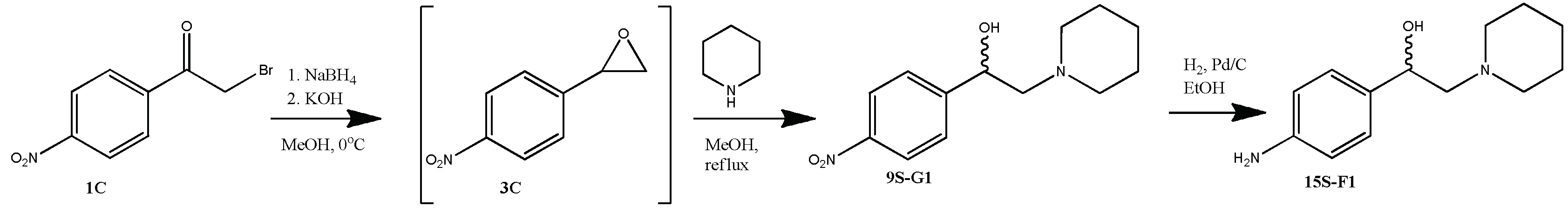
1-(4-nitrophenyl)-2-piperidin-1-yl ethanol (9S-G1)
From 0.5 g (2.05 mmol) of 4-nitrobromoacetophenone , 0.08 g (2.05 mmol) NaBH4, 1.0 ml (10.2 mmol) piperedine and 0.14 g (2.46 mmol) KOH obtained 0.20 g (40%) of product 9S-G1. Rf = 0.67 (5% methanol in chloroform). The content of the target substance according to HPLC data is 97%. 1H NMR spectrum (CDCl3) δ: 8.19-8.16 (m, 2H, Ph); 7.57-7.55 (m, 2H, Ph); 5.05-5.00 (m, 1H, -CH-); 4.40 (br.s., 1H, -OH); 2.91-2.84 (m, 2H, -CH2-CH-); 2.70-2.49 (m, 4H, piperidine 2×CH2); 1.78-1.70 (m, 4H, piperidine 4×CH2); 1.54-1.50 (m, 2H, piperidine CH2). 13C NMR spectrum (CDCl3) δ: 149.41; 147.30; 126.53; 123.65; 67.71; 65.99; 54.63; 25.11; 23.45. For C13H18N2O3 [M+H]+ calculated: 251.1396, found: 251.1398.

2.1.3. General Procedure for Compounds 12S-B2, 2S-B3, 27S-B5, 17S-B6 26S-F2, 16S-F3, 24S-F5, 25S-F6, 15S-F1 Synthesis

10% Pd on carbon (0.05 eq of Pd) was added to a solution 12S-B2, 2S-B3, 27S-B5, 17S-B6 26S-F2, 16S-F3, 24S-F5, 25S-F6, 15S-F1 in ethanol and mixed in an atmosphere of H2 at room temperature overnight. The reaction mass was filtered through a layer of celite, which was washed with 10-15 ml ethanol, and combined organic solutions were evaporated. The product was isolated by column chromatography on silica gel in a chloroform-methanol solvent system (methanol gradient from 0 to 35%).
4-(2-(hexylamino)-1-hydroxyethyl)phenol (12S-B2)
From 0.10 g (0.3 mmol) 10S-E2 0.03 g (41%) of the product 12S-B2 was obtained. Rf = 0.34 (20% methanol in chloroform). The content of the target substance according to HPLC data is 97%. 1H NMR spectrum (DMSO-d6) δ: 7.16-7.12 (m, 2H, Ph); 6.76-6.71 (m, 2H, Ph); 4.73-4.68 (m, 1H, -CH); 3.86-2.71 (m, 4H, -CH2-NH-CH2-); 1.58-1.48 (m, 2H, -NH-CH2-CH2-); 1.30-1.25 (m, 6H, -(CH2)3-CH3); 1.29-0.87-0.84 (m, 3H, -CH3). 13C NMR spectrum (DMSO-d6) δ: 156.75; 133.09; 127.06; 114.91; 69.18; 55.24; 47.78; 30.93; 26.88; 26.02; 21.98; 13.90. For C14H24NO2 [M+H]+ calculated: 238.1807, found: 238.1810.
4-(1-hydroxy-2-((2-hydroxyethyl)amino)ethyl)phenol (2S-B3)
From 0.1 g (0.35 mmol) 19S-C6 0.05 g (73%) of the product 2S-B3 was obtained. Rf = 0.35 (30% methanol in chloroform). The content of the target substance according to HPLC data is 97%.1H NMR spectrum (CD3OD) δ: 7.36-7.33 (m, 2H, Ph); 6.87-6.85 (m, 2H, Ph); 5.01 (br.s, 2H, 2 × OH); 4.33-4.28 (m, 1H, -CH-OH); 3.99-3.86 (m, 2H, -CH2-OH); 3.83-3.69 (m, 2H, -CH2-NH-); 3.05-2.91 (m, 2H, -CH2-CH-); 2.15 (s, 1H, -OH). 13C NMR spectrum (CD3OD) δ: 159.90; 130.97; 124.11; 117.02; 64.74; 63.39; 57.78; 30.76. For C10H15NO3 [M+H]+ calculated: 198.1130, found: 198.1134.
2-((2-hydroxy-2-(4-hydroxyphenyl)ethyl)amino)propane-1,3-diol (27S-B5)
From 0.17 g (0.5 mmol) 20S-E5 0.03 g (25%) of the product 27S-B5 was obtained. Rf = 0.23 (15% methanol in chloroform). The content of the target substance according to HPLC data is 96%. 1H NMR spectrum (DMSO-d6) δ: 7.13-7.09 (m, 2H, Ph); 6.70-6.65 (m, 2H, Ph); 4.47-4.43 (m, 1H, -CH-OH); 3.44-3.20 (m, 5H, -CH-(-CH2-OH)2); 2.69-2.52 (m, 2H, -CH2-NH). 13C NMR spectrum (DMSO-d6) δ: 156.14; 134.72; 126.96; 114.62; 71.67; 61.29; 61.17; 60.98; 55.51. For C11H18NO4 [M+H]+ calculated: 228.1235, found: 228.1239.
3-((2-hydroxy-2-(4-hydroxyphenyl)ethyl)amino)propane-1,2-diol (17S-B6)
From 0.1 g (0.3 mmol) 21S-E6 0.03 g (42%) of the product 17S-B6 was obtained. Rf = 0.37 (20% methanol in chloroform). The content of the target substance according to HPLC data is 98%. 1H NMR spectrum (DMSO-d6) δ: 7.12-7.08 (m, 2H, Ph); 6.70-6.66 (m, 2H, Ph); 4.82 (br.s, 1H, -CH-OH); 4.82 (br.s, 2H, -CH(OH)-CH2-OH); 4.06 (br.s, 1H, -CH-OH); 3.60-3.17 (m, 7H, -CH2-CH(OH)-CH2-OH and -CH-OH and Ph-OH); 2.47-2.19 (m, 2H, -CH2-NH). 13C NMR spectrum (DMSO-d6) δ: 156.38; 156.34; 131.28; 128.44; 128,33; 114,88; 70.70; 69.73; 64,69; 64,40; 63,84; 50,92; 50.10. For C11H18NO4 [M+H]+ calculated: 228.1235, found: 228.1238.
1-(4-aminophenyl)-2-(hexylamino)ethanol (26S-F2)
From 0.1 g (0.4 mmol) 13S-G2 0.035 g (37%) of the product 26S-F2 was obtained. Rf = 0.41 (10% methanol in chloroform). The content of the target substance according to HPLC data is 95%. 1H NMR spectrum (DMSO-d6) δ: 6.96-6.94 (m, 2H, Ph); 6.51-6.47 (m, 2H, Ph); 4.86 (br.s, 2H,-NH2); 4.42-4.37 (m, 1H, -CH); 2.59-2.46 (m, 4H, -CH2-NH-CH2-); 1.40-1.32 (m, 2H, -NH-CH2-CH2-); 1.27-1.22 (m, 6H, -(CH2)3-CH3); 0.87-0.83 (m, 3H, -CH3). 13C NMR spectrum (DMSO-d6) δ: 147.39; 131.63; 126.53; 113.40; 71.27; 57.73; 49.02; 31.18; 29.58; 26.42; 22.02; 13.84. For C14H25N2O [M+H]+ calculated: 237.1967, found: 237.1970.
1-(4-aminophenyl)-2-((2-hydroxyethyl)amino)ethanol (16S-F3)
From 0.09 g (0.4 mmol) 14S-G3 0.062 g (79%) of the product 16S-F3 was obtained. Rf = 0.31 (13% methanol in chloroform). The content of the target substance according to HPLC data is 98%. 1H NMR spectrum (DMSO-d6) δ: 6.97-6.94 (m, 2H, Ph); 6.49-6.47 (m, 2H, Ph); 4.90 (br.s, 2H, 2×-OH); 4.42-4.38 (m, 1H, -CH-OH); 3.43-3.40 (m, 2H, -CH2-OH); 2.61-2.54 (m, 4H, -CH2-NH-CH2-). 13C NMR spectrum (DMSO-d6) δ: 147.52; 131.66; 126.64; 113.46; 71.51; 60.43; 57.75; 51.53. For C10H17N2O2 [M+H]+ calculated: 197.1290, found: 197.1294.
2-((2-(4-aminophenyl)-2-hydroxyethyl)amino)propane-1,3-diol (24S-F5)
From 0.14 g (0.5 mmol) 22S-G5 0.1 g (88%) of the product 24S-F5 was obtained. Rf = 0.26 (10% methanol in chloroform). The content of the target substance according to HPLC data is 98%. 1H NMR spectrum (DMSO-d6) δ: 6.97-6.94 (m, 2H, Ph); 6.49-6.46 (m, 2H, Ph); 4.92-4.90 (m, 3H, -NH2 and -CH-OH); 4.41-4.35 (m, 3H, -CH-OH and 2×-CH2-OH); 3.30-3.23 (m, 5H, -CH-(CH2-OH)2); 2.64-2.56 (m, 2H, -CH2-NH). 13C NMR spectrum (DMSO-d6) δ: 147.49; 131.67; 126.65; 113.44; 71.99; 61.37; 61.28; 61.11; 55.68. For C11H19N2O3 [M+H]+ calculated: 227.1396, found: 227.1400.
3-((2-(4-aminophenyl)-2-hydroxyethyl)amino)propane-1,2-diol (25S-F6)
From 0.2 g (0.8 mmol) 23S-G6 0.095 g (53%) of the product 25S-F6 was obtained. Rf = 0.23 (10% methanol in chloroform). The content of the target substance according to HPLC data is 95%. 1H NMR spectrum (DMSO-d6) δ: 6.97-6.94 (m, 2H, Ph); 6.50-6.47 (m, 2H, Ph); 4.90 (br.s, 3H, -NH2 and -CH-OH); 4.43-4.38 (m, 1H, -CH-OH); 3.53-3.23 (m, 5H, -CH(OH)-CH2-OH and -CH2-NH); 2.63-2.52 (m, 3H, -CH2-NH-CH2-); 2.44-2.37 (m, 1H, -CH2-NH). 13C NMR spectrum (DMSO-d6) δ: 147.53; 131.66; 126.63; 113.48; 71.59; 70.65; 64.63; 58.09; 52.83. For C11H19N2O3 [M+H]+ calculated: 227.1396, found: 227.1399.
1-(4-aminophenyl)-2-piperidin-1-ylethanol (15S-F1)
From 0.1 g (0.4 mmol) 9S-G1 0.03 g (34%) of the product 15S-F1 was obtained. Rf = 0.35 (10% methanol in chloroform). The content of the target substance according to HPLC data is 95%. 1H NMR spectrum (DMSO-d6) δ: 7.04-7.01 (m, 2H, Ph); 6.54-6.51 (m, 2H, Ph); 5.86 (br.s., 1H, -OH); 5.09 (br.s., 2H, -NH2); 4.95-4.91 (m, 1H, -CH-); 3.36-2.98 (m, 6H, piperidine 2×CH2 and -CH-CH2-); 1.77-1.76 (m, 4H, piperidine 4×CH2); 1.51 (br.s., 2H, piperidine CH2). 13C NMR spectrum (DMSO-d6) δ: 148.24; 128.69; 126.70; 113.47; 66.72; 62.98; 52.49; 22.24; 21.46. For C13H21N2O [M+H]+ calculated: 221.1654, found: 221.1656.

2.2. Biology

2.2.1. Cell Cultures

Chronic myeloid leukemia cell line K562 and mantle cell lymphoma cell line Granta were obtained from ATCC and cultured as described [8].

2.2.2. MTT Cytotoxicity Assay

Cell cultures were seeded into 96-well plates and incubated overnight under 5% CO2 at 37°C for 24 h. Then cells were treated with 3S-C1, 6S-C4, 7S-E1, 11S-E4, 4S-C2, 5S-C3, 18S-C5, 19S-C6, 10S-E2, 8S-E3, 20S-E5, 21S-E6, 13S-G2, 14S-G3, 22S-G5, 23S-G6, 12S-B2, 2S-B3, 27S-B5, 17S-B6, 26S-F2, 16S-F3, 24S-F5, 25S-F6, 15S-F, 9S-G1 (10 mM – 100 nM) or vehicle for 24 h and 72 h. Then, 20 μL of MTT solution was added to each well and mixed. After 4 h, the supernatants were removed and 100 μL DMSO was added to each well to dissolve the precipitate. The cells viability was estimated by measuring absorbance at 570 nm using the MultiScan MCC 340 spectrophotometer (Thermo Fisher, Waltham, MA, USA). The IC50 values were determined using GraphPad Prizm software, Ver.7.0. Results are presented as the mean ± standard deviation (SD).

2.2.3. Virtual Docking

Molegro Virtual Docker 6.0 software was used to perform virtual docking. The structure of the GR (PDB ID: 1P93) was chosen as a target. The target structure was prepared automatically using standard procedures of the Molegro Virtual Docker package. Ligand structures were constructed and optimized by molecular dynamic methods in the MMFF94 force field using the Avogadro 1.2.0. MolDock Score was chosen as the scoring function; dexamethasone (CID 5743) served as the reference ligand. Molecular docking was carried out in 40 iterations. MolDock SE was chosen as the docking algorithm following energy minimization and optimization of hydrogen bonds.

2.2.4. Statistical Analysis

Mean and standard deviation values were calculated using Microsoft Excel and GraphPad Prizm (Ver.7.0) software. The treatment effects in each experiment were compared by one-way ANOVA or t-test. Differences between groups were considered significant at p<0.05. All experiments were repeated three times.

3. Results

3.1. Synthesis of 26 Novel Synephrine Derivatives

We focused on evaluation of 4-(1-hydroxy-2-aminoethyl)benzene derivatives substituted at the amino group and the 4-position of the benzoic ring 1 potential as non-steroidal Dex analogues. Two ways of synthesizing synephrine derivatives were used, which differ from each other for primary and secondary amines (Scheme 1).
Scheme 1. Two synthetic routes to non-steroid Dex analogues.
Preprints 121440 sch001
The first synthetic route was direct alkylation of secondary amines with bromoacetophenones 1 followed by reduction of intermediate 2a-d with NaBH4 (Scheme 2). Bromoacetophenones 1 were obtained by α-bromination of corresponding commercially available acetophenones.
Scheme 2. Synthesis of the compounds 3S-C1, 6S-C4, 7S-E1, 11S-E4.
Preprints 121440 sch002
Direct alkylation of primary amines with bromoacetophenones 1 turned out to be less preferable due to the lower selectivity of the reaction, therefore they were synthesized using another synthetic pathway.
The second way was to obtain epoxides 3A,B,C in situ by sequentially reducing bromoacetophenones 1 NaBH4 in methanol and further epoxidation of the corresponding intermediates by adding 1.2 alkali equivalent. The target compounds 4S-C2, 5S-C3, 18S-C5, 19S-C6, 10S-E2, 8S-E3, 20S-E5, 21S-E6 and 13S-G2, 14S-G3, 22S-G5, 23S-G6 were obtained by adding a fivefold excess of primary amines to epoxides 3A,B,C in accordance with scheme 3. The 9S-G1 analogue was obtained in a similar way to the compounds 4S-C2, 5S-C3, 18S-C5, 19S-C6, 10S-E2, 8S-E3, 20S-E5, 21S-E6 and 13S-G2, 14S-G3, 22S-G5, 23S-G6 way – according to scheme 4. The first synthetic pathway applicable for obtaining of 3S-C1, 6S-C4, 7S-E1, 11S-E4 (Scheme 2) is not suitable for 9S-G1 obtaining due to the low reactivity of the starting bromide . The compounds 12S-B2, 2S-B3, 27S-B5, 17S-B6 and 26S-F2, 16S-F3, 24S-F5, 25S-F6, 15S-F were obtained by hydrogenation of 10S-E2, 8S-E3, 20S-E5, 21S-E6 and 13S-G2, 14S-G3, 22S-G5, 23S-G6, 9S-G1 respectively (Scheme 3 and Scheme 4) using palladium on carbon.
For the initial study of biological properties, the compounds were synthesized as a mixture of enantiomers.
As a result, 26 synephrine analogues were obtained. The obtained analogues structures were confirmed by 1H and 13C NMR spectroscopy and high-resolution mass spectrometry. The purity of the obtained compounds was characterized using HPLC with a mass spectrometric detector. Compounds with a purity greater than 95% were allowed to biological evaluation.
Scheme 3. Synthesis of the compounds 4S-C2, 5S-C3, 18S-C5, 19S-C6, 10S-E2, 8S-E3, 20S-E5, 21S-E6, 13S-G2, 14S-G3, 22S-G5, 23S-G6 and 12S-B2, 2S-B3, 27S-B5, 17S-B6, 26S-F2, 16S-F3, 24S-F5, 25S-F6, 15S-F1.
Preprints 121440 sch003
Scheme 4. Synthesis of the compounds 9S-G1 and 15S-F.
Preprints 121440 sch004

3.2. The Evaluation of Cytotoxicity of Novel Synephrine Derivatives

The cytotoxic effects of newly synthesized synephrine derivatives was evaluated by MTT assay in chronic myelocytic leukemia cells K562 and mantle cell lymphoma cells Granta, well-characterized cell lines used in our previous studies [8,10,13,18]. Cells were treated with synephrine derivatives in the wide range of concentrations 10 mM – 100 nM for the 24 h. The most prominent cytotoxic effect was demonstrated for 8S-E3, 10S-E2, 13S-G2 and 26S-F2 in K562 cells with the IC50 values of 120±20 µM, 13,1±1,5 µM, 184±95 µM, 670±148 µM, correspondingly (Figure 1A). Granta-519 cells were more sensitive to the treatment: the decrease in the number of viable cells were shown for 4S-C2, 8S-E3, 10S-E2, 12S-B2, 13S-G2, 21S-E6 and 26S-F2 with the IC50 values of 201±32 µM, 89,1±22,4 µM, 13±0,7µM, 246±6 µM, 26,8±1,2 µM, 725±392 µM and 215±71 µM, correspondingly (Figure 1B). IC50 values for the remaining synephrine derivatives were not reached and probably exceeded 1 mM. The highest cytotoxicity of 13 µM in both cell lines was demonstrated for the compound 1-[4-(benzyloxy)phenyl]-2-(hexylamino)ethanol (10S-E2). These results together with the data on 10S-E2 affinity to GR made this synephrine derivative attractive for the further studies. Another potential target for the further studies was 2-(hexylamino)-1-(4-nitrophenyl)ethanol (13S-G2) with IC50 value of 26,8±1,2 µM specifically for Granta cells.
In the 72 h study of cytotoxic activity at the similar concentration range (10 mM – 100 nM), we demonstrated that the IC50 values were reached for 12S-B2, 20S-E5, 21S-E6, 26S-F2 for K562 cells: 77,3±34,1 µM, 31,0±0,4 µM, 130±30 µM and 56,1±0,9 µM, correspondingly. In Granta cells IC50 value for 20S-E5 was not reached, for 12S-B2, 21S-E6, 26S-F2 these values were 53,1±18,9 µM, 80,1±41,7 µM and 108±14,1 µM, respectively (Figure 2). In this experiment with the prolonged treatment time, we found that 4-(2-(hexylamino)-1-hydroxyethyl)phenol (12S-B2) revealed 50-70 µM cytotoxicity in the 3 days after the treatment that could be associated with gradual induction of mechanisms leading to inhibition of cell survival. That could be a rationale for the development of dosage regimen for the experiments in vivo based on the less regular application than daily treatment. Detailed stability and pharmacokinetic studies will be on demand to prove this approach.
All findings on cytotoxic effects of synephrine derivatives in K562 and Granta cells after 24 h and 72 h of incubation are summarized in Table 1.

3.3. The Evaluation of GR Affinity In Silico

To rank the synthesized and tested for cytotoxicity compounds by the affinity to GR we performed the virtual docking with GR structure from the ProteinBank database (PDB ID: 1P93) on the ligand binding domain (LBD) of GR using Dex (CID 5743) as a reference ligand. According to the results of molecular docking, the greatest potential affinity for GR was shown for derivative 10S-G2: the MolDock Score correlating with the steric orientation and hydrogen bonds in active site of 10S-E2 was similar to this index of Dex (-143 and -146, correspondingly, Table 2 and Table S1). Next top five compounds included 21S-E6, 8S-E3, 20S-E5, 7S-E1 and 13S-G2. All mentioned compounds occupied a sterically advantageous site in GR binding site formed by amino acid residues Thr 739, Asn564 and Gln642 (see Figure 3 and Supplementary Figure S53 for the representative images of virtual docking of novel compounds of the Dex background). The binding of Dex in LBD proceeds mainly along Thr 739, Asn564 and Gln642 residues (Figure 3 and Supplementary Figure S53).
The remaining synephrine derivatives revealed more than 1.2-fold lower affinity to GR in silico (Table 2 and Supplementary Table S1). Interestingly, that template molecule synephrine demonstrated the weakest MolDock Score and potentially the lowest GR affinity.

4. Discussion

Biological effects of synephrine, its molecular targets, safe pharmacological profile of synephrine and its possible mechanisms of action, could fit the hypothesis of its potential as selective glucocorticoid receptor agonist (SEGRA) and as the template for synthesis of putative SEGRAs [11]. SEGRAs could be safer alternatives to GCs in the treatment of cancer [4,5,8,9,11,13,14,19,20,21]. Therefore, searching for synephrine derivatives opens up new avenues for the development of novel SEGRA.
In the present work, we synthesized 26 synephrine derivatives as potential selective glucocorticoid receptor agonists (SEGRA) by two synthetic routs: direct alkylation of secondary amines with bromoacetophenones or by the interaction between obtained from alcohols corresponding to bromoacetophenones epoxides with primary amines. Anticancer effect in vitro in leukemia and lymphoma cells of synthesized compounds was studied as well as their potential affinity to glucocorticoid receptor (GR) in silico. The design of non-steroidal GR structures was based by the idea of preserving the dimensional arrangement of the functional groups of the molecule and, if it is possibly eliminating the dexamethasone (Dex) sterane core.
Novel derivative 1-[4-(benzyloxy)phenyl]-2-(hexylamino)ethanol (10S-E2) revealed the micromolar range cytotoxicity in both cell lines after 24 h of treatment. Correlation with the highest affinity to GR in silico gives us a reason for the assumption of GR-dependent 10S-E2. The synephrine derivative 2-(hexylamino)-1-(4-nitrophenyl)ethanol (13S-G2) with high GR affinity demonstrated 50-70 µM cytotoxicity.
These results implies that synephrine derivatives have potential to the development and application as anticancer agents from the SEGRA class. Further investigations are needed for clarification of detailed mechanism of action of the most cytotoxic compounds in vitro including their direct interaction with GR as well as anticancer activity in vivo. Importantly, the absence of atrophic or metabolic GC-related side effects should be specifically studied.

Supplementary Materials

The following supporting information can be downloaded at the website of this paper posted on Preprints.org., Figures S1-S52: copies of NMR spectra; Figure S53: Structure of the GR active site with analogue of synephrine; Table S1: Synephrine derivatives affinity to GR in silico.

Author Contributions

Conceptualization, A.V.M., E.M.Z., M.G.Y. and E.A.L.; methodology, E.S.O., E.A.M., E.D.S., D.D.G., D.V.S., K.V.G. and L.E.G.; investigation, E.M.Z., E.S.O., E.A.M., E.D.S., D.D.G., D.V.S., K.V.G. and L.E.G.; resources, A.V.M., K.V.G., E.Z.M., and E.A.L.;; writing—original draft preparation, A.V.M., K.V.G., E.Z.M., M.G.Y. and E.A.L.; writing—review and editing, A.V.M., E.Z.M., M.G.Y. and E.A.L.; visualization, A.V.M., K.V.G., E.Z.M., L.E.G. and E.A.L.; supervision, A.V.M., E.Z.M., M.G.Y. and E.A.L.; project administration, E.A.L.; funding acquisition, E.A.L. All authors have read and agreed to the published version of the manuscript.

Funding

This research was funded by Russian Science Foundation, grant No 23-15-00321 (to E.A.L.).

Institutional Review Board Statement

Not applicable.

Informed Consent Statement

Not applicable.

Data Availability Statement

Data are contained within the article and Supplementary Materials.

Acknowledgments

The part of this study was performed using the equipment of the Shared Science and Training Center for Collective Use RTU MIREA and supported by the Ministry of Science and Higher Education of the Russian Federation.

Conflicts of Interest

The authors declare no conflicts of interest

References

  1. Faggiano, A.; Mazzilli, R.; Natalicchio, A.; Adinolfi, V.; Argentiero, A.; Danesi, R.; D’Oronzo, S.; Fogli, S.; Gallo, M.; Giuffrida, D.; et al. Corticosteroids in Oncology: Use, Overuse, Indications, Contraindications. An Italian Association of Medical Oncology (AIOM)/Italian Association of Medical Diabetologists (AMD)/Italian Society of Endocrinology (SIE)/Italian Society of Pharmacology (SIF) Multidisciplinary Consensus Position Paper. Critical Reviews in Oncology/Hematology 2022, 180, 103826. [Google Scholar] [CrossRef] [PubMed]
  2. Eiers, A.-K.; Vettorazzi, S.; Tuckermann, J.P. Journey through Discovery of 75 Years Glucocorticoids: Evolution of Our Knowledge of Glucocorticoid Receptor Mechanisms in Rheumatic Diseases. Ann Rheum Dis 2023, ard-2023-225371. [Google Scholar] [CrossRef] [PubMed]
  3. Clarisse, D.; Offner, F.; De Bosscher, K. Latest Perspectives on Glucocorticoid-Induced Apoptosis and Resistance in Lymphoid Malignancies. Biochimica et Biophysica Acta (BBA) - Reviews on Cancer 2020, 1874, 188430. [Google Scholar] [CrossRef] [PubMed]
  4. Lesovaya, E.A.; Chudakova, D.; Baida, G.; Zhidkova, E.M.; Kirsanov, K.I.; Yakubovskaya, M.G.; Budunova, I.V. The Long Winding Road to the Safer Glucocorticoid Receptor (GR) Targeting Therapies. Oncotarget 2022, 13, 408–424. [Google Scholar] [CrossRef] [PubMed]
  5. De Bosscher, K.; Haegeman, G.; Elewaut, D. Targeting Inflammation Using Selective Glucocorticoid Receptor Modulators. Current Opinion in Pharmacology 2010, 10, 497–504. [Google Scholar] [CrossRef]
  6. De Bosscher, K.; Beck, I.M.; Ratman, D.; Berghe, W.V.; Libert, C. Activation of the Glucocorticoid Receptor in Acute Inflammation: The SEDIGRAM Concept. Trends in Pharmacological Sciences 2016, 37, 4–16. [Google Scholar] [CrossRef]
  7. Zare, F.; Solhjoo, A.; Sadeghpour, H.; Sakhteman, A.; Dehshahri, A. Structure-Based Virtual Screening, Molecular Docking, Molecular Dynamics Simulation and MM/PBSA Calculations towards Identification of Steroidal and Non-Steroidal Selective Glucocorticoid Receptor Modulators. Journal of Biomolecular Structure and Dynamics 2023, 41, 7640–7650. [Google Scholar] [CrossRef]
  8. Lesovaya, E.; Yemelyanov, A.; Kirsanov, K.; Popa, A.; Belitsky, G.; Yakubovskaya, M.; Gordon, L.I.; Rosen, S.T.; Budunova, I. Combination of a Selective Activator of the Glucocorticoid Receptor Compound A with a Proteasome Inhibitor as a Novel Strategy for Chemotherapy of Hematologic Malignancies. Cell cycle (Georgetown, Tex.) 2013, 12, 133–144. [Google Scholar] [CrossRef]
  9. Lesovaya, E.; Yemelyanov, A.; Swart, A.C.; Swart, P.; Haegeman, G.; Budunova, I. Discovery of Compound A - a Selective Activator of the Glucocorticoid Receptor with Anti-Inflammatory and Anti-Cancer Activity. Oncotarget 2015, 6, 30730–30744. [Google Scholar] [CrossRef]
  10. Lesovaya, E.A.; Yemelyanov, A.Yu.; Kirsanov, K.I.; Yakubovskaya, M.G.; Budunova, I.V. Antitumor Effect of Non-Steroid Glucocorticoid Receptor Ligand CpdA on Leukemia Cell Lines CEM and K562. Biochemistry Moscow 2011, 76, 1242–1252. [Google Scholar] [CrossRef]
  11. Dodonova, S.A.; Zhidkova, E.M.; Kryukov, A.A.; Valiev, T.T.; Kirsanov, K.I.; Kulikov, E.P.; Budunova, I.V.; Yakubovskaya, M.G.; Lesovaya, E.A. Synephrine and Its Derivative Compound A: Common and Specific Biological Effects. Int J Mol Sci 2023, 24, 17537. [Google Scholar] [CrossRef] [PubMed]
  12. Wüst, S.; Tischner, D.; John, M.; Tuckermann, J.P.; Menzfeld, C.; Hanisch, U.-K.; Van Den Brandt, J.; Lühder, F.; Reichardt, H.M. Therapeutic and Adverse Effects of a Non-Steroidal Glucocorticoid Receptor Ligand in a Mouse Model of Multiple Sclerosis. PLoS ONE 2009, 4, e8202. [Google Scholar] [CrossRef] [PubMed]
  13. Zhidkova, E.M.; Tilova, L.R.; Fetisov, T.I.; Kirsanov, K.I.; Kulikov, E.P.; Enikeev, A.D.; Budunova, I.V.; Badun, G.A.; Chernysheva, M.G.; Shirinian, V.Z.; et al. Synthesis and Anti-Cancer Activity of the Novel Selective Glucocorticoid Receptor Agonists of the Phenylethanolamine Series. Int J Mol Sci 2024, 25, 8904. [Google Scholar] [CrossRef] [PubMed]
  14. Desmet, S.J.; Bougarne, N.; Van Moortel, L.; De Cauwer, L.; Thommis, J.; Vuylsteke, M.; Ratman, D.; Houtman, R.; Tavernier, J.; De Bosscher, K. Compound A Influences Gene Regulation of the Dexamethasone-Activated Glucocorticoid Receptor by Alternative Cofactor Recruitment. Sci Rep 2017, 7, 8063. [Google Scholar] [CrossRef] [PubMed]
  15. Swart, P.; van der Merwe, K.J.; Swart, A.C.; Todres, P.C.; Hofmeyr, J.H. Inhibition of Cytochrome P-450(11)Beta by Some Naturally Occurring Acetophenones and Plant Extracts from the Shrub Salsola Tuberculatiformis. Planta Med 1993, 59, 139–143. [Google Scholar] [CrossRef]
  16. Ruiz-Moreno, C.; Del Coso, J.; Giráldez-Costas, V.; González-García, J.; Gutiérrez-Hellín, J. Effects of P-Synephrine during Exercise: A Brief Narrative Review. Nutrients 2021, 13, 233. [Google Scholar] [CrossRef]
  17. Gottlieb, H.E.; Kotlyar, V.; Nudelman, A. NMR Chemical Shifts of Common Laboratory Solvents as Trace Impurities. J. Org. Chem. 1997, 62, 7512–7515. [Google Scholar] [CrossRef]
  18. Lesovaya, E.A.; Savinkova, A.V.; Morozova, O.V.; Lylova, E.S.; Zhidkova, E.M.; Kulikov, E.P.; Kirsanov, K.I.; Klopot, A.; Baida, G.; Yakubovskaya, M.G.; et al. A Novel Approach to Safer Glucocorticoid Receptor-Targeted Anti-Lymphoma Therapy via REDD1 (Regulated in Development and DNA Damage 1) Inhibition. Mol Cancer Ther 2020, 19, 1898–1908. [Google Scholar] [CrossRef]
  19. De Bosscher, K. Selective Glucocorticoid Receptor Modulators. The Journal of Steroid Biochemistry and Molecular Biology 2010, 120, 96–104. [Google Scholar] [CrossRef]
  20. Tonsing-Carter, E.; Hernandez, K.M.; Kim, C.R.; Harkless, R.V.; Oh, A.; Bowie, K.R.; West-Szymanski, D.C.; Betancourt-Ponce, M.A.; Green, B.D.; Lastra, R.R.; et al. Glucocorticoid Receptor Modulation Decreases ER-Positive Breast Cancer Cell Proliferation and Suppresses Wild-Type and Mutant ER Chromatin Association. Breast Cancer Res 2019, 21, 82. [Google Scholar] [CrossRef]
  21. West, D.C.; Kocherginsky, M.; Tonsing-Carter, E.Y.; Dolcen, D.N.; Hosfield, D.J.; Lastra, R.R.; Sinnwell, J.P.; Thompson, K.J.; Bowie, K.R.; Harkless, R.V.; et al. Discovery of a Glucocorticoid Receptor (GR) Activity Signature Using Selective GR Antagonism in ER-Negative Breast Cancer. Clinical Cancer Research 2018, 24, 3433–3446. [Google Scholar] [CrossRef] [PubMed]
Figure 1. Cytotoxic effects of synephrine derivatives on K562 (A) and Granta (B) cells after 24 h of incubation. Chronic myeloid leukemia cell line K562 (A) and mantle cell lymphoma cell line Granta (B) were seeded into 96-well plates and incubated overnight under 5% CO2 at 37°C for 24 h. Then cells were treated with 3S-C1, 6S-C4, 7S-E1, 11S-E4, 4S-C2, 5S-C3, 18S-C5, 19S-C6, 10S-E2, 8S-E3, 20S-E5, 21S-E6, 13S-G2, 14S-G3, 22S-G5, 23S-G6, 12S-B2, 2S-B3, 27S-B5, 17S-B6, 26S-F2, 16S-F3, 24S-F5, 25S-F6, 15S-F, 9S-G1 (10 mM – 100 nM) or vehicle for 24 h. Then, 20 μL of MTT solution was added to each well and mixed. After 4 h, the supernatants were removed and 100 μL DMSO was added to each well to dissolve the precipitate. The cells viability was estimated by measuring absorbance at 570 nm using the MultiScan MCC 340 spectrophotometer (Thermo Fisher, Waltham, MA, USA). The IC50 values were determined using GraphPad Prizm software, Ver.7.0. Results are presented as the mean ± standard deviation (SD).
Preprints 121440 g001
Figure 2. Cytotoxic effects of synephrine derivatives on K562 (A) and Granta (B) cells after 72 h of incubation. Chronic myeloid leukemia cell line K562 (A) and mantle cell lymphoma cell line Granta (B) were seeded into 96-well plates and incubated overnight under 5% CO2 at 37°C for 24 h. Then cells were treated with 3S-C1, 6S-C4, 7S-E1, 11S-E4, 4S-C2, 5S-C3, 18S-C5, 19S-C6, 20S-E5, 21S-E6, 14S-G3, 22S-G5, 23S-G6, 12S-B2, 2S-B3, 27S-B5, 17S-B6, 16S-F3, 24S-F5, 25S-F6, 15S-F, 9S-G1 (10 mM – 100 nM) or vehicle for 72 h. Then, 20 μL of MTT solution was added to each well and mixed. After 4 h, the supernatants were removed and 100 μL DMSO was added to each well to dissolve the precipitate. The cells viability was estimated by measuring absorbance at 570 nm using the MultiScan MCC 340 spectrophotometer (Thermo Fisher, Waltham, MA, USA). The IC50 values were determined using GraphPad Prizm software, Ver.7.0. Results are presented as the mean ± standard deviation (SD).
Preprints 121440 g002
Figure 3. Structure of the GR active site with analogue of synephrine 10S-C2 (C), 13S-G2 (D) and synephrine (B) in comparison with Dex (A). Molegro Virtual Docker 6.0 software was used to perform virtual docking. The structure of the GR (PDB ID: 1P93) was chosen as a target. The target structure was prepared automatically using standard procedures of the Molegro Virtual Docker package. Ligand structures were constructed and optimized by molecular dynamic methods in the MMFF94 force field using the Avogadro 1.2.0. MolDock Score was chosen as the scoring function; dexamethasone (CID 5743) served as the reference ligand. Molecular docking was carried out in 40 iterations. MolDock SE was chosen as the docking algorithm following energy minimization and optimization of hydrogen bonds.
Preprints 121440 g003
Table 1. Summary of cytotoxic effects of synephrine derivatives in K562 and Granta cells after 24 h and 72 h of incubation.
Code IC50, µM
K562 Granta
24h 72h 24h 72h
4S-C2 201±32
8S-E3 120±20 89.1±22.4
10S-E2 13.1±1.5 13.0±0.7
12S-B2 77.3±34.1 246±6 53.1±18.9
13S-G2 184±95 36.0±14.0 26.8±1.2 -
20S-E5 31.0±0.4
21S-E6 130±30 725±392 80.1±41.7
26S-F2 670±148 56.1±0.9 215±71 108±14
Table 2. Synephrine derivatives affinity in silico to GR.
Compound MolDock Score Compound MolDock Score
Dex -146.169 19S-C6 -102.139
10S-E2 -142.988 9S-G1 -99.2892
21S-E6 -136.472 18S-C5 -99.2424
8S-E3 -125.395 17S-B6 -95.9832
20S-E5 -123.111 25S-F6 -95.955
7S-E1 -118.911 5S-C3 -95.2164
13S-G2 -117.383 3S-C1 -94.9736
11S-E4 -116.985 6S-C4 -94.1991
4S-C2 -106.142 24S-F5 -93.3857
26S-F2 -105.588 2S-B3 -90.1894
12S-B2 -103.893 15S-F1 -85.8472
23S-G6 -103.408 16S-F3 -89.1399
14S-G3 -102.897 27S-B5 -89.0255
22S-G5 -103.044 Synephrine -75.4565
Disclaimer/Publisher’s Note: The statements, opinions and data contained in all publications are solely those of the individual author(s) and contributor(s) and not of MDPI and/or the editor(s). MDPI and/or the editor(s) disclaim responsibility for any injury to people or property resulting from any ideas, methods, instructions or products referred to in the content.
Copyright: This open access article is published under a Creative Commons CC BY 4.0 license, which permit the free download, distribution, and reuse, provided that the author and preprint are cited in any reuse.
Prerpints.org logo

Preprints.org is a free preprint server supported by MDPI in Basel, Switzerland.

Subscribe

Disclaimer

Terms of Use

Privacy Policy

Privacy Settings

© 2025 MDPI (Basel, Switzerland) unless otherwise stated