
Submitted:
08 October 2024
Posted:
09 October 2024
You are already at the latest version
Abstract
Pseudopregnancy (PPG) is a common reproductive disorder in dairy goats, characterized by the accumulation of sterile fluid in the uterus and persistence of a corpus luteum, leading to temporary infertility and reduced farm efficiency. This study mainly aimed to determine the profile of pregnancy-associated glycoproteins (PAGs) in dairy goats, and to evaluate the potential of the PAGs/P4 ratio as a novel biomarker for PPG diagnosis. A total of 605 Saanen and crossbreed goats were evaluated via transabdominal ultrasonography, at 34.5 ± 0.5 days after bucks´ removal, in four intensive dairy farms, and presenting an overall incidence of 7.8%. Blood samples from PPG (n = 47), non-pregnant (NPG; negative control; n = 11) and pregnant (PG; positive control; n=10) does were collected for PAG and P4 analysis. Plasmatic levels of PAGs of PPG was similar (0.08 ± 0.02) to NPG (0.13 ± 0.04; p >0.05), but lower than PG group (1. 45 ± 0.04 S-N OD; p <0.001). PAGs/P4 ratio was lower in PPG (0.01 ± 0.11; p < 0.001)) than in NPG (0.24 ± 0.23) and PG (0.18 ± 0.23) groups. The sensitivity and specificity of this biomarker to diagnosis PPG was 98.8% and 100%, respectively. The area under the survival curve (AUC) was 99.6 for cut-off of 0.04 and 97.9% of sensibility and specificity. This study suggests that the PAGs/P4 ratio can serve as a reliable biomarker for PPG diagnosis, helping to distinguish it from pregnancy and non-pregnancy, and improving reproductive management in dairy goats.
Keywords:
hydrometra
; goats
; PAGs
; progesterone
; biomarker
; reproductive disorders
1. Introduction
Pseudopregnancy (PPG), or hydrometra, is a reproductive disorder particularly frequent in dairy goats [1]. PPG is characterized by the abnormal intrauterine accumulation of sterile fluid and persistence of a functional corpus luteum (CL) [2] which is responsive to maintain the cervix closed and a pathological anestrus. PPG is one of the relevant causes for non-return to estrus. This temporary infertility increases birth interval, decreasing milk production and farm reproductive efficiency [1,3,4].
Failure to return to cyclical activity and gradual abdominal distension leads to the diagnosis being made during routine pregnancy ultrasonography (B-mode), 30-40 days after artificial insemination (AI) or natural breeding [5,6]. To differentiate PPG from pregnancy, the examination is based on the recognition of an enlarged anechoic fluid-filled uterus in the absence of fetuses or placentomes [2,7]. PPG can be observed in goats with spontaneous or synchronized ovulation and in mated or unmated anestrus does, during breeding and non-breeding seasons [4,8].
Although etiology and pathophysiology still poor understander, the persistent functional CL makes from PPG either a disturbance in luteotrophic or luteolytic mechanism during the ovarian cycle, happening after a non-fertilization or early embryonic death [4,8,9,10]. A high plasma progesterone (P4) concentration (above 1-2 ng/ml) seems to be a prerequisite for the development and maintenance of the condition. However, high P4 levels (higher than 1 ng/ml) only indicate the presence of functional CL, usually associated either with pregnancy or diestrus of cycling does, but also in abnormal conditions such as large length of the estrous cycle, early embryonic death, luteal cysts or PPG.
Pregnancy-associated glycoproteins (PAGs) are antigens secreted by the placenta that enters the maternal bloodstream around the time of implantation, being detected in the peripheral circulation of pregnant does. After being secreted by the placenta trophectoderm cells, migrate from the fetal tissue to fuse with maternal uterine epithelial cells and form hybrid feto-maternal trinucleate cells, responsible for the release of glycoproteins in the maternal organism, presupposing the presence of healthy trophoblastic tissue and, therefore, a healthy embryo [11,12]. As so, the presence of PAGs in the mother’s blood serum represents not only a useful pregnancy diagnostic tool, from 21-24 days post-conception, but also give information about embryonic and/or fetal viability [13]. In fact, PAGs allow to predict early pregnancy failures, as their serum levels decrease after embryos death [14,15,16,17], presenting a serum half-life about 7.5 days in goats [18].
Although PAGs and P4 profiles have been described before in pregnant does, PAGs profile and its association with P4 has not been reported in PPG at our best knowledge. Hence, the aims of the present study were to: (i) characterize PAG profile in PPG; (ii) determine if there is an association between PAGs and PPG, and; (iii) evaluate if PAGs/P4 ratio can serve as a novel biomarker for PPG diagnosis. We hypothesize that the PAGs/P4 ratio biomarker can accurately differentiate PPG from PG and NPG does.
2. Materials and Methods
This study was conducted in accordance with the Declaration of Helsinki. Animal management and blood collections from does were part of the breeding program and reproductive herd health plan of the farms regarding the veterinary services of Cruzvet - Medicina e Produção, Lda (Coimbra, Portugal).
2.1. Animals and Management, and Sample Size
This study was performed between July 13th 2023 and January 16th 2024 in four dairy goat farms, located in the center region of Portugal (40°N 8°W and 39°N 8°W), a temperate Mediterranean climate region. The animals, Saanen and related crossbreeds, were reared under intensive system for dairy production, fed under proper feeding schedules with commercial balanced diet and water ad libitum. All farms were free of brucellosis, regularly dewormed and vaccinated for Enterotoxemia and contagious agalactia diseases.
Goats were confined in different pens according to production and reproductive status and separated from bucks. In all these farms, the breeding programs were based by the establishment of goat’s bands for reproduction purposes every three months to ensure the stable milk yield production throughout the year. Breeding programs involved female estrous synchronization protocols, i.e., progestogen-based protocols or melatonin subcutaneous implants, followed by natural mating. Matting was made by bucks´ introduction (BI) in each band, with a ratio 1:5 or 1:15 according to anestrus or breeding season, respectively during 31 to 52 days.
To estimate PPG incidence in the farms, the sample size was calculated for a finite population of 700 does (expected breeding does during the studied period) and according the following equation [19,20]:
n = [Z2 x P x (1 - P)] / ε2
and adjusted for small/finite populations: n(adj) = (Nxn)/(N+n).
The sample size (adjusted for finite) n(adj) = 583, for a 95% confidence level (Z = 1.96), 1% margin of error (ε), 10% of population proportion with PPG (P), and population size (N) of 700 does. A total of 605 does, i.e., all does for breeding purposes in the four farms, were used during the studied period.
2.3. Ultrasonographic Evaluation, Data Obtention and Groups Definition
The pregnancy and PPG diagnosis were made by B-mode transabdominal ultrasonography (UTR) between 40 and 60 days after bucks´ removal (BR) from pens. The UTR uterus assessment was performed, by the same operator, in all the 605 goats using a portable scanner Iberscan A9® (CCPA, Janzé, France) with a convex multifrequency probe (2.5, 3.5 and 5.0MHz). Goats were maintained in standing position, and the transducer was positioned on the inguinal area across the cranial abdomen to the pelvic brim [7].
PPG diagnosis is made by the presence of enlarged intrauterine anechoic area, representing free fluid and hyperechoic lines representing the juxtaposition of uterine wall folds, without the presence of embryo or fetus. These traits correspondent to grade 3 and 4 (scale: 0 to 4) of the UTR classification reported by [2] for transrectal UTR accoupling a 5.0 MHz transducer. Positive and negative pregnancy diagnosis was made by the presence and absence of fetal heartbeats or fetus, respectively [7]. The kidding was confirmed in all PG does, PPG does were treated moved with NPG does were to the next band for re-breeding.
A total of 47 does was classified as PPG group. For groups´ formation and blood sampling purposes, a total of 10 pregnant does (PG; positive control group) and 11 non-pregnant (NPG; negative control group) were randomly selected. At least one pregnant and one non-pregnant with were sampled at each farm visit for UTR session (approximately 4% of non-PPG does) when one or more PPG does were detected.
Parity, kidding – UTR interval, BI – UTR interval, and BR – UTR interval of each does were systematically registered.
2.4. Sampling and Laboratorial Evaluation of Hormones
Blood samples were collected through jugular venopuncture to a dry tube at the time of diagnosis and sent to a commercial accredited laboratory (Vetdiagnos, Diagnóstico Veterinário®, Cantanhede, Portugal; NP EN ISO/IEC 17025:2018) in the next four hours under refrigeration (approximately at 4º C). Plasma was separated by centrifugation (2000 rpm; 15 minutes) and samples stored at -20ºC until the end of study.
P4 plasma levels were obtained through electrochemiluminescence immunoassay using Elecsys Progesterone III® (Roche Diagnostics, Mannheim, Germany) [21,22]. A cut-off 0.1 ng/mL of P4 was used to the determine the presence or absence (< 0.1 ng/mL) of active CL [23].
For PAG determination, was used an enzymatic immunoassay with a sensitivity of 100% and specificity of 91.4% (Alertys Ruminant Pregnancy Test®, Idexx, Hoofddorp, The Netherlands).
The Netherlands) according to manufacturer’s instructions [24,25]. In this assay type, the correction (S-N) of the optic density (OD) is done by the OD subtraction of the sample (S) to the OD of the negative control (N) measured with 450 nm wavelength. The S-N cut-off value of ≥ 0.3 optical density (OD) for pregnancy [24,26].
2.5. Statistical Analysis
All continuous variables were evaluated for normality distribution using the Shapiro-Wilk test. PAGs and PAGs/P4 ratio were Sqrt and log transformed, respectively, for inferential analysis purposes. One-way ANOVA and Tukey HSD were applied to test differences between groups. The Pearson correlations were used between continuous variables. The threshold of PAGs/P4 ratio was assessed by survival analysis. Logistic regression models followed by receiver operating characteristic (ROC) analysis to determine sensibility and specificity of PAGs/P4 ratio for pseudopregnancy diagnostic test were used.
A significance level of 0.05 was considered to evaluate differences between groups. JMP® version Pro 16 statistical software was used for all analysis, including the evaluation of the statistical power for P4, PAGs and PAGs/P4 inferential analysis. All results presented as LSmean ± SEM for a 0.05 level of significance.
3. Results
The incidence of PPG observed in this study was 7.8% (47/605; 95% interval of confidence: 5.9 to 10.2%). No significant differences (p = 0.50) of mean parity were observed between PPG (2.3 ± 0.2; n = 47), NPG (2.6 ± 0.4; n = 11) and PG (1.9 ± 0.5; n = 10) groups.
It was observed a tendency (p = 0.08) for a higher kidding – UTR interval in PPG (599.6 ± 56.1 days) than in NPG (346.4 ± 114.8 days) or PG (399.6 ± 56.1 days) (n = 10) does. The BI – UTR interval was similar (p = 0.92) between PPG (76.8 ± 1.5 days), NPG (75.8 ± 3.0 days) and PG (77.5 ± 3.1 days) for groups. Also, no significant differences (p = 0.52) of BR– UTR interval were observed between PPG (34.5 ± 0.5 days), NPG (34.1 ± 1.0 days) and PG (33.2 ± 1.0 days) does.
3.1. Hormone Profile
An effect (p < 0.001) of reproductive status on serum PAGs concentration was observed, with PG does presenting the highest levels. No significant differences were founded in PAGs values in PPG and NP does (Table 1). The amplitude of PAGs was 0.05 to 0.28 OD in PPG does. Lower P4 levels were observed in NPG than in PG or PPG does (p < 0.001), but were similar between PG and PPG groups. The amplitude of P4 was 1.3 to 18.1 ng/mL in PPG does. Plasmatic P4 levels ≥ 1.0 ng/mL were observed in 27.3% (3/11) NPG does.
The correlation between PAGs and P4 for pregnant goats was r= 0.78; r2=0.61; RMSE = 0.56; n =10; p < 0.01). No significant correlations between these two variables were observed for NPG (p = 0.94) and PPG (p = 0.20) groups.
A negative correlation r= - 0.69 (r2 = 0.47, RMSE = 0.06; n = 11; p < 0.05) between BI – UTR interval and serum PAG levels was observed in NP animals. No other correlation (P > 0.05) was observed for PAG, P4 or PAG/P4 ratio and this interval was observed
3.2. PAGs/P4 Ratio Biomarker
The logistic regression model regarding the UTR diagnosis of the goats´ reproductive status was significant (p < 0.001).
The receiver operating characteristic curve (ROC) to estimate sensitivity and specificity of PAGs/P4 ratio according to the UTR examination is reported in Figure 1. For PPG diagnosis, the sensitivity was 98.8% for a specificity of 100%; if the sensitivity of PAGs/P4 ratio increase to 100% the specificity decreases to 80%. The sensitivity is 100% for PG and NPG dairy does for a specificity of 89.3% and 80.8%, respectively. To reach a specificity of 100%, the sensitivity was 89.0% and 79.6% for PG and NPG dairy does, respectively.
According to the survival analysis, the selected threshold of PAGs/P4 ratio to consider a positive diagnosis of PPG was 0.044, the highest value with a survival standard error = 0 (Figure 2).
According to the ROC analysis, both the sensibility and specificity of PAGs/P4 biomarker ratio for a cut-off of 0.044 to detect pseudopregnancy was 97.9% with an AUC = 99.6.
4. Discussion
Pseudopregnancy is a reproductive disorder affecting fertility and productivity in dairy goats, with reported mean incidences of less than 9-10%, [3,4,10,27] in agreement with the observed in our study (7.8%). However, few studies recorded variations between 3.0% [3,10,27] and 54% [28]. [29], found, frequency of 30,4% in Saanen goats, much higher than in ours with same breed. The different rates of pseudopregnancy documented may be justified by considering factors such as a higher dairy breeds-specific susceptibilities [1,30,31], an environmental and geographical influence or even differences in reproductive management practices. In some breeds, such as Saanen, it may be an advantage due to a higher milk production and persistence of lactational curve, under certain management circumstances [32]. However, the use of extended lactation as a management practice seems to favor itself the incidence of PPG [28]. This is also suggested in our study, by the large kidding – UTR interval, mainly in PPG group.
Regarding the interval between kidding and ultrasound diagnose, and although not statistically significant, we were able to observe a tendency for a longer period, 200 days or more in animals with PPG, than in NP or P does, in accordance with other authors [28]. This period coincides with days in lactation, and therefore an extended generational interval, confirming the role of PPG as a cause of infertility and productivity (when reared for meat and milk dual-purpose).
In our study, the mean plasma PAGs concentration was 1. 45 ± 0.04 ng/ml in PG group diagnosed between about >1 and 2.5 months after matting. This concentration was 10 times higher than the other two groups. Plasmatic PAGs concentration of is a pregnancy and trophoblastic well-being indicator [33]. In goats, a threshold of 1.5 ng/ml, measured by radioimmunoassay, was considered to determine the occurrence of pregnancy [13]. Nonetheless, for the ELISA test used in our study a cut-off of 0.3 OD is recommended by the manufacture [24] and confirmed by [25] in beef cattle (0.26 ± 0.036). Using (IDEXX) ELISA test, [26] observed that the plasma PAGs concentration in PG Jakhrana goats were 2.14 ± 0.40 OD at day 51 of pregnancy. In NPG goats this level was 0.06 ± 0.03 ng/mL. Also, plasma PAGs levels of 0.42 ± 0.12 OD and 0.09 ± 0.03 OD at day 35 after artificial insemination were observed in PG and NPG East Friesian ewes [34] At day 56 after matting, serum PAGs concentration was 0.80 ± 0.12 OD in Awassi ewes, which levels increased from 0.24 ± 0.03 OD at day 28 [35]. More recently, this research group observed that the men serum PAGs concentrations, regarding the firsts 11 weeks post-matting, were 0.51 ± 0.04 OD for Karya and Konya Merino ewes, with a significant (p <0.001) time effect [36].
The PAGs profile of these local breeds were similar to the observed in in PG and NPG groups of our study.
No data about plasma PAGs levels regarding PPG in goats or other species were found in scientific literature. According to our results, PAGs values below 0.3 OD threshold cannot differentiate PPG from NPG does.
Since the BI - UTR interval was 76.8 ± 1.5 days and after embryonic and/or fetal viability, or kidding, the blood levels of PAGs decrease more quickly in goats than on cows [13,37], it was also not possible to obtain information about potential pregnancy loss from our study. A mean OD of 0.11 ± 0.06 was observed in ewes presenting late embryonic mortality at day 35 after artificial insemination, evidencing the quickly PAGs decrease after pregnancy loss [34]. However, no similar information was retrieved in the main scientific databases for goat species, and further research to cover this gap is required.
In our study, a negative correlation (r= - 0.69) between BI – UTR interval (average of 75.8 ± 3.0 days) and plasma PAG levels were observed in NPG group. The kidding – UTR interval was 346.4 ± 114.8 days and the BR-UTR interval was 34.1 ± 1.0 days in this group, and it was reported that the serum PAGs half-life in goats is approximately 7.5 days with complete elimination between 14-28 days post kidding [18]. Nonetheless, there is a possibility that higher PAG values, but always < 0.3 OD, can be due to potential embryonic death in some NPG goats, for no apparent or known reasons. This is an open question that can be answered with early and successive UTR examinations in further research.
Plasma P4 levels were higher in our PPG than NPG group, confirming the presence of a functional CL as a typical condition for this disease [38]. P4 blood measure ≤ 0.1 ng/mL accurately detect non-pregnancy in early gestation [39,40,41]. However, above this threshold, P4 values were not enough to differentiate PPG from normal pregnancy. In accordance with our findings, Llewelyn et al., 1992 observed similar P4 profiles for pregnant and PPG during the first 80 days of pregnancy. During the breeding season (September to January), Saanen goats and their crossbreeding with similar strong seasonal genotypes (e.g., Alpine bucks) are cycling, but their anestrus persist during non-breeding season, even in our latitudes (< 45º). The reproductive behavior can justify the high variance of P4 levels observed in NPG does, as well as the 27.3 % of does with an active CL, i.e., in diestrus phase of the estrous cycle.
The high and significant correlation between plasma levels of P4 and PAGs hormones in our PG does indicates a close relationship during pregnancy, likely due to their production by the placenta and CL, respectively. PAGs levels may positively impact serum P4 and in vitro research has revealed that the administration of PAGs to luteal cell cultures may increase progesterone output by these cells [42]. Although a strong positive association between P4 and PAGs was observed in Barbari goats by [43], these hormones have a distinct temporal pattern with PAG´s concentrations reached a peak earlier in gestation (8 weeks) than P4 (10-14 weeks). In goats with failed pregnancy, PAGs concentration start to decline 12 days prior to P4 concentration [26].
The high sensibility and specificity of PAGs/P4 ratio observed in our study, using ultrasonography as a gold standard method for pseudopregnancy diagnosis associated with AUC = 1 confirms that this ratio can be used as a biomarker. This biomarker may be useful for dubious situations associated with early pregnancies or depending on the veterinarian experience. The lower PAGs/P4 ratio in PPG group observed in our study was due to different combination of each hormone according to the reproductive status of the does. PPG, PG groups evidenced low and high, and high and high levels of PAGs and P4, respectively. This allows to establish PAGs/P4 ratio as an accurate biomarker to diagnosis PPG with a high sensibility and specificity regarding the positive control group as reported in Figure 1. In NPG, both plasma PAGs and P4 remains lower in average, but overtake the low cut-off value (0.044) PAG/P4 ratio. Since the serum or plasmatic P4 levels of PPG, pregnancy or diestrus are, by definition of active CL, ≥ 1 ng/mL, and the cut-off for PAG only is low (> 0.3 DO) in PPG and NPG does, a few numbers of false negatives or false positives PPG can surge, if the UTR diagnosis is made more than about 30 days after kidding and matting. These false negatives are probably mainly due to cycling goats in diestrus or eventually does presenting any other disease with persistence of CL or luteal tissue (e.g., luteinized ovarian cysts) [44]. PPG false positives can happen if the goat is diagnosed shortly after giving birth, when PAG levels are still high and the PAG/P4 ratio may fall below the cut-off value, or if embryonic mortality occurs less than 25 days before sample blood [41,45].
5. Conclusions
The significant differences in hormone levels across reproductive statuses, with notable increases in PAG during pregnancy, elevated P4 during pregnancy and pseudopregnancy, and significant differences in the PAG/P4 ratio. These differences are highly statistically significant, supporting their potential use as biomarkers for reproductive status.
Implementing this methodology in reproductive management could help reduce reproductive inefficiency and improve the performance of dairy goat farms by avoiding incorrect diagnoses and inappropriate treatments. Thus, the use of the PAG/P4 index could become a valuable practice in the early PPG diagnosis, contributing to better herd management and increased productivity.
Author Contributions
Conceptualization, methodology, investigation, formal analysis, writing—original draft preparation, C.C., G.M. and J.S.; resources, C.C. and M.S.; writing—review and editing, C.C., G.M., M.S. and J.S.; data curation, C.C. and J.S.; supervision, G.M. and J.S.; project administration, funding acquisition, C.C.; All authors have read and agreed to the published version of the manuscript.
Funding
This research was funded by the veterinary services of Cruzvet - Medicina e Produção, Lda (Coimbra, Portugal). The APC was supported by MDPI and by the projects UIDB/00772/2020 (Doi:10.54499/UIDB/00772/2020) funded by the Portuguese Foundation for Science and Technology (FCT).
Institutional Review Board Statement
The study was conducted in accordance with the Declaration of Helsinki, and made as normal veterinary services of Cruzvet - Medicina e Produção, Lda (Coimbra, Portugal).
Informed Consent Statement
Informed consent was obtained from all farmers involved in the study.
Data Availability Statement
The data that support the findings of this study are available on request from the corresponding author.
Acknowledgments
We thank producers for their collaboration with this study. This study was made regarding the doctoral program (UTAD) of the first author.
Conflicts of Interest
The authors declare no conflicts of interest.
References
- Almubarak, A.M.; Abass, N.A.E.; Badawi, M.E.; Ibrahim, M.T.; Elfadil, A.A.; Abdelghafar, R.M. Pseudopregnancy in Goats: Sonographic Prevalence and Associated Risk Factors in Khartoum State, Sudan. Vet World 2018, 11, 525–529. [Google Scholar] [CrossRef] [PubMed]
- Maia, A.L.R.S.; Brandão, F.Z.; Souza-Fabjan, J.M.G.; Veiga, M.O.; Balaro, M.F.A.; Siqueira, L.G.B.; Facó, O.; Fonseca, J.F. Hydrometra in Dairy Goats: Ultrasonic Variables and Therapeutic Protocols Evaluated during the Reproductive Season. Animal Reproduction Science 2018, 197, 203–211. [Google Scholar] [CrossRef] [PubMed]
- Hesselink, J.W. Hydrometra in Dairy Goats: Reproductive Performance after Treatment with Prostaglandins. Veterinary Record 1993, 133, 186–187. [Google Scholar] [CrossRef] [PubMed]
- Souza, J.M.G.; Maia, A.L.R.S.; Brandão, F.Z.; Vilela, C.G.; Oba, E.; Bruschi, J.H.; Fonseca, J.F. Hormonal Treatment of Dairy Goats Affected by Hydrometra Associated or Not with Ovarian Follicular Cyst. Small Ruminant Research 2013, 111, 104–109. [Google Scholar] [CrossRef]
- Desire, S.; Mucha, S.; Coffey, M.; Mrode, R.; Broadbent, J.; Conington, J. Pseudopregnancy and Aseasonal Breeding in Dairy Goats: Genetic Basis of Fertility and Impact on Lifetime Productivity. Animal 2018, 12, 1799–1806. [Google Scholar] [CrossRef]
- Goel, A.K.; Agrawal, K.P. A Review of Pregnancy Diagnosis Techniques in Sheep and Goats. Small Ruminant Research 1992, 9, 255–264. [Google Scholar] [CrossRef]
- Hesselink, J.W.; Taverne, M.A.M. Ultrasonography of the Uterus of the Goat. Veterinary Quarterly 1994, 16, 41–45. [Google Scholar] [CrossRef]
- Taverne, M.; Hesselink, J.W.; Bevers, M.M.; Van Oord, H.A.; Kornalijnslijper, J.E. Aetiology and Endocrinology of Pseudopregnancy in the Goat. Reprod Domestic Animals 1995, 30, 228–230. [Google Scholar] [CrossRef]
- Taverne, M.A.M.; Lavoir, M.C.; Bevers, M.M.; Pieterse, M.C.; Dieleman, S.J. Peripheral Plasma Prolactin and Progesterone Levels in Pseudopregnant Goats during Bromocryptine Treatment. Theriogenology 1988, 30, 777–783. [Google Scholar] [CrossRef]
- Wittek, T.; Erices, J.; Elze, K. Histology of the Endometrium, Clinical–Chemical Parameters of the Uterine Fluid and Blood Plasma Concentrations of Progesterone, Estradiol-17β and Prolactin during Hydrometra in Goats. Small Ruminant Research 1998, 30, 105–112. [Google Scholar] [CrossRef]
- Garbayo, J.M.; Green, J.A.; Manikkam, M.; Beckers, J.-F.; Kiesling, D.O.; Ealy, A.D.; Roberts, R.M. Caprine Pregnancy-Associated Glycoproteins (PAG): Their Cloning, Expression, and Evolutionary Relationship to Other PAG. Mol. Reprod. Dev. 2000, 57, 311–322. [Google Scholar] [CrossRef] [PubMed]
- Barbato, O.; Menchetti, L.; Brecchia, G.; Barile, V.L. Using Pregnancy-Associated Glycoproteins (PAGs) to Improve Reproductive Management: From Dairy Cows to Other Dairy Livestock. Animals 2022, 12, 2033. [Google Scholar] [CrossRef] [PubMed]
- González, F.; Cabrera, F.; Batista, M.; Rodrı́guez, N.; Álamo, D.; Sulon, J.; Beckers, J.-F.; Gracia, A. A Comparison of Diagnosis of Pregnancy in the Goat via Transrectal Ultrasound Scanning, Progesterone, and Pregnancy-Associated Glycoprotein Assays. Theriogenology 2004, 62, 1108–1115. [Google Scholar] [CrossRef]
- Batalha, E.S.; Sulon, J.; Figueiredo, J.R.; Beckers, J.F.; Espeschit, C.J.B.; Martins, R.; Silva, L.D.M. Plasma Profile of Pregnancy Associated Glycoprotein (PAG) in Pregnant Alpine Goats Using Two Radioimmunoassay (RIA) Systems. Small Ruminant Research 2001, 42, 111–118. [Google Scholar] [CrossRef]
- Faye, D.; Sulon, J.; Kane, Y.; Beckers, J.-F.; Leak, S.; Kaboret, Y.; De Sousa, N.M.; Losson, B.; Geerts, S. Effects of an Experimental Trypanosoma Congolense Infection on the Reproductive Performance of West African Dwarf Goats. Theriogenology 2004, 62, 1438–1451. [Google Scholar] [CrossRef]
- González, F.; Sulon, J.; Calero, P.; Batista, M.; Gracia, A.; Beckers, J.F. Pregnancy-Associated Glycoproteins (PAG) Detection in Milk Samples for Pregnancy Diagnosis in Dairy Goats. Theriogenology 2001, 56, 671–676. [Google Scholar] [CrossRef]
- zarrouk, A.; drion, P.V.; Drame, E.D.; Beckers, J.F. Pseudograstation Chez La Chèvre: Facteur d’infecondité. Ann. Med. Vet. 2000, 19–21. [Google Scholar]
- Haugejorden, G.; Waage, S.; Dahl, E.; Karlberg, K.; Beckers, J.F.; Ropstad, E. Pregnancy Associated Glycoproteins (PAG) in Postpartum Cows, Ewes, Goats and Their Offspring. Theriogenology 2006, 66, 1976–1984. [Google Scholar] [CrossRef]
- Thrusfield, M. Veterinary Epidemiology, 2nd ed.; Blackwell Science: Oxford, UK, 2005. [Google Scholar]
- Epitools, 2024. Available online: https://epitools.ausvet.com.au/oneproportion (accessed on 22 September 2024).
- Silveira, D.C.; Vargas, S.F.; Oliveira, F.C.; Barbosa, R.M.; Knabah, N.W.; Goularte, K.L.; Vieira, A.D.; Baldassarre, H.; Gasperin, B.G.; Mondadori, R.G.; et al. Pharmacological Approaches to Induce Follicular Growth and Ovulation for Fixed-Time Artificial Insemination Treatment Regimens in Ewes. Animal Reproduction Science 2021, 228, 106734. [Google Scholar] [CrossRef]
- Alrawy, I.; Hussain, S. Impact of Both Growth Hormone and Gonadotropin Releasing Hormone on Puberty Based on Serum Progesterone and Insulin-Like Growth Factor-1 Level in Iraqi Local Breed Ewe Lambs. Egyptian Journal of Veterinary Sciences 2025, 56, 739–746. [Google Scholar] [CrossRef]
- Balaro, M.F.A.; Santos, A.S.; Moura, L.F.G.M.; Fonseca, J.F.; Brandão, F.Z. Luteal Dynamic and Functionality Assessment in Dairy Goats by Luteal Blood Flow, Luteal Biometry, and Hormonal Assay. Theriogenology 2017, 95, 118–126. [Google Scholar] [CrossRef]
- IDEXX Laboratories Inc. Alertys* Ruminant Pregnancy Test Kit, 2019. Available online: https://s3.amazonaws.com/idexx-ws-web-documents/org/lpd-web-insert/alertys%20ruminant%20pregnancy%20test-06-41169-14.pdf (accessed on 15 September 2024).
- Kline, A.C.; Menegatti Zoca, S.; Epperson, K.M.; Quail, L.K.; Ketchum, J.N.; Andrews, T.N.; Rich, J.J.J.; Rhoades, J.R.; Walker, J.A.; Perry, G.A. Evaluation of Pregnancy Associated Glycoproteins Assays for on Farm Determination of Pregnancy Status in Beef Cattle. PLoS ONE 2024, 19, e0306325. [Google Scholar] [CrossRef] [PubMed]
- Sharma, N.; Singh, S.P.; Bharadwaj, A. Temporal Changes in Circulating Progesterone and Pregnancy-Associated Glycoprotein Concentrations in Jakhrana Goats with Failed Pregnancy. Indian J of Anim Sci 2020, 90, 861–864. [Google Scholar] [CrossRef]
- Batista, M.; Medina, J.; Calero, P.; González, F.; Quesada, E.; Gracia, A. Incidence and Treatment of Hydrometra in Canary Island Goats. Veterinary Record 2001, 149, 329–330. [Google Scholar] [CrossRef] [PubMed]
- Van Den Brom, R.; Klerx, R.; Vellema, P.; Lievaart-Peterson, K.; Hesselink, J.W.; Moll, L.; Vos, P.; Santman-Berends, I. Incidence, Possible Risk Factors and Therapies for Pseudopregnancy on Dutch Dairy Goat Farms: A Cross-sectional Study. Veterinary Record 2019, 184, 770–770. [Google Scholar] [CrossRef]
- Lopes Júnior, E.S.; Cruz, J.F.; Teixeira, D.I.A.; Lima Verde, J.B.; Paula, N.R.O.; Rondina, D.; Freitas, V.J.F. Pseudopregnancy in Saanen Goats (Capra hircus) Raised in Northeast Brazil. Vet Res Commun 2004, 28, 119–125. [Google Scholar] [CrossRef]
- Martel, J.L.M. Vector Plus; 2001; pp. 28–34. [Google Scholar]
- Moraes, E.P.B.X.; Santos, M.H.B.; Arruda, I.J.; Bezerra, F.Q.G.; Aguiar, F.C.R.; Neves, J.P.; Lima, P.F.; Oliveira, M.A.L. Hydrometra and Mucometra in Goats Diagnosed by Ultrasound and Treated with PGF2α. Medicina Veterinária (UFRPE) 2007, 1, 33–39. [Google Scholar]
- Nadolu, D.; Zamfir, C.; Anghel, A.; Ilisiu, E. Quantitative and Qualitative Variation of Saanen Goat Milk Keeped in Extended Lactation for Two Years. Czech Journal of Animal Science 2023.
- Zarrouk, A.; Engeland, I.; Sulon, J.; Beckers, J.F. Determination of Pregnancy-Associated Glycoprotein Concentrations in Goats (Capra hircus) with Unsuccessful Pregnancies: A Retrospective Study. Theriogenology 1999, 51, 1321–1331. [Google Scholar] [CrossRef]
- Yotov, S.; Branimir, S. Effect of GnRH Administration on Pregnancy-Associated Glycoproteins in Dairy Sheep with Different Reproductive Status. Acta Scientiae Veterinariae 2023. [CrossRef]
- Akköse, M.; Çinar, E.M.; Yazlik, M.O.; Çebi̇-Şen, Ç.; Polat, Y. Pregnancy-Associated Glycoprotein Concentrations during Early Gestation in Pregnant Awassi Sheep. Medycyna Weterynaryjna 2021, 77, 6564–2021. [Google Scholar] [CrossRef]
- Akköse, M.; Çınar, E.M.; Yazlık, M.O.; Kaya, U.; Polat, Y.; Çebi, Ç.; Özbeyaz, C.; Vural, M.R. Serum Pregnancy–Associated Glycoprotein Profiles during Early Gestation in Karya and Konya Merino Sheep. Veterinary Medicine & Sci 2024, 10, e1345. [Google Scholar] [CrossRef] [PubMed]
- Szenci, O. Recent Possibilities for the Diagnosis of Early Pregnancy and Embryonic Mortality in Dairy Cows. Animals 2021, 11, 1666. [Google Scholar] [CrossRef] [PubMed]
- Llewelyn, C.A.; Ogaa, J.S.; Obwolo, M.J. Plasma Progesterone Concentrations during Pregnancy and Pseudopregnancy and Onset of Ovarian activityPost Partum in Indigenous Goats in Zimbabwe. Trop Anim Health Prod 1992, 24, 242–250. [Google Scholar] [CrossRef]
- Karadaev, M. Pregnancy Diagnosis Techniques in Goats – a Review. BJVM 2015, 18, 183–193. [Google Scholar] [CrossRef]
- Matsas, D. Pregnancy Diagnosis in Goats. In Current Therapy in Large Animal Theriogenology; Youngquist RS, Threlfall WR: Saunders, Philadelphia, 2007; pp. 547–554. [Google Scholar]
- Sousa, N.M.; Garbayo, J.M.; Figueiredo, J.R.; Sulon, J.; Gonçalves, P.B.D.; Beckers, J.F. Pregnancy-Associated Glycoprotein and Progesterone Profiles during Pregnancy and Postpartum in Native Goats from the North-East of Brazil. Small Ruminant Research 1999, 32, 137–147. [Google Scholar] [CrossRef]
- Del Vecchio, R.P.; Sutherland, W.D.; Sasser, R.G. Bovine Luteal Cell Production in Vitro of Prostaglandin E2, Oxytocin and Progesterone in Response to Pregnancy-Specific Protein B and Prostaglandin F2. Reproduction 1996, 107, 131–136. [Google Scholar] [CrossRef]
- Tandiya, U.; Nagar, V.; Yadav, V.P.; Ali, I.; Gupta, M.; Dangi, S.S.; Hyder, I.; Yadav, B.; Bhakat, M.; Chouhan, V.S.; et al. Temporal Changes in Pregnancy-Associated Glycoproteins across Different Stages of Gestation in the Barbari Goat. Animal Reproduction Science 2013, 142, 141–148. [Google Scholar] [CrossRef]
- Simões, J.; Baril, G.; Azevedo, J.; Mascarenhas, R. Lifespan of a Luteinised Ovarian Cyst, Hormonal Profile and Uterine Ultrasonographic Appearance in a Cyclic Nulliparous Serrana Goat. In Reproduction in domestic animals; 2008; p. 43 (suppl 5), 88. [Google Scholar]
- Sousa, N.M.; Ayad, A.; Beckers, J.F.; Gajewski, Z. Pregnancy-Associated Glycoproteins (PAG) as Pregnancy Markers in the Ruminants. J Physiol Pharmacol 2006, 57 Suppl 8, 153–171. [Google Scholar]
Figure 1.
ROC plot for PAGs/P4 ratio value compared with ultrasonographic diagnosis of pseudopregnancy, non-pregnancy and pregnancy diagnosis.
Figure 1.
ROC plot for PAGs/P4 ratio value compared with ultrasonographic diagnosis of pseudopregnancy, non-pregnancy and pregnancy diagnosis.
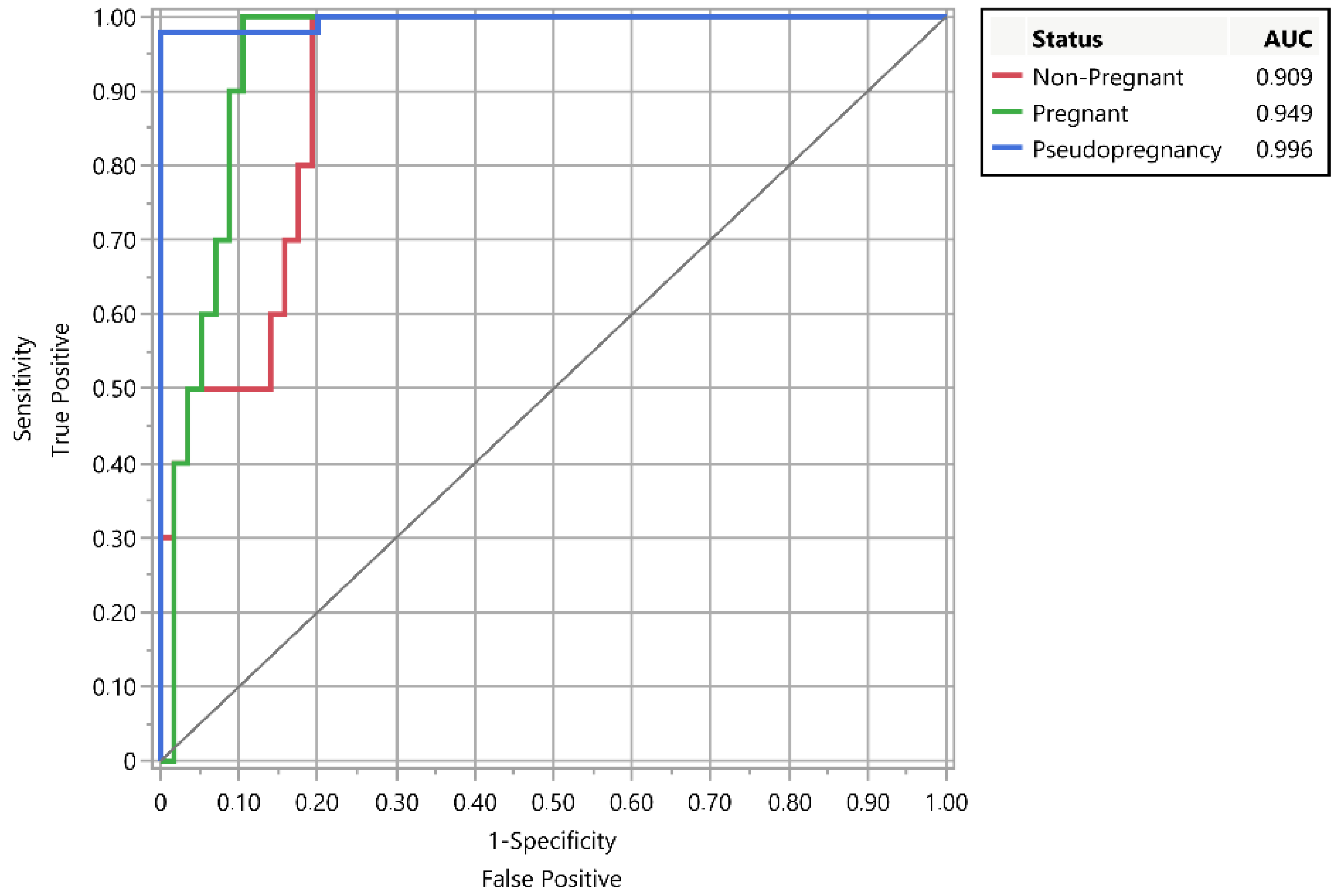
Figure 2.
Survival plot according to PAGs/P4 ratio value. Censor: pseudopregnancy = positive.
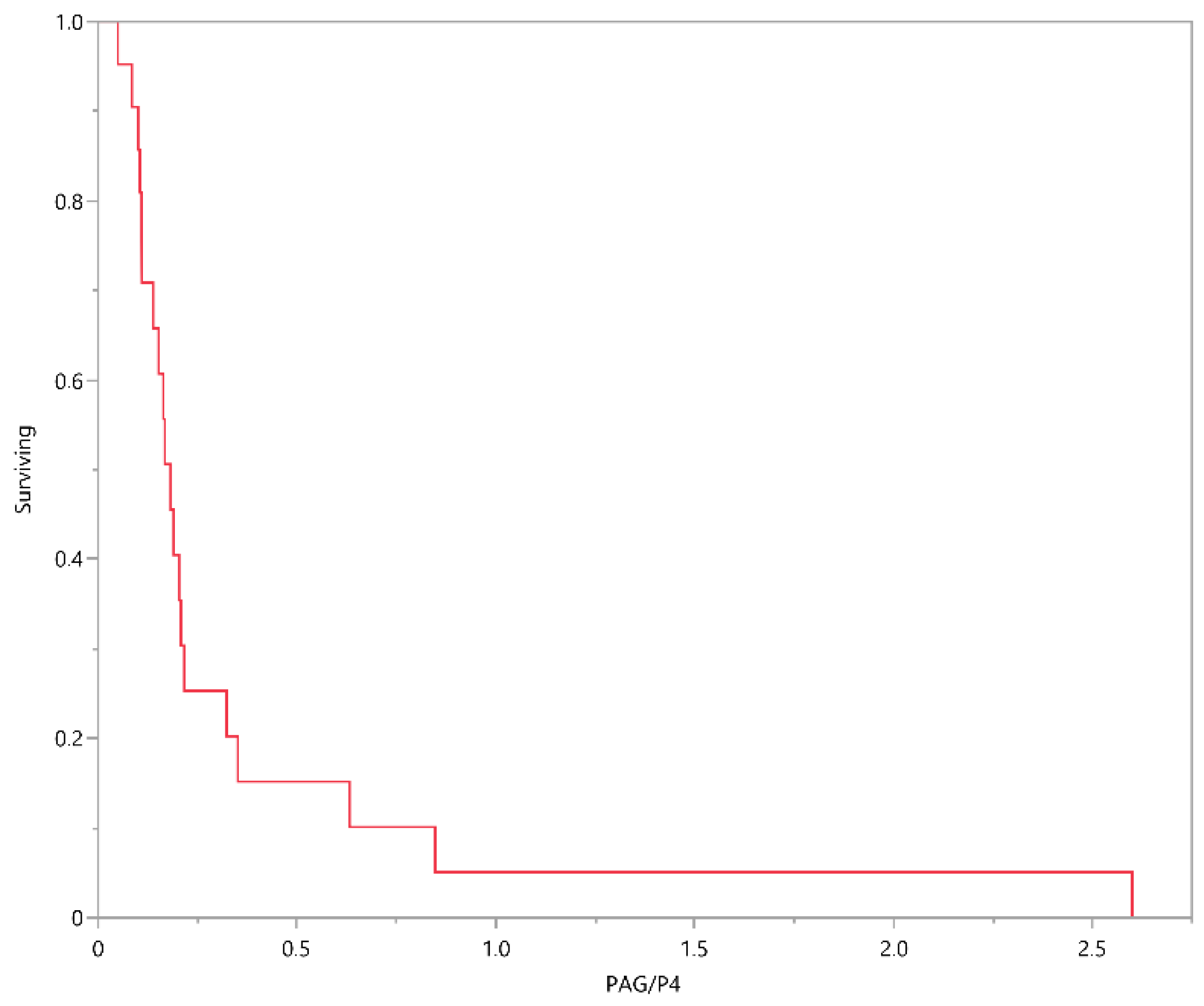

Table 1.
LSmean ± SEM of pregnancy-associated protein B (PAG), progesterone (P4) and PAG/P4 ratio according to the reproductive status of dairy goats.
Table 1.
LSmean ± SEM of pregnancy-associated protein B (PAG), progesterone (P4) and PAG/P4 ratio according to the reproductive status of dairy goats.
| Hormone | Reproductive status | P-value | Statistical power (α, σ, δ) |
||
|---|---|---|---|---|---|
| PPG | NPG | PG | |||
| PAGs (ng/mL) | 0.08 ± 0.02 a | 0.13 ± 0.04 a | 1. 45 ± 0.04 b | < 0.001 | 1 (0.05, 0.13, 0.32) |
| P4 (S-N OD) | 6.76 ± 0.49 a | 0.69 ± 1.00 b | 8.15 ± 1.05 a | < 0.001 | 1 (0.05, 3.32, 2.38) |
| PAGs/P4 ratio | 0.01 ± 0.11 a | 0.24 ± 0.23 b | 0.18 ± 0.23 b | < 0.001 | 1 (0.05, 0.72, 1.25) |
PPG: Pseudopregnancy; NPG: Non-Pregnancy; PG: Pregnancy. S-N OD: Corrected S-N for optic density (ELISA). a-b different superscript letters in the same line: p < 0.001.
Disclaimer/Publisher’s Note: The statements, opinions and data contained in all publications are solely those of the individual author(s) and contributor(s) and not of MDPI and/or the editor(s). MDPI and/or the editor(s) disclaim responsibility for any injury to people or property resulting from any ideas, methods, instructions or products referred to in the content. |
© 2024 by the authors. Licensee MDPI, Basel, Switzerland. This article is an open access article distributed under the terms and conditions of the Creative Commons Attribution (CC BY) license (http://creativecommons.org/licenses/by/4.0/).
Copyright: This open access article is published under a Creative Commons CC BY 4.0 license, which permit the free download, distribution, and reuse, provided that the author and preprint are cited in any reuse.


