
Submitted:
27 November 2024
Posted:
28 November 2024
You are already at the latest version
Abstract
The kidneys contribute to the overall health of the organism by maintaining systemic homeostasis. This process involves the regulation of various biological mechanisms, in which the Krüppel-like factor (KLF) transcription factors are essential for regulating development, differentiation, proliferation, and cellular apoptosis. They also play a role in the metabolic regulation of essential nutrients, such as glucose and lipids. The dysregulation of these transcription factors is associated with the development of various pathologies, which can ultimately lead to renal fibrosis, a process that severely compromises the function of these organs. In this context, the present article provides a comprehensive review of the existing literature, offering an enriching analysis of the findings related to the role of KLFs in the renal field, while also highlighting their potential therapeutic role in the treatment of renal diseases.
Keywords:
Krüppel-like factors
; Kidney
; Development
; Metabolism
; Disease
; Regulation
Introduction
The kidneys are intricate organs that have a vital function in maintaining an overall balance of waste and fluids in the body [1]. By preserving the body's electrolyte and acid-base balance by filtering and reabsorbing substances and eliminating toxins from the bloodstream[2] . Additionally, the kidneys perform endocrine functions such as hormone production e.g., erythropoietin, 1-α hydroxylase, and renin, which are involved in erythropoiesis, calcitriol synthesis, and blood pressure regulation, respectively [3]. It is important to note that to maintain their proper cellular function, particular responses are determined by effectors such as transcriptions factors who respond to external signals resulting in the modulation of gene expression, thereby tightly controlling physiological function[4]. Amongst the responses from different transcription factors, in recent years the Krüppel-like factor (KLF) family of transcription factors have acquired much attention for their roles in development and functional maintenance of the kidney[5]
Briefly, the Krüppel-like factor (KLF) family is a 17-member family of transcription factors characterized by three zinc fingers (Cys2/His2) with highly conserved C-terminal domains [6]. Regarding renal function KLFs have been associated with several biological processes including differentiation, terminal maturation, as well as the maintenance of the structure and function of the glomerular filtration barrier, protecting glomerular endothelial cells and podocytes from inflammatory damage and the development of fibrosis [7]. Members such as KLF5 have been associated with the promotion of inflammation and the development of tubulointerstitial fibrosis by positively regulating pro-inflammatory cytokines[8,9], as well as promoting cell proliferation by activating pathways that induce cell growth [10]..
The structural variations in their regulatory domains determine the variety of roles that KLFs play in renal biological processes. Through their interactions with various coactivators and/or corepressors, these domains enable them activation or repression of the promoter activity of their target genes[11,12]. Deregulation of the expression of KLFs can affect the physiological functions in which they are involved, promoting the development of different diseases [13,14]
This article aims to provide an overview, based on a comprehensive review, of the most relevant findings related to the involvement of KLFs in various biological processes of the kidney. We emphasize KLFs during renal development, their role in regulating metabolic processes of essential macronutrients such as glucose and lipids, as well as their implications in the onset and progression of significant renal diseases such as acute kidney injury (AKI), chronic kidney disease (CKD), and diabetic kidney disease (DKD), highlighting those KLFs that exhibit renal-protective effects.
Krüppel Like Factors
Krüppel-like factors make up a family of zinc-finger motif DNA-binding transcription regulators. Their name derives from the initial discovery of Krüppel protein in Drosophila melanogaster in 1993. A protein which participates in regulating fly embryo segmentation [15]. A study on non-bilateral lineages included 48 species from the Eukarya kingdom. The data presented are of relevance as they trace the evolutionary history of this family of transcription factors, determining that KLF genes originated in the opistocont stem lineage, and SP genes in metazoans, excluding ctenophores[16]. In humans, the KLFs consist of 17 members, which, as mentioned, exhibit three highly conserved Cys2His2-type zinc finger motifs in their carboxyl-terminal regions. Each zinc finger is formed by two cysteine residues and two histidine residues that coordinate a zinc ion. Typically, the amino acid sequence that forms the zinc finger otif is CX(2-4)-CX(12)-HX(3-5)-H, which maintain<ns a fixed length of 23 or 21 amino acids for their motifs [17]. The DNA-binding domain structure, evolutionarily conserved, shows structural homology with Specificity Proteins (SP), which leads to considering KLFs as a subgroup of the SP/KLF family [18]. These motifs allow binding to both the GC-rich proximal promoter regions and the CACCC elements (GT boxes) within the promoter regions of multiple genes [19]. The zinc finger motifs can bind to DNA through a specific recognition code, determined by the non-conserved amino acid residues in their alpha-helical region. The positions of the amino acid residues in the zinc fingers that interact with the DNA bases wrap around the DNA and connect with three nucleotides[20]
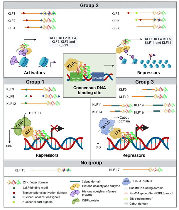
The phylogenetic features that mark the evolutionary distance amongst KLF family members, coupled with specific structural traits in their less conserved amino-terminal regions, result in the division of KLFs into three distinct groups [21]. The first group is composed of KLF3, KLF8, and KLF12, which harbor a Pro-X-Asp-Leu-Ser (PXDLS) motif, where X represents a hydrophobic amino acid. This motif facilitates interaction with the amino-terminal substrate binding domain (SBD) of C-terminal binding proteins (CtBP), aiming to repress transcription[11,22,23,24]. The second group includes KLF1, KLF2, KLF4, KLF5, KLF6, and KLF7, who, except for KLF7, along with KLF8 from group 1 and KLF15 (the latter by homology), possess a transcriptional activation domain (TAD) in their N-terminal regions [11,23]. Both KLF1-2, KLF4-6, and KLF13 (the latter from group 3), hey can, in addition to binding to consensus sites in DNA, interact with histone acetyltransferase enzymes such as cAMP response element-binding protein (CREB) binding protein (CBP), p300, and p300/CBP-associated factor, facilitating chromatin remodeling and promoting transcriptional activity in DNA regions regulated by KLFs[11,23,25,26]. The interaction of KLFs with epigenome-modifying enzymes can occur in different ways, such as when they are acetylated by histone acetyltransferases, promoting the acetylation of genes and inducing their expression, or conversely, when KLFs bind to specific promoters to recruit histone deacetylase enzymes in order to locally promote the deacetylation of histone proteins and repress gene expression. An example of this is how KLF4 can recruit p300 and acetylate itself, promoting the activation of genes such as the intestinal alkaline phosphatase gene in LS174T colon cancer cells, or repress gene expression by deacetylating p53 through the recruitment of HDAC3 to its promoter region[27]. The binding of KLF1, KLF4, KLF5, and KLF11 (the latter from group 3) to histone deacetylase enzymes suppresses their transcriptional activity. Therefore, this second group of KLFs can modulate both activation and re-pression of gene expression at the transcriptional level depending on a biological context and the gene regulatory region through which they are operating[19]. Lastly, in the third group, members KLF9, KLF10, KLF11, KLF13, KLF14, and KLF16 contain a Cabut domain in their N-terminal section that encompasses a Sin3 interaction domain (SID) and their activities as transcriptional repressors [7]. Both KLF15 and KLF17 are not classified within these three phylogenetic groups because their protein interaction domains have not yet been determined. Finaly, researchers have identified that various KLFs also contain nuclear localization signals (NLS) and nuclear export signals (NES) which regulate their subcellular localization, as illustrated in Figure 1.
3. KLFs in kidney physiology
The kidneys play essential roles in maintaining homeostasis in the body, mediated by hormonal signaling processes and their interaction with transcription factors [28,29]. One family of transcription factors involved in regulating renal function is the KLFs [28,30,31], which are expressed in various parts of the nephron, contributing to both its structure and cellular composition [32]. These aspects are fundamental in the cellular biology of the kidney and understanding them provides a better insight of the role of KLFs in each of these aspects and helps elucidate theiinfluence on determining cellular function and behavior.
The kidneys are divided into two specific regions: the renal cortex and the renal medulla. These two regions make up the renal parenchyma, which is the functional tissue of the kidney. The functional and structural unit of the kidney is the nephron, which is composed of the glomerulus and the tubular system [1,33].
Surrounded by a cup-shaped structure called Bowman's capsule, the glomerulus is where blood filtration occurs. The parietal epithelial layer is composed of parietal cells that play a structural maintenance role [1], while the visceral layer is formed by podocytes, which are perivascular cells enveloping the outer layer of the basal membrane of the glomerular capillaries (GC), together forming the glomerular filtration barrier (GFB) [34,35]. The integrity of this barrier, maintained in part by the surface proteins expressed by endothelial cells, the selective adhesion molecules of endothelial cells that mediate cell-to-cell adhesion and regulate vascular permeability [35,36] and the slit diaphragms between the foot processes of podocytes, ensures proper glomerular filtration [37,38,39]. KLF4 collaborates with histone deacetylases (HDACs), specifically HDAC1 and HDAC3, to upregulate the expression of E-cadherin, podocin, and nephrin. E-cadherin is an adhesion molecule, while podocin and nephrin are crucial for the formation and maintenance of the slit diaphragm (SD), a specialized adhesion structure in podocytes [36,37,38]. KLF4 also induces the expression of cytokeratin 8 (K8) and K18, which, like podocins, participates in cytoskeletal organization [36]. Together, the expression of these proteins maintains the structure and functionality of podocytes by modulating cellular adhesion and polarity.
Maintaining cell adhesion and polarity is crucial to prevent acquisition of mesenchymal characteristics. KLF4 is also involved in downregulating mesenchymal markers, such as vimentin and α-smooth muscle actin (α-SMA) in podocytes [36]. In addition to being expressed under normal conditions in podocytes, vimentin is also expressed in mesenchymal cells, which generally lack both intercellular adhesion and polarity, providing them with resistance to migration-related stress. [39,40]. In contrast, α-SMA contributes to motility and contraction of the cytoskeleton [41], allowing greater cell mobility and the ability to migrate to sites of injury or inflammation. Therefore, KLF4 prevents structural damage and the progression of diseases by inhibiting epithelial-to-mesenchymal transition (EMT). Moreover, KLF6 has further been associated with maintaining mitochondrial function and preventing cell death in podocytes. This is because of KLF6's ability to bind to the promoter region of the cytochrome c oxidase assembly gene (SCO2) and positively regulate it. Continuous expression of SCO2, a metallo-chaperone, is crucial for transporting copper ions to electron carriers in the mitochondrial electron transport chain. This process, including cytochrome c, is vital for preventing the activation of the intrinsic apoptotic pathway in podocytes. By doing so, it avoids any harm to the glomerulus and maintains its filtration capacity [31].
In terms of energy metabolism, SCO2 can modulate the metabolic switch from glycolysis to OXPHOS in hematopoietic stem cells with Fanconi anemia, not depending on glycolysis as usual, but on OXPHOS as a compensatory mechanism in these affected cells to ensure functional energy metabolism. Therefore, in a renal context, these data could suggest proper maintenance of OXPHOS mediated in part by SCO2 in kidney cells that rely on fatty acid oxidation to meet their energy needs [42].
In GC, which are made up of fenestrated endothelial cells (ECs) possessing transcellular pores of 60 to 100 nm in diameter, selective permeability for molecules according to their size and charge is achieved [1,43]. KLF2 contributes to regulating the size and distribution of these pores, preventing uncontrolled solute permeability by inhibiting the phosphorylation of the myosin light chain, thus avoiding the contraction of the cytoskeleton that would reduce the size of the ECs and lead to the formation of gaps between them [44]. Another way to maintain proper glomerular filtration is through the modulation of angiogenesis mediated by VEGF-A, where KLF2 downregulates its expression. This prevents the excess of unnecessary or dysfunctional blood vessels that could disrupt hemodynamic balance and affect blood pressure [45].
The role of KLF4 in endothelial cells relates to the reduction of inflammation through the downregulation of adhesion molecules, such as VCAM1, induced by TNF-α. This is achieved by inhibiting the expression of the p65 subunit of nuclear factor kB (NF-κB), which is necessary for the activation of this transcription factor and its binding to the VCAM1 promoter. In this way, KLF4 modulates the adhesion and recruitment of lymphocytes to endothelial cells, preventing chronic inflammation [46]. Moreover, this anti-inflammatory effect of KLF4 in endothelial cells has been shown to improve the condition in patients with vascular lesions induced by ischemic stroke by regulating the endothelial expression of other inflammatory cell adhesion molecules such as E-selectin, intercellular adhesion molecule 1 (ICAM-1), as well as VCAM-1, NF-kB, and tight junction proteins[47] .Mesangial cells are part of the mesenchymal stromal cells, which include fibroblasts, pericytes, and vascular smooth muscle cells [48]. As stromal cells, they organize the structure of the glomerulus and contribute to the homeostasis of endothelial cells and podocytes by directing the immune response and repair after glomerular injury [48,49]. Mesangial cells not only support the glomerular capillaries but also the mesangial cells extending into the extraglomerular mesangium, which are part of the juxtaglomerular apparatus, contributing to the regulation of blood pressure and fluid volume by secreting renin [50].
In a study using C57BL/KsJ db/db mice that overexpressed KLF4 via a lentiviral vector in podocytes, researchers demonstrated through histological staining with periodic acid-Schiff that renal injury was mitigated by attenuating, on one hand, the expansion of the mesangial matrix and the proliferation of mesangial cells in the glomerulus. It is noteworthy to mention that a more thorough analysis of these results was not presented. Therefore, a question on how overexpression of KLF4 in podocytes appears to affect both the proliferation of mesangial cells and the production of mesangial matrix continues.
This event suggests a possible prevention of the overproduction of type IV and V collagens and fibronectin by mesangial cells, thereby preventing glomerular fibrosis and the development of fibrotic kidney diseases [51].
[52]
Between the parietal layer and the visceral layer of Bowman’s capsule, a urinary space known as the "Bowman space" is formed. This space represents the beginning of the urinary system and is contiguous with the proximal convoluted tubule (PCT), in the renal cortex [53]. The PCT, which is the first segment of the nephron's tubular system, receives glomerular ultrafiltrate through epithelial cells (EpC) that allow permeability to both water and luminal fluid, reabsorbing glucose, amino acids, and minerals such as phosphate, chloride, and bicarbonate, as well as secreting hydrogen ions and toxins produced by cellular metabolism and xenobiotics present in the filtrate [54]. The expression of KLF4 and KLF11 mitigates inflammation and fibrosis by decreasing the expression of cytokines MCP-1, MIP-3α, and IL-8 induced by TGF-β1. This is possibly related to the phosphorylation of KLF4 that induces SMAD and p38/MAPK signaling pathways in vascular smooth muscle cells (VSMCs) [55]or through binding to p65 and -inhibiting NF-kB signaling. The decrease of these inflammatory cytokines limits the production of collagen type I and tissue fibrosis[56].
KLF15 in these EpC can decrease the expression of these fibrogenic components by negatively regulating the MAPK pathways, which, when activated, contribute to the production of TGF-β1 and other pro-fibrogenic factors. Thus, KLF15 also modulates the fibrogenic response and helps prevent the accumulation of an extracellular matrix[57].
The specific expression of genes within each segment of the renal tubule determines its respective role [32], unfortunately, given what we know, the expression and function of KLFs in the subsequent segments of the nephron's tubular system remain incompletely understood. Following the proximal PCT, there are several segments, one of which is the loop of Henle; consisting of a thin descending branch, a thin ascending branch, and a thick ascending limb, contributes to the regulation of urine concentration by passively reabsorbing water into the medullary interstitiumIn both the ascending thin branch and the thick ascending branch, approximately 25% of sodium chloride (NaCl) is reabsorbed, while the descending thin branch does not reabsorb NaCl. Additionally, 15% of water and potassium are reabsorbed. Urea reabsorption in the ascending branch of the loop of Henle helps increase solute concentration in the extracellular space, while its excretion is determined not only by glomerular filtration but also by tubular reabsorption, allowing it to play an important role in promoting concentrated urine production and maintaining body water [58] The distal convoluted tubule performs the functions of reabsorbing sodium, potassium, and chloride, besides secreting hydrogen and potassium ions. Finally, the cortical collecting tubule, a specific region of the collecting duct, connects to a collecting duct that drains into the renal papillae. This segment reabsorbs the same electrolytes as the distal convoluted tubule [1,53].
Therefore, the main function of KLFs in the kidneys can be briefly summarized as the preservation of structure, cell adhesion, the glomerular filtration barrier, and regulating the extracellular matrix (ECM) and inflammation of the cell lineage that composes them. This has the effect of modulating glomerular filtration, secretion, and elimination of unnecessary toxins from the body. Figure 2 illustrates the basic structure of the nephron, along with the location of its main cell groups that integrate the specific gene expression of KLFs within it. Table 1 summarizes the specific functions of each of the KLFs involved in the kidney's functional regulation processes.
3.1. Klf in kidney development
The process of organogenesis involves the division and organization of cells to establish the foundational structures of the embryo. Cell cycle arrest plays a crucial role in this process as it enables the progression towards terminal differentiation, which is a vital step in acquiring specialized functions [59].
The intricate progression of the cell cycle and cellular differentiation at a molecular level is regulated by signaling pathways and transcription factors[60]. This principle highlights the importance of the KLF family in contributing to kidney development. In embryonic cell cycle regulation, in vitro studies suggest Klf5 promotes podocyte survival by blocking the mitogen-activated protein (MAP) kinase ERK/p38 pathway, as well as decreasing the expression of apoptosis-related proteins such as Bax, caspase-3, caspase-8, and caspase-9, while increasing the expression of the antiapoptotic protein Bcl-2, which inhibits both cell cycle arrest and apoptosis by preventing Bax from translocating to the mitochondria[61,62,63]. By contrast, in patient serum with diabetic nephropathy (DN) or in a model of HK2 cells with nephritis induced by high glucose concentrations, researchers demonstrated that the inhibition of KLF5 through miR-214-5p, which survives by inhibiting the expression of a long non-coding RNA that functions as an endogenous competitive RNA for microRNAs, specifically the differentiation antagonizing non-coding RNA by means of a small interfering RNA directed towards it, protects against tubular injury under these physiological conditions by inhibiting apoptosis and restoring cell proliferation. Additionally, protein levels of inflammatory factors such as IL-6, TNF-α, and IL-1β are reduced, along with the production of ROS and malondialdehyde content[64]. A possible mechanism by which the inhibition of KLF5 promotes apoptosis has even been demonstrated in prostate cancer cells, where this inhibition, along with the protein Stat5a/b and ICAM-1 through bioactive plant-derived agents, has been associated with a positive regulation of the Bax/Bcl-2 ratio. This relationship regulates apoptosis; specifically, the inhibition of Klf5 induces an increase in the expression of Bax, which translocate to the mitochondria, promoting the release of cytochrome c and consequently the activation of caspase-3 and PARP, which are key proteins in the apoptotic process. The expression of Bcl-2, is not significantly affected during KLF5 downregulation[65].
The complete maturation of the nephrons occurs postnatally and is essential for the kidney to develop its maximum urinary concentrating ability. Various mechanisms are outlined below to illustrate how both KLF12 and KLF15 are fundamental in this process. KLF15 acts as a negative regulator of the chloride channel ClC-K1, which is expressed in the EpC thin ascending branch of the loop of Henle during postnatal development. This KLF15/ClC-K1 repression prevents the formation of channels that facilitate the passage of chloride into the urine, avoiding dysregulation in the organism's electrolyte balance, since the concentration of chloride in urine directly affects its osmolarity[30]. KLF12 is also overexpressed between 15 and 22 days after birth in the EpC of the medullary collecting ducts (IMCD) undergoing maturation. The specificity of KLF12 expression was determined by comparing it with the expression of aquaporin 2 (AQP2), which is upregulated in the IMCD and shares similar DNA-binding sites to those of the KLFs. The co-localization of KLF12 and AQP2 suggests KLF12 plays a key role in the positive regulation of AQP2 expression and its target gene, the urea transporter (UT-A1) by binding to a CACCC element in the UT-A1 promoter [66,67]. This is relevant because amongst the nine aquaporins, including AQP1–8 and AQP11, that are differentially expressed along the renal tubules and collecting ducts, AQP2 has the transport capability and is identified as one of the selective water channels[68] This positive regulation influences the transport of water and urea into the IMCD, resulting in the accumulation of urea necessary for proper urine concentration[69]. Figure 3 illustrates the involvement of KLFs in regulating renal organogenesis and postnatal maturation of nephrons.
KLF15 has been also associated with podocyte differentiation, since it upregulates the expression of nephrin, podocin, synaptopodin, and Wt1; all essential genes for maintaining a differentiated phenotype and preventing the loss and detachment of podocytes [34,70] Meanwhile, KLF4 regulates the differentiation of specific nephron segments and individual cell types by cooperating with p53 and CREB, as evidenced by the p53-CRE-KLF binding sites in the promoter regions of renal function genes, such as AQP2, bradykinin receptor B2 (B2R) and epithelial sodium channel (ENaC) during terminal nephron differentiation[60,71].
To summarize, KLFs are vital for the proper development, structure, and function of the glomerular filtration barrier. They safeguard endothelial cells (ECs), facilitate the differentiation of podocytes, maintain their specialized integrity, and regulate cell cycle and apoptosis.
3.2. Klf in kidney metabolism
Metabolic processes enable the kidney to use, produce, and reabsorb nutrients, fulfilling its own energy demands while also ensuring homeostasis. To regulate these metabolic processes effectively, transcription factors and coactivators are involved in modulating the expression of genes that code for the enzymes involved. This ensures the proper occurrence of these processes.
Transcription factors such as KLFs have been closely associated with regulating metabolism, especially in the hepatic context [72,73,74,75,76,77,78], where they regulate genes involved in lipid metabolism such as CREB, Carbohydrate Response Element Binding Protein (ChREBP), Sterol Regulatory Element Binding Protein 1 (SREBP-1), Peroxisome Proliferator-Activated Receptor (PPAR). Moreover, they further regulate genes involved in glycolytic metabolism such as Peroxisome Proliferator-Activated Receptor Gamma Coactivator 1 Alpha (PGC-1α) [72,79,80,81]because they share the same DNA binding sites in conserved CACCC -sequences and GC-rich elements [82].
The variations in metabolic activity among KLFs remain unexplored. However, in vitro, studies have shown that KLF6 overexpression in PTCs (Hk-2) exposed to high glucose concentrations increases the expression of the protein that interacts with thioredoxin (Txnip) [83]. This protein is particularly intriguing due to its involvement in glucose oxidation, which is vital for energy acquisition. One of the most significant factors is related to the glucose transporter GLUT1. It is regulated by Txnip and could influence its localization by directly binding on the transporter. This binding induces internalization through clathrin-coated pits[84]. Therefore, its expression suggests that peripheral glucose uptake and utilization of this substrate could be compromised, leading to metabolic disturbances in conditions of high and persistent glucose consumption, such as in diabetes mellitus. Thus, silencing KLF6 may represent a therapeutic target, as its inhibition significantly attenuates Txnip expression and mitigates metabolic consequences.
Within the Hk-2 cellular model, KLF14 is an important player in lipid metabolism. The absence of KLF14 has been linked to a decrease in mitochondrial activity by reducing the expression of PPARα, since KLF14 can specifically bind to the region -222 to -209 of the PPARα promoter region and regulate its expression, suggesting an insufficient energy supply to renal tubular cells, lipid accumulation, and the subsequent development of tubulointerstitial fibrosis[85]. Furthermore, it inhibits the expression of CPT1, an enzyme that facilitates the transfer of acyl groups from acyl-CoA to carnitine in the intermembrane space of the mitochondria, leading to the production of acyl-carnitine esters. By utilizing this process, the inner mitochondrial membrane allows for the transportation of long-chain fatty acids. In the next step, CPT2 takes over and transfers the acyl groups from acyl-carnitine back to CoA, ultimately leading to the restoration of acyl-CoA. Subsequently, this acyl-CoA can be metabolized through a series of enzymatic reactions within the β-oxidation pathway of fatty acids [86,87]
Thus, the downregulation of PPARα causes the accumulation of lipids inside cells, a process that can occur in a wide range of renal cell types, including mesangial cells, podocytes, and proximal PCT[88]. The latter cells, which rely on FAO for energy production, are the ones that this applies to [89]. Renal damage can occur when lipids accumulate in cells that are not meant for storage. The build-up of this substance promotes lipoperoxidation, which generates reactive oxygen species (ROS), leading to oxidative stress and contributing to the development of different diseases like acute kidney injury (AKI) and fibrosis [90]. Nevertheless, the overexpression of KLF14 counteracts these effects by enhancing mitochondrial activity, reducing lipogenesis, and decreasing lipid accumulation. In experiments conducted on living subjects, it has been found that KLF15 can positively regulate both CPT1 and Acyl-CoA Acyltransferase 2 (ACAA2) by tightly binding to the PPARα binding sites [91]. The closeness of DNA suggests that KLF15 and PPARα may coordinate in governing the expression of these genes.
Thus, it is crucial to have high levels of both KLF14 and KLF15 to ensure sufficient energy supply, specifically to the PTCs, by utilizing lipids through FAO. Figure 4 illustrates the implication of KLFs in renal metabolism both under healthy conditions and their effect on states of dysregulation.
3.3. Klfs in kidney disease
When the expression of KLFs is dysregulated, it can disrupt various physiological processes in which they are involved. This disruption can cause poor management of inflammation, tissue repair, regeneration, and other cellular adaptations to stress.
Throughout this review, it has been discussed that KLFs demonstrate distinct patterns of expression in different renal cells, greatly influencing their various functions in maintaining overall balance within the body[92]. The dysregulation of KLFs is associated with kidney diseases, including CKD, AKI, and DKD, where the pathophysiology affects the balance of glomerular, tubular, and inflammatory functions, and this variability is commonly observed. Take into consideration that specific KLFs have protective properties for the kidneys and blocking them frequently leads to the deterioration and advancement of the disease. Conversely, the disease can also be exacerbated by the excessive expression of other KLFs.
It is important to mention that AKI, which is characterized by a sudden decrease in kidney function, has the potential to progress to chronic injury and eventually develop into CKD[93]. In this context, KLF2, KLF4, KLF9, KLF10, and KLF15 have beneficial effects [94,95,96,97,98] while KLF5 promotes cell proliferation, tubular damage, and inflammation [99]. Various pathological conditions, such as diabetic nephropathy, which is characterized by albuminuria and progressive renal insufficiency [100], precipitate the development of CKD. Here, the KLFs that stand out are primarily KLF3 and KLF6, which induce inflammation and fibrosis and facilitate epithelial-mesenchymal transition, respectively. Ultimately, all these diseases converge on a common outcome of renal fibrosis, characterized by excessive deposition of ECM [101,102]. The activity of each KLF in these three diseases is detailed in Table 2.
5. Perspectives
Ongoing research is advancing our understanding of KLFs and how they affect the development of the kidneys, metabolic activity, and kidney diseases. With increasing understanding of these factors, it is apparent that a more thorough exploration of the cellular functions in which KLFs might be involved is necessary. Thus, it is of utmost importance to examine the specific interactions of KLFs with other transcription factors and signaling pathways. Understanding how these factors regulate gene expression in different contexts can provide valuable insights into their role in renal homeostasis and response to injury. Moreover, a more detailed approach is required regarding the effects of KLFs on regulating renal metabolism. Given that kidneys are crucial organs for the metabolism of nutrients such as glucose and lipids, investigating how KLFs modulate these metabolic pathways could reveal new therapeutic strategies for diseases like diabetes and chronic kidney disease.
Developing experimental models that permit the study of KLF function in pathological conditions is also of great significance. The creation of in vivo and in vitro models will facilitate the identification of biomarkers and the evaluation of therapeutic interventions that could improve renal health. Ultimately, incorporating cutting-edge technologies such as genetic editing, through CRISPR/Cas9, by manipulating KLF genes to study their effects on both promoting health and the development of kidney disease. Additionally, systems biology, through large data analysis with simple and accessible bioinformatics tools, can facilitate the identification of interaction networks and biomarkers of kidney diseases. By combining both approaches, more precise models can be developed, and therapeutic strategies can be personalized.
This could open new avenues for identifying therapeutic targets and intervention strategies in kidney diseases.
In summary, studying KLFs has the potential to significantly expand our knowledge of renal development and disease. A multidisciplinary approach will be key to unlocking these mechanisms and contributing to the development of new therapies that improve renal health. For this, the valuable collaboration of experts from various fields is essential. Specifically, the participation of molecular biologists and geneticists brings theoretical and practical knowledge of gene manipulation to identify its direct impact on kidney function and the regulation of homeostasis. Renal physiologists help determine the specific physiological processes of the kidneys under certain circumstances. Bioinformaticians are essential in analyzing large volumes of data, facilitating the identification of interaction networks and biomarkers associated with kidney diseases. Nephrology specialists and nutritionists, through their clinical experience and focus on nutrition and overall well-being, contribute their knowledge of the external factors influencing renal health, such as diet and lifestyle. This interdisciplinary collaboration, as a whole, allowing- for a deeper understanding of how KLFs regulate renal homeostasis, promoting health and helping to prevent or treat diseases such as chronic kidney,
Author Contributions
Conceptualization: ISS-C, AEE-R, investigation: ISS-C, MAJ-G, GGG, DFB-C, writing—original draft preparation: ISS-C, AEE-R, DAG-K, writing—review editing: OR-N, MGS-S ENG-T GRP-R JFI, supervision: J.F.I.
Funding
“This research received no external funding”.
Acknowledgments
The authors acknowledge mutual collaboration of all involved institutions.
Conflicts of Interest
“The authors declare no conflicts of interest.”
References
- Wallace, M.A. Anatomy and Physiology of the Kidney. AORN J 1998, 68, 799–820. [Google Scholar] [CrossRef]
- Eckardt, K.-U. The Noblesse of Kidney Physiology. Kidney Int 2019, 96, 1250–1253. [Google Scholar] [CrossRef]
- Acharya, V.; Olivero, J. The Kidney as an Endocrine Organ. Methodist Debakey Cardiovasc J 2018, 14, 305. [Google Scholar] [CrossRef]
- Vaquerizas, J.M.; Kummerfeld, S.K.; Teichmann, S.A.; Luscombe, N.M. A Census of Human Transcription Factors: Function, Expression and Evolution. Nat Rev Genet 2009, 10, 252–263. [Google Scholar] [CrossRef]
- Chiplunkar, A.R.; Lung, T.K.; Alhashem, Y.; Koppenhaver, B.A.; Salloum, F.N.; Kukreja, R.C.; Haar, J.L.; Lloyd, J.A. Krüppel-Like Factor 2 Is Required for Normal Mouse Cardiac Development. PLoS One 2013, 8, e54891. [Google Scholar] [CrossRef]
- Oishi, Y.; Manabe, I. Krüppel-Like Factors in Metabolic Homeostasis and Cardiometabolic Disease. Front Cardiovasc Med 2018, 5, 1–14. [Google Scholar] [CrossRef]
- Rane, M.J.; Zhao, Y.; Cai, L. Krϋppel-like Factors (KLFs) in Renal Physiology and Disease. EBioMedicine 2019, 40, 743–750. [Google Scholar] [CrossRef]
- Fujiu, K.; Manabe, I.; Nagai, R. Renal Collecting Duct Epithelial Cells Regulate Inflammation in Tubulointerstitial Damage in Mice. Journal of Clinical Investigation 2011, 121, 3425–3441. [Google Scholar] [CrossRef]
- Li, Z.; Lv, L.; Wang, B.; Tang, T.; Feng, Y.; Cao, J.; Jiang, L.; Sun, Y.; Liu, H.; Zhang, X.; et al. The Profibrotic Effects of MK-8617 on Tubulointerstitial Fibrosis Mediated by the KLF5 Regulating Pathway. The FASEB Journal 2019, 33, 12630–12643. [Google Scholar] [CrossRef]
- Chen, W.-C.; Lin, H.-H.; Tang, M.-J. Matrix-Stiffness–Regulated Inverse Expression of Krüppel-Like Factor 5 and Krüppel-Like Factor 4 in the Pathogenesis of Renal Fibrosis. Am J Pathol 2015, 185, 2468–2481. [Google Scholar] [CrossRef]
- Swamynathan, S.K. Krüppel-like Factors: Three Fingers in Control. Hum Genomics 2010, 4, 263–270. [Google Scholar] [CrossRef]
- Santoyo-Suarez, M.G.; Mares-Montemayor, J.D.; Padilla-Rivas, G.R.; Delgado-Gallegos, J.L.; Quiroz-Reyes, A.G.; Roacho-Perez, J.A.; Benitez-Chao, D.F.; Garza-Ocañas, L.; Arevalo-Martinez, G.; Garza-Treviño, E.N.; et al. The Involvement of Krüppel-like Factors in Cardiovascular Diseases. Life 2023, 13, 420. [Google Scholar] [CrossRef] [PubMed]
- Xiang, T.; Yang, C.; Deng, Z.; Sun, D.; Luo, F.; Chen, Y. Krüppel-like Factors Family in Health and Disease. MedComm (Beijing) 2024, 5. [Google Scholar] [CrossRef]
- Fletcher, B.R.; Damery, S.; Aiyegbusi, O.L.; Anderson, N.; Calvert, M.; Cockwell, P.; Ferguson, J.; Horton, M.; Paap, M.C.S.; Sidey-Gibbons, C.; et al. Symptom Burden and Health-Related Quality of Life in Chronic Kidney Disease: A Global Systematic Review and Meta-Analysis. PLoS Med 2022, 19, e1003954. [Google Scholar] [CrossRef]
- Zhang, J.; Li, G.; Feng, L.; Lu, H.; Wang, X. Krüppel-like Factors in Breast Cancer: Function, Regulation and Clinical Relevance. Biomedicine & Pharmacotherapy 2020, 123, 109778. [Google Scholar] [CrossRef]
- Presnell, J.S.; Schnitzler, C.E.; Browne, W.E. KLF/SP Transcription Factor Family Evolution: Expansion, Diversification, and Innovation in Eukaryotes. Genome Biol Evol 2015, 7, 2289–2309. [Google Scholar] [CrossRef]
- Wang, H.; Han, J.; Dmitrii, G.; Ning, K.; Zhang, X. KLF Transcription Factors in Bone Diseases. J Cell Mol Med 2024, 28. [Google Scholar] [CrossRef]
- Pei, J.; Grishin, N. V; Xu, E.Y. A New Family of Predicted Krüppel-Like Factor Genes and Pseudogenes in Placental Mammals. PLoS One 2013, 8, 81109. [Google Scholar] [CrossRef]
- García-Loredo, J.A.; Santoyo-Suarez, M.G.; Rodríguez-Nuñez, O.; Benitez Chao, D.F.; Garza-Treviño, E.N.; Zapata-Morin, P.A.; Padilla-Rivas, G.R.; Islas, J.F. Is the Cis-Element CACCC-Box a Master Regulatory Element during Cardiovascular Disease? A Bioinformatics Approach from the Perspective of the Krüppel-like Family of Transcription Factors. Life 2024, 14, 493. [Google Scholar] [CrossRef]
- Wolfe, S.A.; Nekludova, L.; Pabo, C.O. DNA Recognition by Cys 2 His 2 Zinc Finger Proteins. Annu Rev Biophys Biomol Struct 2000, 29, 183–212. [Google Scholar] [CrossRef]
- McConnell, B.B.; Yang, V.W. Mammalian Krüppel-Like Factors in Health and Diseases; 2010; Vol. 90; ISBN 4047275638.
- Abe, M.; Saeki, N.; Ikeda, Y.; Ohba, S. Kruppel-like Factors in Skeletal Physiology and Pathologies. Int J Mol Sci 2022, 23, 15174. [Google Scholar] [CrossRef] [PubMed]
- Pearson, R.; Fleetwood, J.; Eaton, S.; Crossley, M.; Bao, S. Krüppel-like Transcription Factors: A Functional Family. International Journal of Biochemistry and Cell Biology 2008, 40, 1996–2001. [Google Scholar] [CrossRef] [PubMed]
- Pollak, N.M.; Hoffman, M.; Goldberg, I.J.; Drosatos, K. Krüppel-Like Factors: Crippling and Uncrippling Metabolic Pathways. JACC Basic Transl Sci 2018, 3, 132–156. [Google Scholar] [CrossRef] [PubMed]
- Dang, D.T.; Pevsner, J.; Yang, V.W. The Biology of the Mammalian Krüppel-like Family of Transcription Factors. Int J Biochem Cell Biol 2000, 32, 1103–1121. [Google Scholar]
- Kaczynski, J.; Cook, T.; Urrutia, R. Sp1- and Krüppel-like Transcription Factors. Genome Biol 2003, 4, 1–8. [Google Scholar] [CrossRef]
- Evans, P.M.; Zhang, W.; Chen, X.; Yang, J.; Bhakat, K.K.; Liu, C. Krüppel-like Factor 4 Is Acetylated by P300 and Regulates Gene Transcription via Modulation of Histone Acetylation. Journal of Biological Chemistry 2007, 282, 33994–34002. [Google Scholar] [CrossRef]
- Li, J.; Liu, L.; Zhou, W.; Cai, L.; Xu, Z.; Rane, M.J. Roles of Krüppel-like Factor 5 in Kidney Disease. J Cell Mol Med 2021, 25, 2342–2355. [Google Scholar] [CrossRef]
- Dumas, S.J.; Meta, E.; Borri, M.; Luo, Y.; Li, X.; Rabelink, T.J.; Carmeliet, P. Phenotypic Diversity and Metabolic Specialization of Renal Endothelial Cells. Nat Rev Nephrol 2021, 17, 441–464. [Google Scholar] [CrossRef]
- Uchida, S.; Tanaka, Y.; Ito, H.; Saitoh-Ohara, F.; Inazawa, J.; Yokoyama, K.K.; Sasaki, S.; Marumo, F. Transcriptional Regulation of the CLC-K1 Promoter by Myc-Associated Zinc Finger Protein and Kidney-Enriched Krüppel-Like Factor, a Novel Zinc Finger Repressor. Mol Cell Biol 2000, 20, 7319–7331. [Google Scholar] [CrossRef]
- Mallipattu, S.K.; Horne, S.J.; D’Agati, V.; Narla, G.; Liu, R.; Frohman, M.A.; Dickman, K.; Chen, E.Y.; Ma’ayan, A.; Bialkowska, A.B.; et al. Krüppel-like Factor 6 Regulates Mitochondrial Function in the Kidney. Journal of Clinical Investigation 2015, 125, 1347–1361. [Google Scholar] [CrossRef]
- Lee, J.W.; Chou, C.-L.; Knepper, M.A. Deep Sequencing in Microdissected Renal Tubules Identifies Nephron Segment–Specific Transcriptomes. Journal of the American Society of Nephrology 2015, 26, 2669–2677. [Google Scholar] [CrossRef] [PubMed]
- Balzer, M.S.; Rohacs, T.; Susztak, K. How Many Cell Types Are in the Kidney and What Do They Do? Annu Rev Physiol 2022, 84, 507–531. [Google Scholar] [CrossRef] [PubMed]
- Müller-Deile, J.; Schiffer, M. Podocytes from the Diagnostic and Therapeutic Point of View. Pflugers Arch 2017, 469, 1007–1015. [Google Scholar] [CrossRef]
- Tian, X.; Bunda, P.; Ishibe, S. Podocyte Endocytosis in Regulating the Glomerular Filtration Barrier. Front Med (Lausanne) 2022, 9. [Google Scholar] [CrossRef]
- Hayashi, K.; Sasamura, H.; Nakamura, M.; Azegami, T.; Oguchi, H.; Sakamaki, Y.; Itoh, H. KLF4-Dependent Epigenetic Remodeling Modulates Podocyte Phenotypes and Attenuates Proteinuria. Journal of Clinical Investigation 2014, 124, 2523–2537. [Google Scholar] [CrossRef]
- Li, X.; He, J.C. An Update: The Role of Nephrin inside and Outside the Kidney. Sci China Life Sci 2015, 58, 649–657. [Google Scholar] [CrossRef]
- Huber, T.B.; Schermer, B.; Benzing, T. Podocin Organizes Ion Channel-Lipid Supercomplexes: Implications for Mechanosensation at the Slit Diaphragm. Nephron Exp Nephrol 2007, 106, e27–e31. [Google Scholar] [CrossRef]
- Kokkinos, M.I.; Wafai, R.; Wong, M.K.; Newgreen, D.F.; Thompson, E.W.; Waltham, M. Vimentin and Epithelial-Mesenchymal Transition in Human Breast Cancer – Observations in Vitro and in Vivo. Cells Tissues Organs 2007, 185, 191–203. [Google Scholar] [CrossRef]
- Yasuda, H.; Fukusumi, Y.; Zhang, Y.; Kawachi, H. 14-3-3 Proteins Stabilize Actin and Vimentin Filaments to Maintain Processes in Renal Glomerular Podocyte. The FASEB Journal 2023, 37. [Google Scholar] [CrossRef]
- Yuan, S.-M. α-Smooth Muscle Actin and ACTA2 Gene Expressions in Vasculopathies. Revista Brasileira de Cirurgia Cardiovascular 2015. [Google Scholar] [CrossRef]
- Du, W.; Amarachintha, S.; Wilson, A.F.; Pang, Q. SCO2 Mediates Oxidative Stress-Induced Glycolysis to Oxidative Phosphorylation Switch in Hematopoietic Stem Cells. Stem Cells 2016, 34, 960–971. [Google Scholar] [CrossRef] [PubMed]
- Scott, R.P.; Quaggin, S.E. The Cell Biology of Renal Filtration. Journal of Cell Biology 2015, 209, 199–210. [Google Scholar] [CrossRef] [PubMed]
- Lin, Z.; Natesan, V.; Shi, H.; Dong, F.; Kawanami, D.; Mahabeleshwar, G.H.; Atkins, G.B.; Nayak, L.; Cui, Y.; Finigan, J.H.; et al. Kruppel-Like Factor 2 Regulates Endothelial Barrier Function. Arterioscler Thromb Vasc Biol 2010, 30, 1952–1959. [Google Scholar] [CrossRef] [PubMed]
- Bhattacharya, R.; SenBanerjee, S.; Lin, Z.; Mir, S.; Hamik, A.; Wang, P.; Mukherjee, P.; Mukhopadhyay, D.; Jain, M.K. Inhibition of Vascular Permeability Factor/Vascular Endothelial Growth Factor-Mediated Angiogenesis by the Kruppel-like Factor KLF2. Journal of Biological Chemistry 2005, 280, 28848–28851. [Google Scholar] [CrossRef] [PubMed]
- Yoshida, T.; Yamashita, M.; Iwai, M.; Hayashi, M. Endothelial Krüppel-Like Factor 4 Mediates the Protective Effect of Statins against Ischemic AKI. Journal of the American Society of Nephrology 2016, 27, 1379–1388. [Google Scholar] [CrossRef]
- Zhang, X.; Wang, L.; Han, Z.; Dong, J.; Pang, D.; Fu, Y.; Li, L. KLF4 Alleviates Cerebral Vascular Injury by Ameliorating Vascular Endothelial Inflammation and Regulating Tight Junction Protein Expression Following Ischemic Stroke. J Neuroinflammation 2020, 17, 1–16. [Google Scholar] [CrossRef]
- Chung, J.-J.; Goldstein, L.; Chen, Y.-J.J.; Lee, J.; Webster, J.D.; Roose-Girma, M.; Paudyal, S.C.; Modrusan, Z.; Dey, A.; Shaw, A.S. Single-Cell Transcriptome Profiling of the Kidney Glomerulus Identifies Key Cell Types and Reactions to Injury. Journal of the American Society of Nephrology 2020, 31, 2341–2354. [Google Scholar] [CrossRef]
- Koliaraki, V.; Prados, A.; Armaka, M.; Kollias, G. The Mesenchymal Context in Inflammation, Immunity and Cancer. Nat Immunol 2020, 21, 974–982. [Google Scholar] [CrossRef]
- Lai, K.N.; Leung, J.C.K.; Lai, K.B.; To, W.Y.; Yeung, V.T.F.; Lai, F.M.-M. Gene Expression of the Renin-Angiotensin System in Human Kidney. J Hypertens 1998, 16, 91–102. [Google Scholar] [CrossRef]
- Gong, J.; Zhan, H.; Li, Y.; Zhang, W.; Jin, J.; He, Q. Kr�ppel-like Factor�4 Ameliorates Diabetic Kidney Disease by Activating Autophagy via the MTOR Pathway. Mol Med Rep 2019. [Google Scholar] [CrossRef]
- Zhao, J.-H. Mesangial Cells and Renal Fibrosis. In; 2019; pp. 165–194.
- Falkson, S.R.; Bordoni, B. Anatomy, Abdomen and Pelvis: Bowman Capsule, 2024.
- Levassort, H.; Essig, M. Le Rein, Son Anatomie et Ses Grandes Fonctions. Soins Gerontol 2024, 29, 10–20. [Google Scholar] [CrossRef] [PubMed]
- Mreich, E.; Chen, X.; Zaky, A.; Pollock, C.A.; Saad, S. The Role of Krüppel-like Factor 4 in Transforming Growth Factor- β –Induced Inflammatory and Fibrotic Responses in Human Proximal Tubule Cells. Clin Exp Pharmacol Physiol 2015, 42, 680–686. [Google Scholar] [CrossRef] [PubMed]
- De Lorenzo, S.B.; Vrieze, A.M.; Johnson, R.A.; Lien, K.R.; Nath, K.A.; Garovic, V.D.; Khazaie, K.; Grande, J.P. KLF11 Deficiency Enhances Chemokine Generation and Fibrosis in Murine Unilateral Ureteral Obstruction. PLoS One 2022, 17, e0266454. [Google Scholar] [CrossRef] [PubMed]
- Gao, X.; Wu, G.; Gu, X.; Fu, L.; Mei, C. Kruppel-Like Factor 15 Modulates Renal Interstitial Fibrosis by ERK/MAPK and JNK/MAPK Pathways Regulation. Kidney Blood Press Res 2013, 37, 631–640. [Google Scholar] [CrossRef] [PubMed]
- Koeppen, B.M.; Stanton, B.A. Renal Transport Mechanisms. In Renal Physiology; Elsevier, 2013; pp. 45–71.
- Zhu, L.; Skoultchi, A.I. Coordinating Cell Proliferation and Differentiation. Curr Opin Genet Dev 2001, 11, 91–97. [Google Scholar] [CrossRef]
- El-Dahr, S.S.; Aboudehen, K.; Saifudeen, Z. Transcriptional Control of Terminal Nephron Differentiation. American Journal of Physiology-Renal Physiology 2008, 294, F1273–F1278. [Google Scholar] [CrossRef]
- Rao, L.; White, E. Bcl-2 and the ICE Family of Apoptotic Regulators: Making a Connection. Curr Opin Genet Dev 1997, 7, 52–58. [Google Scholar] [CrossRef]
- Cuadrado, A.; Nebreda, A.R. Mechanisms and Functions of P38 MAPK Signalling. Biochemical Journal 2010, 429, 403–417. [Google Scholar] [CrossRef]
- Zhao, W.; Ma, L.; Cai, C.; Gong, X. Caffeine Inhibits NLRP3 Inflammasome Activation by Suppressing MAPK/NF-ΚB and A2aR Signaling in LPS-Induced THP-1 Macrophages. Int J Biol Sci 2019, 15, 1571–1581. [Google Scholar] [CrossRef]
- Kuang, Y.; Yang, J.; Sun, M.; Rui, T.; Yang, Z.; Shi, M. Depression of LncRNA DANCR Alleviates Tubular Injury in Diabetic Nephropathy by Regulating KLF5 through Sponge MiR-214-5p. BMC Nephrol 2024, 25, 130. [Google Scholar] [CrossRef]
- ZHANG, Y.; LIU, K.; ZHANG, Y.; QI, J.; LU, B.; SHI, C.; YIN, Y.; CAI, W.; LI, W. ABL-N May Induce Apoptosis of Human Prostate Cancer Cells through Suppression of KLF5, ICAM-1 and Stat5b, and Upregulation of Bax/Bcl-2 Ratio: An in Vitro and in Vivo Study. Oncol Rep 2015, 34, 2953–2960. [Google Scholar] [CrossRef] [PubMed]
- Lamontagne, J.O.; Zhang, H.; Zeid, A.M.; Strittmatter, K.; Rocha, A.D.; Williams, T.; Zhang, S.; Marneros, A.G. Transcription Factors AP-2α and AP-2β Regulate Distinct Segments of the Distal Nephron in the Mammalian Kidney. Nat Commun 2022, 13, 2226. [Google Scholar] [CrossRef] [PubMed]
- Suda, S.; Rai, T.; Sohara, E.; Sasaki, S.; Uchida, S. Postnatal Expression of KLF12 in the Inner Medullary Collecting Ducts of Kidney and Its Trans-Activation of UT-A1 Urea Transporter Promoter. Biochem Biophys Res Commun 2006, 344, 246–252. [Google Scholar] [CrossRef] [PubMed]
- Su, W.; Cao, R.; Zhang, X.; Guan, Y. Aquaporins in the Kidney: Physiology and Pathophysiology. American Journal of Physiology-Renal Physiology 2020, 318, F193–F203. [Google Scholar] [CrossRef] [PubMed]
- Chou, C.-L.; Hwang, G.; Hageman, D.J.; Han, L.; Agrawal, P.; Pisitkun, T.; Knepper, M.A. Identification of UT-A1- and AQP2-Interacting Proteins in Rat Inner Medullary Collecting Duct. American Journal of Physiology-Cell Physiology 2018, 314, C99–C117. [Google Scholar] [CrossRef]
- Guo, Y.; Pace, J.; Li, Z.; Ma’ayan, A.; Wang, Z.; Revelo, M.P.; Chen, E.; Gu, X.; Attalah, A.; Yang, Y.; et al. Podocyte-Specific Induction of Krüppel-Like Factor 15 Restores Differentiation Markers and Attenuates Kidney Injury in Proteinuric Kidney Disease. Journal of the American Society of Nephrology 2018, 29, 2529–2545. [Google Scholar] [CrossRef]
- Saifudeen, Z.; Dipp, S.; Fan, H.; El-Dahr, S.S. Combinatorial Control of the Bradykinin B2 Receptor Promoter by P53, CREB, KLF-4, and CBP: Implications for Terminal Nephron Differentiation. American Journal of Physiology-Renal Physiology 2005, 288, F899–F909. [Google Scholar] [CrossRef]
- Sun, N.; Shen, C.; Zhang, L.; Wu, X.; Yu, Y.; Yang, X.; Yang, C.; Zhong, C.; Gao, Z.; Miao, W.; et al. Hepatic Krüppel-like Factor 16 (KLF16) Targets PPARα to Improve Steatohepatitis and Insulin Resistance. Gut 2021, 70, 2183–2195. [Google Scholar] [CrossRef]
- Cuthbert, C.E.; Foster, J.E.; Ramdath, D.D. A Maternal High-Fat, High-Sucrose Diet Alters Insulin Sensitivity and Expression of Insulin Signalling and Lipid Metabolism Genes and Proteins in Male Rat Offspring: Effect of Folic Acid Supplementation. British Journal of Nutrition 2017, 118, 580–588. [Google Scholar] [CrossRef]
- Oishi, Y.; Manabe, I.; Tobe, K.; Ohsugi, M.; Kubota, T.; Fujiu, K.; Maemura, K.; Kubota, N.; Kadowaki, T.; Nagai, R. SUMOylation of Krüppel-like Transcription Factor 5 Acts as a Molecular Switch in Transcriptional Programs of Lipid Metabolism Involving PPAR-δ. Nat Med 2008, 14, 656–666. [Google Scholar] [CrossRef]
- Chen, J.-L.; Lu, X.-J.; Zou, K.-L.; Ye, K. Krüppel-like Factor 2 Promotes Liver Steatosis through Upregulation of CD36. J Lipid Res 2014, 55, 32–40. [Google Scholar] [CrossRef] [PubMed]
- Zhang, H.; Chen, Q.; Jiao, T.; Cui, A.; Sun, X.; Fang, W.; Xie, L.; Liu, Y.; Fang, F.; Chang, Y. Involvement of KLF11 in Hepatic Glucose Metabolism in Mice via Suppressing of PEPCK-C Expression. PLoS One 2014, 9, e89552. [Google Scholar] [CrossRef]
- Zheng, D.; Hong, X.; He, X.; Lin, J.; Fan, S.; Wu, J.; Liang, Z.; Chen, S.; Yan, L.; Ren, M.; et al. Intermittent Fasting–Improved Glucose Homeostasis Is Not Entirely Dependent on Caloric Restriction in Db/Db Male Mice. Diabetes 2024, 73, 864–878. [Google Scholar] [CrossRef] [PubMed]
- Yu, S.; Meng, S.; Xiang, M.; Ma, H. Phosphoenolpyruvate Carboxykinase in Cell Metabolism: Roles and Mechanisms beyond Gluconeogenesis. Mol Metab 2021, 53, 101257. [Google Scholar] [CrossRef]
- Rowe, G.C.; Arany, Z. Genetic Models of PGC-1 and Glucose Metabolism and Homeostasis. Rev Endocr Metab Disord 2014, 15, 21–29. [Google Scholar] [CrossRef]
- Iizuka, K.; Takeda, J.; Horikawa, Y. Krüppel-like Factor-10 Is Directly Regulated by Carbohydrate Response Element-Binding Protein in Rat Primary Hepatocytes. Biochem Biophys Res Commun 2011, 412, 638–643. [Google Scholar] [CrossRef]
- Tang, Y.; Li, K.; Hu, B.; Cai, Z.; Li, J.; Tao, H.; Cao, J. Fatty Acid Binding Protein 5 Promotes the Proliferation, Migration, and Invasion of Hepatocellular Carcinoma Cells by Degradation of Krüppel-like Factor 9 Mediated by MiR-889-5p via CAMP-Response Element Binding Protein. Cancer Biol Ther 2022, 23, 424–438. [Google Scholar] [CrossRef]
- Brey, C.W.; Nelder, M.P.; Hailemariam, T.; Gaugler, R.; Hashmi, S. Krüppel-like Family of Transcription Factors: An Emerging New Frontier in Fat Biology. Int J Biol Sci 2009, 622–636. [Google Scholar] [CrossRef]
- Qi, W.; Chen, X.; Holian, J.; Tan, C.Y.R.; Kelly, D.J.; Pollock, C.A. Transcription Factors Krüppel-Like Factor 6 and Peroxisome Proliferator-Activated Receptor-γ Mediate High Glucose-Induced Thioredoxin-Interacting Protein. Am J Pathol 2009, 175, 1858–1867. [Google Scholar] [CrossRef]
- Wu, N.; Zheng, B.; Shaywitz, A.; Dagon, Y.; Tower, C.; Bellinger, G.; Shen, C.-H.; Wen, J.; Asara, J.; McGraw, T.E.; et al. AMPK-Dependent Degradation of TXNIP upon Energy Stress Leads to Enhanced Glucose Uptake via GLUT1. Mol Cell 2013, 49, 1167–1175. [Google Scholar] [CrossRef]
- Chen, L.; Sha, M.-L.; Chen, F.-T.; Jiang, C.-Y.; Li, D.; Xu, C.-L.; Pan, D.-S.; Xu, Z.-J.; Tang, Q.-L.; Xia, S.-J.; et al. Upregulation of KLF14 Expression Attenuates Kidney Fibrosis by Inducing PPARα-Mediated Fatty Acid Oxidation. Free Radic Biol Med 2023, 195, 132–144. [Google Scholar] [CrossRef] [PubMed]
- Hong, F.; Pan, S.; Guo, Y.; Xu, P.; Zhai, Y. PPARs as Nuclear Receptors for Nutrient and Energy Metabolism. Molecules 2019, 24, 2545. [Google Scholar] [CrossRef] [PubMed]
- Schlaepfer, I.R.; Joshi, M. CPT1A-Mediated Fat Oxidation, Mechanisms, and Therapeutic Potential. Endocrinology 2020, 161. [Google Scholar] [CrossRef] [PubMed]
- Gai, Z.; Wang, T.; Visentin, M.; Kullak-Ublick, G.; Fu, X.; Wang, Z. Lipid Accumulation and Chronic Kidney Disease. Nutrients 2019, 11, 722. [Google Scholar] [CrossRef] [PubMed]
- Gewin, L.S. Sugar or Fat? Renal Tubular Metabolism Reviewed in Health and Disease. Nutrients 2021, 13, 1580. [Google Scholar] [CrossRef]
- Jang, H.-S.; Noh, M.R.; Kim, J.; Padanilam, B.J. Defective Mitochondrial Fatty Acid Oxidation and Lipotoxicity in Kidney Diseases. Front Med (Lausanne) 2020, 7. [Google Scholar] [CrossRef]
- Piret, S.E.; Attallah, A.A.; Gu, X.; Guo, Y.; Gujarati, N.A.; Henein, J.; Zollman, A.; Hato, T.; Ma’ayan, A.; Revelo, M.P.; et al. Loss of Proximal Tubular Transcription Factor Krüppel-like Factor 15 Exacerbates Kidney Injury through Loss of Fatty Acid Oxidation. Kidney Int 2021, 100, 1250–1267. [Google Scholar] [CrossRef]
- Ghajar-Rahimi, G.; Agarwal, A. Endothelial KLF11 as a Nephroprotectant in AKI. Kidney360 2022, 3, 1302–1305. [Google Scholar] [CrossRef]
- Niculae, A.; Gherghina, M.-E.; Peride, I.; Tiglis, M.; Nechita, A.-M.; Checherita, I.A. Pathway from Acute Kidney Injury to Chronic Kidney Disease: Molecules Involved in Renal Fibrosis. Int J Mol Sci 2023, 24, 14019. [Google Scholar] [CrossRef]
- Zhong, F.; Lee, K.; He, J.C. Role of Krüppel-like Factor-2 in Kidney Disease. Nephrology 2018, 23, 53–56. [Google Scholar] [CrossRef]
- Wang, Z.; Zhou, Z.; Zhang, Y.; Zuo, F.; Du, J.; Wang, M.; Hu, M.; Sun, Y.; Wang, X.; Liu, M.; et al. Diacylglycerol Kinase Epsilon Protects against Renal Ischemia/Reperfusion Injury in Mice through Krüppel-like Factor 15/Klotho Pathway. Ren Fail 2022, 44, 902–913. [Google Scholar] [CrossRef] [PubMed]
- Sadrkhanloo, M.; Paskeh, M.D.A.; Hashemi, M.; Raesi, R.; Bahonar, A.; Nakhaee, Z.; Entezari, M.; Beig Goharrizi, M.A.S.; Salimimoghadam, S.; Ren, J.; et al. New Emerging Targets in Osteosarcoma Therapy: PTEN and PI3K/Akt Crosstalk in Carcinogenesis. Pathol Res Pract 2023, 251, 154902. [Google Scholar] [CrossRef] [PubMed]
- Zhengbiao, Z.; Liang, C.; Zhi, Z.; Youmin, P. Circular RNA_HIPK3-Targeting MiR-93-5p Regulates KLF9 Expression Level to Control Acute Kidney Injury. Comput Math Methods Med 2023, 2023. [Google Scholar] [CrossRef] [PubMed]
- Zhao, J.; Wang, X.; Wu, Y.; Zhao, C. Krüppel-like Factor 4 Modulates the MiR-101/COL10A1 Axis to Inhibit Renal Fibrosis after AKI by Regulating Epithelial–Mesenchymal Transition. Ren Fail 2024, 46. [Google Scholar] [CrossRef]
- Liu, Y.; Wang, Y.; Xu, C.; Zhang, Y.; Wang, Y.; Qin, J.; Lan, H.-Y.; Wang, L.; Huang, Y.; Mak, K.K.; et al. Activation of the YAP/KLF5 Transcriptional Cascade in Renal Tubular Cells Aggravates Kidney Injury. Molecular Therapy 2024, 32, 1526–1539. [Google Scholar] [CrossRef]
- Gupta, S.; Dominguez, M.; Golestaneh, L. Diabetic Kidney Disease. Medical Clinics of North America 2023, 107, 689–705. [Google Scholar] [CrossRef] [PubMed]
- Nastase, M. V.; Zeng-Brouwers, J.; Wygrecka, M.; Schaefer, L. Targeting Renal Fibrosis: Mechanisms and Drug Delivery Systems. Adv Drug Deliv Rev 2018, 129, 295–307. [Google Scholar] [CrossRef]
- Li, Q.; Liu, J.; Su, R.; Zhen, J.; Liu, X.; Liu, G. Small Extracellular Vesicles-Shuttled MiR-23a-3p from Mesenchymal Stem Cells Alleviate Renal Fibrosis and Inflammation by Inhibiting KLF3/STAT3 Axis in Diabetic Kidney Disease. Int Immunopharmacol 2024, 139, 112667. [Google Scholar] [CrossRef]
- Zhong, F.; Mallipattu, S.K.; Estrada, C.; Menon, M.; Salem, F.; Jain, M.K.; Chen, H.; Wang, Y.; Lee, K.; He, J.C. Reduced Krüppel-Like Factor 2 Aggravates Glomerular Endothelial Cell Injury and Kidney Disease in Mice with Unilateral Nephrectomy. Am J Pathol 2016, 186, 2021–2031. [Google Scholar] [CrossRef]
- Wen, Y.; Lu, X.; Ren, J.; Privratsky, J.R.; Yang, B.; Rudemiller, N.P.; Zhang, J.; Griffiths, R.; Jain, M.K.; Nedospasov, S.A.; et al. KLF4 in Macrophages Attenuates TNFα-Mediated Kidney Injury and Fibrosis. Journal of the American Society of Nephrology 2019, 30, 1925–1938. [Google Scholar] [CrossRef]
- Gujarati, N.A.; Frimpong, B.O.; Zaidi, M.; Bronstein, R.; Revelo, M.P.; Haley, J.D.; Kravets, I.; Guo, Y.; Mallipattu, S.K. Podocyte-Specific KLF6 Primes Proximal Tubule CaMK1D Signaling to Attenuate Diabetic Kidney Disease. Nat Commun 2024, 15, 8038. [Google Scholar] [CrossRef] [PubMed]
- Holian, J.; Qi, W.; Kelly, D.J.; Zhang, Y.; Mreich, E.; Pollock, C.A.; Chen, X.-M. Role of Krüppel-like Factor 6 in Transforming Growth Factor-Β1-Induced Epithelial-Mesenchymal Transition of Proximal Tubule Cells. American Journal of Physiology-Renal Physiology 2008, 295, F1388–F1396. [Google Scholar] [CrossRef] [PubMed]
- Gao, X.; Wu, G.; Gu, X.; Fu, L.; Mei, C. Kruppel-Like Factor 15 Modulates Renal Interstitial Fibrosis by ERK/MAPK and JNK/MAPK Pathways Regulation. Kidney Blood Press Res 2013, 37, 631–640. [Google Scholar] [CrossRef] [PubMed]
- Gao, X.; Huang, L.; Grosjean, F.; Esposito, V.; Wu, J.; Fu, L.; Hu, H.; Tan, J.; He, C.; Gray, S.; et al. Low-Protein Diet Supplemented with Ketoacids Reduces the Severity of Renal Disease in 5/6 Nephrectomized Rats: A Role for KLF15. Kidney Int 2011, 79, 987–996. [Google Scholar] [CrossRef]
- Zhao, J.; Wang, X.; Wu, Y.; Zhao, C. Krüppel-like Factor 4 Modulates the MiR-101/COL10A1 Axis to Inhibit Renal Fibrosis after AKI by Regulating Epithelial–Mesenchymal Transition. Ren Fail 2024, 46. [Google Scholar] [CrossRef]
- Li, Z.; Wang, B.; Lv, L.; Tang, T.; Wen, Y.; Cao, J.; Zhu, X.; Feng, S.; Crowley, S.D.; Liu, B. FIH-1-Modulated HIF-1α C-TAD Promotes Acute Kidney Injury to Chronic Kidney Disease Progression via Regulating KLF5 Signaling. Acta Pharmacol Sin 2021, 42, 2106–2119. [Google Scholar] [CrossRef]
- Piret, S.E.; Guo, Y.; Attallah, A.A.; Horne, S.J.; Zollman, A.; Owusu, D.; Henein, J.; Sidorenko, V.S.; Revelo, M.P.; Hato, T.; et al. Krüppel-like Factor 6–Mediated Loss of BCAA Catabolism Contributes to Kidney Injury in Mice and Humans. Proceedings of the National Academy of Sciences 2021, 118. [Google Scholar] [CrossRef]
- Nath, K.A.; Singh, R.D.; Croatt, A.J.; Ackerman, A.W.; Grande, J.P.; Khazaie, K.; Chen, Y.E.; Zhang, J. KLF11 Is a Novel Endogenous Protectant against Renal Ischemia-Reperfusion Injury. Kidney360 2022, 3, 1417–1422. [Google Scholar] [CrossRef]
- Suzuki, N.; Kanai, A.; Suzuki, Y.; Ogino, H.; Ochi, H. Adrenergic Receptor Signaling Induced by Klf15, a Regulator of Regeneration Enhancer, Promotes Kidney Reconstruction. Proceedings of the National Academy of Sciences 2022, 119. [Google Scholar] [CrossRef]
- Hung, P.-H.; Hsu, Y.-C.; Chen, T.-H.; Lin, C.-L. Recent Advances in Diabetic Kidney Diseases: From Kidney Injury to Kidney Fibrosis. Int J Mol Sci 2021, 22, 11857. [Google Scholar] [CrossRef]
- Chen, S.; Lv, L.; Liu, B.; Tang, R. Crosstalk between Tubular Epithelial Cells and Glomerular Endothelial Cells in Diabetic Kidney Disease. Cell Prolif 2020, 53. [Google Scholar] [CrossRef] [PubMed]
- Jiang, S.; Luo, M.; Bai, X.; Nie, P.; Zhu, Y.; Cai, H.; Li, B.; Luo, P. Cellular Crosstalk of Glomerular Endothelial Cells and Podocytes in Diabetic Kidney Disease. J Cell Commun Signal 2022, 16, 313–331. [Google Scholar] [CrossRef] [PubMed]
- Zhang, X.; Chen, J.; Lin, R.; Huang, Y.; Wang, Z.; Xu, S.; Wang, L.; Chen, F.; Zhang, J.; Pan, K.; et al. Lactate Drives Epithelial-Mesenchymal Transition in Diabetic Kidney Disease via the H3K14la/KLF5 Pathway. Redox Biol 2024, 75, 103246. [Google Scholar] [CrossRef] [PubMed]
- Hsu, Y.-C.; Ho, C.; Shih, Y.-H.; Ni, W.-C.; Li, Y.-C.; Chang, H.-C.; Lin, C.-L. Knockout of KLF10 Ameliorated Diabetic Renal Fibrosis via Downregulation of DKK-1. Molecules 2022, 27, 2644. [Google Scholar] [CrossRef]
- Lin, C.; Hsu, Y.; Huang, Y.; Shih, Y.; Wang, C.; Chiang, W.; Chang, P. A KDM6A–KLF10 Reinforcing Feedback Mechanism Aggravates Diabetic Podocyte Dysfunction. EMBO Mol Med 2019, 11. [Google Scholar] [CrossRef]
- Mou, X.; Zhou, D.; Liu, Y.; Liu, K.; Zhou, D. Identification of Potential Therapeutic Target Genes in Mouse Mesangial Cells Associated with Diabetic Nephropathy Using Bioinformatics Analysis. Exp Ther Med 2019. [Google Scholar] [CrossRef]
Figure 2.
The basic structure of the nephron consists of the glomerulus and the tubule. The tubule is divided into several segments: the proximal tubule, where early solute reabsorption occurs to prevent accumulation and nephrotoxicity; the loop of Henle; the distal convoluted tubule; and finally, the collecting duct, where urine is concentrated through coordinated processes of reabsorption and secretion. The location of the main cellular groups within the nephron is crucial for accurately identifying the specific gene expression of KLFs. It has been shown that KLF10 is the most prevalent transcription factor in the glomerulus, inner medulla (LDLIM), thin ascending limb of the loop of Henle, and inner medullary collecting duct (IMCD). KLF4 follows KLF10, being detected in large quantities in both the glomerulus and loop of Henle. Additionally, KLF2, KLF4, and KLF11 are expressed in renal endothelial cells.
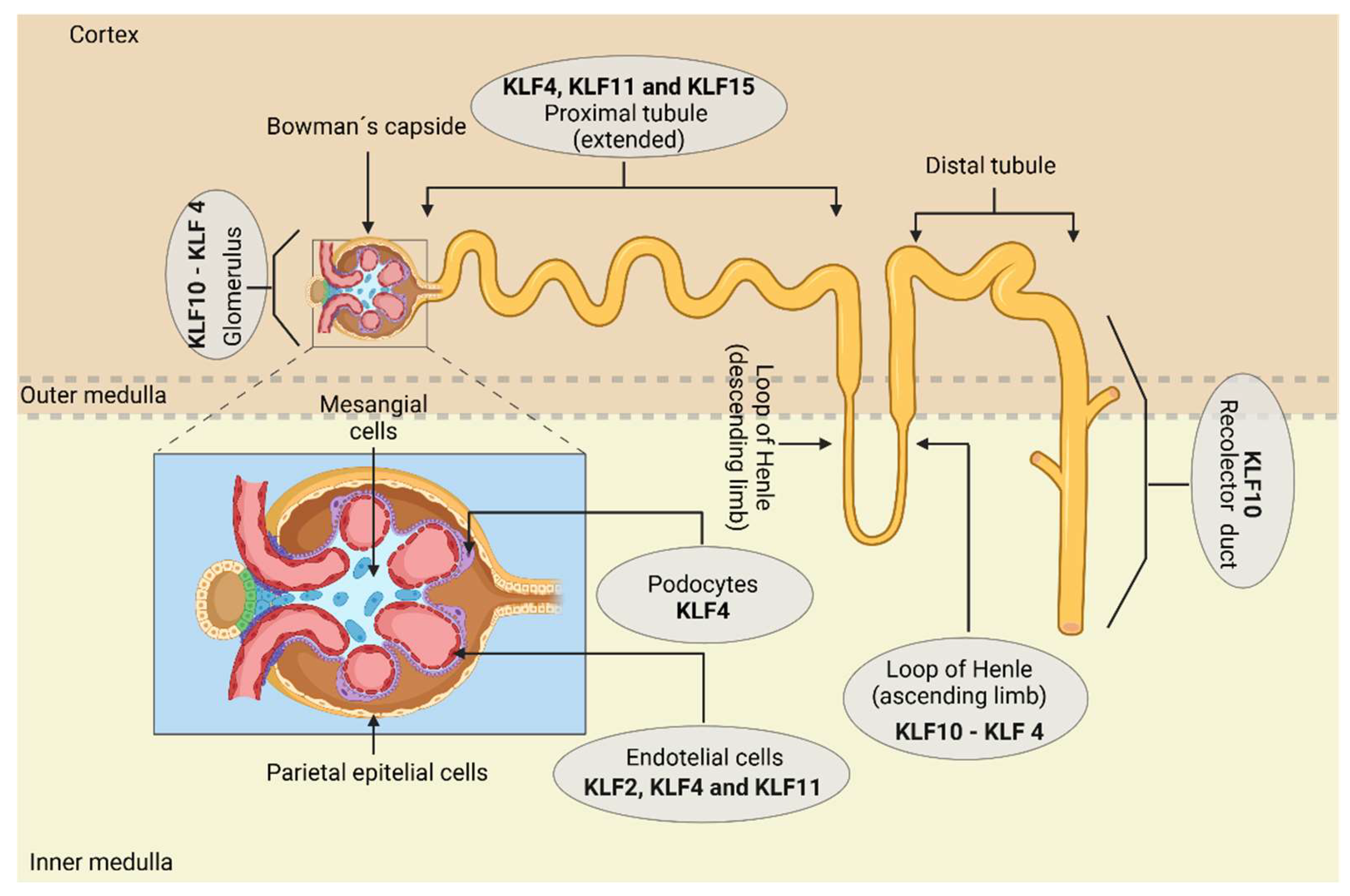
Figure 3.
KLFs involvement in regulating renal organogenesis and postnatal maturation of nephrons. The KLFs promote the survival of epithelial cells and podocytes; interesting, the survival of podocytes mediated by KLF5, through the inhibition of the mitogen-activated protein (MAP) kinase pathway, may seem like a contradictory description, as ERK has been shown to positively induce the expression of anti-apoptotic proteins such as Bcl-2. Therefore, ERK is typically associated with promoting cellular survival. However, its interaction with p38 can lead to apoptosis under severe stress conditions. For this reason, KLF5 may influence the balance between the activation of ERK and p38, so that, by modulating this signaling, cellular survival is favored. They also participate in the completion of postnatal maturation, electrolyte balance, and proper urine concentration.
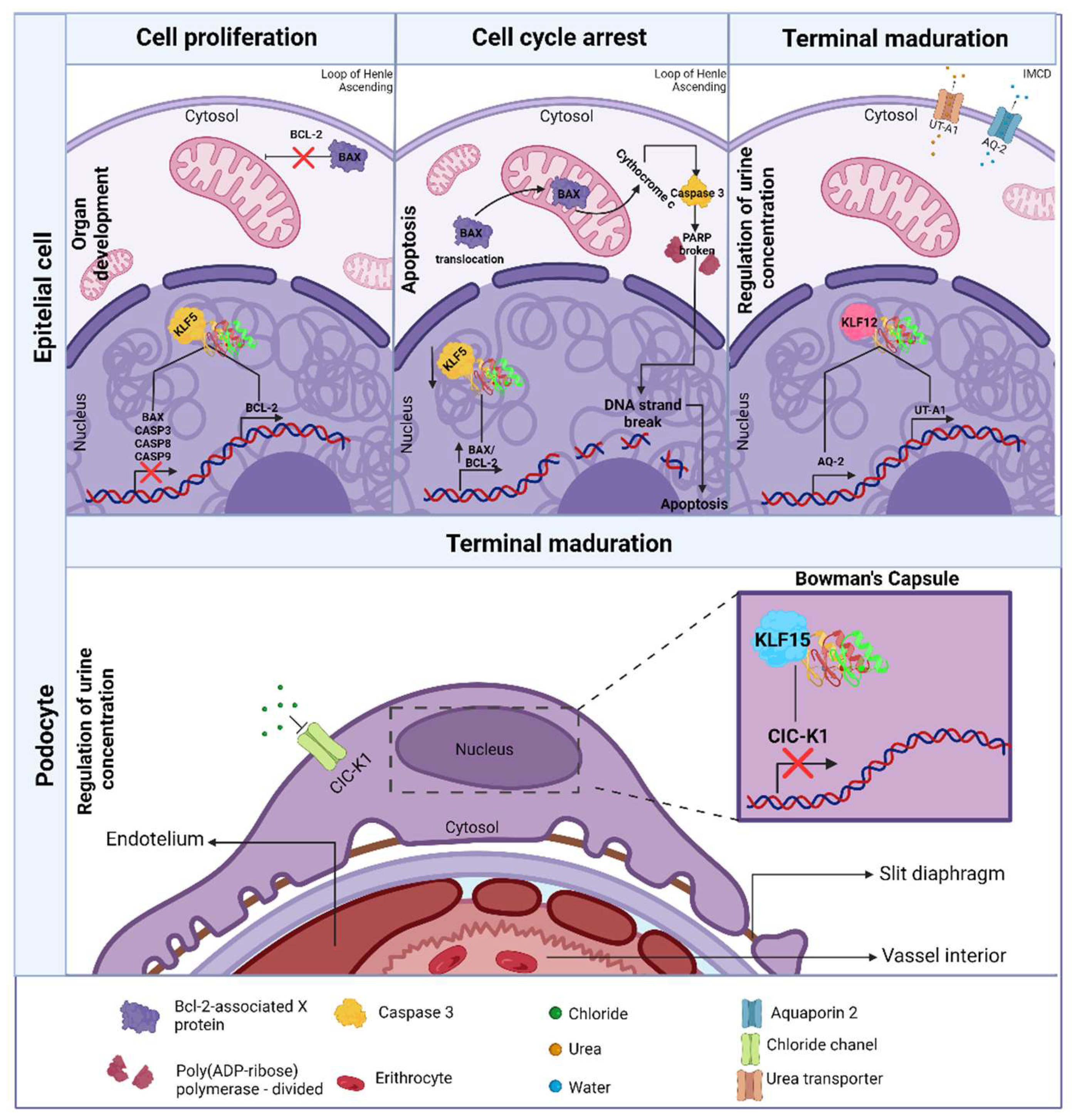
Figure 4.
Implication of KLFs in renal metabolism both under healthy conditions and their effect in states of dysregulation.
Figure 4.
Implication of KLFs in renal metabolism both under healthy conditions and their effect in states of dysregulation.
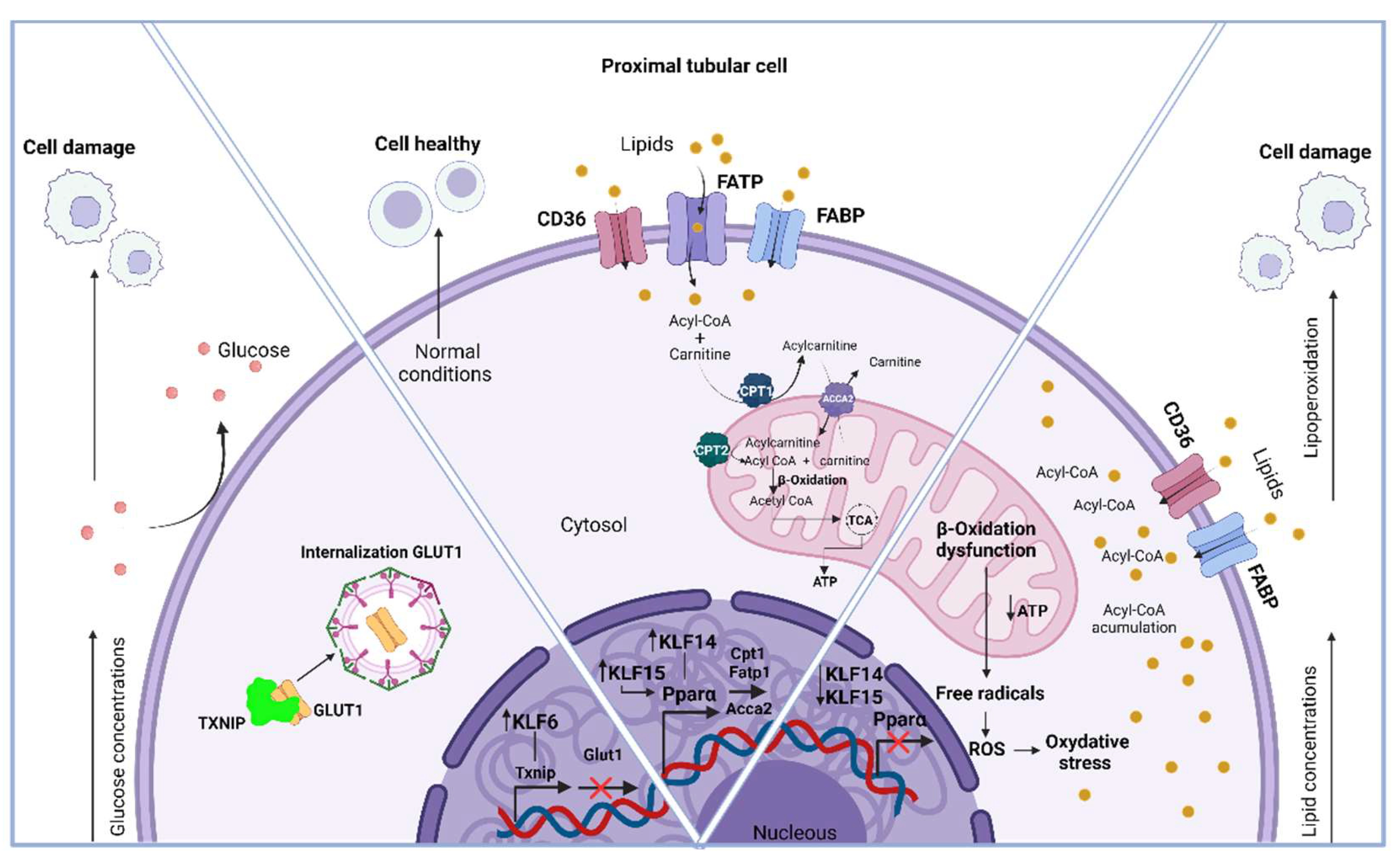

Table 1.
Briefly shows the role of KLF in nephron cells and its implication in kidney physiology. -.
Table 1.
Briefly shows the role of KLF in nephron cells and its implication in kidney physiology. -.
| Cell of nephron | KLF | Role | Reference |
|---|---|---|---|
| Podocytes | [34] | ||
| KLF4 | Induces positive expression of E-cadherin, podocin, and nephrin through interactions with HDACs, for the maintenance of tight junctions and the slit diaphragm. ____________________________________________________ Induces the expression of cytokeratins (K8 and K18) that help in the cytoskeleton's organization. _____________________________________________________ Downregulates mesenchymal markers such as vimentin and α-SMA, preventing EMT and structural damage. ____________________________________________________ Improves the conditions of diabetic nephropathy through the activation of podocyte autophagy via negative regulation of the mTOR pathway, achieved by the reduction of phosphorylated protein levels (p) mTOR and pS6K, preventing excessive extracellular ECM production |
[36,51] | |
| Glomerular endothelial cells | KLF2 | Regulates the size and distribution of transcellular pores in the ECs by inhibiting the phosphorylation of the myosin light chain. ____________________________________________________________ Modulates VEGF-A-mediated angiogenesis by downregulating its expression, preventing an excess of blood vessels. |
[44] |
| KLF4 | Mediates inflammation by downregulating VCAM1 induced by TNF-α, inhibiting the p65 subunit of NF-κB. | [51] | |
| Mesangial cells | KLF4 | Attenuates the expansion of the mesangial matrix and its proliferation by negatively regulating the mTOR pathway, downregulating the expression of phosphorylated (p) mTOR and p S6K proteins, preventing excessive extracellular ECM production, | [51] |
| Proximal tubule cells | KLF4 | Mitigates inflammation and fibrosis by decreasing the expression of pro-inflammatory cytokines, such as MCP-1, MIP-3α, and IL-8. | [55] |
| KLF11 | Like KLF4, it participates in the mitigation of inflammation and fibrosis by reducing the expression of the same pro-inflammatory cytokines. | [56] | |
| KLF15 | Decreases the expression of fibronectin by negatively regulating the MAPK pathways. | [57] |
Table 2.
shows the role of KLF’s on kidney disease.
| Disease | Group Klf | Role | Reference |
|---|---|---|---|
| Chronic kidney disease | Group 1 (Klf 3, 8 and 12) |
Not Available | |
| Group 2 (Klf 1, 2, 4, 5, 6 and 7) |
KLF2 protects endothelial cell injury through anti-inflammatory, anti-thrombotic, and anti-angiogenic effects, as it maintains the proper function of glomerular endothelial cells. Its deficiency has been shown to lead to the progression of renal disease | [94,103] | |
| KLF4 suppression causes the polarization of infiltrating macrophages into myeloid cells that accumulate in the glomerulus and tubular interstitium in CKD to shift to an M1 phenotype. The M1 phenotype of macrophages promotes the production of pro-inflammatory cytokines, such as TNFα and IL-1β. These cytokines exacerbate renal parenchymal injury and accelerate disease progression. Conversely, KLF4 expression suppresses the differentiation of infiltrating macrophages, mitigating renal damage by inhibiting TNFα expression in myeloid cells. Thus, KLF4 is considered a protective transcription factor. In addition, KLF4 mitigates inflammation and fibrosis caused by the TGF-β1-induced release of cytokines MCP-1, MIP-3α and IL-8 in human PTCs, possibly relating to the phosphorylation of KLF4 that TGF-β1 induces via SMAD and p38/MAPK signaling in vascular smooth muscle cells (VSMCs). It has even been linked to the inhibition of podocyte apoptosis through regulating the mTOR signaling pathway, which is involved in regulating cell growth, proliferation, and survival. | [55,104] | ||
| KLF5 participates in the initiation and progression of tubulointerstitial inflammation, and its expression is increased in proliferating renal tubule cells in the cortex and medulla of fibrotic kidneys. KLF5 regulates renal fibrosis through activation of HIF-1α-KLF5-TGF-β1 pathway, renal cell proliferation through activation of ERK/YAP1/KLF5/cyclin D1 pathway, and tubulointerstitial inflammation with upregulation of pro-inflammatory cytokines which promotes kidney injury. | [7,28] | ||
|
KLF6 under conditions that promote renal damage and fibrosis, such as its overexpression enables TGF-β1 to induce the loss of E-cadherin, gain in vimentin expression, and EMT of PTCs. In CKD, TGF-β promotes renal fibrosis by enhancing matrix formation, cell proliferation, and cell migration via MAPK, phosphatidylinositol 3-kinase/protein kinase B, and Smad2/3/4 pathways, subsequently elevating fibronectin, collagen, and α-SMA |
[105] | ||
| [106] | |||
| Group 3 (Klf 9, 10, 11, 13, 14 and 16) |
KLF11 deficiency is associated with increased renal atrophy, fibrosis, and interstitial inflammation in a mouse model of chronic renal obstruction (UUO). In KLF11 KO-UUO mice, this deficiency is linked to the upregulation of genes such as collagen type I, fibronectin, TGF-β1, as well as IL-6 and TNF-α. These genes are associated with TGF-β signaling, fibrosis, and inflammation. |
[56] | |
| No group (15 and 17) |
KLF15 is downregulated by TGF-β1, which activates multiple intracellular signal transduction systems and MAPK pathways, including ERK and JNK, leading to renal fibrosis. Thus, KLF15 may play an anti-fibrotic factor in renal interstitial fibrosis by decreasing extracellular matrix fibronectin, type III collagen and CTGF expression in renal fibroblast. Furthermore, the overexpression of KLF15 in mesangial cells also reduced the mRNA and protein expression of fibronectin and type IV collagen, as well as in HEK293 cells, suggesting that the inhibition of extracellular matrix expression mediated by KLF15 may not be a cell type-specific effect | [107,108] | |
| Acute kidney injury | Group 1 (Klf 3, 8 and 12) |
Not Available | |
| Group 2 (Klf 1, 2, 4, 5, 6 and 7) |
Overexpression of KLF4 in PTCs (HK-2) upregulates the expression of miR-101. This increase in miR-101, downregulates the expression of COL10A1, thereby suppressing EMT and renal fibrosis during the pathogenic process of renal fibrosis associated with acute kidney injury. In contrast, the inhibition of KLF4 expression, directly mediated by epigenetic regulatory enzymes such as DNA methyltransferase 1 (Dnmt1), which hypermethylates the KLF4 promoter region, contributes to the progression of EMT in renal EpC. | [109] | |
|
KLF5 is regulated by YAP and promotes the expression of Mst1/2, which are proteins involved in the Hippo signaling pathway. Activation of this pathway leads to over proliferation of tubular cells, tubular injury, and inflammation. KLF5 can be upregulated in severe acute kidney injury because of the activation of HIF-1α, which facilitates the transition to chronic kidney disease. The overexpression of KLF5 promotes renal fibrosis and tubular dysfunction, exacerbating acute kidney injury. ____________________________________________________________________ KLF6 is rapidly expressed in PTCs after AKI, contributing to the exacerbation of the disease by repressing genes such as Bckdha, Bckdhb, Acadm, Mut, Hibch, Ivd, Mccc1, and Mccc2 involved in branched-chain amino acid catabolism, which, through this metabolic process, provide intermediates necessary for the tricarboxylic acid cycle in the absence of FAO to produce ATP and meet the energy needs of the cells. In contrast, the decreased expression of KLF6 improves AKI and fibrosis through the particular preservation of the expression of the BCAA catabolic enzyme, Bckdhb. |
[99,110,111] | ||
| Group 3 (Klf 9, 10, 11, 13, 14 and 16) |
KLF9, which is upregulated by miR-93-5p, inhibited the expression of circHIPK3, leading to alleviation of oxidative stress and apoptosis in an in vivo model of AKI established by ischemia/reperfusion (I/R) in C57BL/6 mice or hypoxia/reoxygenation (H/R) in HK-2 cells. The circular RNA HIPK3 (circHIPK3), derived from the HIPK3 gene, is important because of its pro-inflammatory activity. | [97] | |
| KLF10 is downregulated in tubular cells during acute kidney injury. This finding suggests that KLF10 acts as a renoprotective protein and provides protection against acute kidney injury, as its induction improves tubular regeneration through the ZBTB7A-KLF10-PTEN axis. PTEN is important because it can inhibit the PI3K/Akt pathway, which regulates cell growth, death, migration, and differentiation. | [96] | ||
| KLF11 depletion in proximal epithelial cells increases the expression of endothelin-1 and IL-6, leading to elevated serum creatinine and blood urea nitrogen levels, as well as damage and death of tubular capillaries in the deep cortex and outer medulla, tubular cast formation, vascular dilation, and congestion, thereby exacerbating AKI. However, KLF11 expression reduces endothelin-1-dependent renal vasoconstriction, inflammation, and aberrant renal hemodynamics. | [112] | ||
| No group (15 and 17) |
KLF15 acts as a bridge connecting the signaling of diacylglycerol kinase epsilon (DGKE) and Klotho. This DGKE/KLF15/Klotho pathway protects against renal ischemia/reperfusion injury (IRI) and AKI in a murine model. In a Xenopus laevis model, it was showed that KLF15 directly binds to enhancers and stimulates the expression of regenerative genes, including adrenoreceptor α 1A (adra1α), suggesting that KLF15 might even promote the regeneration of nephric tubules. As KLF15 attenuates damage and development of glomerulosclerosis, tubulointerstitial fibrosis, inflammation, and stabilizes the actin cytoskeleton, thereby improving renal function. | [95,113] | |
| Diabetic kidney disease | Group 1 (Klf 3, 8 and 12) |
KLF3 directly regulates the transcription of STAT3. In proximal tubular cells (HK-2) exposed to high glucose concentrations, the suppression of KLF3 mediated by miR-23a-3p resulted in the inhibition of STAT3, a protein crucial for regulating inflammation and fibrosis associated with metabolic diseases. Thus, the inhibition of KLF3 leads to a protective effect in renal disease. | [102] |
| Group 2 (Klf 1, 2, 4, 5, 6 and 7) |
KLF2 is upregulated by insulin treatment and downregulated by high glucose concentrations in cultured endothelial cells (EC) from diabetic mice. Even in a KLF2 KO +/- EC model, it was determined that reduced KLF2 expression induced more endothelial cell damage. Additionally, in glomerular endothelial cell (GEC)-specific KLF2 knockout mice with streptozotocin-induced diabetes, it was found that the expression of podocyte-specific genes encoding nephrin, podocin, and synaptopodin in the kidney was decreased compared to wild-type diabetic mice. This suggests the possibility of an interaction mechanism between GECs and podocytes mediated by KLF2. The deletion of KLF2 (knockout, KO) in the glomeruli reduces the expression of several of its target genes, including endothelial nitric oxide synthase (eNOS), zonula occludens-1 (ZO-1), the glycocalyx, fms-related tyrosine kinase 1 (Flt1), tyrosine kinase with immunoglobulin-like and EGF-like domains 2 (Tie2), and angiopoietin 1 (Angpt1). These genes are primarily involved in the function and integrity of the vascular endothelium, which is why KLF2 is considered a vasoprotective factor. In fact, a potential mechanism by which KLF2 may decrease its expression under high glucose conditions has been demonstrated in human umbilical vein endothelial cells, where KLF2 transcriptional silencing is mediated by FOXO1, which binds to the KLF2 promoter region. ____________________________________________________________________KLF4 overexpression induces podocyte autophagy, protecting the tissue from damage in DKD. Suppresses cell proliferation and differentiation during fibrosis and inhibits EMT processes. Hyperglycemia also decreases KLF4 expression and increases TGF-β expression leading to unregulated inflammation in renal tissue. ____________________________________________________________________ KLF5 is overexpressed in the collecting duct EpC found in diabetic kidney and tubulointerstitial disease and associated with alterations like an expansion of mesangial matrix and tubular interstitial space, podocyte damage, and glomerular basement membrane thickening, showing that KLF5 plays a pivotal role in the initiation and progression of renal inflammation. In fact, the inverse expression of KLF4 and KLF5 in the pathogenesis of renal fibrosis is modulated by a matrix stiffness-regulated extracellular signal-regulated kinase (ERK), which increases the protein level and nuclear translocation of mechanosensitive YAP1, preventing the degradation of KLF5. KLF5 is upregulated under hyperglycemic conditions through lactylation of lysine 14 on histone H3 (H3K14la). KLF5 binds to the promoter of the gene encoding E-cadherin (Cadherin 1, cdh1) and inhibits its transcription, promoting disease progression. This lactylation results from the accumulation of lactate because of the metabolic reprogramming that renal PCT undergo in a hyperglycemic state, specifically the shift from oxidative phosphorylation (OXPHOS) to glycolysis. ____________________________________________________________________KLF6, under conditions that promote renal damage and fibrosis, such as diabetic nephropathy, its overexpression enables TGF-β1 to induce the loss of E-cadherin, gain in vimentin expression, and EMT of proximal tubule cells. In CKD, TGF-β promotes renal fibrosis by enhancing matrix formation, cell proliferation, and cell migration via MAPK, phosphatidylinositol 3-kinase/protein kinase B, and Smad2/3/4 pathways, subsequently elevating fibronectin, collagen, and α-SMA. |
[7,114,115,116] [7,51,114,115] [10,28,117] [106] |
|
| Group 3 Klf 9, 10, 11, 13, 14 and 16) |
KLF 10 Activates KDM6A and induces proteinuria, kidney damage and fibrosis under diabetic conditions. Represses nephrin, WT1, podocin, and synaptophysin in podocytes. Increases expression of type I and III collagen, fibronectin, and metalloproteinases. | [114,118,119] | |
| No group and (KLF15 and KLF17) |
KLF15 modulates mitochondrial biogenesis and homeostasis through the SIRT1-PGC-1α pathway in mouse mesangial cells associated with diabetic nephropathy. This finding was determined through enrichment analysis, which identifies KLF15 as a therapeutic target. | [120] |
|
Disclaimer/Publisher’s Note: The statements, opinions and data contained in all publications are solely those of the individual author(s) and contributor(s) and not of MDPI and/or the editor(s). MDPI and/or the editor(s) disclaim responsibility for any injury to people or property resulting from any ideas, methods, instructions or products referred to in the content. |
© 2024 by the authors. Licensee MDPI, Basel, Switzerland. This article is an open access article distributed under the terms and conditions of the Creative Commons Attribution (CC BY) license (http://creativecommons.org/licenses/by/4.0/).
Copyright: This open access article is published under a Creative Commons CC BY 4.0 license, which permit the free download, distribution, and reuse, provided that the author and preprint are cited in any reuse.


