
Submitted:
16 September 2024
Posted:
17 September 2024
You are already at the latest version
Abstract
Superficial acral fibromyxoma (SAF) is a benign fibromyxoid tumor that presents as a slow-growing nodule on the subungual and periungual areas of the hands and feet. Superficial acral fibromyxoma needs to be better documented in the literature, and thus, increased research and education are required to aid clinicians in recognizing its appearance. To review the current literature on superficial acral fibromyxoma and present its epidemiology, pathogenesis, clinical presentation, diagnosis, treatment, and common differential diagnosis. A literature review was performed on Pubmed and Scopus for case reports, case series, clinical studies, and reviews that described epidemiology, clinical presentation, pathogenesis, histological examination, and treatment of superficial acral fibromyxoma. The gathered literature was analyzed to extract concise information on superficial acral fibromyxoma. Results: Superficial acral fibromyxoma is associated with Rb1 gene deletion and grows as a 0.5 to 5.0-centimeter gelatinous-to-firm nodular mass on periungual or subungual regions of middle-aged males. It is also noted to disrupt nail architecture and cause lytic or erosive lesions of bone. Histopathologically, superficial acral fibromyxoma comprises stellate and spindle cells surrounded by a myxo-collagenous matrix and is strongly positive for CD34, CD99, nestin, and vimentin. Complete surgical excision is the first-line treatment for superficial acral fibromyxoma; however, the tumor has a post-operative recurrence rate of 20-25%. Mohs surgery has been attempted to decrease the rate of recurrence. After excluding other cutaneous neoplasms, superficial acral fibromyxoma should be considered in patients with a periungual or subungual slow-growing mass and other soft tissue. Complete surgical excision can lead to recurrence; therefore, patient follow-up is necessary.
Keywords:
superficial acral fibromyxoma
; Rb 1 deletion
; ICH
; sclerosing perineuroma
; dermatofibrosarcoma protuberans
Introduction
Superficial acral fibromyxoma (SAF) is a benign fibromyxoid tumor that presents as a slow-growing nodule on the subungual and periungual areas of the hands and feet. It occurs predominantly in middle-aged men. Due to the rarity of superficial acral fibromyxoma, increased research and education are required to aid clinicians in recognizing its non-specific appearance when presented with a fibromyxoid tumor of the acral site [1].

Table 1.
Common differential diagnoses of superficial acral fibromyxoma.
| Pathology | Color and Appearance | Growth Rate | Area of Predicilition | Associated with Pain | Average age | Male or Female Predominance |
|---|---|---|---|---|---|---|
| Superficial acral fibromyxoma | Firm flesh colored mass | Slow-growing; around 3 to 30 years | Fingers and toes | Painless | Mean age of 43 (4-86) | Male |
| Dermatofibrosarcoma protuberans | pink-to-skin-color plaque that slowly grows into a painless to painful polypoid to multinodular mass | Slow-growing | Trunk | Painless | Present in all ages; Commonly seeen between ages 20-50 | Male |
| Sclerosing perineuroma | Soft grey colored lesion with thickened collagen patterns | Slow growing ~10 years | Extremeties | Painless | Young adults | Male |
| Acquired digital fibrokeratoma | Benign fibrous flesh colored-yellow textureed tumor around 3-5 mm | Growth for several months then with growth phase for several weeks | Fingers and toes | Painless | Adults between the age of 12-70 | Slight predominance in males |
| Myxoid neurofibroma | Skin colored nodular plaque | Slow growing from months to years | Face, shoulders, arms, periungual and in the fee | Painless | Young adults | No gender predominance |
| Superficial angiomyxoma | red-to-pink or skin-colored papule, nodule, or papule | Light to skin colored nodule with a translucent surface | scalp and neck | Painless | Middle aged adults | Male |
| Low-grade fibromyxoid sarcoma | Light tan smooth colored tumor | Slow | shoulders, trunk, and thigh | Painless | Median age of 33 years | Male |
Table 2.
Immunohistochemical stains of common differential diagnoses of superficial acral fibromyxoma.
Table 2.
Immunohistochemical stains of common differential diagnoses of superficial acral fibromyxoma.
| Pathology | Positive Immunohistochemical Stains | Negative Immunohistochemica Stains |
|---|---|---|
| Superficial acral fibromyxoma | CD34 CD99 Nestin Vimentin |
Keratin Claudin-1 Glial fibrillary acidic protein MUC4 AE1/AE3 Cam5.2 STAT6 S100 HMB-45 |
| Dermatofibrosarcoma protuberans | Vimentin Nestin CD34 |
EMA |
| Sclerosing perineuroma | EMA Vimentin Collagen IV Claudin-1 |
S100 |
| Acquired digital fibrokeratoma | FXIIIA | EMA S100 |
| Myxoid neurofibroma | S100 Mucin |
- |
| Superficial angiomyxoma | CD34 | S-100 Smooth muscle actin |
| Low-grade fibromyxoid sarcoma | MUC4 | - |
Objective
This review discusses the clinical presentation, pathogenesis, diagnosis, treatment, and common differential diagnosis of superficial acral fibromyxoma.
Methods
Pubmed and Scopus databases were searched to identify case reports, case series, clinical studies, and reviews that illustrated the epidemiology, clinical presentation, pathogenesis, histological examination, and treatment of superficial acral fibromyxoma. The results section analyzed and summarized the gathered literature on superficial acral fibromyxoma.
Results
Epidemiology
Superficial acral fibromyxoma, more commonly known as digital fibromyxoma, is a rare but benign solitary soft tissue tumor of mesenchymal origin that frequently presents as a slow-growing nodular mass on the toes (Figure 1) [2] and fingers of males in their 5th to 6th decade [1,3]. In 2001, Fetsch et al. examined 37 cases to describe superficial acral fibromyxoma as a distinct entity [1,4]. From 2001 till 2011, only 50 patients with superficial acral fibromyxoma have been examined and documented in the literature [1,4,5]. A study in 2012 observed around 124 total superficial acral fibromyxoma cases, further corroborating superficial acral fibromyxoma’s infrequent nature, primarily due to its nonspecific clinical picture coupled with clinicians’ lack of knowledge surrounding fibromyxoma [6,7,8]. Superficial acral fibromyxoma has a two-to-one male-to-female ratio, with the average age of diagnosis being 48 years and the median age being 49 years [6,8]. Albeit the average age of diagnosis, it has been reported to surface in patients 4 to 86 years old [5,6,7,8]. A review of 314 cases done by Crepaldi et al. found that 47 years and seven months was the mean age of diagnosis, with an age range of 4 and 91 years. They also found that out % of 306 cases, 61% were males and 39% were females [3].
Clinical Presentation
On physical examination, superficial acral fibromyxoma presents as skin-colored with occasional pink-to-white or red hue, gelatinous-to-firm solitary nodular mass on periungual or subungual regions of the toes and fingers of middle-aged adults [1,2,3,4,5,6]. Physicians additionally describe the tumor surface as polypoid, verrucous, or dome-shaped. A study by Hollmann et al. reported on the typical locations of the superficial acral fibromyxoma, with 82% of foot lesions presenting on the toes and 94% of hand lesions presenting on the digits [7,8]. Hollmann et al. also state that around 96% of superficial acral fibromyxoma on the toes and 97% of superficial acral fibromyxoma located on the fingers were near the nail apparatus [8]. Though a majority of superficial acral fibromyxoma lesions are present on the digits of the hands and feet, other less commonly affected areas include the wrist, palms, webspace, posterior thighs, calf, ankles, heels, neck (Figure 2) [9] and lower legs [3,6,7,8,9]. The tumor has been documented to range from 0.5 to 5.0 centimeters in diameter (with the average size being 1.7 cm) [3,6,8]. Since the lesion is slow-growing, patients may present anywhere from 3 months to 30 years, as the lesion may have been previously complex to identify [6,7,8]. Crepaldi et al. found that the average time from symptom presentation to the diagnosis of superficial acral fibromyoxoma was over ten years [3]. Hence, unnecessary delay in evaluation and subsequent medical treatment is typical. The lesion can be asymptomatic to extremely painful, as around 41% of superficial acral fibromyxoma are associated with pain [6,7,8]. Furthermore, superficial acral fibromyxoma cases have also been accompanied by concurrent infection and bleeding [6], disruption of nail architecture with subsequent deformity [7], and lytic or erosive lesions of bone due to periosteum involvement [10]. Hollmann et al. found that 36% of the cases involved erosive or lytic lesions of the bone [8]. Prior trauma may be a rare risk factor, as reported in 25% of cases, though no heritable or secondary medical conditions have been documented as potential correlating risk factors [6,7].
Pathogenesis
Recent studies contend that an Rb1 deletion may be an underlying factor in developing superficial acral fibromyxoma [10]. Some scientists believe that if Rb1 (a tumor suppressor encoded by chromosome 13q14.2) is deleted, a downstream dysregulation of the cell cycle can occur, leading to a proliferation of fibromyxoid neoplasm [10]. Agaimy et al. observed the loss of Rb1 immunoexpression in 90% of studied cases and successfully demonstrated Rb1 gene deletion by fluorescence in situ hybridization. This study highlighted the importance of using Rb1 immunohistochemistry and FISH in diagnosing superficial acral fibromyxoma and possible insight into the pathogenesis of this cutaneous manifestation [10].
Imaging
Sundaramurthy et al. describe a case where digital superficial acral fibromyxoma presented as a homogeneous soft tissue swelling under x-ray imaging [1]. T1 weighted MRI revealed a hypotensive complex multilocular mass with associated fibrous septations, and T2 weighted MRI displayed a hyperintense complex multiloculated mass. Bindra et al. describe another case of digital superficial acral fibromyxoma presenting as a tiny focal soft tissue mass with erosion and scalloping features underlying the bone cortex on x-ray [11]. On T2 weighted MRI, the cutaneous manifestation presented as a hyperintense lobulated mass, versus T1 weighted MRI, which showed the lesion to be isointense compared to the underlying skeletal muscle. A post-contrast T1 weighted image demonstrated significant central enhancement of the lesion, alluding to the vasculature nature of the lesion [11].
Macroscopic Examination
On macroscopic examination, superficial acral fibromyxoma is often a well-circumcised, non-encapsulated, dome-shaped, verrucous, or polypoid mass that diffusely involves the dermis [1,4]. It is usually described as a gray-to-white, gelatinous-to-firm solid lesion on sectioning. Occasional cases have been shown to extend into the subcutaneous tissue on gross examination [5,6,7,8].
Microscopic Examination
The microscopic examination of superficial acral fibromyxoma is described as a prominent dermal proliferation of a moderate amount of star to spindle-shaped, fibroblast-like, and uniform stellate cells with pale eosinophilic cytoplasm arranged in storiform or, less commonly, a loose fascicular pattern (Figure 3). The background stroma can be collagenous to myxoid, with many cases displaying an alteration of myxoid, fibrous, and cartilaginous background stroma [3,5,6,7,8]. Occasional extension to the subcutaneous fat, periosteal layer, and fascia can be observed [3,5,6,7]. Hollmann et al. found that around 27% of superficial acral fibromyxoma cases infiltrated underlying fat, and 3% invaded underlying bone [8]. Superficial acral fibromyxoma is morphologically a lobular mass. It is often described as either having a “pushing” margin or an ill-defined and infiltrative margin with dispersed mast cells and occasional multinucleated giant cells [5,6,8]. Hollmann et al. described that 88% of the studied cases had associated mast cells [8]. Rare cases have also shown lymphocyte infiltration, possibly due to nonspecific chronic inflammation [6]. Ulceration of the mass has been noted to occur, which can increase the infiltration of neutrophils, plasma cells, and histiocytes.
Hyperkeratosis of the epidermis can occur as a result, with an epithelial collarette often present even when the lesion does not involve the epidermis [5,6]. Mitosis and nuclear atypia are typically absent in standard superficial acral fibromyxoma, but mild atypia has been identified in severe cases [6,7]. Similarly, although necrosis is not a prominent feature in standard superficial acral fibromyxoma, rare cases have been documented to have associated necrosis and osseous and cartilaginous metaplasia [8]. Severe lesions involving a predominance of myxoid stroma are often accompanied by accentuated microvasculature, leading to hypersensitivity of the area and an increased propensity for bleeding. One rare case with admixed benign and mature adipose cells has been described. Hence, the above scenario must exclude lipoma, liposarcoma, and dedifferentiated liposarcoma [5,6,7].
Immunohistochemistry
Immunohistochemistry is vital to the diagnosis of superficial acral fibromyxoma. Superficial acral fibromyxoma cells are strongly positive for CD34, CD99, nestin, and vimentin [7]. In contrast, superficial acral fibromyxoma has variable focal positivity for EMA. Hollmann et al. found 7.5% of cases to have focal reactivity for EMA [8]. Rare cases have shown positive staining for desmin and SMA but can be considered non-specific findings. As mentioned, immunohistochemistry will also show a loss of the tumor suppressor gene Rb1 (Figure 3). CD10 immunohistochemical stain has been weakly to strongly positive in rare cases of superficial acral fibromyxoma that involve peri adnexal mesenchymal cells and onychoblasts [1,6,7,10]. Superficial acral fibromyxoma is negative for keratin, claudin-1, glial fibrillary acidic protein, MUC4, AE1/AE3 , Cam5.2, STAT6, S100, and HMB-45. Fetsch et al. describe one case of superficial acral fibromyxoma with weak S100 immunoreactivity, though this weak response can be excluded as it was deemed a non-specific outlier [4,5,6,7].
Differential Diagnosis
The differential diagnosis of superficial acral fibromyxoma is broad, comprising malignant and benign pathologies. Clinicians and dermatopathologists should keep dermatofibrosarcoma protuberans, perineuroma, acquired digital fibrokeratoma, myxoid neurofibroma, superficial angiomyxoma, and low-grade fibromyxoid sarcoma as top differentials are given certain similar features.
Dermatofibrosarcoma protuberans (DFSP) is a rare, superficial, and locally aggressive mesenchymal soft tissue tumor that presents as pink-to-skin-colored plaque that slowly grows into a painless to painful polypoid to multinodular mass with occasional atrophic changes [11,12]. In contrast to superficial acral fibromyxoma, dermatofibrosarcoma protuberans occur in young to middle-aged adults but, like superficial acral fibromyxoma, tend to occur slightly more in male patients. Unlike superficial acral fibromyxoma, it tends to involve the trunks and extremities with rare involvement of hands, feet, and nailbeds. On microscopic examination, dermatofibrosarcoma protuberans involves the monotonous proliferation of dermal spindle cells arranged in a cartwheel, whorled, or storiform pattern with the development of a honeycomb appearance with significant infiltration of dermal appendages, subcutis, and adipocytes. Even though it is positive for vimentin, nestin, and CD34, it is negative for EMA, which can be focally positive in SAF [6,13]. Molecular studies can confirm dermatofibrosarcoma protuberans and exclude superficial acral fibromyxoma because dermatofibrosarcoma protuberans are positive for t(17;22)(q22;q13) translocation with COL1A1-PDGFB fusion by FISH. Nuclear RB1 expression should be retained in dermatofibrosarcoma protuberans in contrast to absent expression in superficial acral fibromyxoma [5,6,11,12].
Sclerosing perineuroma (SP) is a rare variant of perineurioma, a tumor that arises from perineural cells [14]. Sclerosing perineuroma presents similarly to superficial acral fibromyxoma as it is an asymptomatic, slow-growing pink-to-skin-colored fibrous lesion that presents as a papule or nodule on the digits [6,14]. Sclerosing perineuroma, however, can commonly present on the palms and is preferred by young adults [5,6]. A study performed by Fetsch et al. showed a male predominance with a median age of 24.5 and an age range of 9-55 years [15]. Histology can help differentiate the two pathologies in that sclerosing perineuroma is a well-circumcised lesion composed of a hypocellular population of small epithelioid and spindle cells with tapered nuclei and bipolar cytoplasm in a corded, whorled, trabecular or onion skin pattern with a background of dense collagen and is found in either the dermis or hypodermis. Sclerosing perineuroma tumor cells, like superficial acral fibromyxoma tumor cells, are positive for EMA and vimentin and negative for S100; however, sclerosing perineuroma is also favorable for collagen IV and has a membranous granular positivity for Claudin-1. Unlike superficial acral fibromyxoma, sclerosing perineuroma tends not to reoccur after excision [5,6,14,15,16,17].
Acquired digital fibrokeratoma is a rare benign cutaneous pathology that, like SPF, appears on the digits of the hands and feet but uniquely presents as a painless skin-colored or red to pink hyperkeratotic dome-shaped papule with scaling [5,18,19]. It tends to occur in middle-aged men and has been associated with regional trauma. Acquired digital fibrokeratoma is a small tumor that tends to be less than 1 cm and can be histologically differentiated from superficial acral fibromyxoma as it has prominent acanthosis with cores of thick bundles of collagen oriented vertically in the hypocellular dermis [18,19]. Acquired digital fibrokeratoma can have variable CD34 staining but harms EMA and S100. It has positive staining for FXIIIA(+). In contrast to Superficial acral fibromyxoma, it has no recurrence after surgical excision [5,6,18].
Myxoid neurofibroma, a benign perineural tumor, presents as a solid, painless, slow, growing flesh-colored solitary nodule and can occur on the feet and periungual regions [6,20]. However, unlike SPF, it can also be commonly found in the upper extremities and on the face of young adults. It is histologically composed of spindle cells with wavy nuclei that are S100 positive with associated mucin, in contrast to Superficial acral fibromyxoma. Similar to superficial acral fibromyxoma, it may reoccur with incomplete excision [4,6,20].
Superficial angiomyxoma, also known as cutaneous myxoma, is a poorly circumcised benign myxoid tumor. It presents up to a 5 cm red-to-pink or skin-colored papule, nodule, or papule lesion [6]. However, it has a prediction for the scalp and neck of men. It can also occur in the trunk, lower extremities, and genital areas. It tends to occur in middle-aged men. In a study by Calonje et al., the median age of diagnosis was 45.5 years [21]. It is also composed of dermal spindled and stellate cells with an extensive myxoid stroma but is arranged in a multilobular pattern with prominent mixed stromal inflammatory cells and vasculature. Similar to superficial acral fibromyxoma, superficial angiomyxoma is negative for S-100 protein and smooth muscle actin and positive for CD34 [5,6,21].
Lastly, low-grade fibromyxoid sarcoma (LGFS) is a malignant, painless, slow-growing mass that is deep with a predisposition to young adults’ shoulders, trunks, and thighs [7]. The age range is 10 to 69 years, with the average age being 33 years, and the tumor predominantly occurs in males. Most low-grade fibromyxoid sarcomas are deep-seated, whereas superficial types are more common in children. Like superficial acral fibromyxoma, LGFS also comprises spindle cells arranged in a whorled pattern with a predominant transition from myxoid to fibrous stroma. In contrast to superficial acral fibromyxoma, LGFS is positive for MUC4 [6,22].
Treatment
Complete surgical excision is the first-line treatment for Superficial acral fibromyxoma; however, it has a recurrence rate of 20-25%, with a positive margin being the dominant risk factor for recurrence [10,23]. They tend to recur on an average of 27 months post-excision. The overall prognosis is good as no metastases nor malignant transformation has been described through the increased rate of recurrence results in disfigurement and additional surgery. Hankinson et al. treated a Superficial acral fibromyxoma and destroyed the nail fold and matrix with Mohs surgery to prevent recurrence and disfigurement [22]. A section was frozen using cryostat to check for remaining positive margins. A positive medial margin was noted on examination, and a second stage was performed, resulting in negative margins of the lesion. Six weeks post-surgery, the lesion had healed by the second intention, and the patient reported no pain but regained function in the treated area. Through this procedure, 75% of his nail bed was salvaged with a naturally appearing digit. Hankinson et al. recommended using Mohs surgery to prevent superficial acral fibromyxoma recurrence through margin control, improving the functionality and cosmetic appearance of the lesion [24].
Conclusions
Superficial acral fibromyxoma is a rare, well-circumscribed, nonencapsulated tumor with a predilection for the ungual region of the fingers and toes of middle-aged adults. The possibility of a Superficial acral fibromyxoma should be considered when a patient presents with a periungual or subungual slow-growing mass. Given the rare and nonspecific presentation of Superficial acral fibromyxoma, there is a lack of literature and research regarding the pathophysiology and treatment needed to prevent recurrence and deformity.
Superficial acral fibromyxomas may present similarly to dermatofibrosarcoma protuberans, sclerosing perineuromas, acquired digital fibrokeratomas, myxoid neurofibromas, superficial angiomyxomas, and low-grade fibromyxoid sarcomas, so it is of the utmost importance physicians remain aware of the similarities and differences amongst the cutaneous pathologies. Complete surgical excision is the recommended method of therapy; however, there are also chances of recurrence. Therefore, patient follow-up is necessary.
Funding
No funding sources were utilized in the composition of this research.
Conflicts of Interest
The authors report no conflicts of interest.
References
- Sundaramurthy, N.; Parthasarathy, J.; Mahipathy, S.R.; Durairaj, A.R. Superficial Acral Fibromyxoma: A Rare Entity—A Case Report. J Clin Diagn Res. 2016, 10, PD03–PD05. [Google Scholar] [CrossRef] [PubMed]
- Kura, M.M.; Jindal, S.R. Solitary superficial acral angiomyxoma: an infrequently reported soft tissue tumor. Indian J Dermatol. 2014, 59, 529. [Google Scholar] [CrossRef] [PubMed]
- Crepaldi, B.E.; Soares, R.D.; Silveira, F.D.; Taira, R.I.; Hirakawa, C.K.; Matsumoto, M.H. Superficial Acral Fibromyxoma: Literature Review. Rev Bras Ortop (Sao Paulo). 2019, 54, 491–496. [Google Scholar] [CrossRef] [PubMed]
- Fetsch, J.F.; Laskin, W.B.; Miettinen, M. Superficial acral fibromyxoma: a clinicopathologic and immunohistochemical analysis of 37 cases of a distinctive soft tissue tumor with a predilection for the fingers and toes. Hum Pathol. 2001, 32, 704–714. [Google Scholar] [CrossRef] [PubMed]
- Ashby-Richardson, H.; Rogers, G.S.; Stadecker, M.J. (2011). Superficial acral fibromyxoma: an overview. Archives of pathology & laboratory medicine, 1066. [Google Scholar] [CrossRef]
- Sawaya, J.L.; Khachemoune, A. Superficial acral fibromyxoma. International Journal of Dermatology 2015, 54, 499–508. [Google Scholar] [CrossRef] [PubMed]
- Choi, J.H.; Ro, J.Y. The 2020 WHO Classification of Tumors of Soft Tissue: Selected Changes and New Entities. Adv Anat Pathol. 2021, 28, 44–58. [Google Scholar] [CrossRef] [PubMed]
- Hollmann, T.J.; Bovée, J.V.; Fletcher, C.D. Digital fibromyxoma (superficial acral fibromyxoma): a detailed characterization of 124 cases. The American journal of surgical pathology 2012, 36, 789–798. [Google Scholar] [CrossRef] [PubMed]
- Hwang, Y.J.; Lee, H.W.; Lee, I.S.; Jung, S.G.; Lee, H.K. Superficial angiomyxoma of the posterior neck. Arch Craniofac Surg. 2021, 22, 62–65. [Google Scholar] [CrossRef] [PubMed]
- Agaimy, A.; Michal, M.; Giedl, J.; Hadravsky, L.; Michal, M. Superficial acral fibromyxoma: clinicopathological, immunohistochemical, and molecular study of 11 cases highlighting frequent Rb1 loss/deletions. Human pathology 2017, 60, 192–198. [Google Scholar] [CrossRef] [PubMed]
- Bindra, J.; Doherty, M.; Hunter, J.C. Superficial acral fibromyxoma. Radiology case reports 2015, 7, 751. [Google Scholar] [CrossRef] [PubMed]
- Brooks, J.; Ramsey, M.L. Dermatofibrosarcoma Protuberans. [Updated 2022 Nov 12]. In: StatPearls [Internet]. Treasure Island (FL): StatPearls Publishing; 2022 Jan-. Available from: https://www.ncbi.nlm.nih. 5133. [Google Scholar]
- Neff, R. , Collins, R., & Backes, F. Dermatofibrosarcoma protuberans: A rare and devastating tumor of the vulva. Gynecologic oncology reports, 2019; 29, 9–11. [Google Scholar] [CrossRef]
- Armengot-Carbo, M.; Millán, F.; Sanjuan, J.; Quecedo, E.; Gimeno, E. Sclerosing perineurioma: case report and literature review. Dermatol Online J. 2014, 20, 13030/qt92s86728. [Google Scholar] [CrossRef]
- Fetsch, J.F.; Miettinen, M. Sclerosing perineurioma: a clinicopathologic study of 19 cases of a distinctive soft tissue lesion with a predilection for the fingers and palms of young adults. Am J Surg Pathol. 1997, 21, 1433–1442. [Google Scholar] [CrossRef] [PubMed]
- Bhat, W.; Akhtar, S.; Teoh, V.; Bourke, G. Sclerosing perineuroma in paediatric fingers: a rare distinct and under-recognised entity. J Plast Reconstr Aesthet Surg. 2014, 67, 1161–1162. [Google Scholar] [CrossRef] [PubMed]
- Tancredi, A.; Graziano, P.; Dimitri, L.; Impagnatiello, E.; Taurchini, M. Left Supraclavicular Swelling: Sclerosing Perineurioma. Eurasian J Med. 2018, 50, 47–49. [Google Scholar] [CrossRef] [PubMed]
- Tabka, M.; Litaiem, N. Acquired Digital Fibrokeratoma. [Updated 2022 May 1]. In: StatPearls [Internet]. Treasure Island (FL): StatPearls Publishing; 2022 Jan-. Available online: https://www.ncbi.nlm.nih. 1 May 5451. [Google Scholar]
- Ali, M.; Mbah, C.A.; Alwadiya, A.; Nur, M.M.; Sunderamoorthy, D. Giant fibrokeratoma, a rare soft tissue tumor presenting like an accessory digit, a case report and review of literature. Int J Surg Case Rep. 2015, 10, 187–90. [Google Scholar] [CrossRef] [PubMed] [PubMed Central]
- Ponce-Olivera, R.M.; Tirado-Sanchez, A.; Peniche-Castellanos, A.; Peniche-Rosado, J.; Mercadillo-Perez, P. Myxoid neurofibroma: an unusual presentation. Indian J Dermatol. 2008, 53, 35–36. [Google Scholar] [CrossRef]
- Calonje, E.; Guerin, D.; McCormick, D.; Fletcher, C.D. Superficial angiomyxoma: clinicopathologic analysis of a series of distinctive but poorly recognized cutaneous tumors with tendency for recurrence. Am J Surg Pathol. 1999, 23, 910–917. [Google Scholar] [CrossRef] [PubMed]
- Mohamed, M.; Fisher, C.; Thway, K. Low-grade fibromyxoid sarcoma: Clinical, morphologic and genetic features. Ann Diagn Pathol. 2017, 28, 60–67. [Google Scholar] [CrossRef] [PubMed]
- Hashimoto, K.; Nishimura, S.; Oka, N.; Tanaka, H.; Kakinoki, R.; Akagi, M. Aggressive superficial acral fibromyxoma of the great toe: A case report and mini-review of the literature. Mol Clin Oncol. 2018, 9, 310–314. [Google Scholar] [CrossRef] [PubMed] [PubMed Central]
- Hankinson, A.; Holmes, T.; Pierson, J. Superficial Acral Fibromyxoma (Digital Fibromyxoma): A Novel Treatment Approach Using Mohs Micrographic Surgery for a Recurrence-Prone Digital Tumor. Dermatol Surg. 2016, 42, 897–899. [Google Scholar] [CrossRef]
Figure 1.
A firm round tender tumor on the lateral aspect of the 4th toe.
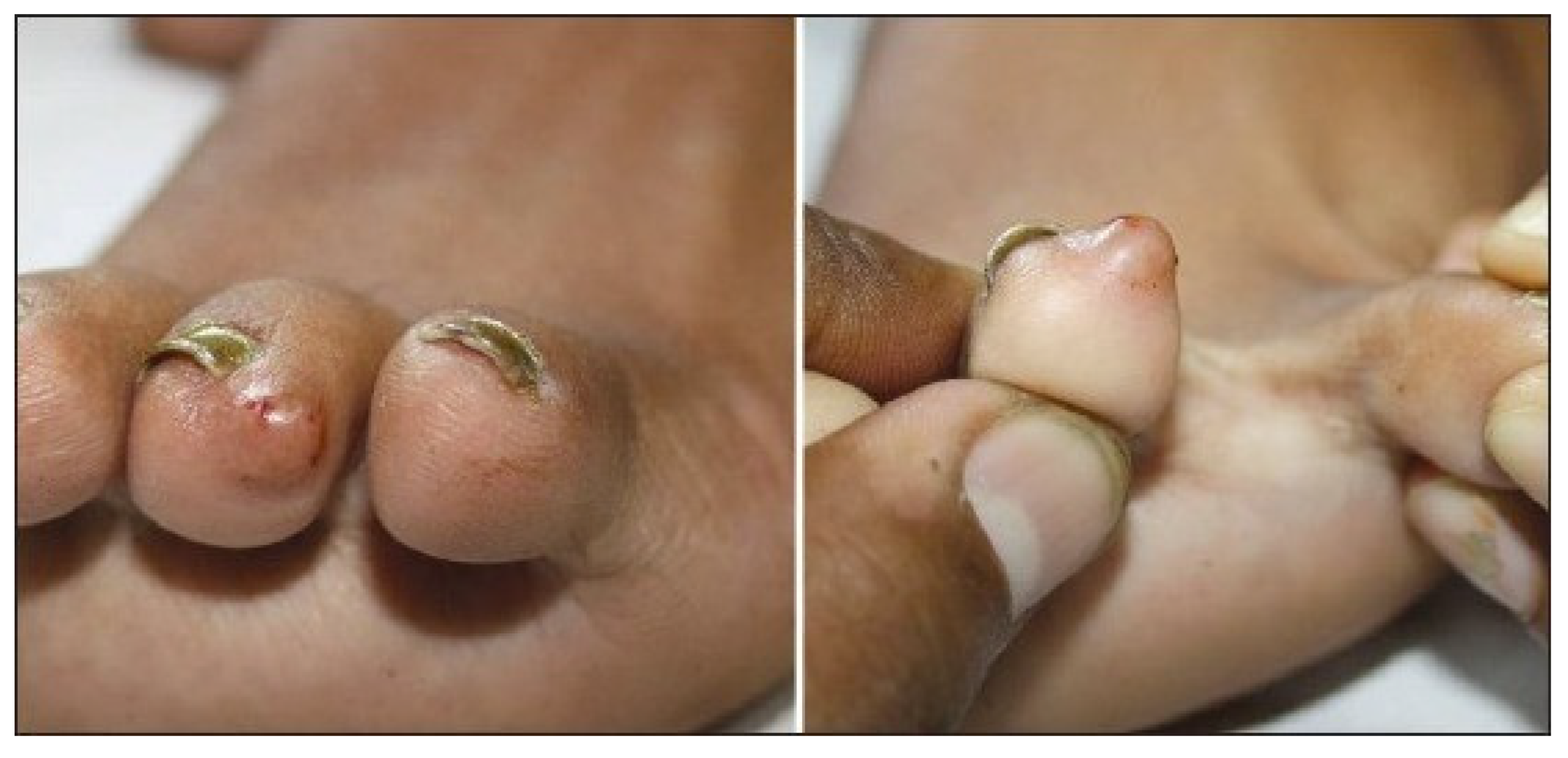
Figure 2.
Preoperative photograph of a solitary mass on the posterior neck of a 6-year-old boy.

Disclaimer/Publisher’s Note: The statements, opinions and data contained in all publications are solely those of the individual author(s) and contributor(s) and not of MDPI and/or the editor(s). MDPI and/or the editor(s) disclaim responsibility for any injury to people or property resulting from any ideas, methods, instructions or products referred to in the content. |
© 2024 by the authors. Licensee MDPI, Basel, Switzerland. This article is an open access article distributed under the terms and conditions of the Creative Commons Attribution (CC BY) license (http://creativecommons.org/licenses/by/4.0/).
Copyright: This open access article is published under a Creative Commons CC BY 4.0 license, which permit the free download, distribution, and reuse, provided that the author and preprint are cited in any reuse.


