
Submitted:
12 December 2024
Posted:
13 December 2024
You are already at the latest version
Abstract
The interest in natural remedies in modern medicine has revealed potent disease-fighting agents, such asCUR(CUR) and nicotinamide (NIC), known for their antioxidant, anti-inflammatory, and immune-boosting properties. This study explores the antibacterial and antimelanoma potential of a CUR-NIC combination and its liposomal formulation. The antibacterial efficacy was assessed against skin infection-causing bacteria, Staphylococcus aureus, while cytotoxicity and migration assays were conducted on the melanoma B16 cancer cell line. The CUR-NIC combination (1:1) demonstrated an inhibition zone of 18±0.8 mm against S. aureus and a MIC of 31.25 µg/ml, outperforming CUR alone. Whereas Lip-CUR-NIC showed an inhibition zone of 12±0.8 mm against S. aureus and a MIC of 37.5 µg/ml. Notably, the cytotoxicity assay revealed that the CUR-NIC duo synergistically inhibited melanoma cell proliferation (CI<1). Liposomal preparations further enhanced this effect, with Lip-CUR-NIC showing a remarkably low IC50 of 8.5 ± 0.3 µM compared to CUR (IC50= 9.8 ± 2.2 µM) and NIC (IC50=135.95 ± 10.2 µM). These findings highlight the synergistic potential of CUR and NIC, especially in their liposomal form, offering a promising strategy for more effective cancer treatment.
Keywords:
Natural compounds
; melanoma
; skin infection
; synergism
; liposomes
1. Introduction
The discovery of bioactive compounds and vitamins in natural nutrients has significantly advanced medicine, showcasing their diverse biological activities, including anti-inflammatory, antioxidant, antimicrobial, and anticancer effects. These compounds support various physiological functions, such as regulatory and catalytic activities, which play vital roles in maintaining health [1,2,3,4,5,6].
The increasing threat of antibiotic resistance presents a global health crisis, where bacteria develop mechanisms to evade the effects of antibiotics, rendering these drugs ineffective. The misuse and overuse of antibiotics have accelerated the emergence of resistant strains, making infections harder to treat. Compounding this issue is the slow pace of new antibiotic development, raising concerns about the future treatment of infections[7,8]. Simultaneously, melanoma, a severe form of skin cancer originating from melanocytes, remains a critical public health challenge. Although chemotherapy is effective, it is often limited by significant systemic toxicity and the emergence of multidrug-resistant cancer cells [9,10]. Given these limitations, there is a growing interest in exploring safer and more effective treatment strategies, particularly those involving natural compounds with known therapeutic properties [9,10].
Curcumin (CUR), a natural compound with strong antioxidant and anti-inflammatory properties, has garnered attention for its potential role in cancer prevention and treatment. By mitigating oxidative stress and chronic inflammation—two key contributors to tumorigenesis—CUR may inhibit tumor initiation and progression. Nicotinamide (NIC), another promising agent, enhances DNA repair through Poly-ADP-ribose Polymerase (PARP) activation, increasing cancer cell sensitivity to treatments such as radiation and certain chemotherapeutic drugs.[11,12]. Several studies have investigated the antibacterial activity of NIC against many strains such as methicillin-resistant Staphylococcus aureus (MRSA) [13], Mycobacterium tuberculosis and Bacille Calmette-Guérin [14,15].
Nicotinamide (NIC), a form of vitamin B3, is a versatile compound known for its roles in enhancing DNA repair, modulating inflammation, and improving cellular resilience, making it a promising candidate for therapeutic applications in cancer and other diseases [16,17,18]. One notable mechanism involves its involvement in DNA repair processes. Nicotinamide enhances the capacity for DNA repair by activating Poly-ADP-ribose Polymerase (PARP), an enzyme critical for the restoration of DNA damage [19,20]. This particular property enables NIC to effectively sensitize cancer cells to DNA-damaging agents, including radiation and specific Chemotherapeutic Drugs [20].
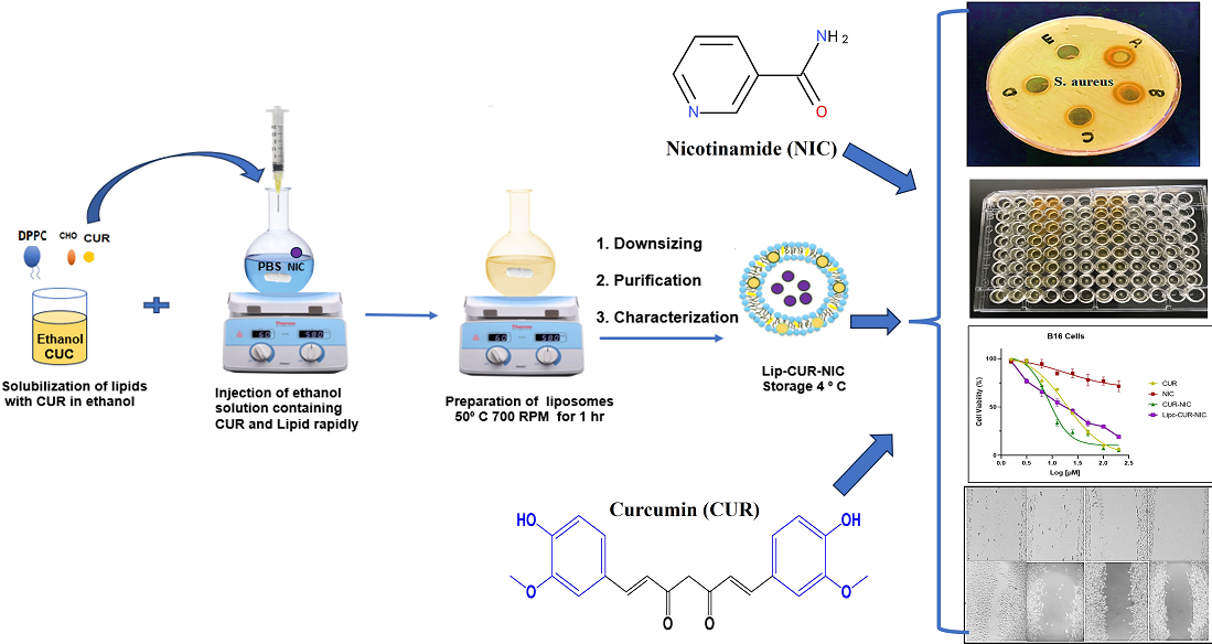
The integration of nanotechnology in medicine has opened avenues for safer and more efficient drug delivery systems. Liposomal formulations improve the stability and bioavailability of poorly soluble drugs like CUR, enhancing therapeutic outcomes. Encapsulating compounds with different solubility profiles in liposomes enhances their ability to target bacteria effectively while addressing solubility and stability challenges. Given the overlapping challenges of antibiotic resistance and melanoma, investigating liposomal formulations is essential for optimizing the therapeutic potential of CUR and NIC. [21,22]. This study explores the combined use of CUR and NIC in a liposomal formulation as a dual therapeutic approach against bacterial infections and melanoma. The liposomal delivery system, prepared using the ethanol injection method, is expected to enhance the skin penetration and therapeutic efficacy of CUR and NIC, making this combination a promising candidate for adjuvant treatment in skin infections and melanoma.
2. Material and Methods
Nicotinamide (NIC) was obtained from Sigma-Aldrich (USA),CUR(CUR) was obtained from ICT (Japan). 1,2-dipalmitoyl-sn-glycero-3-phosphocholine (DPPC) was obtained from Avanti Polar Lipids, Inc. (Alabaster, Alabama, USA) and Cholesterol (CHO) was obtained from Carbosynth (UK). Phosphate Buffer Saline (PBS) was purchased from LONZA® (USA). PLC grade Methanol was from Sigma-Aldrich (USA). HPLC-grade Ethanol was from the carbon group- England. All chemicals and solvents were of high purity.
2.1. Liposomal Preparation
Liposomes of CUR and NIC were prepared using the conventional ethanol injection technique as described in our previous article [23]. Briefly, 10 mg of DPPC, CHO 2.5 mg, and 2.0 mg CUR were dissolved ethanol, which was then warmed at 40 °C water bath. NIC 2mg/ml was dissolved PBS was heated using hot plate at ∼50 °C with string 700 rpm. Then the warm drug lipid ethanol mixture was injected rapidly into the PBS with heating and continuous stirring. After were prepped, the free of CUR and NIC were removed and washed and stored at 4°C [24].
The average particle size, Zeta Potential (charge) and Polydispersity Index (PDI) for liposomes were measured by DLS experiments using Zetasizer, Nano-ZS (Malvern Instruments Ltd., Malvern, UK). The Stability of the loaded liposomes was performed at 4 °C with storage times of one month. The EE % of CUR and NIC into liposomes was expressed as the percentage of drug complex encapsulated inside liposomes [25,26].
Entrapment efficiency (EE%) was determined to quantify the amount of CUR and NIC successfully encapsulated within the liposomal formulations. Briefly, the liposomes were separated from unencapsulated drugs by ultracentrifugation at 15,000 rpm for 45 minutes at 4°C. The supernatant containing the unencapsulated drug was collected, and its concentration was measured using UV-Vis spectrophotometry at specific wavelengths for CUR and NIC. The encapsulated drug amount was calculated by subtracting the free drug in the supernatant from the total drug added during formulation. The entrapment efficiency was then expressed as a percentage using the following equation:
This approach ensures precise quantification of drug encapsulation, providing insight into the formulation’s efficiency and stability. Optimization parameters such as lipid composition, solvent concentration, and preparation methods were fine-tuned to achieve high EE% for both CUR and NIC
2.2. Screening of the Antibacterial Activity
2.2.1. Agar Well-Diffusion Method
The Antibacterial activity screening was performed by the agar well-diffusion method according to guidelines of the Clinical and Laboratory Standards Institute. Muller-Hinton Agar Plates were prepared according to the manufacturer instructions (Thermo Fisher Scientific, Waltham, MA, USA). Wells were made into the agar using a 6 mm diameter sterile borer. The selected bacteria were mainly S. aureus and p. aeruginosa (ATCC® 29213). Bacterial cultures started in Muller-Hinton (MH) broth, and incubated overnight in the shaker at 37o C. Before use, the inoculum turbidity was standardized at OD520 nm = 0.1 (contrasted to 0.5 M McFarland). Bacteria were swabbed uniformly on the agar plates using sterile cotton applicators dipped into the standardized inoculum. Each well had 100 µL of the desired drug and liposomes concentration, while the control well contained the same volume of DMSO. Plates were then incubated at 37°C for 24 h, and the diameter of the inhibition zones was measured in millimeters (mm). Data are presented as the means for readings obtained from three different wells for each concentration.
2.2.2. Minimum Inhibitory Concentration (MIC)
If the bacteria were inhibited by the 1000 µg/mL at the primary screening, a determination for the MIC will take place using two-fold lower concentrations starting from 500, 250, 125, and 62.5 µg/ml. Five wells were made into the agar before streaking the plates with a standardized bacterial inoculum. Each well was filled with 100 µL of a selected drug, liposomes or with a control solvent which was used to solubilize the drugs and liposomes (DMSO or PBS). The plates were then incubated at 37°C for 24 h, and the inhibition zones were measured in mm.
2.2.3. S. aureus Susceptibility
The antibacterial activity of free CUR, NIC and loaded liposomes were evaluated by the broth Microdilution Method, according to guidelines of the Clinical and Laboratory Standards Institute [27], followed by turbidity evaluation. Briefly, the formulations were diluted in PBS to produce a serial dilution with concentrations ranging from: 0.1875 to 200 µg/mL of both drugs and their liposomes. Bacterial Suspensions were performed from a MSSA overnight culture diluted in broth media until reaching a value of 0.5 in a McFarland scale equivalent to 108 colony forming unit/ml by measuring the optical density at 600 nm. Bacterial Suspension was cultured in 96-well cell culture plates at 5 × 10⁵ bacterial density and incubated with CUR, NIC, Lip-CUR-NIC, at 37 °C during 24 h. A negative control containing a Suspension of bacteria in broth, without treatment, and a sterile control containing broth only without bacteria, were included. Minimum Inhibitory Concentration (MIC: the lowest Antibiotic concentration able to prevent visible bacterial growth, resulting in the absence of turbidity) was determined Spectrophotometrically, at 570 nm in a microplate reader (Bio-Rad laboratories, Inc., Hercules, CA, USA).
2.3. Cell Culture
The B16 mouse cancer cell line was obtained from the American Type Culture Collection (ATCC, Manassas, VA, USA). These cells were cultivated as attached monolayers and preserved in DMEM medium (EuroClone, Italy), enriched with 10% (v/v) heat-inactivated Fetal Bovine Serum (FBS) (Euro Clone, Italy), 1% Penicillin-Streptomycin (Euro Clone, Italy), and 2 mM L-glutamine. Incubation of the cells was carried out at 37°C within a Tissue Culture Incubator (Memmert, Schwabach, Germany) containing 5% CO2.
2.3.1. Cell Viability Assay (MTT)
The MTT assay was conducted to evaluate the cytotoxicity of both free drugs (CUR and NIC) and their liposomal formulations (Lip-CUR, Lip-NIC, and Lip-CUR-NIC) on B16 melanoma cells. Briefly, 5 × 10³ cells/well were seeded into 96-well plates and incubated for 24 hours at 37°C in a 5% CO₂ incubator. Cells were then treated with varying concentrations (3.125–200 µM) of free CUR, free NIC, Lip-CUR, Lip-NIC, and Lip-CUR-NIC for 72 hours. After the incubation period, the medium was removed, and 100 µL of fresh medium containing MTT reagent (0.5 mg/mL) was added to each well. The plates were further incubated for 3 hours at 37°C. Subsequently, 50 µL of DMSO was added to solubilize the formazan crystals, and the absorbance was measured at 560 nm using an ELISA plate reader. The IC50 values for each treatment were determined using dose-response curves. The free drugs (CUR and NIC) were included to compare their cytotoxic effects with those of the liposomal formulations. These results provided insights into the potential advantages of liposomal delivery systems over free drug formulations.
2.3.2. Cell Migration Assay
Melanoma B16 cell line was seeded in sterile 6-well cell culture plates at a density of 800,000 cells per well and then incubated for 24 hours at 37°C with 5% CO2 to attach. The following day, a vertical scratch was created at the center of the cell’s monolayer using a sterile 1,000 µl micropipette tip for either free drugs or nanoliposome’s treatment (Day 1, baseline). Subsequently, each well was washed twice with sterile PBS. After 1 day, cells were exposed to CUR, NIC CUR-NIC and Lip-CUR-NIC at three different concentrations near IC50, as determined by the MTT test. Finally, images of the scratches were captured before and during cell treatment using a phase contrast microscope (model P. MICRO-001, Nikon) equipped with a 4× magnification objective. The Images J software was employed to calculate the wound closure area (µm2). DMSO and free media served as negative controls. The wound closure rate was observed on day 1 (before treatment) and day 2 after 48h of cell therapy [28]. Empty liposomes and media were employed as a negative control.
The wound closure (%) was calculated using equation
Where Area day 1 is the initial wound area, and day 2 represents the wound area at the subsequent time points (Day 2 (24h)).
2.4. DPPH Assay
The free radical scavenging activities of four treatments—Vitamin C, NIC, CUR, and CUR-NIC, were evaluated using the 2,2-diphenyl-1-picrylhydrazyl (DPPH) free radical scavenging assay [29]. For the calibration curve, a stock solution of vitamin C was prepared by dissolving 1 mg in 1 mL of methanol to create standard stock solutions of Vitamin C, CUR, NIC, and CUR-NIC. Serial dilutions were made by mixing 500 μL of each sample with 500 μL of methanol. The DPPH solution was prepared by dissolving 8 mg of DPPH in 10 mL of methanol, producing a deep violet-colored solution. In a 96-well plate, 100 μL of each sample was combined with 100 μL of the freshly prepared DPPH solution. Each assay was performed in triplicate and incubated in the dark for 30 minutes. Controls included DPPH and CUR in methanol. Absorbance was then measured at 517 nm using an ELISA plate reader. The Vitamin C calibration curve was used to calculate both the percentage of free radical inhibition and the concentration of Vitamin C, CUR, NIC, and CUR-NIC formulations required to achieve 50% free radical inhibition (IC50).
(%) Inhibition of Free Radical = (Ab Control – Ab Sample) / Ab Control *100 %
2.5. Statistical Analysis
The findings were expressed as the mean ± standard deviation from a minimum of three separate trials. Statistical analysis and graph plotting were performed using GraphPad Prism 8 (GraphPad Software Inc., USA), CompuSyn.exe (Version 1) for combination index evaluation, and Microsoft Office Excel (Microsoft, USA) for additional data handling and visualization
3. Results and Discussion
3.1. Liposomal Preparation and Characterization
The liposomal formulation of Lip-CUR-NIC was developed and fully characterized using UV spectrophotometry and HPLC analysis, as previously reported by Fahdawi et al. [23]. In this study, the Lip-CUR-NIC formulation was biologically evaluated alongside the pure compounds CUR and NIC. The average particle size for all preparations was under 200 nm (Figure 1A), and the particle size distribution was assessed using the polydispersity index (PDI). A lower PDI indicates a more uniform size distribution, with values below 0.4 considered good for monodispersed nanoparticles (Figure 1B) [30].The stability of the liposomal formulations was monitored over 30 days, focusing on size distribution (Figure 1C) and zeta potential (Figure 1D,E). These parameters, including average diameter, PDI, surface charge, encapsulation efficiency (EE%), and loading efficiency (LE%), were consistent with those reported in the previous study for the same liposomal formulation [23]. Additionally, the in vitro release profiles of CUR and NIC were evaluated over 72 hours, showing that CUR had a slower release from the Lip-CUR-NIC formulation compared to NIC. This difference is likely due to the higher solubility of NIC in PBS compared to CUR.
As illustrated in Figure 1F, CUR exhibited a slower and more incomplete release profile from the Lip-CUR-NIC formulation compared to NIC. This finding indicates that CUR’s hydrophobic nature and potential interactions with the liposomal bilayer matrix restrict its diffusion, leading to a sustained release. Conversely, NIC, which is more hydrophilic, showed a faster and more cumulative release, likely due to its higher solubility in the aqueous medium [31,32,33].
3.2. Antioxidant Activity and DPPH Assay
A DPPH assay, which measures the free radical scavenging activity of CUR, NIC, CUR- NIC and Vit C as a standard (Figure 2A,B). The assay results are plotted as percentage inhibition of the DPPH radical against the concentration of the tested substances in mg/ml. NIC exhibited the lowest activity among the tested samples, with a more gradual increase in inhibition percentage as the concentration increases, while CUR demonstrated higher activity than NIC, with a steeper curve that approaches the activity level of Vit C at higher concentrations. The combination of CUR and NIC showed an improved scavenging effect compared to NIC alone and slightly better than CUR alone, suggesting a potential synergistic effect between the two on radical scavenging activity. As expected, Vitamin C exhibited the highest radical scavenging activity, nearing 100% inhibition, since it is a well-known potent antioxidant used as the positive control in the assay.
3.3. Antibacterial Activity of CUR, NIC and lip- CUR-NC
An initial assessment of the antibacterial activity of CUR, NIC, and their combination CUR-NIC, along with their respective liposomal formulations, against S. aureus is shown in Figure 3A. CUR demonstrated effective inhibition of S. aureus growth, while NIC alone, at various concentrations, did not show significant antibacterial activity. However, when NIC was combined with CUR, it enhanced CUR’s antibacterial effect. These findings indicate that the antibacterial activity of the CUR-NIC combination is concentration-dependent (Figure 3A,B), highlighting a potential synergistic interaction between the two compounds.
The findings regarding the antibacterial activity against S. epidermidis are presented in Figure 4A,B. The results indicate that liposomal formulations showed no significant activity against S. epidermidis, while CUR alone exhibited notable inhibitory effects. This suggests that CUR’s selectivity may vary depending on the bacterial strain, and further investigation is required to balance therapeutic efficacy with the preservation of normal skin microbiota.
When evaluating the antibacterial activity of CUR, CUR-NIC, Lip-CUR, and Lip-CUR-NIC across concentrations ranging from 3.13 to 400 µg/ml, CUR exhibited an MIC of 62.5 µg/ml, while Lip-CUR showed a slightly lower MIC of 75.5 µg/ml. This suggests that the liposomal formulation of CUR does not significantly improve its antibacterial efficacy against S. aureus. Similarly, CUR-NIC had an MIC of 31.25 µg/ml, whereas Lip-CUR-NIC displayed an MIC of 37.7 µg/ml at the same concentrations (Figure 5A and Figure 4B), indicating a comparable trend between the free and liposomal formulations. The apparent reduction in activity observed with the liposomal formulations compared to the free components (CUR, NIC, and CUR-NIC) can be attributed to differences in their release profiles and cellular uptake mechanisms. Liposomal formulations often exhibit a controlled or sustained release of encapsulated compounds, which may lead to a delayed therapeutic effect compared to the immediate availability of free compounds. Additionally, encapsulation in liposomes may slightly reduce the immediate bioavailability of the active agents, as the compounds need to be released from the lipid bilayer before exerting their biologicac
As noted by Harush-Frenkel in 2010, positively charged nanoparticles were associated with increased side effects and toxicity. In contrast, the prepared liposomes in this study exhibited a negative average zeta potential, which is more favorable for their safety profile. [34].
Inhibitors of bacterial resistance present promising treatment options for patients with antibiotic-resistant infections. The use of natural inhibitors could enhance the effectiveness of retreatment in patients who previously received ineffective antibiotics and help prevent the emergence of new antibiotic-resistant bacterial strains. Various studies have consistently demonstrated that CUR exhibits antimicrobial effects, with no contradictory findings reported on this topic [35,36].
Teow and Ali (2015) conducted a study to examine the combined antibacterial effects of CUR and eight different antibiotic groups. Using disc diffusion assays, they found synergistic effects between CUR and most of the antibiotics against S. aureus. However, in microdilution assays, synergy was only observed with three antibiotics: ciprofloxacin, gentamicin, and amikacin. The other tested antibiotics showed no significant interaction, though no antagonism was detected. These findings align with this study, likely due to the use of similar experimental methods. [37].
The antibacterial activity of CUR was evaluated using the broth microdilution method, checkerboard dilution test, and time-kill assay. CUR demonstrated antimicrobial activity against all tested strains. In the checkerboard test, CUR significantly reduced the MIC of antibiotics such as oxacillin, ampicillin, ciprofloxacin, and norfloxacin, which are commonly used to treat methicillin-resistant Staphylococcus aureus (MRSA)[38].
Like this study, Zhou et al. demonstrated that the combined treatment of CUR and Erythromycin effectively suppressed bacterial growth and alleviated bone infection. The combination showed stronger efficacy against MRSA-induced osteomyelitis in rats compared to monotherapy [39].
In a different study, Wang et al. utilized CUR as a natural antibacterial and antifungal Agent against various foodborne pathogens, including Staphylococcus Aureus, Escherichia coli, Yersinia enterocolitica, Bacillus cereus, and Aspergillus niger. They improved the stability and solubility of CUR by using Microcapsules. The study demonstrated a broad-spectrum inhibitory effect of CUR against all tested organisms using the Oxford Cup Method. The results also indicated that CUR had greater antibacterial activity against Gram-positive bacteria than Gram-negative bacteria, while its Antifungal Activity was significantly higher than its antibacterial activity [40].
Gunes et al. investigated the effect of CUR on standard bacterial strains at high concentrations and demonstrated its strong antibacterial activity at high doses on animals. This study was conducted in Turkey, and the similarity in results could be attributed to the potential presence of the same bacterial strains and resistance genes [41].
In a study conducted by Shailendiran et al. in (2011), the antibacterial properties of CUR and non-formulated CUR were examined against both a gram-positive bacterial strain (Cocci) and a gram-negative bacterial strain such as E. coli. The study applied the agar disc assay to observe the size of the inhibition zone over time. Results showed that after 10 hours, a clearly visible inhibition zone was observed, indicating inhibition of bacterial growth. However, this zone became less distinct after 24 hours for both CUR and nanocurcumin-treated discs. These findings suggest that the tested samples exhibited bacteriostatic properties, inhibiting bacterial growth rather than killing the bacteria outright [42]. In another study conducted by Hu et al., the antimicrobial activity of CUR against S. Mutans was examined, and the inhibitory ability of CUR on purified Sortase A was evaluated using Western blot and Real-time PCR. The study revealed that CUR can effectively inhibit purified S. Mutans Sortase A at a concentration equivalent to half of the minimum inhibitory concentration (MIC), leading to a reduction in S. Mutans biofilm formation [43].
Furthermore, Lzui et al. demonstrated that CUR exhibited a dose-dependent inhibition of the growth of Prevotella intermedia, P. gingivalis, Treponema denticola, and Fusobacterium nucleatum. Even at very low concentrations, CUR significantly suppressed bacterial development [44].
Additionally, testing the liposomal formulations against Staphylococcus epidermidis, a key component of the skin’s normal flora, revealed that CUR alone inhibited bacterial growth. This raises concerns about CUR’s selectivity for pathogenic bacteria versus beneficial bacteria and highlights the need to assess the safety of such formulations in clinical applications. While CUR shows promise as an antibacterial agent, its impact on normal skin flora could affect skin health, suggesting further investigation is needed to balance therapeutic efficacy with the preservation of healthy microbiota.
In this study, CUR exhibited an MIC of 62.5 µg/mL, consistent with prior reports that have demonstrated CUR’s antimicrobial effects against a range of bacterial pathogens. For instance, Wang et al. (2009) reported a similar MIC range (50–100 µg/mL) for CUR against Gram-positive bacteria, including S. aureus. Additionally, Mun et al. (2013) observed CUR’s MIC against methicillin-resistant S. aureus (MRSA) to be approximately 64 µg/mL, which aligns closely with our findings. These comparisons confirm the reproducibility of CUR’s antibacterial efficacy, further validating its potential as a natural antimicrobial agent. The slight variations in MIC across studies may result from differences in experimental conditions, bacterial strains, and methods used for assessment.
3.4. Cytotoxicity Study and Anticancer Activity
When assessing the anticancer potential of CUR and NIC, and their combination CUR-NIC, it was found that CUR-NIC had the most significant effect in reducing cancer cell viability, particularly at moderate concentrations. This indicates a possible synergistic effect between CUR and NIC in combating cancer, making their combination a promising candidate for further exploration in cancer therapy. Whereas, in the case of drug-loaded liposomes, encapsulating CUR increased its IC50 to 50 ± 9.2 µM, demonstrating reduced effectiveness compared to free CUR. NIC, on the other hand, remained relatively ineffective with an IC50 value still above 200 µM (Figure 5A). When considering the liposomal formulations, CUR had an IC50 of 31.4 ± 3.2 µM, and NIC exhibited an IC50 of 190 ± 7.3 µM. These results show that while liposomal CUR and NIC both improved in effectiveness compared to their single-drug liposome counterparts, they were still less effective than the free CUR-NIC combination (Figure 6A,B).
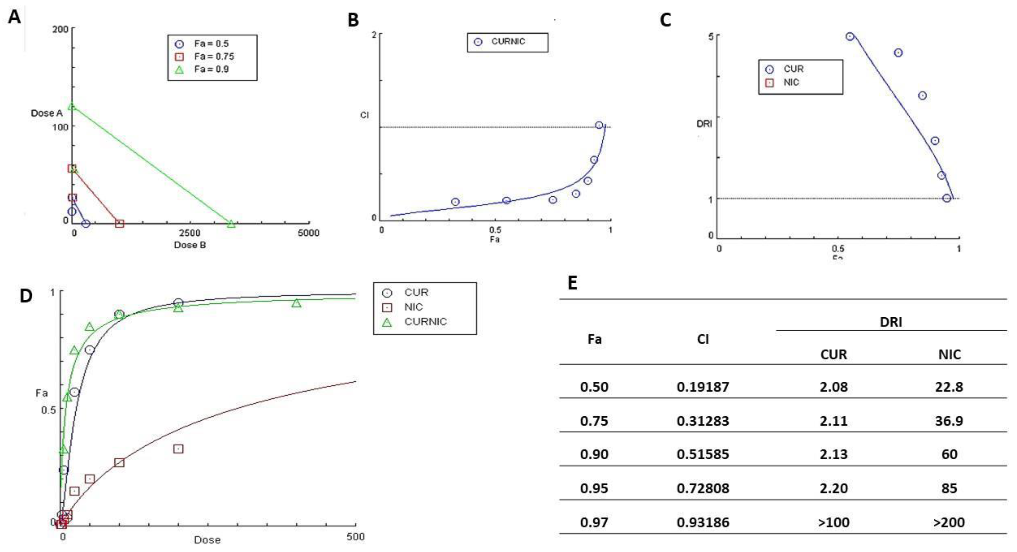
The IC50 values for CUR and NIC, when administered alone and in combination with B16 cells, are shown in Figure 6C. For the free drugs alone, CUR had an IC50 value of 27.3 ± 2.3 µM, indicating moderate potency. NIC had an IC50 value exceeding 200 µM, showing low effectiveness as a single agent against B16 cells. When used in combination, the IC50 for CUR significantly decreased to 9.8 ± 2.2 µM, showing increased potency. Similarly, the IC50 for NIC dropped to 135.95 ± 10.2 µM, indicating enhanced effectiveness when used with CUR. The isobologram analysis for the CUR-NIC combination (Figure 7A) plots three iso-effective combinations of Dose A (CUR) and Dose B (NIC) corresponding to Fraction Affected (Fa) values of 0.5, 0.75, and 0.9. This analysis shows that either drug alone can achieve a 50% effect at a certain dose, but the combination provides a more potent effect at lower doses.
The Isobologram indicates a synergistic interaction between CUR and NIC when combined at a 1:1 ratio, as the combination points fall below the line of additivity. This synergy implies that lower doses of each drug can be used in combination to achieve a high level of effect, potentially reducing side effects, and increasing treatment efficacy, allowing for dose reduction while maintaining or improving therapeutic outcomes.
The Combination Index (CI) plot for the CUR and NIC Free Powder Combination, quantifies the interaction between two drugs, with CI<1 indicating synergy, CI=1 indicating an additive effect, and CI >1 indicating antagonism (Figure 7A–E).
the Combination Index (CI) values at various Fraction Affected (Fa) levels, ranging from 0 (no effect) to 1 (complete effect). For the CUR+NIC combination, most CI values fall below 1 across the Fa range, indicating increasing synergy, particularly at higher Fa levels.
Figure 7D illustrates the dose-effect curves for CUR, NIC, and their combination (CUR-NIC), plotting Fa against dose to show the impact on cell viability. CUR exhibits a steep dose-effect relationship, effectively inhibiting cell viability even at low doses and reaching near-complete inhibition (Fa close to 1) at lower doses. In contrast, NIC displays a more gradual dose-effect curve with less inhibition of cell viability, even at higher doses, and does not achieve the same level of inhibition as CUR. The CUR-NIC combination has a dose-effect curve similar to CUR, suggesting that the combination is as effective as CUR alone and that NIC does not negatively impact CUR’s inhibitory effect.
The Dose Reduction Index (DRI) quantifies the potential dose reduction in combination therapy compared to using each drug alone. CUR shows a significant increase in DRI with Fa, indicating up to a fivefold or greater dose reduction when used in combination. In contrast, data points for NIC are absent, suggesting NIC does not contribute to dose reduction in the combination. The DRI curve for CUR indicates a strong synergistic interaction, enabling significant dose reduction while maintaining anticancer efficacy. At the highest Fa level (0.97), the DRI for CUR exceeds 100, and for NIC surpasses 200, indicating the doses required to achieve 97% inhibition of cell viability in combination.
The CUR-NIC combination has a notably lower IC50 of 10, a slope of 0.867, and a high correlation coefficient of 0.981, reflecting a potent effect with a well-fitting dose-response curve. When delivered via Lip-CUR and Lip-NIC, Lip-CUR shows moderate anticancer activity with an IC50 of 20, while Lip-NIC has an IC50 above 50. However, Lip-CUR-NIC demonstrates the most significant decline in cancer cell viability among all treatments, highlighting the potential of liposomal delivery to enhance cancer treatment efficacy.
Wang et al. studied curcumin-loaded MPEG-PLA micelles for melanoma treatment, both in lab tests (in vitro) and in animal models (in vivo). They found that the spherical curcumin/MPEG-PLA micelles disperse well in normal saline and provide a sustained release of the drug. These micelles also showed stronger cell-killing effects. Further histochemical analysis in animal studies confirmed their ability to trigger melanoma cell death and block new blood vessel formation in tumors. Their conclusion is that these curcumin/MPEG-PLA micelles hold promise for clinical melanoma treatment.
Fontes et al. explored the combination of CUR and disulfiram for treating B16 melanoma cells. Their research, both in vitro and in vivo, showed that combining CUR and disulfiram had a synergistic effect at specific ratios, increasing cell death (apoptosis) and oxidative stress. This combination was more effective at slowing tumor growth compared to either compound alone. The synergy likely results from the combined action of CUR and disulfiram on the NF-κB and PI3K/Akt signaling pathways, which enhances apoptosis and slows cell growth.
3.5. Migration Test (Scratch Assay)
A Scratch Assay was performed on B16 melanoma cells over two days to assess cell migration after treatment with different concentrations of CUR-NIC and liposomal formulations. At the highest concentration of CUR-NIC (52 µM), the scratch area stayed mostly clear by Day 2, showing strong inhibition of cell migration. At 26 µM, migration was moderately inhibited, while at 13 µM, some migration occurred, but it was still less than the control group (p- value <0.05. This demonstrates that CUR-NIC inhibits cell migration in a concentration-dependent manner, highlighting its potential to prevent tumor metastasis.
Figure 8.
Effect of CUR-NIC and Lip-CUR-NIC on cell migration and invasion capability. (A) The migration of B16 melanoma cells in the Matrigel surface after 48 h of scrape in different concentration 1/2 *IC50, IC50 and 2*IC50 [26], 2IC50 (13 ,26, 52 μ M) of CUR-NIC combination groups. (B) show the migration of B16 melanoma cells in the matrigel surface after 48 h of scrape in different concentration ½* IC50, IC50 and 2*IC50 (25, 50, 100 μM) of Lip-CUR-NIC. (C) The cells migration inhibited distance for all the formulations and free drugs were calculated. (Quantification of the wound scratch assay shown Data are expressed as a mean ± SD, for 24 hrs. and n=2).
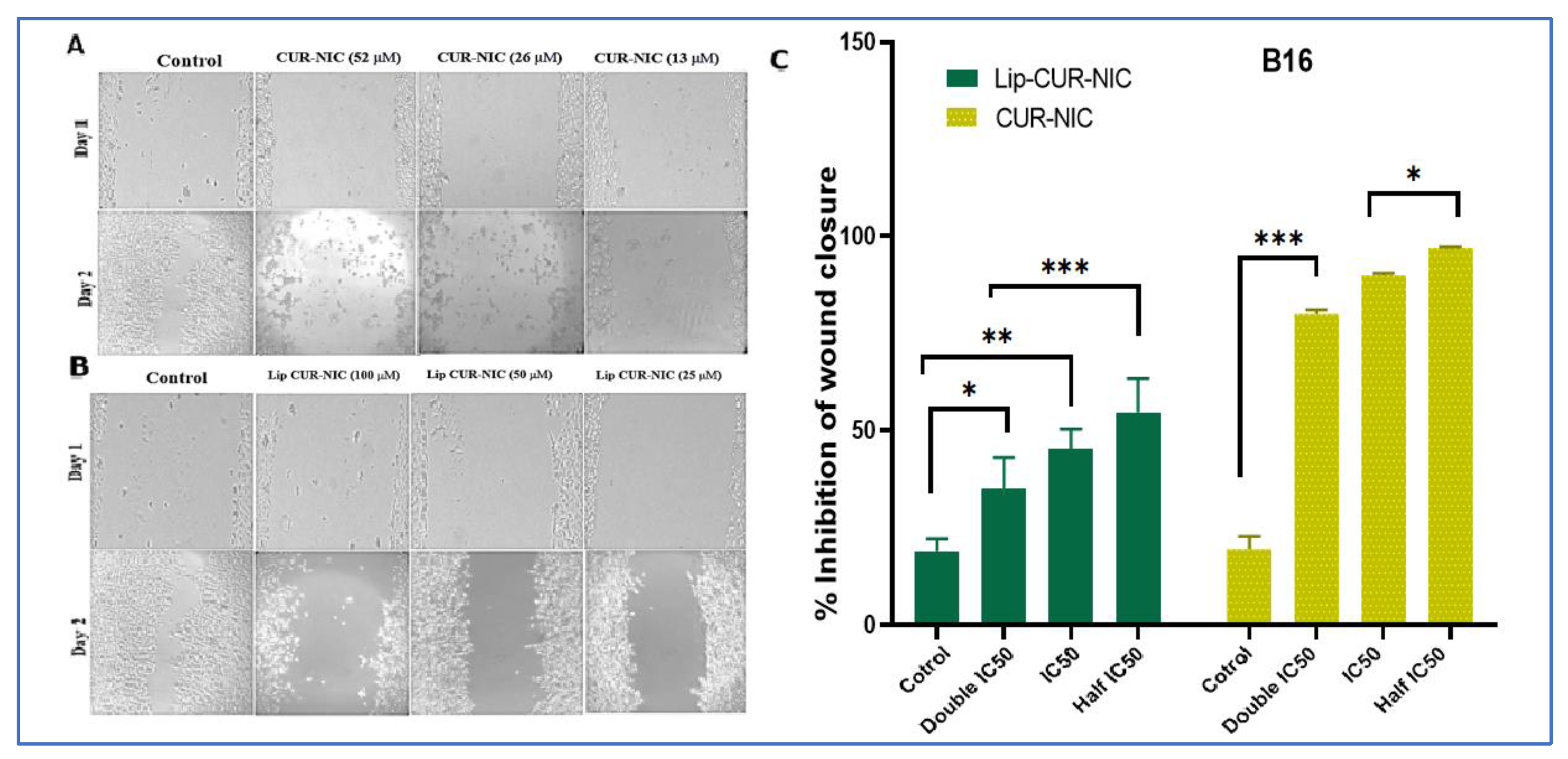
Similarly, Lip-CUR-NIC was tested at various concentrations over two days. Control samples showed significant cell migration by Day 2, while Lip-CUR-NIC at 100 µM showed a strong inhibition of migration. At 50 µM, the migration was moderate, and at 25 µM, slight migration was observed. These results indicate that Lip-CUR-NIC can effectively block B16 melanoma cell migration in a dose-dependent manner (Figure 7A,B). Control bars in the figures represent the baseline migration of untreated cells, providing a point of comparison for the treated samples (Figure 7C).
The liposomal formulation may not have shown enhanced inhibition at lower concentrations due to a combination of factors such as slower drug release, lower cellular uptake, and suboptimal encapsulation efficiency. Additionally, the sustained release profile of the liposomes may require a longer observation period to fully reveal their therapeutic potential. Therefore, longer observation period might reveal greater efficacy for Lip-CUR-NIC, as its sustained release could take longer to show significant inhibition compared to the free drug, which is more rapidly bioavailable.
4. Conclusions
In summary, the CUR-NIC combination demonstrated significant anticancer activity in the MTT assay, highlighting its synergistic potential as a promising cancer treatment strategy. The DPPH assay further validated the antioxidant properties of the CUR-NIC combination, which may contribute to its enhanced anticancer efficacy.
The ethanol injection method was successfully optimized for encapsulating CUR and NIC by carefully adjusting critical parameters, including the lipid-to-drug ratio, ethanol concentration, and injection rate. A key challenge was reconciling the hydrophobic nature of CUR with the hydrophilic properties of NIC to create stable liposomes with high encapsulation efficiency. Through formulation optimization, the study achieved efficient drug encapsulation and stable particle sizes, overcoming solubility disparities between the two compounds.
Author Contributions
Conceptualization, Zainab Lafi and Naeem Shalan.; Methodology, Zainab Lafi and Omar Markab; Software, Omar Markab, Ali Fahdawi.; Validation, Hala Al Daghistani. And Zainab Lafi and Naem Shalan; Formal Analysis, Omar Markab and Zainab Lafi ; Investigation, Ali Fahdawi and Omar Markab; Resources, ; Data Curation, Naeem Shalan and Zainab Lafi; Writing – Original Draft Preparation, Omar Markab and Ali Fahdawi; Writing – Review & Editing, Naeem Shalan, Zainab Lafi and Hala Al Daghistani; Visualization, Naeem Shalan, Zainab Lafi, and Hala Al Daghistani;; Supervision, *, Naeem Shalan, Zainab Lafi, and Hala Al Daghistani; Project Administration, Zainab Lafi.; Funding Acquisition, Al-Ahliyya Amman University.
References
- Sorrenti V, Burò I, Consoli V, Vanella L. Recent Advances in Health Benefits of Bioactive Compounds from Food Wastes and By-Products: Biochemical Aspects. Int J Mol Sci. 2023;24(3). [CrossRef]
- Nsairat H, Lafi Z, Al-Sulaibi M, Gharaibeh L, Alshaer W. Impact of nanotechnology on the oral delivery of phyto-bioactive compounds. Food Chemistry. 2023;424:136438. [CrossRef]
- Lafi Z, Alshaer W, Ma’mon MH, Zihlif MA, Asha NY, Abdelnabi H, et al. A review Echinomycin: A Journey of Challenges. Jordan Journal of Pharmaceutical Sciences. 2023;16(3):640-54.
- Hammad HM, Imraish A, Al-Hussaini M, Zihlif M, Harb AA, Abu Thiab TM, et al. Ethanol extract of Achillea fragrantissima enhances angiogenesis through stimulation of VEGF production. Endocrine, Metabolic & Immune Disorders-Drug Targets (Formerly Current Drug Targets-Immune, Endocrine & Metabolic Disorders). 2021;21(11):2035-42. [CrossRef]
- Lafi ZM, Irshaid YM, El-Khateeb M, Ajlouni KM, Hyassat D. Association of rs7041 and rs4588 polymorphisms of the vitamin D binding protein and the rs10741657 polymorphism of CYP2R1 with vitamin D status among Jordanian patients. Genetic testing and molecular biomarkers. 2015;19(11):629-36. [CrossRef]
- Matalqah S, Lafi Z, Asha SY. Hyaluronic Acid in Nanopharmaceuticals: An Overview. Current Issues in Molecular Biology. 2024;46(9):10444-61. [CrossRef]
- Watkins RR, Bonomo RA. Overview: Global and Local Impact of Antibiotic Resistance. Infect Dis Clin North Am. 2016;30(2):313-22. [CrossRef]
- Hussain Y, Alam W, Ullah H, Dacrema M, Daglia M, Khan H, et al. Antimicrobial Potential of Curcumin: Therapeutic Potential and Challenges to Clinical Applications. Antibiotics (Basel). 2022;11(3). [CrossRef]
- Chinemerem Nwobodo D, Ugwu MC, Oliseloke Anie C, Al-Ouqaili MTS, Chinedu Ikem J, Victor Chigozie U, et al. Antibiotic resistance: The challenges and some emerging strategies for tackling a global menace. J Clin Lab Anal. 2022;36(9):e24655. [CrossRef]
- Laikova KV, Oberemok VV, Krasnodubets AM, Gal’chinsky NV, Useinov RZ, Novikov IA, et al. Advances in the understanding of skin cancer: ultraviolet radiation, mutations, and antisense oligonucleotides as anticancer drugs. Molecules. 2019;24(8):1516. [CrossRef]
- Abd El-Hack ME, de Oliveira MC, Attia YA, Kamal M, Almohmadi NH, Youssef IM, et al. The efficacy of polyphenols as an antioxidant agent: An updated review. International Journal of Biological Macromolecules. 2023:126525. [CrossRef]
- Munef A, Lafi Z, Shalan N. Investigating anti-cancer activity of dual-loaded liposomes with thymoquinone and vitamin C. Therapeutic Delivery. 2024;15(4):267-78. [CrossRef]
- AlSaleh A, Shahid M, Farid E, Kamal N, Bindayna K. Synergistic antimicrobial effect of ascorbic acid and nicotinamide with rifampicin and vancomycin against SCCmec type IV methicillin-resistant Staphylococcus aureus (MRSA). Access Microbiol. 2023;5(2). [CrossRef]
- Simmons JD, Peterson GJ, Campo M, Lohmiller J, Skerrett SJ, Tunaru S, et al. Nicotinamide Limits Replication of Mycobacterium tuberculosis and Bacille Calmette-Guérin Within Macrophages. J Infect Dis. 2020;221(6):989-99. [CrossRef]
- Murray MF. Nicotinamide: an oral antimicrobial agent with activity against both Mycobacterium tuberculosis and human immunodeficiency virus. Clinical infectious diseases. 2003;36(4):453-60. [CrossRef]
- Starr P. Oral Nicotinamide Prevents Common Skin Cancers in High-Risk Patients, Reduces Costs. Am Health Drug Benefits. 2015;8(Spec Issue):13-4.
- Allen NC, Martin AJ, Snaidr VA, Eggins R, Chong AH, Fernandéz-Peñas P, et al. Nicotinamide for Skin-Cancer Chemoprevention in Transplant Recipients. New England Journal of Medicine. 2023;388(9):804-12. [CrossRef]
- Tosti G, Pepe F, Gnagnarella P, Silvestri F, Gaeta A, Queirolo P, et al. The Role of Nicotinamide as Chemo-Preventive Agent in NMSCs: A Systematic Review and Meta-Analysis. Nutrients. 2024;16(1):100. [CrossRef]
- Surjana D, Halliday GM, Damian DL. Role of nicotinamide in DNA damage, mutagenesis, and DNA repair. Journal of nucleic acids. 2010;2010. [CrossRef]
- Salech F, Ponce DP, Paula-Lima AC, SanMartin CD, Behrens MI. Nicotinamide, a poly [ADP-ribose] polymerase 1 (PARP-1) inhibitor, as an adjunctive therapy for the treatment of Alzheimer’s disease. Frontiers in Aging Neuroscience. 2020;12:255. [CrossRef]
- Lafi Z, Hiba T, Hanan A. An updated assessment on anticancer activity of screened medicinal plants in Jordan: Mini review. Journal of Pharmacognosy and Phytochemistry. 2020;9(5):55-8. [CrossRef]
- Lafi Z, Aboalhaija N, Afifi F. Ethnopharmacological importance of local flora in the traditional medicine of Jordan:(A mini review). Jordan Journal of Pharmaceutical Sciences. 2022;15(1):132-44. [CrossRef]
- Fahdawi A, Shalan N, Lafi Z, Markab O. Analytical Approaches for Assessing Curcumin and Nicotinamide Co-Encapsulated in Liposomal Formulation: UV Spectrophotometry and HPLC Validation. Jordan Journal of Pharmaceutical Sciences. 2024;17(3):468-80. [CrossRef]
- Lafi Z, Alshaer W, Ma’mon MH, Zihlif M, Alqudah DA, Nsairat H, et al. Aptamer-functionalized pH-sensitive liposomes for a selective delivery of echinomycin into cancer cells. RSC Advances. 2021;11(47):29164-77.
- Alshaer W, Zraikat M, Amer A, Nsairat H, Lafi Z, Alqudah DA, et al. Encapsulation of echinomycin in cyclodextrin inclusion complexes into liposomes: in vitro anti-proliferative and anti-invasive activity in glioblastoma. RSC advances. 2019;9(53):30976-88. [CrossRef]
- Allateef A, Shalan N, Lafi Z. Anticancer activity of liposomal formulation co-encapsulated with coumarin and phenyl butyric acid. Journal of Applied Pharmaceutical Science. 2024. [CrossRef]
- Weinstein MP, Lewis JS. The clinical and laboratory standards institute subcommittee on antimicrobial susceptibility testing: background, organization, functions, and processes. Journal of clinical microbiology. 2020;58(3):e01864-19. [CrossRef]
- Grimmig R, Babczyk P, Gillemot P, Schmitz K-P, Schulze M, Tobiasch E. Development and evaluation of a prototype scratch apparatus for wound assays adjustable to different forces and substrates. Applied Sciences. 2019;9(20):4414. [CrossRef]
- Baliyan S, Mukherjee R, Priyadarshini A, Vibhuti A, Gupta A, Pandey RP, et al. Determination of Antioxidants by DPPH Radical Scavenging Activity and Quantitative Phytochemical Analysis of Ficus religiosa. Molecules. 2022;27(4). [CrossRef]
- Allateef A, Shalan N, Lafi Z. Anticancer activity of liposomal formulation co-encapsulated with coumarin and phenyl butyric acid. Journal of Applied Pharmaceutical Science. 2024;14(11):208-15. [CrossRef]
- De Leo V, Milano F, Mancini E, Comparelli R, Giotta L, Nacci A, et al. Encapsulation of Curcumin-Loaded Liposomes for Colonic Drug Delivery in a pH-Responsive Polymer Cluster Using a pH-Driven and Organic Solvent-Free Process. Molecules. 2018;23(4). [CrossRef]
- Zhang Q, Li J, Zhong H, Xu Y. The mechanism of nicotinamide on reducing acute lung injury by inhibiting MAPK and NF-κB signal pathway. Mol Med. 2021;27(1):115. [CrossRef]
- De Leo V, Milano F, Mancini E, Comparelli R, Giotta L, Nacci A, et al. Encapsulation of curcumin-loaded liposomes for colonic drug delivery in a pH-responsive polymer cluster using a pH-driven and organic solvent-free process. Molecules. 2018;23(4):739. [CrossRef]
- Harush-Frenkel O, Bivas-Benita M, Nassar T, Springer C, Sherman Y, Avital A, et al. A safety and tolerability study of differently-charged nanoparticles for local pulmonary drug delivery. Toxicology and Applied Pharmacology. 2010;246(1):83-90. [CrossRef]
- Shajari M, Rostamizadeh K, Shapouri R, Taghavi L. Eco-friendly curcumin-loaded nanostructured lipid carrier as an efficient antibacterial for hospital wastewater treatment. Environmental Technology & Innovation. 2020;18:100703. [CrossRef]
- Stavri M, Piddock LJ, Gibbons S. Bacterial efflux pump inhibitors from natural sources. J Antimicrob Chemother. 2007;59(6):1247-60. [CrossRef]
- Teow SY, Ali SA. Synergistic antibacterial activity of Curcumin with antibiotics against Staphylococcus aureus. Pak J Pharm Sci. 2015;28(6):2109-14.
- Mun SH, Joung DK, Kim YS, Kang OH, Kim SB, Seo YS, et al. Synergistic antibacterial effect of curcumin against methicillin-resistant Staphylococcus aureus. Phytomedicine. 2013;20(8-9):714-8. [CrossRef]
- Zhou Z, Pan C, Lu Y, Gao Y, Liu W, Yin P, et al. Combination of erythromycin and curcumin alleviates Staphylococcus aureus induced osteomyelitis in rats. Frontiers in cellular and infection microbiology. 2017;7:379. [CrossRef]
- Wang Y, Lu Z, Wu H, Lv F. Study on the antibiotic activity of microcapsule curcumin against foodborne pathogens. Int J Food Microbiol. 2009;136(1):71-4. [CrossRef]
- Gunes H, Gulen D, Mutlu R, Gumus A, Tas T, Topkaya AE. Antibacterial effects of curcumin: An in vitro minimum inhibitory concentration study. Toxicol Ind Health. 2016;32(2):246-50.
- Shailendiran D, Pawar N, Chanchal A, Pandey RP, Bohidar HB, Verma AK, editors. Characterization and Antimicrobial Activity of Nanocurcumin and Curcumin. 2011 International Conference on Nanoscience, Technology and Societal Implications; 2011 8-10 Dec. 2011.
- Hu P, Huang P, Chen MW. Curcumin reduces Streptococcus mutans biofilm formation by inhibiting sortase A activity. Arch Oral Biol. 2013;58(10):1343-8. [CrossRef]
- Izui S, Sekine S, Maeda K, Kuboniwa M, Takada A, Amano A, et al. Antibacterial Activity of Curcumin Against Periodontopathic Bacteria. J Periodontol. 2016;87(1):83-90. [CrossRef]
Figure 1.
Characterization of the prepared liposomes A) Size Distribution and morphology of particles from TEM image B) Average Zeta Potential C) Stability Test for liposomal Hydrodynamic Diameter for one month D) Stability Test for liposomal Zeta Potential for one month E) Summary table for average size, zeta potential, encapsulation and loading efficiency F) In vitro release assay for CUR and NIC from Lip-CUR-NIC (mean ± SD, n = 3).
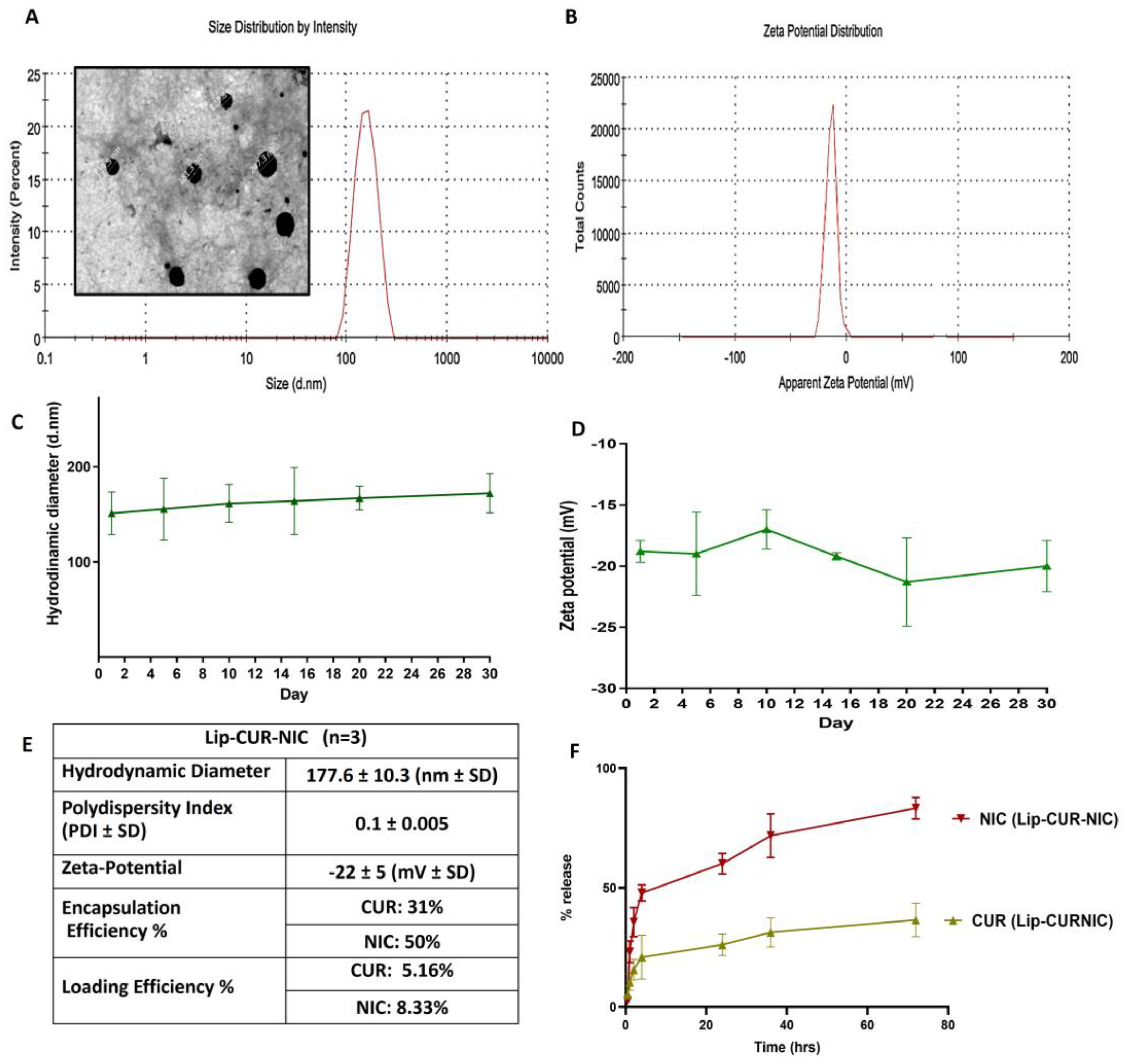
Figure 2.
A) Percent Inhibition of DPPH free radical using CUR, NIC and their combination B) IC50 of DPPH radical CUR, NIC, CUR-NIC and lip-CUR-NIC (mean ±SD, and n=3).
Figure 2.
A) Percent Inhibition of DPPH free radical using CUR, NIC and their combination B) IC50 of DPPH radical CUR, NIC, CUR-NIC and lip-CUR-NIC (mean ±SD, and n=3).
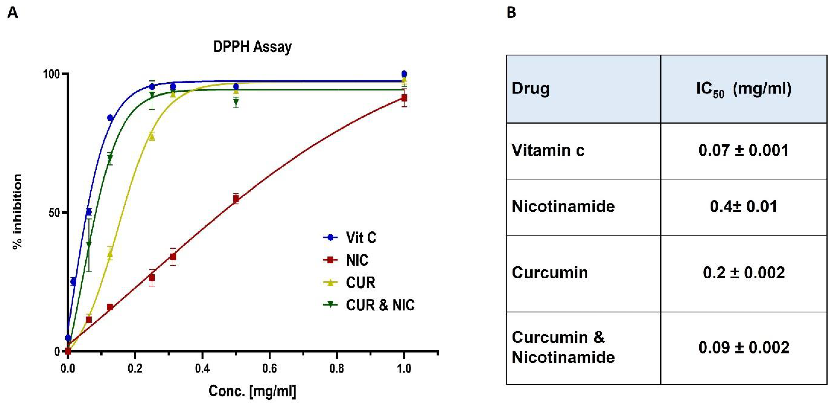
Figure 3.
Screening of antibacterial activity by well-Diffusion method A) CUR-NIC demonstrated moderate inhibition zone against S. aureus across four serial dilutions (1000, 125, 62. 5, and 31.5 μg/ml). B) The Lip-CUR-NIC tested across six serial dilutions (1000, 250, 125, 62.5 and 31.5 μg/ml.
Figure 3.
Screening of antibacterial activity by well-Diffusion method A) CUR-NIC demonstrated moderate inhibition zone against S. aureus across four serial dilutions (1000, 125, 62. 5, and 31.5 μg/ml). B) The Lip-CUR-NIC tested across six serial dilutions (1000, 250, 125, 62.5 and 31.5 μg/ml.
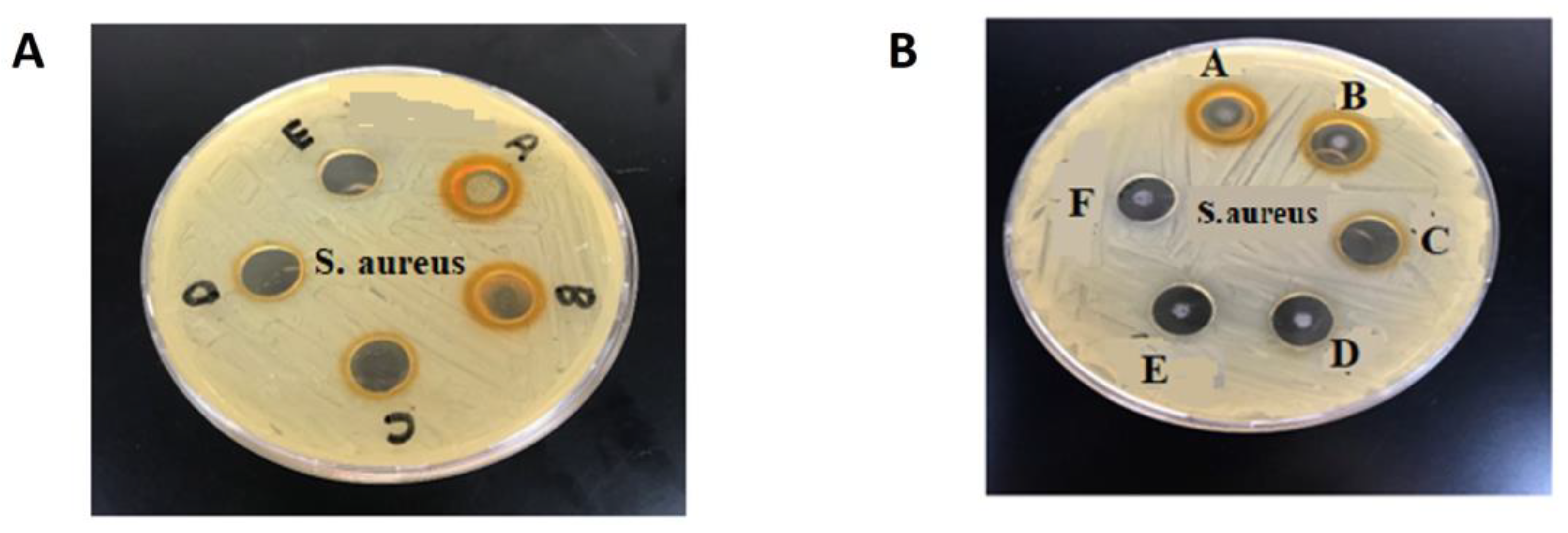
Figure 4.
Screening of antibacterial activity by well-Diffusion method A) CUR-NIC demonstrated moderate inhibition zone against S. epidermises across four serial dilutions (1000, 125, 62. 5, and 31.5 μg/ml). B) The Lip-CUR-NIC tested across six serial dilutions (1000, 250, 125, 62.5 and 31.5 μg/ml).
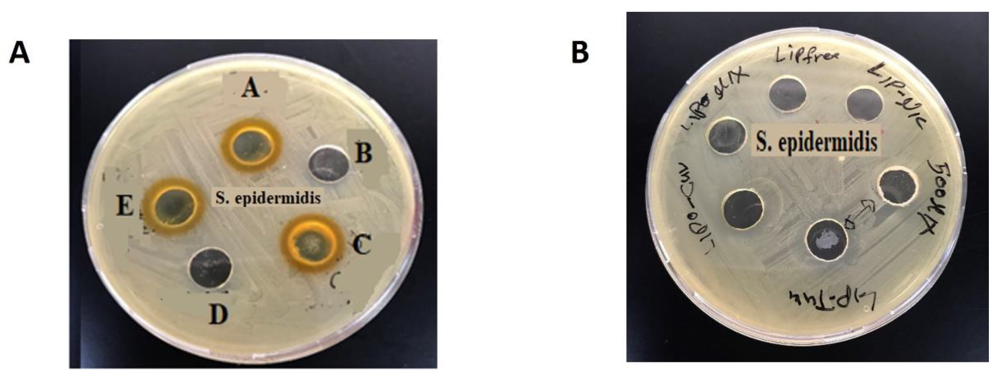
Figure 5.
A) Plate titter method for S. Aureus Susceptibility Test using CUR, Lip-CUR, CUR-NIC, Lip-CUR-NIC and NIC, at a concentration range of (400-3.13 μg/ml) B) Table of inhibition zone and MIC of the CUR, Lip-CUR, CUR-NIC, Lip-CUR-NIC and NIC.
Figure 5.
A) Plate titter method for S. Aureus Susceptibility Test using CUR, Lip-CUR, CUR-NIC, Lip-CUR-NIC and NIC, at a concentration range of (400-3.13 μg/ml) B) Table of inhibition zone and MIC of the CUR, Lip-CUR, CUR-NIC, Lip-CUR-NIC and NIC.
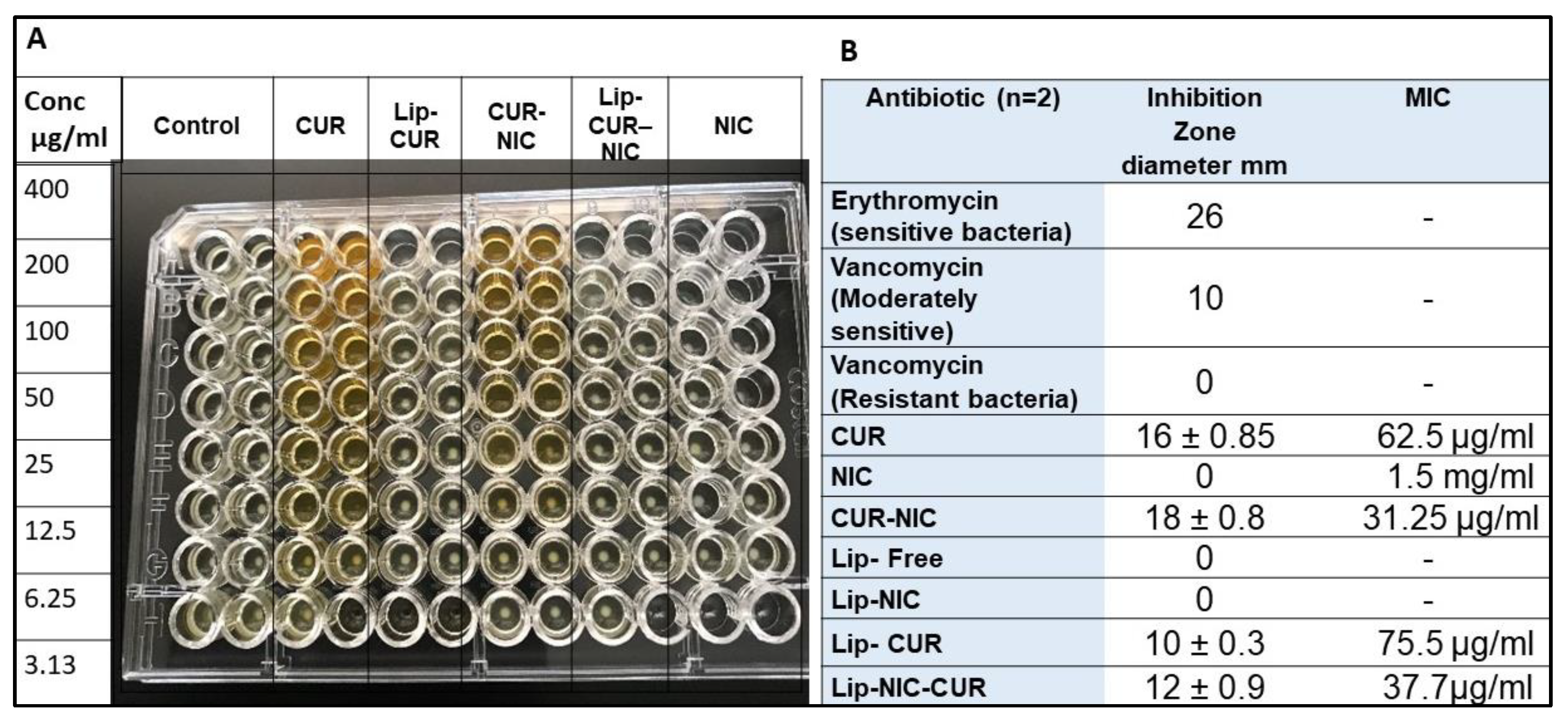
Figure 6.
(A)The Dose Response Curve for B16 cancer cells treated with Free CUR, NIC and their mixture (0.4-200 μM, n=3) (B) The Dose Response Curve for B16 cancer cells treated with Lip-CUR, Lip-NIC and Lip-CUR-NIC (0.4-200 μM, n=3) (C) Summary of IC50 values for single and combination of CUR & NIC against B16 cells.
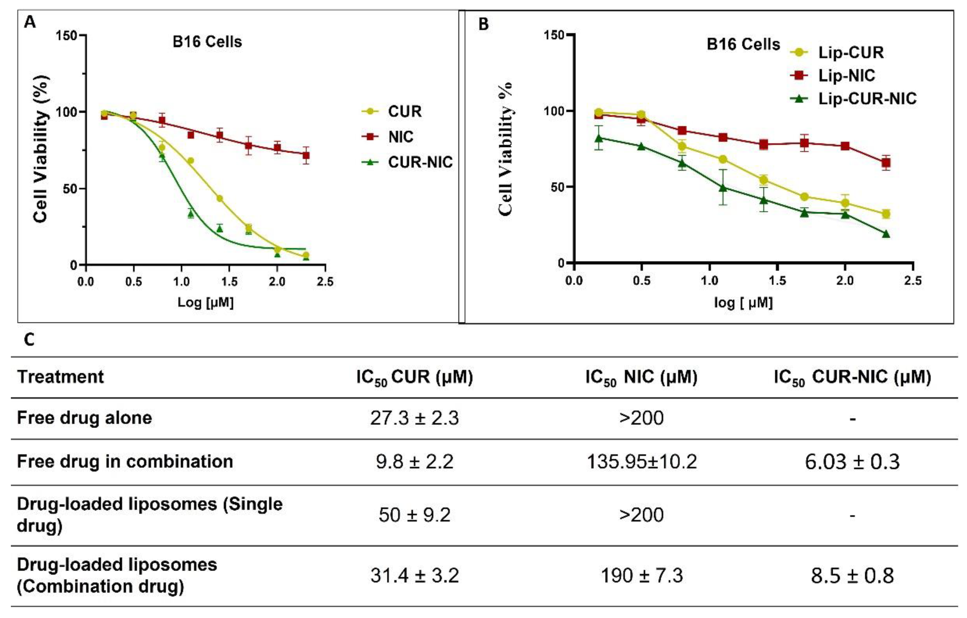
Figure 7.
(A) Isobologram for combination: CUR-NIC (CUR+NIC [1:1]) (B) Combination Index Plot of CUR and NIC Free Powder (C) Dose-reducing Index Curve of CUR and NIC combination (D) Dose-effect Curve of CUR and NIC combination (E) Summary of Combination Index (CI) and dose Reducing Index of (DRI) CUR-NIC.
Disclaimer/Publisher’s Note: The statements, opinions and data contained in all publications are solely those of the individual author(s) and contributor(s) and not of MDPI and/or the editor(s). MDPI and/or the editor(s) disclaim responsibility for any injury to people or property resulting from any ideas, methods, instructions or products referred to in the content. |
© 2024 by the authors. Licensee MDPI, Basel, Switzerland. This article is an open access article distributed under the terms and conditions of the Creative Commons Attribution (CC BY) license (http://creativecommons.org/licenses/by/4.0/).
Copyright: This open access article is published under a Creative Commons CC BY 4.0 license, which permit the free download, distribution, and reuse, provided that the author and preprint are cited in any reuse.


