
Submitted:
19 August 2024
Posted:
21 August 2024
You are already at the latest version
Abstract
Trematode infections cause long-term suffering and debilitation, posing a significant threat to global animal health and production and leading to considerable economic losses. Studies on the epidemiology and control of these infections in Tanzania are limited, and most have been conducted in abattoir settings. The study aims to fill the knowledge gap by determining the prevalence, distribution, and risk factors for trematode infections in domesticated ruminants in two ecological zones of Tanzania. A cross-sectional study was conducted in Lake Victoria and the southern highlands of Tanzania. Rectal fecal samples were collected and examined for Fasciola spp., paramphistomes, and schistosome infections using the sedimentation technique. A total of 1367 domesticated ruminants were sampled and examined for trematode infections. Prevalence of trematode infections was found to be 65.7%. The individual frequency of F. gigantica, paramphistomes, and S. bovis (based on egg morphology only,) was 35.1%, 60.2% and 3.1%, respectively. Adult cattle were more likely to be infected with paramphistomes (AOR: 1.98; 95% CI: 1.40-2.78) and S. bovis (AOR: 8.5; 95% CI: 1.12–64.19) than weaners. It was found that trematode infections in domesticated ruminants are prevalent across Tanzania, therefore effective and community-acceptable prevention and control strategies are highly needed.
Keywords:
Domesticated ruminants
; fasciola
; paramphistome
; trematode
; schistosoma
; Tanzania
1. Introduction
Trematode infections, particularly fasciolosis and schistosomiasis, are among the most common helminth infections in domestic ruminants worldwide [1,2]. Adult trematodes are sometimes referred to as "flukes," and the families that contain parasites of veterinary interest include Schistosomatidae, Fasciolidae, Paramphistomatidae, and Dicrocoeliidae [4,5]. The genera Fasciola (liver fluke), Paramphistomes (rumen/stomach fluke), and Schistosoma (blood fluke) [5] have similar life cycles in which domestic ruminants and humans (except rumen flukes) are definitive hosts, while wild animals serve as reservoirs [6]. The species F. gigantica [7], F. hepatica [8], S. bovis [9], paramphistomes species and Dicrocoelium hospes [10] have been widely reported in Tanzania.
The presence of these parasites is widespread in tropical and subtropical regions globally, thriving in areas with favorable climatic, ecological, and hygienic conditions [11,12]. In most African countries south of the Sahara, these infections are considered to be endemic [13]. The presence and distribution of freshwater snail intermediate hosts often influence the occurrence of trematode infections [14,15], which in turn vary with climatic circumstances [16].
Globally, trematode infections are a considerable veterinary and public health burden [17]. Moreover, they threaten domesticated ruminant health and production due to reduced fertility and productivity, liver condemnation, stunted growth, and premature death [18,19,20]. In the southern highlands of Tanzania, almost 100% of the total condemnations of bovine liver in slaughter slabs, poor growth rate, reduced milk production, and infertility are attributed to these infections [21,22].
Bovine trematode infections in Tanzania have been reported in all geographical zones by abattoir surveys [23,24,25,26,27], and studies on the epidemiology and control of these parasites in cattle have been carried out in the southern highlands [5,6,7,8,28]. In these studies, the prevalence of fasciolosis and schistosomiasis ranged from 18%-94% in domesticated ruminants depending on the production system used [21,22].
Despite these findings, there is a paucity of information regarding the magnitude, distribution, and risk factors for trematode infections in domesticated ruminants in many parts of Tanzania. This study was therefore conducted to address this gap to generate baseline data that will provide the scientific evidence needed to design effective and locally accepted control interventions against trematode infections.
2. Materials and Methods
2.1. Study Area
The study was conducted from March to May 2023 in the Misungwi, Bariadi, and Iringa District Councils of the Mwanza, Simiyu, and Iringa regions, respectively. Within the Misungwi district, the study was carried out in Kanyelele, Koromije, and Ibongoya B villages. In the Bariadi district, the study was conducted in Pugu and Itubukilo A villages. On the other hand, in the Iringa rural district, the study was conducted in Lupembelwasenga, Usengelindeti, and Migori villages (See Figure 1). The study districts and villages were selected purposively based on the available domesticated ruminant population, disease history, and disease ecology described in routine reports from the respective local District Veterinary Offices.
The Bariadi and Misungwi district councils are located in the northwestern part of Tanzania to the southeast of Lake Victoria. Both districts experience a low rain season from October to December and a high rain season between March and May of each year, while the dry period usually runs from January to the end of February and from June to the end of September of each year. The districts receive 700 mm to 950 mm of rain annually [29]. The temperatures in the two districts range from190°C to 290°C [29].
The Iringa Rural District Council is located in the southern highlands of Tanzania. The highland region in the eastern part of the district consists of many hills and valleys, along with numerous permanent rivers, streams, and ponds. In contrast, the flat lowland on the western side is semi-arid and characterized by dry grazing land with thickets and scattered bushes, as described by Mahoo [30]. The annual rainfall in highland areas ranges from 500 mm to 2700 mm, while in lowland areas; the rainfall is less than 600 mm [31].
2.2. Study Design and Sampling Procedure
A cross-sectional study was conducted around Lake Victoria and the southern highland Zone of Tanzania. Rectal fecal samples collected by veterinary technicians were concealed inside sterile surgical gloves that had been turned inside out. Domesticated ruminants information such as age (classified as weaners and adults), sex, breed (local-breed and cross-breed), and body condition (lean, median, fat) were recorded. Unique numbers and dates were used to label the samples, after which they were stored in an ice-packed cool box and transported to the laboratory of the National Institute for Medical Research (NIMR), Mwanza Centre, for laboratory examination.
The formula n = (Z2 * p * (1-p))/d2 was used to determine the sample size. This was based on an expected prevalence (p) of bovine fascioliasis of 33% as reported by Nzalawahe et al., [32], and a desired absolute precision (d) of 3% with a 95% confidence interval (CI) as per Thrusfield's [33]. A total of 1367 domesticated ruminants were sampled and examined.
2.3. Coprological Examination
Fecal samples from domesticated ruminants were processed by the fecal sedimentation method [34]. In brief, approximately 10 grams of fecal material was placed in a plastic container and mixed with 30 ml of tap water. Using 500 ml conical beakers, the fecal suspension was sieved through a wire mesh with an aperture of 250 μm and then filled with tape water. The suspension in a beaker was then allowed to settle for approximately 15 minutes.
Thereafter, the process of decanting the supernatants and re-suspending the sediments in tape water was repeated three times. Eventually, all the sediment was placed into a Petri dish, stained with 1% methylene blue, and observed under a microscope at 10x magnification for parasitic eggs [11]. The number of trematode eggs was determined by thoroughly examining every section of the Petri dish. To ensure accuracy, two lab technicians independently examined the same samples. The eggs were counted and identified based on specific morphological characteristics [35]. To calculate the egg per gram of feces, the number of specific parasite eggs present in a 10 g sample was divided by ten.
2.4. Mapping of Study Villages and Study Farms
The study villages and farms participating in the research were geographically mapped using handheld differential geographic global positioning system (GPS) units from Trimble Navigation Ltd. in California, USA, which had an approximate accuracy of ± 1 meter. The GPS data was downloaded with differential correction into a GPS database (GPS Pathfinder Office 2.8 from Trimble Navigation Ltd., California, USA), and the mapping process was carried out using ArcView version 9.2 software from Environmental Systems Research Institute, Inc. in Redlands, CA (refer to Figure 2 for details).
2.5. Statistical Analysis
The information obtained during the collection of samples and the findings from the coprological analysis were entered into the Census and Survey Processing System (CSPro) software by the U.S. Census Bureau in the United States. The cleaned dataset was transferred to Stata version 15.1 developed by Stata Corporation located in College Station, TX, USA. The data was summarized using descriptive statistics, and proportions were used to summarize categorical variables, which were then compared using the chi-square test. The egg count data (EPGs) underwent log transformation [log10(EPG+1)], and differences in EPGs between sex, age, and breed were examined using two-sample t-tests, while one-way analysis of variance (ANOVA) was utilized to compare the EPGs between different body conditions. Positive for at least one egg was the criteria for considering an animal to be infected. Statistical significance was considered for P values ≤ 0.05. Associations between parasitic infections and risk factors were tested using multiple logistic regression analysis. QGIS® spatial software version 2.2 was utilized to represent the distribution of trematode infections.
3. Results
3.1. Animal Population Characteristics
A total of 1367 domesticated ruminants (739 cattle, 319 goats, and 309 sheep) were sampled and examined for trematode infections. Most of the sampled animals were female; 57.9% (428/739) were cattle, and 64.4% (199/309) were sheep. A total of 83.7% (267/319) of the goats were males. Compared with crossbred breeds, local breeds were more common in each group of examined animals, accounting for 98.4% (729/739) of cattle, 100% (319/319) of goats and 100% (309/309) of sheep. In terms of age, adult animals constituted the majority of the animals sampled: 73.5% (543/739) for cattle, 61.1% (195/319) for goats, and 63.1% (195/309) for sheep.
3.2. Prevalence of F. gigantica, Paramphistomes and S. bovis
Prevalence of trematode infections was found to be 65.7%. The individual frequency of F. gigantica, paramphistomes, and S. bovis (based on egg morphology only, Figure 2) was 35.1%, 60.2% and 3.1%, respectively. (Table 1). All animal species (cattle, goats and sheep) were co-infected with all three parasite species, but sheep were more often co-infected than other animal species (Table 1). The most frequently observed co-infections were between F. gigantica and paramphistome at 29.7% (95% CI: 27.3-32.2%).
The Simiyu region was found to have high burden of F. gigantica (43% (95% CI: 36.2-50.0), paramphistomes (84.1% (95% CI: 78.3-88.8) and S. bovis infections (7.7% (95% CI: 4.4-12.2) in cattle compared to the Mwanza and Iringa regions (Table 2). Comparatively, the Simiyu region exhibited the highest prevalence of F. gigantica, paramphistomes, and S. bovis in goats and sheep compared to the other study regions (Table 2).
The associations between trematode infections and demographic characteristics (sex, age, and breed) of cattle, goats, and sheep are shown in Table 3, Table 4, and Table 5, respectively. Adult cattle (24+ months) were at higher risk of infection with paramphistomes (AOR: 1.98; 95% CI: 1.40-2.78) and S. bovis (AOR: 8.5; 95% CI: 1.12–64.19) than younger cattle (Table 3). Locally bred cattle were more susceptible to paramphistomes infection (AOR: 8.19; 95% CI: 1.76-38.09) than were crossbred cattle and low risk of infection with F. gigantica (AOR: 0.61; 95% CI: O.19-1.92) and S. bovis (AOR: 0.25; 95% CI: 0.03-2.21) (Table 3).
Adult goats (24+ months) were more susceptible to F. gigantica infection (AOR: 1.59; 95% CI: 0.92-2.77), paramphistomes (AOR: 1.07; 95% CI: 0.8-1.68) and S. bovis (AOR: 3.9; 95% CI: 0.46-32.82) than younger goats, however these associations were not statistically significant (Table 4). Compared with male goats, female goats were less likely to be infected with F. gigantica (AOR: 0.83; 95% CI: 0.47-1.46) and S. bovis (AOR: 0.97; 95% CI: 0.18-5.12) (Table 4).
Female sheep had a 1.63 times higher likelihood of being infected with S. bovis compared to male sheep (AOR: 1.63; 95% CI: 0.43-6.17) (Table 5). Adult sheep had higher odds of being infected with F. gigantica (AOR: 2.11; 95% CI: 1.28-3.51), paramphistomes (AOR: 1.93; 95% CI: 1.20-3.10), and S. bovis (AOR: 2.94; 95% CI: 0.63-13.71) compared to younger sheep. The differences in infection rates between age groups were statistically significant for F. gigantica (P < 0.004) and paramphistomes (P < 0.007), but not for S. bovis (Table 5).
3.3. Spatial Distribution of Infections with F. gigantica, Paramphistomes and S. bovis
Infections with F. gigantica, paramphistomes, and S. bovis showed varying prevalence across regions and districts, with the Lake Victoria zone exhibiting the highest prevalence (Table 2 and Figure 3). Pugu village in the Simiyu region had the highest prevalence rates of S. bovis in cattle, goats, and sheep, with rates of 7.8% (95% CI: 3.4-14.9%), 7.4% (95% CI: 2.0-17.9%), and 12.7% (95% CI: 5.3-24.5%) respectively.
F. gigantica was found to be more prevalent in Kanyelele village (Mwanza region) in sheep, at a prevalence of 70.0% (95% CI: 50.6-85.2), while for cattle, the highest prevalence was found in Usengelindete village (Iringa region), at 54.6% (95% CI: 44.8-64.2), and for goats, in Itubukilo village (Simiyu region), at 42.1% (95% CI: 29.1- 55.0)
The highest prevalence of paramphistomes was found in Itubukilo A Village for both cattle and sheep, at 88.6% (95% CI: 80.9-93.9%) and 88.7% (95% CI: 77.0-95.7%), respectively. The greatest percentage of paramphistomes was found in Lupembelwasenga village (80.0%, 95% CI: 51.9-95.7%).
3.4. Fecal Egg Count
The average (±SE) fasciola, paramphistomes, and schistosoma egg counts in cattle were 2.4 (0.06), 3.0 (0.06), and 0.8 (0.05), respectively, with ranges of 0.7 to 6.1, 0.7 to 6.3, and 0.7 to 1.4. In goats, the mean epg were 2.5 (0.14), 2.6 (0.09), and 0.8 (0.10), with ranges of 0.7 to 5.9, 0 to 6.2, and 0.7 to 1.4. For sheep, the averages were 2.6 (0.11), 2.9 (0.10), and 0.9 (0.09), with ranges of 0.7 to 6.6, 0.7 to 6.5, and 0.7 to 1.4, respectively.
In goats, there was a significant correlation between age and the average epg for fasciola (P<0.011), and paramphistomes showed a significant correlation with body condition (P<0.028). Paramphistomes had a higher mean fecal egg count per gram of feces compared to fasciola and schistosoma (Table 6 and Table 7).
4. Discussion
The burden of trematode infections of F. gigantica, paramphistomes, and S. bovis in domesticated ruminants in various ecological zones (specifically the Lake Zone (Mwanza and Simiyu) and Southern Highlands) of Tanzania has been investigated for the first time in this study.
The results indicated that domesticated ruminants in the study regions of Tanzania have a high prevalence of S. bovis, paramphistomes, and F.gigantica infections. Cattle showed a higher prevalence of S. bovis, paramphistomes, and F.gigantica compared to small ruminants, which is consistent with findings from the study conducted in Côte d’Ivoire by Kouadio et al.,[13]. This could be attributed to the fact that goats primarily consume leaves and heaths in elevated areas, while sheep graze on open land, and cattle graze near water bodies. As a result, cattle are at a greater risk of being exposed to water, where the infective parasite stage is present, leading to transmission.
It was found that paramphistomes and F. gigantica are the predominant trematode infections in cattle, goats, and sheep. This aligns with earlier research conducted in Tanzania and other parts of Africa [7,32,36]. The study found that paramphistomes had the highest average fecal egg count per gram of feces (epg) followed by F. gigantica and S. bovis. A similar pattern of infection load for these three trematodes has been documented in various regions of Tanzania and other parts of Africa [32,37].
In this study, the high occurrence of paramphistomes could be attributed to the fact that the adult parasite is highly productive and capable of surviving in the host for extended periods [38]. Paramphistome parasites are known for their high reproductive capacity and their ability to thrive in harsh conditions [39]. Additionally, many broad-spectrum anthelmintics, such as albendazole, ivermectin, and triclabendazole, which are commonly used to prevent important nematode and trematode parasites, have minimal or no impact on paramphistomes [40].
The prevalence of S. bovis infection in this study was lower compared to other trematodes, aligning with the findings of Nzalawahe study [32]. Furthermore, schistosomal eggs are not always excreted in feces because they are sometimes left trapped in tissues, which may result in a low prevalence in fecal samples [41]. Additionally, the host organism immune response against schistosomes is directed towards the suppression of worm fecundity, resulting in reduced egg output rather than elimination of adult worms [42].
The higher occurrence of F. gigantica may be attributed to the higher prevalence of Fasciola in the study regions. In contrast, a study conducted in southern Ethiopia also demonstrated a higher prevalence of F. gigantica in cattle [43]. The predominant co-infection of trematodes in this study was observed between F. gigantica and paramphistomes. This could be attributed to the similarities in the life cycles of these parasites, which both rely on lymnaeid snails as intermediate hosts [44,45].
The observed distribution of trematode infections in this study is likely determined by the natural ecology of the study areas. These water bodies not only provide habitats for intermediate snail hosts of parasites [46], but also animal-water contact sites, and aquatic plants that can be consumed by domesticated animals, all of which play essential roles in maintaining the transmission cycle of trematode infections. The results of previous study conducted in the southern highlands of Tanzania indicated that the spread of trematode infection is impacted by the management of domesticated ruminants [7]. This finding aligns with a study carried out in Mali, which demonstrated that factors such as climate, the presence of water bodies, and the type of domesticated ruminant rearing systems can affect the prevalence of trematode infections [47].
Adult cattle were more infected by paramphistome and S. bovis infections compared to younger animals, likely due to prolonged exposure to contaminated water while grazing. This pattern has also been observed in Nigeria [48] and Tanzania [46].
The study limitations include the small sample size for goats and sheep in comparison to cattle across all study villages. Additionally, the study did not document the treatment history, which could have impacted the prevalence of trematode infections. The identification of parasite species relied solely on egg morphology. Nonetheless, this study underscores the significance of trematode infections of veterinary importance in Tanzania. It is also the first study to provide a comprehensive overview of the magnitude and distribution of these trematode infections among domesticated ruminants in the Lake Victoria Zone (Mwanza and Simiyu regions) of Tanzania.
5. Conclusions
The study revealed that paramphistomes and F. gigantica were more prevalent trematode infections in cattle, goats, and sheep sampled from the Lake Victoria zone (Mwanza and Simiyu) and southern highlands (Iringa region) of Tanzania. The findings clearly show that trematode infections are prevalent across the two study areas, with the highest prevalence found in the lake zone (Mwanza and Simiyu regions); thus, effective and community-acceptable prevention and control interventions are needed to break the transmission cycle and hence reduce fecal egg contamination of the environment and open water sources.
Author Contributions
GSM, SK, JN, AK, MEV, AS BJV conceptualized and designed the study; GSM, SK and JN planned and conducted the field work; GSM and JN examined the fecal specimens; GSM analyzed the data and drafted the manuscript; and GSM, JN, AK, SK, MEV, AS and BJV revised and improved the final version of the manuscript. The authors have read and agreed the final version of the manuscript.
Funding
This research was funded by the European Union's Horizon 2020 as part of the PREPARE4VBD research and innovation program under grant agreement No. 101000365.
Institutional Review Board Statement
This research was a part of a project called "PREPARE4VBDs" aimed at identifying, predicting, and preparing for emerging vector-borne diseases. The study adhered to the ethical approval guidelines set forth by the Medical Research Coordination Committee (MRCC) of the National Institute for Medical Research (NIMR), which serves as the national ethics review board in Tanzania (ethics clearance certificate number NIMR/HQ/R.8a/Vol. IX/3860).
Informed Consent Statement
Informed consent was obtained from the owners of all animals involved in the study.
Data Availability Statement
The data presented in this study are available on request from the corresponding author.
Acknowledgments
The authors express their gratitude to the Regional Commissioners of the Mwanza, Simiyu, and Iringa regions, the District Executive Directors of the Bariadi, Misungwi, and Iringa Rural districts, the Regional and Districts Veterinary Officers, and the Village and Subvillage leaders for their cooperation during fieldwork. Salim Bwata, Revocatus Silayo, Aruni Haruya, Martin Anditi, and Kalebu Kihongosi are acknowledged for their technical assistance during field and laboratory work.
Conflicts of Interest
No competing interests were declared.
References
- Mage, C.; Bourgne, H.; Toullieu, J.M.; Rondelaud, D.; Dreyfuss, G. Fasciola hepatica and Paramphistomum daubneyi: changes in prevalences of natural infections in cattle and in Lymnaeatruncatula from central France over the past 12 years. Veterinary research 2002, 33, 439–447. [Google Scholar] [CrossRef] [PubMed]
- Swai, E.S.; Ulicky, E. An evaluation of the economic losses resulting from condemnation of cattle livers and loss of carcass weight due to fasciolosis: a case study from Hai town abattoir, Kilimanjaro region, Tanzania. Domesticated ruminants Res Rural Dev 2009, 21, 186. [Google Scholar]
- Urquhart, G.M.; Armour, J.; Duncan, J.; Dunn, A.; Jennings, F.W. "Veterinary parasitology 2003, 2nd ed. Black well science Ltd." 252.
- Mas-Coma, S.; Valero, M.A.; Bargues, M.D. Fasciola, lymnaeids and human fascioliasis, with a global overview on disease transmission, epidemiology, evolutionary genetics, molecular epidemiology and control. Advances in parasitology 2009, 69, 41–146. [Google Scholar]
- Dreyfuss, G.; Alarion, N.; Vignoles, P.; Rondelaud. D. A retrospective study on the metacercarial production of Fasciola hepatica from experimentally infected Galba truncatula in central France. Parasitology Research 2006, 98, 162–166. [Google Scholar] [CrossRef] [PubMed]
- Chen, J.; Chen, M.; Ai, L.; Xu, X.N.; Jiao, J.; Zhu, T.; Su, H.; Zang, W.; Luo, J.; Guo, Y.; Lv, S.; Zhou, X. “An Outbreak of Human Fascioliasis Gigantica in Southwest China.” PLoS ONE, 2023, 8.
- Keyyu, J.D.; Monrad, J.; Kyvsgaard, N.C.; Kassuku, A.A. Epidemiology of F. gigantica and amphistomes in cattle on traditional, small-scale dairy and large-scale dairy farms in the southern highlands of Tanzania. Trop Anim Health Pro 2005, 37, 303–314. [Google Scholar] [CrossRef] [PubMed]
- Walker, S.M.; Makundi, A.E.; Namuba, F.V.; Kassuku, A.A.; Keyyu, J.; Hoey, E.M.; Prodohl, P.; Stothard, J.R.; Trudgett, A. The distribution of Fasciola hepatica and F. gigantica within southern Tanzania–constraints associated with the intermediate host. Parasitology 2008, 135, 495–503. [Google Scholar] [CrossRef]
- Kassuku, A.A.; Christensen, N.O.; Monrad, J.; Nansen, P.; Knudsen, J. Epidemiological studies of S. bovisinIringa Region, Tanzania. Acta Trop 1986, 43, 153–163. [Google Scholar]
- Mahlau, E.A. Liver fluke survey in zebu cattle of Iringa Region, Tanzania and first finding of the small fluke Dicrocoeliumhospes/Loos. Bull Epizootic Dis Afr 1970, 18:21–28.
- Mas-Coma, S.; Bargues, M.D.; Valero, M.A. Human fascioliasis infection sources, their diversity, incidence factors, analytical methods and prevention measures Parasitology 2018,145, 1665–99.
- Lai, Y.S.; Biedermann, P.; Ekpo, U.F.; Garba, A.; Mathieu, E, Midzi, N. ; Pauline, M. P.; N'Goran, E.K.; Raso, G.; Assaré, R.K.; Sacko, M.; Schur, N.; Talla, I.; Tchuenté, L.T.; Touré, S.; Winkler, M.S.; Utzinger, J,; Vounatsou, P. Spatial distribution of schistosomiasis and treatment needs in sub-Saharan Africa: a systematic review and geostatistical analysis. Lancet Infect Dis 2015, 15, 927–40. [Google Scholar]
- Kouadio, J.N.; Evack, J.G.; Achi, L.Y.; Fritsche, D.; Ouattara, M.; Silué, K.D.; Bonfoh, B.; Hattendorf, J.; Utzinger, J.; Zinsstag, J.; Balmer, O.; N’Goran, E.K. “Prevalence and Distribution of livestockSchistosomiasis and Fascioliasis in Côte d’Ivoire: Results from a Cross-Sectional Survey. ” BMC Veterinary Research 2020, 16, 1–13. [Google Scholar]
- Schillhorn van Veen, T.W.; Folaranmi, D.O.B.; Usman, S.; Ishaya, T. Incidence of liver fluke infections (Fasciola gigantic and Dicrocoeliumhospes) in ruminants in Northern Nigeria. Tropical Animal Health and Production 1980, 12, 97–104. [Google Scholar] [CrossRef]
- Brown, D.S. Freshwater Snails of Africa and their Medical Importance,1994 2nd Edn. Taylor and Francis Ltd, London.
- Mungube, E.O.; Bauni, S.M.; Tenhagen, B.A.; Wamae, L.W.; Nginyi, J.M.; Mugambi, J.M. The prevalence and economic significance of F. giganticaand Stilesia hepatica in slaughtered animals in the semiarid coastal Kenya. Tropical Animal Health and Production 2006, 38, 475–483. [Google Scholar] [CrossRef] [PubMed]
- GBD 2017 DALYs and HALE Collaborators Global, regional, and national disability-adjusted life-years (DALYs) for 359 diseases and injuries and healthy life expectancy (HALE) for 195 countries and territories, 1990–2017: a systematic analysis for the Global Burden of Disease Study 2017. Lancet Lond Engl 2018, 392, 1859–922.
- Saleha, A.A. Liver fluke disease (fascioliasis): epidemiology, economic impact and public health significance Southeast Asian J Trop Med Public Health 1991, 22(Suppl), 361–4.
- You, H.; Cai, P.; Tebeje, B.; Li, Y.; McManus, D. Schistosome vaccines for domestic animals. Trop Med Infect Dis 2018, 3, 68. [Google Scholar] [CrossRef]
- Yusuf, M.; Ibrahim, N.; Tafese, W.; Deneke, Y. Prevalence of bovine fasciolosis in municipal abattoir of Haramaya, Ethiopia. Food SciQualManag 2016, 48, 38–43. [Google Scholar]
- Nonga, H.E.; Mwabonimana, M.F.; Ngowi, H.A.; Mellau, L.S.; Karimuribo, E.D. “A Retrospective Survey of Liver Fasciolosis and Stilesiosis in Domesticated ruminants Based on Abattoir Data in Arusha, Tanzania. ” Tropical Animal Health and Production 2009, 41, 1377–80. [Google Scholar] [CrossRef]
- Nzalawahe, J.; Hannah, R.; Kassuku, A.A.; Stothard, J.R.; Coles, G.C.; Eisler, M.C. “Evaluating the Effectiveness of Trematocides against F. gigantica and Amphistomes Infections in Cattle, Using Fecal Egg Count Reduction Tests in Iringa Rural and Arumeru Districts, Tanzania.” Parasites and Vectors 2018, 11, 384. [Google Scholar]
- Hyera, J.M.K. Prevalence, seasonal variation and economic significance of fascioliasis in cattle as observed at Iringa abattoir between 1976–1980. Bull Anim Health Pro Afr 1984, 32, 356–359. [Google Scholar]
- Komba, E.V.G.; Mkupasi, E.M.; Mbyuzi, A.O.; Mshamu, S.; Luwumba, D.; Busagwe, Z.; Mzula, A. Sanitary practices and occurrence of zoonotic conditions in cattle at slaughter in Morogoro Municipality, Tanzania: implications for public health. Tanzania J Health Res 2012, 14, 131–8. [Google Scholar] [CrossRef]
- Mellau, L.S.B.; Nonga, H.E.; Karimuribo, E.D. A slaughterhouse survey of liver lesions in slaughtered cattle, sheep and goats at Arusha, Tanzania. Res J Vet Sci 2010, 3, 179–188. [Google Scholar] [CrossRef]
- Msanga, J.F. Prevalence and economic importance of F. gigantica and Stilesia hepatica in Sukuma land, Tanzania. Tanzania Vet Bull 1985, 7, 9–16. [Google Scholar]
- Swai, E.S.; Mtui, P.F.; Mbise, A.N.; Kaaya, E.; Sanka, P.; Loomu, P.M. Prevalence of gastrointestinal parasite infections in Maasai cattle in Ngorongoro District, Tanzania. Domesticated ruminants Res Rural Dev 2006, 18, 107. [Google Scholar]
- Makundi, A.E.; Kassuku, A.A.; Maselle, R.M.; Boa, M.E. Distribution, prevalence and intensity of S. bovis in cattle in Iringa District, Tanzania. Vet Parasitol 1998, 75, 59–6. [Google Scholar] [CrossRef] [PubMed]
- Tanzania Climate, Retrieved from The Global Historical Weather and Climate Data, https://weatherandclimate.com/tanzania (accessed 29 May 2024).
- Mahoo, H. Improving research strategies to assist scaling-up of pro-poor management of natural resources in semiarid areas 2005, 82pp.
- Tanzania Climate, Retrieved from The Global Historical Weather and Climate Data, https://weatherandclimate.com/tanzania (accessed 29 May 2024).
- Nzalawahe, J.; Kassuku, A.A.; Stothard, J.R.; Coles, G.C.; Eisler, M.C. “Trematode Infections in Cattle in Arumeru District, Tanzania are Associated with Irrigation” Parasites & Vectors 2014, 7, 107. 7.
- Thrusfield, M. Veterinary epidemiology. 2018, John Wiley & Sons. Fourth edition.
- Sirois, M. Principles and practice of veterinary technology 2016, 4th edition. St. Louis, Missouri.
- Soulsby, E.J.L. Helminths, arthropods and protozoa of domesticated animals 1982, 7th Ed. London: Baillere Tindall.
- Pfukenyi, D.M.; Monrad, J.; Mukaratirwa, S. Epidemiology and control of trematode infections in cattle in Zimbabwe: a review. J S Afr Vet Assoc 2005, 76, 9–17. [Google Scholar] [CrossRef]
- Yeneneh, A.; Kebede, H.; Fentahun, T.; Chanie, M. Prevalence of cattle flukes infection at Andassa domesticated ruminants research center in north‒west of Ethiopia. Veterinary Research Forum 2012, 3, 85–89. [Google Scholar] [PubMed]
- Dorchies, P.H. Flukes: Old parasites but new emergence. Proceedings of the XXIV World Buiatrics Congress 2006, Vol. 16 Consultada.
- Hansen, J.; Perry, B. The epidemiology, diagnosis and control of helminth parasites of ruminants, a hand book. Nairobi, Kenya: International Laboratory for Research on Animal Disease (ILRAD) 1994.
- Rolfe, P.F.; Boray, J.C.; Nichols, P.; Collins, G.H. Epidemiology of paramphistomosis in cattle. International Journal of Parasitology 1991, 21, 813–819. [Google Scholar] [CrossRef]
- Melkamu, S. Study on prevalence and associated risk factors for bovine and human schistosomiasis in Bahir Dar and its surrounding areas. J Anim Res 2016, 6, 967–75. [Google Scholar] [CrossRef]
- Lawrence, J.A. Schistosomamattheei in sheep: the host-parasite relationship. Res Vet Sci 1974, 17, 263–4. [Google Scholar] [CrossRef]
- Chakiso, B.; Menkir, S.; Desta, M. On farm study of bovine fasciolosis in Lemo district and its economic loss due to liver condemnation at Hossana municipal abattoir, southern Ethiopia. Int J CurrMicrobiolAppl Sci, 2014, 3, 1122–32. [Google Scholar]
- Food and Agriculture Organization. The Epidemiology of helminth parasites.1993 2nd ed. p. 1–30.
- Keyyu, J.D.; Kassuku, A.A.; Msalilwa, L.P.; Monrad, J.; Kyvsgaard, N.C. Cross-sectional prevalence of helminth infections in cattle on traditional, small-scale and largescale dairy farms in Iringa district, Tanzania. Vet Res Commun 2006, 30, 45–55. [Google Scholar] [CrossRef]
- Nzalawahe, J.; Kassuku, A.A.; Russell, S.J.; Coles, G.C.; Eisler, M.C. Associations between trematode infections in cattle and freshwater snails in highland and lowland areas of Iringa. Parasitology 2015, 142, 1430–9. [Google Scholar] [CrossRef]
- Tembely, S.; Galvin, T.J.; Craig, T.M.; Traore, S. Liver fluke infections of cattle in Mali. An abattoir survey on prevalence and geographic distribution. Trop Anim Health Prod 1988, 20, 117–21. [Google Scholar] [CrossRef] [PubMed]
- Elelu, N.; Ambali, A.; Coles, G.C.; Eisler, M.C. Cross-sectional study of F. gigantica and other trematode infections of cattle in Edu local government area, Kwara state, north-Central Nigeria. Parasit Vectors 2016, 9, 470. [Google Scholar] [CrossRef] [PubMed]
Figure 1.
A map showing the study areas: A=Tanzania map, B=Bariadi District, C=Misungwi District, D=Iringa Rural District.
Figure 1.
A map showing the study areas: A=Tanzania map, B=Bariadi District, C=Misungwi District, D=Iringa Rural District.
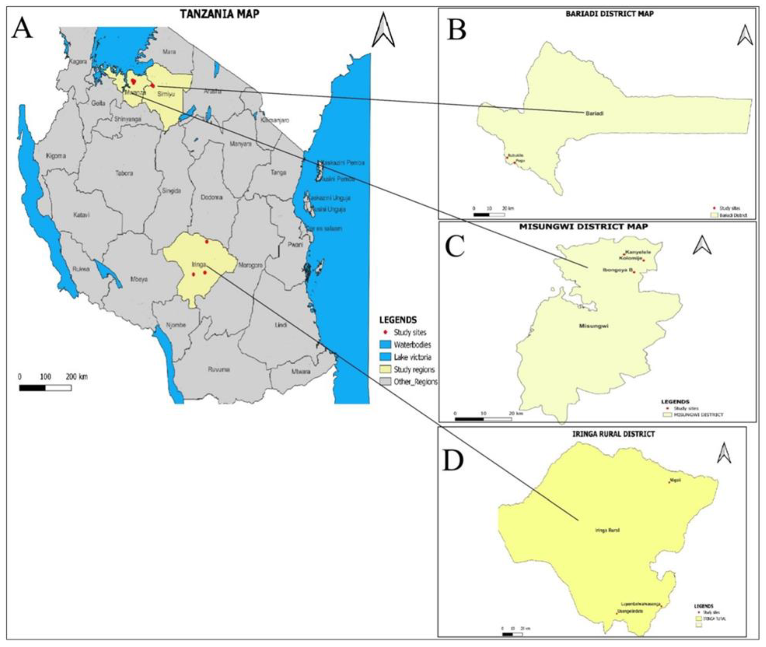
Figure 2.
Fasciola egg (A), S. bovis egg (B) and paramhistome egg (C).
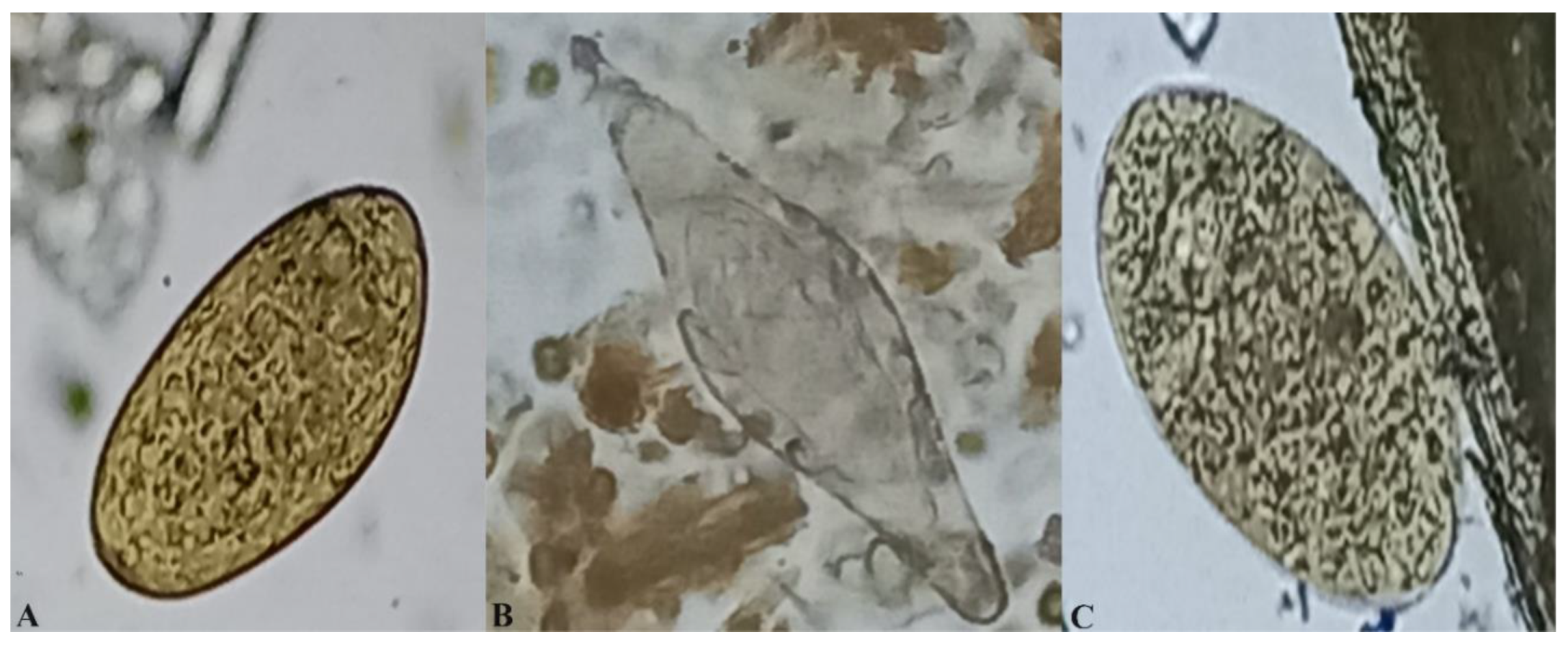
Figure 3.
A map showing trematode infections distribution across villages sampled in the Bariadi, Misungwi and Iringa rural district councils of Tanzania.
Figure 3.
A map showing trematode infections distribution across villages sampled in the Bariadi, Misungwi and Iringa rural district councils of Tanzania.
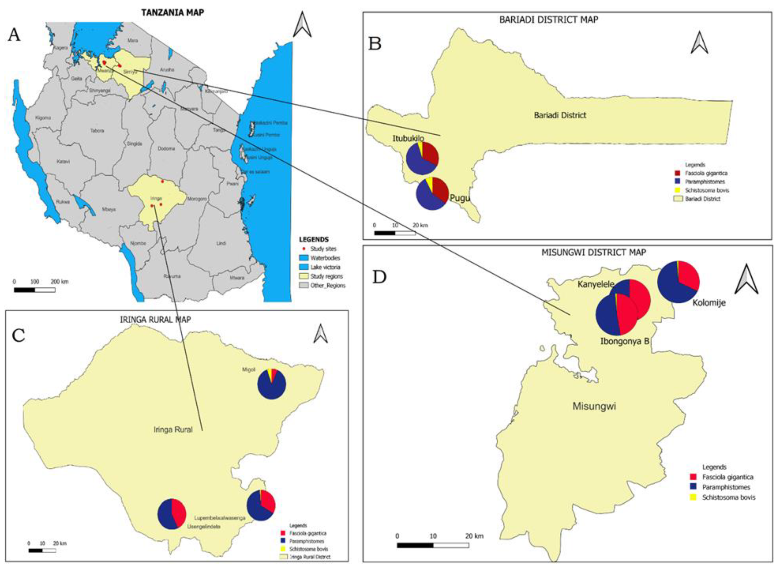

Table 1.
The prevalence of trematode infections (F. gigantica, Paramphistomes and S bovis) by animal species.
Table 1.
The prevalence of trematode infections (F. gigantica, Paramphistomes and S bovis) by animal species.
| Cattle N=739 |
Goats N=319 |
Sheep N=309 |
Total N=1367 |
|||||
|---|---|---|---|---|---|---|---|---|
| ++ | %(95%CI) | ++ | %(95%CI) | ++ | %(95%CI) |
++ |
%(95%CI) |
|
| F.gigantica | 289 | 39.1 (35.6-42.7) | 75 | 23.5 (19.0-28.6) | 116 | 37.5 (32.1-43.2) | 480 | 35.1 (32.6-37.7) |
| paramphistomes | 484 | 65.5(61.9-68.9) | 155 | 48.6 (43.0-54.2) | 184 | 59.6 (53.8-65.1) | 823 | 60.2 (57.6-62.8) |
| S.bovis | 24 | 3.3 (2.1-4.8) | 7 | 2.2(0.9-4.5) | 12 | 3.9 (2.0-6.7) | 43 | 3.1(2.3-4.2) |
| F. gigantica+ paramhistomes | 247 | 33.4 (30.0-37.0) | 65 | 20.4(16.1-25.2) | 94 | 30.4(25.3-35.9) | 406 | 29.7(27.3-32.2) |
| F. gigantica+ S.bovis | 16 | 2.2 (1.2-3.5) | 5 | 1.6(0.5-3.6) | 11 | 3.6 (1.8-6.3) | 32 | 2.3 (1.6-3.3) |
| Paramphistomes + S. bovis | 23 | 3.1(1.9-4.6) | 7 | 2.2(0.9-4.5) | 11 | 3.6 (1.8-6.3) | 41 | 3.0 (2.2-4.0) |
| F. gigantica+ paramphistomes + S. bovis | 16 | 2.2 ((1.2-3.5) | 5 | 1.6 (0.5-3.6) | 10 | 3.2(1.6-5.9) | 31 | 2.3(1.5-3.2) |
| Overall Prevalence | 527 | 71.3 (67.9-74.5) | 165 | 51.7(46.1-57.3) | 206 | 66.7(61.1-71.9) | 898 | 65.7(63.1-68.2) |
N: Examined domesticated ruminants, ++: infected domesticated ruminants, CI: confidence interval.
Table 2.
The prevalence of trematode infections (F. gigantica, paramphistomes and S. bovis) by region.
Table 2.
The prevalence of trematode infections (F. gigantica, paramphistomes and S. bovis) by region.
| F.gigantica | Paramphistomes | S. bovis | ||||||||||||||||
|---|---|---|---|---|---|---|---|---|---|---|---|---|---|---|---|---|---|---|
| Cattle | Goat | Sheep | Cattle | Goat | Sheep | Cattle | Goat | Sheep | ||||||||||
| Region | ++ | %(95%CI) | ++ | %(95%CI) | ++ | %(95%CI) | ++ | %(95%CI) | ++ | %(95%CI) | ++ | %(95%CI) | ++ | %(95%CI) | ++ | %(95%CI) | ++ | %(95%CI) |
| SIMIYU | 89 | 43(36.2-50.0) | 42 | 37.8(28.8-47.5) | 52 | 48.1(38.4-58.0) | 174 | 84.1(78.3-88.8) | 71 | 64.0(54.3-72.9) | 81 | 75(65.7-82.8) | 16 | 7.7(4.4-12.2) | 5 | 4.5(1.5-10.2) | 12 | 11.1(0.6-18.6) |
| IRINGA | 110 | 35.7 (30.4-41.3) | 3 | 2.6 (0.5-7.4) | 23 | 20.7(13.6-29.4) | 186 | 60.4(54.7-65.9) | 40 | 34.8((26.1-44.2) | 57 | 51.4(41.7-60.9) | 6 | 2(0.7-4.2) | 1 | 0.9(0.0-4.7) | 0 | 0(0.0-0.3) |
| MWANZA | 90 | 40.2 (33.7-46.9) | 30 | 32.3(22.9-42.7) | 41 | 45.6(35.0-56.4) | 124 | 55.4(48.6-62.0) | 44 | 47.3(36.9-57.9) | 46 | 51.1(40.3-61.8) | 2 | 0.9(0.1-3.2) | 1 | 1.1(0.0-5.8) | 0 | 0(0.0-0.4) |
N: Examined domesticated ruminants, ++: infected domesticated ruminants, CI: confidence interval.
Table 3.
Multiple logistic regression analysis of variables associated with F.gigantica, Paramphistomes and S. bovis infections among cattle.
Table 3.
Multiple logistic regression analysis of variables associated with F.gigantica, Paramphistomes and S. bovis infections among cattle.
| F.gigantica | Paramphistomes | S. bovis | ||||||||||||||
|---|---|---|---|---|---|---|---|---|---|---|---|---|---|---|---|---|
| N | ++ | % | AOR | 95% CI | p value | ++ | % | AOR | 95% CI | p value | ++ | % | AOR | 95% CI | p value | |
| Sex | ||||||||||||||||
| Male | 311 | 114 | 36.7 | 207 | 66.6 | 6 | 1.93 | |||||||||
| Female | 428 | 175 | 40.9 | 1.17 | 0.87-1.59 | 0.303 | 277 | 64.7 | 0.86 | 0.63-1.19 | 0.368 | 18 | 4.21 | 1.93 | 0.75-4.98 | 0.171 |
| Age | ||||||||||||||||
| 6-24 months | 195 | 71 | 36.4 | 104 | 53.3 | 1 | 0.51 | |||||||||
| 24+ months | 543 | 218 | 40.2 | 1.16 | 0.83-1.64 | 0.380 | 379 | 69.8 | 1.98* | 1.40-2.78 | <0.001 | 23 | 4.24 | 8.5* | 1.12-64.19 | 0.038 |
| Breed | ||||||||||||||||
| Cross | 12 | 6 | 50 | 2 | 16.7 | 1 | 8.33 | |||||||||
| Local | 727 | 283 | 38.9 | 0.61 | 0.19-1.92 | 0.395 | 482 | 66.3 | 8.19* | 1.76-38.09 | 0.007 | 23 | 3.16 | 0.25 | 0.03-2.21 | 0.211 |
N: Examined domesticated ruminants, ++: infected domesticated ruminants, CI: Confidence interval, AOR: Odds ratio, * p<0.05.
Table 4.
Multiple logistic regression analysis of variables associated with F. gigantica, Paramphistomes and S. bovis infection among goats.
Table 4.
Multiple logistic regression analysis of variables associated with F. gigantica, Paramphistomes and S. bovis infection among goats.
| F.gigantica | Paramphistomes | S. bovis | ||||||||||||||
|---|---|---|---|---|---|---|---|---|---|---|---|---|---|---|---|---|
| N | ++ | % | AOR | 95% CI | p value | ++ | % | AOR | 95% CI | p value | ++ | % | AOR | 95% CI | p value | |
| Sex | ||||||||||||||||
| Male | 267 | 69 | 25.8 | 135 | 50.6 | 7 | 2.6 | |||||||||
| Female | 52 | 6 | 11.5 | 0.83 | 0.47-1.46 | 0.512 | 20 | 38.5 | 1.19 | 0.73-1.95 | 0.486 | 0 | 0.0 | 0.97 | 0.18-5.12 | 0.972 |
| Age | ||||||||||||||||
| 6-24 months | 124 | 23 | 18.6 | 59 | 47.6 | 1 | 0.8 | |||||||||
| 24+ months | 195 | 52 | 26.7 | 1.59 | 0.92-2.77 | 0.099 | 96 | 49.2 | 1.07 | 0.68-1.68 | 0.764 | 6 | 3.1 | 3.9 | 0.46-32.82 | 0.210 |
| Breed | ||||||||||||||||
| Cross | 0 | |||||||||||||||
| Local | 319 | 75 | 23.5 | NA | 155 | 48.6 | NA | 7 | 2.2 | NA | ||||||
N: Examined domesticated ruminants, ++: infected domesticated ruminants, CI: Confidence interval, AOR: Odds ratio.
Table 5.
Multiple logistic regression analysis of variables associated with F. gigantica, paramphistomes and S. bovis infection among sheep.
Table 5.
Multiple logistic regression analysis of variables associated with F. gigantica, paramphistomes and S. bovis infection among sheep.
| F. gigantica | Paramphistomes | S. bovis | ||||||||||||||
|---|---|---|---|---|---|---|---|---|---|---|---|---|---|---|---|---|
| N | ++ | % | AOR | 95% CI | p value | ++ | % | AOR | 95% CI | p value | ++ | % | AOR | 95% CI | p value | |
| Sex | ||||||||||||||||
| Male | 108 | 42 | 38.9 | 69 | 63.9 | 3 | 2.7 | |||||||||
| Female | 199 | 73 | 36.7 | 0.89 | 0.55-1.46 | 0.650 | 115 | 57.8 | 0.76 | 0.46-1.24 | 0.265 | 9 | 4.5 | 1.63 | 0.43-6.17 | 0.472 |
| Age | ||||||||||||||||
| 6-24 months | 112 | 30 | 26.8 | 56 | 50.0 | 2 | 1.8 | |||||||||
| 24+ months | 195 | 85 | 43.6 | 2.11* | 1.28-3.51 | 0.004 | 128 | 65.7 | 1.93* | 1.20-3.10 | 0.007 | 10 | 5.1 | 2.94 | 0.63-13.71 | 0.168 |
| Breed | ||||||||||||||||
| Cross | 0 | 0 | 0 | 0 | 0 | 0 | 0 | |||||||||
| Local | 309 | 116 | 37.5 | NA | 184 | 59.6 | NA | 12 | 3.9 | NA | ||||||
N: Examined domesticated ruminants, ++: infected domesticated ruminants, CI: Confidence interval, AOR: Odds ratio, * p<0.05.
Table 6.
The mean (±SE) count of Fasciola, paramphistomes and Schistosoma eggs per gram by risk factor in cattle.
Table 6.
The mean (±SE) count of Fasciola, paramphistomes and Schistosoma eggs per gram by risk factor in cattle.
| Variable name | N | Fasciola | Paramphistomes | Schistosome | |||
|---|---|---|---|---|---|---|---|
| Mean (SE) | P value | Mean (SE) | P value | Mean (SE) | P value | ||
| Sex | |||||||
| Male | 311 | 2.5(0.87) | 0.362 | 3.0(0.92) | 0.833 | 0.7(0.00) | 0.223 |
| Female | 428 | 2.4(0.80) | 3.0(0.07) | 0.8(0.06) | |||
| Animal age | |||||||
| Weaners | 195 | 2.3(0.13) | 0.186 | 3.0(0.13) | 0.921 | 0.7(0.00) | |
| Adult | 543 | 2.4(0.07) | 3.1(0.06) | 0.8(0.05) | |||
| Breed type | |||||||
| Local | 727 | 2.4(0.06) | 0.673 | 3.0(0.05) | 0.937 | 0.8(0.05) | |
| Cross breed | 12 | 2.2(0.20) | 2.9(1.15) | 1.4(0.00) | |||
| Animal body condition | |||||||
| Fat | 191 | 2.3(0.12) | 0.435 | 2.9(0.09) | 0.234 | 0.6(0.00) | |
| Medium | 309 | 2.5(0.06) | 2.8(0.06) | 0.8(0.05) | |||
| Lean | 239 | 2.4(0.07) | 3.1(0.09) | 1.1(0.17) | |||
| Overall | 739 | 2.4(0.06) | 3.0(0.06) | 0.8(0.05) | |||
Table 7.
The mean (±SE)) count of Fasciola, paramphistomes and Schistosoma eggs per gram by risk factor in goats and sheep.
Table 7.
The mean (±SE)) count of Fasciola, paramphistomes and Schistosoma eggs per gram by risk factor in goats and sheep.
| Goat | Sheep | |||||||||||||
|---|---|---|---|---|---|---|---|---|---|---|---|---|---|---|
| Variable | N | Fasciola | Paramphistomes | Schistosome | N | Fasciola | Paramphistomes | Schistosome | ||||||
| Mean (SE) | P value | Mean (SE) | P value | Mean (SE) | P value | Mean (SE) | P value | Mean (SE) | P value | Mean (SE) | P value | |||
| Sex | ||||||||||||||
| Male | 88 | 2.6(0.28) | 0.791 | 2.5(0.16) | 0.606 | 0.6(0.00) | 0.576 | 108 | 2.8(0.18) | 0.379 | 2.8(0.15) | 0.743 | 0.9(0.23) | 0.731 |
| Female | 231 | 2.5(0.15) | 2.6(0.11) | 0.8(0.13) | 202 | 2.6(0.15) | 2.9(0.12) | 0.8(0.10) | ||||||
| Age | ||||||||||||||
| Weaners | 124 | 3.0(0.28) | 0.011 | 2.6(0.13) | 0.987 | 1.4 | 115 | 2.9(0.29) | 0.101 | 2.8(0.17) | 0.947 | 0.7(0.00) | 0.417 | |
| Adult | 195 | 2.3(0.14) | 2.6(0.12) | 0.7(0.00) | 195 | 2.5(0.11) | 2.9(0.11) | 0.9(0.11) | ||||||
| BCS | ||||||||||||||
| Lean | 8 | 2.1 | 3.1(1.33) | 20 | 2.5(0.6) | 2.7(0.43) | 0.7(0.00) | |||||||
| Medium | 247 | 2.5(0.13) | 2.7(0.09) | 0.028 | 0.8(0.66) | 216 | 2.7(0.13) | 0.765 | 2.7(0.11) | 0.067 | 0.9(0.11) | |||
| Fat | 64 | 0.7 | 1.9(0.26) | 71 | 2.2(0.16) | 3.2(0.17) | ||||||||
| Overall | 319 | 2.5(0.14) | 2.6(0.09) | 0.8(0.10) | 309 | 2.6(0.11) | 2.9(0.10) | |||||||
Disclaimer/Publisher’s Note: The statements, opinions and data contained in all publications are solely those of the individual author(s) and contributor(s) and not of MDPI and/or the editor(s). MDPI and/or the editor(s) disclaim responsibility for any injury to people or property resulting from any ideas, methods, instructions or products referred to in the content. |
© 2024 by the authors. Licensee MDPI, Basel, Switzerland. This article is an open access article distributed under the terms and conditions of the Creative Commons Attribution (CC BY) license (https://creativecommons.org/licenses/by/4.0/).
Copyright: This open access article is published under a Creative Commons CC BY 4.0 license, which permit the free download, distribution, and reuse, provided that the author and preprint are cited in any reuse.


