Submitted:
06 August 2024
Posted:
06 August 2024
You are already at the latest version
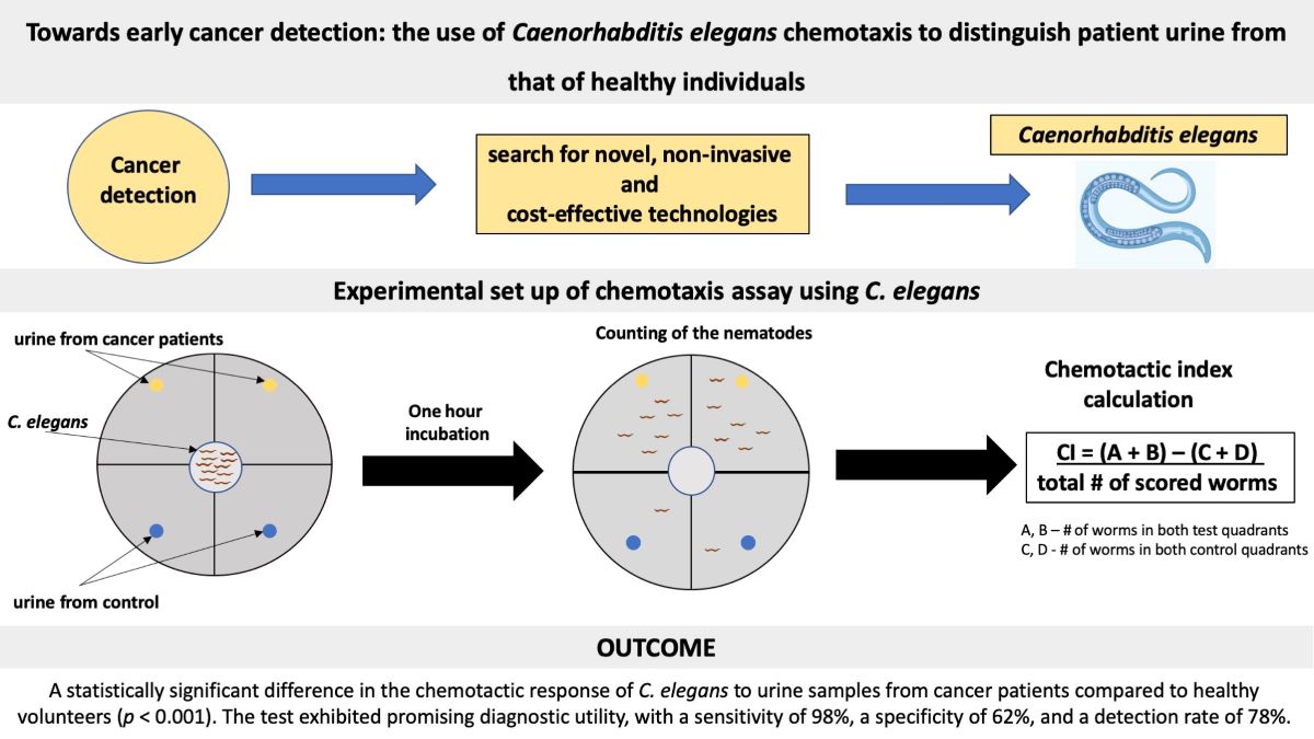
Abstract
The nematode Caenorhabditis elegans, with its highly sensitive olfactory system, has emerged as a promising tool for testing chemotaxis. In the field of cancer diagnostics, there is a growing interest in the development of non-invasive screening methods for the detection of volatile organic compounds in a patient's urine. The objective of this study was to contribute to the existing body of knowledge by evaluating the ability of a Caenorhabditis elegans-based chemotaxis assay to discriminate between urine samples from healthy individuals and patients diagnosed with breast or colon cancer. Following synchronization of the developmental stages of C. elegans, nematodes were exposed to the urine of cancer patients and healthy individuals. Subsequently, chemotactic indices were calculated for each urine sample. Our results demonstrated a statistically significant difference in the chemotactic response of C. elegans to urine samples from cancer patients compared to healthy volunteers (p < 0.001). Moreover, the test exhibited promising diagnostic utility, with a sensitivity of 98%, a specificity of 62%, and a detection rate of 78%. Our findings expand on previous observations, confirming the remarkable sensitivity of C. elegans hermaphrodites to discriminating cancer-related volatile organic compounds in urine samples.
Keywords:
Caenorhabditis elegans
; cancer
; chemotaxis
; chemotactic tests
1. Introduction
Caenorhabditis elegans is a non-pathogenic, free-living, microscopic nematode. In recent decades, it has become one of the most popular model organisms for studying human diseases because of its unique characteristics, including its rapid development cycle, high number of offspring, and ease of cultivation. A notable contribution to the molecular, cellular, developmental, and behavioral biology of this nematode was made by Sydney Brenner, a Nobel Prize-winning researcher, during the 1960s and 1970s [1]. Furthermore, the genome of C. elegans was subsequently sequenced in 1998, and it is the first sequenced genome of a multicellular organism [2]. Currently, researchers engaged in a range of biological and medical disciplines, including the development of cancer screening tests, derive considerable benefit from the use of this invaluable tool.
Cancer represents one of the most significant global causes of mortality and morbidity among non-communicable diseases [3]. Consequently, early diagnosis is crucial to simplify treatment and increase the chances of a cure. The development and implementation of cost-effective, non-invasive cancer screening tests could play a significant role in the effective management of this disease. The efficacy of oncological treatments is optimized when the disease is identified and addressed at its earliest stage, before the emergence of noticeable symptoms. Current research in the field of oncology is focused on the development of multiplex screening tests for the early detection of various types of cancer from a single sample. These tests analyze fragments of circulating tumor DNA, extracellular DNA, circulating microRNAs, or proteins released by cancer cells into the patient’s bodily fluids. The utility of such tests for identifying early-stage cancer has been demonstrated in several studies [4,5,6,7,8,9]. However, the high cost of these screening tests presents a significant obstacle to their extensive implementation as cancer preventive measures [10].
On the contrary, the use of minimally invasive tests that employ tumor markers shows a relatively low sensitivity for the early detection of cancer. Moreover, these markers are specific to tumors and therefore require a distinct evaluation for each cancer type. Previous research has indicated a correlation between tumor growth and the emission of volatile organic compounds (VOCs), which can be detected by certain vertebrate species, including canines and rodents [11,12]. However, the use of these animals in clinical settings presents considerable logistical challenges. For large-scale screening applications, it is essential to identify a suitable organism that is relatively simple to maintain and amenable to high-throughput processing. In this regard, the approximately 1 mm-long nematode C. elegans appears to be a compelling candidate due to its ease of cultivation, well-defined nutritional requirements, rapid life cycle, and the possibility of laboratory manipulation. Moreover, C. elegans possesses a highly sophisticated chemosensory system that enables the detection of a diverse range of volatile substances [13,14]. The analysis of VOCs in urine represents a promising area of research with the potential to identify novel biomarkers for the early detection of cancer [15]. The potential of nematode-based chemotaxis assays for early cancer detection was initially highlighted by Hirotsu et al. (2015) [16], followed by subsequent publications that addressed this topic [17,18,19,20].
In our study, we investigated the potential of utilizing nematode-based chemotactic assays for the detection of cancer. The differences in chemotactic indices of C. elegans nematodes exposed to the urine of individuals with cancer and the urine of healthy young adults were compared. To evaluate the diagnostic efficacy of the assay, its sensitivity, specificity, and detection rate were calculated. The objective of this research was to contribute to the existing body of knowledge in the emerging field of cancer diagnostics.
2. Materials and Methods
2.1. Ethical Approval
The study was approved by the Ethics Committee for Human Research at the Faculty of Health Care and Social Work, Trnava University in Trnava, Slovakia (EK-1/1K/2024).
2.2. Urine Samples
Urine samples were provided by Medirex, a.s., Bratislava, Slovakia, and collected over a 12-month period, from December 2022 to December 2023. The samples were promptly frozen at -20°C and stored in a freezer until they were used.
The cohort of cancer patients (n = 42, median age 66 years, interquartile range (IQR): 53-73, range: 44-85) was diagnosed in oncological clinics in Slovakia. According to the International Classification of Diseases (ICD-10), the diagnoses were classified into categories C18 (malignant neoplasm of the colon) and C19 (malignant neoplasm of the rectosigmoid junction), collectively referred to as colorectal carcinoma (n = 18, median age 58 years, IQR: 51-69, range: 45-79) and C50 (malignant neoplasm of the breast, including connective tissue of the breast) (n = 24, median age 68 years, IQR: 58-75, range: 44-85). The control group consisted of healthy young volunteers who had not been diagnosed with the disease (n = 53, median age 21 years, IQR: 19-23, range: 18-35).
2.3. C. elegans Strain
The wild-type strain of C. elegans (N2) was obtained from the Carolina Biological Supply Company, New York, USA. The Nematode Growth Media (NGM) plates used for the cultivation of C. elegans were prepared according to the methodology previously described [21]. A specific strain of Escherichia coli OP 50, which is unable to synthesize uracil and exhibits restricted growth in NGM, was utilized as a food source for this nematode.
2.4. Synchronization of Developmental Stages of C. elegans
Given the distinctive characteristics exhibited by each stage of development of C. elegans, biological experiments have employed synchronization techniques to reach a single stage of development, thus facilitating uniformity between experimental conditions. In the present study, nematode synchronization was achieved by the use of the bleaching method [22]. The nematodes were gently removed from the NGM plate using a buffered M9 solution (5.8 g of Na2HPO4.7H2O, 3.0 g of KH2PO4, 5.0 g of NaCl, and 0.25 g of MgSO4.7H2O dissolved in 1 L of distilled water). The resulting suspension was subsequently transferred to a sterile 15-ml conical bottom tube. To eliminate all developmental stages except for eggs, the nematodes were subjected to multiple rounds of centrifugation and washing with M9 solution. Subsequently, the pellet was dissolved in a freshly prepared bleaching solution (8.25 ml of sterile distilled water, 3.75 ml of 1 M NaOH, and 3 ml of bleach) for approximately 5 minutes. This treatment was effective in eliminating all stages of the motile worm. Finally, the isolated eggs were thoroughly washed with M9 solution to remove bleach residues and impurities. The suspension containing the eggs was incubated at 20°C for a period of approximately 24 hours with gentle agitation until the first-stage larvae (L1) hatched.
2.5. Preparation of the Test Plates for Chemotactic Tests
The chemotactic test was performed in accordance with the methodology previously described [23]. Briefly, four identical quadrants were designated on 5 cm agar plates with NGM medium. A circle with a radius of 0.5 cm was then marked at the point of intersection between the quadrant-limiting lines. In each quadrant, a "T" or "C" point was marked at a distance of 2 cm from the center of the plate. Urine from oncology patients or control subjects (designated as "T") or sterile distilled water (designated as "C") was applied to these points in a volume of 2 μl, respectively. Urine samples were diluted 1:3 with M9 buffer. Before use, urine samples were subjected to centrifugation (5 minutes, RCF 400) and filtration (0.22 μm, Merck Millipore). A suspension of L1 nematodes in M9 solution, with a concentration of 25-125 nematodes/μl, was added to the center of the plates in a volume of 5 μl. Following the absorption of the nematode suspension and odorant drops into the agar, the plates were inverted and left at room temperature for 60 minutes. Subsequently, the number of young worms (L1) in each quadrant that had crossed the inner circle entirely was recorded. The chemotaxis index (CI) was calculated according to the following equation: CI = (# worms in both test quadrants - # worms in both control quadrants) / (total # of scored worms) [23].
2.6. Statistical Analysis
R-Project version 4.3.3 was employed for the processing of data and statistical analyses. The fundamental characteristics of the cohort were detailed using descriptive statistics, including the mean, median, range, and interquartile range, to provide a robust summary of the central tendency and variability. A statistical analysis was performed to verify the normality of the data distribution using the Shapiro test. Subsequently, assuming that the data followed a normal distribution, the Welch’s t-test was used to compare the means of two groups, and an Anova test was employed to compare the means of three or more groups. A p-value of less than or equal to 0.05 was considered statistically significant.
Following the examination of 42 tumor samples and 53 control samples, a threshold for elevated cancer risk was established, corresponding to the upper 99.5 percentile derived from the Student’s t-distribution of the tested control samples. According to the threshold mentioned above, the samples were classified into two categories: "low risk" and "increased risk". Furthermore, the sensitivity, specificity, positive predictive value, negative predictive value, and detection rate of the tests were calculated.
3. Results
A statistical analysis of the results indicated a statistically significant difference in CI between the control and test tumor samples (p < 0.001) (Figure 1).
No statistically significant differences were observed in CI values between respondents of working age (i.e., up to 64 years) and those of post-working age (i.e., 65 years and older) (p > 0.05). Moreover, a subgroup analysis was conducted on urine samples from cancer patients, categorizing them according to diagnoses C18 and C19 (malignant neoplasm of the colon + malignant neoplasm of the rectosigmoid junction) and C50 (malignant neoplasm of the breast, including connective tissue of the breast). A comparison of CI values between these subgroups and control subjects revealed statistically significant differences for both tumor types, respectively (p < 0.001).
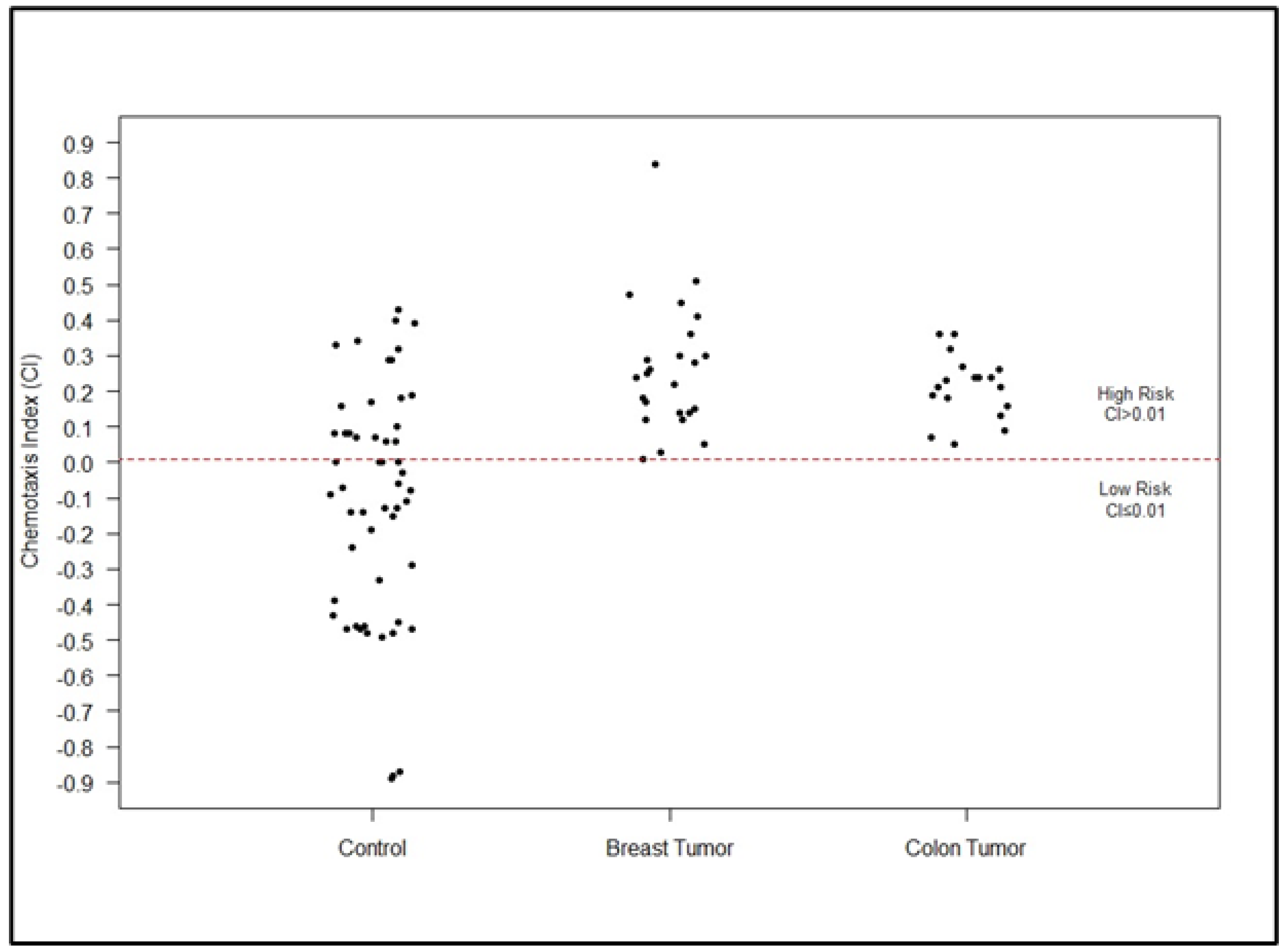
The threshold for elevated cancer risk was established at CI = 0.01, which corresponds to the upper 99.5 percentile derived from the Student’s t-distribution of the control subjects examined. Samples with mean values less than or equal to 0.01 were classified as "low risk", indicating a lack of cancerous characteristics. On the contrary, values greater than 0.01 were classified as high-risk samples (Figure 2).
The test used in this study demonstrated a sensitivity of 98% and a specificity of 62% in detecting cancer and ruling out its presence in a sample, respectively. The test demonstrated a positive predictive value (PPV) of 67%, a negative predictive value (NPV) of 97%, and an overall detection rate of 78%. Furthermore, a comprehensive examination of these statistical parameters for breast and colon cancer reveals the following: breast cancer: sensitivity 96%, specificity 62%, PPV 53%, NPV 97%, detection rate 73%; colon cancer: sensitivity 100%, specificity 62% PPV 47%, NPV 100%, detection rate 72%.
4. Discussion
The ability of C. elegans to sense attractive or repulsive odors is primarily facilitated by three pairs of olfactory neurons: AWA, AWB, and AWC. In the majority of cases, worms detect attractive compounds using AWA and AWC olfactory neurons, while AWB neurons are responsible for sensing repulsive volatile compounds [24,25]. Although chemotaxis is controlled by a complex set of neural and cellular mechanisms, it can be objectively quantified using chemotaxis assays with C. elegans nematodes [13,23,26,27,28]. Some vertebrates have been shown to be capable of detecting tumor growth [12,29]; however, the use of these animals in clinical applications is not feasible due to a number of limitations. To facilitate potential screening, it is essential to utilize a simple model organism that is suitable for high-throughput analysis. C. elegans meets these criteria because it has a highly developed chemosensory system, which allows it to detect a broad spectrum of volatile compounds. For example, Hirotsu et al. (2015) conducted a study examining the chemotactic activity of C. elegans toward urine samples, serum, and tumor tissues of cancer patients [16]. The highest level of attractive chemotaxis was observed in nematodes in response to urine samples, while the lowest level was detected in response to serum samples. According to these findings, the researchers postulated that the serum of cancer patients may also contain other odors that could repel C. elegans nematodes [16]. Furthermore, the researchers observed that olfactory neuron-ablated nematodes were not attracted to cancer patient urine, indicating that C. elegans is capable of detecting specific odors present in such samples. This finding provides a rationale for the N-NOSE (Nematode Nose) multi-cancer screening test, which has been shown to exhibit high sensitivity in the detection of early-stage cancers [17,18,19,20,30]. In addition to these findings, the objective of this study was to validate the documented results in the context of our institutional setting, with a particular focus on colorectal and mammary carcinomas. A significant difference in CI was observed between the control and tumor samples (p < 0.001, Figure 1). A subgroup analysis of urine samples from the patients with colon, rectosigmoid junction (C18/C19), and breast cancer (C50) also revealed significant differences compared to controls (p < 0.001). The test demonstrated high sensitivity, correctly identifying nearly all individuals with cancer (98% true positive results). However, the test demonstrated a lower specificity (62%), indicating that it correctly identified 62% of healthy individuals (i.e., true negative results). Conversely, 38% of individuals who tested positive in the control group did not have cancer, indicating a false positive result. This phenomenon can be attributed to the potential involvement of VOCs in non-cancerous pathologies such as tuberculosis [31], diabetes [32], or other as yet unidentified pathologies, which could compromise the assay’s specificity. The positive predictive value (67%) indicates that in 67% of positive tests, the presence of cancer was confirmed. In contrast, the negative predictive value (97%) indicates a high degree of accuracy in the exclusion of cancer when the test result is negative. Taken together, the test demonstrated satisfactory performance, with a 78% detection rate (73% for breast cancer and 72% for colorectal cancer, respectively).
While mammography remains the primary method for breast cancer screening in women over the age of 40 (e.g., the European Commission recommends mammography screening starting at the age of 45), its use in younger cohorts is limited [33]. The limited use of mammography in younger cohorts is attributed to the presence of dense glandular tissue and the potential exposure to ionizing radiation associated with the procedure [34]. However, the increasing prevalence of breast cancer in younger women and the inherently aggressive nature of breast cancer in younger patients require the investigation of alternative approaches to detection [35,36]. Since our results did not show a significant difference in CI between age groups (p > 0.05), it can be hypothesized that the aforementioned test could represent a potential benefit for the early detection of malignancies in all age categories. Furthermore, methods such as the C. elegans chemotaxis assay have the potential to serve as a valuable marker for breast cancer detection, even in younger women for whom mammography is often contraindicated. However, this assertion requires further validation through additional experimental studies and consideration of other important factors, such as cancer stage, tumor histology, patient age, and comorbidities that may influence laboratory results.
Another prevalent malignant neoplasm in the Western population is colorectal cancer. It is the second most common diagnosis in women, following only breast cancer, and the third most common in men, surpassed only by prostate and lung cancer [37]. Currently, fecal occult blood testing (FOBT) is the primary screening modality for colorectal cancer [38]. However, the limitations associated with FOBT have prompted researchers to explore alternative methods. These initiatives are guided by two key objectives: first, to foster patient participation in screening programs, and second, to minimize the burden of unnecessary colonoscopies resulting from false positive FOBT outcomes. Given the potential for increased patient compliance, urine-based assays hold significant promise. Consequently, scientific research is currently being conducted in order to develop new colorectal cancer screening tests that utilize metabolome and volatilome analyses of urine samples [39]. In a study by Kusumoto et al. (2020), the feasibility of using a cancer screening method based on the chemotaxis of C. elegans (N-NOSE tests) was evaluated in post-surgical patients with colon or stomach cancer [17]. The findings demonstrated that N-NOSE exhibited superior performance compared to conventional tests (based on the detection of carcinoembryonic and carbohydrate antigens) in evaluating the completeness of cancer resection. Researchers concluded that the use of C. elegans chemosensory responses facilitated the detection of gastrointestinal malignancies in urine samples with a high degree of sensitivity [17]. Similarly, Lanza et al. (2021) observed that C. elegans exhibited a significant preference for samples collected from women with breast cancer, while urinary controls demonstrated the capacity to act as avoidance-promoting chemorepellents [40]. The sensitivity of the test in the experiment was 75%, while the specificity was 97.22%, resulting in an accuracy of 86.11% [40].
The findings of our research and numerous other studies regarding the exceptional sensitivity of C. elegans chemotaxis assays indicate their potential utility in the early identification of cancer, a critical stage for therapeutic intervention. For example, C. elegans-based detection tests demonstrated 100% sensitivity for breast cancer and 88.9% sensitivity for colorectal cancer [19]. However, it is important to note, that the methodology used in the study mentioned above differed slightly from that used in the present study. It should be noted that the current study used a single dilution of the urine sample, whereas Inaba et al. (2021) used data from two dilutions (10-fold and 100-fold). However, our results were consistent with those of previous studies, indicating that C. elegans hermaphrodites can accurately identify urine samples from colorectal and breast cancers with a high degree of sensitivity compared to control samples. Although our study focused on the identification of only two distinct cancers, breast and colorectal, a novel assay that makes use of C. elegans’ behavioral responses shows promise in the identification of a wide range of tumor types from urine samples [18,41,42]. Compared to current screening techniques, which are typically cancer-specific and require distinct tests for each form of malignancy, this could represent a paradigm shift. Furthermore, di Luccio et al. (2022) propose that C. elegans-based cancer screening assays offer several advantages over the "one-test-one-cancer" approach, including protein tumor markers and cfDNA/ctDNA technologies [10]. Such tests may prove to be more cost-effective and easier to implement on a large scale within cancer screening programs. Future research aimed at identifying the urinary chemoattractants responsible for the response of the helminth may provide new insights into the mechanisms of carcinogenesis. This is of particular importance in light of the increasing global prevalence of cancer, with an alarming tendency for early-stage malignancies. Early detection of cancer can be an effective tool, as it allows the administration of the most appropriate treatment before patient’s present symptoms. This indicates the potential significance of C. elegans-based screening in this context.
5. Conclusions
The early diagnosis of cancer is a key factor in the successful treatment of the disease. The findings of our study confirm the remarkable sensitivity of C. elegans nematodes to discriminating volatile organic compounds associated with cancer in urine samples. It appears that this tiny nematode may help in the development of a rapid, painless, and cost-effective diagnostic screening test for cancer detection with high sensitivity based on the simple collection of a readily available biological sample (urine).
Author Contributions
Conceptualization, A.K.; methodology, A.K., P.H., R.R., S.K.; formal analysis, A.K., Z.B., S.K.; investigation, A.K., S.K.; resources, R.R.; data curation, A.K., Z.B., S.K.; writing—original draft preparation, A.K., Z.B.; writing—review and editing, A.K., P.H., Z.B., R.R., K.M., S.K; visualization, A.K, Z.B., S.K; supervision, A.K., S.K.; project administration, A.K., S.K.; funding acquisition, Z.B. All authors have read and agreed to the published version of the manuscript.
Funding
This research was funded by Trnava University in Trnava (grant number 20/TU/2023). The grant was awarded to ZB.
Institutional Review Board Statement
The study was conducted in accordance with the Declaration of Helsinki and approved by the Ethics Committee for Human Research at the Faculty of Health Care and Social Work, Trnava University in Trnava, Slovakia (EK-1/1K/2024).
Informed Consent Statement
The requirement for patient consent was waived due to the retrospective nature of the study, which involved the analysis of pre-existing, anonymized urine samples. As such, it was not feasible to obtain informed consent from patients.
Data Availability Statement
The data analyzed in this current study are available from the corresponding author upon reasonable request.
Acknowledgments
We would like to thank Iveta Adámková for her excellent technical assistance in the laboratory.
Conflicts of Interest
The authors declare no conflicts of interest. The funders had no role in the design of the study; in the collection, analyses, or interpretation of data; in the writing of the manuscript; or in the decision to publish the results.
References
- Brenner, S. The genetics of Caenorhabditis elegans. Genetics 1974, 77, 71–94. [Google Scholar] [CrossRef] [PubMed]
- The C. elegans Sequencing Consortium Genome Sequence of the Nematode C. elegans: A Platform for Investigating Biology. Science (80-. ) 1998, 282, 2012–2018. [CrossRef]
- Truong, L.-L.; Scott, L.; Pal, R.S.; Jalink, M.; Gunasekara, S.; Wijeratne, D.T. Cancer and cardiovascular disease: Can understanding the mechanisms of cardiovascular injury guide us to optimise care in cancer survivors? Ecancermedicalscience 2022, 16. [Google Scholar] [CrossRef] [PubMed]
- Fehlmann, T.; Kahraman, M.; Ludwig, N.; Backes, C.; Galata, V.; Keller, V.; Geffers, L.; Mercaldo, N.; Hornung, D.; Weis, T.; et al. Evaluating the Use of Circulating MicroRNA Profiles for Lung Cancer Detection in Symptomatic Patients. JAMA Oncol. 2020, 6, 714. [Google Scholar] [CrossRef] [PubMed]
- Hinestrosa, J.P.; Kurzrock, R.; Lewis, J.M.; Schork, N.J.; Schroeder, G.; Kamat, A.M.; Lowy, A.M.; Eskander, R.N.; Perrera, O.; Searson, D.; et al. Early-stage multi-cancer detection using an extracellular vesicle protein-based blood test. Commun. Med. 2022, 2, 1–9. [Google Scholar] [CrossRef] [PubMed]
- Jamshidi, A.; Liu, M.C.; Klein, E.A.; Venn, O.; Hubbell, E.; Beausang, J.F.; Gross, S.; Melton, C.; Fields, A.P.; Liu, Q.; et al. Evaluation of cell-free DNA approaches for multi-cancer early detection. Cancer Cell 2022, 40, 1537–1549. [Google Scholar] [CrossRef] [PubMed]
- Mencel, J.; Slater, S.; Cartwright, E.; Starling, N. The Role of ctDNA in Gastric Cancer. Cancers 2022, 14, 5105. [Google Scholar] [CrossRef] [PubMed]
- Nicholson, B.D.; Oke, J.; Virdee, P.S.; Harris, D.A.; O’Doherty, C.; Park, J.E.; Hamady, Z.; Sehgal, V.; Millar, A.; Medley, L.; et al. Multi-cancer early detection test in symptomatic patients referred for cancer investigation in England and Wales (SYMPLIFY): A large-scale, observational cohort study. Lancet Oncol. 2023, 24, 733–743. [Google Scholar] [CrossRef]
- Xin, L.; Yue, Y.; Zihan, R.; Youbin, C.; Tianyu, L.; Rui, W. Clinical application of liquid biopsy based on circulating tumor DNA in non-small cell lung cancer. Front. Physiol. 2023, 14, 1200124. [Google Scholar] [CrossRef] [PubMed]
- di Luccio, E.; Morishita, M.; Hirotsu, T.C. elegans as a Powerful Tool for Cancer Screening. Biomedicines 2022, 10, 2371. [Google Scholar] [CrossRef]
- Price, M. These ‘supersniffer’ mice could one day detect land mines, diseases. Science (80-. ) 2016. [Google Scholar] [CrossRef]
- Guerrero-Flores, H.; Apresa-García, T.; Garay-Villar, Ó.; Sánchez-Pérez, A.; Flores-Villegas, D.; Bandera-Calderón, A.; García-Palacios, R.; Rojas-Sánchez, T.; Romero-Morelos, P.; Sánchez-Albor, V.; et al. A non-invasive tool for detecting cervical cancer odor by trained scent dogs. BMC Cancer 2017, 17, 1–8. [Google Scholar] [CrossRef] [PubMed]
- Bargmann, C.I.; Horvitz, H.R. Chemosensory neurons with overlapping functions direct chemotaxis to multiple chemicals in C. elegans. Neuron 1991, 7, 729–742. [Google Scholar] [CrossRef] [PubMed]
- Bargmann, C.I. Chemosensation in C. elegans. WormBook 2006, 1–29. [Google Scholar] [CrossRef] [PubMed]
- Wen, Q.; Boshier, P.; Myridakis, A.; Belluomo, I.; Hanna, G.B. Urinary Volatile Organic Compound Analysis for the Diagnosis of Cancer: A Systematic Literature Review and Quality Assessment. Metabolites 2020, 11, 17. [Google Scholar] [CrossRef] [PubMed]
- Hirotsu, T.; Sonoda, H.; Uozumi, T.; Shinden, Y.; Mimori, K.; Maehara, Y.; Ueda, N.; Hamakawa, M. A highly accurate inclusive cancer screening test using Caenorhabditis elegans scent detection. PLoS ONE 2015, 10, 1–15. [Google Scholar] [CrossRef] [PubMed]
- Kusumoto, H.; Tashiro, K.; Shimaoka, S.; Tsukasa, K.; Baba, Y.; Furukawa, S.; Furukawa, J.; Niihara, T.; Hirotsu, T.; Uozumi, T. Efficiency of gastrointestinal cancer detection by nematode-NOSE (N-NOSE). In Vivo (Brooklyn). 2020, 34, 73–80. [Google Scholar] [CrossRef] [PubMed]
- Asai, A.; Konno, M.; Ozaki, M.; Kawamoto, K. Scent test using Caenorhabditis elegans to screen for early- stage pancreatic cancer. Oncotarget 2021, 12, 1687–1696. [Google Scholar] [CrossRef] [PubMed]
- Inaba, S.; Shimozono, N.; Yabuki, H.; Enomoto, M.; Morishita, M.; Hirotsu, T.; di Luccio, E. Accuracy evaluation of the C. elegans cancer test (N-NOSE) using a new combined method. Cancer Treat. Res. Commun. 2021, 27, 100370. [Google Scholar] [CrossRef]
- Thompson, M.; Feria, N.S.; Yoshioka, A.; Tu, E.; Civitci, F.; Estes, S.; Wagner, J.T. A Caenorhabditis elegans behavioral assay distinguishes early stage prostate cancer patient urine from controls. Biol. Open 2021, 10. [Google Scholar] [CrossRef]
- Merrow, M.; Olmedo, M. In situ Chemotaxis Assay in Caenorhabditis elegans (for the Study of Circadian Rhythms). BIO-PROTOCOL 2014, 4. [Google Scholar] [CrossRef]
- Porta-de-la-Riva, M.; Fontrodona, L.; Villanueva, A.; Cerón, J. Basic Caenorhabditis elegans Methods: Synchronization and Observation. J. Vis. Exp. 2012, e4019. [Google Scholar] [CrossRef]
- Margie, O.; Palmer, C.; Chin-Sang, I.C. elegans Chemotaxis Assay. J. Vis. Exp. 2013, 729–742. [Google Scholar] [CrossRef]
- Zhang, C.; Yan, J.; Chen, Y.; Chen, C.; Zhang, K.; Huang, X. The olfactory signal transduction for attractive odorants in Caenorhabditis elegans. Biotechnol. Adv. 2014, 32, 290–295. [Google Scholar] [CrossRef] [PubMed]
- Zhang, C.; Zhao, N.; Chen, Y.; Zhang, D.; Yan, J.; Zou, W.; Zhang, K.; Huang, X. The signaling pathway of Caenorhabditis elegans mediates chemotaxis response to the attractant 2-heptanone in a Trojan Horse-like pathogenesis. J. Biol. Chem. 2016, 291, 23618–23627. [Google Scholar] [CrossRef]
- Ward, S. Chemotaxis by the nematode Caenorhabditis elegans: Identification of attractants and analysis of the response by use of mutants. Proc. Natl. Acad. Sci. USA 1973, 70, 817–821. [Google Scholar] [CrossRef]
- Worthy, S.E.; Haynes, L.; Chambers, M.; Bethune, D.; Kan, E.; Chung, K.; Ota, R.; Taylor, C.J.; Glater, E.E. Identification of attractive odorants released by preferred bacterial food found in the natural habitats of C. elegans. PLoS ONE 2018, 13, 1–14. [Google Scholar] [CrossRef]
- Queirós, L.; Marques, C.; Pereira, J.L.; Gonçalves, F.J.M.; Aschner, M.; Pereira, P. Overview of Chemotaxis Behavior Assays in Caenorhabditis elegans. Curr. Protoc. 2021, 1, e120. [Google Scholar] [CrossRef]
- Guirao Montes, Á.; Molins López-Rodó, L.; Ramón Rodríguez, I.; Sunyer Dequigiovanni, G.; Viñolas Segarra, N.; Marrades Sicart, R.M.; Hernández Ferrández, J.; Fibla Alfara, J.J.; Agustí García-Navarro, Á. Lung cancer diagnosis by trained dogs†. Eur. J. Cardio-Thoracic Surg. 2017, 52, 1206–1210. [Google Scholar] [CrossRef]
- Namgong, C.; Kim, J.H.; Lee, M.H.; Midkiff, D. Non-invasive cancer detection in canine urine through Caenorhabditis elegans chemotaxis. Front. Vet. Sci. 2022, 9. [Google Scholar] [CrossRef]
- Küntzel, A.; Oertel, P.; Fischer, S.; Bergmann, A.; Trefz, P.; Schubert, J.; Miekisch, W.; Reinhold, P.; Köhler, H. Comparative analysis of volatile organic compounds for the classification and identification of mycobacterial species. PLoS ONE 2018, 13, e0194348. [Google Scholar] [CrossRef]
- Zaim, O.; Bouchikhi, B.; Motia, S.; Abelló, S.; Llobet, E.; El Bari, N. Discrimination of Diabetes Mellitus Patients and Healthy Individuals Based on Volatile Organic Compounds (VOCs): Analysis of Exhaled Breath and Urine Samples by Using E-Nose and VE-Tongue. Chemosensors 2023, 11, 350. [Google Scholar] [CrossRef]
- European Commission, Directorate-General for Health and Consumers; von Karsa, L.; Holland, R.; Broeders, M.; de Wolf, C.; Perry, N.; Törnberg, S. European guidelines for quality assurance in breast cancer screening and diagnosis: Fourth edition, supplements; European Commission, 2013; ISBN 978-92-79-32970-8. [Google Scholar]
- Shah, T.; Guraya, S. Breast cancer screening programs: Review of merits, demerits, and recent recommendations practiced across the world. J. Microsc. Ultrastruct. 2017, 5, 59. [Google Scholar] [CrossRef] [PubMed]
- Radecka, B.; Litwiniuk, M. Breast cancer in young women. Ginekol. Pol. 2016, 87, 659–663. [Google Scholar] [CrossRef] [PubMed]
- Fernandes, U.; Guidi, G.; Martins, D.; Vieira, B.; Leal, C.; Marques, C.; Freitas, F.; Dupont, M.; Ribeiro, J.; Gomes, C.; et al. Breast cancer in young women: A rising threat: A 5-year follow-up comparative study. Porto Biomed. J. 2023, 8. [Google Scholar] [CrossRef] [PubMed]
- Bray, F.; Ferlay, J.; Soerjomataram, I.; Siegel, R.L.; Torre, L.A.; Jemal, A. Global cancer statistics 2018: GLOBOCAN estimates of incidence and mortality worldwide for 36 cancers in 185 countries. CA. Cancer J. Clin. 2018, 68, 394–424. [Google Scholar] [CrossRef] [PubMed]
- Song, L.-L.; Li, Y.-M. Current noninvasive tests for colorectal cancer screening: An overview of colorectal cancer screening tests. World J. Gastrointest. Oncol. 2016, 8, 793. [Google Scholar] [CrossRef] [PubMed]
- Mallafré-Muro, C.; Llambrich, M.; Cumeras, R.; Pardo, A.; Brezmes, J.; Marco, S.; Gumà, J. Comprehensive Volatilome and Metabolome Signatures of Colorectal Cancer in Urine: A Systematic Review and Meta-Analysis. Cancers 2021, 13, 2534. [Google Scholar] [CrossRef]
- Lanza, E.; Di Rocco, M.; Schwartz, S.; Caprini, D.; Milanetti, E.; Ferrarese, G.; Lonardo, M.T.; Pannone, L.; Ruocco, G.; Martinelli, S.; et al. C. elegans-based chemosensation strategy for the early detection of cancer metabolites in urine samples. Sci. Rep. 2021, 11, 17133. [Google Scholar] [CrossRef]
- Daulton, E.; Wicaksono, A.N.; Tiele, A.; Kocher, H.M.; Debernardi, S.; Crnogorac-Jurcevic, T.; Covington, J.A. Volatile organic compounds (VOCs) for the non-invasive detection of pancreatic cancer from urine. Talanta 2021, 221, 121604. [Google Scholar] [CrossRef]
- Lett, L.; George, M.; Slater, R.; De Lacy Costello, B.; Ratcliffe, N.; García-Fiñana, M.; Lazarowicz, H.; Probert, C. Investigation of urinary volatile organic compounds as novel diagnostic and surveillance biomarkers of bladder cancer. Br. J. Cancer 2022, 127, 329–336. [Google Scholar] [CrossRef] [PubMed]
Figure 1.
A comparative analysis of chemotactic indices (CI) was conducted between urine samples obtained from control subjects and patients diagnosed with breast and colon tumors. The Welch’s t-test revealed a statistically significant difference between the CI of control subjects (healthy individuals) and breast tumors (malignant neoplasm of the breast, including connective tissue of the breast) and colon tumors (colorectal carcinoma), respectively (***p < 0.001). The boxplots illustrate the data distribution and pivotal statistical measures of CI in individual groups, including control samples, breast and colon tumors, respectively. The boxplots show the interquartile range (IQR), which encompasses 50% of the most frequently occurring values in the data, specifically from the 25th percentile to the 75th percentile. The line located at the center of the box represents the median value. The whiskers indicate the range of 1.5 times the IQR of the quartiles. The average value of CI is indicated by a plus (+) symbol located at the center of the box. The red line indicates the threshold for elevated cancer risk, corresponding to the upper 99.5 percentile derived from the Student’s t-distribution of the tested control samples.
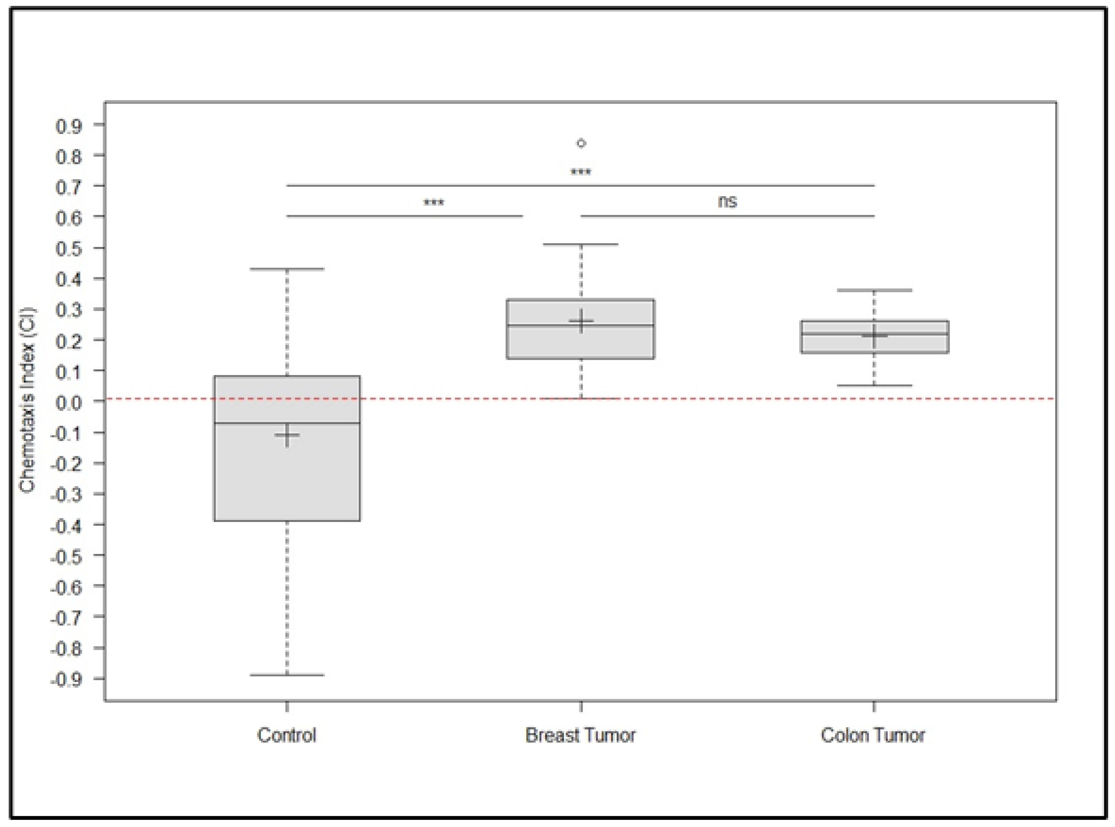
Figure 2.
An overview of chemotactic indices (CI) detected in samples from healthy individuals (control samples) and patients diagnosed with breast (malignant neoplasm of the breast, including connective tissue of the breast) and colon tumors (colorectal carcinoma). The dot plots demonstrate the CI index for each individual sample. The red line illustrates the threshold for increased risk of cancer, corresponding to the upper 99.5 percentile obtained from the Student’s t-distribution of the tested samples from healthy individuals (controls). Individuals with a low risk of developing cancer are indicated by data points that are below the indicated threshold. On the contrary, values greater than 0.01 indicate samples with a high probability of cancer development.
Disclaimer/Publisher’s Note: The statements, opinions and data contained in all publications are solely those of the individual author(s) and contributor(s) and not of MDPI and/or the editor(s). MDPI and/or the editor(s) disclaim responsibility for any injury to people or property resulting from any ideas, methods, instructions or products referred to in the content. |
© 2024 by the authors. Licensee MDPI, Basel, Switzerland. This article is an open access article distributed under the terms and conditions of the Creative Commons Attribution (CC BY) license (http://creativecommons.org/licenses/by/4.0/).
Copyright: This open access article is published under a Creative Commons CC BY 4.0 license, which permit the free download, distribution, and reuse, provided that the author and preprint are cited in any reuse.


