Submitted:
23 July 2024
Posted:
24 July 2024
You are already at the latest version
Abstract
All-inorganic perovskite materials are promising in optoelectronics but their morphology is a key parameter for achieving high device efficiency. We prepared CsPbBr3 perovskite microcrystals with different shape, grown directly on planar substrate by conventional drop casting. We observed the formation of CsPbBr3 microcubes on bare indium tin oxide (ITO) coated glass. Interestingly, with the same technique CsPbBr3 microrods were obtained on (3-Aminopropyl) triethoxysilane (APTES) modified ITO-glass, which we ascribe to modification of formation kinetics. The obtained micro-crystals exhibit orthorhombic structure. Green photoluminescence (PL) emission is revealed from the CsPbBr3 microrods. Contact angle measurements, Fourier-transform infrared and PL spectroscopies confirmed that APTES linked successfully to the ITO-glass substrate. We propose a qualitative mechanism to explain the anisotropic growth. The microrods exhibited improved PL and slower PL lifetime compared to the microcubes, likely due to diminished occurrence of defects. This work demonstrates the importance of the substrate surface to control the growth of perovskite single crystals and to boost the radiative recombination in view of high performance optoelectronic devices.
Keywords:
perovskite
; microrods
; aminosilane
; optical properties
; surface modification
1. Introduction
Due to the solution processability [1], low cost [2], high stability [3] and narrow emission line [4], all-inorganic cesium halide perovskites CsPbX3 (X=Cl, Br and I) have recently attracted high interest in optoelectronic applications [4,5]. These members of the perovskite family appear to be a promising alternative to traditional semiconductors, yet require improvement in synthesis and understanding of their properties. Several synthesis protocols have been developed in order to obtain all-inorganic perovskite in different form, such as microcrystals [6], thin films [1,7] and nanocrystals (NCs) [4]. However, most of those strategies do not allow a proper control of shape and size. Synthesis of material with high quality and well-defined morphology not only benefits fundamental research but also offers great promise for practical application [8]. In the arena of NCs, the synthesis protocols based on solution phase such as hot injection [9] or ligand assisted precipitation [10] have shown their potential in regulating the final morphology of the CsPbBr3 NCs, promoting the selective formation of nanocubes, nanoplates and nanowires, by acting on the acid-base equilibrium mechanism [11]. NCs with a variety of shapes have been synthesized by kinetically controlling the growth of particular facets [12] or through an oriented attachment mechanism [13]. The surface chemistry of NCs is decisive in determining the final morphology of the material [14]. Surfactant or surface ligands such as alkylamine and carboxylic acid have been proven essential to alter the kinetic pathways and give anisotropically developed nanostructures, forming hydrogen bonds with bromide anionic surface sites and alkyl carboxylate binding the surface cationic sites [12]. However, the shape control of single microcrystals is less explored.
In this study, we investigated the effects of the (3-aminopropyl)triethoxysilane (APTES) surface ligand-modified substrate on the growth of CsPbBr3 perovskite single microcrystals. A silane layer is usually employed to control the physical and chemical properties of a solid surface [11]. APTES is a surface ligand extensively employed to functionalize surface with amino silane via covalent interaction [15]. Attachment of primary amine seems an effective technique to control the growth of all inorganic CsPbBr3 perovskite. Our work shows unpreceded method to prepare CsPbBr3 single microcrystals via a solution processing strategy, which gave rise to uniaxial directional growth of elongated crystals that we called microrods (MRs) crystallizing in orthorhombic phase. The presence of functional groups of silane layer and the possible chemical bonding to CsPbBr3 perovskite was confirmed by Fourier-transform infrared spectroscopy (FTIR). The synthesis technique in this study is facile and offers a new procedure to grow CsPbBr3 perovskite MRs structure with improved properties for optoelectronic application.
2. Materials and Methods
2.1. Surface Treatment
If not otherwise specified, all chemicals and materials were acquired from Sigma-Aldrich. ITO-coated glass wafers to be used as the substrates were rinsed sequentially with absolute ethanol and acetone in a bath ultra-sonicator. The cleaned wafers were hydroxylated via Piranha treatment at room temperature (RT) for 20 min and with an H2O2/H2SO4 (3:7) at 60 °C, and then rinsed with water and dried with argon flux. For silanization process the substrates were immersed in 2% toluene solution during 3 min. Finally, the substrates were rinsed with toluene and deionized water and then dried in a nitrogen stream.
2.2. Perovskite Preparation
We prepared CsPbBr3 precursor solution by mixing CsBr and PbBr2 in dimethyl sulfoxide (DMSO). The solution of precursor was magnetically stirred before deposition. The precursor solution was deposited on silane-functionalized surface by drop casting. After the deposition onto the substrate, a short annealing was conducted.
2.3. Characterization
X-ray diffraction (XRD) patterns were obtained using a PAN analytical Empyrean X-ray diffractometers equipped with 1.8 Kw CuKα ceramic X-ray tube and operating at 45 kV and 40 mA.
Optical microscopy images and 3D profilometry measurements were obtained with a confocal Zeta-20 profilometer (Zeta instruments, USA).
Atomic force microscopy (AFM) measurements were performed with a XE 100 (Park Systems, Korea) microscope under ambient atmosphere.
PL microscopy was used to obtain images and to acquire spectrum of individual microcrystals under 488 nm wavelength (blue light) excitation, with collection in the cyan-green window (500–550 nm).
UV-Visible absorbance spectra were measured using a SPECORD 210-PLUS spectrophotometer, equipped with a deuterium D2E lamp (185 nm-350 nm), and a halogen lamp (330 nm-1100 nm).
Steady state PL spectra were measured at 300 K using laser excitation wavelength of 360 nm. Spectral analysis of the luminescence measurements was dispersed using JOBIN YVON HRDI (iHR320) monochromator and detected by a silicon photodiode.
Time resolved PL (TRPL) measurements of the recombination dynamics were carried out by time correlated single photon counting setup based on the pulsed laser diode ( operated at 80 MHz and time Harp 200 electronic card from Pico Quant company16. The excitation laser pulse width was 70 ps, the time resolution of the experimental setup was 40 ps. The lifetimes t were obtained by fitting the experimental data of PL intensity IPL, as compared to initial intensity I0, to a mono-exponential decay:
Contact angle (CA) measurements were carried out by the CA goniometer Digidrop GBX, using the Laplace- Young method [17].
FTIR spectra were recorded using Perkin Elmer spectrometer equipped with a universal attenuated total reflectance mode and scanned at RT in transmission mode over the wave number range of 4000-850 cm-1.
3. Results and Discussion
CsPbBr3 microcrystals were prepared on solid substrates in two forms: the novel MRs (elongated in one planar direction) and the planar-symmetric microcrystals already presented in previous works [18,19], which we call here microcubes (MCs). Actually, the name MCs is to simplify the description, as the microcrystals are anyway not full cubes, yet have a hollow pyramidal pit on top, discussed extensively in our previous reports [18,19]. The MCs will be used here as a term of comparison for the novel MRs structures. A morphological characterization of the MCs is reported in Figure 1a–c. An optical micrograph of the sample (Figure 1a) shows the presence of squares shaped structures (see also the 3D optical profilometer image of the same region in Figure 1b). The MCs are quite homogenous in size (see distribution in Figure 1c), with an edge length of ~64 µm. The preparation parameters are critical for the final microstructure size. The XRD pattern (Figure 1g) evidences the crystalline nature of the MCs, which matches the orthorhombic perovskite phase. The UV-visible absorption spectrum of the as-prepared sample presented in Figure 1i shows the typical profile of CsPbBr3 microcrystals, characterized by an absorption peak at ~517 nm. The PL spectrum of the MCs shows a narrow and intense band (fullwidth at half maximum ~17 nm) centered at ~519 nm (2.39 eV). The emission band is associated to the bulk bandgap recombination of CsPbBr3 [19].
Figure 1d,e present some perovskite MRs deposited on APTES-modified ITO-glass substrate. The images show the formation of well-defined rectangular single rods. Obviously the effect of the APTES modified substratewas the transformation of crystals from MCs to MRs. In the given fabrication conditions, the aspect ratio of the MRs (ratio of long side to short side) is ~5, while the longest diagonal is ~200 µm. The size distribution histogram for the latter parameter is presented in Figure 1f. According to the histogram, the spread around the peak size value is described by a standard deviation of ~30 µm. For the off-plane size of the MRs, optical profilometer measurements (see a representative 3D image in Figure 1d) showed a MRs thickness in the range of 10 to 25 µm.
The bulk crystallinity of the MRs was confirmed by XRD measurements (see Figure 1h). The majority of peaks, green dots in the diffractogram, fits well with CsPbBr3 orthorhombic perovskite phase (PDF#45-0752, standard diffraction pattern), confirming the formation of CsPbBr3 crystals. The sharp peaks indicate high crystallinity of the MRs. The XRD pattern revealed also the presence of other phases such as Pb (cubic, F m-3 m), and BrN (tetragonal, P 4/n m m).
High resolution confocal microscopy images (2D and 3D) of the novel MRs have been acquired (Figure 2a,b). Clearly, one can see that the MRs fluoresce in the green region of the visible electromagnetic spectrum. Two PL spectra extracted from different regions of the rods in the confocal image (red and green circles in Figure 2b) are presented in Figure 2c. It appears that the edge of the MR yields a higher PL as compared to the center, and a peak shift towards the red (~530 nm vs ~520 nm). This effect has been confirmed by testing several different MRs. We should point out that our MRs present hollow top with inverted pyramid shape similar to the MCs (see Figure 1b,e). We have shown previously by using PL microscopy that in single CsPbBr3 MCs [18] the emission from the upper surface (near the edge) is different compared to that one from the center. So, The PL intensity change by changing the excitation surface position for single CsPbBr3 microcrystal.
For a deeper understanding of the optical properties of the MRs, a more advanced optical characterization has been carried out. In Figure 2d,e typical absorbance and steady state PL spectrum of the MRs recorded at 300 K are presented, respectively. As we can see, the PL spectrum revealed the presence of two peaks. One peak, named , is located at 442 nm and can be assigned to APTES emission [20], in accordance with the presence of APTES absorption edge feature at 320 nm (inset in Figure 2d). The important emission peak that presents an asymmetrical profile with extended shoulder on the long wavelength side was fitted by two Gaussians with maxima located at 523 and 540 nm, which were denoted P2 and P3, respectively. This feature can be attributed to the PL from the CsPbBr3 MRs. Correspondingly, a strong absorbance band at ~520 nm is shown in Figure 2d, which is a characteristic peak of bulk CsPbBr3 perovskite [21]. Figure 2f displays normalized TRPL emission profiles recorded for the sample at RT, at different energies of the emission peaks. It can be seen that profile of decays faster than that of . The PL decays presented the following lifetimes: t1~1.62 ns, t2~4.12 ns and t3~7.86 ns, for peaks , and , respectively. Obviously, the longer carrier lifetime results from the delayed recombination process via non-radiative pathways, which means that the PL peak can be assigned to the emission of shallow defects in the CsPbrBr3 material [21].
CA measurements were carried out to determine the wettability of substrates before and after APTES modification, which are shown in Figure 3a. The obtained CA value for bare ITO-glass was found to be ~ (Figure 3a1). A significant increase in CA, ~ (Figure 3a2) was observed for the APTES modified surface (Figure 3b,c). Thus, the surface becomes less hydrophilic on coating with APTES, due to the presence of hydrophobic alkyl chains of this molecule [22,23] (see Figure 3b). The APTES is anchoring the ITO-glass surface by replacing the terminating OH groups of the substrate with organic groups attached to amine groups (see Figure 3c), which changes the surface energy and thus its wettability.
Typically, the silanization process consists of two steps: hydrolysis and condensation. In the first step, hydrolysis of the substrate leads to the formation of the reactive silanol group (R-Si-OH) and ethanol molecules [22]. In the second step, condensation of these silanol groups takes place leading to the formation of siloxane (Si-O-Si) linkages over the surface [22,23]. Thus, attachment of APTES takes place with OH groups on the substrate, and a NH2 terminated layer is created on the surface (Figure 3c). These amino (-NH2) groups on APTES surface react with perovskite via chemical bonding. According to literature [24,25], attachment of APTES to CsPbBr3 perovskite takes place through three possible binding mechanisms [15]: (1) formation of dative-covalent bond between amines (NH2) of silanes network and undercoordinated Pb2+ sites; (2) hydrogen bond formation between remaining silanol and surface bromide (Br-) sites; (3) formation of hydrogen bonds through protonated amines (NH+) and bromine (Br-) dangling bonds.
For ideal silanization, all amino group of the APTES should be oriented away from the substrate surface [26]. The typical APTES surface quality was as shown in the representative image in Figure 3d, showing a flat and smooth background with some aggregates up to ~1 mm size, likely due to excess material. In fact, the thickness of the APTES layer was also estimated, by using AFM across hand-made scratches in the molecular coating, which resulted in a value around ~150 nm (data not shown). The thickness of one silane layer is ~2 nm [27], which confirms the formation of multisilane layers on the top of ITO-glass substrate.
The surface chemical composition of CsPbBr3 MRs was characterized by FTIR measurements (see Figure 3e). The resulting peaks assignment is reported in Table 1. FTIR results confirm the presence of asymmetric stretching vibration of surface hydroxyl groups (OH) by the broad band at 3464 cm-1, due to ethanol. This large band shows a small peak located at 3258 cm-1, due to the N-H symmetric stretching vibration of APTES [28]. Bands at 2856 cm-1, 2925 cm-1 and 2959 cm-1 corresponds to C-H vibration from both ethanol and APTES [28]. Bands at 1638-1664 cm-1 are attributed to binding vibration of primary amines (NH2) in APTES [28]. The appearance of these bands demonstrated that amine functional groups in organosilane were successfully grafted onto the ITO-glass substrate [28].
Furthermore, peaks corresponding to Si-O-Si bond were observed at around 1012 cm-1-1066 cm-1, indicating the condensation reaction between silanol groups [28]. These bands confirm condensation reaction between ethoxy groups of APTES and the ITO surface hydroxyl groups, namely ITO-glass modification with APTES. The FTIR spectrum shows another weak peak at 1575 cm−1, which was assigned to the NH+ bond [9]. In conclusion, FTIR spectroscopy confirms the presence of characteristic functions of APTES on the sample surface and the attachment of perovskite to the APTES through three different binding due to the presence of NH2, NH+ and silanol groups on the surface of the rods (see Figure 3f).
To figure out the origin of anisotropic growth of CsPbBr3 MRs, we propose a plausible mechanism. In precursor solution, the Br- ions coordinate with Pb2+ to form [PbBr6]4- octahedra in DMSO. After precursor solution deposition, the solvent is evaporated under heat treatment to allow crystallization of perovskite [29], and the Cs+ ions occupy the octahedral sites to form CsPbBr3 perovskite MCs above bare ITO-glass substrate. This occurs by using the same precursor solution for both MRs and MCs, which means that MRs evolve from MCs during heating process. The crystallization process (nucleation and growth) plays an important role in determining the crystal structure, shape and size [30]. According to literature, nano/micro or macro crystals synthesis with various size and shapes can described via supersaturation chemistry approach (Lamer theory) [30,31]. Shape control of CsPbX3 perovskite NCs has been tuned either by kinetically controlling the growth of particular crystal facets [12] or through an oriented attachment mechanism [13]. In contrast, few studies deal with anisotropic microparticles formation mechanism. Consequently, a hypothetical approach to understand the mechanism of microcrystals construction provides a greater control over the size, shape and structure of single crystals and consequent the ability to tune the properties by varying the crystallization conditions.
The growth rate of the single crystal increases with the increase of CA. The less hydrophilic APTES-modified surface makes precursor ions and solvent to get closer, which will increase the diffusions of ions, leading to a faster crystal growth. In fact, it is known that, at high growth rates, organic molecules (ligands) that selectively adhere to particular NC facets decrease their energy and slow down their growth [32]. High-energy facets grow faster than low-energy ones, giving rise to the formation of anisotropic shape [33]. Previously, Pan et al. [9] suggested preferential binding of amine toward certain facets during the growth of CsPbBr3 nanowires and nanoplatelets. Also, it is demonstrated that ammonium binding with Cs+ ions on the surfaces allows the growth of platelets [33]. So, we suppose here that the amine groups compete with the Cs+ ions on the surface of the growing rods, and thus it slows the growth along the orthogonal direction, leading to the observed anisotropic rod structures. Also, it is suggested that halide-rich environment may result in oriented crystal growth along special facets.
In support of our hypothesis that preferential binding of APTES takes place through amine groups giving rise to the formation of perovskite MRs, we have compared the PL properties of MCs and MRs. The PL spectrum of MRs is slightly redshifted with respect to MCs ones (see Figure 4). The PL maximum slightly redshifts from the 519 nm for MCs to 523 nm for MRs. MRs also show PL enhancement compared to MCs. The origin of enhancement may arise from the effective surface passivation by APTES molecules. TRPL spectra of the microcrystals were also acquired and fitted using a biexponential decay function where Ai and i refer to the decay amplitudes and respective PL lifetimes, for i=1,2. The average lifetime can thus be obtained by the following formula:
The time-resolved measurements also show an enhancement of the PL lifetime upon APTES treatment (see Figure 4b), from ~2.3 to ~7.4 ns). This behavior may arise from enhanced passivation of the CsPbBr3 MRs surfaces.
4. Conclusions
In summary, we presented a comparative study of CsPbBr3 perovskite microcrystals with different form, grown directly on planar substrate by conventional drop casting method. We observed the formation of CsPbBr3 MCs on bare ITO-coated glass substrate. With the same technique, CsPbBr3 single MRs were obtained on APTES surface ligand modified ITO-glass substrate, which we ascribed to the modification of the kinetic regime by using APTES. Green emission was revealed from the CsPbBr3 microcrystals by using PL spectroscopy. CA measurements, FTIR and PL spectroscopies confirmed that APTES linked successfully to ITO-coated glass substrate. The fabricated perovskite MRs exhibited improved PL thanks to the introduced APTES molecules effectively passivating surface defects and controlling the crystal growth. We conclude that, even for microcrystals, introduction of certain organic molecules that selectively adhere to a particular crystal facet can be used to slow the growth of that side relative to others, leading to the formation of various shapes singles crystals. Thus, we have identified a new strategy to enhance the photoluminescence and to control the shape of CsPbBr3 microcrystals for optoelectronic application. Further research work is needed to confirm these findings, still our work points out that careful management of the perovskite MCs and substrate surface engineering are important factors in this promising class of optoelectronic materials.
Author Contributions
Conceptualization and methodology, K.A. and Z.Z.; formal analysis, K.A: and F.H.; investigation, K.A. and M.S.; resources, F.H. and Z.Z.; data curation, K.A.; writing—original draft preparation, K.A.; writing—review and editing, M.S., F.H. and Z.Z.; visualization, K.A.; supervision and funding acquisition, F.H. and Z.Z.. All authors have read and agreed to the published version of the manuscript.
Funding
This research received no external funding.
Institutional Review Board Statement
Not applicable.
Informed Consent Statement
Not applicable.
Data Availability Statement
Data available on request to K.A.
Acknowledgments
KA wishes to thank Dr. Roman Krahne of the Italian institute of technology (iit) for the confocal microscope measurements.
Conflicts of Interest
The authors declare no conflicts of interest.
References
- Li, Y.; Shi, Z.F.; Li, S.; Lei, L.Z.; Ji, H.F.; Wu, D.; Xu, T.T.; Tian, Y.T.; Li, X.J. High-performance perovskite photodetectors based on solution-processed all-inorganic CsPbBr3 thin films. J. Mater. Chem. C 2017, 5, 8355–8360. [Google Scholar] [CrossRef]
- Cui, J.; Xu, F.; Dong, Q.; Jia, J.; Liu, L.; Liu, S.; Yang, F.; Ye, X. Facile, low-cost, and large-scale synthesis of CsPbBr3 nanorods at room-temperature with 86% photoluminescence quantum yield. Mater. Res. Bull. 2020, 124, 110731. [Google Scholar] [CrossRef]
- Zhou, Y.; Zhao, Y. Chemical stability and instability of inorganic halide perovskites. En. & Envir. Sci. 2019, 12, 1495–1511. [Google Scholar]
- Protesescu, L.; Yakunin, S.; Bodnarchuk, M.I.; Krieg, F.; Caputo, R.; Hendon, C.H.; Yang, R.X.; Walsh, A.; Kovalenko, M.V. Nanocrystals of Cesium Lead Halide Perovskites (CsPbX3, X=Cl, Br, and I): Novel Optoelectronic Materials Showing Bright Emission with Wide Color Gamut. Nano Lett. 2015, 15, 3692–3696. [Google Scholar] [CrossRef] [PubMed]
- Liu, X.; Wang, Y.; Wang, Y.; Zhao, Y.; Yu, J.; Shan, X.; Tong, Y.; Lian, X. Recent advances in perovskites-based optoelectronics. Nanotech. Rev. 2022, 11, 3063–3094. [Google Scholar] [CrossRef]
- Zhang, H.; Liu, X.; Dong, J.; Yu, H.; Zhou, C.; Zhang, B.; Xu, Y.; Jie, W. Centimeter-Sized Inorganic Lead Halide Perovskite CsPbBr3 Crystals Grown by an Improved Solution Method. Cryst. Growth Des. 2017, 17, 6426–6431. [Google Scholar] [CrossRef]
- Yu, J.; Liu, G.; Chen, C.; Li, Y.; Xu, M.; Wang, T.; Zhao, G.; Zhang, L. Perovskite CsPbBr3 crystals: growth and applications. J. Mater. Chem. C 2020, 8, 6326–6341. [Google Scholar] [CrossRef]
- Ding, J.; Du, S.; Zuo, Z.; Zhao, Y.; Cui, H.; Zhan, X. High Detectivity and Rapid Response in Perovskite CsPbBr3 Single-Crystal Photodetector. J. Phys. Chem. C 2017, 121, 4917–4923. [Google Scholar] [CrossRef]
- Pan, A.; He, B.; Fan, X.; Liu, Z.; Urban, J.J.; Alivisatos, A.P.; He, L.; Liu, Y. Insight into the Ligand-Mediated Synthesis of Colloidal CsPbBr3 Perovskite Nanocrystals: The Role of Organic Acid, Base, and Cesium Precursors. ACS Nano 2016, 10, 7943–7954. [Google Scholar] [CrossRef]
- Mu, Y.; He, Z.; Wang, K.; Pi, X.; Zhou, S. Recent progress and future prospects on halide perovskite nanocrystals for optoelectronics and beyond. iScience 2022, 25, 105371. [Google Scholar] [CrossRef]
- Zhu, M.; Li, C.; Li, B.; Zhang, J.; Sun, Y.; Guo, W.; Zhou, Z.; Pang, S.; Yan, Y. Interaction engineering in organic–inorganic hybrid perovskite solar cells. Mater. Horizons 2020, 7, 2208–2236. [Google Scholar] [CrossRef]
- Bealing, C.R.; Baumgardner, W.J.; Choi, J.J.; Hanrath, T.; Hennig, R.G. Predicting Nanocrystal Shape through Consideration of Surface-Ligand Interactions. ACS Nano 2012, 6, 2118–2127. [Google Scholar] [CrossRef]
- Cho, K.; Talapin, D.V.; Gaschler, W.; Murray, C.B. Designing PbSe nanowires and nanorings through oriented attachment of nanoparticles. JACS 2005, 19, 7140–7147. [Google Scholar] [CrossRef] [PubMed]
- Fanizza, E.; Cascella, F.; Altamura, D.; Giannini, C.; Panniello, A.; Triggiani, L.; Panzarea, F.; Depalo, N.; Grisorio, R.; Suranna, G.P.; Agostiano, A.; Curri, M.L.; Striccoli, M. Post-synthesis phase and shape evolution of CsPbBr3 colloidal nanocrystals: The role of ligands. Nano Research 2019, 12, 1155–1166. [Google Scholar] [CrossRef]
- Wen, X.; Wu, J.; Gao, D.; Lin, C. Interfacial engineering with amino-functionalized graphene for efficient perovskite solar cells. J. Mater. Chem. A 2016, 4, 13482–13487. [Google Scholar] [CrossRef]
- Abiedh, K.; Zaaboub, Z.; Hassen, F. Mixed monomolecular and bimolecular-like recombination processes in CsPbBr3 perovskite film revealed by time-resolved photoluminescence spectroscopy. Appl. Phys. A 2021, 127, 1–9. [Google Scholar] [CrossRef]
- Boutar, Y.; Naïmi, S.; Mezlini, S.; Da Silva, L.F.M.; Hamdaoui, M.; Ben Sik Ali, M. Effect of adhesive thickness and surface roughness on the shear strength of aluminium one-component polyurethane adhesive singlelap joints for automotive applications. J. Adhes. Sci. Technol. 2016, 30, 1913–1929. [Google Scholar] [CrossRef]
- Abiedh, K.; Dhanabalan, B.; Kutkan, S.; Lauciello, S.; Pasquale, L.; Toma, A.; Salerno, M.; Arciniegas, M.P.; Hassen, F.; Krahne, R. Surface-Dependent Properties and Tunable Photodetection of CsPbBr3 Microcrystals Grown on Functional Substrates. Adv. Opt. Mater. 2021, 10, 2101807. [Google Scholar] [CrossRef]
- Khouloud, A.; Hassen, F.; Zaaboub, Z.; Salerno, M. Evaluating the optoelectronic properties of individual CsPbBr3 microcrystals by electric AFM techniques. Curr. Appl. Phys. 2024, 62, 1–6. [Google Scholar] [CrossRef]
- Gammoudi, H.; Belkhiria, F.; Sahlaoui, K.; Zaghdoudi, W.; Daoudi, M.; Helali, S.; Morote, F.; Saadaoui, H.; Amlouk, M.; Jonusauskas, G.; Cohen-Bouhacina, T.; Chtourou, R. Enhancement of the photoluminescence property of hybrid structures using single-walled carbon nanotubes/pyramidal porous silicon surface. J. Alloys Compd. 2018, 731, 978–984. [Google Scholar] [CrossRef]
- Rakita, Y.; Kedem, N.; Gupta, S.; Sadhanala, A.; Kalchenko, V.; Böhm, M.L.; Kulbak, M.; Friend, R.H.; Cahen, D.; Hodes, G. Low-Temperature Solution-Grown CsPbBr3 Single Crystals and Their Characterization. Cryst. Growth Des. 2016, 16, 5717–5725. [Google Scholar] [CrossRef]
- Lee, C.W.; Kim, J.G.; Gong, M.S. Humidity-Sensitive Properties of Self-Assembled Polyelectrolyte System. Macromol. Res. 2005, 13, 265–272. [Google Scholar] [CrossRef]
- Arya, S.K.; Datta, M.; Singh, S.P.; Malhotra, B.D. Biosensor for total cholesterol estimation using N-(2-aminoethyl)-3-aminopropyltrimethoxysilane self-assembled monolayer. Anal. Bioanal. Chem. 2007, 389, 2235–2242. [Google Scholar] [CrossRef] [PubMed]
- Zhang, M.; Tian, Z.Q.; Zhu, D.L.; He, H.; Guo, S.W.; Chen, Z.L.; Pang, D.W. Stable CsPbBr3 perovskite quantum dots with high fluorescence quantum yields. New J. Chem. 2018, 42, 9496–9500. [Google Scholar] [CrossRef]
- González-Pedro, V.; Veldhuis, S.A.; Begum, R.; Bañuls, M.J.; Bruno, A.; Mathews, N.; Mhaisalkar, S.; Maquieira, Á. Recovery of Shallow Charge-Trapping Defects in CsPbX3 Nanocrystals through Specific Binding and Encapsulation with Amino-Functionalized Silanes. ACS Energy Lett. 2018, 3, 1409–1414. [Google Scholar] [CrossRef]
- Saengdee, P.; Chaisriratanakul, W.; Bunjongpru, W.; Sripumkhai, W.; Srisuwan, A.; Jeamsaksiri, W.; Hruanun, C.; Poyai, A.; Promptmas, C. Surface modification of silicon dioxide, silicon nitride and titanium oxynitride for lactate dehydrogenase immobilization. Biosens. Bioelectron. 2015, 67, 134–138. [Google Scholar] [CrossRef] [PubMed]
- Gunda, N.S.K.; Singh, M.; Norman, L.; Kaur, K.; Mitra, S.K. Optimization and characterization of biomolecule immobilization on silicon substrates using (3-aminopropyl)triethoxysilane (APTES) and glutaraldehyde linker. Appl. Surf. Sci. 2014, 305, 522–530. [Google Scholar] [CrossRef]
- Peña-Alonso, R.; Rubio, F.; Rubio, J.; Oteo, J.L. Study of the hydrolysis and condensation of γ-Aminopropyltriethoxysilane by FT-IR spectroscopy. J. Mater. Sci. 2007, 42, 595–603. [Google Scholar] [CrossRef]
- Ray, A.; Maggioni, D.; Baranov, D.; Dang, Z.; Prato, M.; Akkerman, Q.A.; Goldoni, L.; Caneva, E.; Manna, L.; Abdelhady, A.L. Green-Emitting Powders of Zero-Dimensional Cs4PbBr6: Delineating the Intricacies of the Synthesis and the Origin of Photoluminescence. Chem. Mater. 2019, 31, 7761–7769. [Google Scholar] [CrossRef]
- Arshadi, S.; Moghaddam, J.; Eskandarian, M. LaMer diagram approach to study the nucleation and growth of Cu2O nanoparticles using supersaturation theory. Korean J. Chem. Eng. 2014, 31, 2020–2026. [Google Scholar] [CrossRef]
- Polte, Fundamental growth principles of colloidal metal nanoparticles – a new perspective. J. CrystEngComm 2015, 17, 6809–6830. [CrossRef]
- Yin, Y.; Alivisatos, A.P. Colloidal nanocrystal synthesis and the organic–inorganic interface. Nature 2005, 437, 664–670. [Google Scholar] [CrossRef] [PubMed]
- Akkerman, Q.A.; Motti, S.G.; Srimath Kandada, A.R.; Mosconi, E.; D’Innocenzo, V.; Bertoni, G.; Marras, S.; Kamino, B.A.; Miranda, L.; De Angelis, F.; Petrozza, A.; Prato, M.; Manna, L. Solution Synthesis Approach to Colloidal Cesium Lead Halide Perovskite Nanoplatelets with Monolayer-Level Thickness Control. J. Am. Chem. Soc. 2016, 138, 1010–1016. [Google Scholar] [CrossRef] [PubMed]
Figure 1.
Structural characterization of CsPbBr3 microcrystals. (a) Optical micrograph (scale bar 28 µm) and (b) 3D optical micrograph image of some MCs. (c) Histogram of the edge size parameter of MCs. (d) Optical micrograph (scale bar 28 µm) and (e) 3D optical micrograph image of some MRs. (f) Histogram of the in-plane size diagonal length (1) and (2) lateral size. (g-h) Typical XRD pattern of a MC and MR, respectively. (i) UV-visible absorption and PL spectra of an ensemble of MCs.
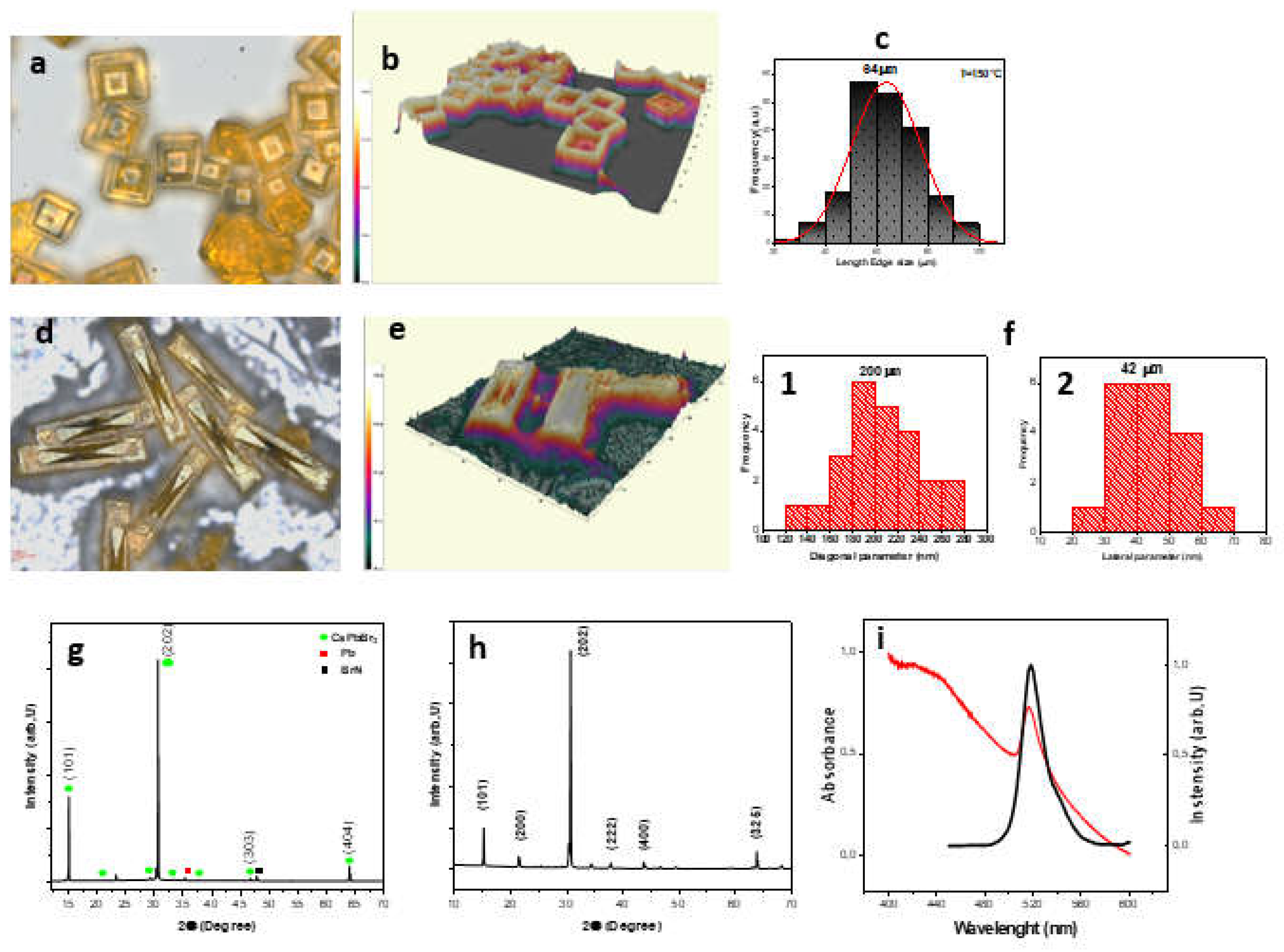
Figure 2.
Optical measurements of the MRs. (a) 3D rendering and (b) top-view confocal microscopy image of two MRs. (c) PL spectra obtained from one MR at the spots marked in (b). (d) Absorbance spectrum, (e) steady state PL spectrum and (f) TRPL decay profiles.
Figure 2.
Optical measurements of the MRs. (a) 3D rendering and (b) top-view confocal microscopy image of two MRs. (c) PL spectra obtained from one MR at the spots marked in (b). (d) Absorbance spectrum, (e) steady state PL spectrum and (f) TRPL decay profiles.
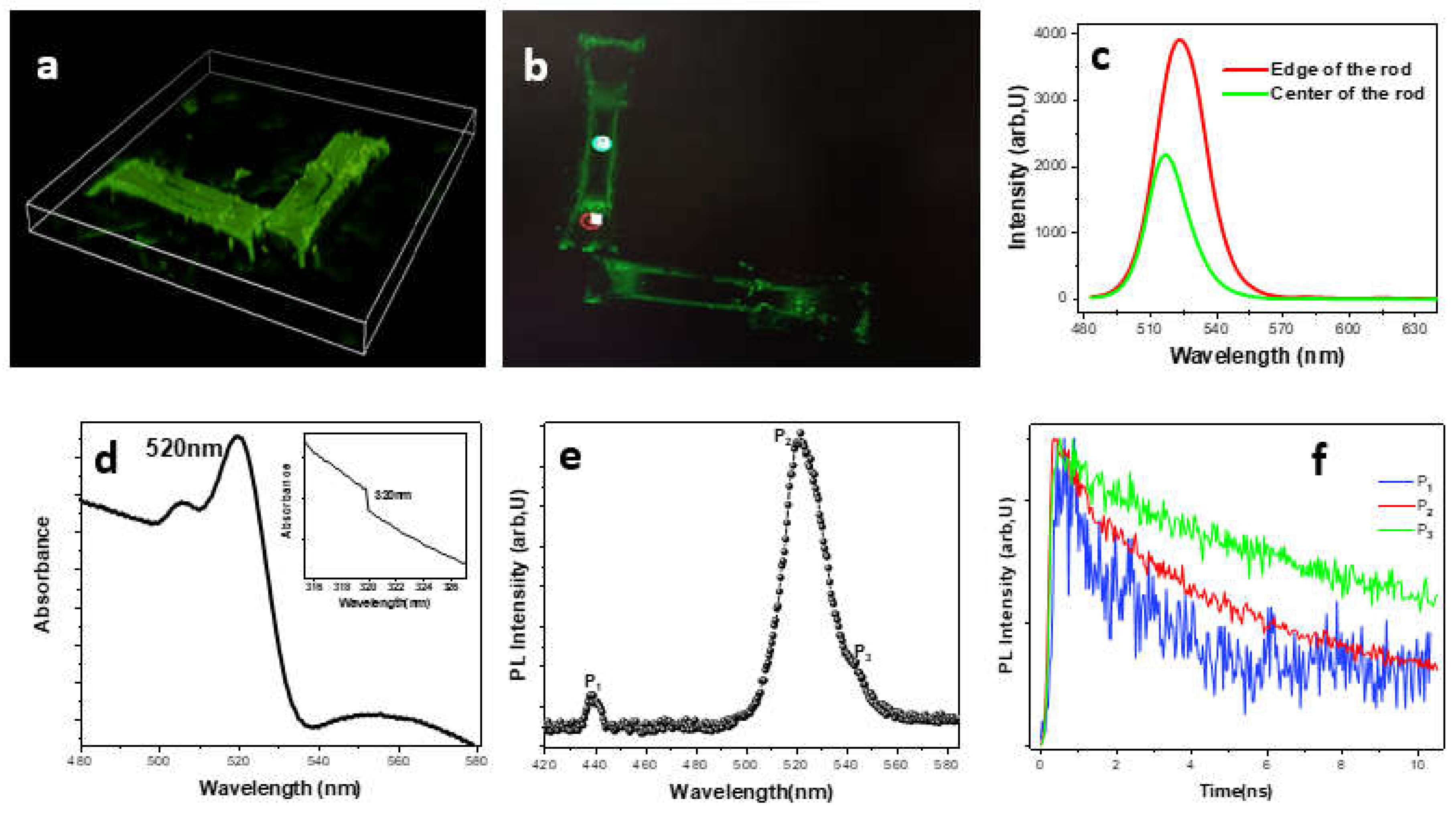
Figure 3.
(a) CA measurements of (1) ITO coated glass and (2) APTES/ITO-glass. (b) APTES chemical structure. (c) Silanization process of ITO patterned glass substrate. (d) AFM image of APTES modified-ITO glass substrate. (e) FTIR spectrum of CsPbBr3 MRs deposited on APTES ITO-coated glass substrate. (f) The schematic structure of the possible binding of the obtained rods with aminosilane layers.
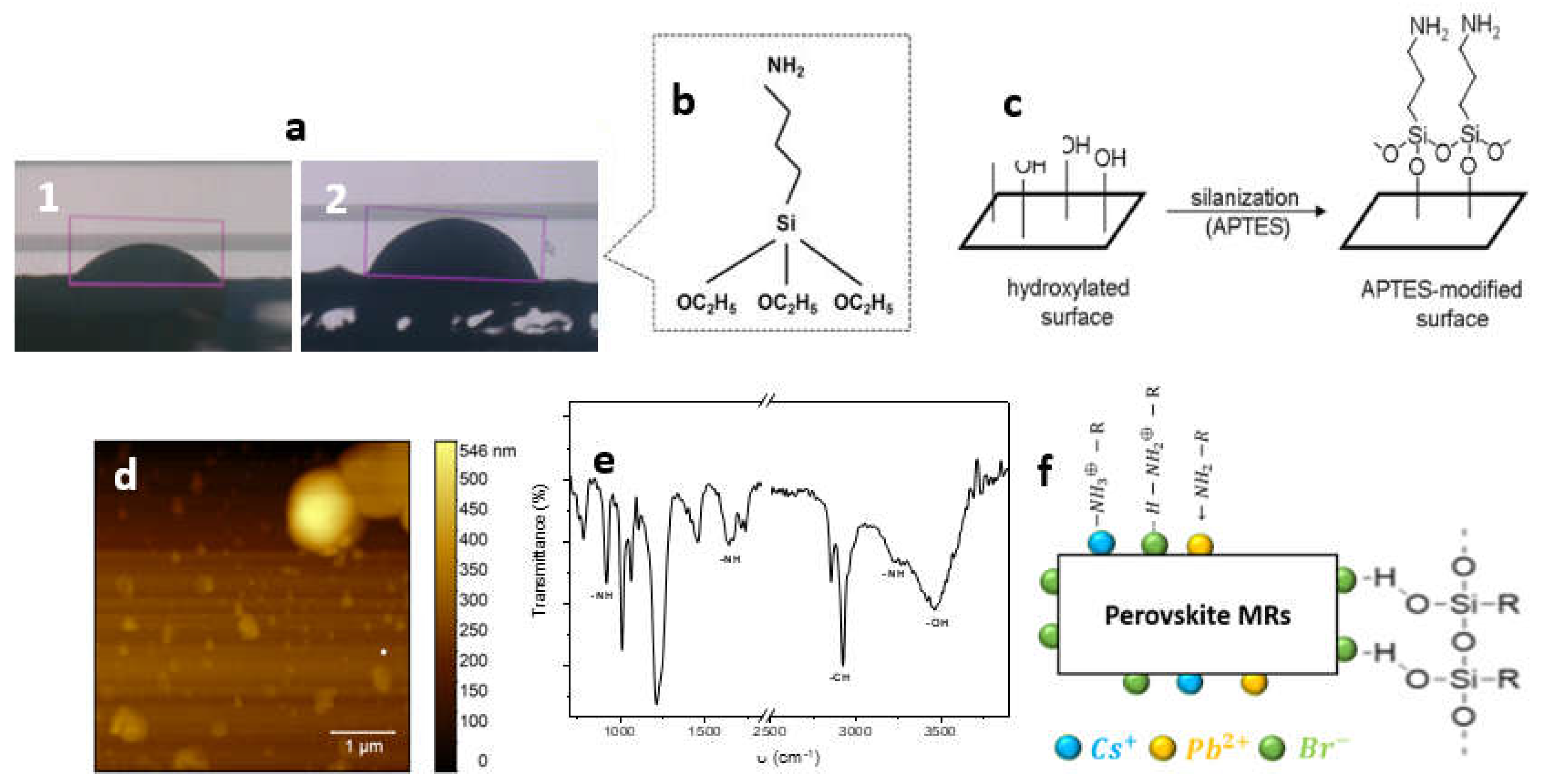
Figure 4.
(a) CA measurements of (1) ITO coated glass and (2) APTES/ITO-glass. (b) APTES chemical structure. (c) Silanization process of ITO patterned glass substrate. (d) AFM image of APTES modified-ITO glass substrate. (e) FTIR spectrum of CsPbBr3 MRs deposited on APTES ITO-coated glass substrate. (f) The schematic structure of the possible binding of the obtained rods with aminosilane layers.
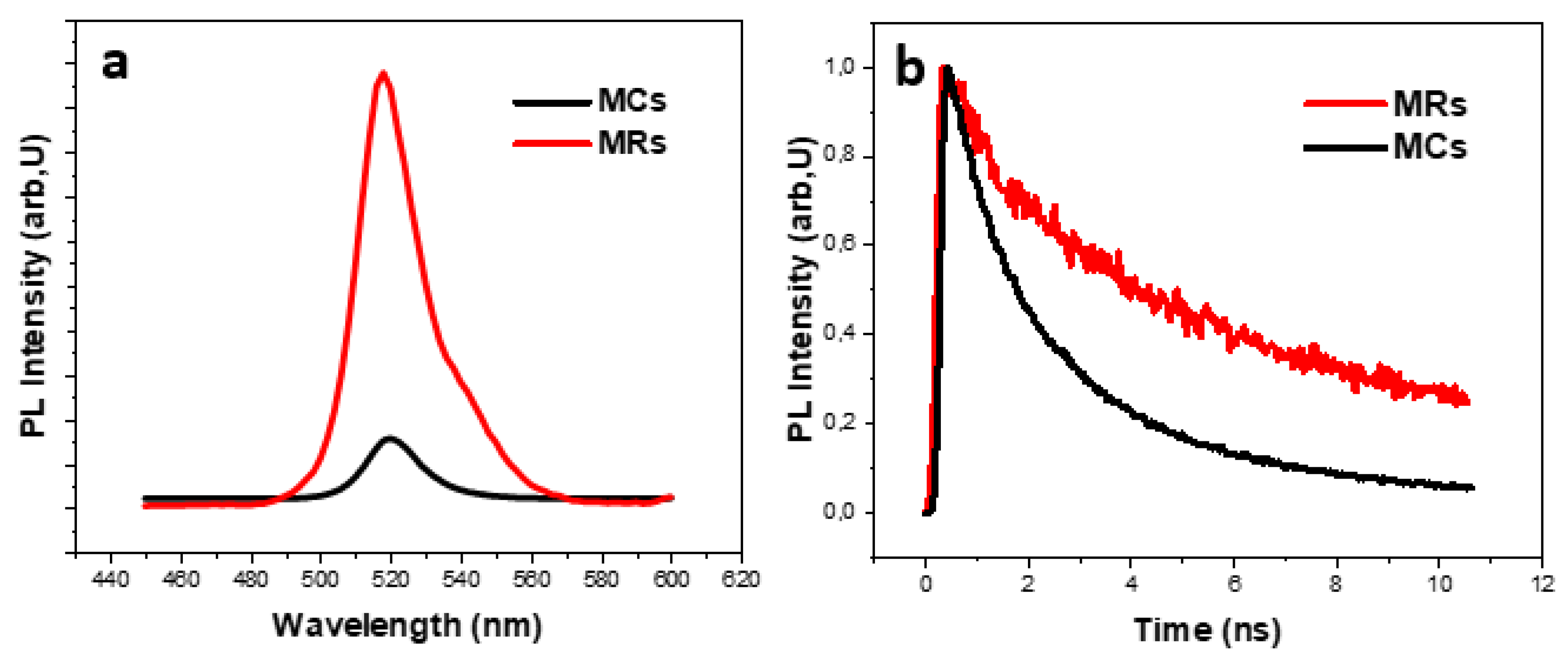

Table 1.
FTIR bands and peak assignments.
| Wavenumber (cm-1) | Functional group |
| 3464 | -OH |
| 3258 | -NH |
| 2959-2924-2856 | -CH |
| 1715-1734 | -CH |
| 1638-1664 | -NH2 |
| 1453 | C-H |
| 1244-1213 | C-H |
| 1012-1066 | Si-O-Si |
| 922 | CH3 rocking of APTES |
| 755-788 | Ethoxy group of APTES |
Table 2.
PL lifetimes of CsPbBr3 MCs and MRs.
| sample | (ns) | (ns) | (ns) | ||
|---|---|---|---|---|---|
| MCs | 54.65 | 3,39 | 45.35 | 1.01 | 2.31 |
| MRs | 72.56 | 9.95 | 19.79 | 1.10 | 7.43 |
Disclaimer/Publisher’s Note: The statements, opinions and data contained in all publications are solely those of the individual author(s) and contributor(s) and not of MDPI and/or the editor(s). MDPI and/or the editor(s) disclaim responsibility for any injury to people or property resulting from any ideas, methods, instructions or products referred to in the content. |
© 2024 by the authors. Licensee MDPI, Basel, Switzerland. This article is an open access article distributed under the terms and conditions of the Creative Commons Attribution (CC BY) license (http://creativecommons.org/licenses/by/4.0/).
Copyright: This open access article is published under a Creative Commons CC BY 4.0 license, which permit the free download, distribution, and reuse, provided that the author and preprint are cited in any reuse.


