
Submitted:
17 July 2024
Posted:
18 July 2024
You are already at the latest version
Abstract
Background. Myocardial involvement mediated by chronic obstructive pulmonary disease (COPD) is a common cause of morbidity and mortality. Conventional transthoracic echocardiography (TTE) parameters are poor in the detection of subclinical myocardial dysfunction.
Aim.To investigate the contribution of strain in the early detection of cardiac damage in stable COPD patients
Methods. Group A and B patients with COPD were enrolled in this study. The COPD assessment test, spirometry, 6-minute walk test, and both conventional TTE and strain were performed in these patients.
Results. 80 COPD patients, with a mean age 65.6 ± 8.9 years, were included. The. Left ventricular ejection fraction (LVEF) was 60.7 ± 5.1%. Right atrium and right ventricle (RV) strain were 24.5 ± 6.6% and -19.9 ± 3.7%, respectively. Additionally, left ventricle global longitudinal strain (LV GLS) was -21.1 ± 2. Forty-eight patients had impaired RV strain. Compared to COPD patient with normal RV strain, those with RV reduced starin had a lower 6meter walk distance (6MWD) (p=0.001) and forced expiratory volume in the first second (FEV1) (p=0.012), and a higher CAT score (p=0.012). A reduced RV strain was correlated with a higher risk of hospitalizations for acute exacerbation in the post inclusion year (55% versus 25%; p=0.024). No deaths were recorded during the follow-up period. No significant factors causing neither RA starin alteration nor LV GLS reduction were revealed.
Conclusion. Group A and B COPD patients having normal conventional TTE parameters, speckle tracking is a key parameter in the detection of subclinical myocardial dysfunction.
Keywords:
chronic obstructive pulmonary disease
; subtle myocardial involvement
; echocardiography
; strain
Introduction
Chronic obstructive pulmonary disease (COPD) is a public health problem worldwide[1]. It is one of the main causes of high morbidity and mortality [2]. The development of heart failure during COPD is a major anticipating factor of exacerbation, hospital readmission, and mortality [3]. Both right- and left-sided heart failure could frequently be noted in COPD patients. Several studies have revealed that this comorbidity is related, on the one hand, to the structural and physiological changes in the pulmonary vascularization predisposing to the right heart failure even before pulmonary hypertension (PH) [4] and, on the other hand, to the common cardiovascular risk factors, such as smoking, chronic systemic inflammation, and endothelial dysfunction that are leading factors of heart failure [5]. Subtle heart failure has no symptomatic repercussion on COPD with overlapping symptoms, such as dyspnea, leading to an under diagnosis of incipient myocardial damage [6] . Right ventricle (RV) and left ventricle (LV) subclinical dysfunctions remain challenging. The conventional echocardiographic measurements are poor in detecting subtle myocardial injury [7]. Speckle tracking study, a technique of myocardial deformation measurement, provides promising results in the early detection of LV and RV dysfunction[8,9]. Thus, the aim of the current study was to identify subtle RV and LV dysfunction using two-dimensional strain in stable COPD patients and its correlation with hospital readmission.
Methods
Study Design
This is an analytical cross-sectional prospective study performed during the period from January 2023 to May 2024. All stable group A and B COPD patients without PH were included in this study.
Study Population
Inclusion criteria: Patients with confirmed diagnosis of group A and B COPD and those aged more than 18 years were included in this study [10]. Participants in the study had to be free of COPD exacerbations for three months before enrollment.
Non-inclusion criteria: Patients with evidence of LV ejection fraction (LVEF)<50% on echocardiography and those with severe valvular heart disease, pulmonary embolism, PH, coronary artery disease, conduction abnormalities, and atrial fibrillation on electrocardiogram were not included in the present study. Patients with contraindications for the 6-minute walk test were not also included in the study[10].
Exclusion criteria: Patients with abnormalities noted while performing echocardiography were excluded.
Sample Size
The sample size was calculated according to the following predictive equation[11]: N = (Zα p (1-p))/i2 and based on the prevalence of heart failure in COPD patients and the expected sensitivity and specificity. For a prevalence of 54%, an expected sensitivity and specificity of respectively 97% and 99%, a desired precision of 0.1, the minimum sample size was estimated at 23 patients.
Variables and Data Collection
Diagnosis of COPD was confirmed by a spirometry using the GOLD 2023 criteria, indicating a non-reversible ventilatory deficit (FEV1 /FVC < 0.7 following bronchodilation) [12]. Patients’ recruitment was carried out from January to March 2023. During this period, patients filled out a questionnaire written in the local Arabic dialect. The questionnaire had three components. The first part involved the patients’ social and demographic characteristics (e.g., age, gender, medical ATCD). The second part included COPD data (e.g., treatment, number of hospitalizations). The third part involved the COPD assessment test (CAT), including questions about eight areas, to assign an overall score ranging from 0 to 40. Higher scores indicates that COPD has a greater impact on the patient’s health and well-being [13]. After filling out the questionnaire and on the same day , spirometry was performed by an experienced technician using a portable spirometer (SpirobankG MIR, delMaggiolino 12500155 Roma, Italy) according to the international recommendations[14]. Spirometry data, including expiratory forced vital capacity (FVC, L), (FEV1, L), maximal mid-expiratory flow (L/s), and FEV1/FVC ratio (absolute value), were reported as absolute values and percentages of reference international value[15]. The 6-minute walk test was performed for all the patients. The test was conducted on a flat, straight corridor and the patients were required to walk as far as possible for six minutes to calculate the 6-minute walk distance (6MWD). The directions given to the patients throughout the test were in compliance with the International Standards Guidelines [16].
Trans thoracic echocardiography (TTE) was performed between April 2023 and May 2024. All the included COPD patients underwent a standard TTE using a Vivid E9 echocardiography system (General Electric Medical System). The same operator performed all echocardiographies to limit inter-operator variations. All conventional TTE parameters were performed, including LV diameters, wall thickness and volumes. LVEF was estimated using the Simpson method. Peak mitral E and A waves in pulse Doppler, e’ wave in tissue Doppler imaging, E/e’ ratio as well as the left atrial area (LAA) and volume were measured. Right ventricle (RV) function was evaluated using peak of right ventricular systolic myocardial velocity (S wave) and tricuspid annular plane systolic excursion (TAPSE). The right and left atria area and volume were measured. Systolic pulmonary artery pressure (SPAP) was calculated on the peak of tricuspid regurgitation. Speckle tracking analyses using Echopac software version 112 and automated functional imaging (AFI) was used to evaluate both LV and RV strain, and RA reservoir function. A RA strain <25% is considered impaired[17]. An altered RV free wall strain is >-19 [18] and an impaired LV global longitudinal strain (LV GLS) is >-20 [19]
One year after TTE performing, all patients were contacted to check the number of hospitalizations during this year and deaths.
Statistical Analysis
Quantitative data were expressed as means and medians, and qualitative data as percentages. The Chi-square test and Fisher’s exact test were appropriately used for qualitative variables and percentage comparisons. The Student’s t-test was utilized to compare the means of quantitative variables. Non-parametric tests (Mann-Whitney test) were used as needed. Values were considered significant when p was ≤ 0.05.
A univariate analysis was initially conducted. The association between dependent and each independent variable was analyzed to include variables that could be highly predictive a priori in a multivariate analysis model.
During multivariate analysis, variables with p < 0.25 were included in the multivariate model and they were analyzed using backward stepwise logistic regression. The Backward stepwise procedure included all selected variables and progressively removed those that did not contribute sufficient information to the model at each step. Thus, only the independent variables remained in the final step. Variables with a significant Odds Ratio (p ≤ 0.05) still present in the final step were considered significant independent variables in the observed multivariate model. To determine the association between quantitative variables, correlation was used to determine the correlation coefficient (r) and regression analysis was used to study the regression equation: Y = a + b X, in which Y is dependent, X is independent, b is slope, and a is intercept.
All statistical analyses were performed using SPSS (Statistical Package for the Social Sciences) version 21.0 IBM.
Ethical Consideration
This study was approved by the medical and research ethics committee at Fattouma Bourguiba University Hospital (Approval number IORG 0009738 N 160 OMB 0990-0279) All the patients signed an informed consent to participate in this study.
Results
A total of 80 patients with a mean age of 65.6 ± 8.9 years were included. Male predominance was noted (83.3%). The number of active smokers was 29 (36.2%). The mean FEV1 value was 1954 ± 639 mL in the exploration of respiratory function. Using the CAT score, nine patients were scored >30 (Table 1).
Figure 1.
RV strain in stable COPD patient.
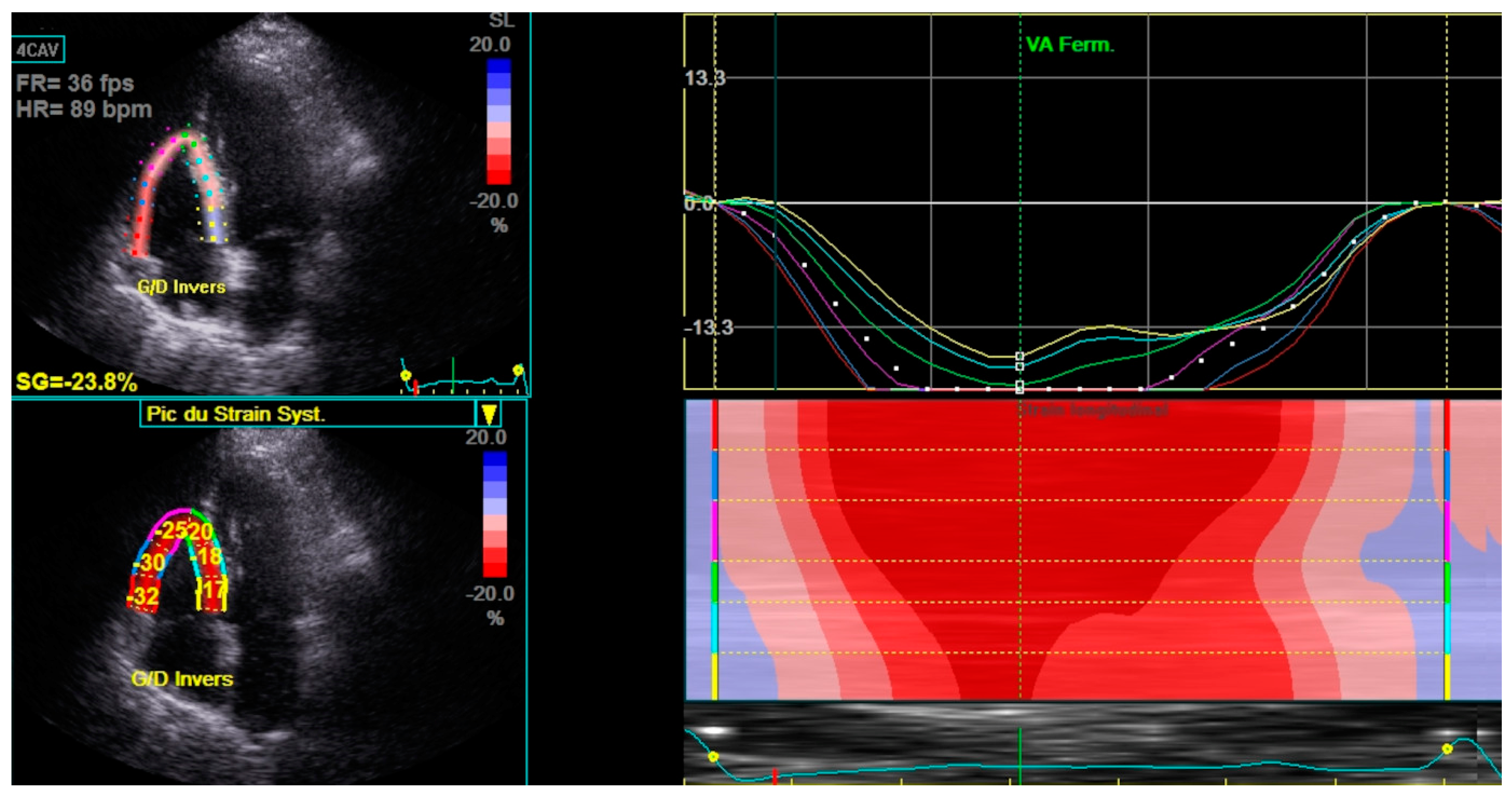
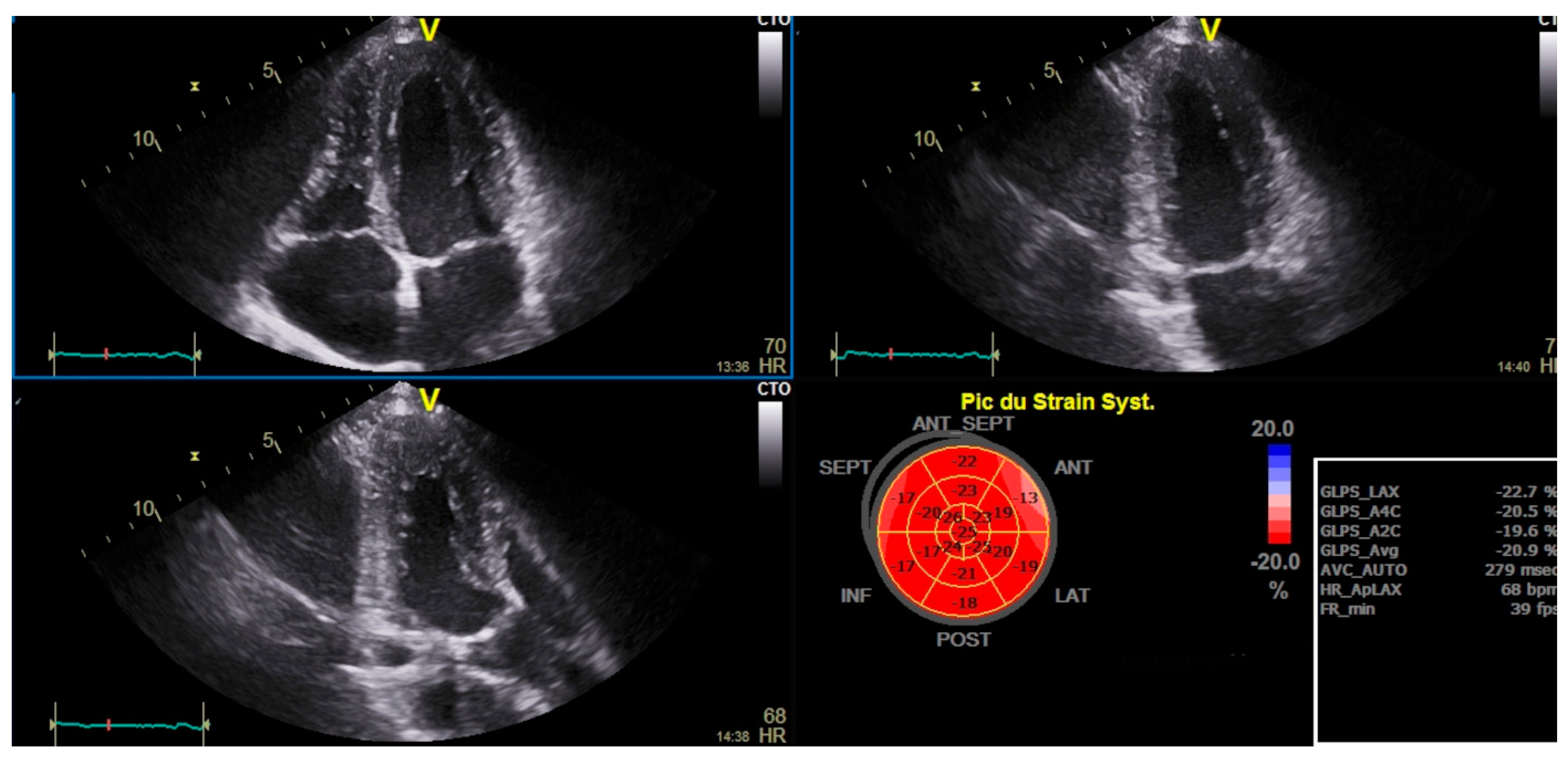
Figure 2.
LV GLS and bull’s eye in stable COPD patient.
Among the eighty participants, forty-eight patients had impaired RV strain, with a lower 6MWD (p=0.001) and FEV1 (p=0.012), and a higher CAT score (p=0.012) compared to those with normal RV strain (Table 3).
Univariate analysis revealed a significant association between damaged RV Strain and FAC, S wave RV, TAPSE, and sPAPS (Table 4).
Haut du Formulaire
The multivariate analysis identified three factors associated with the reduction of RV strain. The sPAP was found to be a significant factor for damaged RV strain, with an adjusted OR of 1.2 (p=0.001) (Table 5).
A moderate and statistically significant positive association was noted between sPAPS and the alteration in RV Strain. An increase in sPAP was associated with the damage in RV strain (r=0.561 P=0.000) (Figure 1).
A moderate and statistically significant negative relationship was noted between S wave RV and RV strain. An increase in S wave RV was associated with the improvement in RV Strain (r=-0.485 P=0.000) (Figure 3).
Only, reduced RV strain was correlated with a higher risk of hospitalizations for acute exacerbation in the post inclusion year (55% versus 25%; p=0.024). No deaths were recorded during the follow-up period.
This study revealed no significant factors causing neither RA reservoir function alteration nor LV GLS reduction.
Discussion
Right heart failure is a major cause of mortality and morbidity in patients with COPD. The prognosis of these patients can be affected by prompt diagnosis, effective therapy, and careful observation[20] .
This study highlighted that stable COPD without PH could cause subtle LV and RV dysfunction before developing symptoms of heart failure. Progressive cardiovascular impairment related to COPD increases the mortality and morbidity rates[21]. According to previous studies, the possible mechanisms leading to this damage are chronic multisystemic inflammation, high oxidative stress, high levels of inflammatory markers such as TNF-α, interleukins, and C-reactive protein, endothelial alteration, and the interaction between the heart and the lungs (22,4, 5). Cardiovascular damages include pulmonary hypertension, and left and right heart failure [22]. Heart failure is a major cause of mortality in patients with COPD (1). Initially, ventricular dysfunction is asymptomatic or oligosymptomatic, especially for the right ventricle in the early stage of COPD without pulmonary hypertension [23]. Cardiac involvement in patients with recurrent exacerbations in group E has already been documented [5,24,25]. According to Freixa et al., nearly one in every eight COPD patients requiring hospitalization develop severe RV dysfunction three months following the initial exacerbation [26]. However, cardiac involvement is underdiagnosed in group A and B patients with COPD [27]. Chronic hypoxemia and low blood oxygen levels can engender pulmonary hypertension or elevated blood pressure in the pulmonary arteries through several mechanisms (6,7) .Chronic hypoxemia can be undiagnosed for a long time in COPD patients since it is initially limited to intensive exertion and can be hidden by adaptive processes, especially in group A patients with low dyspnea manifestations. Classifying the cause of respiratory problems among patients with both conditions can be challenging. Systematic screening for cardiac involvement is therefore beneficial. Indeed, early diagnosis of subtle left and right dysfunction could change the therapeutic strategy and the prognosis of COPD patients.
As it is available and non-invasive, echocardiography is currently used to diagnose the effects of COPD on the heart. However, the parameters of standard echocardiography are not effective in screening subtle cardiac damage. Several studies have revealed the geometry of the LV, the function changes encompassing systolic and diastolic dysfunction, the hypertrophy of LV, and the reduced volumes [28]. However, systolic and diastolic LV function could be totally normal in standard echocardiography parameters as shown in the present study. Speckle tracking is a promising method for the early detection of LV dysfunction. Pizarro et al. showed a significant damage in LV GLS in COPD patients with a reduced regional strain in the apical and septal walls that is correlated with COPD severity [29]. In this study, despite the normal parameters of LV standard echocardiography, subclinical LV alteration was revealed by GLS. RV dysfunction in COPD patients is associated with worse outcomes and an increased mortality [3]. Previous studies have shown that RV hypertrophy, dilatation, and systolic dysfunction are common in COPD patients regardless of pulmonary hypertension and the increased RV afterload (30,31). These findings may be due to an elevated pulmonary vascular resistance and a reduced pulmonary artery/arterial compliance [32]. RV remodeling develops early during COPD, leading to subclinical RV dysfunction [32]. RV GLS is a powerful parameter in RV in subclinical dysfunction secondary to chronic respiratory diseases, such as COPD and fibrotic interstitial lung diseases [33]. Speckle tracking echocardiography allows the quantification of RV dysfunction and the screening of discrete and localized contractile loss [33]. The current study demonstrated that despite normal RV systolic parameters (S wave RV, TAPSE), RV free wall strain was damaged. Right atrium exercise intolerance may also be altered by heart dysfunction [5]. Therefore, in the case of reduced 6MWD, a subclinical cardiac involvement should be screened, especially in non-exacerbated COPD. A high CAT score was also noted in patients with altered RV Strain (p=0.012). Since many factors contribute to the impact of the quality of life in individuals with COPD, the significance of cardiac involvement is often neglected , particularly in groups A and B [12] .This emphasizes how crucial it is to provide COPD patients with global management plans that take into account both cardiac and pulmonary issues even in group A and B patients with COPD. The quality of life of such patients can be enhanced by integrating care approaches that take into account the complex character of these illnesses. Besides, cardiovascular disorders are correlated with a higher risk of hospitalizations as shown in this meta-analysis where right heart failure was a potential risk factor for the 30-day readmission of COPD patients [34]. COPD patients with heart failure have a far higher risk of being hospitalized, which exacerbates their already complicated medical needs. Early detection of heart failure plays a crucial role in reducing the hospitalization rates and improving patient outcomes. Therefore, an early identification of heart failure enables prompt intervention, which can slow the evolution of the disease and improve symptoms.
Study Limitaions
One of the limitations of the present study is the absence of magnetic resonance imaging as a gold standard for the diagnosis of RV and LV subtle dysfunction as well as the short term follow-up and the lack of data. Moreover, LV and RV strain could be affected by other factors beyond COPD. Indeed, despite the high sensitivity of speckle tracking in the early detection of cardiac involvement, its low specificity remains a major limitation.
Conclusions
Cardiac damage is a common complication in COPD patients. It could worsen the prognosis and increase mortality. Due to the overlapping symptoms of cardiac failure and COPD, myocardial damage is often overlooked. Based on this study, LV and RV strain could predict silent myocardial involvement at early stages. Close follow-up using speckle tracking allows the detection of subtle cardiac damage and the indication of the appropriate strategy to prevent advanced myocardial dysfunction.
Abbreviations
| IVS | inter ventricular septum |
| LVEDD | left ventricle end diastolic diameter |
| LVEF | Left ventricular ejection fraction |
| PW | posterior wall |
| RV | right ventricular |
| TAPSE | tricuspid annular plane systolic excursion |
| TTE | Conventional transthoracic echocardiography |
| CAT | COPD assessment test |
| COPD | chronic obstructive pulmonary disease |
| FAC | fractional area change |
| FEV1 | forced expiratory volume in the first second |
| GLS | global longitudinal strain |
| IVC | inferior vena cava |
| LA | Left atrium |
| LAA | left atrial area |
| m | meter |
| ml | milliliter |
| PH | Pulmonary hypertension |
| RA | Right atrium |
| S wave | right ventricular systolic myocardial velocity |
| RAA | right atrium area |
| RVBD | Right ventricle basal diameter |
| RV MCD | Right ventricle mid cavity diameter |
| RVLD | Right ventricle longitudinal diameter |
| sPAP | Systolic pulmonary artery pressure |
| TAD | tricuspid annulus diameter |
| TAPSE | tricuspid annular plane systolic excursion |
References
- Murray, C.J.; Lopez, A.D. Global mortality, disability, and the contribution of risk factors: Global Burden of Disease Study. Lancet 1997, 349, 1436–1442. [Google Scholar] [CrossRef]
- Rabe, K.F.; Hurd, S.; Anzueto, A.; Barnes, P.J.; Buist, S.A.; Calverley, P. Global Strategy for the Diagnosis, Management, and Prevention of Chronic Obstructive Pulmonary Disease: GOLD Executive Summary. Am J Respir Crit Care Med. 2007, 176, 532–55. [Google Scholar]
- Almagro, P.; Barreiro, B.; de Echagüen, A.O.; Quintana, S.; Carballeira, M.R.; Heredia, J.L.; Garau, J. Risk Factors for Hospital Readmission in Patients with Chronic Obstructive Pulmonary Disease. Respiration 2006, 73, 311–317. [Google Scholar] [CrossRef]
- MacNee, W. Pathophysiology of cor pulmonale in chronic obstructive pulmonary disease. Part One. Am J Respir Crit Care Med. 1994, 150, 833–52. [Google Scholar]
- Hesse, K.; Bourke, S.; Steer, J. Heart failure in patients with COPD exacerbations: Looking below the tip of the iceberg. Respir. Med. 2022, 196, 106800. [Google Scholar] [CrossRef]
- Gupta, M.; Chhabra, S.K. Coexistent Chronic Obstructive Pulmonary Disease-Heart Failure: Mechanisms, Diagnostic and Therapeutic Dilemmas. Indian J. Chest Dis. Allied Sci. 2022, 52, 225–238. [Google Scholar] [CrossRef]
- Elçioğlu, B.C.; Kamat, S.; Yurdakul, S.; Şahin, .T.; Sarper, A.; Yıldız, P.; Aytekin, S. Assessment of subclinical left ventricular systolic dysfunction and structural changes in patients with chronic obstructive pulmonary disease. Intern. Med. J. 2021, 52, 1791–1798. [Google Scholar] [CrossRef]
- Schoos, M.M.; Dalsgaard, M.; Kjærgaard, J.; Moesby, D.; Jensen, S.G.; Steffensen, I.; Iversen, K.K. Echocardiographic predictors of exercise capacity and mortality in chronic obstructive pulmonary disease. BMC Cardiovasc. Disord. 2013, 13, 84–84. [Google Scholar] [CrossRef] [PubMed]
- Smolarek, D.; Gruchała, M.; Sobiczewski, W. Echocardiographic evaluation of right ventricular systolic function: The traditional and innovative approach. Cardiol. J. 2017, 24, 563–572. [Google Scholar] [CrossRef] [PubMed]
- Singh SJ, Puhan MA, Andrianopoulos V, Hernandes NA, Mitchell KE, Hill CJ, et al. An official systematic review of the European Respiratory Society/American Thoracic Society: measurement properties of field walking tests in chronic respiratory disease. Eur Respir J. déc 2014;44(6):1447-78.
- Serhier Z, Bendahhou K, Ben Abdelaziz A, Bennani MO. Methodological sheet n°1: How to calculate the size of a sample for an observational study? Tunis Med. janv 2020;98(1):1-7.
- Agustí A, Celli BR, Criner GJ, Halpin D, Anzueto A, Barnes P, et al. Global Initiative for Chronic Obstructive Lung Disease 2023 Report: GOLD Executive Summary. Eur Respir J. avr 2023;61(4):2300239.
- Houben-Wilke, S.; Janssen, D.J.A.; Franssen, F.M.E.; Vanfleteren, L.E.G.W.; Wouters, E.F.M.; Spruit, M.A. Contribution of individual COPD assessment test (CAT) items to CAT total score and effects of pulmonary rehabilitation on CAT scores. Heal. Qual. Life Outcomes 2018, 16, 1–8. [Google Scholar] [CrossRef] [PubMed]
- Graham, B.L.; Steenbruggen, I.; Miller, M.R.; Barjaktarevic, I.Z.; Cooper, B.G.; Hall, G.L.; Hallstrand, T.S.; Kaminsky, D.A.; McCarthy, K.; McCormack, M.C.; et al. Standardization of Spirometry 2019 Update. An Official American Thoracic Society and European Respiratory Society Technical Statement. Am. J. Respir. Crit. Care Med. 2019, 200, e70–e88. [Google Scholar] [CrossRef]
- Firnhaber, J.M. Performance and Interpretation of Office Spirometry. Prim. Care: Clin. Off. Pr. 2021, 48, 645–654. [Google Scholar] [CrossRef]
- Singh SJ, Puhan MA, Andrianopoulos V, Hernandes NA, Mitchell KE, Hill CJ, et al. An official systematic review of the European Respiratory Society/American Thoracic Society: measurement properties of field walking tests in chronic respiratory disease. Eur Respir J. déc 2014;44(6):1447-78.
- Krittanawong C, Maitra NS, Hassan Virk HU, Farrell A, Hamzeh I, Arya B, et al. Normal Ranges of Right Atrial Strain. JACC Cardiovasc Imaging. mars 2023;16(3):282-94.
- Morris, D.A.; Krisper, M.; Nakatani, S.; Köhncke, C.; Otsuji, Y.; Belyavskiy, E.; Radha Krishnan, A.K.; Kropf, M.; Osmanoglou, E.; Boldt, L.-H.; et al. Normal range and usefulness of right ventricular systolic strain to detect subtle right ventricular systolic abnormalities in patients with heart failure: A multicentre study. Eur. Heart J. Cardiovasc. Imaging 2017, 18, 212–223. [Google Scholar] [CrossRef] [PubMed]
- Pio, S.M.; Medvedofsky, D.; Stassen, J.; Delgado, V.; Namazi, F.; Weissman, N.J.; Grayburn, P.; Kar, S.; Lim, D.S.; Zhou, Z.; et al. Changes in Left Ventricular Global Longitudinal Strain in Patients With Heart Failure and Secondary Mitral Regurgitation: The COAPT Trial. J. Am. Hear. Assoc. 2023, 12, e029956. [Google Scholar] [CrossRef] [PubMed]
- Elçioğlu, B.C.; Kamat, S.; Yurdakul, S.; Şahin, .T.; Sarper, A.; Yıldız, P.; Aytekin, S. Assessment of subclinical left ventricular systolic dysfunction and structural changes in patients with chronic obstructive pulmonary disease. Intern. Med. J. 2021, 52, 1791–1798. [Google Scholar] [CrossRef]
- Anthonisen, N.R.; Connett, J.E.; Enright, P.L.; Manfreda, J. Hospitalizations and Mortality in the Lung Health Study. Am. J. Respir. Crit. Care Med. 2002, 166, 333–339. [Google Scholar] [CrossRef] [PubMed]
- Hunninghake, D.B. Cardiovascular Disease in Chronic Obstructive Pulmonary Disease. Proc. Am. Thorac. Soc. 2005, 2, 44–49. [Google Scholar] [CrossRef] [PubMed]
- Chaouat, A.; Bugnet, A.-S.; Kadaoui, N.; Schott, R.; Enache, I.; Ducoloné, A.; Ehrhart, M.; Kessler, R.; Weitzenblum, E. Severe Pulmonary Hypertension and Chronic Obstructive Pulmonary Disease. Am. J. Respir. Crit. Care Med. 2005, 172, 189–194. [Google Scholar] [CrossRef] [PubMed]
- Kovacs, G.; Avian, A.; Bachmaier, G.; Troester, N.; Tornyos, A.; Douschan, P.; Foris, V.; Sassmann, T.; Zeder, K.; Lindenmann, J.; et al. Severe Pulmonary Hypertension in COPD. Chest 2022, 162, 202–212. [Google Scholar] [CrossRef] [PubMed]
- Hurst JR, Skolnik N, Hansen GJ, Anzueto A, Donaldson GC, Dransfield MT, et al. Understanding the impact of chronic obstructive pulmonary disease exacerbations on patient health and quality of life. Eur J Intern Med. mars 2020;73:1-6.
- Freixa, X.; Portillo, K.; Paré, C.; Garcia-Aymerich, J.; Gomez, F.P.; Benet, M.; Roca, J.; Farrero, E.; Ferrer, J.; Fernandez-Palomeque, C.; et al. Echocardiographic abnormalities in patients with COPD at their first hospital admission. Eur. Respir. J. 2012, 41, 784–791. [Google Scholar] [CrossRef] [PubMed]
- Rahman, H.H.; Rashid, M.H.; A Miah, N.; Israt, S.; Atiqullah, S.; Akbar, M.S. Correlation Study between COPD and Heart Failure in Elderly Patient. . 2022, 31, 498–505. [Google Scholar]
- Jörgensen, K.; Müller, M.F.; Nel, J.; Upton, R.N.; Houltz, E.; Ricksten, S.-E. Reduced Intrathoracic Blood Volume and Left and Right Ventricular Dimensions in Patients With Severe Emphysema. Chest 2007, 131, 1050–1057. [Google Scholar] [CrossRef]
- Pizarro, C.; van Essen, F.; Linnhoff, F.; Schueler, R.; Hammerstingl, C.; Nickenig, G.; Skowasch, D.; Weber, M. Speckle tracking echocardiography in chronic obstructive pulmonary disease and overlapping obstructive sleep apnea. Int. J. Chronic Obstr. Pulm. Dis. 2016, ume 11, 1823–1834. [Google Scholar] [CrossRef]
- Marti, S.; Muñoz, X.; Rios, J.; Morell, F.; Ferrer, J. Body weight and comorbidity predict mortality in COPD patients treated with oxygen therapy. Eur. Respir. J. 2006, 27, 689–696. [Google Scholar] [CrossRef] [PubMed]
- Ito, S.; Pislaru, S.V.; Soo, W.M.; Huang, R.; Greason, K.L.; Mathew, V.; Sandhu, G.S.; Eleid, M.F.; Suri, R.M.; Oh, J.K.; et al. Impact of right ventricular size and function on survival following transcatheter aortic valve replacement. Int. J. Cardiol. 2016, 221, 269–274. [Google Scholar] [CrossRef] [PubMed]
- Hilde JM, Skjørten I, Grøtta OJ, Hansteen V, Melsom MN, Hisdal J, et al. Right Ventricular Dysfunction and Remodeling in Chronic Obstructive Pulmonary Disease Without Pulmonary Hypertension. J Am Coll Cardiol. sept 2013;62(12):1103-11.
- Hilde, J.M.; Skjørten, I.; Grøtta, O.J.; Hansteen, V.; Melsom, M.N.; Hisdal, J.; Humerfelt, S.; Steine, K. Right Ventricular Dysfunction and Remodeling in Chronic Obstructive Pulmonary Disease Without Pulmonary Hypertension. Circ. 2013, 62, 1103–1111. [Google Scholar] [CrossRef] [PubMed]
- Buonauro, A.; Santoro, C.; Galderisi, M.; Canora, A.; Sorrentino, R.; Esposito, R.; Lembo, M.; Canonico, M.E.; Ilardi, F.; Fazio, V.; et al. Impaired Right and Left Ventricular Longitudinal Function in Patients with Fibrotic Interstitial Lung Diseases. J. Clin. Med. 2020, 9, 587. [Google Scholar] [CrossRef]
- Ruan, H.; Zhang, H.; Wang, J.; Zhao, H.; Han, W.; Li, J. Readmission rate for acute exacerbation of chronic obstructive pulmonary disease: A systematic review and meta-analysis. Respir. Med. 2022, 206, 107090. [Google Scholar] [CrossRef]
Figure 3.
Regression and Correlation Analysis between RV Strain and sPAPS, and RV Strain S wave RV.
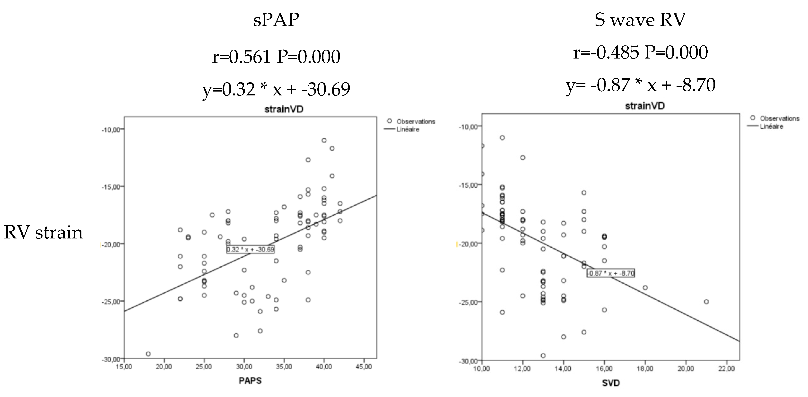

Table 1.
Socio-demographic and respiratory characteristics of stable goup A and B COPD patients.
| Characteristics | Total (N=80 ) N(%) or Mean ±SD |
|---|---|
| Age (Years) | 65.6±8.9 |
| Gender (n, %) | |
| Male | 67(83.8) |
| Female | 13(16.3) |
| Living Habits (n, %) | |
| Current smoker | 29(36 .2) |
| Former smoker | 42(52.5) |
| Never smoked | 9(11.3) |
| FEV1 (ml) | 1954 ±639 |
| 6MWD (meter) | 396±121 |
| CAT score (n, %) | |
| <10 | 24(30.0) |
| 10-20 | 30(37.5) |
| 21-30 | 17(21.3) |
| >30 | 9(11.3) |
LVEF was 60.7 ± 5.1%. Right atrium reservoir and RV strain were 24.5 ± 6.6% (Figure 1) and -19.9 ± 3.7%, respectively. Additionally, LV GLS was -21.1 ± 2.4.
Table 2.
Echocardiographic parameters in stable COPD patients.
| Cardiac echography | Total(n=80) Mean±SD |
|---|---|
| LVEF (%) | 60.7±5.1 |
| FAC (%) | 45.4±7.7 |
| TAD ( mm) | 27.7±2.7 |
| RVBD (mm) | 32.3±3.6 |
| RV MCD (mm) | 24.4±2.5 |
| RVLD (mm) | 62.5±5.7 |
| RAA (cm2) | 12.6±2.1 |
| S RV (cm/s) | 12.8±2.1 |
| TAPSE (mm) | 20.8±3.28 |
| sPAP (mmHg) | 32.8±6.4 |
| RV wall thickness (mm) | 6.6±0.8 |
| IVC (mm) | 14.9±1.3 |
|
E/A ratio E/e’ ratio |
0.98±0.29 6.2±3.3 |
| LAA (cm2) | 14.5±2.4 |
| IVS thickness (mm) | 9.0±0.8 |
| LVEDD (mm) | 45.8±3.4 |
| PW thickness (mm) | 8.7±0.9 |
| RV Strain (%) | -19.9±3.7 |
| RA Strain (%) | 24.5±6.6 |
| LV GLS (%) | -21.1±2.4 |
FAC: fractional area change, IVC: inferior vena cava, IVS: inter ventricular septum, LAA: left atrial area, LV: left ventricle, LVEF: left ventricle ejection fraction, LVEDD: left ventricle end diastolic diameter, PW: posterior wall, RA: Right atrium, RAA: RA area, RV: right ventricle, RVBD: RV basal diameter, RV MCD: RV mid cavity diameter, RVLD: RV longitudinal diameter, sPAP: systolic pulmonary artery pressure, TAD: tricuspid annulus diameter, TAPSE: tricuspid annular plane systolic excursion.
Table 3.
Associated factors with a reduced RV strain in group A and B patients with COPD.
| Characteristics | Normal RV Strain (N=32) N (%) Or Mean±SD | Impaired RV Strain (N=48) N (%) Or Mean±SD |
P |
|---|---|---|---|
| Age (Years) | 65.4±.8 | 65.7±9.5 | 0.87 |
| Gender | 0.083 | ||
| Male | 24(35.8) | 43(64.2) | |
| Female | 8(61.5) | 5(38.5) | |
| Living Habits | 0.005 | ||
| Current smoker | 17(9.3) | 12(40.7) | |
| Former smoker | 9(21.4) | 33(78.6) | |
| Never smoked | 6(66.7) | 3(33.3) | |
| FEV1(ml) | 2182±407 | 1626±727 | 0.012 |
| 6MWD(m) | 470±104 | 310±113 | 0.001 |
| CAT | 13±6 | 21±10 | 0.012 |
| <10 | 16(66.6) | 8(33.3) | |
| 10-20 | 16(53.3) | 14(46.6) | |
| 21-30 | 0(0.0) | 17(100) | |
| >30 | 0(0.0) | 9(100) |
CAT: COPD Assessment Test, FEV1: forced expiratory volume in one second; 6MWD:6 minute walk distance, m: meter, ml: milliliter.
Table 4.
Association between conventional echocardiographic parameters and reduced RV Strain in COPD patients in univariate analysis.
Table 4.
Association between conventional echocardiographic parameters and reduced RV Strain in COPD patients in univariate analysis.
| Characteristics | Normal RV strain (N=32) Mean±SD | Reduced RV Strain (N=48) Mean±SD | p |
|---|---|---|---|
| LV Ejection fraction (%) | 60.75±4.819 | 60.77±9.599 | 0.986 |
| FAC (%) | 50.343±5.839 | 42.166±7.143 | 0.000 |
| TAD (mm) | 27.593±2.949 | 27.854±2.576 | 0.677 |
| RVBD (mm) | 32.593±3.653 | 32.208±3.649 | 0.645 |
| RVMD (mm) | 23.875±2.485 | 24.750±2.621 | 0.140 |
| RVLD (mm) | 60.875±6.282 | 63.583±5.089 | 0.037 |
| RAA (cm2) | 12.531±2.361 | 12.693±2.041 | 0.744 |
| S wave RV (cm/s) | 13.906±1.956 | 12.062±1.803 | 0.000 |
| TAPSE (mm) | 22.937±3.426 | 19.500±2.352 | 0.000 |
| sPAP (mmHg) | 29.000±5.364 | 35.375±5.796 | 0.000 |
| RV wall thickness (mm) | 6.343±1.035 | 6.708±0.682 | 0.061 |
| IVC (mm) | 14.66±1.537 | 15.17±1.226 | 0.104 |
| E/A | 1.060±0.290 | 0.938±0.282 | 0.065 |
| LAA (cm2) | 14.931±2.597 | 14.333±2.364 | 0.290 |
| IVS thickness (mm) | 8.906±0.928 | 9.208±0.742 | 0.111 |
| LV EDD (mm) | 45.312±3.335 | 46.229±3.520 | 0.248 |
| LV PW (mm) | 8.625±0.975 | 8.791±0988 | 0.460 |
FAC: fractional area change, IVC: inferior vena cava, IVS: inter ventricular septum, LAA: left atrial area, LV: left ventricle, LVEF: left ventricle ejection fraction, LVEDD: left ventricle end diastolic diameter, PW: posterior wall, RA: Right atrium, RAA: RA area, RV: right ventricle, RVBD: RV basal diameter, RV MCD: RV mid cavity diameter, RVLD: RV longitudinal diameter, sPAP: systolic pulmonary artery pressure, TAD: tricuspid annulus diameter, TAPSE: tricuspid annular plane systolic excursion.
Table 5.
Multivariate analysis of the factors associated with the changes in RV strain.
| aOR | 95%CI | P | ||
|---|---|---|---|---|
| 6MWD | 0.985 | 0.978 | 0.993 | 0.001 |
| sPAP | 1.214 | 1.079 | 1.366 | 0.000 |
| S wave RV | 0.526 | 0.338 | 0.818 | 0.004 |
aOR: adjusted Odds Ratio, 6MWD: 6-minute walk distance, RV: right ventricle sPAP: systolic pulmonary artery pressureHaut du formulaire.
Disclaimer/Publisher’s Note: The statements, opinions and data contained in all publications are solely those of the individual author(s) and contributor(s) and not of MDPI and/or the editor(s). MDPI and/or the editor(s) disclaim responsibility for any injury to people or property resulting from any ideas, methods, instructions or products referred to in the content. |
© 2024 by the authors. Licensee MDPI, Basel, Switzerland. This article is an open access article distributed under the terms and conditions of the Creative Commons Attribution (CC BY) license (http://creativecommons.org/licenses/by/4.0/).
Copyright: This open access article is published under a Creative Commons CC BY 4.0 license, which permit the free download, distribution, and reuse, provided that the author and preprint are cited in any reuse.


