
Submitted:
20 June 2024
Posted:
24 June 2024
You are already at the latest version
Preprints on COVID-19 and SARS-CoV-2
Abstract
Introduction: The disease caused by Coronavirus-2019 (COVID-19) was initially described in December 2019. Severe acute respiratory syndrome type 2 (Sars-Cov-2) is responsible for an important multisystem inflammatory spectrum. Cardiovascular conditions have been reported with an estimated frequency of 8-28%. In this context, transthoracic echocardiography associated with the analysis of the two-dimensional Global Longitudinal Strain (GLS) of the left ventricle derived from Speckle Tracking emerges as a promising modality. Objective: This study aims to evaluate the applicability of GLS and segmental deformity assessment in detecting subclinical myocardial dysfunction in patients that recovered from COVID-19 infection. Methods: The observational study involved 18 patients (mean age 52 years) with recent evidence of COVID-19 infection that underwent detailed echocardiographic evaluation with GLS, and cardiac magnetic resonance (CMR) according to segmental assessment results. The degree of correlation between the methods was analyzed and a systematic literature review was also conducted. Results: The mean left ventricular ejection fraction (LVEF) was 57.50 ± 9.98%. Only 16.6% of patients had reduced LVEF. The average GLS was – 18.70 ± 3.54% and in 44% of cases only one myocardial segment was affected, particularly the inferolateral basal. CMR revealed inflammatory cardiomyopathy. Discussion: Subclinical myocardial involvement is associated with major adverse cardiac events. Literature review supports the use of GLS for detecting early cardiac involvement. Conclusion: Speckle-tracking echocardiography has been shown to have clinical utility in a variety of settings and to offer superior prognostic value with a potential to enhance subclinical detection of myocardial changes in patients after COVID-19 infection.
Keywords:
Global Longitudinal Strain
; COVID-19
; Echocardiography
Introduction
The disease caused by Coronavirus-2019 (COVID-19) was initially described in December 2019 in Wuhan, China [1]. The novel coronavirus, has since been identified as the causative agent of a global pandemic, significantly altering the cultural, economic, and scientific atmospheres of our community [2]. Later named Severe Acute Respiratory Syndrome Coronavirus 2 (SARS-CoV-2), it is responsible for an important multisystem inflammatory spectrum due to a complex pathophysiology. Named after the Latin word “corona” for their crown-like spikes as seen under a microscope, coronaviruses are not a new occurrence in the virological universe with previously caused large-scale pandemics in 2002 and 2012 [3,4]. SARS-CoV-2 largely circulates within mammal and bird populations, of which bats constitute the main carrier for human infections [5]. Human-to-human transmission on the other hand has been established to be mainly via respiratory droplets. Exposure to coughing and sneezing from an infected individual increases the risk of transmission [6]. By mid-March 2020, the spread of Covid-19 had rapidly snowballed to include more than 160 countries, hitting both developed and developing countries alike [7]. In the next three years, numerous vaccines as well as new variants of SARS-CoV-2 emerged, setting off multiple waves of infection across countries. By the time the WHO declared that the COVID-19 was over on 5 May 2023, it had resulted in over 765 million confirmed cases and 6.9 million attributed deaths [8]. Fast-tracked vaccine development and strict public health measures gradually controlled the pandemic. One of the factors contributing to the spread of Covid-19 was the infectivity of asymptomatic individuals. The latent period can last up to 14 days, during which they are just as likely to transmit infection silently [9]. Following this period, clinical features can be spread across a spectrum of mild, moderate or severe disease [10]. One significant aspect of COVID-19’s impact is its effect on the cardiovascular system. Cardiovascular conditions related to COVID-19 have been reported with an estimated frequency ranging from 8% to 28%. These conditions include myocarditis, acute myocardial injury, heart failure, arrhythmias, and thromboembolic events [11,12]. The mechanisms underlying these cardiovascular manifestations are multifactorial, involving direct viral invasion, systemic inflammation, endothelial dysfunction, and the effects of the hypercoagulable state induced by the infection [13,14]. In this context, the assessment of cardiac function becomes critical. Transthoracic echocardiography (TTE) is a non-invasive imaging modality that has emerged as a valuable tool in evaluating cardiac involvement in COVID-19 patients [15]. TTE allows for comprehensive assessment of cardiac structure and function, providing essential information that can guide clinical management. A particularly promising aspect of TTE in the context of COVID-19 is the analysis of the two-dimensional Global Longitudinal Strain (GLS) of the left ventricle, derived from Speckle Tracking Echocardiography (STE) [16,17]. GLS is a measure of myocardial deformation, representing the percentage of shortening of the myocardium during the cardiac cycle. This technique offers superior sensitivity compared to traditional measures of left ventricular function, such as ejection fraction (EF), in detecting subclinical myocardial dysfunction [18]. In COVID-19 patients, GLS has shown potential in identifying early myocardial involvement before significant changes in ejection fraction are observed. Studies have reported that reduced GLS is associated with worse outcomes in COVID-19 patients, highlighting its prognostic value. Furthermore, GLS can help differentiate between COVID-19-related myocardial injury and other causes of cardiac dysfunction, guiding appropriate therapeutic interventions.In summary, the integration of transthoracic echocardiography and two-dimensional GLS derived from Speckle Tracking provides a powerful diagnostic and prognostic tool in the management of these patients [19]. In this study we aim to evaluate the applicability of GLS and segmental deformity assessment in detecting subclinical myocardial dysfunction in patients who have recovered from COVID-19 infection, via a case series and literature review.
Methods
A cross-sectional observational study of 18 patients with confirmed diagnosis of COVID-19 infection was conducted at a tertiary care specialized hospital in Brazil with 9 males (50%) and 9 females (50%). The mean age of the participants was 52 +/- 10 years. Patients underwent detailed history, physical examination, resting electrocardiogram (ECG), and conventional bidimensional echocardiography with Doppler followed by GLS analysis using speckle tracking and cardiac magnetic resonance imaging (CMR). Patients underwent serologic confirmation of infection using polymerase chain reaction for coronavirus detection (Ortho Clinical Diagnostic, USA). Echocardiography was performed using Philips Epiq 7C with a 3.4 MHz sector transducer. Measurements for left ventricular parameters were made according to recommendations by the American Society of Echocardiography and the European Association of Cardiovascular Imaging [20]. LVEF was calculated by the modified Simpson’s biplane method using the apical four and two-chamber views. Left ventricle segmental and longitudinal strain analysis were conducted using QLAB system (Philips, Netherlands). A scoping review of literature was performed on major medical sciences databases, including Pubmed and Scopus. Keyword inclusion criteria was selected for the following: COVID-19 infection and left ventricle global longitudinal strain. The results were filtered to include full-text case reports, meta-analysis, and systematic reviews written in English. A total of 27 records were obtained during the initial search. Two reviewers screened the titles and abstracts to determine if they met the inclusion criteria. During the screening, 6 records were excluded. A total of 21 records proceeded to the full-text assessment. Four reviewers assessed the content of the case reports to determine if they had enough information about the patients and their disease. Written consent was obtained from study participants and also approved by the institutional ethics review board. Baseline characteristic and echocardiographic parameters for the patients enrolled in the study are presented in Table 1.
Case Series Presentation
This cross-sectional observational study included 18 participants selected from a cohort of patients who presented with mild COVID-19 symptoms at a tertiary care center in Brazil. The cohort consisted of 9 males (50%) and 9 females (50%), with a mean age of 52 ± 10 years. Patients presented with mild symptoms such as cough, sore throat, headache, and fever. Diagnosis was confirmed by performing RT-PCR tests on nasal and throat swab samples. None of the patients required hospitalization, and all were treated symptomatically as per the suggested guidelines. Serum biomarkers such as brain natriuretic peptide (BNP) and high-sensitivity troponin were also obtained with values below the lower limit of normality for all patients. This study provides significant insights into the cardiac involvement in patients with mild COVID-19 symptoms. Despite the mild nature of their symptoms, screening echocardiography was performed followed by cardiac magnetic resonance imaging if at least one segment was altered. Echocardiographic analysis was performed and reviewed by two board certified echocardiographers. Left ventricle segmental and longitudinal strain analysis were calculated using the QLAB semi-automatic system (Philips, Netherlands), which considered normal values > - 18%. Echocardiographic evaluation revealed important findings: the mean LVEF was 57.50 ± 9.98%, with only 16.6% of patients showing reduced LVEF. However, the mean GLS was -18.70 ± 3.54%, indicating that GLS is a more sensitive marker for detecting myocardial dysfunction. In 44% of the patients, GLS abnormalities were localized to a single myocardial segment, particularly the inferolateral basal segment (image 1), which may not be detected by traditional LVEF measurements (mean values for echocardiographic and baseline parameters are displayed on Table 2). CMR imaging confirmed the presence of inflammatory cardiomyopathy in several patients (image 2), validating the findings from GLS analysis.
Image 1: In 61% of the patients a preferential involvement of the basal inferolateral segment was observed. Bull’s eye (graphic manifestation of STE derived from speckle tracking).
Image 2: Cardiac magnetic resonance (CMR) with late gadolinium enhancement suggesting inflammatory cardiomyopathy in the same segments altered on longitudinal strain analysis.
These findings suggest that even in asymptomatic or mildly symptomatic COVID-19 patients, there may be underlying myocardial inflammation that can be detected using advanced imaging techniques. The integration of TTE with GLS analysis provides a comprehensive approach to evaluating cardiac involvement in COVID-19 patients. This approach can identify subclinical myocardial dysfunction early, which is crucial for timely intervention and potentially improving outcomes. These findings highlight the need for comprehensive cardiovascular assessment in all COVID-19 patients, irrespective of symptom severity, to ensure early detection and management of potential cardiac complications.
Discussion
Coronavirus disease (Covid-19) is caused by a novel coronavirus called severe acute respiratory syndrome coronavirus-2 (SARS-CoV-2). It was initially discovered in Wuhan, China and has subsequently spread quickly to become a global epidemic [22]. Clinical features of this pathology can be spread across a spectrum of mild, moderate or severe disease, as represented in Table 3. Although the respiratory system is the primary organ system affected by SARS-CoV-2, its pathogenesis also involves several other organ systems, including the cardiovascular system. Pathogenesis for myocardial damage varies from myocardial infection caused directly by viruses to reactive inflammation that occurs indirectly as well as micro or macrovascular thrombosis [23,24].
Although the respiratory system is the primary organ system affected by SARS-CoV-2, its pathogenesis also involves several other organ systems, including the cardiovascular system 24. A meta-analysis conducted in 2021 found that among laboratory-confirmed cases, approximately 35.1% of cases were found to be entirely asymptomatic [25]. Meanwhile, in a large-scale study in China, 81% of patients had mild manifestations, 14% had severe manifestations, and 5% had critical manifestations [26]. Cardiac involvement can present as arrhythmias, shock, acute myocardial injury, chest pain, or ST-depression on electrocardiogram. Myocardial injury attributed to Covid-19 can be attributed to three possible mechanisms: direct viral effect on myocytes, indirectly as a result of the immune inflammatory response, and as a product of acute respiratory damage resulting in oxidative stress [27]. The cardiovascular involvement by COVID-19 has been determined primarily by elevated Serum troponin, LV dysfunction on transthoracic echocardiography [28]. The findings of significant cardiac involvement and ongoing myocardial inflammation that occur independent of preexisting conditions, severity, overall course of the acute illness, time from the diagnosis in patients with recent COVID-19 illness indicate the importance of detecting myocardial injury before the long term cardiovascular consequences occur. The subclinical myocardial involvement in COVID-19 can be detected as abnormal GLS measurements that precede a significant reduction in LVEF [29,30]. As there is evidence of subclinical dysfunction in COVID-19 hospitalized patients despite preserved LVEF, normal serum troponin, and BNP levels, myocardial deformation analysis is considered a sensitive indicator of cardiac injury and appears superior to biomarkers in COVID-19 patients [31]. Up to 80% of patients who have recovered from COVID-19 are found to have an aberrant cardiac MRI (CMRI) results, mostly related to pericardial enhancement, regional scarring, and myocardial inflammation. These results suggest that even in patients who do not appear to have had any cardiovascular involvement in acute infection, long-term cardiac sequelae may be identified [28]. Elevated serum troponin or a lower left ventricular ejection fractionon TTE or elevated levels of brain natriuretic peptide (BNP) indicate myocardial involvement, which is linked to worse outcomes and higher mortality [29,30]. As a result, it appears critical to concentrate on identifying cardiac injury early in the course of SARS-CoV-2 infection. At the moment, the gold standard for acute myocarditis detection is cardiac magnetic resonance imaging (CMR). But the availability of CMR, the risk of viral transmission, and the condition of the critically sick patients restrict its usage. As a result, imaging modalities such as echocardiography may be preferred in these circumstances due to their availability and greater suitability for repeated examinations, particularly in the event that these patients require monitoring in emergency rooms or critical care units [32]. Through a case series, we intend to determine whether bidimensional speckle tracking echocardiography can be helpful in identifying subclinical myocardial dysfunction in individuals diagnosed with COVID-19 by looking at left ventricular global longitudinal strain. By doing this, we raised the prospect of utilizing LV-GLS as an additional value in the early detection of myocardial damage. The information provided on this article is summarized in the central figure.
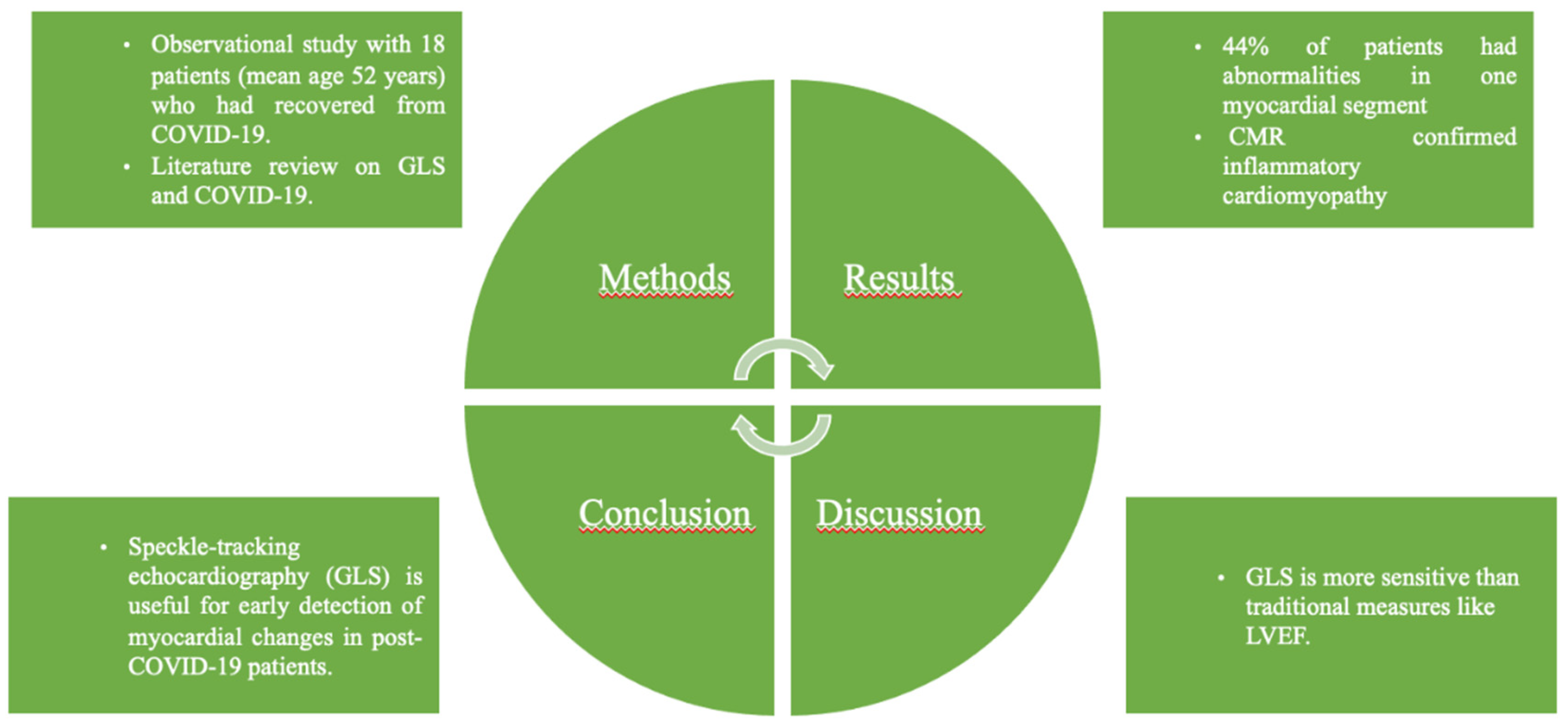
Central Figure: Central figure that summarizes data presented in the article.
Conclusion
The study presents significant insights into the cardiovascular implications of COVID-19, particularly through the use of advanced echocardiographic techniques such as Global Longitudinal Strain (GLS) analysis derived from Speckle Tracking Echocardiography (STE). Our findings underline the utility of GLS in detecting subtle myocardial dysfunction that may not be apparent through traditional measures such as Left Ventricular Ejection Fraction (LVEF). Despite a mean LVEF of 57.50 ± 9.98%, indicating preserved overall systolic function in the majority of patients, only 16.6% exhibited reduced LVEF. This underscores the limitation of relying solely on LVEF to assess myocardial health in COVID-19 patients. In contrast, GLS provided a more nuanced assessment, with an average value of -18.70 ± 3.54%. Notably, in 44% of the cases, GLS identified impairment in just one myocardial segment, specifically the inferolateral basal segment. This regional strain reduction highlights the ability of GLS to detect early and localized myocardial involvement, which may precede global systolic dysfunction detectable by LVEF. Moreover, Cardiac Magnetic Resonance (CMR) imaging revealed inflammatory cardiomyopathy, confirming the presence of myocardial inflammation. The concordance between CMR findings and reduced GLS further validates GLS as a sensitive marker for detecting inflammatory myocardial involvement in the context of COVID-19. These results emphasize the importance of incorporating GLS into routine echocardiographic evaluation for COVID-19 patients. This approach could be pivotal in improving prognostic outcomes and tailoring specific treatments for affected patients. As we continue to navigate the ongoing long term effects of this condition, the adoption of advanced echocardiographic techniques like GLS will be crucial in understanding and mitigating its cardiovascular impacts.
References
- Zhu N, Zhang D, Wang W, Li X, Yang B, Song J, Zhao X, Huang B, Shi W, Lu R, Niu P. A novel coronavirus from patients with pneumonia in China, 2019. New England journal of medicine. 2020 Feb 20;382(8):727-33.
- Cucinotta D, Vanelli M. WHO declares COVID-19 a pandemic. Acta bio medica: Atenei parmensis. 2020;91(1):157.
- Cheng VC, Lau SK, Woo PC, Yuen KY. Severe acute respiratory syndrome coronavirus as an agent of emerging and reemerging infection. Clinical microbiology reviews. 2007 Oct;20(4):660-94.
- Zaki AM, Van Boheemen S, Bestebroer TM, Osterhaus AD, Fouchier RA. Isolation of a novel coronavirus from a man with pneumonia in Saudi Arabia. New England Journal of Medicine. 2012 Nov 8;367(19):1814-20.
- Anthony SJ, Johnson CK, Greig DJ, Kramer S, Che X, Wells H, Hicks AL, Joly DO, Wolfe ND, Daszak P, Karesh W. Global patterns in coronavirus diversity. Virus evolution. 2017 Jan;3(1):vex012.
- Sharma A, Ahmad Farouk I, Lal SK. COVID-19: a review on the novel coronavirus disease evolution, transmission, detection, control and prevention. Viruses. 2021 Jan 29;13(2):202.
- Ochani R, Asad A, Yasmin F, Shaikh S, Khalid H, Batra S, Sohail MR, Mahmood SF, Ochani R, Hussham Arshad M, Kumar A. COVID-19 pandemic: from origins to outcomes. A comprehensive review of viral pathogenesis, clinical manifestations, diagnostic evaluation, and management. Infez Med. 2021 Mar 1;29(1):20-36.
- Sarker R, Roknuzzaman AS, Hossain MJ, Bhuiyan MA, Islam MR. The WHO declares COVID-19 is no longer a public health emergency of international concern: benefits, challenges, and necessary precautions to come back to normal life. International Journal of Surgery. 2023 Sep 1;109(9):2851-2.
- Parasher, A. COVID-19: Current understanding of its Pathophysiology, Clinical presentation and Treatment. Postgraduate medical journal. 2021 May;97(1147):312-20.
- Yuki K, Fujiogi M, Koutsogiannaki S. COVID-19 pathophysiology: A review. Clinical immunology. 2020 Jun 1;215:108427.
- Guo, T., Fan, Y., Chen, M., Wu, X., Zhang, L., He, T., ... & Wang, H. (2020). Cardiovascular Implications of Fatal Outcomes of Patients With Coronavirus Disease 2019 (COVID-19). JAMA Cardiology, 5(7), 811-818. [CrossRef]
- Zhou, F., Yu, T., Du, R., Fan, G., Liu, Y., Liu, Z., ... & Cao, B. (2020). Clinical course and risk factors for mortality of adult inpatients with COVID-19 in Wuhan, China: a retrospective cohort study. The Lancet, 395(10229), 1054-1062. [CrossRef]
- Bavishi, C., Maddox, T. M., & Messerli, F. H. (2020). Coronavirus Disease 2019 (COVID-19) Infection and Renin Angiotensin System Blockers. JAMA Cardiology, 5(7), 745-747. [CrossRef]
- Clerkin, K. J., Fried, J. A., Raikhelkar, J., Sayer, G., Griffin, J. M., Masoumi, A., ... & Schwartz, A. (2020). COVID-19 and Cardiovascular Disease. Circulation, 141(20), 1648-1655. [CrossRef]
- Dweck, M. R., Bularga, A., Hahn, R. T., Bing, R., Lee, K. K., Chapman, A. R., ... & Newby, D. E. (2020). Global evaluation of echocardiography in patients with COVID-19. European Heart Journal - Cardiovascular Imaging, 21(9), 949-958. [CrossRef]
- Szekely, Y., Lichter, Y., Taieb, P., Banai, A., Hochstadt, A., Merdler, I., ... & Topilsky, Y. (2020). Spectrum of Cardiac Manifestations in COVID-19. Circulation, 142(4), 342-353. [CrossRef]
- Fried, J. A., Ramasubbu, K., Bhatt, R., Topkara, V. K., Clerkin, K. J., Horn, E., ... & Leon, M. B. (2020). The Variety of Cardiovascular Presentations of COVID-19. Circulation, 141(23), 1930-1936. [CrossRef]
- Jensen, J., Ma, L. P., Fu, M., & Røsjø, H. (2020). Prognostic Value of Echocardiography and Speckle-Tracking Echocardiography in COVID-19. JACC: Cardiovascular Imaging, 13(11), 2472-2474. [CrossRef]
- Li, Y., Li, H., Zhu, S., Xie, Y., Wang, B., He, L., ... & Cao, B. (2020). Prognostic Value of Right Ventricular Longitudinal Strain in COVID-19 Patients. JACC: Cardiovascular Imaging, 13(11), 2287-2299. [CrossRef]
- Lang RM, Badano LP, Mor-Avi V, Afilalo J, Armstrong A, Ernande L, Flachskampf FA, Foster E, Goldstein SA, Kuznetsova T, Lancellotti P, Muraru D, Picard MH, Rietzschel ER, Rudski L, Spencer KT, Tsang W, Voigt JU. Recommendations for cardiac chamber quantification by echocardiography in adults: an update from the American Society of Echocardiography and the European Association of Cardiovascular Imaging. J Am Soc Echocardiogr. 2015 Jan;28(1):1-39.e14. [CrossRef] [PubMed]
- Lairez O, Blanchard V, Houard V, Vardon-Bounes F, Lemasle M, Cariou E, Lavie-Badie Y, Ruiz S, Cazalbou S, Delmas C, Georges B, Galinier M, Carrié D, Conil JM, Minville V. Cardiac imaging phenotype in patients with coronavirus disease 2019 (COVID-19): results of the cocarde study. Int J Cardiovasc Imaging. 2021 Feb;37(2):449-457. [CrossRef] [PubMed]
- Guan, W., Ni, Z., Hu, Y., Liang, W., Ou, C., He, J., & Zhong, N. (2020). Clinical characteristics of coronavirus disease 2019 in China. New England Journal of Medicine, 382(18), 1708-1720. [CrossRef]
- Zhu, N., Zhang, D., Wang, W., Li, X., Yang, B., Song, J., & Tan, W. (2020). A novel coronavirus from patients with pneumonia in China, 2019. New England Journal of Medicine, 382(8), 727-733. [CrossRef]
- Lewnard, J. A., Hong, V. X., Patel, M. M., Kahn, R., Lipsitch, M., Tartof, S. Y., & Jodar, L. (2022). Clinical outcomes among patients infected with Omicron (B.1.1.529) SARS-CoV-2 variant in southern California. Nature Medicine, 28(7), 1463-1472. [CrossRef]
- Sah P, Fitzpatrick MC, Zimmer CF, Abdollahi E, Juden-Kelly L, Moghadas SM, Singer BH, Galvani AP. Asymptomatic SARS-CoV-2 infection: A systematic review and meta-analysis. Proceedings of the National Academy of Sciences. 2021 Aug 24;118(34):e2109229118.
- Surveillances V. The epidemiological characteristics of an outbreak of 2019 novel coronavirus diseases (COVID-19)—China, 2020. China CDC weekly. 2020 Apr;2(8):113-22.
- Bielecka-Dąbrowa A, Cichocka-Radwan A, Lewek J, Pawliczak F, Maciejewski M, Banach M. Cardiac manifestations of COVID-19. Reviews in cardiovascular medicine. 2021;22(2).
- Hezzy Shmueli, Maulin Shah, Joseph E. Ebinger. Left ventricular global longitudinal strain in identifying subclinical myocardial dysfunction among patients hospitalized with COVID-19. [CrossRef]
- Valentina, O. Puntmann, M. Ludovica Carerj, Imke Wieters. Outcomes of Cardiovascular Magnetic Resonance Imaging in Patients Recently Recovered From Coronavirus Disease 2019 (COVID-19). [CrossRef]
- Jin Joo Park, Jun-Bean Park, Jae-Hyeong Park. Global Longitudinal Strain to Predict Mortality in Patients With Acute Heart Failure. [CrossRef]
- Rui Li, Hong Wang, Fei Ma. Widespread myocardial dysfunction in COVID-19 patients detected by myocardial strain imaging using 2-D speckle-tracking echocardiography. [CrossRef]
- Hosseiny, M., Kooraki, S., Gholamrezanezhad, A., Reddy, S., Myers, L., & Hajighasemi, F. (2020). Clinical characteristics of hospitalized patients with SARS-CoV-2 infection: A single arm meta-analysis. Journal of Medical Virology, 92(6), 612–617. [CrossRef]

Table 1.
Baseline characteristic and echocardiographic parameters for the patients enrolled in the study. LVEF left ventricle ejection fraction, LV GLS Left Ventricular Global Longitudinal Strain, ANT-SEPT LS Anterior-Septal Longitudinal Strain, ANT LS Anterior Longitudinal Strain, ANT-LAT LS Anterior-Lateral Longitudinal Strain, INF-LAT LS Inferior-Lateral Longitudinal Strain, INF LS Inferior Longitudinal Strain, INF-SEPT LS Inferior-Septal Longitudinal Strain.
| Patient No. | Age (years) | LVEF (%) | LV GLS (%) | ANT-SEPT LS (%) | ANT LS (%) | ANT-LAT LS (%) | INF-LAT LS (%) | INF LS (%) | INF-SEPT LS (%) |
|---|---|---|---|---|---|---|---|---|---|
| 1 | 63 | 70 | -21.2 | -23 | -15 | -12 | -22 | -16 | -15 |
| 2 | 50 | 54 | -17.2 | -16 | -22 | -13 | -17 | -14 | -11 |
| 3 | 55 | 62 | -19.6 | -16 | -21 | -15 | -16 | -18 | -15 |
| 4 | 69 | 67 | -22.5 | -20 | -22 | -19 | -26 | -11 | -8 |
| 5 | 53 | 69 | -21.1 | -16 | -20 | -23 | -22 | -14 | -17 |
| 6 | 46 | 55 | -18.3 | -7 | -15 | -14 | -17 | -22 | -7 |
| 7 | 50 | 55 | -19.2 | -17 | -19 | -23 | -19 | -14 | -12 |
| 8 | 48 | 29 | -8.9 | -8 | -12 | -7 | -7 | -7 | -6 |
| 9 | 59 | 42 | -14.9 | -15 | -14 | -14 | -13 | -12 | -12 |
| 10 | 44 | 59 | -17 | -8 | -16 | -19 | -12 | -11 | -13 |
| 11 | 47 | 55 | -16.9 | -15 | -10 | -12 | -18 | -14 | -10 |
| 12 | 49 | 59 | -22 | -17 | -28 | -20 | -24 | -19 | -12 |
| 13 | 58 | 60 | -18.5 | -17 | -18 | -18 | -20 | -12 | -18 |
| 14 | 45 | 60 | -18.8 | -13 | -20 | -15 | -16 | -16 | -12 |
| 15 | 71 | 63 | -21.1 | -17 | -22 | -19 | -18 | -20 | -13 |
| 16 | 44 | 61 | -24.8 | ||||||
| 17 | 47 | 49 | -15.2 | -14 | -20 | -15 | -15 | -13 | -14 |
| 18 | 28 | 66 | -19.4 |
Table 2.
Mean values for the echocardiographic and baseline parameters for the participants. LVEF left ventricular ejection fraction, LV GLS Left Ventricular Global Longitudinal Strain, ANT-SEPT LS Anterior-Septal Longitudinal Strain, ANT LS Anterior Longitudinal Strain, ANT-LAT LS Anterior-Lateral Longitudinal Strain, INF-LAT LS Inferior-Lateral Longitudinal Strain, INF LS Inferior Longitudinal Strain, INF-SEPT LS Inferior-Septal Longitudinal Strain.
| Variable | Values |
| N | 18 |
| Male (%) | 50 |
| Female (%) | 50 |
| Age (years) | 52 ± 10 |
| LVEF (%) | 57.5 ± 9.986 |
| ANT-SEPT LS (%) | 18.22± 4.84 |
| ANT LS (%) | 17.83± 4.86 |
| ANT-LAT LS (%) | 15.91±4,68 |
| INF LS (%) | 14.33±3.85 |
| INF-SEPT LS (%) | 11.5 ± 3.20 |
| INF-LAT (%) | 17.75± 5.16 |
| LV GLS (%) | 18.7 ± 3.54 |
Table 3.
Classification of COVID-19 patients according to symptom severity.
| Asymptomatic | COVID nucleic acid test positive. Without any clinical symptoms and signs and the chest imaging is normal. |
| Mild | Symptoms of acute upper respiratory tract infection (fever, fatigue, myalgia, cough, sore throat, runny nose, sneezing) or digestive symptoms (nausea, vomiting, abdominal pain, diarrhea). |
| Moderate | Pneumonia (frequent fever, cough) with no obvious hypoxemia, chest CT with lesions. |
| Severe | Pneumonia with hypoxemia (Peripheral oxygen saturation < 92%). |
| Critical | Acute respiratory distress syndrome (ARDS), may have shock, encephalopathy, myocardial injury, heart failure, coagulation dysfunction and acute kidney injury. |
Disclaimer/Publisher’s Note: The statements, opinions and data contained in all publications are solely those of the individual author(s) and contributor(s) and not of MDPI and/or the editor(s). MDPI and/or the editor(s) disclaim responsibility for any injury to people or property resulting from any ideas, methods, instructions or products referred to in the content. |
© 2024 by the authors. Licensee MDPI, Basel, Switzerland. This article is an open access article distributed under the terms and conditions of the Creative Commons Attribution (CC BY) license (http://creativecommons.org/licenses/by/4.0/).
Copyright: This open access article is published under a Creative Commons CC BY 4.0 license, which permit the free download, distribution, and reuse, provided that the author and preprint are cited in any reuse.


