Submitted:
08 July 2024
Posted:
09 July 2024
Read the latest preprint version here
Abstract
People with aphantasia exhibit the inability to voluntarily generate or form mental imagery in their minds. Since the term “aphantasia” was proposed by Zeman et al. in 2015 to describe this, it has gained increasing attention from psychiatrists, neuroscientists, and clinicians. Previous studies have mainly focused on definition, prevalence, and measurement, its impacts on individuals’ cognitive and emotional processing, and theoretical frameworks synthesizing existing findings, which have contributed greatly to our understanding of aphantasia. However, there are still some debates regarding the conclusions derived from existing research and the theories that were constructed from various sources of evidence. Building upon existing endeavors, the current systematic review emphasizes that future research is much needed to refine definition and diagnosis of aphantasia, strengthen empirical investigations at behavioral and neural levels, and more importantly, develop or update theories. These multiple lines of efforts could lead to a deeper understanding of aphantasia and further guide researchers in future research directions.
Keywords:
aphantasia
; visual imagery
; mental imagery
; vividness
; cognitive functioning
; mental health
1. Introduction
In 1880, Galton first systematically elucidated and studied individual differences in visual imagery, designing specific questions to investigate related phenomena. Visual imagery ability refers to the capacity to create mental images of objects or scenes that are not in front of their eyes [1], such as closing one’s eyes and imagining a brightly colored apple. Visual imagery has been shown to be closely associated with various cognitive functions and everyday activities. The active generation of visual imagery is an important cognitive ability in humans. However, the human capacity for visual imagery can be considered a spectrum with some individuals exhibiting exceptionally vivid visual imagery, which is referred to as hyperphantasia [2], but other individuals can only produce limited or even deficient visual imagery. In 2015, Zeman and colleagues introduced the term “aphantasia” to describe absent or markedly reduced mental imagery in individuals.
Presently, investigations into aphantasia concentrate on delineating its nature, assessing its frequency, devising measurement methods, examining its influence on cognitive and emotional functions, identifying related disorders, probing its neural underpinnings, and developing conceptual models. In particular, studies examining cognitive functions primarily explore how aphantasia impacts memory retention, non-temporal and prospective imagination, spatial visualization, the capacity for mental rotation, and the proficiency in visual searches (e.g., references [3,4]). Neuroimaging studies indicate a possible link between the lack of visual imagery and activity in the visual cortex (e.g., references [4,5,6,7]).
The purpose of this review is to encapsulate the current empirical research and theoretical perspectives pertaining to aphantasia. First, our discussion will concentrate on the explicit delineation, frequency, inheritability, and evaluation of aphantasia. Second, we will synthesize findings regarding aphantasia's influence on cognitive functions. Third, we will examine its potential associations with related disorders. Fourth, we will summarize existing evidence on the neural basis of aphantasia. Fifthly, we will critically appraise the principal theoretical models. Lastly, we will outline prospective avenues for future research on aphantasia, with the intention of steering scholars towards potential investigative trajectories.
2. Literature retrieval and screening
The present study undertook a systematic search of English-language articles published from 2015 to the current date, as depicted in Figure 1. The commencement year of 2015 was selected due to its significance as the year Zeman et al. first coined the term “aphantasia” to characterize the individual variance in lacking or significantly diminished visual imagery. Keyword searches employing “aphantasia” were executed across databases, including Scopus, PubMed, ProQuest, and Web of Science. All journal articles, master’s and doctoral theses, conference papers, and preprints were included in the search. As of April 6, 2024, an initial retrieval yielded 255 literature sources. Upon the exclusion of 113 duplicates, the remaining corpus comprised 142 literature sources. Further examination of the references cited in pertinent articles added 20 more literature sources to the pool. An in-depth review of titles, abstracts, and full texts from the 162 literature sources led to the exclusion of 10 inaccessible papers and 76 papers not centrally concerned with aphantasia, culminating in a curated collection of 76 pertinent literature sources. Drawing on these 76 papers, this review delivers an in-depth dissection, critique, and synthesis of the extant body of research concerning aphantasia.
It should be emphasized that pivotal research predating 2015, which did not employ the term “aphantasia”, was also taken into account where relevant. In this review, we used “aphantasia” to refer to an absence of mental imagery in any modality for consistency, although the majority of the reviewed studies focused on visual imagery deficits. The detailed content is summarized as follows (see Supplementary Materials for the details of each study included).
3. Definition, measurement, and prevalence of aphantasia
Aphantasia is conceptually defined as the inability to generate mental imagery. However, variations exist in the specific details of these conceptual definitions [8,9]. Blomkvist [8] reviewed various definitions of aphantasia [1,2,3,4,10,11,12,13,14,15,16] and identified three primary areas of disagreement: whether aphantasics exhibit impairments solely in visual imagery, whether a distinction is made between the absence of voluntary and involuntary imagery, and whether aphantasia primarily concerns the generation of imagery. Regarding the first aspect, aphantasia has also been associated with deficits in non-visual modalities (see Section 4.1). Some studies do not emphasize "visual imagery" when describing its absence, instead using the broader term "mental imagery" to refer to various modalities [4,13,16]. It is recommended to consistently use the term "aphantasia" and specify the modality when necessary, such as "auditory aphantasia" [17,18]. Concerning voluntariness, some researchers explicitly define aphantasia as the absence of voluntary visual imagery [1,3,4], while others do not specifically address this aspect [12,14]. Krempel and Monzel call for simply using a broader characterization of aphantasia in the field as there is no solid evidence of preserved involuntary imagery in aphantasics [19]. Regarding the third aspect, increasing evidence suggests that aphantasia is linked to other deficits in cognitive and emotional processes (see Section 4).
In measuring visual imagery deficits, most research utilizes scores from the Vividness of Visual Imagery Questionnaire (VVIQ) [20] as a primary identification and diagnostic tool [9]. The VVIQ comprises 16 questions and employs a 5-point Likert scale. Participants rate their visual imagery ability on a scale from "No imagery at all, you only 'know' that you are thinking of the object" to "Perfectly clear and as vivid as real seeing," to distinguish between individuals with visual imagery deficits, normal imagery abilities, and excessive visual imagery [20]. According to the criteria, a score of 16 indicates a complete absence of visual imagery, while a score of 17-32 indicates vague or dim imagery. According to the criteria, a score of 16 indicates a complete absence of visual imagery, while a score of 17-32 indicates vague or dim imagery. Instructions to imagine one of the images increase the likelihood of perceiving that image during the subsequent binocular rivalry task. Participants with visual imagery deficits exhibit minimal evidence of image-based binocular rivalry priming [21]. The VVIQ-2, an extended version of the VVIQ with 32 items [22], and a variant of the VVIQ [2] have also been utilized in the field. Recent research has found that pupillary responses to light can indicate visual imagery deficits, as affected individuals lack an imagery pupillary light response but still exhibit a perceptual pupillary light response [23]. Additionally, rhythmic visual flickering typically induces illusory percepts, and compared to control groups, individuals with visual imagery deficits are less likely to experience complex and vivid illusions [24,25]. These findings further highlight the physiological differences between individuals with visual imagery deficits and control groups, offering an objective measure for assessing visual imagery intensity.
The vividness of mental imagery can be viewed as a continuous characteristic forming a normal distribution curve, with aphantasia occupying the left tail and hyperphantasia occupying the right tail. Before the term "aphantasia" was introduced, the most cited study based on a single question about visual imagery indicated a prevalence rate of 2.1–2.7% for a total absence of visual imagery in the general population [26]. In the field, the VVIQ has been widely used to measure the vividness of visual imagery. Different operational definitions have led to varying prevalence rates. Zeman et al. found that the prevalence rate of aphantasia, where imagery is completely absent, was 0.7% (VVIQ = 16), and that of moderate aphantasia with a VVIQ score of 16–23 was 2.6% [10]. Using a VVIQ score of 16-32, covering "total absence" to "vague/dim" imagery ratings, Dance et al. found a prevalence rate of 3.9% across two separate samples [27]. When using a score of 16, the rate was only 0.8%. Later, Monzel et al. conducted meta-analyses and found a prevalence rate of 4.8% across all included studies and 3.5% in studies using the VVIQ [28]. Recently, Takahashi et al. found that in a large sample of Japanese individuals, the rate was 0.07% when using a score of 16 and 3.6% when using a score of 17-32 [29]. A study with a large sample of Brazilian university students reported a prevalence rate of 5.9% based on VVIQ scores of 16-32 [30]. However, another study noted a self-reported prevalence rate of 8.9% in the general adult population, but not all self-reported aphantasics showed low imagery scores on the VVIQ, resulting in a prevalence of only 1.5% [31]. Overall, the prevalence rates of aphantasia are inconsistent due to the use of different criteria across studies. If the strictest criterion is used, the rate would be extremely low. It is crucial to use consistent standards when discussing prevalence rates.
Studies have not found any gender differences in the prevalence of aphantasia [27,32]. Although previous studies found that women tend to score higher on object imagery measures compared to men [33,34], this difference is not statistically significant enough to conclude that men are more likely to be aphantasic. Age has also been considered a factor influencing aphantasia. However, there is no consensus on how age affects aphantasia [32,35]. Additionally, whether the prevalence of aphantasia varies across different professions remains controversial [2,35,36], despite Galton's initial suggestion that scientists might have weaker visual imagery abilities as a group [37]. This controversy could be explained by Blajenkova et al.'s opinion that scientists may not lack all types of visual imagery but may be specifically deficient in object imagery [38,39]. Similarly, visual artists tend to excel in spatial imagery rather than object imagery [38].
Aphantasia can be either congenital or acquired [40]. It is more common in individuals with a family history of lacking visual imagery, suggesting a potential genetic basis. However, a recent study found no significant genetic association, indicating the need for further research to explore the genetic underpinnings of aphantasia [41]. Acquired aphantasia can provide insights into the causes and mechanisms of the condition. This form of aphantasia can be triggered by various events, including craniocerebral injuries, emotional disorders, stroke, and postoperative complications [40,42,43]. A case study reported a woman who developed aphantasia after contracting COVID-19 [44]. Psychological and psychiatric factors should be carefully considered in the assessment of aphantasia [45,46], as mental illnesses such as depression and anxiety can significantly impact the vividness of visual imagery [45].
4. Aphantasia and Cognitive Processing
4.1. Visual and Non-visual Imagery Ability
Visual imagery ability is associated with other forms of imagery, such as olfactory and auditory imagery. In a pioneering study by Zeman et al., 10 out of 21 individuals with visual imagery deficits reported impairments in all forms of imagery, including auditory, olfactory, gustatory, tactile, motor, and bodily imagery [10]. Some aphantasics reported a lack of all forms of mental imagery, while others only have deficits in visual imagery [29]. In a large-scale study, 54.2% of aphantasics with visual deficits reported deficits in all types of imagery [2]. Similar findings have been confirmed in other studies, revealing that some aphantasics with visual deficits also reported deficits in other forms of mental imagery (34%, [47]; 26%, [3]). Takahashi and Gyoba reported a person with aphantasia who exhibited a complete deficit in various types of imagery (e.g., visual, olfactory, pain, tactile, gustatory, and somatic) in vividness and a substantial deficit in auditory imagery [48]. Furthermore, individuals with hyperphantasia, who exhibit exceptionally vivid visual imagery, tend to experience more vivid imagery in other sensory modalities as well [2]. There may be overlapping mechanisms underlying visual and auditory imagery, as many aphantasia participants report weak or absent auditory imagery [49,50], and individuals who lack auditory imagery, termed "anauralia," often show visual imagery deficits [49]. Using cluster analysis, Dawes et al. recently found that aphantasia is heterogeneous and has two subtypes: visual aphantasia, which selectively shows an absence of visual imagery, and multisensory aphantasia, which shows an inability to generate any sensory modality of mental imagery [51].
Overall, some individuals with aphantasia may experience deficits in all types of mental imagery. However, there is still a lack of large-scale studies to establish a reliable and consistent understanding, as well as plausible explanations of the neural mechanisms underlying these deficits. It is worth noting that all previous measurements of visual and other forms of mental imagery in research have relied on self-report questionnaires (e.g., VVIQ, Auditory Imagery Questionnaire, and The Questionnaire upon Mental Imagery). Although self-report questionnaires are convenient for large samples, there may be a meta-cognitive effect in the data. Participants may not actually lack genuine mental imagery but rather report deficits based on cognitive differences and misunderstandings of the questionnaire items. Although self-report questionnaires are convenient for large samples, there may be a meta-cognitive effect in the data. Participants may not actually lack genuine mental imagery but rather report deficits based on cognitive differences and misunderstandings of the questionnaire items. Alternatively, when using self-report questionnaires, participants should be provided with thorough explanations and clarifications of the items to minimize misunderstandings.
4.2. Aphantasia and Memory
Existing research on the impact of visual imagery impairment on memory has mainly focused on working memory, episodic memory, autobiographical memory, and object and spatial memory. Overall, aphantasics do not exhibit deficits in simple working memory tasks [52,53,54,55]. However, it has been reported that aphantasics perform worse than control groups in tasks requiring fine-grained visual working memory, which are cognitively more demanding and require participants to remember smaller differences [52,53,55]. Jacobs et al. reported an individual with aphantasia who performed worse than controls only on the most difficult visual working memory trials requiring a high level of precision [13]. The difficulty level appears to be a crucial factor in visual working memory tasks. In terms of episodic memory, individuals with aphantasia demonstrated significantly lower performance than control groups [3,56,57]. Similarly, individuals with aphantasia have been reported to experience difficulties in autobiographical memory [2,3,10,14,52,57,58,59]. Regarding spatial memory, which involves the processing of an object's location information, two recent studies did not find any deficits in aphantasics [1,3]. For object memory, aphantasics showed poorer performance in a drawing task that requires object processing [1]. Siena and Simon used both subjective and objective measures of object and spatial memory and found that aphantasia participants showed no objective deficits in memory performance, indicating that some aphantasics might have a deficit in the awareness of mental imagery [60].
It should be noted that new measurements of memory performance have been employed in existing research, deviating from traditional scales and experimental tasks. For instance, drawing and wearable cameras have been used to complement the assessment of memory performance in participants [1,61]. In the case of drawing, aphantasics and control groups were instructed to draw scene images from real-world memory. It was found that aphantasics remembered fewer objects, used fewer colors, and relied more on verbal scaffolding to compensate for the lack of visual imagery [1].
Overall, memory is a relatively well-researched aspect in the field of aphantasia. Many studies have employed questionnaires and experiments to measure participants' memory performance. In visual working memory, aphantasics exhibit similar performance to control groups, which could be explained by their potential use of semantic encoding or other representational strategies to aid them in completing the task [4]. However, some studies indicate that aphantasics perform worse in these visual working memory tasks [13]. The inconsistency of these findings may be attributed to task characteristics, measurement methods, and individual differences among participants. For instance, the heterogeneous outcome included only one individual with aphantasia, suggesting that the observed differences in working memory could be influenced by sample size or the unique characteristics of the case study [13]. As for autobiographical memory, all results seem to converge on the conclusion that individuals with aphantasia have poorer autobiographical memory. Differences in object memory and spatial memory performance between the aphantasics group and the control group may be related to their abilities in object imagery and spatial imagery. These differences may be explained through the neural mechanisms of the ventral and dorsal pathways of visual processing, which will be discussed more comprehensively later.
4.3. Aphantasia and Object and Spatial Imagery
Object imagery refers to visualizing the appearance of objects and scenes, including their shape, color, and brightness, while spatial imagery involves visualizing the spatial relationships, movements of objects and their components, and spatial transformations [62]. Existing research on the performance of individuals with aphantasia in object and spatial imagery abilities has not reached a consensus and can be categorized into three types of studies. The first type of study indicates that individuals with aphantasia score lower in object imagery abilities but show no significant differences from control groups in spatial imagery performance [1,3,50,57]. The second type of study shows that aphantasics score significantly lower in object imagery than control groups but perform better in spatial imagery tests [11,52]. In the third category, individuals with aphantasia demonstrate poorer performance in both object and spatial imagery abilities [63,64]. Based on these findings and a review of the literature, researchers have proposed two types of visual imagery impairments: object aphantasia and spatial aphantasia [39,65].
The finding that object imagery is impaired while spatial imagery remains unaffected can also be explained by the functional division of visual pathways. Aphantasics' ventral pathway for object processing may be impaired, while the dorsal pathway for spatial processing remains intact [4,21] (see below). To better explain spatial aphantasia, researchers have proposed that this type of aphantasia may be related to functional changes in the visual dorsal stream projecting to the frontal lobe [65]. However, this explanation still lacks further support from neuroimaging studies.
4.4. Aphantasia and Atemporal and Future Imagination
Atemporal and future imagination are related to voluntarily imagining general events and future events, respectively. Autobiographical interviews on atemporal and future imagination show that aphantasia participants have poorer abilities in these areas, with fewer details in their imagined scenarios [14,57]. Evidence suggests that atemporal and future imagination abilities are related to autobiographical memory [14]. This may explain why aphantasics show poorer abilities in atemporal and future imagination, as their autobiographical memory abilities are also lower compared to control groups.
4.5. Aphantasia and Mental Rotation Task Performance
The mental rotation task has been widely used to investigate individuals' spatial abilities [66]. It has been reported that, compared to control groups, individuals with aphantasia showed equal [14,55,67] or even higher accuracy in mental rotation tasks [52,63,64]. However, aphantasics also required longer response times in these tasks [55,63].
Pounder and colleagues (2022) suggested that aphantasics may employ non-visual processes in these tasks. For example, the patient in Furman et al.'s study self-reported using a spatial and kinesthetic strategy instead of low-level visual object imagery in a mental rotation task [11,64]. Specifically, he used a first-person path-following strategy (i.e., spatial strategy) containing kinesthetic features, using his body as a reference. This hypothesis is supported by the performance of congenitally or early blind individuals in mental rotation tasks, as they did not show differences from control groups in completing complex spatial tasks, indicating that mental rotation tasks can be completed independently of visual processes [68]. Additionally, the mental rotation task is considered to rely on an individual's spatial imagery ability, and many individuals with aphantasia report having similar or even superior spatial imagery abilities compared to control groups [11,57].
4.6. Aphantasia and Visual Searching Ability
Visual search is a commonly used skill in everyday life that helps us find the things we need, even if the desired target is within our current field of vision. When performing visual search tasks, aphantasics exhibit significantly slower speeds compared to control groups [69,70]. This difference could be explained by the varying involvement of visual imagery in top-down strategies between the two groups [70,71]. However, in Moriya's task, which examines participants' attentional guidance in visual search tasks [72] (where participants are first asked to imagine a color primed by a color word such as "blue" and then to indicate whether one of the two colored squares presented on the left and right sides has an opening at the top or bottom), the priming process appears to be similar between the aphantasia group and the control group [69]. This can be explained by the characteristics of the task, where the instructions in Moriya's task are overly complex, resulting in a lack of visual priming and only non-visual priming [69]. These findings suggest that visual imagery could influence the way people perceive the world. Future research would benefit from designing materials that are more ecologically valid, such as incorporating environmental cues into the experimental design.
5. Aphantasia and Disorders and Emotional Processing
5.1. Emotion
Due to absent or reduced visual imagery, aphantasics experience reduced emotional engagement and less sympathy for characters in stories [73] and exhibit lower levels of fear response when reading scary materials [74]. Similar results were found in the lower empathy scores of individuals with aphantasia when receiving verbal materials [75]. Additionally, aphantasics seem to experience lower emotional intensity when listening to music [76]. These findings align with research by Zeman and his colleagues, suggesting that emotions in individuals with aphantasia are unlikely to be influenced by their imagery abilities [2]. This may be because vivid imagery makes thoughts more realistic, triggering stronger responses in the brain's emotional circuitry and thereby amplifying emotions [77]. Future research should continue to explore different types of emotions and incorporate neuroimaging methods to validate previous findings.
5.2. Mental Health
Although aphantasia may impact individuals' cognition and emotional processing and potentially cause some personal distress, it is not currently included in any common clinical diagnostic systems. Furthermore, the impact of aphantasia on individuals' daily lives and personal distress is relatively minimal and not sufficient to classify it as a psychological disorder [28]. However, research has found associations between aphantasia and certain conditions or diseases.
Existing research has focused on the impact of aphantasia on mental health, including depression, anxiety, distress, and well-being. Research has revealed that the aphantasia group showed no significant differences compared to the control group in terms of depression, anxiety, and state-trait anxiety [14,77]. However, Monzel and colleagues found that 34.7% of aphantasia participants reported distress caused by the lack of visual imagery [28]. Nevertheless, the researchers suggested that these negative emotions were relatively weak and did not affect individuals' daily lives [28].
5.3. Post-Traumatic Stress Disorder (PTSD)
Previous research has established a link between mental imagery and PTSD, a condition characterized by the re-experiencing of traumatic events through unwanted and recurring intrusive memories and nightmares [78]. Individuals with aphantasia were found to experience fewer intrusions and exhibit less avoidance behavior following trauma [3], both of which are predictors of PTSD. Additionally, the aforementioned lack of visual imagery and its association with reduced emotional intensity may also contribute to their lower likelihood of developing PTSD. However, it is important to note that researchers emphasize the limited protective effect of visual imagery deficits when individuals with such deficits encounter stressful events in their daily lives [3].
5.4. Autism
Individuals with aphantasia frequently score higher on the Autism Quotient Questionnaire (AQ) and are more likely to be classified within the autism spectrum [14,47,79]. This association may be attributed to the relationship between imagery and mentalizing. Compared to control groups, individuals with aphantasia exhibit slightly reduced atemporal and future imagination, as well as episodic memory [3,14,57], which are also characteristics of Autism Spectrum Disorder [80]. Additionally, limited or absent visual imagery can impair the theory of mind abilities of individuals with aphantasia, leading to difficulties in social skills [79]. These factors may explain why individuals with aphantasia tend to score higher on the AQ, indicating a potential link between visual imagery deficits and Autism Spectrum Disorder.
5.5. Prosopagnosia
Aphantasia has been associated with reduced facial recognition ability [29]. In a recent study, 5.9% of participants in the spatial aphantasia group were identified as having prosopagnosia [65], and face recognition difficulties were found to be more common among individuals with aphantasia [14,81]. Some individuals with aphantasia also reported difficulties with facial recognition [40]. Additionally, individuals with aphantasia have been associated with lower confidence in tasks involving facial perception [82]. However, aphantasia does not significantly impact accuracy in tasks involving the construction of facial composites, as individuals with aphantasia can still create facial composites from memory similar to control groups [81]. Nevertheless, some researchers have argued that the test scores of individuals with aphantasia are not sufficient to diagnose them with prosopagnosia [64]. Overall, individuals with aphantasia tend to exhibit poorer face recognition abilities. However, this may be influenced by the scales used in experiments, as these scales may elicit a metacognitive effect [14]. In other words, individuals with aphantasia may have reduced confidence in their facial recognition abilities due to their lack of vivid facial imagery, which can manifest as relatively lower scores on self-report measures.
6. Neural Basis of Aphantasia
Although numerous studies have focused on the neural basis of mental imagery [4,5,7,83], there are limited studies utilizing modern neuroimaging techniques such as magnetic resonance imaging (MRI) and electroencephalography (EEG) to explore the neural basis of visual imagery deficits. Using resting-state and task-based MRI, Milton et al. recently compared the brain activities of three groups: individuals with aphantasia, typical controls, and individuals with hyperphantasia [14]. The results showed that individuals with hyperphantasia exhibit stronger connectivity between the prefrontal regions and the visual-occipital network compared to the aphantasia group. When comparing the visualization and perception of famous faces and places, individuals with hyperphantasia and the control group exhibited greater frontal and parietal activation compared to the aphantasia group [14]. Another functional MRI study showed that the aphantasia group exhibited decreased activation in the hippocampus and increased activation in the visual-perceptual cortex during an autobiographical memory task [84]. However, the control group displayed strong negative task-based functional connectivity between the hippocampus and the visual cortex during the task, and the resting-state functional connectivity between these two areas could predict visualization skills [84]. Furthermore, using transcranial magnetic stimulation to induce changes in brain activity, Dupont et al. found that there was no increase in the amplitude of motor-evoked potentials triggered in the target right index finger in the aphantasia group, indicating a lack of corticospinal excitability in individuals with aphantasia during motor simulation [85].
Case studies are also a crucial source of neural evidence. Lesion studies have found that patients retain intact visual imagery after brain lesions restricted to the occipital cortex [86,87,88,89], suggesting that early visual areas are not involved in visual imagery. Moreover, patients with damage to the anterior part of the temporal lobe, particularly in the left hemisphere, often report an inability to generate visual images [90,91]. Zeman et al. reported a patient who exhibited reduced activation in the occipitotemporal regions during an imagery task [92]. In a recent study, a patient with an absence of visual imagery ability exhibited selective lesions in a specific area of the left fusiform gyrus and a portion of the right lingual gyrus, demonstrating a causal role of the left fusiform gyrus in visual imagery [40]. Therefore, the fusiform region “might act as a neural interface between sensory information coming from the occipital cortex and semantic processing in the anterior temporal lobe” during visual perception, and “could endow semantic memories with visual information during visual imagery” [89] (p. 517). An EEG study with source reconstruction reported that during a visual imagery task, an aphantasics begins the evoking phase from the left temporal area while lacking activation in the occipital and parietal lobes, which are associated with visual image vividness [64].
Due to the limited evidence in these studies, it is challenging to draw a consistent picture of aberrant brain activity in aphantasia. However, we can still draw inspiration from neuroimaging studies based on individual differences in mental imagery. Spagna et al. recently conducted a meta-analysis of fMRI studies on visual imagery and found that visual imagery recruits several frontoparietal areas and a specialized area in the left fusiform gyrus [7]. This specialized area was labeled the fusiform imagery node (FIN), referring to a brain network node specifically responsible for voluntary visual mental imagery [7]. Liu et al. found that imagery tasks activated the left frontoparietal regions, the FIN, and areas in the ventral temporal cortex, which were similarly activated in both the aphantasia and control groups [93]. However, the connectivity between the FIN and frontoparietal regions is reduced in individuals with aphantasia [93]. Together, these brain lesion cases and fMRI studies demonstrate that the fusiform gyrus is a core area for visual imagery, and damage or impairment to it could lead to deficits in visual imagery. Additionally, abnormalities in the fusiform gyrus have been associated with prosopagnosia, which may explain the facial recognition deficits observed in individuals with visual imagery disorders [94].
A related debate concerns the involvement of the early visual cortex in (impaired) visual imagery [89,95]. Using stimulation, Kosslyn et al. found that the early visual cortex is causally involved in visual imagery [96]. Bergmann et al. documented that a smaller V1 size is associated with stronger but less precise imagery, indicating an anatomical basis [97]. Keogh et al. [98] suggested a causal relationship between cortical excitability in the early visual cortex and the intensity of visual imagery. However, Meng et al. suggested that an imagery-related representation exists in the primary visual cortex of individuals with aphantasia, despite the absence of visual imagery, though the representation contains less or transformed sensory information [99]. Bartolomeo et al. argued that the left fusiform gyrus plays a crucial role in visual imagery, rather than the early visual cortex, and that the involvement of the early visual cortex [89,96] might be modulated by downstream areas. Dijkstra recently reviewed existing evidence and proposed that “imagery can recruit the early visual cortex, but that does not mean it always does” [83].
7. Theory Development
Individuals with imagery deficits can still possess imagination capabilities similar to those without such deficits, such as creating novels and movies. Based on this, Arcangeli attempts to distinguish between mental imagery and sensory imagination [100]. Mental imagery can be considered a type of mental content (e.g., the appearance of an apple). In contrast, sensory imagination is a special psychological attitude that involves the recreation of perceptual experiences. According to this theory, most individuals previously defined as having aphantasia in past research may actually have a deficiency in sensory imagination rather than mental imagery, which could explain why individuals with visual imagery deficits can still perform imagination tasks.
Nanay explains aphantasia from the perspective of both conscious and unconscious visual imagery [16]. Previous research has found that individuals with aphantasia do not exhibit the imagery priming effect in binocular rivalry tasks, yet some participants can generate vivid dreams and perform visual imagery tasks similar to control groups. To explain this phenomenon, Nanay distinguishes between conscious and unconscious visual imagery abilities, both of which can be generated voluntarily or involuntarily [16]. One type of aphantasia involves a fundamental lack of visual imagery, while another type involves the ability to generate visual imagery without conscious awareness [69]. However, this theory is challenged by Blomkvist, who argues that it does not fully explain the issues of episodic memory or the imagination of atemporal and future events in visual imagery deficits, as it does not provide a link between mental imagery and the episodic processes in episodic memory and episodic imagination [8]. Additionally, research suggests that imagery tasks can be accomplished through cognitive strategies that do not rely on imagery [101,102].
To provide a better explanation for aphantasia, Blomkvist proposed enhancements to the Constructive Episodic Simulation Hypothesis (CESH) model. The original CESH posits that memory and imagination involve three key processes: semantic retrieval, episodic retrieval, and (re)combination (Perrin & Michaelian, 2017) [8]. Blomkvist expanded this model by adding three new components: “memory indices, differing episodic retrieval mechanisms for all kinds of sensory information, and spatial retrieval mechanisms” [8]. These additions aim to offer a new theoretical explanation for visual imagery deficits. According to this updated theory, the mechanism of the episodic system is deficient, resulting in the loss of visual imagery. However, this theory still lacks empirical research data for further support.
The theoretical framework of the ventral and dorsal pathways in the visual system has been proposed for many years [103,104,105], although there are ongoing debates about this framework [106,107]. Pearson proposed that the ventral (or "what") pathway is associated with object information, while the dorsal (or "where") pathway is associated with location and spatial features [4]. The two aspects of visual imagery, object imagery and spatial imagery, are also likely generated through the ventral and dorsal pathways [39]. Damage to the ventral pathway could impair individuals' ability to visualize objects' appearance, while damage to the dorsal pathway is associated with a disrupted ability in spatial imagery. These processes can be dissociated in aphantasia, which may explain why individuals with visual imagery deficits perform similarly to, or even better than, control groups in spatial imagery tasks [4,21]. Bergmann and Ortiz-Tudela also suggested that visual feedback pathways used during episodic and schematic memory retrieval may differ depending on the two visual processing streams: episodic memory retrieval involves both the "what" and "where" streams, while schematic memory retrieval primarily involves the "where" stream [108]. In other words, aphantasia may be associated with differences in the "what" stream.
Inspired by neural models of mental imagery [4,5] and existing sources of neural evidence, Zeman tried to depict candidate neural mechanisms of extreme imageries, aphantasia, and hyperphantasia [6]. In the brain mapping model, there are five functional clusters, each involved in a unique role in mental imagery: the frontal cortex for initiating imagery generation; the parietal cortex for interacting with the frontal cortex to generate imagery, mediating attentional and spatial aspects; the temporal cortex, including limbic structures, for enabling access to semantic and episodic memories that determine what to visualize; and higher-order visual areas (e.g., the FIN) for visualizing imagery. The extent to which activity in the early visual cortex (e.g., the V1) is required for imagery is still debated. Moreover, the connections between these clusters are also essential for imagery generation. From this mapping picture, Zeman proposes five candidates showing neural atypicality in extreme imageries, variations in the strengths of the top-down feedback connection between higher-order regions (the frontal cortex) serving as cognitive control and modality-specific areas (e.g., the visual cortex) activated by sensory imagery, variations in the structure and function of the frontal cortex involved in generating imageries, variations at the level of higher-order visual areas including the FIN proposed by Spagna et al., variation in the anatomical structure of the early visual cortex including the V1, variation in the saliency involves the parietal areas of the frontoparietal control system [6,7]. In this proposal, Zeman provides future directions for researchers in the field. A neural model constructed and refined from accumulated evidence would contribute to a better understanding of aphantasia [6].
8. Summary and Future Directions
This review primarily examines the definition, prevalence, and measurement methods of aphantasia, as well as its impacts on individual cognitive and emotional processing, associated disorders, neural basis, and theoretical development. Research on aphantasia is continuously progressing, and its descriptions are becoming increasingly clear. However, several debated areas remain, and the study of its impacts on individuals and underlying mechanisms faces numerous challenges. Future investigations into aphantasia should focus on the following aspects.
8.1. Clarify Definition and Diagnosis
It is evident that current research on aphantasia is still in its early stages. One of the primary concerns is the definition of aphantasia. Clear definitions serve as the foundation for advancing further research and facilitating effective communication among researchers. The existing literature presents variations in the definitions of aphantasia [9]. For example, some definitions do not explicitly distinguish between voluntary and involuntary imagery (see a review by Krempel & Monzel, 2024 [19]). The cut scores for diagnosing aphantasia varied across studies (see section 3). The inconsistent use of arbitrary cut scores is considered a hindrance to cross-study comparisons and communication among researchers. Moreover, the use of cut scores may discourage efforts to examine individual differences in imagery. Additionally, more effective tools for both non-clinical and clinical purposes should be developed [109], rather than relying predominantly on the VVIQ or its variants.
The terminology in the field has recently been debated [17,18,49]. Other terms have been proposed to refer to the absence of different modalities of imagery. For auditory imagery, Hinwar and Lambert introduced the term “anauralia” to refer to the absence of auditory imagery and “hyperauralia” to refer to the experience of extremely vivid auditory imagery [49]. Dance et al. proposed the term “dysikonesia” to refer to multisensory or global aphantasia [47]. In this context, Monzel et al. argue that these new terms complicate the field, making communication less effective for researchers and the public, and therefore advocate for the use of the simple term “aphantasia,” which is widely known [17,18]. When referring to the absence of a specific modality of imagery, it is straightforward to use modality-specific terms (e.g., “visual/auditory/multisensory aphantasia”).
These divergences across studies create difficulties in the classification of participants and scientific communication among researchers and the public, thereby hindering progress toward a better understanding of aphantasia. Researchers in the field should reach a consensus on these issues to facilitate further development.
8.2. Strengthen Behavioral Research
Aphantasia research is still in its early stages, and there is limited literature in several research directions. Future studies should build upon existing efforts to systematically examine this phenomenon to better understand aphantasia. At the behavioral level, more research is urgently needed within and beyond existing research directions. First of all, together with a clear and consistent definition of aphantasia, researchers should focus on the examination of the nature of aphantasia including but not limited to investigating effects of demographic variables (e.g., age and gender) and their interactions in large datasets, developing new assessment or diagnosis tools with high reliability and validity, elucidating perceptual and cognitive processes required by tasks used for eliciting mental imagery, and determining the comorbidity rate of two or more subtypes of aphantasia in large samples. Second, researchers should investigate how aphantasia is associated with other psychological aspects (e.g., conscious experience [16,110]), the influences of aphantasia on emotional processing and disorders (see Section 4), and the possible mechanisms underlying these relationships (e.g., [111]). Third, research on hyperphantasia, the opposite extreme characterized by vivid mental imagery, can also contribute to a better understanding of aphantasia. Viewing mental imagery ability as a continuum could be more realistic and scientifically useful for understanding the absence or reduction of imagery in individuals with aphantasia. Fourth, researchers should use these different sources of evidence as a foundation to propose or create intervention programs for individuals with aphantasia. The effectiveness of these interventions can also help researchers better understand the nature of this phenomenon, fostering further advances.
8.3. Discover Neural Basis
Unraveling the neural bases of aphantasia has recently become a prominent research topic. Neuroimaging evidence has been instrumental in understanding the manifestations of aphantasia and in constructing neural models. Employing neuroimaging methods, future research can explore various variables to gather neural evidence. Specifically, researchers can utilize the high spatial-resolution of MRI techniques to investigate regional activation and inter-regional connections (structural and functional connectivity) involved in the absence or reduction of mental imagery. Previous functional MRI studies have identified areas involved in mental imagery, based on which neural models have been proposed (e.g., [12,97,98,112]; see a review by Pearson [4]). Although some studies on aphantasia have been conducted [14,92,93], it is difficult to accumulate convergent evidence for localizing aberrant brain regions and connections. Using EEG and magnetoencephalography (MEG) techniques with high temporal resolution [113,114], researchers can examine the neural dynamics of processes involving mental imagery in individuals with and without aphantasia (e.g., [64]). For example, Xie et al. found shared alpha-band neural representations in visual imagery and perception [115]. Combining spatial and temporal neural evidence across different studies, or even using fusion techniques (e.g., EEG-MRI fusion, [116]), can contribute to a better understanding of aphantasia by incorporating different types of information into a single picture, further facilitating the construction of neural models. Recent technical advances in multivariate pattern analysis (e.g., [117,118]) may also be powerful in understanding the neural correlates of aphantasia, as demonstrated in previous studies of mental imagery [99,115]. Non-invasive brain stimulation techniques [121] can be powerful tools for discovering causal evidence of the involvement of brain areas in mental imagery. Together, the convergence of behavioral and neural evidence not only helps elucidate various aspects of aphantasia but is also useful in building behavioral and neural models that explain this phenomenon [6]. In addition, evidence from other sources of investigation should be encouraged. It has been reported that aphantasia has a genetic basis [2] and that dopamine plays an important role in generating mental imagery [121].
8.4. Construct and Refine Theories
Good theories help humans systematically and scientifically understand their minds [122,123]. Although aphantasia has garnered increasing attention over the past decade, empirical studies examining this phenomenon remain limited. The field is still in its nascent stages. A lack of theories for synthesizing existing literature is one of the field's significant characteristics. Although some accounts and models have been proposed to interpret behavioral findings [8,16,100], they have only focused on certain aspects. Neural models for synthesizing neural evidence (e.g., [6]) are largely unknown or require further examination. One of the main reasons is that both behavioral and neural empirical studies are very limited in number and diversity (see above), resulting in insufficient evidence to be integrated into a theory or model.
In addition to proposing new theories or models, we advocate for efforts to use existing theories or models of mental imagery [4,5,8] as a foundation to theoretically guide future research on aphantasia. Modifications to these models or theories will be made as more evidence is collected. We believe these modifications will either refine existing theories or help foster new ones.
8.5. Encourage Direct and Conceptual Replications
In recent years, concerns about replication in psychology have increased (e.g., [124,125]). Replicability is a crucial indicator of the reliability and stability of research findings [126]. Conducting replicable experiments to demonstrate the reliability of findings lays a solid foundation for further investigations. There are two types of replications, each with different requirements. Conceptual replication deliberately modifies key elements of the original study to test the robustness of the phenomenon or the generalizability of the theoretical claims, while direct replication involves recreating the original experiment, which has been questioned for its feasibility [127]. The inconsistency in research findings on aphantasia necessitates considering replicability in existing studies, requiring numerous replication studies to confirm the reliability and stability of results. For example, there are limited studies on the relationship between aphantasia and atemporal and future imagination, as well as visual search. Although consistent conclusions have been reached, different experimental designs are still needed to validate the accuracy and replicability of the results through conceptual replications. Moreover, researchers have explored the relationships between attention [15], sensory sensitivity [47], and aphantasia, but these are isolated studies lacking cross-validation from other research, indicating the necessity of conducting both direct and conceptual replication studies.
9. Conclusions
This article provides a comprehensive review of aphantasia, encompassing its definition, prevalence, measurement methods, empirical research, and relevant theories. The absence of visual imagery can have implications for an individual’s cognitive and emotional processing and is also associated with certain psychological disorders. Research on aphantasia is beneficial for expanding researchers' understanding of cognitive and emotional processes and their relationships with other disorders. However, consensus regarding the theoretical framework has not yet been achieved. Many theories still lack sufficient empirical support, leaving significant theoretical gaps and research opportunities. The study of visual imagery deficits is continually growing, although areas of debate remain.
Supplementary Materials
The following supporting information can be downloaded at the website of this paper posted on Preprints.org.
Author Contributions
F.J.; study design, investigation, methodology, software, visualization, writing - original draft, writing - review and editing; H.S. investigation, writing - review and editing. Y.L.; study design, funding acquisition, investigation, methodology, resources, supervision, writing - original draft, writing - review and editing. All authors have read and agreed to the published version of the manuscript.
Funding
This research was funded by BNU-HKBU United International College Re- search Grants (R72021207, R202102, R202011,) and in part by the Guangdong Provincial Key Laboratory IRADS (2022B1212010006, R0400001-22).
Institutional Review Board Statement
Not applicable.
Informed Consent Statement
Not applicable.
Data Availability Statement
Not applicable.
Conflicts of Interest
The authors declare no conflicts of interest.
References
- *Bainbridge, W.A.; Pounder, Z.; Eardley, A.F.; Baker, C.I. Quantifying aphantasia through drawing: Those without visual imagery show deficits in object but not spatial memory. Cortex 2021, 135, 159–172. [Google Scholar] [CrossRef] [PubMed]
- *Zeman, A.; Milton, F.; Della Sala, S.; Dewar, M.; Frayling, T.; Gaddum, J.; Hattersley, A.; Heuerman-Williamson, B.; Jones, K.; MacKisack, M.; Winlove, C. Phantasia–The psychological significance of lifelong visual imagery vividness extremes. Cortex 2020, 130, 426–440. [Google Scholar] [PubMed]
- *Dawes, A.J.; Keogh, R.; Andrillon, T.; Pearson, J. A cognitive profile of multi-sensory imagery, memory and dreaming in aphantasia. Scientific Reports 2020, 10, 10022. [Google Scholar] [PubMed]
- *Pearson, J. The human imagination: The cognitive neuroscience of visual mental imagery. Nature Reviews Neuroscience 2019, 20, 624–634. [Google Scholar] [CrossRef] [PubMed]
- Dijkstra, N.; Bosch, S.E.; van Gerven, M.A. Shared neural mechanisms of visual perception and imagery. Trends in Cognitive Sciences 2019, 23, 423–434. [Google Scholar] [CrossRef] [PubMed]
- Zeman, A. Aphantasia and hyperphantasia: Exploring imagery vividness extremes. Trends in Cognitive Sciences 2024. [Google Scholar] [CrossRef] [PubMed]
- *Spagna, A.; Hajhajate, D.; Liu, J.; Bartolomeo, P. Visual mental imagery engages the left fusiform gyrus, but not the early visual cortex: A meta-analysis of neuroimaging evidence. Neuroscience & Biobehavioral Reviews 2021, 122, 201–217. [Google Scholar]
- *Blomkvist, A. Aphantasia: In search of a theory. Mind and Language 2022, 38, 866–888. [Google Scholar] [CrossRef]
- *Blomkvist, A.; Marks, D.F. Defining and 'diagnosing' aphantasia: Condition or individual difference? Cortex 2023, 169, 220–234. [Google Scholar] [CrossRef]
- *Zeman, A.; Dewar, M.; Della Sala, S. Lives without imagery - congenital aphantasia. Cortex 2015, 73, 378–380. [Google Scholar]
- *Keogh, R.; Pearson, J. The blind mind: No sensory visual imagery in aphantasia. Cortex 2018, 105, 53–60. [Google Scholar] [PubMed]
- *Fulford, J.; Milton, F.; Salas, D.; Smith, A.; Simler, A.; Winlove, C.; Zeman, A. The neural correlates of visual imagery vividness – an fMRI study and literature review. Cortex 2018, 105, 26–40. [Google Scholar] [CrossRef] [PubMed]
- *Jacobs, C.; Schwarzkopf, D.S.; Silvanto, J. Visual working memory performance in aphantasia. Cortex 2018, 105, 61–73. [Google Scholar] [CrossRef] [PubMed]
- Milton, F.; Fulford, J.; Dance, C.; Gaddum, J.; Heuerman-Williamson, B.; Jones, K.; ... & Zeman, A. Behavioral and neural signatures of visual imagery vividness extremes: Aphantasia versus hyperphantasia. Cerebral Cortex Communications 2021, 2, tgab035. [Google Scholar] [CrossRef] [PubMed]
- Keogh, R.; Pearson, J. Attention driven phantom vision: Measuring the sensory strength of attentional templates and their relation to visual mental imagery and aphantasia: Measuring attentional templates. Philosophical Transactions of the Royal Society B: Biological Sciences 2021, 376. [Google Scholar] [CrossRef] [PubMed]
- Nanay, B. Unconscious mental imagery: Unconscious mental imagery. Philosophical Transactions of the Royal Society B: Biological Sciences 2021, 376. [Google Scholar] [CrossRef] [PubMed]
- Monzel, M.; Mitchell, D.; Macpherson, F.; Pearson, J.; Zeman, A. Aphantasia, dysikonesia, anauralia: Call for a single term for the lack of mental imagery - Commentary on dance et al. (2021) and Hinwar and lambert (2021). Cortex 2022, 150, 149–152. [Google Scholar] [CrossRef] [PubMed]
- Monzel, M.; Mitchell, D.; Macpherson, F.; Pearson, J.; Zeman, A. Proposal for a consistent definition of aphantasia and hyperphantasia: A response to Lambert and Sibley (2022) and Simner and Dance (2022). Cortex 2022, 152, 74–76. [Google Scholar] [CrossRef]
- Krempel, R.; Monzel, M. Aphantasia and involuntary imagery. Consciousness and Cognition 2024, 120, 103679. [Google Scholar] [CrossRef]
- Marks, D.F. Visual imagery differences in the recall of pictures. British Journal of Psychology 1973, 64, 17–24. [Google Scholar] [CrossRef]
- Keogh, R.; Pearson, J. The perceptual and phenomenal capacity of mental imagery. Cognition 2017, 162, 124–132. [Google Scholar] [CrossRef] [PubMed]
- Marks, D.F. New directions for mental imagery research. Journal of Mental Imagery 1995, 19(3e4), 153e167. [Google Scholar]
- *Kay, L.; Keogh, R.; Andrillon, T.; Pearson, J.; Serences, J.T. The pupillary light response as a physiological index of aphantasia, sensory and phenomenological imagery strength. Elife 2022, 11, e72484. [Google Scholar] [PubMed]
- *Konigsmark, V.T.; Bergmann, J.; Reeder, R.R. The Ganzflicker experience: High probability of seeing vivid and complex pseudo-hallucinations with imagery but not aphantasia. Cortex 2021, 141, 522–534. [Google Scholar] [CrossRef]
- *Reeder, R.R. Ganzflicker reveals the complex relationship between visual mental imagery and pseudo-hallucinatory experiences: A replication and expansion. Collabra Psychology 2022, 8, 36318. [Google Scholar] [CrossRef]
- Faw, B. Conflicting intuitions may be based on differing abilities: Evidence from mental imaging research. Journal of Consciousness Studies 2009, 16, 45–68. [Google Scholar]
- *Dance, C.J.; Ipser, A.; Simner, J. The prevalence of aphantasia (imagery weakness) in the general population. Consciousness and Cognition 2022, 97, 103243. [Google Scholar] [PubMed]
- *Monzel, M.; Vetterlein, A.; Reuter, M. No general pathological significance of aphantasia: An evaluation based on criteria for mental disorders. Scandinavian Journal of Psychology 2023, 64, 314–324. [Google Scholar] [CrossRef]
- Takahashi, J.; Saito, G.; Omura, K.; Yasunaga, D.; Sugimura, S.; Sakamoto, S.; ... & Gyoba, J. Diversity of aphantasia revealed by multiple assessments of visual imagery, multisensory imagery, and cognitive style. Frontiers in psychology 2023, 14, 1174873. [Google Scholar] [CrossRef]
- Gouveia, L.A. Imagination capacity in university students: Aphantasia and its possible changes in personal development. 2023. [Google Scholar] [CrossRef]
- *Beran, M.J.; James, B.T.; French, K.; Haseltine, E.L.; Kleider-Offutt, H.M. Assessing aphantasia prevalence and the relation of self-reported imagery abilities and memory task performance. Consciousness and Cognition 2023, 113, 103548. [Google Scholar]
- *Gulyas, E.; Gombos, F.; Sutori, S.; Lovas, A.; Ziman, G.; Kovacs, I. Visual imagery vividness declines across the lifespan. Cortex 2022, 154, 365–374. [Google Scholar] [CrossRef] [PubMed]
- Campos, A. Gender differences in imagery. Personality and Individual Differences 2014, 59, 107–111. [Google Scholar] [CrossRef]
- Aydin, Ç. Gender differences in visual imagery: Object and spatial imagery. Dokuz Eylül Üniversitesi Sosyal Bilimler Enstitüsü Dergisi 2020, 22, 1045–1064. [Google Scholar] [CrossRef]
- *Smyth, R.S. D.; Acharya, P.; Hunt, N.P. Is visual imagery ability higher for orthodontic students than those in other disciplines? A cross-sectional questionnaire-based study. Journal of Orthodontics 2019, 46, 205–211. [Google Scholar] [CrossRef] [PubMed]
- *Fielding, D.; Kahui, V.; Wesselbaum, D. Visual imagination and the performance of undergraduate economics students. New Zealand Economic Papers 2020, 54, 127–137. [Google Scholar] [CrossRef]
- *Galton, F. Statistics of mental imagery. Mind 1880, 5, 301–318. [Google Scholar] [CrossRef]
- Blajenkova, O.; Kozhevnikov, M.; Motes, M.A. Object-spatial imagery: A new self-report imagery questionnaire. Applied Cognitive Psychology: The Official Journal of the Society for Applied Research in Memory and Cognition 2006, 20, 239–263. [Google Scholar] [CrossRef]
- *Blazhenkova, O.; Pechenkova, E. The two eyes of the blind mind: Object vs. spatial aphantasia? . Russian Journal of Cognitive Science 2019, 6, 51–65. [Google Scholar] [CrossRef]
- *Thorudottir, S.; Sigurdardottir, H.M.; Rice, G.E.; Kerry, S.J.; Robotham, R.J.; Leff, A.P.; Starrfelt, R. The architect who lost the ability to imagine: The cerebral basis of visual imagery. Brain Sciences 2020, 10. [Google Scholar] [CrossRef]
- Day, J.; Frayling, T.; Wood, A.; Zeman, A. 130 Does visual imagery vividness have a genetic basis? A genome-wide associa-tion study of 1019 individuals. Journal of Neurology Neurosurgery Psychiatry 2022, 93, A51. [Google Scholar] [CrossRef]
- Bumgardner, A.L.; Yuan, K.; Chiu, A.V. I cannot picture it in my mind: Acquired aphantasia after autologous stem cell transplantation for multiple myeloma. Oxford Medical Case Reports 2021, 8, 158–160. [Google Scholar] [CrossRef] [PubMed]
- *Knowles, L.; Jones, K.; Zeman, A. Acquired aphantasia in 88 cases: A preliminary report. Journal of Neurology Neurosurgery and Psychiatry 2021, 92. [Google Scholar]
- *Gaber, T.A. K.; Eltemamy, M. Post-COVID-19 aphantasia. Progress in Neurology and Psychiatry 2021, 25, 16–17. [Google Scholar]
- *De Vito, S.; Bartolomeo, P. Refusing to imagine? on the possibility of psychogenic aphantasia. A commentary on zeman et al. (2015). Cortex 2016, 74, 334–335. [Google Scholar] [CrossRef] [PubMed]
- *Zeman, A.; Dewar, M.; Della Sala, S. Reflections on aphantasia. Cortex 2016, 74, 336–337. [Google Scholar] [PubMed]
- *Dance, C.J.; Ward, J.; Simner, J. What is the link between mental imagery and sensory sensitivity? insights from aphantasia. Perception 2021, 50, 757–782. [Google Scholar] [PubMed]
- *Takahashi, J.; Gyoba, J. A preliminary single-case study of aphantasia in Japan. Tohoku psychologica folia 2020, 79, 26–32. [Google Scholar]
- *Hinwar, R.P.; Lambert, A.J. Anauralia: The silent mind and its association with aphantasia. Frontiers in Psychology 2021, 12, 744213. [Google Scholar] [CrossRef] [PubMed]
- *Wittmann, B.C.; Satirer, Y. Decreased associative processing and memory confidence in aphantasia. Learning and Memory 2022, 29, 412–420. [Google Scholar] [CrossRef] [PubMed]
- Dawes, A.J.; Keogh, R.; Pearson, J. Multisensory subtypes of aphantasia: Mental imagery as supramodal perception in reverse. Neuroscience Research. 2023. [Google Scholar] [CrossRef]
- Ganczarek, J.; Żurawska-Żyła, R.; Rolek, A. “I remember things, but I can’t picture them.” what can a case of aphantasia tell us about imagery and memory? Psychiatria i Psychologia Kliniczna 2020, 20, 134–141. [Google Scholar] [CrossRef]
- *Keogh, R.; Wicken, M.; Pearson, J. Visual working memory in aphantasia: Retained accuracy and capacity with a different strategy. Cortex 2021, 143, 237–253. [Google Scholar] [PubMed]
- Knight, K.F.; Milton, F. Memory without Imagery: No Evidence of Visual Working Memory Impairment in People with Aphantasia. In Proceedings of the Annual Meeting of the Cognitive Science Society (Vol. 44, No. 44).
- *Pounder, Z.; Jacob, J.; Evans, S.; Loveday, C.; Eardley, A.F.; Silvanto, J. Only minimal differences between individuals with congenital aphantasia and those with typical imagery on neuropsychological tasks that involve imagery. Cortex 2022, 148, 180–192. [Google Scholar] [CrossRef] [PubMed]
- *Dando, C.J.; Nahouli, Z.; Hart, A.; Pounder, Z. Real-world implications of aphantasia: Episodic recall of eyewitnesses with aphantasia is less complete but no less accurate than typical imagers. Royal Society Open Science 2023, 10, 231007. [Google Scholar] [PubMed]
- *Dawes, A.J.; Keogh, R.; Robuck, S.; Pearson, J. Memories with a blind mind: Remembering the past and imagining the future with aphantasia. Cognition 2022, 227, 105192. [Google Scholar] [PubMed]
- *Monzel, M.; Vetterlein, A.; Reuter, M. Memory deficits in aphantasics are not restricted to autobiographical memory – perspectives from the dual coding approach. Journal of Neuropsychology 2022, 16, 444–461. [Google Scholar] [CrossRef] [PubMed]
- *Watkins, N.W. (A)phantasia and severely deficient autobiographical memory: Scientific and personal perspectives. Cortex 2018, 105, 41–52. [Google Scholar] [CrossRef]
- Siena, M.J.; Simons, J. Metacognitive Awareness and the Subjective Experience of Remembering in Aphantasia. 2023. [Google Scholar]
- Nicolas, B.; Wu, X.; Dimiccolli, M.; Sierpowska, J.; Saiz-Masvidal, C.; Soriano-Mas, C.; ... & Fuentemilla, L. Neurophysiological signatures in the retrieval of individual autobiographical memories of real-life episodic events. BioRxiv 2020, 2020-04. [Google Scholar]
- Kozhevnikov, M.; Kosslyn, S.; Shephard, J. Spatial versus object visualizers: A new characterization of visual cognitive style. Memory & cognition 2005, 33, 710–726. [Google Scholar]
- Crowder, A. Differences in spatial visualization ability and vividness of spatial imagery between people with and without aphantasia. 2018.
- *Furman, M.; Fleitas-Rumak, P.; Lopez-Segura, P.; Furman, M.; Tafet, G.; de Erausquin, G.A.; Ortiz, T. Cortical activity involved in perception and imagery of visual stimuli in a subject with aphantasia. an EEG case report. Neurocase 2022, 28, 344–355. [Google Scholar] [CrossRef]
- *Palermo, L.; Boccia, M.; Piccardi, L.; Nori, R. Congenital lack and extraordinary ability in object and spatial imagery: An investigation on sub-types of aphantasia and hyperphantasia. Consciousness and Cognition 2022, 103, 103360. [Google Scholar] [CrossRef] [PubMed]
- Shepard, R.N.; Metzler, J. Mental rotation of three-dimensional objects. Science 1971, 171, 701–703. [Google Scholar] [CrossRef] [PubMed]
- *Zhao, B.; Della Sala, S.; Zeman, A.; Gherri, E. Spatial transformation in mental rotation tasks in aphantasia. Psychonomic Bulletin and Review 2022, 29, 2096–2107. [Google Scholar] [PubMed]
- Eardley, A.F.; Pring, L. Spatial processing, mental imagery, and creativity in individuals with and without sight. European Journal of Cognitive Psychology 2007, 19, 37–58. [Google Scholar] [CrossRef]
- Monzel, M.; Keidel, K.; Reuter, M. Imagine, and you will find – lack of attentional guidance through visual imagery in aphantasics. Attention, Perception, and Psychophysics 2021, 83, 2486–2497. [Google Scholar] [CrossRef] [PubMed]
- Monzel, M.; Reuter, M. Where’s Wanda? the influence of visual imagery vividness on visual search speed measured by means of hidden object pictures. Attention, Perception, and Psychophysics 2024, 1–6. [Google Scholar] [CrossRef] [PubMed]
- Pearson, J.; Clifford, C.W.; Tong, F. The functional impact of mental imagery on conscious perception. Current Biology 2008, 18, 982–986. [Google Scholar] [CrossRef]
- Moriya, J. Visual mental imagery influences attentional guidance in a visual-search task. Attention Perception, & Psychophysics 2018, 80, 1127–1142. [Google Scholar]
- *Speed, L.J.; Eekhof, L.S.; Mak, M. The role of visual imagery in story reading: Evidence from aphantasia. Consciousness and Cognition 2024, 118, 103645. [Google Scholar]
- *Wicken, M.; Keogh, R.; Pearson, J. The critical role of mental imagery in human emotion: Insights from fear-based imagery and aphantasia. Proceedings of the Royal Society B-Biological Sciences 2021, 288, 20210267. [Google Scholar] [CrossRef]
- *Monzel, M.; Keidel, K.; Reuter, M. Is it really empathy? the potentially confounding role of mental imagery in self-reports of empathy. Journal of Research in Personality 2023, 103, 104354. [Google Scholar] [CrossRef]
- Hashim, S.; Pulcini, C.; Jansari, A.; Küssner, M.B.; Omigie, D. The Experience of Music in Aphantasia: Emotion, Reward, and Everyday Functions. Music & Science 2024, 7, 20592043231216259. [Google Scholar]
- *Keogh, R.; Wicken, M.; Pearson, J. Fewer intrusive memories in aphantasia: Using the trauma film paradigm as a laboratory model of PTSD. PsyArxiv 2023, 2023. [Google Scholar]
- Pearson, J.; Naselaris, T.; Holmes, E.A.; Kosslyn, S.M. Mental imagery: Functional mechanisms and clinical applications. Trends in Cognitive Sciences 2015, 19, 590–602. [Google Scholar] [CrossRef] [PubMed]
- Dance, C.J.; Jaquiery, M.; Eagleman, D.M.; Porteous, D.; Zeman, A.; Simner, J. What is the relationship between aphantasia, synaesthesia and autism? Consciousness and Cognition 2021, 89, 103087. [Google Scholar] [CrossRef] [PubMed]
- Crespi, B.; Leach, E.; Dinsdale, N.; Mokkonen, M.; Hurd, P. Imagination in human social cognition, autism, and psychotic-affective conditions. Cognition 2016, 150, 181–199. [Google Scholar] [CrossRef] [PubMed]
- *Dance, C.J.; Hole, G.; Simner, J. The role of visual imagery in face recognition and the construction of facial composites. Evidence from Aphantasia. Evidence from Aphantasia. Cortex 2023, 167, 318–334. [Google Scholar] [PubMed]
- Liu, J.; Bartolomeo, P. Probing the unimaginable: The impact of aphantasia on distinct domains of visual mental imagery and visual perception. Cortex 2023, 166, 338–347. [Google Scholar] [CrossRef] [PubMed]
- Dijkstra, N. Uncovering the Role of the Early Visual Cortex in Visual Mental Imagery. Vision 2024, 8, 29. [Google Scholar] [CrossRef]
- *Monzel, M.; Leelaarporn, P.; Lutz, T.; Schultz, J.; Brunheim, S.; Reuter, M.; McCormick, C. Hippocampal-occipital connectivity reflects autobiographical memory deficits in aphantasia. bioRxiv 2023, 2023–08. [Google Scholar]
- *Dupont, W.; Papaxanthis, C.; Madden-Lombardi, C.; Lebon, F. Explicit and implicit motor simulations are impaired in individuals with aphantasia. BioRxiv 2022, 2022–12. [Google Scholar] [CrossRef] [PubMed]
- Bartolomeo, P.; Bachoud-Lévi, A.C.; De Gelder, B.; Denes, G.; Dalla Barba, G.; Brugières, P.; Degos, J.D. Multiple-domain dissociation between impaired visual perception and preserved mental imagery in a patient with bilateral extrastriate lesions. Neuropsychologia 1998, 36, 239–249. [Google Scholar] [CrossRef] [PubMed]
- Bartolomeo, P. The relationship between visual perception and visual mental imagery: A reappraisal of the neuropsychological evidence. Cortex 2002, 38, 357–378. [Google Scholar] [CrossRef] [PubMed]
- Bartolomeo, P.; Bachoud-Lévi, A.C.; de Schotten, M.T. The anatomy of cerebral achromatopsia: A reappraisal and comparison of two case reports. Cortex 2014, 56, 138–144. [Google Scholar] [CrossRef] [PubMed]
- *Bartolomeo, P.; Hajhajate, D.; Liu, J.; Spagna, A. Assessing the causal role of early visual areas in visual mental imagery. Nature Reviews Neuroscience 2020, 21, 517–517. [Google Scholar] [CrossRef] [PubMed]
- Bartolomeo, P. The neural correlates of visual mental imagery: An ongoing debate. Cortex 2008, 44, 107–108. [Google Scholar] [CrossRef] [PubMed]
- Moro, V.; Berlucchi, G.; Lerch, J.; Tomaiuolo, F.; Aglioti, S.M. Selective deficit of mental visual imagery with intact primary visual cortex and visual perception. Cortex 2008, 44, 109–118. [Google Scholar] [CrossRef] [PubMed]
- Zeman, A.Z.; Della Sala, S.; Torrens, L.A.; Gountouna, V.E.; McGonigle, D.J.; Logie, R.H. Loss of imagery phenomenology with intact visuo-spatial task performance: A case of ‘blind imagination’. Neuropsychologia 2010, 48, 145–155. [Google Scholar] [CrossRef] [PubMed]
- *Liu, J.; Zhan, M.; Hajhajate, D.; Spagna, A.; Dehaene, S.; Cohen, L.; Bartolomeo, P. Ultra-high field fMRI of visual mental imagery in typical imagers and aphantasic individuals. BioRxiv 2023, 2023–06. [Google Scholar]
- McCarthy, G.; Puce, A.; Gore, J.C.; Allison, T. Face-specific processing in the human fusiform gyrus. Journal of Cognitive Neuroscience 1997, 9, 605–610. [Google Scholar] [CrossRef]
- *Pearson, J. Reply to: Assessing the causal role of early visual areas in visual mental imagery. Nature Reviews Neuroscience 2020, 21, 517–518. [Google Scholar] [CrossRef] [PubMed]
- Kosslyn, S.M.; Pascual-Leone, A.; Felician, O.; Camposano, S.; Keenan, J.P.; Ganis, G.; ... & Alpert, N.M. The role of area 17 in visual imagery: Convergent evidence from PET and rTMS. Science 1999, 284, 167–170. [Google Scholar] [CrossRef] [PubMed]
- Bergmann, J.; Genç, E.; Kohler, A.; Singer, W.; Pearson, J. Smaller primary visual cortex is associated with stronger, but less precise mental imagery. Cerebral cortex 2016, 26, 3838–3850. [Google Scholar] [CrossRef] [PubMed]
- Keogh, R.; Bergmann, J.; Pearson, J. Cortical excitability controls the strength of mental imagery. Elife 2020, 9, e50232. [Google Scholar] [CrossRef] [PubMed]
- Meng, M.; Chang, S.; Zhang, X.; Pearson, J. (2023). Imageless imagery in aphantasia: Decoding non-sensory imagery in aphantasia.
- *Arcangeli, M. Aphantasia demystified. Synthese 2023, 201, 31. [Google Scholar] [CrossRef]
- Park, T. Why successful performance in imagery tasks does not require the manipulation of mental imagery. Avant 2019, 10. [Google Scholar] [CrossRef]
- Toftness, A.R. Clarifying aphantasia (Doctoral dissertation, Iowa State University). 2022. [Google Scholar]
- Ungerleider, L.G.; Haxby, J.V. ‘What’and ‘where’in the human brain. Current opinion in neurobiology 1994, 4, 157–165. [Google Scholar] [CrossRef] [PubMed]
- Goodale, M.A.; Milner, A.D. Separate visual pathways for perception and action. Trends in neurosciences 1992, 15, 20–25. [Google Scholar] [CrossRef] [PubMed]
- Milner, A.D.; Goodale, M.A. Two visual systems re-viewed. Neuropsychologia 2008, 46, 774–785. [Google Scholar] [CrossRef] [PubMed]
- de Haan, E.H.; Jackson, S.R.; Schenk, T. Where are we now with ‘What’and ‘How’? Cortex 2018, 98, 1–7. [Google Scholar] [CrossRef]
- Grünbaum, T. The two visual systems hypothesis and contrastive underdetermination. Synthese 2021, 198 (Suppl 17), 4045–4068. [Google Scholar] [CrossRef]
- Bergmann, J.; Ortiz-Tudela, J. Feedback signals in visual cortex during episodic and schematic memory retrieval and their potential implications for aphantasia. Neuroscience & Biobehavioral Reviews 2023, 105335. [Google Scholar]
- Suica, Z.; Behrendt, F.; Gäumann, S.; Gerth, U.; Schmidt-Trucksäss, A.; Ettlin, T.; Schuster-Amft, C. Imagery ability assessments: A cross-disciplinary systematic review and quality evaluation of psychometric properties. BMC Medicine 2022, 20, 166. [Google Scholar] [CrossRef] [PubMed]
- Lau, H.; Michel, M.; LeDoux, J.E.; Fleming, S.M. The mnemonic basis of subjective experience. Nature Reviews Psychology 2022, 1, 479–488. [Google Scholar] [CrossRef]
- Muraki, E.J.; Speed, L.J.; Pexman, P.M. Insights into embodied cognition and mental imagery from aphantasia. Nature Reviews Psychology 2023, 2, 591–605. [Google Scholar] [CrossRef]
- Dijkstra, N.; Bosch, S.E.; van Gerven, M.A. Vividness of visual imagery depends on the neural overlap with perception in visual areas. Journal of Neuroscience 2017, 37, 1367–1373. [Google Scholar] [CrossRef] [PubMed]
- Baillet, S. Magnetoencephalography for brain electrophysiology and imaging. Nature Neuroscience 2017, 20, 327–339. [Google Scholar] [CrossRef] [PubMed]
- Cohen, M.X. Where does EEG come from and what does it mean? Trends in Neurosciences 2017, 40, 208–218. [Google Scholar] [CrossRef]
- Xie, S.; Kaiser, D.; Cichy, R.M. Visual imagery and perception share neural representations in the alpha frequency band. Current Biology 2020, 30, 2621–2627. [Google Scholar] [CrossRef] [PubMed]
- Cichy, R.M.; Oliva, A. AM/EEG-fMRI fusion primer: Resolving human brain responses in space and time. Neuron 2020, 107, 772–781. [Google Scholar] [CrossRef]
- Kriegeskorte, N.; Mur, M.; Bandettini, P.A. Representational similarity analysis-connecting the branches of systems neuroscience. Frontiers in Systems Neuroscience 2008, 2, 249. [Google Scholar] [CrossRef] [PubMed]
- Haynes, J.D. A primer on pattern-based approaches to fMRI: Principles, pitfalls, and perspectives. Neuron 2015, 87, 257–270. [Google Scholar] [CrossRef] [PubMed]
- Polanía, R.; Nitsche, M.A.; Ruff, C.C. Studying and modifying brain function with non-invasive brain stimulation. Nature Neuroscience 2018, 21, 174–187. [Google Scholar] [CrossRef] [PubMed]
- Monzel, M.; Karneboge, J.; Reuter, M. The role of dopamine in visual imagery—An experimental pharmacological study. Journal of Neuroscience Research 2024, 102, e25262. [Google Scholar] [CrossRef] [PubMed]
- Fried, E.I. Lack of theory building and testing impedes progress in the factor and network literature. Psychological Inquiry 2020, 31, 271–288. [Google Scholar] [CrossRef]
- Gigerenzer, G. Personal reflections on theory and psychology. Theory & Psychology 2010, 733–743. [Google Scholar]
- Open Science Collaboration. Estimating the reproducibility of psychological science. Estimating the reproducibility of psychological science. Science 2015, 349, aac4716. [Google Scholar]
- Tackett, J.L.; Brandes, C.M.; King, K.M.; Markon, K.E. Psychology's replication crisis and clinical psychological science. Annual Review of Clinical Psychology 2019, 15, 579–604. [Google Scholar] [CrossRef] [PubMed]
- Nosek, B.A.; Hardwicke, T.E.; Moshontz, H.; Allard, A.; Corker, K.S.; Dreber, A. . & Vazire, S. Replicability, robustness, and reproducibility in psychological science. Annual Review of Psychology 2022, 73, 719–748. [Google Scholar]
- Miłkowski, M.; Hensel, W.M.; Hohol, M. Replicability or reproducibility? On the replication crisis in computational neuroscience and sharing only relevant detail. Journal of Computational Neuroscience. 45 2018, 163–172).*Monzel, M.; Dance, C., Azañón, E., Eds.; Simner, J. Aphantasia within the framework of neurodivergence: Some preliminary data and the curse of the confidence gap. Consciousness and Cognition 2023, 115, 103567. [Google Scholar]
- Arnold, D.H.; Andresen, I.; Anderson, N.; Saurels, B.W. Commonalities between the berger rhythm and spectra differences driven by cross-modal attention and imagination. Consciousness and Cognition 2023, 107, 103436. [Google Scholar] [CrossRef]
- Bartolomeo, P. Visual agnosia and imagery after Lissauer. Brain 2021, 144, 2557–2559. [Google Scholar] [CrossRef] [PubMed]
- Boccia, M.; Sulpizio, V.; Bencivenga, F.; Guariglia, C.; Galati, G. Neural representations underlying mental imagery as unveiled by representation similarity analysis. Brain Structure & Function 2021, 226, 1511–1531. [Google Scholar]
- Cavedon-Taylor, D. Mental imagery: Pulling the plug on perceptualism. Philosophical Studies 2021, 178, 3847–3868. [Google Scholar] [CrossRef]
- Cavedon-Taylor, D. Untying the knot: Imagination, perception and their neural substrates. Synthese 2021, 199, 7203–7230. [Google Scholar] [CrossRef]
- *Cavedon-Taylor, D. Aphantasia and psychological disorder: Current connections, defining the imagery deficit and future directions. Frontiers in Psychology 2022, 13, 822989. [Google Scholar] [CrossRef] [PubMed]
- Cavedon-Taylor, D. Predictive processing and perception: What does imagining have to do with it? Consciousness and Cognition 2022, 106, 103419. [Google Scholar] [CrossRef] [PubMed]
- Cichy, R.M.; Oliva, A. A M/EEG-fMRI fusion primer: Resolving human brain responses in space and time. Neuron 2020, 107, 772–781. [Google Scholar] [CrossRef] [PubMed]
- D’Angiulli, A. Vividness, Consciousness and Mental Imagery: A Start on Connecting the Dots. Brain Sciences 2020, 10, 500. [Google Scholar] [CrossRef] [PubMed]
- D’Aloisio-Montilla, N. Imagery and overflow: We see more than we report. Philosophical Psychology 2017, 30, 545–570. [Google Scholar] [CrossRef]
- D’arcy, T.M. Aphantasia. British Columbia Medical Journal 2021, 63, 408. [Google Scholar]
- Damasio, A.R.; Damasio, H.; Van Hoesen, G.W. Prosopagnosia: Anatomic basis and behavioral mechanisms. Neurology 1982, 32, 331–331. [Google Scholar] [CrossRef]
- De la Cruz, B.M.; Abbatecola, C.; Bergmann, J.; Petro, L.; Muckli, L. Investigating sound content in the early visual cortex of aphantasia participants. Perception 2021, 50, 577. [Google Scholar]
- Duch, W. Imagery agnosia and its phenomenology. Annals of Psychology 2021, 24, 307–323. [Google Scholar] [CrossRef]
- Fox-Muraton, M. Aphantasia and the language of imagination: A wittgensteinian exploration. Analiza i Egzystencja 2021, 55, 5–24. [Google Scholar] [CrossRef]
- Fox-Muraton, M. A world without imagination? consequences of aphantasia for an existential account of self. History of European Ideas 2021, 47, 414–428. [Google Scholar] [CrossRef]
- Gerlach, C.; Robotham, R.J. Object recognition and visual object agnosia. Handbook of Clinical Neurology 2021, 155–173. [Google Scholar]
- Hajhajate, D.; Kaufmann, B.C.; Liu, J.; Siuda-Krzywicka, K.; Bartolomeo, P. The connectional anatomy of visual mental imagery: Evidence from a patient with left occipito-temporal damage. Brain Structure and Function 2022, 227, 3075–3083. [Google Scholar] [CrossRef]
- Keogh, R.; Pearson, J.; Zeman, A. Aphantasia: The science of visual imagery extremes. Handbook of Clinical Neurology 2021, 277–296. [Google Scholar]
- Küssner, M.B.; Taruffi, L. Modalities and causal routes in music-induced mental imagery. Trends in Cognitive Sciences 2023, 27, 114–115. [Google Scholar] [CrossRef]
- Lambert, A.J.; Sibley, C.G. On the importance of consistent terminology for describing sensory imagery and its absence: A response to Monzel et al.(2022). ( 152, 153–156. [CrossRef]
- Lupyan, G.; Uchiyama, R.; Thompson, B.; Casasanto, D. Hidden differences in phenomenal experience. Cognitive Science 2023, 47, e13239. [Google Scholar] [CrossRef] [PubMed]
- *Lorenzatti, J.J. Aphantasia: A philosophical approach. Philosophical Psychology 2023, 1–29. [Google Scholar] [CrossRef]
- Lord, C.; Elsabbagh, M.; Baird, G.; Veenstra-Vanderweele, J. Autism spectrum disorder. The Lancet 2018, 392, 508–520. [Google Scholar] [CrossRef]
- MacKisack, M.; Aldworth, S.; Macpherson, F.; Onians, J.; Winlove, C.; Zeman, A. Plural imagination: Diversity in mind and making. Art Journal 2022, 81, 70–87. [Google Scholar] [CrossRef]
- *Monzel, M.; Vetterlein, A.; Hogeterp, S.A.; Reuter, M. No increased prevalence of prosopagnosia in aphantasia: Visual recognition deficits are small and not restricted to faces. Perception 2023, 03010066231180712. [Google Scholar] [CrossRef]
- Riley, S.N.; Davies, J. Vividness as the similarity between generated imagery and an internal model. Brain and Cognition 2023, 169, 105988. [Google Scholar] [CrossRef] [PubMed]
- Simner, J.; Dance, C.J. Dysikonesia or aphantasia? understanding the impact and history of names. A reply to monzel et al. (2022). Cortex 2022, 153, 220–223. [Google Scholar] [CrossRef]
- Sitek, E.J.; Konieczna, S. Does progressive aphantasia exist? the hypothetical role of aphantasia in the diagnosis of neurodegenerative diseases. Behavioral and Brain Sciences 2022, 45, 49–51. [Google Scholar] [CrossRef] [PubMed]
- Svart, N.; Starrfelt, R. Is it just face blindness? exploring developmental comorbidity in individuals with Self-Reported developmental prosopagnosia. Brain Sciences 2022, 12. [Google Scholar] [CrossRef] [PubMed]
- Whiteley, C.M. K. Aphantasia, imagination and dreaming. Philosophical Studies 2021, 178, 2111–2132. [Google Scholar] [CrossRef]
- Yehuda, R.; Hoge, C.W.; McFarlane, A.C.; Vermetten, E.; Lanius, R.A.; Nievergelt, C.M. . & Hyman, S.E. Post-traumatic stress disorder. Nature Reviews Disease Primers 2015, 1, 1–22. [Google Scholar]
Figure 1.
Flow diagram for illustrating research searching and screening. Adapted from Moher et al. (2009).
Figure 1.
Flow diagram for illustrating research searching and screening. Adapted from Moher et al. (2009).
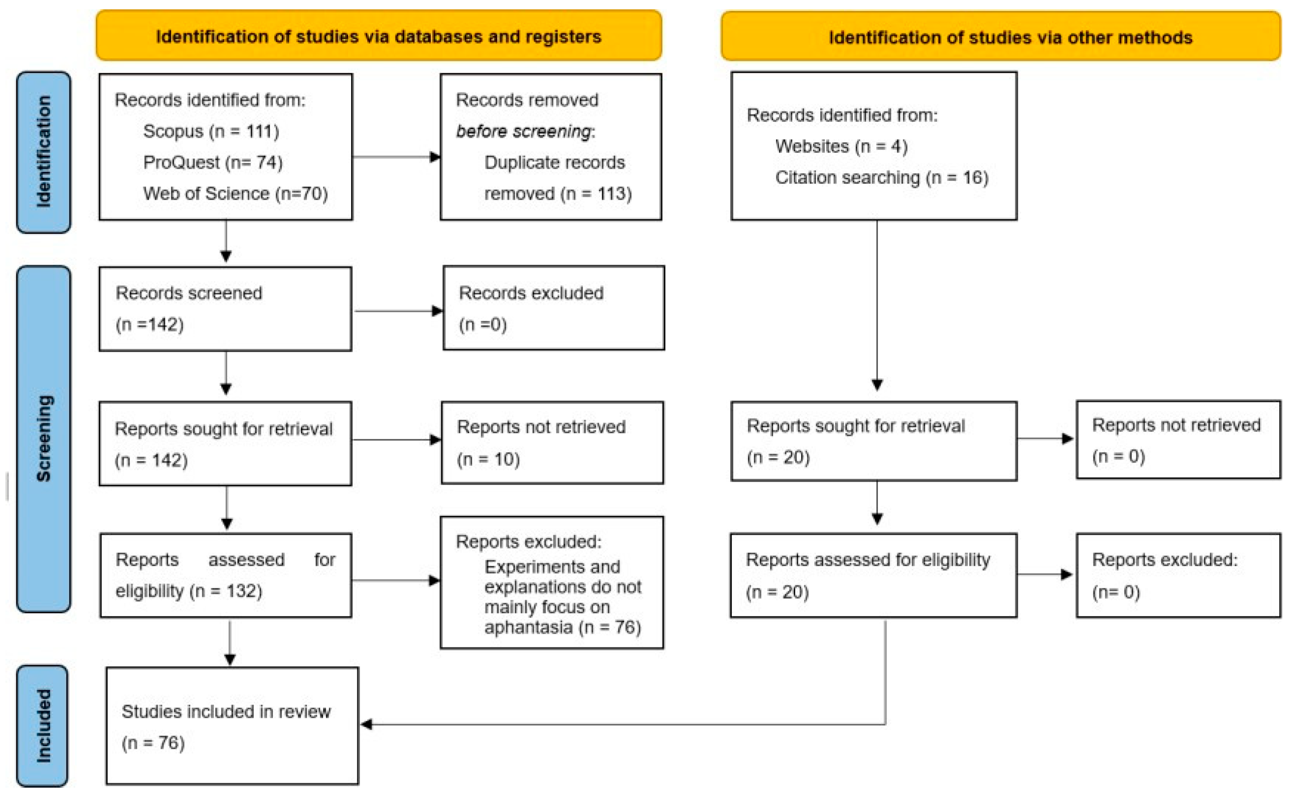
Disclaimer/Publisher’s Note: The statements, opinions and data contained in all publications are solely those of the individual author(s) and contributor(s) and not of MDPI and/or the editor(s). MDPI and/or the editor(s) disclaim responsibility for any injury to people or property resulting from any ideas, methods, instructions or products referred to in the content. |
© 2024 by the authors. Licensee MDPI, Basel, Switzerland. This article is an open access article distributed under the terms and conditions of the Creative Commons Attribution (CC BY) license (http://creativecommons.org/licenses/by/4.0/).
Copyright: This open access article is published under a Creative Commons CC BY 4.0 license, which permit the free download, distribution, and reuse, provided that the author and preprint are cited in any reuse.


