
Submitted:
24 June 2024
Posted:
25 June 2024
Read the latest preprint version here
Abstract
The production of recombinant proteins in Escherichia coli is often challenged by cytoplasmic expression due to proteolytic degradation and inclusion body formation. Extracellular expression can overcome these problems by simplifying downstream processing and improving protein yields. This study aims to compare the efficiency of two Bacillus subtilis chitosanase signal peptides in mediating extracellular secretion in E. coli. We identified a naturally occurring mutant signal peptide (mCsn2-SP) from B. subtilis CH2 chitosanase (CH2CSN), which is characterized by a deletion of six amino acids in the N-region relative to the signal peptide (Csn1-SP) from B. subtilis CH1 chitosanase (CH1CSN). The CH1CSN and CH2CSN genes were cloned into the pET-11a vector and protein secretion was evaluated in E. coli BL21(DE3) host cells. Expression was induced with 0.1 mM and 1 mM IPTG at 30°C for one and three days. CH2CSN showed higher secretion levels compared to CH1CSN under all experimental conditions, especially with 0.1 mM IPTG induction for 3 days, which resulted in a 2.37-fold increase in secretion. These results suggest that signal peptide deletion variants may have an effect on protein secretion efficiency and help to further understand the function of signal peptides.
Keywords:
Keywords signal peptide
; chitosanase
; recombinant protein
; expression
; secretion
; sec pathway
; E. coli
; Bacillus subtilis
1. Introduction
The industrial production of recombinant proteins secreted by E. coli is essential in the biotechnology industry [1]. E. coli has advantageous properties, such as rapid growth to high cell densities on a low-cost carbon source, and serves as a platform for recombinant protein production [2]. Due to its extensive use as a model system, E. coli genetics have been extensively characterized, resulting in the development of numerous tools that aid in chromosome engineering, gene replication, and gene expression [3]. Recombinant proteins can be targeted to different compartments within E. coli, including the cytoplasm, periplasm, and extracellular space. However, cytoplasmic expression of recombinant proteins is challenging. Proteins expressed in the cytoplasm may be susceptible to degradation by cellular proteases or accumulate as insoluble inclusion bodies, rendering them inactive [4]. In addition, cytoplasmic expression of certain proteins can have toxic effects on host cells, leading to reduced protein yields or even cell death [5]. In contrast, extracellular expression in E. coli offers several advantages for the production of recombinant proteins, including simplified downstream processing, reduced host cell contamination, high expression levels, reduced protein degradation, and increased solubility[6]. E. coli has two major secretory pathways, the Sec and Tat pathways. The Sec pathway, which facilitates the transport of proteins from the cytoplasmic membrane, was the first secretory pathway identified in bacteria [7]. This pathway has demonstrated the ability to translocate proteins in an unfolded state, thereby facilitating the efficient export of target proteins [8]. In contrast, the Tat pathway has the distinct ability to transport fully folded proteins across the cytoplasmic membrane [6].
Chitosanase (EC 3.2.1.132) catalyzes the hydrolysis of β-1,4-glucosidic linkages in the polymeric chitosan, a type of polysaccharide. Chitosan is a high molecular weight polymer that requires enzymatic degradation for human consumption. It is broken down into chitosan oligosaccharides (COSs) by extracellular chitosanases found in various bacteria. COSs offer more significant biological functions compared to chitosan, including higher levels of antibacterial, antioxidant, blood cholesterol reduction, blood pressure improvement, infection prevention, arthritis control, and antitumor effects [9]. The GH-46 type chitosanases, especially those derived from Bacillus and Streptomyces, have been extensively studied for their catalytic functions, enzymatic mechanisms, and protein structures [10].
Previously, our studies have shown that chitosanase derived from B. subtilis CH1 and B. subtilis CH2 are secreted into the extracellular space of B. subtilis [11,12]. In this study, we compared the efficiency of two chitosanase signal peptides from B. subtilis in mediating the extracellular secretion of chitosanase when introduced into E. coli.
2. Materials and Methods
2.1. Molecular Characterization of Chitosanase
Signal peptides were analyzed using SignalP version 6.0, available at the Health Tech website (https://services.healthtech.dtu.dk/services/SignalP-6.0/) [13]. The N-region net charge of signal peptides predicted using peptide calculator (https://www.pepcalc.com/). Conserved domains and active sites within the protein sequences were identified using the protein BLAST tool from the National Center for Biotechnology Information (NCBI) (https://blast.ncbi.nlm.nih.gov/Blast.cgi?PROGRAM=blastp&PAGE_TYPE=BlastSearch&BLAST_SPEC=&LINK_LOC=blasttab&LAST_PAGE=blastp). Molecular weights were predicted using the Compute pI/Mw tool available at ExPASy (https://web.expasy.org/compute_pi/). The nucleotide and amino acid sequences of chitosanase from B. subtilis strain CH2 were compared with those of strains 168 and CH1 using Clustal multiple sequence alignments in the DNADynamo program.
2.2. Genomic DNA Extraction
B. subtilis strains CH1 and CH2 were obtained from Jeju National University (Jeju, South Korea). These strains were cultured in 4 mL of Luria-Bertani (LB) broth at 30°C for 16 hours. After incubation, cells were harvested by centrifugation. The cell pellets were then resuspended in 200 μL lysozyme solution (20 mg/mL in 20 mM Tris-Cl, pH 8.0) and incubated at 37°C for 30 minutes. To the lysed cells, 100 μg/mL proteinase K and 30 μL 10% SDS were added. The mixture was further incubated at 56°C for 30 minutes. For DNA extraction, an equal volume of phenol/chloroform/isoamyl alcohol (25:24:1) was added to the lysate, followed by centrifugation at 12,000 rpm for 2 minutes. The aqueous phase was carefully transferred to a new tube to which 3 M Na-acetate (1/10 volume) was added. Two times the volume of 100% ethanol was added, the mixture was inverted several times and stored at -20°C for 2 hours to precipitate the DNA. The samples were then centrifuged at 13,000 × g for 30 minutes at 4 °C, and the supernatant was discarded. The DNA pellets were washed with 70% ethanol and centrifuged again at 13,000 × g at 4 °C for 10 minutes. After discarding the supernatant, the pellets were dried at 20°C for 30 minutes. Finally, the dried genomic DNA was resuspended in 100 μL distilled water.
2.3. Cloning of Chitosanase
Primers were designed to amplify the coding region, including the signal sequences, from the genomic DNA of B. subtilis strains CH1 and CH2. The forward primer, CH-F (5′-GAGACATATGAAAATCAGTATGCAAAAAGC-3′), contained an NdeI site, and the reverse primer, CH-R (5′-GAGAGGATCCTTATTTGATTACAAAATTACCGTA-3′), contained a BamHI site. The PCR mixture consisted of 5 μL of 10X Ex Taq DNA polymerase buffer, 8 μL of 2.5 mM dNTP mixture, 5 μL of 25 mM MgCl2, 100 pmol of each primer, 1 μg of genomic DNA, and 3 units of Ex Taq DNA polymerase (Takara Bio Inc., Shiga, Japan) in a final volume of 50 μL. PCR cycling conditions were set as follows: initial denaturation at 94 °C for 5 minutes, followed by 30 cycles of 94 °C for 1 minute, 55 °C for 30 seconds, and 72 °C for 1 minute, with a final extension at 72 °C for 5 minutes. PCR products were verified by 1% agarose gel electrophoresis and purified using a gel purification kit (Bioneer Co., Daejeon, South Korea). The verified PCR products and the pET-11a vector (Novagen, Madison, USA) were digested with NdeI and BamHI restriction enzymes (Takara Bio Inc., Shiga, Japan) according to the manufacturer’s instructions. The digested fragments were then ligated into the vector using T4 DNA ligase (Takara Bio Inc., Shiga, Japan). The ligation mixture was transformed into E. coli DH5alpha cells by the heat shock method. Transformed colonies were selected on LB agar plates containing 100 μg/mL ampicillin and incubated in 4 mL LB broth at 37°C overnight with shaking at 180 rpm. Plasmid DNA (pET11a-CH1 and pET11a-CH2) was extracted from the cultured cells using an Accuprep Nano-plus Plasmid Mini Extraction Kit (Bioneer Co., Daejeon, South Korea). The nucleotide sequences of the plasmids were confirmed by sequencing at Macrogen (Seoul, South Korea).
2.4. Overexpression of Chitosanase in E. coli
The recombinant plasmids pET11a-CH1 and pET11a-CH2 were transformed into E. coli BL21(DE3) cells. Transformed cells were plated on LB agar containing 100 μg/mL ampicillin and incubated at 37°C for 16 hours. Single colonies were inoculated into 20 mL LB broth supplemented with 100 μg/mL ampicillin and cultured at 37°C with shaking at 180 rpm. When the optical density at 600 nm reached 0.4, recombinant protein expression was induced by the addition of IPTG at concentrations of 0.1 mM and 1 mM. Cultures were then incubated at 30°C with shaking at 180 rpm for three days. Samples were collected on day 1 and day 3 post-induction, with 1 mL aliquots taken for each time point. Samples were centrifuged at 7,000 × g for 5 minutes to separate the pellet and supernatant. The collected pellets were resuspended in 1 mL cold distilled water and disrupted using an ultrasonicator to release intracellular contents. The cell lysates and supernatants were analyzed by 10% SDS-polyacrylamide gel electrophoresis (PAGE) using MOPS buffer. The gels were then stained with Coomassie Brilliant Blue to visualize proteins.
2.5. Protein Measurement
Protein quantification was performed using ImageJ software (NIH, Maryland, USA). Bovine serum albumin (BSA) protein (Thermo Fisher Scientific, Massachusetts, USA) was used as the standard for protein quantification.
2.6. Statistical Analysis
All experiments were performed in triplicate, and the data were analyzed using GraphPad Prism version 8 (GraphPad Software, San Diego, CA, USA). Results are presented as the mean ± standard deviation. Significant differences between the two groups were compared using a t-test, with p < 0.05 indicating a significant difference.
3. Results
3.1. Molecular Characterization of Chitosanase
The nucleotide (nt) sequence of the CH1CSN gene contains an 831-base pair (bp) open reading frame (ORF) encoding a chitosanase protein of 277 amino acid (aa) residues (GenBank accession number GU001718). Conversely, the nt sequence of the CH2CSN gene has an 813-bp ORF encoding a chitosanase protein of 271 aa residues (GenBank accession number GU001716). Figure 1. show the nt and aa sequences in comparison with the chitosanase from B. subtilis strain 168 (168CSN). The signal peptides of CH1CSN and CH2CSN, predicted to be located in the N-terminal region, consist of 35 aa and 29 aa, respectively, and were identified as Sec pathway signal peptides using the SignalP program. Despite an 11 bp difference in the mature chitosanase N-terminal sequences of CH1CSN and CH2CSN, their amino acid sequences remained identical. However, the signal sequences differed by 20 bp in the nt sequence, with a consecutive removal of 6 aa in the aa sequence. Of interest, a repetitive sequence (AAAAAGCAG) was identified in the signal sequences of 168CSN and CH1CSN, and based on these sequences, we were able to confirm that some sequences were deleted in the signal sequence of CH2CSN (Figure 1). The molecular weight of CH1CSN was predicted to be 31.5/27.4 kDa (with/without signal peptide). The molecular weights of CH1CSN and CH2CSN were predicted to be 30.7 kDa and 27.4 kDa, respectively, with the signal peptide and 27.4 kDa for both without the signal peptide. All chitosanase sequences lacked cysteine residues and contained the GH46 domain.
3.2. Expression and Extracellular Secretion Analysis of Chitosanase in E. coli
To express CH1CSN and CH2CSN from B. subtilis in E. coli and evaluate their secretion efficiency, the chitosanase genes including the signal sequences were amplified by PCR and cloned into the pET11a vector. The constructs were then transformed into E. coli BL21(DE3) cells. The chitosanase proteins were induced by different IPTG concentrations (0.1 and 1 mM) and different times (1 and 3 days) at 30°C. SDS-PAGE was employed to differentiate chitosanase proteins based on the cleavage status of their signal peptides. Proteins with intact signal peptides remained in the cytoplasm, while those with cleaved signal peptides were located in the periplasm and secreted extracellularly (Figure 2a). Quantitative analysis of these proteins was conducted using image analysis software, and the detailed results are presented in Table 1. The data confirm that the signal peptides not only facilitated the export of chitosanase from the cytoplasm to the periplasm but also enabled its subsequent secretion outside the cell. For CH1CSN, extracellular secretion levels remained consistent on day 1 across both 0.1 mM and 1 mM IPTG inductions. However, there was a notable increase in secretion on day 3 with the 1 mM IPTG induction (Figure 2). Conversely, for CH2CSN, secretion levels were similar on day 1 for both IPTG concentrations but were higher at 0.1 mM than at 1 mM IPTG on day 3 (Figure 2). CH2CSN consistently showed higher secretion levels than CH1CSN in all experimental conditions. Remarkably, when expression was induced with 0.1 mM IPTG for 3 days, the secretion level of CH2CSN was 65.48 μg/mL, representing a 2.37-fold increase over the 27.6 μg/mL observed for CH1CSN (Figure 2, Table 1). The total expression of chitosanase, which includes cytoplasmic, periplasmic, and extracellular components, was highest at 128.06 μg/mL when CH2CSN was treated with 0.1 mM IPTG for 3 days to induce expression. These results confirmed that a naturally mutated signal peptide from B. subtilis affected the efficiency of recombinant protein secretion in E. coli.
4. Discussion
In the field of biotechnology, it has become increasingly apparent that there is no universal signal peptide that can universally enhance the optimal secretion of desired target proteins across different bacterial expression hosts [14]. The first systematic study to explore the effects of signal peptide variations on the secretion production of heterologous proteins was reported by Brockmeier et al. in 2006 [15]. One surprising outcome of this study was that signal peptides optimal for one protein were often ineffective for another, underscoring the necessity for a specific combination between the signal peptide and the target protein. Therefore, the discovery of new signal peptides provides fresh options for the production of recombinant proteins, highlighting the tailored approach needed in optimizing secretion systems for diverse proteins.
In this study, we investigated the genetic characterization of chitosanase in two B. subtilis strains and produced it as a recombinant protein in E. coli to determine the secretion properties of the signal peptide. We compared the genetic profile of the known 168CSN with that of CH1CSN and CH2CSN, as shown in Figure 1a. The results show that there is a small difference of only 1 bp between 168CSN and CH1CSN, while there is a larger difference of 30 bp in CH2CSN. There is also a difference of 31 bp between CH1CSN and CH2CSN. Looking at the signal sequence alone, there is no difference between Csn168-SP and Csn1-SP, while a 20 bp difference was observed in mCsn2-SP. Interestingly, the sequences of Csn168-SP and Csn1-SP contain a repeat sequence (AAAAAGCAG) that appears to be central to the 18 bp deletion mutation identified in mCsn2-SP. This finding is consistent with the work of Bzymek and Lovett, who showed that genetic diversity in bacteria often results from genetic rearrangements in which simple sequence repeats result in a variety of insertions or deletions [16]. As shown in Figure 1b, a comparison of the amino acid sequences of the chitosanases revealed that the mature chitosanase sequences were all identical. However, mCsn2-SP showed a deletion of six consecutive amino acids (ADFWKK) compared to Csn168-SP and Csn1-SP. Signal peptides are typically divided into three main regions: the N-region, which carries a positive charge and assists in the translocation process; the H-region, which is hydrophobic and binds to the membrane in an alpha-helix structure; and the C-region, which is polar and contains the cleavage site that releases the mature protein [17]. Specially, the N-region plays a crucial role in facilitating protein translocation by interacting with the negatively charged phosphate groups in the lipid bilayer, which is vital for the effective movement of proteins. Despite the H-region and C-region of mCsn2-SP being identical to those of Csn168-SP and Csn1-SP, the N-region of mCsn2-SP exhibited a net charge decrease from +3 to +2 due to the deletion of six amino acids. Although the net positive charge in the N-region of a signal peptide is crucial for the efficient translocation of target proteins, an increase in net positive charge does not always result in beneficial outcomes. In fact, higher positive charges can lead to negative consequences such as protein aggregation, jamming of the secretion translocase, and activation of stress-related genes [18].
We determined the chitosanase secretion efficiency of Csn1-SP and mCsn2-SP as a function of concentration and time of IPTG treatment, a protein expression inducer in E. coli. SDS-PAGE showed that both forms of pre-chitosanase, in which the signal peptide is not cleaved and is located in the cytoplasm, and mature chitosanase, in which the signal peptide has been cleaved and translocated to the periplasmic region, were observed in the total protein of the cells, and some of the protein was released into the culture supernatant (Figure 2a). The difference in length of the signal peptides resulted in the size of pre-CH1CSN (31.5 kDa) being larger in molecular weight than pre-CH2CSN (30.7 kDa), while the mature forms of CH1CSN and CH2CSN were the same size (27.4 kDa). The results show that the signal peptide of the naturally mutated CH2CSN significantly improves the efficiency of protein secretion compared to CH1CSN under all conditions used in the experiment (Figure 2b, Table 1). This result suggests that minor alterations in the signal peptide sequence can have substantial impacts on the protein export process in bacterial systems. Consequently, these results may imply that such mutations contribute to the enhancement of genomic diversity and highlight the potential of adaptive evolutionary processes in microbial genetics [19].
Overall, the secretory capability of E. coli provides valuable advantages in terms of increased yield, simplified purification, reduced cell toxicity, improved post-translational modifications, and scalability, making it a popular choice for recombinant protein expression and production [20]. The Sec pathway is a well-known secretory pathway in E. coli and is involved in the secretion of various proteins. Representative proteins secreted via the Sec pathway in E. coli include MalE [21], OmpA [22], OmpC [23], OmpF [24], PelB [25], and PhoA [26] signal peptides. As signal peptides in the Sec pathway, Csn1-SP and mCsn2-SP were also identified. We also identified a mutant chitosanase signal peptide, mCsn2-SP, and successfully demonstrated secretion of recombinant proteins in E. coli. This study confirmed whether mCsn2-SP has a higher secretion efficiency than Csn1-SP in E. coli. A previous study by another group showed that the E. coli OmpA signal sequence was more effective in both expression and secretion than the native Bacillus signal peptide [22]. In Gram-negative bacteria, such as E. coli, the Sec pathway plays a key role in protein secretion. It is also known to recognize cleavage sites and selectively target the Sec translocase. Nevertheless, these results confirm that the signal peptide, originally from Gram-positive bacteria, operates effectively even when interacting with distinct receptors within the secretion system of Gram-negative bacteria.
5. Conclusions
We compared the nucleotide and amino acid sequences of CH1CSN and CH2CSN from B. subtilis and found that a repetitive sequence in the Csn1-SP resulted in an 18 bp base deletion in the mCsn2-SP. This was manifested as a deletion of 6 aa at the end of the N-region of mCsn2-SP. When CH1CSN and CH2CSN were produced as recombinant proteins in E. coli, the secretion rate of CH2CSN was found to be superior to that of CH1CSN under all expression induction conditions, demonstrating that the deletion of the N-region that affects translocation within mCsn2-SP is an evolutionary consequence of its evolution relative to Csn1-SP. This approach has the potential to reduce costs and improve yields in recombinant protein production, as well as advance our understanding of signal peptide function in microbial genetics.
6. Patents
This section is not mandatory but may be added if there are patents resulting from the work reported in this manuscript.
Supplementary Materials
The following supporting information can be downloaded at the website of this paper posted on Preprints.org, Figure S1: Signal peptide diagnostics of CH1CSN and CH2CSN using SignalP 6.0 tool.
Author Contributions
T.-Y.E. and C.O.: Conceptualization, T.-Y.E., Y.G. and Y.L.: methodology, Y.-H.K. and E.J.: validation, T.-Y.E. and S.D.M.: investigation, T.-Y.E. and Y.G.: data curation, T.-Y.E. and G.-H.P.: writing original draft preparation, G.-H.P. and H.S.P.: writing review and editing, T.-Y.E., and Y.L.: visualization, G.-H.P. and C.O.: supervision, All authors have read and agreed to the published version of the manuscript.
Funding
This research was supported by the Development of Technology for Biomaterialization of Marine Fishery Byproducts of the Korea Institute of Marine Science & Technology Promotion (KIMST), funded by the Ministry of Oceans and Fisheries (KIMST-20220128) and the Korea Institute of Ocean Science and Technology (PEA0124).
Institutional Review Board Statement
Not applicable.
Informed Consent Statement
Not applicable.
Data Availability Statement
Data are contained within the article.
References
- Yoon, S.H.; Kim, S.K.; Kim, J.F. Secretory production of recombinant proteins in Escherichia coli. Recent Pat Biotechnol 2010, 4, 23–29. [Google Scholar] [CrossRef] [PubMed]
- Baneyx, F.; Mujacic, M. Recombinant protein folding and misfolding in Escherichia coli. Nat Biotechnol 2004, 22, 1399–1408. [Google Scholar] [CrossRef] [PubMed]
- Mergulhao, F.J.; Summers, D.K.; Monteiro, G.A. Recombinant protein secretion in Escherichia coli. Biotechnol Adv 2005, 23, 177–202. [Google Scholar] [CrossRef] [PubMed]
- Mergulhao, F.J.; Monteiro, G.A. Periplasmic targeting of recombinant proteins in Escherichia coli. Methods Mol Biol 2007, 390, 47–61. [Google Scholar] [CrossRef] [PubMed]
- Mierendorf, R.C.; Morris, B.B.; Hammer, B.; Novy, R.E. Expression and Purification of Recombinant Proteins Using the pET System. Methods Mol Med 1998, 13, 257–292. [Google Scholar] [CrossRef] [PubMed]
- Choi, J.H.; Lee, S.Y. Secretory and extracellular production of recombinant proteins using Escherichia coli. Appl Microbiol Biotechnol 2004, 64, 625–635. [Google Scholar] [CrossRef] [PubMed]
- Beckwith, J. The Sec-dependent pathway. Res Microbiol 2013, 164, 497–504. [Google Scholar] [CrossRef] [PubMed]
- Crane, J.M.; Randall, L.L. The Sec System: Protein Export in Escherichia coli. EcoSal Plus 2017, 7. [Google Scholar] [CrossRef] [PubMed]
- Yabuki, M.; Uchiyama, A.; Suzuki, K.; Ando, A.; Fujii, T. Purification and Properties of Chitosanase from Bacillus-Circulans Mh-K1. J Gen Appl Microbiol 1988, 34, 255–270. [Google Scholar] [CrossRef]
- Wang, J.; Zhou, W.; Yuan, H.; Wang, Y. Characterization of a novel fungal chitosanase Csn2 from Gongronella sp. JG. Carbohydr Res 2008, 343, 2583–2588. [Google Scholar] [CrossRef]
- Oh, C.-H.; Lee, J.-H. Isolation, purification and characterization of chitosanase from Bacillus subtilis CH1. Journal of Aquaculture 2006, 19, 40–46. [Google Scholar]
- Oh, C.; De Zoysa, M.; Kang, D.H.; Lee, Y.; Whang, I.; Nikapitiya, C.; Heo, S.J.; Yoon, K.T.; Affan, A.; Lee, J. Isolation, purification, and enzymatic characterization of extracellular chitosanase from marine bacterium Bacillus subtilis CH2. J Microbiol Biotechnol 2011, 21, 1021–1025. [Google Scholar] [CrossRef] [PubMed]
- Teufel, F.; Almagro Armenteros, J.J.; Johansen, A.R.; Gíslason, M.H.; Pihl, S.I.; Tsirigos, K.D.; Winther, O.; Brunak, S.; von Heijne, G.; Nielsen, H. SignalP 6.0 predicts all five types of signal peptides using protein language models. Nature biotechnology 2022, 40, 1023–1025. [Google Scholar] [CrossRef] [PubMed]
- Freudl, R. Signal peptides for recombinant protein secretion in bacterial expression systems. Microb Cell Fact 2018, 17. [Google Scholar] [CrossRef] [PubMed]
- Brockmeier, U.; Caspers, M.; Freudl, R.; Jockwer, A.; Noll, T.; Eggert, T. Systematic screening of all signal peptides from:: A powerful strategy in optimizing heterologous protein secretion in gram-positive bacteria. Journal of Molecular Biology 2006, 362, 393–402. [Google Scholar] [CrossRef] [PubMed]
- Bzymek, M.; Lovett, S.T. Instability of repetitive DNA sequences: the role of replication in multiple mechanisms. Proc Natl Acad Sci U S A 2001, 98, 8319–8325. [Google Scholar] [CrossRef] [PubMed]
- Rusch, S.L.; Kendall, D.A. Interactions that drive Sec-dependent bacterial protein transport. Biochemistry 2007, 46, 9665–9673. [Google Scholar] [CrossRef] [PubMed]
- Owji, H.; Nezafat, N.; Negandaripour, M.; Hajiebrahimi, A.; Ghasemi, Y. A comprehensive review of signal peptides: Structure, roles, and applications. Eur J Cell Biol 2018, 97, 422–441. [Google Scholar] [CrossRef] [PubMed]
- Vale, F.F.; Lehours, P.; Yamaoka, Y. Editorial: The Role of Mobile Genetic Elements in Bacterial Evolution and Their Adaptability. Frontiers in Microbiology 2022, 13. [Google Scholar] [CrossRef]
- Baldi, L.; Hacker, D.L.; Adam, M.; Wurm, F.M. Recombinant protein production by large-scale transient gene expression in mammalian cells: state of the art and future perspectives. Biotechnology letters 2007, 29, 677–684. [Google Scholar] [CrossRef]
- Beena, K.; Udgaonkar, J.B.; Varadarajan, R. Effect of signal peptide on the stability and folding kinetics of maltose binding protein. Biochemistry 2004, 43, 3608–3619. [Google Scholar] [CrossRef] [PubMed]
- Pechsrichuang, P.; Songsiriritthigul, C.; Haltrich, D.; Roytrakul, S.; Namvijtr, P.; Bonaparte, N.; Yamabhai, M. OmpA signal peptide leads to heterogenous secretion of B. subtilis chitosanase enzyme from E. coli expression system. Springerplus 2016, 5, 1–10. [Google Scholar] [CrossRef] [PubMed]
- Wang, P.; Ma, J.; Zhang, Y.; Zhang, M.; Wu, M.; Dai, Z.; Jiang, M. Efficient Secretory Overexpression of Endoinulinase in Escherichia coli and the Production of Inulooligosaccharides. Appl Biochem Biotechnol 2016, 179, 880–894. [Google Scholar] [CrossRef] [PubMed]
- Forster, S.; Brandt, M.; Mottok, D.S.; Zschuttig, A.; Zimmermann, K.; Blattner, F.R.; Gunzer, F.; Pohlmann, C. Secretory expression of biologically active human Herpes virus interleukin-10 analogues in Escherichia coli via a modified Sec-dependent transporter construct. BMC Biotechnol 2013, 13, 82. [Google Scholar] [CrossRef] [PubMed]
- Shi, L.; Liu, H.; Gao, S.; Weng, Y.; Zhu, L. Enhanced Extracellular Production of IsPETase in Escherichia coli via Engineering of the pelB Signal Peptide. J Agric Food Chem 2021, 69, 2245–2252. [Google Scholar] [CrossRef] [PubMed]
- Miksch, G.; Ryu, S.; Risse, J.M.; Flaschel, E. Factors that influence the extracellular expression of streptavidin in Escherichia coli using a bacteriocin release protein. Applied microbiology and biotechnology 2008, 81, 319–326. [Google Scholar] [CrossRef]
Figure 1.
(a)Nucleotide sequence analysis of CH2CSN compared to 168CSN and CH1CSN. Red box; signal sequence, gray box; sequence difference, arrow; repeat sequence; (b) Amino acid sequence analysis of CH2CSN compared to 168CSN and CH1CSN. Red box; signal peptide, blue box; GH46 chitosanase domain, #; activity site.
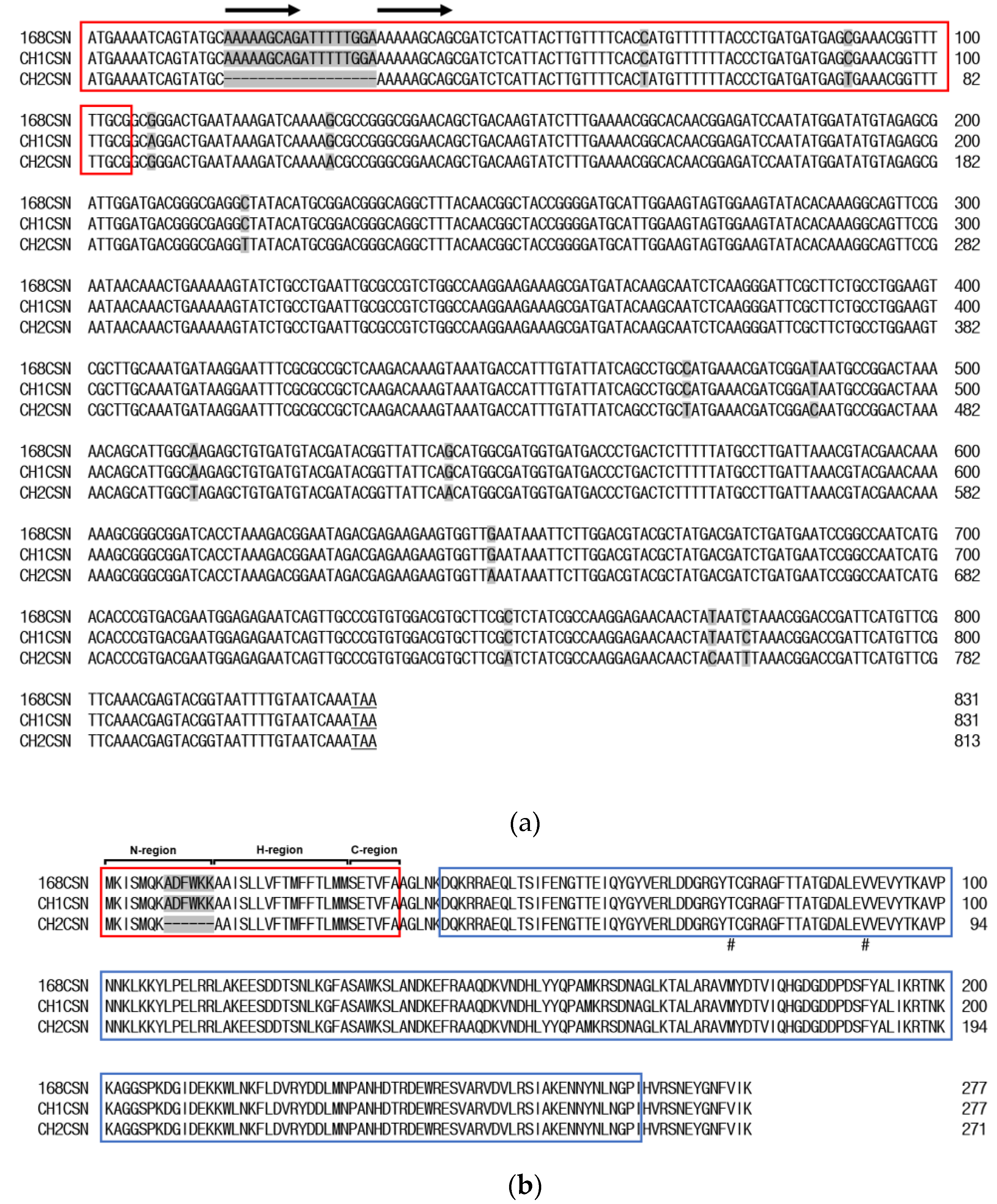
Figure 2.
(a) A comparative study of recombinant chitosanase expression using the Csn1-SP and mCsn2-SP. M, molecular mass standard marker; Standard; BSA protein for protein quantification. T, total cellular protein; Su, supernatant; (b) Comparison of extracellular secreted chitosanase levels between CH1CSN and CH2CSN. Sup, supernatant (mean ± standard deviation, n = 3, p < 0.05).
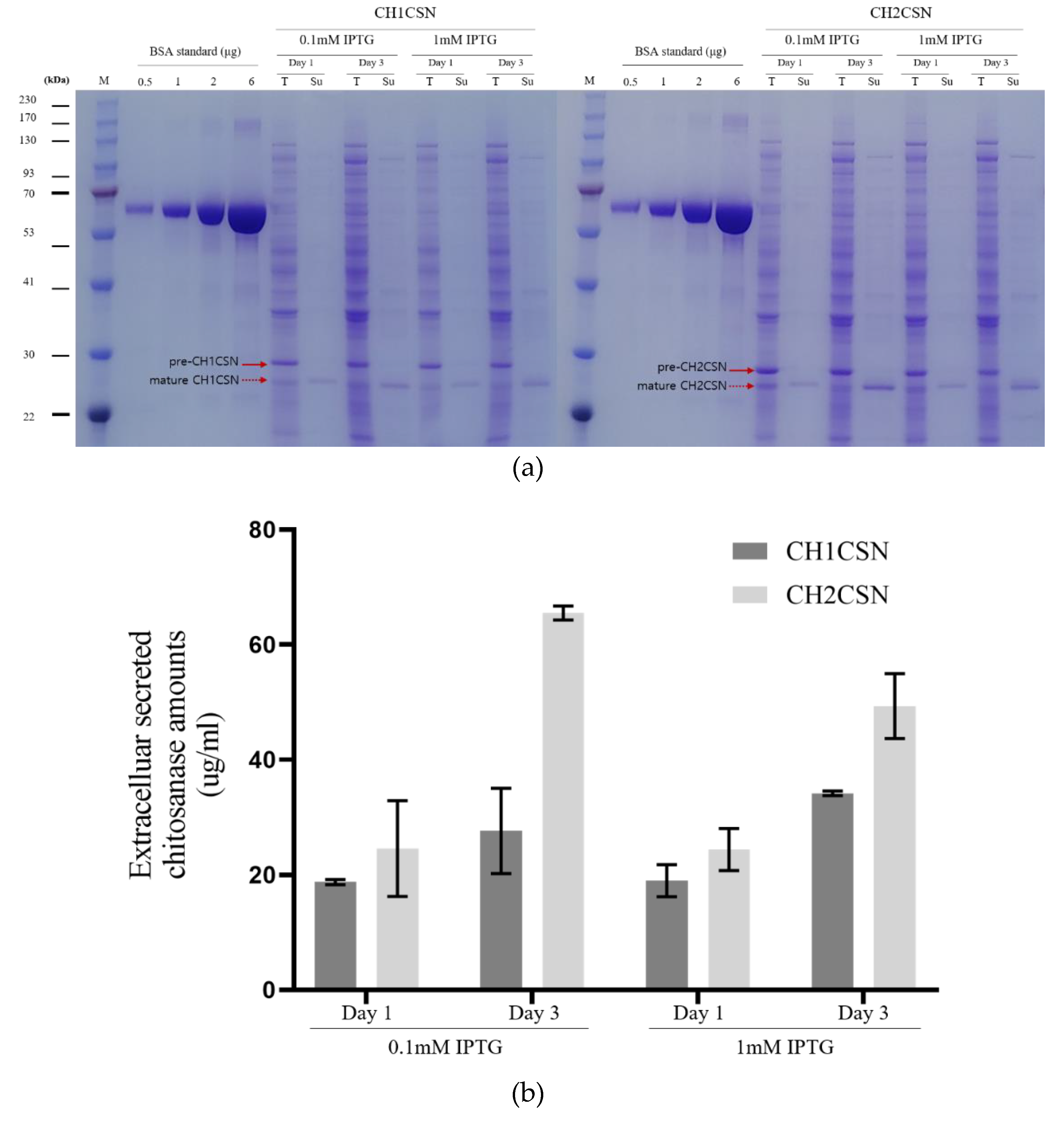

Table 1.
Quantitative measurement of CH1CSN and CH2CSN (mean ± standard deviation, n = 3).
| 0.1mM IPTG | 1mM IPTG | ||||
|---|---|---|---|---|---|
| day 1 | day 3 | day 1 | day 3 | ||
|
CH1CSN (ug/ml) |
Cytoplasm | 48.04 ± 0.87 | 38.80 ± 2.87 | 47.08 ± 1.45 | 33.51 ± 0.51 |
| Periplasm | 16.75 ± 0.39 | 9.73 ± 2.03 | 6.79 ± 5.99 | 6.28 ± 3.75 | |
| Supernatant | 18.75 ± 0.44 | 27.60 ± 7.39 | 18.95 ± 2.77 | 34.12 ± 0.38 | |
|
CH2CSN (ug/ml) |
Cytoplasm | 60.70 ± 1.92 | 51.80 ± 1.93 | 41.08 ± 1.90 | 28.49 ± 2.82 |
| Periplasm | 22.58 ± 1.48 | 10.78 ± 1.25 | 15.54 ± 1.51 | 5.96 ± 1.21 | |
| Supernatant | 24.52 ± 8.29 | 65.48 ± 1.22 | 24.37 ± 3.65 | 49.31 ± 5.65 | |
Disclaimer/Publisher’s Note: The statements, opinions and data contained in all publications are solely those of the individual author(s) and contributor(s) and not of MDPI and/or the editor(s). MDPI and/or the editor(s) disclaim responsibility for any injury to people or property resulting from any ideas, methods, instructions or products referred to in the content. |
© 2024 by the authors. Licensee MDPI, Basel, Switzerland. This article is an open access article distributed under the terms and conditions of the Creative Commons Attribution (CC BY) license (http://creativecommons.org/licenses/by/4.0/).
Copyright: This open access article is published under a Creative Commons CC BY 4.0 license, which permit the free download, distribution, and reuse, provided that the author and preprint are cited in any reuse.


