
Submitted:
28 May 2024
Posted:
29 May 2024
You are already at the latest version
Abstract
Plant DNA extraction techniques are well-established, featuring a variety of protocols tailored to the needs of individual laboratories or research goals. Currently, there is a strong demand for plant DNA extraction methods that are rapid, cost-effective, and uncomplicated. We have devised an optimized protocol for plant DNA extraction that eliminates the need for liquid nitrogen. DNA was extracted from leaves of Inga feuilleei (pacae) in the Cañete Valley using a basic cetyltrimethylammonium bromide method. Subsequently, the extracted DNA was quantified and assessed for quality through agarose gel electrophoresis. This protocol holds the potential to significantly reduce costs in laboratories engaged in routine DNA extraction for genetic diversity and gene expression analyses. It addresses challenges encountered by other conventional methods, particularly in situations where they have proven ineffective due to issues related to explant, sample conditions, and the quantity and quality of nucleic acids. This is especially pertinent for numerous laboratories in developing countries where constraints related to the cost and availability of liquid nitrogen may be a limiting factor. Furthermore, this is the first study reported in this species for genomic studies.
Keywords:
Inga feuilleei
; pacae
; DNA
; nucleic acids
; plant
1. Introduction
Inga feuilleei, also known as pacae, pacay, guaba, guamo or inga, is a legume and is a plant native to tropical areas and temperate areas of Latin America and the Caribbean. The Inga plant genus originated in the Amazon of Brazil, Ecuador, Colombia, Bolivia and Peru; this plant species is also introduced to a large part of Panama and Costa Rica [1].
Pacae has great nutritional value (vitamin A, B and C), its antioxidant, anti-inflammatory, antiseptic and healing properties stand out; in addition to having fiber, calcium, iron and phosphorus. The fruit, stem, and leaves have different types of metabolites, among them we find flavonoids and tannins; These metabolites could be responsible for the anti-inflammatory, analgesic and gastroprotective therapeutic activity. In addition, it is used to improve vision, reduce cholesterol and triglyceride levels, helps maintain the health of the circulatory system and heart diseases, strengthens bones and improves the immune system. It has a high fiber content, so it has detoxifying properties and helps maintain proper functioning of the system [2].
Many difficulties have been reported for isolating good-quality DNA from plants [3]. These difficulties were attributed to the fact that different plant species have varying levels of polysaccharides, polyphenols, and other secondary metabolites. These components are usually hindering the process of DNA purification and its further use in molecular studies. These plant components have a similar structure of nucleic acids that allow secondary metabolites and polysaccharides to interfere with total DNA isolation [4]. They strongly combine with nucleic acids during DNA isolation and affect the quality of the extracted DNA from higher plants. The present work describes a rapid CTAB-based method with modifications for the extraction of high-quality genomic DNA from Inga feuilleei.
In this study
2. Experimental Design
A 3 x 2 fatorial design was established with 15 repetitions, using 3 c.

Table 1.
Factorial design 3 x 2.
| Extraction method | Sample Type | |
|---|---|---|
| Dry Sample (D) | Fresh Sample (F) | |
| Cetyltrimethylammonium bromide (CTAB 2X) (Kit 1) | T1. CTAB-D | T2. CTAB-F |
| GeneJET Plant Genomic DNA Purification Kit (Kit 2) | T3. GenJET-D | T4. GenJET-F |
| EZ-10 Spin Column Genomic | DNA Minipreps Kit (Plant) (Kit 3) | T5. EZGen-D | T6. EZGen-F |
2.1. Materials
- Fresh leaves sample of Inga feuillei
- Cetyltrimethylammonium bromide - CTAB (Promega Corporation, Madison, USA)
- β-mercaptoetanol (Central Drug House, New Delhi, India)
- Tris-HCl 1M (Promega Corporation, Madison, USA)
- EDTA 0.5M (Promega Corporation, Madison, USA)
- NaCl (Sigma-Milipore, Saint Louis, MO, USA).
- PVP-40 (Caisson Labs, Smithfield, UT, USA)
- Chloroform (J.T. Baker, Phillipsburg, NJ, USA)
- Iso amyl alcohol (Central Drug House, New Delhi, India)
- Isopropanol (Central Drug House, New Delhi, India)
- Absolute ethanol (Merck, Darmstadt, Germany)
- Rnase A solution (Thermo Fisher Scientific, Waltham, MA, USA)
- Buffer TE solution (Promega Corporation, Madison, USA)
- Agarose (Promega Corporation, Madison, USA)
- Loading dye 6X (Thermo Fisher Scientific, Waltham, MA, USA)
- GeneRuler 1 Kb DNA Ladder (Thermo Fisher Scientific, Waltham, MA, USA)
2.2. Equipment
- Biological safety cabinet (Labconco, Kansas, MO, USA)
- TissueLyser II (Qiagen, Hilden, Germany)
- Microcentrifuge air-cooled (Labnet International, Edison, NJ, USA)
- Thermomixer C (Eppendorf, Hamburg, Germany)
- Biospectrometer (Eppendorf, Hamburg, Germany)
- Enduro Gel documentation Systems (Labnet International, Edison, NJ, USA)
3. Procedure
3.1. Sample Collection
The samples leaf were collected in the district of Lunahuana, province of Cañete, Region Lima - Peru. And they were prepared in accordance with their respective sampling methodologies:
Fresh Sample: Utilizing sterilized forceps, foliar tissue was extracted from the plant and placed into a plastic bag containing moistened paper with distilled water. Subsequently, the samples were stored at 4°C. The viability for usage of these samples is restricted to a maximum duration of three to four days.
Dry Sample: Employing sterilized forceps, foliar tissue was carefully removed from the plant and placed into a plastic bag lined with completely dry paper, to which 100 grams of silica gel were added. This preparation protocol is implemented when the samples are to be processed subsequent to a period extending from four to eight weeks post-collection.
3.2. Sample Homogenization
Homogenization of the samples was carried out using a equipment TissueLyser II (Qiagen, Hilden, Germany) with steel beads. The process is described as follows:
- The sample of plant material (leaves) is collected in a 2 mL tube with 750 µL extraction buffer (2X CTAB extraction buffer and 2µL of β-mercaptoethanol).
- The tubes are then opened, a steel bead is added to each, closed, and placed in the TissueLyser II disruptor kit.
- The samples are ground for eight minutes at a speed of 30 rps. The tubes are then removed to continue the extraction.
Figure 1.
Homogenization process.

3.3. DNA Extraction
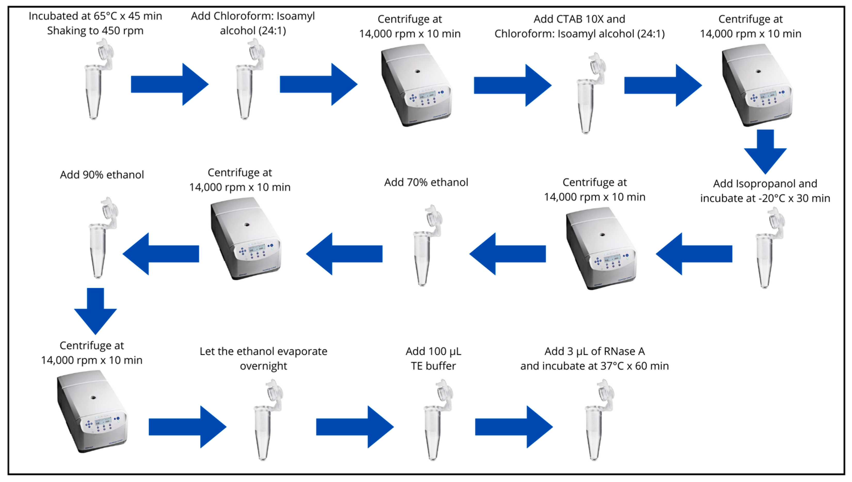
- Incubate samples at 65°C for 45 minutes to 1 hour. Shaking the tubes constantly (by inversion) every 20 minutes in a bain-marie or at 450 rpm in a Thermomixer C (Eppendorf, Hamburg, Germany).
- Remove the samples and let them cool at room temperature for 4 min.
- Add 750 µL of Chloroform: Isoamyl Alcohol (24:1), shake the tubes slightly (by inversion approximately 40 times) until the content is homogenized (milky green).
- Centrifuge at 14,000 rpm for 10 minutes.
- Transfer 600 µL of the supernatant to new 2 mL microtubes, previously labeled.
- Add 150 uL of CTAB 10X (preheated to 65°C), homogenize by inversion (10 times) and add 750 µL of Chloroform: Isoamyl Alcohol (24:1) and homogenize (by inversion approximately 40 times).
- Centrifuge at 14,000 rpm for 10 minutes.
- Transfer 500 µL of the supernatant to 1.5 mL microtubes, previously labeled.
- Add approximately 2/3 volume of Isopropanol (350 µL), shake gently by inversion (10 times) and incubate at -20°C for 30 minutes.
- Centrifuge at 14,000 rpm for 10 minutes.
- Decant the Isopropanol being careful not to lose the pellet.
- Add 500 µL of 70% ethanol. Remove the pellet from the bottom of the tube and gently shake the tube.
- Centrifuge at 14,000 rpm for 10 minutes.
- Decant the 70% ethanol, taking care not to lose the pellet.
- Add 500 µL of 90% Ethanol. Remove the pellet from the bottom of the tube and gently shake the tube.
- Centrifuge at 14,000 rpm for 10 minutes.
- Decant the 90% ethanol, taking care not to lose the pellet.
- Let the ethanol evaporate until the next day or leave it upside down for 2 to 3 hours to ensure that there is no ethanol left in the tube.
- Add 100 µL T10E1. Gently homogenize the DNA sample.
- Add 3 µL of RNase A and incubate in the thermomixer at 37°C. for 60 minutes at 300 rpm.
Figure 2.
DNA extraction process.
3.4. Quantification of DNA
Once genomic DNA is obtained, its purity is determined by the maximum absorbance value of nucleic acids detected at a wavelength of 260 nm, allowing for the estimation of concentration through spectrophotometry [7] Biospectrometer (Eppendorf, Hamburg, Germany):
- The ratio of absorbances A260/A280 helps determine if the obtained DNA is contaminated by the presence of aromatic compounds, as these absorb at a wavelength of 280 nm.
- DNA is considered of optimal quality when the 260/280 ratio is greater than 1.8. A ratio of A260/280 > 2.1 could indicate the presence of RNA in the sample. Conversely, if this ratio is low (A260/280 < 1.6), it could be indicative of contamination by proteins or phenols.
- A second assessment of nucleic acid purity is the 260/230 ratio, as at 230 nm, the maximum absorbance of salts present in the solution, carbohydrates, or other possible contaminants is detected.
- Accepted values fall within the range of 1.5 to 2.2; if the ratio is less than 1.5, it indicates the presence of contaminants in the sample.
3.5. Electrophoresis of DNA
It is crucial to assess the integrity or quality of the obtained DNA, and for this purpose, the agarose gel electrophoresis technique is employed. This technique relies on the utilization of a support matrix created by the polymerization of agarose substances, enabling the selective passage of molecules based on their size [8].
The migration of DNA molecules occurs horizontally due to the application of an electric field. For the visual detection of DNA, a substance sensitive to UV light is used to observe the gel image on a UV transilluminator. The equipment includes a container for the running buffer, two electrodes for the flow of electric current, a mold for gel preparation, and combs to create openings, lanes, or wells where the samples are deposited [9,10].
Sample preparation and electrophoretic migration
- Dispense 1 µl of 6X loading buffer (Loading dye 6X, Thermo scientific) per sample to migrate on a parafilm sheet.
- Add 2 µL of the DNA sample. Mingle.
- Add 1X TAE buffer to the electrophoretic chamber and place the mold with the already polymerized agarose gel.
- From the second well, load the samples previously prepared with the 6X loading buffer.
- Place 2-3 µL of a 1 Kb DNA molecular weight marker (1 kb MP) in the first well of each row of the 1% agarose gel.
- Connect the electrodes correctly and migrate to 100 V for 30 minutes
- See the results with the Enduro Gel documentation Systems (Labnet International, Edison, NJ, USA).
3.6. DNA Amplification by Polymerase Chain Reation (PCR)
Samples with higher concentrations and better quality of DNA were amplified using the PCR technique in a thermocycler (Mastercycler X50S, Eppendorf, Hamburg, Germany), using the primers Inga03 F, 5′- TTCCAAGCTTATACAAACCTCC -3′ and R, 5′- AGATCCGTACGTGTGATGGT-3′ (Invitrogen, Waltham, MA, USA). The reaction was prepared in a final volume of 50 µL, which contained 25 µL of DreamTaq PCR Master Mix (2X) (Thermo Fisher Scientific Waltham, MA, USA), 1 µL of each 10 mM primer, 2 µL of DNA (~120 ng/µL) and sufficient molecular grade water to reach the desired volume. The PCR products were evaluated by electrophoresis on 1% agarose gels in a horizontal electrophoresis chamber and then visually analyzed in a photodocumenter (Enduro GDS Labnet, Tewksbury, MA, USA). The size of the fragments was determined by comparison with a GeneRuler 1 Kb DNA Ladder (Thermo Fisher Scientific, Waltham, MA, USA).
4. Expected Results
Reagents Setup
For this activity, glassware and consumables were prepared through sterilization, glass jars, microcentrifuge tubes, and micropipette tips. Additionally, the preparation of extraction buffers was carried out according to the CTAB method, which was previously recommended based on previous research. For this activity, the buffers were prepared: CTAB 10X, CTAB 2X, T10E1, 1M Tris pH 8.0 and 0.5 M EDTA pH 8.0, which are required in the different stages of plant DNA isolation [11,12].
Buffer CTAB 10X
- Tris-HCl 1M, pH 8.0 100 mM
- EDTA 0.5M, pH 8.0 20 mM
- NaCl 0.7 M
- CTAB 10 %
Bufferde Lisis CTAB 2X
- Tris-HCl 1M, pH 8.0 100 mM
- EDTA 0.5M, pH 8.0 20 mM
- NaCl 1.4 M
- PVP-40 1 % (w/v)
- CTAB 2 %
T10E1
- Tris- pH=8.0 1M 10 mM
- EDTA-8.0 0,5 M 1 mM
- 1M TRIS -pH 8.0 (adjust pH 8.0 with HCl)
- Tris base pH=8.0 1 M
- 0.5M EDTA -pH 8.0 (adjust pH with NaOH)
5. Conclusions
This protocol provides a new guide for more investigations into the Inga feuilleei (pacae). Furthermore, no reports of DNA extraction protocols have been found in this crop, therefore, this study will serve to give other researchers a starting point to carry out new research at the molecular and genomic level.
Funding
This research was fully funded by National University of Cañete from Peru.
Acknowledgments
Thanks to the authorities of the National University of Cañete for allowing the development of this research and to the students who collaborated in the collection of this species.
Conflicts of Interest
The author declare no conflicts of interest.
References
- Abdel-Latif, A., & Osman, G. (2017). Comparison of three genomic DNA extraction methods to obtain high DNA quality from maize. Plant Methods, 13(1), 1. https://doi.org/10.1186/s13007-016-0152-4. [CrossRef]
- Aboul-Maaty, N. A.-F., & Oraby, H. A.-S. (2019). Extraction of high-quality genomic DNA from different plant orders applying a modified CTAB-based method. Bulletin of the National Research Centre, 43(1), 25. [CrossRef]
- Arbi, G., Naceur, B., Chokri, M., & Mohamed, B. (s. f.). A simple, rapid and efficient method for the extraction of genomic DNA from Allium roseum L. (Alliaceae).
- Doyle JJ, Doyle JL. (1990). Isolation of plant DNA from fresh tissue. Focus. 3: 12(13).
- Ferreira, C. F., Gutierrez, D. L., Kreuze, J. F., Iskra-Caruana, M. L., Chabannes, M., Barbosa, A. C. O., Santos, T. A., Silva, A. G. S., Santos, R. M. F., Amorim, E. P., De Oliveira, S. A. S., & Jesus, O. N. (2019). Brief Note Rapid plant DNA and RNA extraction protocol using a bench drill. Genetics and Molecular Research, 18(3). [CrossRef]
- Gupta, N. (2019). DNA extraction and polymerase chain reaction. Journal of Cytology, 36(2), 116. [CrossRef]
- Helmersen, K., & Aamot, H. V. (2020). DNA extraction of microbial DNA directly from infected tissue: An optimized protocol for use in nanopore sequencing. Scientific Reports, 10(1), 2985. [CrossRef]
- Inglis, P. W., Pappas, M. D. C. R., Resende, L. V., & Grattapaglia, D. (2018). Fast and inexpensive protocols for consistent extraction of high quality DNA and RNA from challenging plant and fungal samples for high-throughput SNP genotyping and sequencing applications. PLOS ONE, 13(10), e0206085. [CrossRef]
- Porebski, S., Bailey, L. G., & Baum, B. R. (1997). Modification of a CTAB DNA extraction protocol for plants containing high polysaccharide and polyphenol components. Plant Molecular Biology Reporter, 15(1), 8-15. [CrossRef]
- Sahu, S. K., Thangaraj, M., & Kathiresan, K. (2012). DNA Extraction Protocol for Plants with High Levels of Secondary Metabolites and Polysaccharides without Using Liquid Nitrogen and Phenol. ISRN Molecular Biology, 2012, 1-6. [CrossRef]
- Montaño, Roberto. Tratamientos pregerminativos y sustratos en la germinacion de copoasu (Theobroma grandiflorum (Willd. ex Spreng.) Schum.) y pacay (Inga edulis Martius) en la comunidad de Santa Rosa del Abuna, Pando. Bachelor's tesis. Universidad Mayor de San Andres. Bolivia. 2006. https://repositorio.umsa.bo/handle/123456789/10773.
- Villacorta Santos and Vasquez Angel. Efecto antiinflamatorio y analgésico del extracto etanólico de la cascara Inga feuilleei DC. Pacay en ratones. Universidad Norbert Wiener. Peru. 2022. https://repositorio.uwiener.edu.pe/handle/20.500.13053/6258.
Disclaimer/Publisher’s Note: The statements, opinions and data contained in all publications are solely those of the individual author(s) and contributor(s) and not of MDPI and/or the editor(s). MDPI and/or the editor(s) disclaim responsibility for any injury to people or property resulting from any ideas, methods, instructions or products referred to in the content. |
© 2024 by the authors. Licensee MDPI, Basel, Switzerland. This article is an open access article distributed under the terms and conditions of the Creative Commons Attribution (CC BY) license (http://creativecommons.org/licenses/by/4.0/).
Copyright: This open access article is published under a Creative Commons CC BY 4.0 license, which permit the free download, distribution, and reuse, provided that the author and preprint are cited in any reuse.


