
Submitted:
24 March 2024
Posted:
25 March 2024
You are already at the latest version
Abstract
Sialylated milk oligosaccharides (SMOs) is the most important oligosaccharides in human milk oligosaccharides (HMOs), and have a multifunctional health benefit, and the most abundant SMOs are sialyllactose (SLs). The previous studies indicate that the growth of tumor cells, and angiogenesis on tumor tissues with Lewis lung carcinoma, melanoma, and colon carcinoma cells could be prevented by SL. Thus, it has been proposed that SL might be an ideal candidate for the development of anti-angiogenic drugs without any side effects. It has been proposed that the Sialic acid (Sia) can be produced through SL’s hydrolysis, and the dietary Sia is further transported to the brain and other tissues though blood for formation of CMP-Sia, the activated precursor of Sia, and then is catalyzed by polysialyltransferases (polySTs) for synthesis of polysialic acid (polySia) or polysialylation of neural cell adhesion molecule (NCAM). Thus, it’s necessary to study and develop new inhibitors, which not only modulate a proper balance between the gut health and neuros system development, but also inhibit the overexpression of polySia, and thus block the tumor cell migration. In this study, we proposed that 3’-SL and 6’-SL may be more ideal inhibitors comparing with two previous inhibitors, the low molecular weight heparin (LMWH) and cytidine monophosphate (CMP) in the previous studies.
Keywords:
NMR
; PSTD
; polySia
; NCAM polysialylation
; polyST
; SLs
1. Introduction
The major components of human milk are proteins, fats, and rich diverse oligosaccharides, including human milk oligosaccharides (HMOs), which are indigestible by infants, and play important role in promoting the health and growth of infants, as well as defense against infection by various pathogenic agents [1]. Sialylated milk oligosaccharides (SMOs) is the most important oligosaccharides in HMOs, and have a multifunctional health benefit, and the most abundant SMOs are sialyllactose (SLs) [2,3,4].
The biological functions of SLs have been found in the newborn, such as anti-infective activity, immune function, gut maturation, bifidogenic activity [5,6,7], as well promotion of intestinal development [8,9]. The previous studies indicate that SL inhibits the activation of vascular endothelial growth factor (VEGF)- mediated VEGF receptor-2 (VEGFR-2), and growth of VEGF-stimulated endothelial cells, and diminished tube formation, migration, and the arrangement of actin filament in endothelial cells treated with VEGF. Moreover, it has also been found that the growth of tumor cells, and angiogenesis on tumor tissues in in vivo mice models allotransplanted with Lewis lung carcinoma, melanoma, and colon carcinoma cells could be prevented by SL. Thus, it has been proposed that SL might be an ideal candidate for the development of anti-angiogenic drugs without any side effects [10].
In mammalians, SL is a major source of sialic acid (Sia), a key component of brain gangliosides and an essential nutrient in the development of the brain, synaptic connection, and memory formation [3,11]. It has been proposed that the Sia can be produced through SL’s hydrolyzation, and the dietary Sia is further transported to the brain and other tissues though blood for formation of CMP-Sia, the activated precursor of Sia, and then is catalyzed by polysialyltransferases (polySTs) for synthesis of polysialic acid (polySia) or polysialylation of neural cell adhesion molecule (NCAM) [12,13,14,15,16,17,18,19]. Thus, it’s possible that SL, Sia, CMP-Sia, polySia, and polyST are in one system during the generation process of sialylated glycans, and there may be interactions between them.
The hypotheses of the interactions between the polyST and several ligands such as Sia, CMP-Sia, oligo-sialic acids, polySia and heparin have been proposed, and were further verified by analyzing the interactions between these ligands and the polysialyltransferase domain (PSTD) using NMR spectroscopy [20,21,22,23,24,25,26,27,28]. The PSTD is 32 amino acids unique to the α2,8-polysialyltransferases (polyST), and is essential for polysialylation [17]. Among these ligands, heparin LWMH has been found to be an efficient inhibitor due to its stronger binding to the PSTD [29]. More recently, cytidine monophosphate (CMP) was found that polysialylation could be partially inhibited when CMP-Sia and polySia co-exist in solution, and CMP-Sia may play a role in reducing the gathering extent of polySia chains on the PSTD, and may benefit for the inhibition of polysialylation [30].
There are two main types of SLs, 3’-SL (3'-N-acetylneuraminyl-D-lactose) and 6’SL (6'-N-acetylneuraminyl-D-lactose), both have antimicrobial activity. The contents of 3′- SL are the major SL in infant formula, and 6′- SL is the predominant SL in human breast milk [5,6]. Compared with 3’-SL, 6’-SL concentration is unstable in human breast milk and is significantly declined after the first month of the lactation [9]. This is why 6’-SL is more cared for than 3’-SL when it is added into infant formula.
In this study, our interest is to understand how SL's concentration level effects on NCAM polysialylation through the modulation of the interaction between the PSTD and 3’-SL/6’-SL, and proposed their theoretical concentrations for the supplements of infant formula using NMR spectroscopy. Furthermore, the bifunctional effects of the interaction between SLs and PSTD are proposed, i.e. SLs not only are novel food for the supplement to infant formula and gut health system, but also can inhibit the overexpression of polysialic acids, and thus block tumor cell migration of cancer patients.
2. Results
2.1. CD Data
As shown in Figure 1 and Table 1, our CD spectra display that the α-helical content contains 26.5% of the PSTD in the absence of any ligands. The helical contents in the PSTD were decreased to 18.1% in the presence of CMP-Sia and polySia. The helical contents were slightly decreased to 17.7% after 3’-SL was added to the mixture of the CMP-Sia and polySia (Figure 1a and Table 1). However, the helical contents were significantly decreased to 14.5% after 6’-SL was added into the mixture of the CMP-Sia and polySia (Figure 1b and Table 1). These results suggest that 3’-SL or 6’-SL might be bound to the H2 α-helical domain.
2.2. NMR Results
In this study, the overlaid HSQC spectra of the PSTD for the PSTD-(3’-SL) interaction, showed that the significant changes in chemical shift (Figure 2) or chemical shift perturbation (CSP) are found in residues K246, K248, K250, V251, R252, T253, A254, S257, L258, V264, Y267, W268, L269, N271, V273, I275, R277(Figure 3), which cover the N-terminus of the PSTD, the short helix (L249-V251), the flexible region (R252-S257) between the short helix and the long helix, the C-terminus (V264-N271) of the long helix H2, as well as the C-terminal PSTD (V273-R277) (Figure 4).
As shown in Figure 3, the CSP values in above regions are larger for the PSTD-(1mM 3’-SL) interaction than for the PSTD-(0.5 mM 3’-SL) interaction, but smaller than for the PSTD-(3mM 3’-SL) interaction (Figure 3a). In addition, we also found that the CSP values for PSTD-(0.5 mM 3’-SL) interaction are almost equal to that for the PSTD-(CMP-Sia) interaction in the CMP-Sia binding region (K246-L258) (Figure 3a), which covers the N-terminus of the PDTD, the flexible region between short helix H1 and long helix H2 (Figure 4). These findings suggest that PSTD-(CMP-Sia) binding could be inhibited when 3’-SL concentration is more than 0.5 mM.
The CSP values of the PSTD between the PSTD-polySia interaction and the PSTD-(3’-SL) interaction could be compared using Figure 3b, in which the CSP values for the PSTD-(0.5 mM 3’-SL) are very close to that for the PSTD-polySia binding from the residue range 263 to 271. In addition, polySia binding sites (A263-N271) is also covered by the binding region of 3’-SL binding (A263-R277) (Figure 3b). These results suggest that both PSTD-(CMP-Sia) and the PSTD-polySia interactions could be inhibited when 3’-SL concentration is at least 0.5 mM.
The Interaction between the PSTD and 6’-SL
In order to study the effects of 6’-SL on the polysialylation, the 2D overlaid 1H-15N HSQC experiments of the PSTD in the absence and the presence of 6’-SL (6’-SL’s concentration 0.5 mM, 1 mM, and 2mM, respectively) were carried out. The overlaid HSQC spectra of the PSTD in the presence of 6’-SL (1.0 mM) and 6’-SL (2.0 mM) are shown in Figure 5. Similar to Figure 2, the more significant changes in chemical shift were observed for most amino acids after 6’-SL concentration was increased to 2.0 mM (Figure 5b) from 1.0 mM (Figure 5a), and the CSPs of the amino acids in the PSTD-(CMP-Sia) binding region were also significantly increased with 6’-SL concentration from 0.5 mM to 3 mM (Figure 6a).
An unexpected finding is that the CSPs of the PSTD for the PSTD-(CMP-Sia) interaction are almost same with that for the PSTD-(1.0 mM 6’-SL) interaction in the two residue ranges, from 246 to 258, and 265 to 273 (Figure 6b). However, the CSP values of the PSTD for the PSTD-polySia interaction are very close to the median values of the CSPs between the PSTD-(3mM 6’-SL) binding and the PSTD-(2mM 6’-SL) binding (Figure 6c). Thus, above results suggest that the PSTD-(CMP-Sia) interaction and the PSTD-polySia interaction could be inhibited when 6’-SL concentrations are 1 and 3 mM, respectively.
2.3. The Comparison of the Inhibition of the SLs and Heparin (LMWH) on the Polysialylation
2.3.1. The Comparison of the Inhibition of the SLs and Heparin (LMWH) on the PSTD-CMP-Sia Interaction
The previous NMR studies indicated that heparin LMWH is an effective inhibitor of NCAM polysialylation. Twelve residues, N247, V251, R252, T253, S257, R265, Y267, W268, L269, V273, I275, and K276 in the PSTD were discovered to be the binding sites of the LMWH, and they were mainly located on the long α-helix of the PSTD, and the short 3-residue loop of the C-terminal PSTD [29]. The range of LNWH binding to the PSTD is almost same with that of the SL3 and SL6 (Figure 7). As shown in Figure 7a, the CSPs of the PSTD for the PSTD-LMWH (80uM) and the PSTD-SLs (0.5 mM 3’-SL or 1mM 6’-SL) interactions are larger than that for the PSTD-(CMP-Sia) interaction, indicated the PSTD-(CMP-Sia) binding could be inhibited by 0.5 mM 3’-SL, or 1mM 6’-SL, or 80 uM LMWH. In addition, the CSPs for the PSTD-LMWH (80uM) interaction are larger than that for the PSTD-SLs interactions (Figure 7a), indicating stronger inhibition of heparin LMWH than SLs.
2.3.2. The Comparison of the Inhibition of the SLs and Heparin (LMWH) on the PSTD-polySia Interaction
It has been known that 0.1 mM polySia could be bound to four residues, R265, Y267, W268, L269 in the H2 helix of the PSTD [22,23,24]. In this study, we found that the CSPs for the PSTD-LMWH (80uM), the PSTD-(0.5 mM 3’-SL), and the PSTD-(3 mM 6’-SL) interactions are larger than that for the PSTD-polySia interaction in the residue range R265-N271, particularly from residues V264 to L269 (Figure 7b), suggesting the PSTD-polySia interaction could be inhibited by LMWH or 3’-SL or 6’-SL. In addition, the CSPs for the PSTD-LMWH (80uM) interaction are larger than that for PSTD-(0.5 mM 3’-SL), and the PSTD-(3 mM 6’-SL) interactions in the common binding range A263-N271 between these three ligands and the PSTD.
2.4. The Comparison of the Inhibition of the SLs and CMP on the Polysialylation
2.4.1. The Comparison of the Inhibition of the 3’-SL and CMP on the PSTD-(CMP-Sia) Interaction
The polysialylation of trimer of α-2,8-linked sialic acid (triSia) was inhibited by cytidine monophosphate (CMP) in the presence of ST8SiaII and CMP-Neu5Ac (CMP-Sia) based on in vitro experiments [31,32]. The more recent studies verified that the PSTD-(CMP-Sia) could be inhibited by CMP, but the PSTD-polySia binding could not be inhibited by CMP even in mixture status of CMP-Sia, polySia and the PSTD based on our NMR data [30].
There are two binding regions for CMP-Sia in the PSTD, one is in the residue range K246-L258 [30], and other one in the range Y267-R277 (Figure 4). The former is also the binding region of CMP or SLs (Figure 4), and the latter shares the same binding region with CMP. However, this region (Y267-R277) is covered by the second binding region of SLs (A263-R277) (Figure 4). Although the CSPs for the PSTD-(0.5 mM 3’-SL) interaction are less than that for the PSTD-(1mM CMP) in the residue range K246-L258, they are still larger than that for the PSTD-(1 mM CMP-Sia) interaction in the main binding regions of CMP-Sia (Figure 8a). This suggests that 0.5 mM 3’-SL is a more effective inhibitor in inhibiting the PSTD-(CMP-Sia) interaction.
It has been known that the binding sites of polySia is at A263-N271 of the long helix of the PSTD (Figure 4) [22,23,24,30], and the CSPs for the PSTD-polySia interaction are larger than that for the PSTD-CMP interaction in this binding region (Figure 8b). This result suggests that the PSTD-polySia interaction could not be inhibited by CMP.
However, the CSPs for the PSTD-(0.5 mM 3’-SL) interaction are larger than that for the PSTD-polySia interaction (Figure 8b). This comparison further proposed that 0.5 mM 3’-SL is also an ideal inhibitor in inhibiting PSTD-polySia interaction.
2.4.2. The Comparison of the Inhibition of the 6’-SL and CMP on the Polysialylation
It has been proposed that the PSTD-(CMP-Sia) interaction could be inhibited when 6’-SL concentration is above 1 mM (Figure 6b). However, the CSPs for the PSTD-(1 mM 6’-SL) interaction are less than that for the PSTD- 1mM CMP interaction in the main binding region of CMP-Sia (Fi. 8c). In order to make the CSPs for the PSTD-(6’-SL) larger than that for the PSTD- 1mM CMP interaction, the concentration should be about 3 mM (Figure 6a). In practical application, it’s enough to take above 1 mM 6’-SL for the inhibition of the PSTD-(CMP-Sia) interaction. The previous studies have shown that the PSTD-polySia interaction could not be inhibited by CMP [30]. Comparing with CMP, the CSPs for the PSTD-(3 mM 6’-SL) interaction are larger than that for the PSTD-polySia interactions (Figure 8d). Thus, it has been suggested that the PSTD-polySia interactions could be inhibited by 3 mM 6’-SL.
3. Discussion
The interactions of the PSTD-(CMP-Sia) and the PSTD-polySia have been verified in the previous NMR studies [23,24], and the largest chemical-shift perturbations (CSPs) for the PSTD-(CMP-Sia) interaction were mainly distributed in the N-terminus included the short H1 helix, and the region between the H1 and the N-terminus of the long helix H2 (K246-L258). On the other hand, the largest CSP values for the PSTD-polySia interaction were mainly found in the long H2 helix, particularly at residues 263, 265, 267 and 269 [23,24], which are also consistent with the prediction results using Chou’s Wenxiang diagrams [33,34,35,36,37,38,39,40]. In this study, the helical contents of the PSTD in the absence of any ligands were 26%, and were decreased to 18.4% in the presence of the mixture of CMP-Sia and polySia. It’s possible that the major contribution of α-helical content reduction is due to the formation of polySia chain binding to the long helix H2, and induce the partial unwinding of the helix. Because CMP-Sia couldn’t be bound to the long helix H2. The helical contents of the PSTD were decreased to 17.7% or 14.5%, after 0.5 mM 3’-SL or 3mM 6’-SL was added to the mixture of the CMP-Sia and polySia. Similar to polySia, these results suggest that SLs could be also bound to the long helix H2 of the PSTD, and play an important role in unwinding the long helix H2.
The analysis results of above CD spectra are further supported by our NMR experiments. As shown in Figure 2 to Figure 5, the SL binding regions for the PSTD-(3’-SL) interaction or the PSTD-(6’-SL) interaction are in two ranges, K246-L258 and A263-R277. The former (K246-L258) is also the main binding region of CMP-Sia, and the latter (A263-R277) covers the binding region (A263-N271) of the PSTD-polySia interaction.
In this study, the CSPs for the PSTD-(3’-SL) interaction are not only larger than that for the PSTD-(CMP-Sia) interaction (Figure 3a), but also larger than that for the PSTD-polySia binding region (A263-N271) (Figure 3b) when 3’-SL concentration is more than 0.5 mM, thus it was suggested that both the PSTD-(CMP-Sia) and the PSTD-polySia bindings could be simultaneously inhibited by 0.5 mM 3’-SL (Figure 3 and Table 2). Here, an unexpected finding is that the concentration of 3’-SL in human milk during lactation is 220 mg/L [9], which is very close to 0.5 mM. This consistency also reflects that concentrations of 3’-SL should be stable during lactation period [9,41]. 6’-SL concentration should be taken more than 1 mM in order to make the CSPs for the PSTD-(6’-SL) interaction larger than that for the PSTD-(CMP-Sia) interaction in their common binding region (K246-L258) (Figure 6b), and it take around 3 mM 6’-SL to make the CSPs for the PSTD-(6’-SL) interaction larger than that for the PSTD-polySia interaction in their common binding region (A263-N271) (Figure 6c and Table 2).
Although 3′-SL and 6′-SL have similar biological functions in inhibiting growth of pathogens [9], promoting the growth of beneficial intestinal bacteria [10], and boosting neural function and cognitive development [6], there is obvious difference between these two SLs in the concentration levels for the supplement of the infant formula and the inhibition of the polysialylation. Above results show that the same low 3’-SL concentration (0.5 mM) could be used for inhibiting both the PSTD-(CMP-Sia) and the PSTD-polySia interactions, but concentration of 6’-SL are more than that of 3’-SL for inhibiting these two interactions. This difference suggests that the consumption of 6’-SL might be more than that of 3’-SL, and 6’-SL concentration level’s instability for gut and neural system developments.
In addition, the recent study also suggested that 6′SL in maternal milk adjusts cognitive development through a short-term upregulation of genes modulating neuronal patterning in the prefrontal cortex (PFC) [42]. Thus, this analysis may explain why 6’-SL concentration in human milk is reduced to 250 mg/L at 2 months from 500 mg/L at 1 month (Table 2) [9], and why the supplement of the infant formula for 6’-SL should be more than that for 3’-SL.
As shown in Figure 7a, the CSPs of the PSTD-(CMP-Sia) interaction are less than that of the PSTD-(80 uM LMWH), suggesting PSTD-(CMP-Sia) binding could be inhibited by 80 uM LMWH, besides using 0.5 mM 3’-SL or 1 mM 6’-SL. In the other hand, the CSPs of the PSTD-polySia interaction are less than that of the PSTD-(80 uM LMWH), and thus it is suggested that PSTD-polySia binding could be inhibited by 80 uM LMWH, besides 0.5 mM 3’-SL, or 3 mM 6’-SL (Figure 7b). Among the interactions of the PSTD-(80 uM LMWH), the PSTD-(0.5 mM3’-SL) and the PSTD-(3 mM 6’-SL), the largest CSPs were displayed in the PSTD-(80 uM LMWH) interaction in both CMP-Sia binding region and polySia binding region. Although heparin LMWH seems to be a more powerful inhibitor compared with the SLs, it should be very careful for clinical research and use. Because coagulation, development, stabilization of fibrin clots, and severe bleeding are often related to high dose heparin on the clotting cascade [43].
It has been proposed that NCAM polysialylation inhibition could be made through inhibiting the bindings between the CMP-Sia and the PSTD, or the interaction between polySia and the PSTD [22,23]. In actual operation, it’s difficult to determine when polySia has been formed in the system. Therefore, the control of the inhibition for the PSTD-(CMP-Sia) binding should be more easily and efficient than that for the PSTD-polySia binding in the early stages of the polysialic acid formation. Our recent NMR studies showed that the inhibition of polysialylation could be partially inhibited by CMP [30]. Because CMP only can inhibit the PSTD-(CMP-Sia) interaction, but not the PSTD-polySia interaction. Therefore, CMP has been proposed to be a competitive inhibitor of polySTs, and might be useful in maintaining a proper balance between the neural system health and control of hypersalivation for the life process.
Comparing SLs with CMP, not only the PSTD-(CMP-Sia) interaction but also the PSTD-polySia interaction could be inhibited by SLs (3’-SL or 6’-SL). Moreover, SLs have been verified to be main components of novel food (NF), which could be added to a variety of foods, including infant formula and follow-on formula, food for special medical purposes and food supplements (FS), and does not raise safety concerns under the proposed conditions of use [44,45]. These analyses displayed the bifunctional effects of SLs, which may benefit in a stronger focus on CAMM (Computer Assisted Molecular Modelling) methods [46,47] for the novel drug design.
4. Materials and Methods
4.1. Material Sources
The PSTD (246K-277R) should be a 32 amino acid sequence peptide from ST8Sia IV molecule. However, in order to obtain more accurate 3D structural information by NMR spectroscopy, one amino acid (245L) and two amino acids (278P and 279S) from ST8Sia IV sequence were added into the N- and C- terminals of PSTD, respectively [22,23]. Thus, a 35 amino acid sequence peptide sample containing PSTD was synthesized as follows: 245LKNKLKVRTAYPSLRLIHAVRGYWLTNKVPIKRPS279”. In which, the PSTD sequence is labelled by underline. This intact peptide sample was chemically synthesized by automated solid-phase synthesis using the F-MOC-protection strategy and purified by HPLC (GenScript, NanJing, China). Its molecular weight was determined to be 4117.95 and its purity established to be 99.36%.
3’-SL and 6’-SL were purchased from BIOSYNTH Carbosynth. Their formulas are all C23H38NO19Na, and molecular weights are 655.53 g/mol.
4.2. Circular Dichroism (CD) Spectroscopy
4.3. NMR Sample Preparation
The 35 amino acid peptide containing the PSTD was prepared as described above in a 20 mM phosphate buffer containing 25% TFE. Chemical shifts were referenced with respect to 2-dimethyl-2-silapentane-5-sulfonic acid (DSS) used as the internal standard.
For both the 1-D and 2-D NMR experiments, the concentration of the PSTD peptide in the absence or the presence of the sialylactose (SL) was 2.0 mM. The concentrations of 3’-SL and 6’-SL in the presence of the PSTD were all 0.5, 1.0, 2.0, and 3.0 mM, respectively. The NMR samples were dissolved in 25% TFE (v/v), 10% D2O (v/v), and 65% (v/v) 20 mM phosphate buffer, pH 6.7. All samples contained DSS as the reference standard.
4.4. NMR Spectroscopic Methods
NMR spectroscopy is a powerful tool for studying biomolecule-protein (DNA) or protein-ligand interactions [22,23,24,25,26,27,28,29,30,48,49,50,51,52,53,54,55,56,57]. All NMR spectra were recorded at 298 K using an Agilent DD2 800 MHz spectrometer equipped with a cold-probe in the NMR laboratory at the Guangxi Academy of Sciences. Water resonance was suppressed using pre-saturation. NOESY mixing times were set at 300 msec while the TOCSY experiments were recorded with mixing times of 80 msec [29,30]. All chemical shifts were referenced to the internal DSS signal set at 0.00 ppm for proton, and indirectly for carbon and nitrogen [29,30]. Data were typically apodized with a shifted sine bell window function and zero-filled to double the data points in F1, prior to being Fourier transformed. NMRPipe [29,30]. CcpNmr (www.ccpn.ac.uk/v2-software/analysis) was used for processing the data and spectral analysis. Spin system identification and sequential assignment of individual resonances were carried out using a combination of TOCSY and NOESY spectra, as previously described [29,30], and coupled with an analysis of 1H-15N and 1H-13C HSQC for overlapping resonances. In order to identify and characterize the specificity of the PSTD-ligand binding, the chemical shift perturbation (CSP) of each amino acid in the PSTD was calculated using the formula:
5. Conclusions
The results and findings of this study are summarized in Table 2 and Table 3. When the CSPs of the PSTD for PSTD-(3’-SL) and for the PSTD-(6’-SL) interactions are close to that for PSTD-(CMP-Sia) binding, the concentrations of 3’-SL and 6’-SL are 0.5 mM and 1 mM, respectively. These two values are almost equal to the maximum concentrations of 3’-SL/6’-SL in human milk during lactation period (Table 2). This is why the maximum concentration of 3’-SL and 6’-SL in human milk during lactation are naturally chosen to be 220 mg/L (close to 0.5 mM) and 500 mg/L (about 1.0 mM), respectively? These two theoretical concentrations not only provide enough supplements for human breast milk nutrition, but also ensure proper up regulation of polysialylation. Although heparin LMWH is a powerful inhibitor of the polysialylation compared with CMP and SLs according to the data in Table 3, the use of its high dose use often induces the serious problems on the clotting cascade and neural system development. SLs are more powerful inhibitors than CMP, and do not raise any safety concerns whether as a novel food or inhibitor of polysialylation in a larger concentration range. The bifunctional effects of SLs in maintaining a proper balance between the neural system health and control of hypersalivation for the life process may play a crucial role. Such balance could avoid the formation of the excessed polySia chains, and thus may block tumor metastasis of cancer patients [59]. As Toman’s Law states: “Enough of Anything will Inhibit Anything” [60].
Author Contributions
The manuscript was written through contributions of all authors. All authors have given approval to the final version of the manuscript. Conceptualization, GP. Z and RB. H.; Methodology, B.L., JX.L. and XH. L; Validation, SM. L.; Formal Analysis, GP. Z.; Investigation, B.L., SJ. L and GP.Z.; Writing – Original Draft Preparation, GP.Z.; Writing – Review & Editing, GP.Z.; Visualization, B.L; Supervision, GP.Z & RB. H.; Project Administration, SM. L; Funding Acquisition, SM. L., B. L & GP.Z.
Funding
This research was supported National Natural Science Foundation of China(32060216), Guangxi Science and Technology Base and Talent Project(GuiKe AA20297007), Nanning Scientific Research and Technology Development Project(20223038), Guangxi Major science and technology Innovation base construction project(2022-36-Z06-01), Central Guidance Fund for Local Scientific and Technological Development Project (Guike ZY23055011).
Institutional Review Board Statement
Not applicable.
Informed Consent Statement
Not applicable.
Data Availability Statement
Not applicable.
Acknowledgments
The authors thank the 800 MHz NMR facility of Guangxi Academy of Sciences for the support in using NMR spectra acquirement.
Conflicts of Interest
The authors declare no conflict of interest.
Abbreviations
| 1. polySia | 2. polysialic acid |
| 3. Sia | 4. mono-sialic acid |
| 5. CMP-Sia | 6. cytidine monophosphate-sialic acid |
| 7. NCAMs | 8. neural cell adhesion molecules |
| 9. polySTs | 10. polysialyltransferases (ST8Sia II (STX) & ST8Sia IV (PST) |
| 11. PSTD | 12. polysialyltransferase domain |
| 13. SL | 14. Sialyllactose |
| 15. SMOs | 16. sialylated milk oligosaccharides |
| 17. HMOs | 18. human milk oligosaccharides |
| 19. CSP | 20. chemical shift perturbation |
References
- Craft, K.M.; Townsend, S.D. The Human Milk Glycome as a Defense Against Infectious Diseases: Rationale, Challenges, and Opportunities. ACS Infect Dis 2018, 4, 77–83. [Google Scholar] [CrossRef] [PubMed]
- Sandra; J. M ten Bruggencate; Ingeborg MJ Bovee-Oudenhoven; Anouk L Feitsma; Els van Hoffen; Schoterman, M.H. Functional role and mechanisms of sialyllactose and other sialylated milk oligosaccharides. Nutrition Reviews 2014, 72, 377–389. [Google Scholar] [CrossRef] [PubMed]
- Martin-Sosa, S.; Martin, M.J.; Garcia-Pardo, L.A.; Hueso, P. Sialyloligosaccharides in human and bovine milk and in infant formulas: variations with the progression of lactation. J Dairy Sci 2003, 86, 52–9. [Google Scholar] [CrossRef]
- Yan, J.; Ding, J.; Jin, G.; Yu, D.; Yu, L.; Long, Z.; Guo, Z.; Chai, W.; Liang, X. Profiling of Sialylated Oligosaccharides in Mammalian Milk Using Online Solid Phase Extraction-Hydrophilic Interaction Chromatography Coupled with Negative-Ion Electrospray Mass Spectrometry. Anal Chem 2018, 90, 3174–3182. [Google Scholar] [CrossRef]
- Facinelli, B.; Marini, E.; Magi, G.; Zampini, L.; Santoro, L.; Catassi, C.; Monachesi, C.; Gabrielli, O.; Coppa, G.V. Breast milk oligosaccharides: effects of 2'-fucosyllactose and 6'-sialyllactose on the adhesion of Escherichia coli and Salmonella fyris to Caco-2 cells. J Matern Fetal Neonatal Med 2019, 32, 2950–2952. [Google Scholar] [CrossRef]
- Yu, Z.T.; Chen, C.; Newburg, D.S. Utilization of major fucosylated and sialylated human milk oligosaccharides by isolated human gut microbes. Glycobiology 2013, 23, 1281–92. [Google Scholar] [CrossRef]
- Kawashima, N.; Yoon, S.J.; Itoh, K.; Nakayama, K. Tyrosine kinase activity of epidermal growth factor receptor is regulated by GM3 binding through carbohydrate to carbohydrate interactions. J Biol Chem 2009, 284, 6147–55. [Google Scholar] [CrossRef] [PubMed]
- Kuntz, S.; Rudloff, S.; Kunz, C. Oligosaccharides from human milk influence growth-related characteristics of intestinally transformed and non-transformed intestinal cells. Br J Nutr 2008, 99, 462–71. [Google Scholar] [CrossRef]
- Yang, C.; Zhang, P.; Fang, W.; Chen, Y.; Zhang, N.; Qiao, Z.; Troy, F.A., 2nd; Wang, B. Molecular Mechanisms Underlying How Sialyllactose Intervention Promotes Intestinal Maturity by Upregulating GDNF Through a CREB-Dependent Pathway in Neonatal Piglets. Mol Neurobiol 2019, 56, 7994–8007. [Google Scholar] [CrossRef]
- Chung, T.W.; Kim, E.Y.; Kim, S.J.; Choi, H.J.; Jang, S.B.; Kim, K.J.; Ha, S.H.; Abekura, F.; Kwak, C.H.; Kim, C.H.; et al. Sialyllactose suppresses angiogenesis by inhibiting VEGFR-2 activation, and tumor progression. Oncotarget 2017, 8, 58152–58162. [Google Scholar] [CrossRef]
- Alassane-Kpembi, I.; Canlet, C.; Tremblay-Franco, M.; Jourdan, F.; Chalzaviel, M.; Pinton, P.; Cossalter, A.M.; Achard, C.; Castex, M.; Combes, S.; et al. (1)H-NMR metabolomics response to a realistic diet contamination with the mycotoxin deoxynivalenol: Effect of probiotics supplementation. Food Chem Toxicol 2020, 138, 111222. [Google Scholar] [CrossRef] [PubMed]
- Angata, K.; Suzuki, M.; McAuliffe, J.; Ding, Y.; Hindsgaul, O.; Fukuda, M. Differential biosynthesis of polysialic acid on neural cell adhesion molecule (NCAM) and oligosaccharide acceptors by three distinct alpha 2,8-sialyltransferases, ST8Sia IV (PST), ST8Sia II (STX), and ST8Sia III. J Biol Chem 2000, 275, 18594–601. [Google Scholar] [CrossRef] [PubMed]
- Troy, F.A. , 2nd, Polysialylation: from bacteria to brains. Glycobiology 1992, 2, 5–23. [Google Scholar] [CrossRef] [PubMed]
- Rosenberg, A. The Beginnings of Sialic Acid. Biology of the Sialic Acids. Springer: Berlin, Germany.
- Munro, S. Essentials of Glycobiology. Trends in Cell Biology 2000, 10, 552–553. [Google Scholar] [CrossRef]
- Zhou, G.P.; Huang, R.B.; Troy, F.A. , 2nd, 3D structural conformation and functional domains of polysialyltransferase ST8Sia IV required for polysialylation of neural cell adhesion molecules. Protein Pept Lett 2015, 22, 137–48. [Google Scholar] [CrossRef] [PubMed]
- Nakata, D.; Zhang, L.; Troy, F.A. Molecular basis for polysialylation: A novel polybasic polysialyltransferase domain (PSTD) of 32 amino acids unique to the α2,8-polysialyltransferases is essential for polysialylation. Glycoconjugate Journal.
- Foley, D.A.; Swartzentruber, K.G.; Colley, K.J. Identification of sequences in the polysialyltransferases ST8Sia II and ST8Sia IV that are required for the protein-specific polysialylation of the neural cell adhesion molecule, NCAM. J Biol Chem 2009, 284, 15505–16. [Google Scholar] [CrossRef] [PubMed]
- Bhide, G.P.; Prehna, G.; Ramirez, B.E.; Colley, K.J. The Polybasic Region of the Polysialyltransferase ST8Sia-IV Binds Directly to the Neural Cell Adhesion Molecule, NCAM. Biochemistry 2017, 56, 1504–1517. [Google Scholar] [CrossRef] [PubMed]
- Zhou, G.-P.; Liao, S.-M.; Chen, D.; Huang, R.-B. The Cooperative Effect between Polybasic Region (PBR) and Polysialyltransferase Domain (PSTD) within Tumor-Target Polysialyltranseferase ST8Sia II. Current Topics in Medicinal Chemistry 2020, 19, 2831–2841. [Google Scholar] [CrossRef] [PubMed]
- Huang, R.B.; Cheng, D.; Liao, S.M.; Lu, B.; Wang, Q.Y.; Xie, N.Z.; Troy Ii, F.A.; Zhou, G.P. The Intrinsic Relationship Between Structure and Function of the Sialyltransferase ST8Sia Family Members. Curr Top Med Chem 2017, 17, 2359–2369. [Google Scholar] [CrossRef]
- Liao, S.M.; Lu, B.; Liu, X.H.; Lu, Z.L.; Liang, S.J.; Chen, D.; Troy Ii, F.A.; Huang, R.B.; Zhou, G.P. Molecular Interactions of the Polysialytransferase Domain (PSTD) in ST8Sia IV with CMP-Sialic Acid and Polysialic Acid Required for Polysialylation of the Neural Cell Adhesion Molecule Proteins: An NMR Study. Int J Mol Sci 2020, 21. [Google Scholar] [CrossRef]
- Liao, S.M.; Liu, X.H.; Peng, L.X.; Lu, B.; Huang, R.B.; Zhou, G.P. Molecular Mechanism of Inhibition of Polysialyltransferase Domain (PSTD) by Heparin. Curr Top Med Chem 2021, 21, 1113–1120. [Google Scholar] [CrossRef]
- Lu, B.; Liu, X.H.; Liao, S.M.; Lu, Z.L.; Chen, D.; Troy Ii, F.A.; Huang, R.B.; Zhou, G.P. A Possible Modulation Mechanism of Intramolecular and Intermolecular Interactions for NCAM Polysialylation and Cell Migration. Curr Top Med Chem 2019, 19, 2271–2282. [Google Scholar] [CrossRef]
- Zhou, G.P. The Medicinal Chemistry of Structure-based Inhibitor/Drug Design: Current Progress and Future Prospective. Curr Top Med Chem 2021, 21, 1097–1098. [Google Scholar] [CrossRef] [PubMed]
- Zhou, G.P.; Chou, K.C. Two Latest Hot Researches in Medicinal Chemistry. Curr Top Med Chem 2020, 20, 264–265. [Google Scholar] [CrossRef]
- Zhou, G.P. The Latest Researches of Enzyme Inhibition and Multi-Target Drug Predictors in Medicinal Chemistry. Med Chem 2019, 15, 572–573. [Google Scholar] [CrossRef]
- Zhou, G.P. Current Advances of Drug Target Research in Medicinal Chemistry. Curr Top Med Chem 2019, 19, 2269–2270. [Google Scholar] [CrossRef] [PubMed]
- Peng, L.X.; Liu, X.H.; Lu, B.; Liao, S.M.; Zhou, F.; Huang, J.M.; Chen, D.; Troy, F.A. , II; Zhou, G.P.; Huang, R.B. The Inhibition of Polysialyltranseferase ST8SiaIV Through Heparin Binding to Polysialyltransferase Domain (PSTD). Med Chem 2019, 15, 486–495. [Google Scholar] [CrossRef]
- Lu, B.; Liao, S.M.; Liu, X.H.; Liang, S.J.; Huang, J.; Lin, M.; Meng, L.; Wang, Q.Y.; Huang, R.B.; Zhou, G.P. The NMR studies of CMP inhibition of polysialylation. J Enzyme Inhib Med Chem 2023, 38, 2248411. [Google Scholar] [CrossRef] [PubMed]
- Al-Saraireh, Y.M.; Sutherland, M.; Springett, B.R.; Freiberger, F.; Ribeiro Morais, G.; Loadman, P.M.; Errington, R.J.; Smith, P.J.; Fukuda, M.; Gerardy-Schahn, R.; et al. Pharmacological inhibition of polysialyltransferase ST8SiaII modulates tumour cell migration. PLoS One 2013, 8, e73366. [Google Scholar] [CrossRef]
- Ortiz, A.I.; Reglero, A.; Rodriguez-Aparicio, L.B.; Luengo, J.M. In vitro synthesis of colominic acid by membrane-bound sialyltransferase of Escherichia coli K-235. Kinetic properties of this enzyme and inhibition by CMP and other cytidine nucleotides. Eur J Biochem 1989, 178, 741–9. [Google Scholar] [CrossRef]
- Zhou, G.P.; Huang, R.B. The Graphical Studies of the Major Molecular Interactions for Neural Cell Adhesion Molecule (NCAM) Polysialylation by Incorporating Wenxiang Diagram into NMR Spectroscopy. Int J Mol Sci 2022, 23. [Google Scholar] [CrossRef]
- Zhou, G.P.; Chen, D.; Liao, S.; Huang, R.B. Recent Progresses in Studying Helix-Helix Interactions in Proteins by Incorporating the Wenxiang Diagram into the NMR Spectroscopy. Curr Top Med Chem 2016, 16, 581–90. [Google Scholar] [CrossRef]
- Zhou, G.P. The disposition of the LZCC protein residues in wenxiang diagram provides new insights into the protein-protein interaction mechanism. J Theor Biol 2011, 284, 142–8. [Google Scholar] [CrossRef]
- Chou, K.-C. The Significant and Profound Impacts of Chou’s “wenxiang” Diagram. Voice of the Publisher 2020, 06, 102–103. [Google Scholar] [CrossRef]
- Chou, K.-C.; Lin, W.-Z.; Xiao, X. Wenxiang: a web-server for drawing wenxiang diagrams. Natural Science 2011, 03, 862–865. [Google Scholar] [CrossRef]
- Chou, K.-C.; Zhang, C.-T.; Maggiora, G.M. Disposition of amphiphilic helices in heteropolar environments. Proteins: Structure, Function, and Genetics 1997, 28, 99–108. [Google Scholar] [CrossRef]
- Zhou, G.P.; Zhong, W.Z. Perspectives in Medicinal Chemistry. Curr Top Med Chem 2016, 16, 381–2. [Google Scholar] [CrossRef]
- Zhou, G.P.; Huang, R.B. The pH-triggered conversion of the PrP(c) to PrP(sc.). Curr Top Med Chem 2013, 13, 1152–63. [Google Scholar] [CrossRef]
- Sprenger, N.; Lee, L.Y.; De Castro, C.A.; Steenhout, P.; Thakkar, S.K. Longitudinal change of selected human milk oligosaccharides and association to infants' growth, an observatory, single center, longitudinal cohort study. PLoS One 2017, 12, e0171814. [Google Scholar] [CrossRef] [PubMed]
- Hauser, J.; Pisa, E.; Arias Vasquez, A.; Tomasi, F.; Traversa, A.; Chiodi, V.; Martin, F.P.; Sprenger, N.; Lukjancenko, O.; Zollinger, A.; et al. Sialylated human milk oligosaccharides program cognitive development through a non-genomic transmission mode. Mol Psychiatry 2021, 26, 2854–2871. [Google Scholar] [CrossRef] [PubMed]
- Gray, E.; Hogwood, J.; Mulloy, B. The anticoagulant and antithrombotic mechanisms of heparin. Handb Exp Pharmacol 2012, 43–61. [Google Scholar]
- Efsa Panel on Nutrition, N.F.; Food, A.; Turck, D.; Bohn, T.; Castenmiller, J.; De Henauw, S.; Hirsch-Ernst, K.I.; Maciuk, A.; Mangelsdorf, I.; McArdle, H.J.; et al. Safety of 6'-sialyllactose (6'-SL) sodium salt produced by derivative strains of Escherichia coli BL21 (DE3) as a novel food pursuant to Regulation (EU) 2015/2283. EFSA J 2022, 20, e07645. [Google Scholar]
- Efsa Panel on Nutrition, N.F.; Food, A.; Turck, D.; Bohn, T.; Castenmiller, J.; De Henauw, S.; Hirsch-Ernst, K.I.; Maciuk, A.; Mangelsdorf, I.; McArdle, H.J.; et al. Safety of 3'-sialyllactose (3'-SL) sodium salt produced by derivative strains of Escherichia coli BL21 (DE3) as a Novel Food pursuant to Regulation (EU) 2015/2283. EFSA J 2022, 20, e07331. [Google Scholar]
- Schauer, R.; Kamerling, J.P. Exploration of the Sialic Acid World. Adv Carbohydr Chem Biochem 2018, 75, 1–213. [Google Scholar]
- Zhang, R.; Loers, G.; Schachner, M.; Boelens, R.; Wienk, H.; Siebert, S.; Eckert, T.; Kraan, S.; Rojas-Macias, M.A.; Lutteke, T.; et al. Molecular Basis of the Receptor Interactions of Polysialic Acid (polySia), polySia Mimetics, and Sulfated Polysaccharides. ChemMedChem 2016, 11, 990–1002. [Google Scholar] [CrossRef]
- Fu, Q.; Piai, A.; Chen, W.; Xia, K.; Chou, J.J. Structure determination protocol for transmembrane domain oligomers. Nat Protoc 2019, 14, 2483–2520. [Google Scholar] [CrossRef]
- Schnell, J.R.; Zhou, G.P.; Zweckstetter, M.; Rigby, A.C.; Chou, J.J. Rapid and accurate structure determination of coiled-coil domains using NMR dipolar couplings: application to cGMP-dependent protein kinase Ialpha. Protein Sci 2005, 14, 2421–8. [Google Scholar] [CrossRef]
- Sharma, A.K.; Zhou, G.P.; Kupferman, J.; Surks, H.K.; Christensen, E.N.; Chou, J.J.; Mendelsohn, M.E.; Rigby, A.C. Probing the interaction between the coiled coil leucine zipper of cGMP-dependent protein kinase Ialpha and the C terminus of the myosin binding subunit of the myosin light chain phosphatase. J Biol Chem 2008, 283, 32860–9. [Google Scholar] [CrossRef]
- Berardi, M.J.; Shih, W.M.; Harrison, S.C.; Chou, J.J. Mitochondrial uncoupling protein 2 structure determined by NMR molecular fragment searching. Nature 2011, 476, 109–13. [Google Scholar] [CrossRef] [PubMed]
- OuYang, B.; Xie, S.; Berardi, M.J.; Zhao, X.; Dev, J.; Yu, W.; Sun, B.; Chou, J.J. Unusual architecture of the p7 channel from hepatitis C virus. Nature 2013, 498, 521–5. [Google Scholar] [CrossRef] [PubMed]
- Oxenoid, K.; Dong, Y.; Cao, C.; Cui, T.; Sancak, Y.; Markhard, A.L.; Grabarek, Z.; Kong, L.; Liu, Z.; Ouyang, B.; et al. Architecture of the mitochondrial calcium uniporter. Nature 2016, 533, 269–73. [Google Scholar] [CrossRef]
- Chen, W.; OuYang, B.; Chou, J.J. Critical Effect of the Detergent:Protein Ratio on the Formation of the Hepatitis C Virus p7 Channel. Biochemistry 2019, 58, 3834–3837. [Google Scholar] [CrossRef]
- Fu, Q.; Fu, T.M.; Cruz, A.C.; Sengupta, P.; Thomas, S.K.; Wang, S.; Siegel, R.M.; Wu, H.; Chou, J.J. Structural Basis and Functional Role of Intramembrane Trimerization of the Fas/CD95 Death Receptor. Mol Cell 2016, 61, 602–613. [Google Scholar] [CrossRef]
- Joseph, P.R.; Poluri, K.M.; Sepuru, K.M.; Rajarathnam, K. Characterizing protein-glycosaminoglycan interactions using solution NMR spectroscopy. Methods Mol Biol 2015, 1229, 325–33. [Google Scholar]
- Bjorndahl, T.C.; Zhou, G.P.; Liu, X.; Perez-Pineiro, R.; Semenchenko, V.; Saleem, F.; Acharya, S.; Bujold, A.; Sobsey, C.A.; Wishart, D.S. Detailed biophysical characterization of the acid-induced PrP(c) to PrP(beta) conversion process. Biochemistry 2011, 50, 1162–73. [Google Scholar] [CrossRef]
- Vaynberg, J.; Qin, J. Weak protein-protein interactions as probed by NMR spectroscopy. Trends Biotechnol 2006, 24, 22–7. [Google Scholar] [CrossRef] [PubMed]
- Scheer, M.; Bork, K.; Simon, F.; Nagasundaram, M.; Horstkorte, R.; Gnanapragassam, V.S. Glycation Leads to Increased Polysialylation and Promotes the Metastatic Potential of Neuroblastoma Cells. Cells 2020, 9. [Google Scholar] [CrossRef] [PubMed]
- Chenoweth, M.B.; Ellman, G.L. Tissue metabolism (pharmacological aspects). Annu Rev Physiol 1957, 19, 121–50. [Google Scholar] [CrossRef] [PubMed]
Figure 1.
Circular dichroism (CD) spectra of the PSTD peptide from ST8Sia IV in the absence of any ligand (blue), and in the presence of polySia/CMP-Sia (red), or in the presence of polySia/CMP-Sia/3’-SL (green)(a); and CD spectra of the PSTD peptide in the absence of any ligand (blue), and in the presence of polySia/CMP-Sia (red), or in the presence of polySia/CMP-Sia/6’-SL (green)(b).
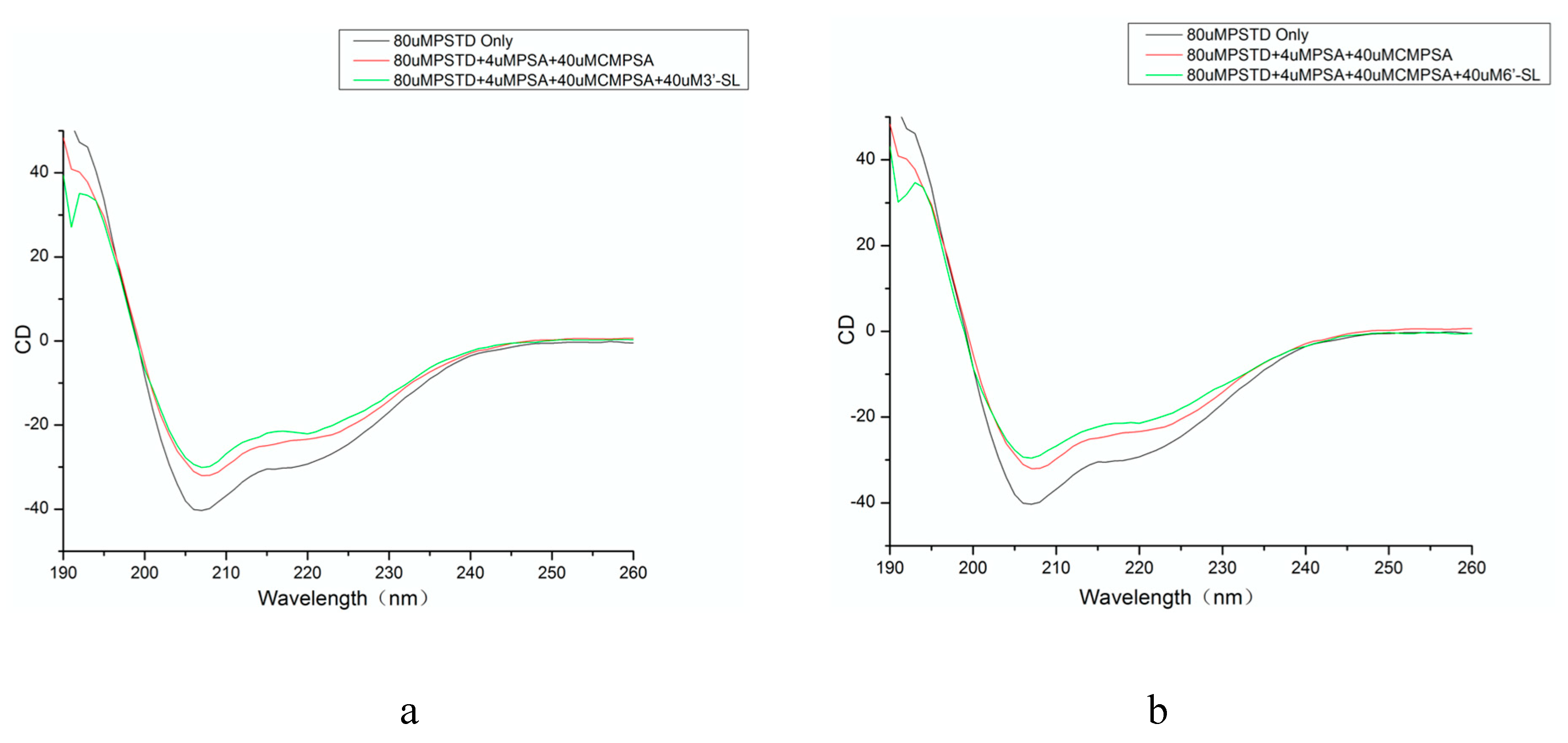
Figure 2.
The overlaid 1H-15N HSQC spectra of the PSTD in the absence and the presence of 0.5mM 3’-SL (a), and 1mM 3’-SL (b), respectively.
Figure 2.
The overlaid 1H-15N HSQC spectra of the PSTD in the absence and the presence of 0.5mM 3’-SL (a), and 1mM 3’-SL (b), respectively.
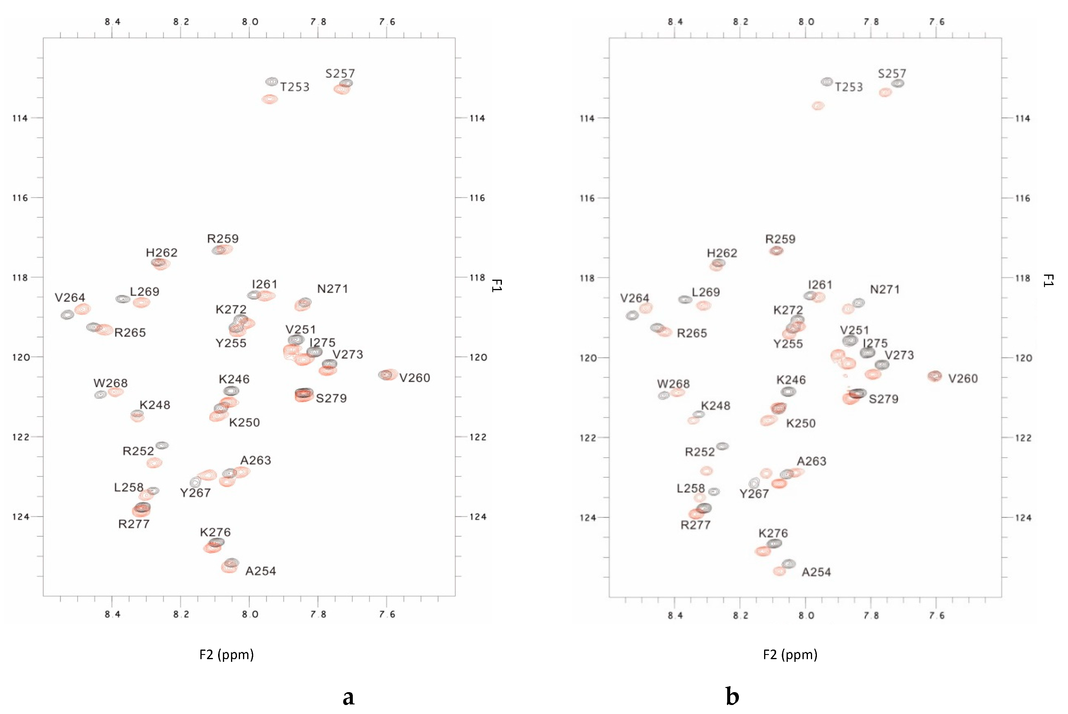
Figure 3.
Chemical shift perturbations (CSPs) of the PSTD when the PSTD interacted with CMP-Sia (1mM), 0.5 mM 3’-SL, 1mM 3’-SL, and 3mM 3’-SL, respectively (a); and the CSPs of the PSTD when it interacted with 0.1 mM PSA, 0.5 mM 3’-SL and 2 mM 3’-SL, respectively (b).
Figure 3.
Chemical shift perturbations (CSPs) of the PSTD when the PSTD interacted with CMP-Sia (1mM), 0.5 mM 3’-SL, 1mM 3’-SL, and 3mM 3’-SL, respectively (a); and the CSPs of the PSTD when it interacted with 0.1 mM PSA, 0.5 mM 3’-SL and 2 mM 3’-SL, respectively (b).
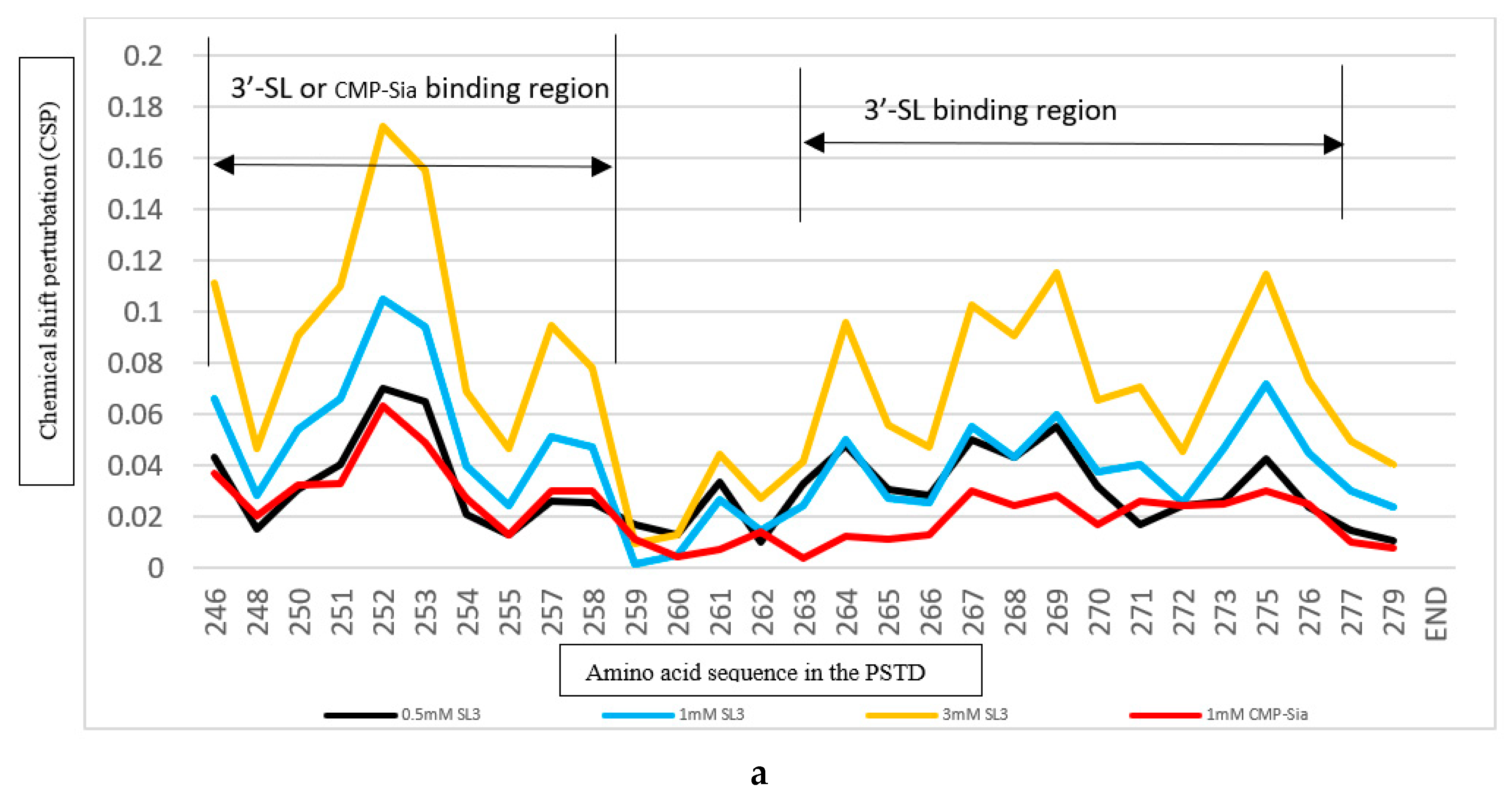
Figure 4.
The binding regions of CMP-Sia, 3’-SL and 6’-SL to the PSTD model based on the results shown in Figure 2, 3, 5, and 6, respectively.
Figure 4.
The binding regions of CMP-Sia, 3’-SL and 6’-SL to the PSTD model based on the results shown in Figure 2, 3, 5, and 6, respectively.
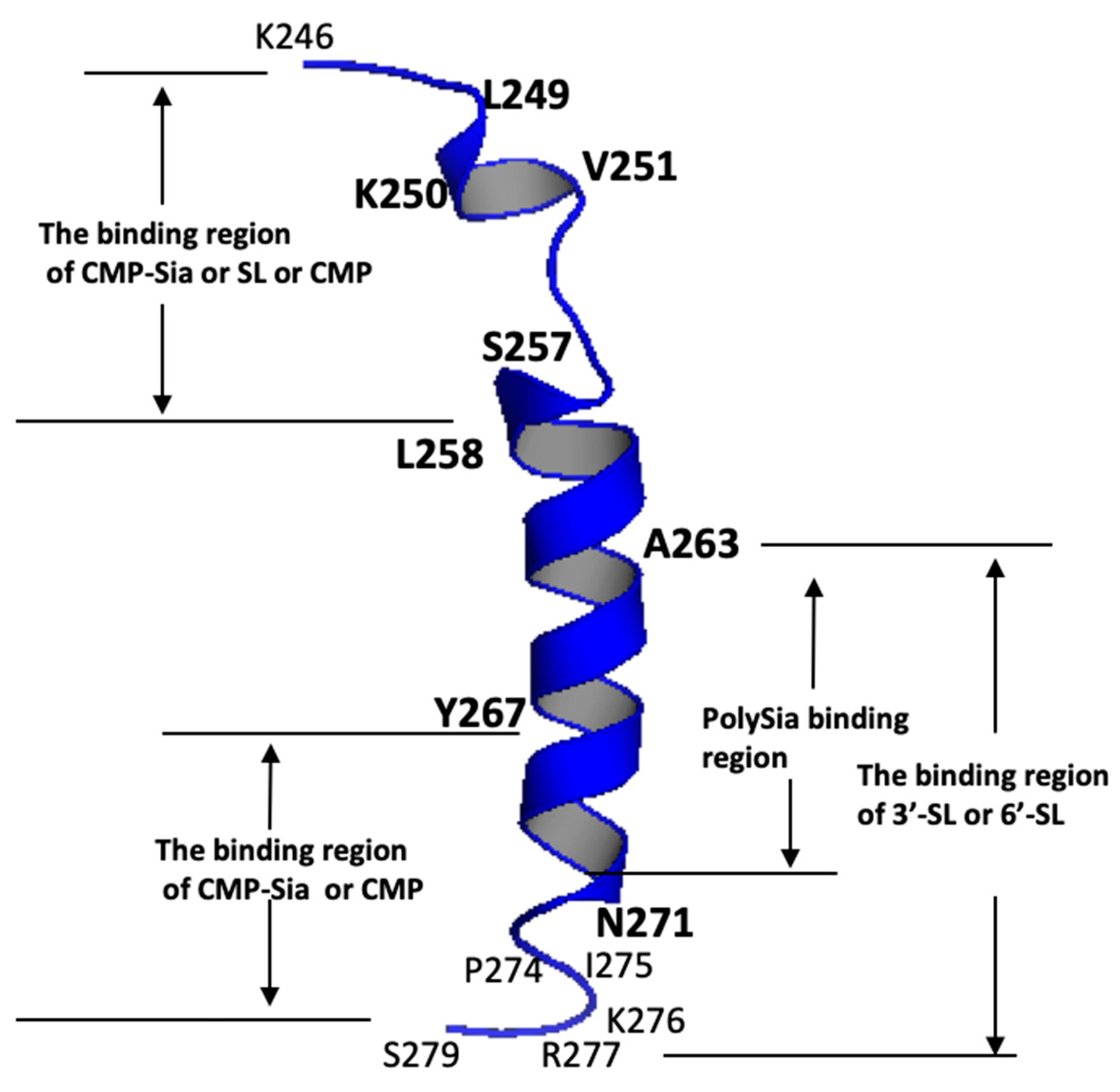
Figure 5.
The overlaid 1H-15N HSQC spectra of the PSTD in the absence and the presence of 1.0 mM 6’-SL (a), and 2.0 mM 6’-SL (b), respectively.
Figure 5.
The overlaid 1H-15N HSQC spectra of the PSTD in the absence and the presence of 1.0 mM 6’-SL (a), and 2.0 mM 6’-SL (b), respectively.
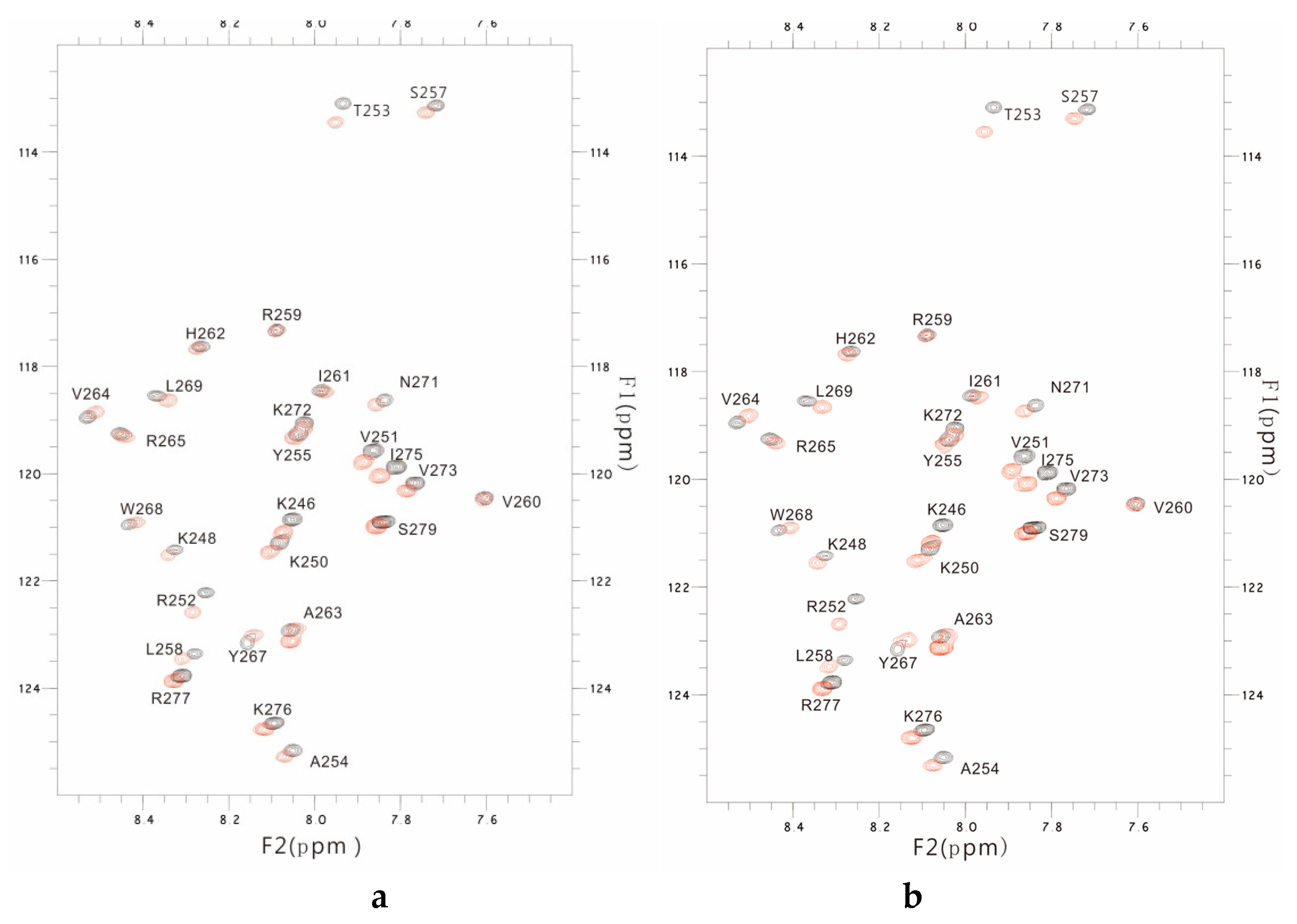
Figure 6.
The Chemical shift perturbations (CSPs) of the PSTD in the presence of 0.5, 1.0, 2.0, and 3.0 mM 6’-SL (a); The CSPs of the PSTD when it interacted with 1mM CMP-Sia, and 1.0 mM 6-SL, respectively (b); and the CSPs of the PSTD when it interacted with 0.1 mM PSA, 2.0 mM and 3 mM 6’-SL, respectively (c).
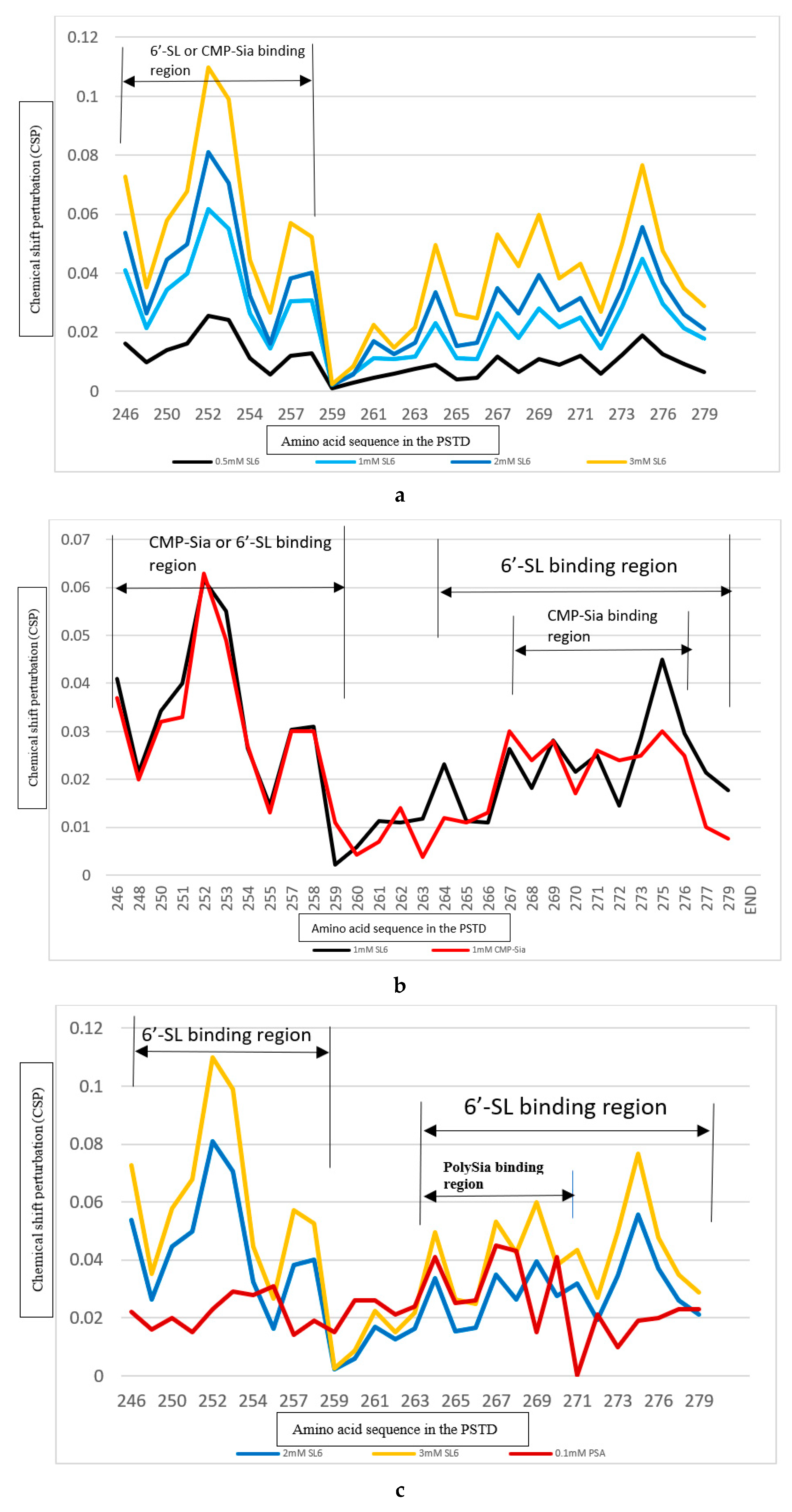
Figure 7.
The Comparison of the inhibition of the SL (3’-SL and 6’-SL), heparin (LMWH) on the PSTD-(CMP-Sia) and the PSTD-polySia Interactions. The CSPs of the PSTD for the PSTD-0.5 mM 3’-SL, the PSTD-1 mM 6’-SL, the PSTD-1mM (CMP-Sia), and the PSTD-80 uM LMWH interactions (a); The CSPs of the PSTD for the PSTD-0.5 mM 3’-SL, the PSTD-3 mM 6’-SL, the PSTD- 80 uM LMWH, and the PSTD-0.1 mM polySia interactions (b).
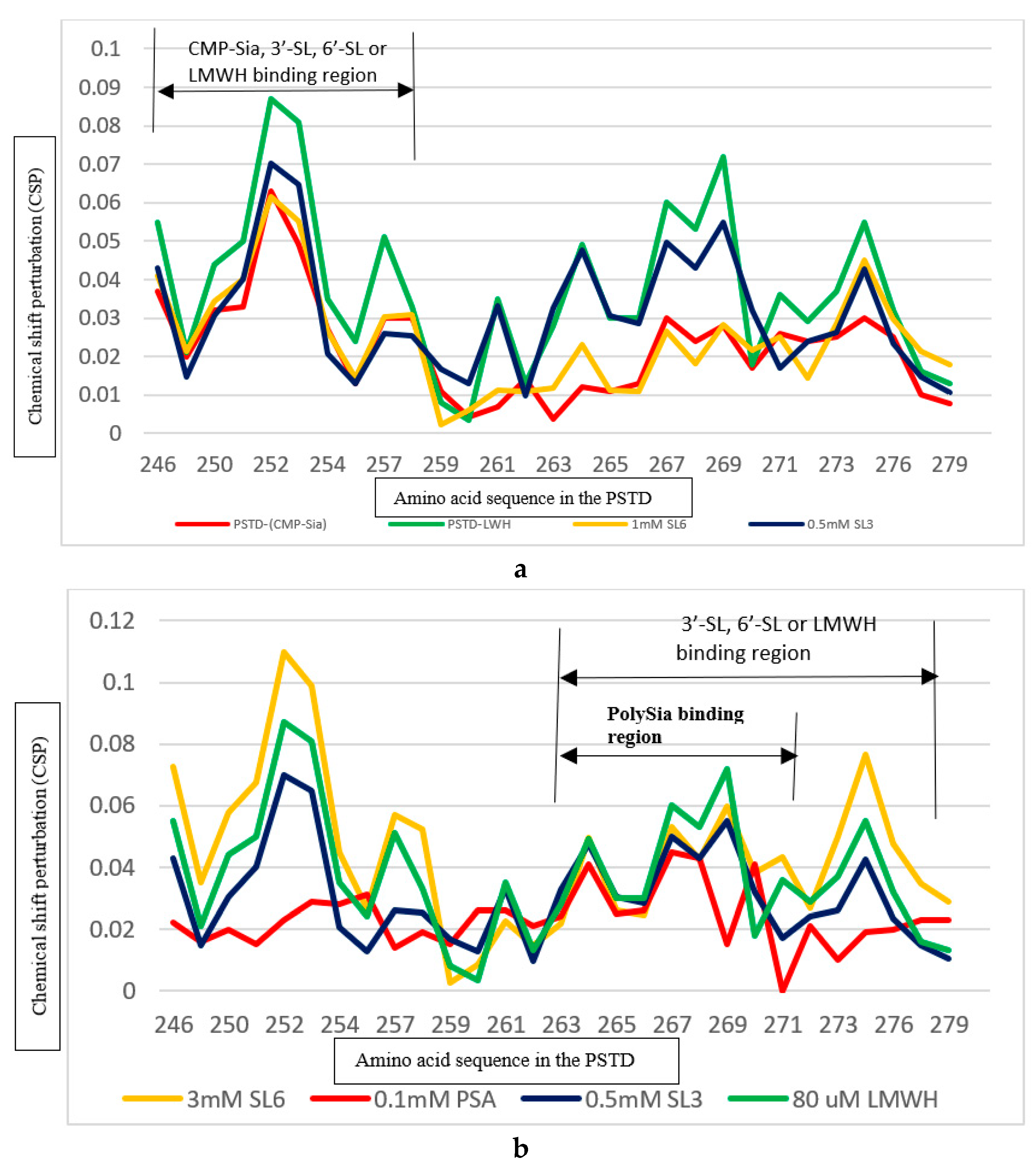
Figure 8.
The Comparison of the inhibitions of the SLs and CMP on the PSTD-(CMP-Sia) interactions and the PSTD-polySia interactions. The CSPs of the PSTD for the PSTD-(0.5 mM 3’-SL), the PSTD- 1 mM CMP, and the PSTD-(1mM CMP-Sia) interactions (a); The CSPs of the PSTD for the PSTD-(0.5 mM 3’-SL), the PSTD- 1 mM CMP, and the PSTD-(0.1 mM polySia) interactions (b); The CSPs of the PSTD for the PSTD-(1 mM 6’-SL), the PSTD- 1 mM CMP, and the PSTD-(1mM CMP-Sia) interactions (c); The CSPs of the PSTD for the PSTD-(3 mM 3’-SL), the PSTD- 1 mM CMP, and the PSTD-(0.1 mM polySia) interactions (d).
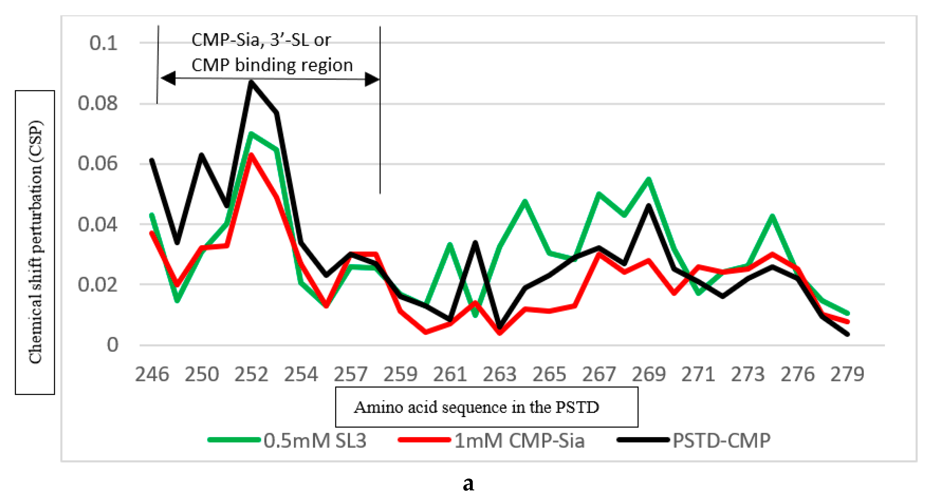
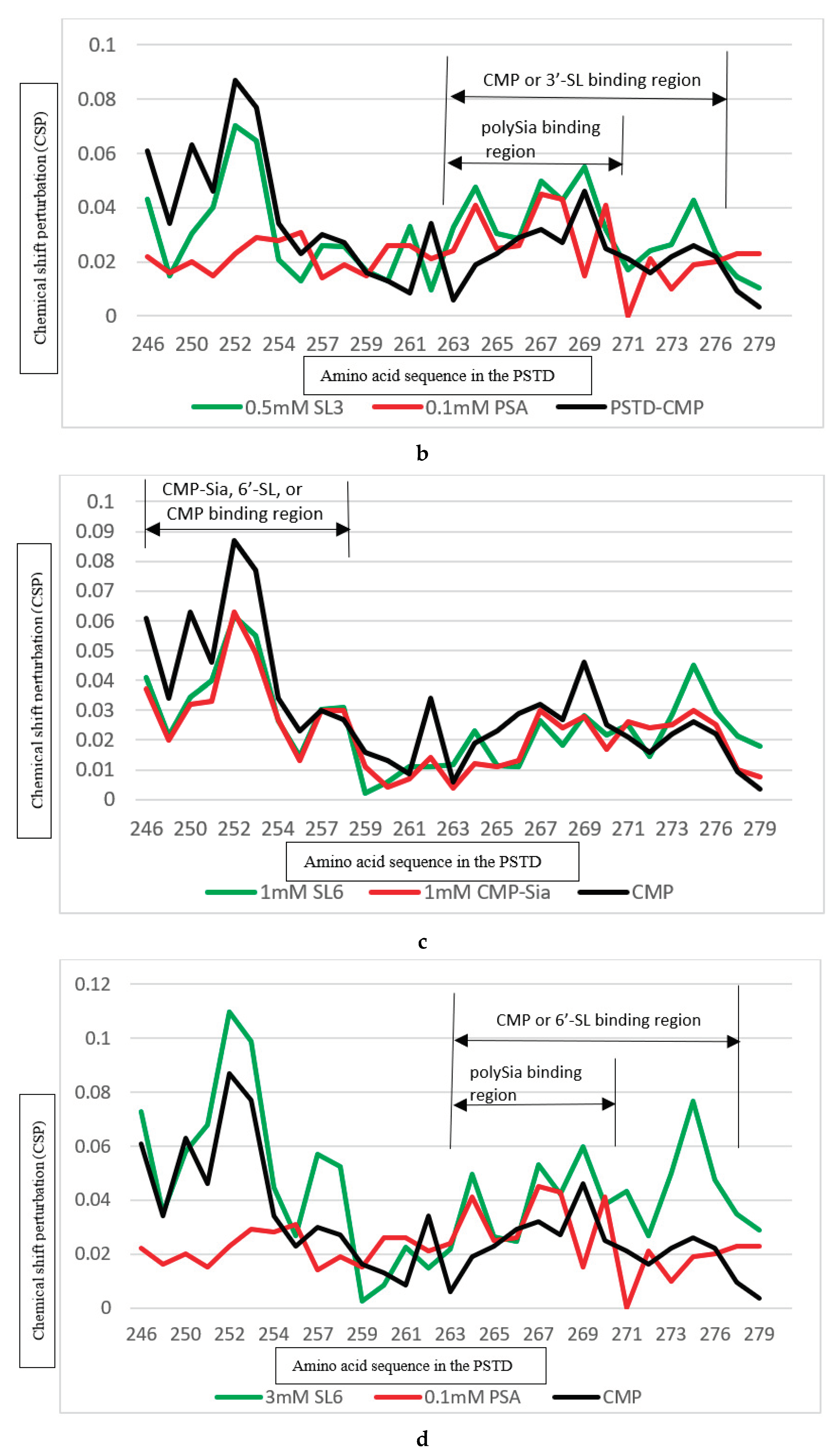

Table 1.
The helix content of the PSTD in the absence and the presence of the ligands (CMP-Sia, polySia), and SL (3’-SL or 6’-SL).
Table 1.
The helix content of the PSTD in the absence and the presence of the ligands (CMP-Sia, polySia), and SL (3’-SL or 6’-SL).
| The helical content of the PSTD in the absence of any ligands (%) | The helical content of the PSTD in the presence of CMP-Sia and polySia (%) | The helical content of the PSTD in the presence of CMP-Sia, polySia and 3’-SL (%) | The helical content of the PSTD in the presence of CMP-Sia, polySia and 6’-SL (%) |
|---|---|---|---|
| 26.5 | 18.4 | 17.7 | 14.5 |
Table 2.
The measured maximum concentrations of SLs during 1st month of the lactation, and the SL's concentrations when the chemical shift perturbations (CSPs) for the PSTD-SL (3'-SL or 6'-SL) binding are close to that of the PSTD-(CMP-Sia) and the PSTD-polySia bindings, respectively. CSPs were obtained based on the 2D 1H-15N HSQC experiments.
| Sialylactose (SL) interacted with the PSTD | SL’s concentration when CSPs of PSTD-SL binding are close to that of PSTD-(CMP-Sia) binding in same amino acid range of the PSTD (mM) | SL’s concentration when CSPs of PSTD-SL binding are close to that of PSTD-polySia binding in same amino acid range of the PSTD (mM) | The maximum concentration of the SL in human milk during 1st month of lactation (mM)[9] |
|---|---|---|---|
| 3’-SL | 0.5 | 0.5 | Ca. 0.5 |
| 6’-SL | 1.0 | Ca. 3.0 | Ca. 1.0 |
Table 3.
The summary of the binding regions of the ligands CMP-Sia, polySia, heparin LMWH, CMP, 3’-SL (0.5 mM) and 6’-SL (1 mM) on the PSTD, the maximum CSPs in each binding region, and CMP-Sia and polySia binding regions are covered by the binding regions of other ligands on the PSTD. CSPs were obtained based on the 2D 1H-15N HSQC experiments.
| Ligands binding to the PSTD | The main binding region of the ligands in the PSTD | The maximum CSPs of each binding region | Is CMP-Sia binding inhibited by the following ligand? | Is polySia binding inhibited by the following ligand? |
|---|---|---|---|---|
| CMP-Sia | K246-S257 V267-276 |
0.070 0.026 |
N/A N/A |
No No |
| polySia | A263-N271 | 0.049 | No | N/A |
| Heparin LMWH | K246-S257 A263-R277 |
0.087 0.072 |
Yes N/A |
N/A Yes |
| CMP | K246-S257 A263-R277 |
0.087 0.046 |
Yes N/A |
N/A No |
| 3’-SL (0.5 mM) |
K246-S257 A263-R277 |
0.070 0.043 |
Yes, when 3’-SL concentration more than 0.5 mM N/A |
N/A Yes, when 3’-SL concentration more than 0.5 mM |
| 6’-SL (1mM) |
K246-S257 A263-R277 |
0.070 0.026 |
Yes, when 6’-SL concentration more than 1 mM N/A |
N/A Yes, when 6’-SL concentration is about 3 mM |
Disclaimer/Publisher’s Note: The statements, opinions and data contained in all publications are solely those of the individual author(s) and contributor(s) and not of MDPI and/or the editor(s). MDPI and/or the editor(s) disclaim responsibility for any injury to people or property resulting from any ideas, methods, instructions or products referred to in the content. |
© 2024 by the authors. Licensee MDPI, Basel, Switzerland. This article is an open access article distributed under the terms and conditions of the Creative Commons Attribution (CC BY) license (http://creativecommons.org/licenses/by/4.0/).
Copyright: This open access article is published under a Creative Commons CC BY 4.0 license, which permit the free download, distribution, and reuse, provided that the author and preprint are cited in any reuse.


