
Submitted:
21 September 2023
Posted:
22 September 2023
You are already at the latest version
Abstract
This hypothesis demonstrates that the efficiency of Loop-Mediated Isothermal Amplification (LAMP) for nucleic acid detection can be positively influenced by preconcentration of microbial cells onto hydrophobic paper surfaces. The mechanism of this model is based on the high attraction of microbes towards hydrophobic surfaces. Extensive studies have demonstrated that hydrophobic surfaces exhibit enhanced bacterial and fungal adhesion. The preconcentration approach by exploiting this inherent affinity of hydrophobic paper substrates enables the adherence of a greater number of target cells, resulting in a higher concentration of target templates for amplification directly from urine samples. In contrast to conventional methods, which often involve complex procedures, this approach offers a simpler, cost-effective, and user-friendly alternative. Moreover, the integration of cell adhesion, LAMP amplification, and signal readout within paper origami-based devices can provide a portable, robust, and highly efficient platform for rapid nucleic acid detection. This innovative hypothesis holds significant potential for point-of-care (POC) diagnostics and field surveillance applications. Further research and development in this field will advance the implementation of this technology, contributing to improved healthcare systems and public health outcomes.
Keywords:
preconcentration
; loop-mediated isothermal amplification (LAMP)
; Urine
; microbial adhesion
; hydrophobic
; paper-based analytical devices (μPADs)
; cell surface hydrophobicity (CSH)
; UTI
Introduction
The human body acts as favourable environment for pathogenic organisms to invade, grow and reproduce. Our body comprises of cells in the order of 1013 as well as thousands of microbial species, containing approximately 1014-1015 bacterial cells [1]. The microbial colonization in distinct regions of the human body, specifically the skin, oral cavity, gastrointestinal tract, and vaginal mucosa, exerts a profound impact on the development of a wide range of infections [2]. Notably, urinary tract infection (UTI) emerges as a pervasive and clinically significant infection among them [3,4]. It is worth highlighting that, around 50% of the individuals have encountered the distressing experience of UTI at least once in their lifetime [5]. Also, it is crucial to acknowledge the significant financial burden imposed by UTIs, as evidenced by an expenditure of nearly 2.8 billion USD in 2011 [6]. The etiology of these infections involves a diverse range of microbial agents, encompassing gram-positive bacteria, gram-negative bacteria, and fungi [7].
The uropathogenic Escherichia coli, a member of the Enterobacteriaceae family, stands out as the primary causative agent, accounting for more than 80% of UTI cases [3]. Other pathogens include Klebsiella species, Proteus species, Pseudomonas aeruginosa, and Enterococcus species [8]. These infections exert a substantial impact on the morbidity rates of different patient populations, including infants, elderly males, and females of all age groups [7]. UTI is divided into two categories based on disease progression among vulnerable individuals. The first category is known as "uncomplicated UTI”, which primarily affects individuals without underlying health conditions. The second category is called "complicated UTI, which is associated with factors such as catheterization, immunosuppression, or prior antibiotic usage [9]. Technically, for a patient presenting symptoms of a urinary tract infection, any microbial concentration in the test results may indicate an infection. However, in order to clinically diagnosed as UTI, the microbial load should surpass a threshold of 105 colony-forming units per millilitre (CFU/ml) of urine [10]. In the realm of urinary tract infection detection, the gold standard diagnostic method involves the collection of urine samples, followed by subsequent culture [11]. Although this approach is highly reliable, it requires a turnaround time of 2-3 days for result acquisition and relies on the expertise of trained personnel. Commercial urinary-tract-infection dipstick tests are available as an alternative; however, their results are less reliable compared to urine culture tests [7].
Recently, there has been a growing interest and importance surrounding Microfluidic Paper-based Analytical Devices (µPADs) and Microfluidic Thread-based Analytical Devices (µTADs) for the detection of pathogen biomarkers [12,13,14,15,16,17,18,19,20], food adulterants [21,22,23] and chemical analyte detection [24]. These devices focus on the unique properties of paper such as uniform thickness, high hygroscopicity, optimal wicking, desirable flow rates, consistent fluid flow, efficient sample absorption, and reliable analyte transportation [25]. Furthermore, paper can be easily modified and functionalized with specific chemicals or coatings, allowing for selective detection of disease specific biomarkers or pathogens through visual color changes or other readout methods [26]. The World Health Organization (WHO) has established essential guidelines for future diagnostics through the ASSURED criteria, which stands for affordable, sensitive, specific, user-friendly, rapid, and robust, equipment-free and deliverable to end users [27]. Point-of-care (POC) diagnostics offer a promising solution that aligns with these defined criteria, presenting numerous advantages such as affordability, sustainability, portability, disposability, simplicity, and the ability to handle very small volumes of samples [28]. The simplicity and robustness of µPADs facilitate effortless operation without the need for extensive training [29]. Furthermore, their cost-effective manufacturing renders them well-suited for large-scale production, making them accessible to a wide population, who may not have the means to afford expensive and highly sophisticated diagnostic alternatives [30]. They also eliminate the need for complex machinery, making them ideal for clinical use [27] .
In the past few years, the benefits of Loop-Mediated Isothermal Amplification (LAMP) on Microfluidic Paper-based Analytical Devices (µPADs) are opening new avenues for point-of-care nucleic acid testing in resource-limited settings [31]. LAMP can be considered as a low-cost alternative to PCR. It is highly specific due to the involvement of five distinct primers [32]. Besides, this method is easy to use, as it enables the amplification to be carried out in a dry block heater or an incubator. Despite its numerous advantages, the susceptibility of LAMP to amplification inhibitors necessitates a range of reaction additives and enhancement strategies to improve its sensitivity [33]. These strategies include the use of crowding agents, stabilizing nucleic acid structures, enzyme stabilization, oligonucleotide modifications, and template blockers [33]. However, compared to the simple and facile approaches to increase the amplification efficiency, these strategies have several inherent disadvantages. Firstly, they add complexity to the LAMP protocol, involving additional steps, reagents, and optimization which complicate the workflow and increase the chance of errors. Secondly, the use of sensitivity enhancement techniques often leads to higher costs due to the need for specialized and potentially expensive reagents and modifications, making them less accessible in resource-limited settings. Schematic illustration showing dynamic paper substrates with tuned hydrophobicity for capturing pathogens from human urine samples is shown in Figure 1.
In light of the limitations mentioned above, our hypothesis aims to explore the potential of preconcentrating microbial cells onto paper surfaces as a means of enhancing the efficiency of LAMP amplification. Generally, different pathogens especially bacteria exhibit varying degrees of binding affinities to abiotic surfaces based on their diverse hydrophobic characteristics [34]. Also, it has been demonstrated that, surfaces with intermediate wettability (hydrophobic) demonstrate a higher affinity for bacterial or cellular binding compared to surfaces that are extremely hydrophobic or hydrophilic [35]. Building upon these compelling observations, we formulate the hypothesis that most of the pathogens in the urine sample would adhere to hydrophobic paper-based devices compared to its hydrophilic or superhydrophobic counterparts. By leveraging this characteristic, we can facilitate the concentration of microbial cells onto hydrophobic paper surfaces, thereby improving efficiency of the subsequent amplification, as the result, specific pathogen detection.
The Hypotheses
Our hypothesis deals with a robust method of using paper-based adsorbents to concentrate microbial cells present in the urine sample, upon which loop-mediated isothermal amplification (LAMP) can be performed. In this model, we exploited the concept of cell surface hydrophobicity (CSH) exhibited by pathogens to effectively concentrate the cells from the urine samples. Drawing insights from existing literatures, we observed that bacterial and fungal pathogens generally display a higher affinity for binding towards moderately hydrophobic surfaces, having a water contact angle of 90o for adhesion [35]. Paper being a versatile material can be easily tuned to produce these hydrophobic surfaces. We anticipate that the dynamic interaction between the pathogenic cells and the hydrophobic paper will facilitate the adhesion process, resulting in effective cell concentration from the sample. This method of pre-concentrating the cells would help us to further improve the efficiency of the LAMP process.
Evaluation of the Hypotheses
Our hypothesis seeks to explore the potential of preconcentrating microbial cells in urine onto hydrophobic paper surfaces as a means of improving LAMP amplification efficiency. We draw upon the understanding that surfaces with intermediate wettability, specifically hydrophobicity, demonstrate a higher affinity for bacterial or cellular binding compared to extremely hydrophobic or hydrophilic surfaces [35]. The adhesive forces between an hydrophobic abiotic substrate and microbes arise through van der Waals and electrostatic double-layer interactions [36]. Numerous studies have documented comparable results and reported similar findings. For instance, Tegoulia and Cooper utilized thiol surfaces with differing functional end groups to study the effect of surface hydrophilicity and hydrophobicity on Staphylococcus aureus adhesion and found that the bacterial adhesion was higher on the hydrophobic surfaces [37]. In a research study conducted by Yuan et al., demonstrated that, moderate hydrophobicity with Water Contact Angle (WCA) of about 90ο showed enhanced adhesion of E.coli on polymeric substrates, whereas the adhesion of the bacteria on hydrophilic surfaces and superhydrophobic surfaces was found to be limited [35]. The reduced affinity of the bacteria to hydrophilic surface was attributed to diminished hydrophobic interaction and the repulsive interaction between the bacteria and the substrate [35]. Conversely, when the bacteria were exposed to a superhydrophobic surface, the contact area fraction between the pathogen and the surface was reduced due to the air entrapment at the interface, leading to lower bacterial adhesion [35]. Previously, the relationship between surface energy/interfacial interaction energy and bacterial adhesion has been elucidated by various models, including the thermodynamic theory [36]. According to this theory, bacteria with hydrophobic cell surfaces exhibit a preference for hydrophobic surfaces, which have a lower surface energy, while bacteria with hydrophilic cell surfaces tend to favour hydrophilic surfaces, which possess higher surface energy [38].
The adherence of pathogens onto hydrophobic surfaces is attributed to various underlying mechanisms that are specific to different pathogens. These mechanisms enable pathogens to interact and bind more effectively to hydrophobic surfaces. One contributing factor to pathogen adhesion on hydrophobic surfaces is the development of specific adaptive mechanisms for their survival [39]. These mechanisms enable bacteria to modify their cell surfaces in response to toxicity and limited availability of nutrients [39]. By adjusting their hydrophobicity, bacteria facilitate direct hydrophobic-hydrophobic interactions with the substrates, promoting adhesion [39]. In addition to structural adaptations, some pathogens exhibit changes in their cell surface hydrophobicity (CSH); a biophysical measurement of a cell’s affinity for a hydrophobic versus hydrophilic environment [38]. Cells with higher CSH prefer a hydrophobic environment while those with lower CSH will preferentially remain in an aqueous or hydrophilic environment. Furthermore, the presence of hydrophobins, which are small secreted proteins produced by fungi, significantly impacts adhesion [40]. Hydrophobins possess moderate to high hydrophobicity and have been extensively studied for their involvement in facilitating hydrophobic interactions [40]. Table 1 provides a comprehensive overview of the common pathogens found in urine along with their respective favourable surface hydrophobicity for adhesion. A Compilation of existing literatures on microbial adhesion to various hydrophobic material surfaces is depicted in Table 2.

Table 2.
Existing literatures on microbial adhesion to various hydrophobic material surfaces.
| Sl. No | Hydrophobic Material | Pathogen | Detection Method | Ref |
|---|---|---|---|---|
| 1 | Polymeric substrate film | S. aureus and E. coli | Fluorescence assay with Green Fluorescent Protein (GFP) and Bright Field Microscopy | [35] |
| 2 | Knitted polypropylene (PP) and poly-4-hydroxybutyrate (P4HB) | S. aureus and E. coli | Scanning Electron Microscopy (SEM) | [47] |
| 3 | Titanium dioxide (TiO2) surface | S. epidermidis | Fluorescence | [48] |
| 4 | Plastic surface | C. albicans | Hemacytometer measurement | [49] |
| 5 | Silane surface | Two strainsof E. coli, JM109 and D21 and two strains of B. cepacia, G4and Env435 | Column adhesion tests | [50] |
| 6 | Hydrophobic Steel Surface | E. coli | Scanning Electron Microscopy (SEM) | [51] |
Extensive research findings have unveiled the remarkable potential of paper substrates in achieving precise tuning of hydrophobicity [52,53]. This arises from factors that render paper uniquely amenable to such modifications. Firstly, the inherent porosity of paper facilitates facile integration of hydrophobic materials or coatings, thereby augmenting its hydrophobic properties [54]. Secondly, the surface roughness of paper can be finely controlled or tailored to exert a profound influence on its hydrophobic characteristics. To date, several methods have been developed to create hydrophobic paper surfaces using various physical and chemical techniques [55,56]. These techniques include plasma treatment [57], construction of micro-structured surfaces using micro-sized CaCO3 and fatty acid [58], rapid extension of supercritical CO2 containing alkyl ketene dimer (AKD) through spraying [59], chemical vapor deposition (CVD) of silica particles and polymers [60], dip-coating with AKD [61], and surface-coating by grafting polymers [62]. By implementing these techniques, it is possible to modify the wettability of paper substrates to achieve the desired level of hydrophobicity. This enables the effective preconcentration of microbial cells from urine through the process of microbial adhesion.
Based on the well-established observation that microbes exhibit a higher affinity towards different hydrophobic substrates, we propose a hypothesis that, the microbes would attach in a similar fashion to paper-based devices. By preconcentrating microbial cells in urine onto hydrophobic paper surfaces, a greater number of target cells will be adhered, leading to higher concentration of nucleic acids for amplification. This in turn expected to enhance the overall efficiency of the LAMP assay. In contrast to conventional methods employed to increase LAMP sensitivity, which often involve complex and time-consuming procedures, the preconcentration of microbes onto hydrophobic paper substrates offers a simpler and more cost-effective alternative. By focusing on the innate affinity of microbial cells for hydrophobic surfaces, selective and efficient cell capture can be achieved. Moreover, the reduced reliance on complex equipment and the elimination of laborious manual steps makes this approach more user-friendly and accessible for molecular diagnostics, particularly in resource-limited or remote settings. The proposed approach also holds significant implications for the specific colorimetric detection of pathogens in urine samples [63,64], highlighting its importance in advancing diagnostic capabilities for timely and accurate identification of microbial infections. The simplicity and cost-effectiveness of colorimetric methods make them highly suitable for resource-limited environments and field applications [65,66].
Furthermore, the approach can be effectively implemented in paper origami-based devices for rapid and onsite nucleic acid detection by integrating key components such as cell adhesion, cell lysis, amplification, and signal readout. By incorporating cell adhesion mechanisms into the design, the paper device allows for the targeted capture and concentration of pathogenic cells. This preconcentration step enhances the sensitivity of the assay by ensuring a higher concentration of the target cells. Subsequently, the LAMP amplification step, known for its isothermal and rapid nature, facilitates the amplification of the target nucleic acids, resulting in improved sensitivity and specificity of the detection. The integration of amplification directly within the paper device streamlines the workflow and eliminates the need for additional equipment. Finally, the signal readout component of the device allows for the interpretation and analysis of the amplified signals, enabling rapid and onsite nucleic acid detection. An Artificial intelligence (AI) powered smart-phone app with machine learning algorithm capture and analyse the output signals generated by nucleic acid samples in real-time, providing accurate and reliable results while identifying and rectifying sources of interference. Thus, by combining the benefits derived from cell adhesion-based preconcentration, LAMP amplification, and user-friendly AI-based signal readout mechanisms within a paper origami-based device, a resilient and portable platform is anticipated to be established. This platform holds immense potential for facilitating sensitive and rapid nucleic acid detection across diverse applications, such as point-of-care diagnostics and field surveillance.
Limitations
- (1)
- In the initial phase of adhesion, there is a possibility for capturing non-target microbes or metabolites. This is because when two different pathogens share similar hydrophobicity and diffusion coefficients, they typically exhibit similar adherence patterns to hydrophobic paper substrates.
- (2)
- Small variations in the fabrication of hydrophobic paper surfaces, or the surface properties of fabricated hydrophobic paper may lead to inconsistencies in microbial adhesion and pre-concentration efficiency. This would affect the reproducibility of results between different batches or experiments.
- (3)
- There can be various other factors apart from cell surface hydrophobicity (CSH) which could impact the adhesion of the cells to a particular surface. The impact of these confounding factors also needs to be considered. The rate of adhesion can differ depending on the culture conditions of the sample, and hence reproducibility of the results might become an issue.
- (4)
- Due to limited research in the field of microbial adhesion onto hydrophobic paper adsorbents, there may be significant aspects of the topic that have not been sufficiently addressed in this manuscript. Consequently, it is essential to conduct experimental verification of the hypothesis in future studies to ensure its validity.
- (5)
- Consequence of the Hypotheses and Discussion
Our hypothesis investigates the potential of preconcentrating microbial cells from urine onto hydrophobic paper surfaces as a promising approach to enhance the efficiency of LAMP amplification. Through the innate affinity of microbial cells for hydrophobic substrates, a higher concentration of target cells is adhered, leading to increased concentration of nucleic acids for amplification. This preconcentration approach on hydrophobic paper substrates presents several advantages over conventional methods, including simplicity, cost-effectiveness, and suitability for resource-limited settings. By integrating cell adhesion-based preconcentration, LAMP amplification, and user-friendly AI-based signal readout within a paper origami-based device, a robust and portable platform can be established for sensitive and rapid nucleic acid detection. This innovative approach holds significant potential for various applications, ranging from point-of-care diagnostics to field surveillance, and represents a major stride towards advancing molecular diagnostics in diverse settings. Further research and development in this area will undoubtedly contribute to the refinement and widespread implementation of this technology, ultimately benefiting healthcare systems and improving public health outcomes.
Consent statement/Ethical approval
Not required.
Data statement
Not applicable.
Declaration of Competing Interest
The authors declare that they have no known competing financial interests or personal relationships that could have appeared to influence the work reported in this paper.
Funding
This research received no specific grant from any funding agency in the public, commercial, or not-for-profit sectors.
References
- Sender R, Fuchs S, Milo R. Revised Estimates for the Number of Human and Bacteria Cells in the Body. PLoS Biol 2016, 14, e1002533. [Google Scholar] [CrossRef]
- Hou K, Wu ZX, Chen XY, Wang JQ, Zhang D, Xiao C, et al. Microbiota in health and diseases. Signal Transduct Target Ther 2022, 7. [Google Scholar] [CrossRef]
- McLellan LK, Hunstad DA. Urinary Tract Infection: Pathogenesis and Outlook. Trends Mol Med 2016, 22, 946–957. [Google Scholar] [CrossRef] [PubMed]
- Hasandka A, Singh AR, Prabhu A, Singhal HR, Nandagopal MSG, Mani NK. Paper and thread as media for the frugal detection of urinary tract infections (UTIs). Anal Bioanal Chem 2022, 414, 847–865. [Google Scholar] [CrossRef]
- Adrover-Jaume C, Rojo-Molinero E, Clemente A, Russell SM, Arranz J, Oliver A, et al. Mobile origami immunosensors for the rapid detection of urinary tract infections. Analyst 2020, 145, 7916–7921. [Google Scholar] [CrossRef]
- Simmering JE, Tang F, Cavanaugh JE, Polgreen LA, Polgreen PM. The Increase in Hospitalizations for Urinary Tract Infections and the Associated Costs in the United States, 1998-2011. Open Forum Infect Dis 2017, 4, ofw281. [CrossRef]
- Flores-Mireles AL, Walker JN, Caparon M, Hultgren SJ. Urinary tract infections: epidemiology, mechanisms of infection and treatment options. Nat Rev Microbiol 2015, 13, 269–284. [Google Scholar] [CrossRef]
- Urinary tract infection caused by bacteria other than Escherichia coli. Arch Dis Child 2006, 91, 168.
- Sabih A, Leslie SW. Complicated Urinary Tract Infections., Treasure Island (FL): 2023.
- Torres-Sangiao E, Lamas Rodriguez B, Cea Pájaro M, Carracedo Montero R, Parajó Pazos N, García-Riestra C. Direct Urine Resistance Detection Using VITEK 2. Antibiot (Basel, Switzerland) 2022, 11. [CrossRef]
- Huang B, Zhang L, Zhang W, Liao K, Zhang S, Zhang Z, et al. Direct Detection and Identification of Bacterial Pathogens from Urine with Optimized Specimen Processing and Enhanced Testing Algorithm. J Clin Microbiol 2017, 55, 1488–1495. [Google Scholar] [CrossRef]
- Sudarsan S, Prabhu A, Prasad D, Mani NK. DNA Compaction Enhances Sensitivity of Fluorescence-Based Nucleic Acid Assays: Game changer in Point of Care Sensors? Analyst 2023.
- Kelkar N, Prabhu A, Prabhu A, Giri Nandagopal MS, Mani NK. Sensing of body fluid hormones using paper-based analytical devices. Microchem J 2022, 174, 107069. [Google Scholar] [CrossRef]
- Hasandka A, Prabhu A, Prabhu A, Singhal HR, Nandagopal M. S. G, Shenoy R, et al. “Scratch it out”: carbon copy based paper devices for microbial assays and liver disease diagnosis. Anal Methods 2021, 13, 3172–3180. [Google Scholar] [CrossRef]
- Prabhu A, Singhal H, Giri Nandagopal MS, Kulal R, Peralam Yegneswaran P, Mani NK. Knitting Thread Devices: Detecting Candida albicans Using Napkins and Tampons. ACS Omega 2021, 6, 12667–12675. [Google Scholar] [CrossRef]
- Singhal HR, Prabhu A, Giri Nandagopal MS, Dheivasigamani T, Mani NK. One-dollar microfluidic paper-based analytical devices: Do-It-Yourself approaches. Microchem J 2021, 165, 106126. [Google Scholar] [CrossRef]
- Prabhu A, Nandagopal M. S. G, Peralam Yegneswaran P, Prabhu V, Verma U, Mani NK. Thread integrated smart-phone imaging facilitates early turning point colorimetric assay for microbes. RSC Adv 2020, 10, 26853–26861. [Google Scholar] [CrossRef] [PubMed]
- Prabhu A, Giri Nandagopal MS, Peralam Yegneswaran P, Singhal HR, Mani NK. Inkjet printing of paraffin on paper allows low-cost point-of-care diagnostics for pathogenic fungi. Cellulose 2020, 27, 7691–7701. [Google Scholar] [CrossRef]
- Mani NK, Das SS, Dawn S, Chakraborty S. Electro-kinetically driven route for highly sensitive blood pathology on a paper-based device. Electrophoresis 2020, 41, 615–620. [Google Scholar] [CrossRef]
- Mani NK, Prabhu A, Biswas SK, Chakraborty S. Fabricating Paper Based Devices Using Correction Pens. Sci Rep 2019, 9, 1752. [Google Scholar] [CrossRef]
- Ray R, Goyal A, Prabhu A, Parekkh S, Maddasani S, Mani NK. Paper-based dots and smartphone for detecting counterfeit country eggs. Food Chem 2023, 403, 134484. [Google Scholar] [CrossRef]
- Ray R, Noronha C, Prabhu A, Mani NK. Latex-Based Paper Devices with Super Solvent Resistance for On-the-Spot Detection of Metanil Yellow in Food Samples. Food Anal Methods 2022, 15, 2664–2674. [Google Scholar] [CrossRef]
- Ray R, Prabhu A, Prasad D, Garlapati V kumar, Aminabhavi TM, Mani NK, et al. Paper-based microfluidic devices for food adulterants: Cost-effective technological monitoring systems. Food Chem 2022, 390, 133173. [Google Scholar] [CrossRef] [PubMed]
- Abdollahi-Aghdam A, Majidi MR, Omidi Y. Microfluidic paper-based analytical devices (µPADs) for fast and ultrasensitive sensing of biomarkers and monitoring of diseases. Bioimpacts 2018, 8, 237–240. [Google Scholar] [CrossRef] [PubMed]
- Bhattarai RK, Pudasaini S, Sah M, Neupane BB, Giri B. Handmade Paper as a Paper Analytical Device for Determining the Quality of an Antidiabetic Drug. ACS Omega 2022, 7, 14074–14081. [Google Scholar] [CrossRef] [PubMed]
- Campbell JM, Balhoff JB, Landwehr GM, Rahman SM, Vaithiyanathan M, Melvin AT. Microfluidic and Paper-Based Devices for Disease Detection and Diagnostic Research. Int J Mol Sci 2018, 19, 2731. [Google Scholar] [CrossRef]
- A. , Shafiq CWNSSJRB. Paper-based analytical devices for clinical diagnosis. Physiol Behav 2017, 176, 139–148. [Google Scholar] [CrossRef]
- Sher M, Zhuang R, Demirci U, Asghar W. Paper-based analytical devices for clinical diagnosis: recent advances in the fabrication techniques and sensing mechanisms. Expert Rev Mol Diagn 2017, 17, 351–366. [Google Scholar] [CrossRef]
- Martinez AW, Phillips ST, Whitesides GM, Carrilho E. Diagnostics for the Developing World: Microfluidic Paper-Based Analytical Devices. Anal Chem 2010, 82, 3–10. [Google Scholar] [CrossRef]
- St John A, Price CP. Existing and Emerging Technologies for Point-of-Care Testing. Clin Biochem Rev 2014, 35, 155–167. [Google Scholar]
- Notomi T, Mori Y, Tomita N, Kanda H. Loop-mediated isothermal amplification (LAMP): principle, features, and future prospects. J Microbiol 2015, 53, 1–5. [Google Scholar] [CrossRef]
- Geddes-McAlister J, Shapiro RS. New pathogens, new tricks: emerging, drug-resistant fungal pathogens and future prospects for antifungal therapeutics. Ann N Y Acad Sci 2019, 1435, 57–78. [Google Scholar] [CrossRef]
- Özay B, McCalla SE. A review of reaction enhancement strategies for isothermal nucleic acid amplification reactions. Sensors and Actuators Reports 2021, 3. [Google Scholar] [CrossRef]
- Zheng S, Bawazir M, Dhall A, Kim HE, He L, Heo J, et al. Implication of Surface Properties, Bacterial Motility, and Hydrodynamic Conditions on Bacterial Surface Sensing and Their Initial Adhesion. Front Bioeng Biotechnol 2021, 9, 1–22. [Google Scholar] [CrossRef]
- Yuan Y, Hays MP, Hardwidge PR, Kim J. Surface characteristics influencing bacterial adhesion to polymeric substrates. RSC Adv 2017, 7, 14254–14261. [Google Scholar] [CrossRef]
- Oh JK, Yegin Y, Yang F, Zhang M, Li J, Huang S, et al. The influence of surface chemistry on the kinetics and thermodynamics of bacterial adhesion. Sci Rep 2018, 8, 1–13. [Google Scholar] [CrossRef]
- Tegoulia VA, Cooper SL. Staphylococcus aureus adhesion to self-assembled monolayers: effect of surface chemistry and fibrinogen presence. Colloids Surfaces B Biointerfaces 2002, 24, 217–228. [Google Scholar] [CrossRef]
- Krasowska A, Sigler K. How microorganisms use hydrophobicity and what does this mean for human needs? Front Cell Infect Microbiol 2014, 4, 1–7. [Google Scholar] [CrossRef]
- Heipieper HJ, Pepi M. Handbook of Hydrocarbon and Lipid Microbiology. Handb Hydrocarb Lipid Microbiol 2010. [CrossRef]
- Bayry J, Aimanianda V, Guijarro JI, Sunde M, Latgé J-P. Hydrophobins--unique fungal proteins. PLoS Pathog 2012, 8, e1002700. [Google Scholar] [CrossRef]
- Farniya F, Jamalli A, Dadgar T. Physicochemical surface characteristics in different pathogenic bacteria. Cogent Biol 2019, 5, 1638572. [Google Scholar] [CrossRef]
- 42. Nogueira BA, Olivella JGB, Sued-Karam BR, Ribeiro PMAP, Neves FPG, Fracalanzza SEL, et al. Biofilm formation, interaction and survival within A549 pneumocytes of Klebsiella pneumoniae clinical strains: identification of pulsotypes, multidrug-resistance and genes coding for adhesins. Brazilian J Dev 2022, 55259–55287. [CrossRef]
- Czerwonka G, Guzy A, Kałuża K, Grosicka M, Dańczuk M, Lechowicz Ł, et al. The role of Proteus mirabilis cell wall features in biofilm formation. Arch Microbiol 2016, 198, 877–884. [Google Scholar] [CrossRef] [PubMed]
- Maikranz E, Spengler C, Thewes N, Thewes A, Nolle F, Jung P, et al. Different binding mechanisms of: Staphylococcus aureus to hydrophobic and hydrophilic surfaces. Nanoscale 2020, 12, 19267–19275. [Google Scholar] [CrossRef] [PubMed]
- Reifsteck F, Wee S, Wilkinson BJ. Hydrophobicity-hydrophilicity of staphylococci. J Med Microbiol 1987, 24, 65–73. [Google Scholar] [CrossRef] [PubMed]
- Ellepola ANB, Samaranayake LP. Investigative methods for studying the adhesion and cell surface hydrophobicity of candida species: An overview. Microb Ecol Health Dis 2001, 13, 46–54. [Google Scholar] [CrossRef]
- Verhorstert KWJ, Guler Z, De Boer L, Riool M, Roovers JPWR, Zaat SAJ. In Vitro Bacterial Adhesion and Biofilm Formation on Fully Absorbable Poly-4-hydroxybutyrate and Nonabsorbable Polypropylene Pelvic Floor Implants. ACS Appl Mater Interfaces 2020, 12, 53646–53653. [Google Scholar] [CrossRef]
- Wassmann T, Kreis S, Behr M, Buergers R. The influence of surface texture and wettability on initial bacterial adhesion on titanium and zirconium oxide dental implants. Int J Implant Dent 2017, 3. [Google Scholar] [CrossRef]
- Klotz SA, Drutz DJ, Zajic JE. Factors governing adherence of Candida species to plastic surfaces. Infect Immun 1985, 50, 97–101. [Google Scholar] [CrossRef]
- Salerno MB, Logan BE, Velegol D. Importance of molecular details in predicting bacterial adhesion to hydrophobie surfaces. Langmuir 2004, 20, 10625–10629. [Google Scholar] [CrossRef]
- Arkan-Ozdemir S, Cansever N, Ilhan-Sungur E. Biofilm Formation of Escherichia coli on Hydrophobic Steel Surface Provided by Laser-Texturing. Johnson Matthey Technol Rev 2022, 186–196. [CrossRef]
- Kim HT, Jung SK, Kim D-E, Park CY, Lee S-Y. Wettability control of paper through substitution between the hydroxyl group and carbon elements using argon-carbon plasma treatment. Vacuum 2022, 205, 111398. [Google Scholar] [CrossRef]
- Gómez N, Quintana E, Villar JC. Effect of Paper Surface Properties on Coated Paper Wettability with Different Fountain Solutions. BioResources 2014, 9, 4226–4241. [Google Scholar] [CrossRef]
- Modaressi H, Garnier G. Mechanism of Wetting and Absorption of Water Droplets on Sized Paper: Effects of Chemical and Physical Heterogeneity. Langmuir 2002, 18, 642–649. [Google Scholar] [CrossRef]
- Wen Q, Guo F, Yang F, Guo Z. Green fabrication of coloured superhydrophobic paper from native cotton cellulose. J Colloid Interface Sci 2017, 497, 284–289. [Google Scholar] [CrossRef]
- Baidya A, Ganayee MA, Jakka Ravindran S, Tam KC, Das SK, Ras RHA, et al. Organic solvent-free fabrication of durable and multifunctional superhydrophobic paper from waterborne fluorinated cellulose nanofiber building blocks. ACS Nano 2017, 11, 11091–11099. [Google Scholar] [CrossRef] [PubMed]
- Balu B, Kim JS, Breedveld V, Hess DW. Tunability of the adhesion of water drops on a superhydrophobic paper surface via selective plasma etching. J Adhes Sci Technol 2009, 23, 361–380. [Google Scholar] [CrossRef]
- Hu Z, Zen X, Gong J, Deng Y. Water resistance improvement of paper by superhydrophobic modification with microsized CaCO3 and fatty acid coating. Colloids Surfaces A Physicochem Eng Asp 2009, 351, 65–70. [Google Scholar] [CrossRef]
- Werner O, Quan C, Turner C, Pettersson B, Wågberg L. Properties of superhydrophobic paper treated with rapid expansion of supercritical CO 2 containing a crystallizing wax. Cellulose 2010, 17, 187–198. [Google Scholar] [CrossRef]
- Yang H, Deng Y. Preparation and physical properties of superhydrophobic papers. J Colloid Interface Sci 2008, 325, 588–593. [Google Scholar] [CrossRef]
- Arbatan T, Zhang L, Fang X-Y, Shen W. Cellulose nanofibers as binder for fabrication of superhydrophobic paper. Chem Eng J 2012, 210, 74–79. [Google Scholar] [CrossRef]
- Carlmark A, Malmström EE. ATRP grafting from cellulose fibers to create block-copolymer grafts. Biomacromolecules 2003, 4, 1740–1745. [Google Scholar] [CrossRef]
- David S, Munteanu RE, Tițoiu AM, Petcu IC, Cernat IC, Leancu C, et al. Direct, Rapid Detection of Pathogens from Urine Samples. Materials (Basel) 2022, 15, 7640. [Google Scholar] [CrossRef] [PubMed]
- Wallis C, Melnick JL, Longoria CJ. Colorimetric method for rapid determination of bacteriuria. J Clin Microbiol 1981, 14, 342–346. [Google Scholar] [CrossRef] [PubMed]
- Luka GS, Nowak E, Kawchuk J, Hoorfar M, Najjaran H. Portable device for the detection of colorimetric assays. R Soc Open Sci 2017, 4, 171025. [Google Scholar] [CrossRef] [PubMed]
- Qian S, Cui Y, Cai Z, Li L. Applications of smartphone-based colorimetric biosensors. Biosens Bioelectron X 2022, 11, 100173. [Google Scholar] [CrossRef]
Figure 1.
Schematic illustration showing dynamic paper substrates with tuned hydrophobicity for capturing pathogens from human urine samples.
Figure 1.
Schematic illustration showing dynamic paper substrates with tuned hydrophobicity for capturing pathogens from human urine samples.
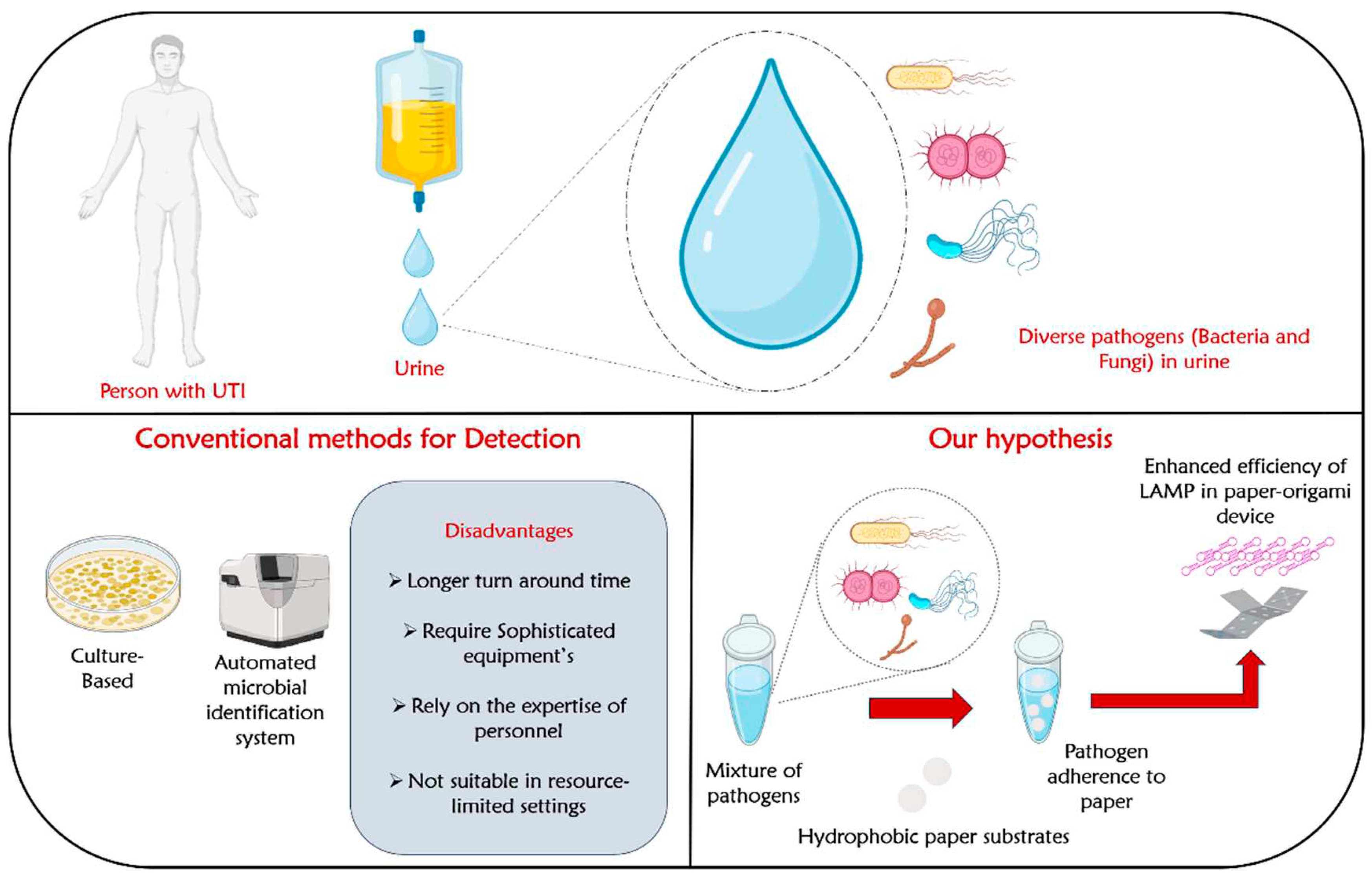
Table 1.
Name of pathogens in urine and their favourable surface hydrophobicity’s for adhesion.
| Sl. No | Type of pathogen | Name of pathogen | Favourable Surface | Ref |
|---|---|---|---|---|
| 1 | Gram negative | E. coli | Hydrophobic | [41] |
| 2 | Gram negative | Klebsiella pneumoniae | Hydrophobic | [42] |
| 3 | Gram negative | Pseudomonas aeruginosa | Hydrophobic | [41] |
| 4 | Gram negative | Proteus mirabilis | Hydrophobic | [43] |
| 5 | Gram positive | Staphylococcus aureus | Hydrophobic and Hydrophilic | [37,44,45] |
| 6 | Gram positive | Listeria monocytogenes | Hydrophobic | [41] |
| 7 | Yeast | Candida albicans | Hydrophobic | [46] |
Disclaimer/Publisher’s Note: The statements, opinions and data contained in all publications are solely those of the individual author(s) and contributor(s) and not of MDPI and/or the editor(s). MDPI and/or the editor(s) disclaim responsibility for any injury to people or property resulting from any ideas, methods, instructions or products referred to in the content. |
© 2023 by the authors. Licensee MDPI, Basel, Switzerland. This article is an open access article distributed under the terms and conditions of the Creative Commons Attribution (CC BY) license (http://creativecommons.org/licenses/by/4.0/).
Copyright: This open access article is published under a Creative Commons CC BY 4.0 license, which permit the free download, distribution, and reuse, provided that the author and preprint are cited in any reuse.


