
Submitted:
16 June 2023
Posted:
19 June 2023
You are already at the latest version
Abstract
Abstract: The morphological characteristics of the tongue of the Rahmani sheep (Ovis aries) were described using gross inspection, morphometry analysis, and scanning electron microscopy. Nine mature male sheep heads were slaughtered, and samples were taken right away. The tongue was dissected, and various samples were obtained for SEM. The tongue had two different types of lingual papillae: mechanical (filiform, conical, and lenticular papillae) and gustatory (fungiform and vallate papillae). The main papillae and several secondary and tertiary papillae combine to produce each filiform papilla. On the dorsal surface of the tongue, there were five different types of conical papillae dispersed throughout, as well as three huge conical papillae and various-sized flattening papillae in front of the frenulum linguae. Furthermore, on the torus linguae, two different types of lenticular papillae were found. The majority of fungiform papillae possessed prominent taste buds' pores and were button-shaped with a central depression region, particularly in the rostral section of the tongue. On either side of the torus linguae, there are two rows of round, elliptical, pyramidal, and oval circumvallate papillae. Each papilla is surrounded by flat, broad, undulating annular rods and papillary grooves. The lingual surface also included numerous lin-gual gland openings in a variety of forms, which increased in number at the root. Finally, variations in the form, size, and number of lingual papillae, as well as the distribution of lingual gland openings along the lingual surface, may provide efficiency to the Rahmani sheep's tongue when feeding hard forages and high fiber foods.
Keywords:
papillae
; salivary glands
; SEM
; sheep
; tongue
1. Introduction
Animals’ unique ways of living are reflected in the tongues’ distinctive physical features. Mammals’ tongues vary greatly in shape and structure[1,2,3]. These tongue traits differ from species to species based on how they handle and capture their food, as shown by studies [1,4] There are species-specific differences in the morphological feature of the tongue and the distribution of their lingual papillae, as evidenced by the literature on such animals as dogs and foxes. [5], small ruminants: goat [3,6,7], and sheep [8,9,10], regarding large ruminants: buffalo [11], and cattle [12,13], while camel [14], equines: donkey [15], and horse [16], pig [17], rodents [1,18].
Sheep and goats are characterized as intermediate feeders, between grass eaters and roughage eaters [19], because they graze on herbs and shrubs utilizing their upper and bottom lips and tongues to pluck the leaves of the grass[20].
The tongue’s musculature allows it to play a variety of roles, including food prehension, mastication, lapping, drinking, and grooming [21], this role is performed by pressing the tongue to the food against the hard palate [22]. Additionally, the tongue’s receptors are responsible for determining flavor[23], , allowing it to pick and identify foods based on their deliciousness. [24]. The tongue of ruminants possesses a unique structure called the torus linguae, which is located in the caudal region of the lingual body. The torus linguae is a thickening of the lingual muscle that extends from the premolar teeth to the caudal region of the oral cavity. [25]. This structure is crucial for bringing food up to the mouth and esophagus, and it also aids in the ruminating process by bringing food back up from the rumen through regurgitation.Lip, hard palate, and tongue anatomy, in addition to diet, influenced how animals consumed their food. [26]. This study was previously discussed by Madkour and Mohammed [27] on the lip and Madkour, Mohammed [28] on the hard palate of Rahmani sheep. Thus, the structure of the tongue of Rahmani sheep has been interested to complete this study. Additionally, the most decisive factor influencing the productive achievement of the mammals is its feeding behavior and the ingestive mechanisms [29]. The current work focused on the gross characteristic, morphometrical analysis of the tongue, and some traits like shape, number, and size of the lingual papillae using a scanning electron microscope to provide detailed characteristic features of the tongue of Rahmani sheep.
2. Materials and Methods
2.1. Sampling
Adult male Rahmani sheep were slaughtered, and their severed heads (n=9) were brought from the butcher. Institutional Ethical Committee of Faculty of Veterinary Medicine, SVU, Qena, Egypt (Approval number:82/10.10.2022) approved the procedures for animal killing and sample handling.
2.2. Gross anatomy and morphometric analysis
Following the separation of the upper and lower jaws, wash the tongues with distal water and saline and fix them with 10% formalin. The tongues’ anatomical descriptions were examined and photographed with an (iPhone 6S) camera. The parts of the fixed tongues were measured separately by using a digital caliper in millimeters. The mean value ± standard deviation (SD) was used by Microsoft Excel V.2016.
2.3. Scanning electron microscopy examination
SEM analysis was performed on many pieces of tongue tissue. (Figure 1). Briefly, after dehydration and fixation in 4% glutaraldehyde and 2% buffered osmium tetroxide, gold palladium was applied to the specimens, as described by Madkour and Mohammed [27] . A JEOL SEM (JSM 5,500 LV) operating at 10 kV was used to examine and take pictures of the samples.
2.4. Coloring of scanning electron microscopic images
3. Results
3.1. Gross anatomy and morphometric analysis of the tongue
Rahmani sheep have lengthy tongues (147.66 ± 2.14 mm long), which cover the entire bottom of the mouth. The tongue can be further subdivided into its apex (23.82%), its body (57.57%), and its root (14.60%) (Figure 2). Tongue widths varied from20.85 ± 1.25 at the lingual tip to, 32.13 ± 0.61 in the transverse groove (fossa linguae), and 31.7 ± 4.31 mm at lingual root.
The tip of the tongue was shaped like a wedge, but it was flattened dorsoventrally, and it thickened dramatically from there to the frenulum linguae. There was a shallow median longitudinal groove (34 ± 2.52 mm in length) on the dorsum of the apex.The body was divided into a small flat anterior part and a large raised posterior part by a transverse groove opposite the 3rd premolar teeth called the fossa linguae, which was about 20.33 ± 1.78 mm long. The former part was 69.95 ± 4 mm away from the tip of the tongue, representing an elliptical muscular elevation known as torus linguae. It made up 37.05% of the total length of the tongue and 64.35% of the length of the lingual body, respectively (Figure 2). The morphometrical data of the tongue of Rahmani sheep were summarized in Table 1.The entire tongue’s dorsal and lateral sides were covered in lingual papillae. The largest papillae, both filiform and fungiform, were found on the lingual body’s apex and the anterior flat region. Fungiform papillae, which were evenly dispersed longitudinally on the apex and the lateral aspect of the body and grew larger toward the cadual part, were present. Dominant filiform papillae now cover the whole lingual apex’s dorsal surface. Before the transverse fossa, these papillae enlarge, become more prominent, and cluster together. In addition, filiform papillae added a velvety feel to the tongue’s surface. (Figure 2). There was a V-shaped strip of fungiform and filiform papillae on the ventral surface of the lingual apex, whereas the rest of the surface was smooth but for a few conical papillae in front of the frenulum linguae. (Figure 3a).There were lenticular, conical, fungiform, and vallate papillae on the big raised posterior part of the body (torus linguae), as well as a few filiform papillae on either side of the torus linguae. (Figure 2). In the middle of its dorsal surface, the animal displayed a dense concentration of lenticular and conical papillae. Only the dorsolateral aspect of the first third of the torus linguae displayed fungiform papillae. The palatoglossal fold is sandwiched between two rows of valleculate papillae, one on each side of the torus linguae. The papillae in the medial row were larger than those in the lateral row, numbering 7 to 9, respectively. Several V-shaped rows of fungiform papillae were seen in the caudal region of the torus linguae. There were no papillae or rough spots on the lingual root, either. (Figure 3b).
3.2. Scanning electron microscopy of the tongue and their lingual papillae
Mechanical papillae (filiform, lenticular, and conical) and gustatory papillae (fungiform, and circumvallate) on the tongues of Rahmani sheep come in a wide range of shapes, sizes, numbers, and distributions that reflect the animals’ varied diets.
3.3. Lingual apex
Filiform papillae surrounded fungiform papillae at the lingual apex, both dorsally and ventrally. (Figure 4a–e). Each filiform papilla consisted of a big central papilla with a conical shape surrounded by 3-6 secondary papillae of varying sizes and heights. Secondary papillae did not reach the same height as the primary papillae. The main and secondary filiform papillae were encircled by a shallow groove (Figure 4c,d). On the lateral side of the lingual tip, however, the main filiform papillae were sharper at the tip, densely packed with smaller secondary papillae, and surrounded by a deep groove. (Figure 4f,g). Fungiform papillae were larger and button-shaped with central deep depression on the dorsal aspect of the lingual apex (Figure 4a,b), but smaller and shallower on the ventral and lateral sides. (Figure 4e,f). The taste bud pores were located in these indentations. A few fungiform papillae, with a convex appearance, were displayed on the dorsal surface. (Figure 4e). Three large conical papillae, one with a pointed apex and the other with blunt ends directed medially, were also located on the ventral part of the lingual apex in front of the frenulum papillae. (Figure 5a). In addition to the three large conical papillae, there were numerous smaller flattened papillae surrounding them, as well as longitudinal and transverse mucosal folds (Figure 5a,b). This structure is known as the frenulum linguae. (Figure 5c,d).
Numerous filiform and fungiform papillae filled the dorsal median longitudinal groove. (Figure 6a). The primary filiform papillae were segmented into two, three, or five processes at their apex. (Figure 6b,c). Additionally, several anterior lingual gland orifices were seen on both sides of the longitudinal groove. (Figure 6c,d). Some of these openings were guarded by spiral mucosal fold (Figure 6d).
3.4. Lingual body and torus linguae
There were numerous filiform papillae of comparable size and a few fungiform papillae on either side occupying the dorsum of the anterior half of the lingual body. The fungiform papillae’s dorsal surface featured 5-6 taste bud pores (Figure 7a). At a high microscopic level, overlapping keratinized epithelial layers were seen protecting the taste bud openings. (Figure 7b,c). Secondary and tertiary papillae, shaped like paper qirtas, were nested within the larger, main filiform papilla. (Figure 7d,e). The anterior lingual glands have a rounded opening within overlapping keratinized layers at the base of the big filiform papilla. (Figure 7f,g).
Short, straight, conical papillae with rounded apices made up the first of three mechanical papillae different types that protected the transverse fossa. There was a concentration of this variety right in front of the fossa. The second form of papillae were broad, curved cones with sharp tips. An impression was found on the distal end of the shaft of some of these papillae. These expanded caudally toward the torus linguae and filled the transverse depression. Shallow grooves around the bases of the first and second types. (Figure 8a,b). The highly keratinized stratified squamous epithelium and connective tissue core of the conical papilla were made clear in a cut-section view (Figure 8c). As for the third type, known as filiform papillae, its shared base was divided into several smaller secondary papillae of varying heights. (Figure 8d). Under a microscope by higher magnification, numerous keratinized rows lined the papilla. (Figure 8e). Near the transverse fossa, the anterior lingual glands’ rounded orifices were dispersed. (Figure 8f).
Larger fungiform papillae with distinct necks were seen dispersed among conical papillae with a wide base and pointed apices on the lateral side of the torus linguae of Rahmani sheep’s tongue. (Figure 9a–c). However, the majority of lingual papillae types were located on the dorsum of the torus linguae. Conical papillae of different sizes and fungiform papillae (large elongated papillae with rounded apices, and some small papillae with pointed tips were raised from the base of the large conical papilla). Additional lenticular papillae included both rounded and tongue-shaped varieties. (Figure 9d,e). There were also various shaped papillae located throughout the circumvallate papillae; while the majority of these papillae were oval with an elongated depressed area, some were elliptical, pyramidal, or round. A papillary groove and flat, broad, undulating, annular rods with uneven surfaces encircled each circumvallate papilla. (Figure 9f–h). Each of the two circumvallate papillae was separated from the other by a ridge of the mucosa. (Figure 9f). These papillae and rods had secretory granules covering their stratified squamous epithelium. (Figure 9h). The microplicae on the surface of the circumvallate papillae were both huge, short and stubby when viewed under a microscope by high magnification view(Figure 9i).
3.5. Lingual root
Corrugated mucosal folds separated by grooves form the glossopalatine arch, which served as the boundary between the lingual body and root. (Figure 10a). On the glossopalatine arch, there were a few flattened conical papillae that resembled nodules, surrounded by shallow grooves. (Figure 10b). A scan electron micrograph of the lingual root reveals an arrangement of a few dome-shaped papillae and openings of the posterior lingual glands. (Figure 11a). Some of these openings were rounded-shaped and positioned within rounded depressions, covered with abundant secretory granules (Figure 11b,c), while others were volcanic crater-shaped and surrounded by desquamated keratinized cells. (Figure 11d,e).
4. Discussion
The anatomy of the digestive tract of the mammals is affected by the style of food intake, the way of its mastication in the buccal cavity, andthe method of its passage to their parts. The tongue is one of the most important digestive organs that plays a role in these factors. Thus, the morphological features of the tongue may be used as a sign of ruminants’ development with regard to their diet [35]. The current study showed that Rahmani sheep’s tongue had three parts and a special structure (torus linguae) in line with many published anatomical data in other ruminants such as sheep [8], goat [3], fallow deer [36], cattle [13,37], and camel [14]. According to the findings of Mahdy, Abdalla [3] in goat, the torus linguae accounts for 37% of the total length of the tongue, while it accounts for 30% in deer [36]. In ruminants, it may compensate for incomplete dentition [19].The torus linguae is found in animals that eat herbs and browse on the leaves of trees, and it plays an important role in food mastication in the mouth.As a result, it has not been observed in omnivores such as swine [17], canines such as fox and dog [5], cat [38], or American mink [39].
Corresponding to previously published anatomical articles, a dorsal longitudinal groove was observed at the dorsum of the studied tongue [2,3,8,14,36,40,41,42,43]. On the contrary, this groove is absent in different animal species , as mentioned by authors [13,37,44,45,46,47].
Distribution patterns, as well as the form, size, type, and quantity of lingual papillae, vary from species to species[2,48], and are influenced by the animals’ diet and the foods they eat [3,49]. As seen in the ram[8], goat [3], fallow deer [36], buffalo [44] , the investigated tongue featured three mechanical and two gustatory papillae.. Similar to the Nile fox, it has three gustatory papillae (fungiform, vallate, foliate) and two mechanical (filiform, conical) papillae [50]. Japanese badgers have four papillae: two mechanical (filiform and conical) and two gustatory (fungiform and vallate) [51]. Like the European hedgehog [52], and the rabbit [1] , it has one mechanical (filiform) and three gustatory (fungiform, vallate, and foliate) papillae.. Bats have a single type of gustatory papillae (vallate) and two types of mechanical papillae (filiform and conical) [18].
In contrast to the V-shaped strip of fungiform and filiform papillae at the apical section and the few conical papillae in front of the frenulum linguae, the dorsal aspect of the lingual apex was smooth. The findings of Erdoğan, Villar Arias [2] in Alpaca are comparable to some extent to these. In this regard, , Jackowiak, Skubis [36] observed a semilunar strip of fungiform and filiform papillae on the ventral surface of the tip of the deer tongue. When looking at the dorsal surface of the lingual apex of buffalo,. El-Bakary and Abumandour [44], noticed a U-shaped line of fungiform papillae. According to SEM, there are three conical papillae on the ventral surface in front of the frenulum linguae, and they are flanked by smaller, flattened papillae of varying diameters. When sheep graze for extended periods of time, this trait is crucial because of its role in manipulating the sheep’s diet According to SEM, there are three conical papillae on the ventral surface in front of the frenulum linguae, and they are flanked by smaller, flattened papillae of varying diameters, and surrounded by longitudinal and transverse mucosal folds. When sheep graze for extended periods of time, this trait is crucial because of its role in manipulating the sheep’s diet.During prehension of food, the dorsal surface of the tongue makes touch with the ingested food and transmits taste sensations because of the larger and more numerous fungiform papillae on the dorsum of the lingual apex compared to its ventral aspect. On the other hand, the fungiform papillae are more frequent and larger on the ventral side of the lingual apex than they are on the dorsal one, as shown by Mahdy, Abdalla [3] in goat, Jackowiak, Skubis [36] in fallow deer, Atoji, Yamamoto [43] in Formosan Serow, and Erdoğan and Pérez [53] in pampas deer. Iraqi goats, on the other hand, don’t have any fungiform papillae on the underside of their tongues near the very tip [54]. Not only were fungiform papillae seen on the lingual apex, but also on the dorsal aspect of the anterior lingual body, the lateral surface of the tongue, and even extending to the torus linguae.. This results coincides with that of Erdoğan and Sağsöz [55] in sheep, and Mahdy, Abdalla [3] in goat. It has been noted that the fungiform papillae in ruminants are encircled by a deep groove [44,56], , whereas in goats [3] and pampas deer, this groove is shallow or nonexistent. [53].
Several taste bud openings were seen on the dorsal surface of the fungiform papillae, which is consistent with the literature on ruminants, [3,13,44,50]. These buds were first seen by Jabbar [54] in the Iraqi goat’s fungiform papillae on the dorsolateral side. Some of the fungiform papillae in the cattle tongue include taste buds, while others do not; this suggests that they may be divided into gustatory and mechanical papillae, as discovered by. Scala, Mirabella [4]. On the other hand, as [57] Iranian buffalo, and [7] in Saanen goat lack taste buds. the fungiform papillae serve primarily a mechanical purpose. Number of taste buds is correlated with flavor quality [58,59,60]. So, the Rahmani sheep’s ability to detect grass may be aided by the arrangement of the taste buds’ pores on the surface of the fungiform papillae in the back part of the tongue. Where, Goats and sheep feed on herbs and shrubs by picking the leaves with their top and lower lips and tongues [20].
The current research found a high concentration of filiform papillae along the lingual dorsolateral surface and the lingual tip’s ventral surface. The lingual surface showed morphological variation. Main filiform papillae near the median longitudinal groove contained 2-5 processes, and subsidiary papillae of varying sizes formed a ring around the main papillae at the lingual apex. Whereas, the major filiform papilla in the rostral half of the lingual body encloses secondary and tertiary papillae of varying sizes, and the filiform papillae are folded like qirtas of paper. Because of this variant, forages are better able to transport food on the lingual dorsum and stick to the lingual surface, which improves mastication efficiency.. This variation increased the capability of the forages to adhere the lingual surface and transport the food on the lingual dorsum, in addition increase the efficiency of mastication. In this connection, the pattern of the main filiform papillae surrounded by a various number of secondary papillae are discussed by several authors [55] in sheep [3,6] in goat, and [36] in fallow deer all have a pattern of major filiform papillae surrounded by a variable number of subsidiary papillae, and this has been explored by a number of authors. The secondary papillae serve to protect and reinforce the major filiform papillae, creating a durable and pliable lingual surface. We proposed that the lingual apex and dorsum of the rostral half of the lingual body are the most exposed for forages contact because the quantity, form, and distribution of the secondary papillae vary according to exposure to mechanical impacts [61], The present experiment and that of Mahdy, Abdalla [3] in goat both showed that a groove around the base of the filiform papillae
The dorsal surface of the tongue under study featured a distribution of five different forms of conical papillae. Short, straight, conical papillae with rounded apices and long, curving, conical papillae with somewhat pointed apices can be found in the transverse fossa region. The lateral aspect of the torus linguae featured conical papillae with a wide base and pointed apices, whereas the dorsal portion featured conical papillae of varying sizes. Additionally, a single variety ((flattened conical papillae) on the lingual root and glosso-palatine arch. .. El-Bakary and Abumandour [44]only recognized one form of conical papillae, however Mahdy, Abdalla [3] in goat, Mahdy [47] in Nile fox, and Shao, Long [59] in yak exhibited two varieties of conical papillae. However, rabbits[1], and Bactrian camels [14] lack these papillae on their tongues.
Like other ruminants, they have a cluster of lenticular papillae on their torus linguae [3,10,11,44,62]. The torus linguae of a lamb [9],a barbary sheep [63], a fallow deer [36], a deer from the species Mazama americana or Mazama gouzoubira [64], a pampas deer [53], or a Formosan serow [43] lacks these papillae. Both rounded and tongue-shaped lenticular papillae were seen in this investigation. Lenticular papillae in Karacabey Merino sheep, on the other hand, are reported to be convex and pyramidal [48], while those in Egyptian water buffalo are reported to be triangular and to have blunted ends [44]. Herbivore animals (ruminants) that consume tough food and food with a high fiber content have a greater concentration of mechanical papillae (conical, lenticular, filiform) on the torus linguae of the tongue to facilitate the mastication process and the forward movement of the bolus inside the oral cavity and into the pharynx.
Just like in most ruminants, the circumvallate papillae were arranged in two rows, one on either side of the torus linguae. They came in a variety of shapes, including elliptical, pyramidal, rounded, and oval with an extended depression area. Abd Murad, Hassan [8] in ram, , Tadjalli and Pazhoomand [9] in lamb, and Mahdy, Abdalla [3] in goat all reported roughly comparable findings. In contrast, the papillae of the Egyptian water buffalo [11,44], Bactrian camel [14], and fallow deer [36] are spherical. The ruminants mentioned above all had a large number of these papillae. As described in several non-ruminant animals, the circumvallate papillae are relatively low in number. The Nile fox [50], American mink [39], and cat [38], all have six circumvallate papillae, while agouti [65], the golden-headed lion [66], and horses [16] and bats [67], each have three. Calomys callosus [68] only has one circumvallate papilla. In contrast, the cape hyrax lacks these papillae [69].
The circumvallate papillae of Rahmani sheep were round and surrounded by papillary grooves and annular rods. Several studies on ruminant and non-ruminant animals have found similar outcomes [5,8,14,36,63]. According to buffalo [44], , the surface of these papillae is uneven, and the irregularity aids in the dissemination of the saliva secretions. This led the current research team to conclude that the circumvallate papillae and rods were covered in many secretory granules. Additionally,, Chamorro, De Paz [70] hypothesized that the papillae’s surrounding groove served a gustatory purpose.
According to our knowledge, the first report of flattened conical papillae on the gloss palatine arch and the lingual root and different shaped apertures of the lingual glands on the dorsum surface of the tongue in Rahmani sheep comes from our scanning electron microscopy research.. Some of these openings were observed on the basal parts of the filiform papillae,.. Mahdy [50] and Kobayashi, Miyata [71] both found similar holes on the base of the filiform papillae, confirming this finding. Posterior lingual gland apertures in fallow deer are either circular or slit-shaped, and are found in abundance on the lingual root surface [36]. The bolus is moved and swallowed toward the pharynx thanks to the presence of many apertures of the lingual glands on the root. Furthermore, the lingual gland apertures may vary in shape depending on the tongue’s morphology.
5. Conclusions
The current study describes the morphology of the tongue of Rahmani sheep as well as the distribution of their lingual papillae, which include mechanical (filiform, conical, and lenticular) and gustatory (fungiform and circumvallate) papillae. The filiform papillae, along with their secondary and tertiary papillae, as well as the varied forms and sizes of the conical and lenticular papillae, improve food adhesion on the tongue’s surface and mastication effectiveness. Because sheep are grazers, they used their lips and tongue to select and taste the grass, and the pores of the fungiform papillae were conspicuous at the rostral third of the tongue. Various shapes of lingual gland apertures were dispersed over the lingual surface, increasing in frequency near the lingual root. These physical characteristics provide a high functional adaptability to the animal’s feeding habits and food type.
Funding
Mr. Hazem Barakat, a technician in the Scanning Electron Microscopee Unit, is thanked by the author. The authors extend their appreciation to the South Valley University for funding the electron microscopic work.
Institutional Review Board Statement
Ethics approval: Institutional Ethical Committee of Faculty of Veterinary Medicine, SVU, Qena, Egypt (Approval number:82/10.10.2022) approved the procedures for animal killing and sample.
Informed Consent Statement
Not applicable.
Data Availability Statement
The results and analyses presented in this paper are freely available upon request from Dr. Fatma A. Madkour.
Acknowledgments
The authors extend their appreciation to the Deanship of Scientific Research at King Khalid University for funding this work through large group Research Project under grant number (R.G.P.2/66/44).
Conflicts of Interest
“The authors declare no conflict of interest.”
References
- Abumandour, M. and R. El-Bakary, Anatomic reference for morphological and scanning electron microscopic studies of the New Zealand white rabbits tongue (Orycotolagus cuniculus) and their lingual adaptation for feeding habits. Journal of morphological sciences, 2013. 30(4): p. 260-271.
- Erdoğan, S., S. Villar Arias, and W. Pérez, Morphofunctional structure of the lingual papillae in three species of South American Camelids: alpaca, guanaco, and llama. Microscopy research and technique, 2016. 79(2): p. 61-71. [CrossRef]
- Mahdy, M.A.A., K.E. Abdalla, and S.A. Mohamed, Morphological and scanning electron microscopic studies of the lingual papillae of the tongue of the goat (Capra hircus). Microscopy Research and Technique, 2021. 84(5): p. 891-901. [CrossRef]
- Scala, G., N. Mirabella, and G. Pelagalli, Morphofunctional study of the lingual papillae in cattle (Bos taurus). Anatomia, histologia, embryologia, 1995. 24(2): p. 101-105.
- Emura, S., et al., Morphology of the lingual papillae in the raccoon dog and fox. Okajimas folia anatomica Japonica, 2006. 83(3): p. 73-76. [CrossRef]
- Goodarzi, N. and T. Hoseini, Fine structure of lingual papillae in the Markhoz goat (Iranian Angora): a scanning electron microscopic study. International Journal of Zoological Research, 2015. 11(4): p. 160-168. [CrossRef]
- Kurtul, I. and S. Atalgın, Scanning electron microscopic study on the structure of the lingual papillae of the Saanen goat. Small Ruminant Research, 2008. 80(1-3): p. 52-56. [CrossRef]
- Abd Murad, N., N.H. Hassan, and T.A. Abid, Anatomical study of the tongue in adult rams. Kufa Journal for Veterinary Medical Sciences, 2010. 1(2): p. 48-57.
- Tadjalli, M. and R. Pazhoomand, Tongue papillae in lambs: a scanning electron microscopic study. Small Ruminant Research, 2004. 54(1-2): p. 157-164. [CrossRef]
- Unsal, S., et al., The number and distribution of fungiform papillae and taste buds in the tongue of young and adult Akkaraman sheep. Revue de médecine vétérinaire, 2003. 154(11): p. 709-714.
- Farrag, F.A., et al., Ultrastructural features on the oral cavity floor (tongue, sublingual caruncle) of the Egyptian water buffalo (Bubalus bubalis): gross, histology and scanning electron microscope. Folia Morphologica.10.5603/FM.a2021.0061., 2021. [CrossRef]
- Fu, J., Z. Qian, and L. Ren, Morphologic Effects of Filiform Papilla Root on the Lingual Mechanical Functions of Chinese Yellow Cattle. International Journal of Morphology, 2016. 34(1): p. 63–70. [CrossRef]
- Sari, E.K., M.K. Harem, and I.S. Harem, Characteristics of dorsal lingual papillae of Zavot cattle. Journal of animal and veterinary advances, 2010. 9(1): p. 123-130.
- EErdunchaolu and Baiyin, Characteristics of dorsal lingual papillae of the Bactrian camel (Camelus bactrianus). Anatomia, Histologia, Embryologia, 2001. 30(3): p. 147-151.
- Abd-Elnaeim, M.M., A.E. Zayed, and R. Leiser, Morphological characteristics of the tongue and its papillae in the donkey (Equus asinus): a light and scanning electron microscopical study. Annals of Anatomy-Anatomischer Anzeiger, 2002. 184(5): p. 473-480. [CrossRef]
- Kobayashi, K., et al., Comparative morphological study on the tongue and lingual papillae of horses (Perissodactyla) and selected ruminantia (Artiodactyla). Italian journal of anatomy and embryology= Archivio italiano di anatomia ed embriologia, 2005. 110(2 Suppl 1): p. 55-63.
- Kumar, S. and L.A. Bate, Scanning electron microscopy of the tongue papillae in the pig (Sus scrofa). Microscopy research and technique, 2004. 63(5): p. 253-258. [CrossRef]
- Massoud, D. and M.M. Abumandour, Descriptive studies on the tongue of two micro-mammals inhabiting the Egyptian fauna; the Nile grass rat (Arvicanthis niloticus) and the Egyptian long-eared hedgehog (Hemiechinus auritus). Microscopy research and technique, 2019. 82(9): p. 1584-1592. [CrossRef]
- Hofmann, R.R., Evolutionary steps of ecophysiological adaptation and diversification of ruminants: A comparative view of their digestive system. Oecologia, 1989. 78(4): p. 443–457. [CrossRef]
- Pugh, D.G., Sheep & Goat Medicine-E-book 2001, London, UK: Elsevier Health Sciences.
- Dyce, K.M., W.O. Sack, and C.J.G. Wensing, Textbook of veterinary anatomy (4th ed.). 2010, St. Louis, MO: Saunders/Elsevier.
- Akers, R.M. and D.M. Denbow, Anatomy and physiology of domestic animals. 2013, Oxford, England: John Wiley & Sons.
- König, H.E., et al., G.Digestive system (apparatus digestorius). In H. E. König & H.-G. Liebich (Eds.),Veterinary anatomy of domestic mammals: Textbook and colour atlas (7th ed., pp. 327–395). 2020, Stuttgart, Germany: Thieme.
- Ginane, C., R. Baumont, and A. Favreau-Peigné, Perception and hedonic value of basic tastes in domestic ruminants. Physiology & behavior, 2011. 104(5): p. 666-674. [CrossRef]
- Thome, H., Mundhöhle und Schlundkopf. In: Nickel, R., Schummer, A., Seiferle, E. (Eds.), Lehrbuch der Anatomie der Haustiere, 8. Aufl., Bd. II. . Lehrbuch der Anatomie der Haustiere. Bd. II, Aufl. Vol. (Vol. 8. 1999, Berlin: Parey Buchverlag. [CrossRef]
- Currie, W.B., Structure and function of domestic animals. 1992, Boca Raton: CRC Press. [CrossRef]
- Madkour, F.A. and E.S. Mohammed, Histomorphological investigations on the lips of Rahmani sheep (Ovis aries): A scanning electron and light microscopic study. Microscopy Research and Technique, 2021. 84(5): p. 992-1002. [CrossRef]
- Madkour, F.A., et al., Morphometrical, histological, and scanning electron microscopic investigations on the hard palate of Rahmani sheep (Ovis aries). Microscopy Research and Technique, 2022. 85(1): p. 92–105.
- Silva, T.M., et al., Ingestive behavior and physiological parameters of goats fed diets containing peanut cake from biodiesel. Tropical animal health and production, 2016. 48(1): p. 59-66. [CrossRef]
- Madkour, F.A. and M. Abdelsabour-Khalaf, Scanning electron microscopy of the nasal skin in different animal species as a method for forensic identification. Microscopy Research and Technique, 2022a. 85(5): p. 1643-1653. [CrossRef]
- Madkour, F.A. and M. Abdelsabour-Khalaf, Performance scanning electron microscopic investigations and elemental analysis of hair of the different animal species for forensic identification. Microscopy research and technique, 2022b. [CrossRef]
- Soliman, S.A., et al., Role of Uterine Telocytes During Pregnancy. Microscopy and Microanalysis, 2023. 29(1): p. 283-302. [CrossRef]
- Sayed, M., et al., Sperm tendency to agglutinate in motile bundles in relation to sperm competition and fertility duration in chickens. Scientific Reports, 2022. 12(1): p. 18860. [CrossRef]
- El-Sherry, T.M., H.H. Abd-Elhafeez, and M. Sayed, New insights into sperm rheotaxis, agglutination and bundle formation in Sharkasi chickens based on an in vitro study. Scientific Reports, 2022. 12(1): p. 13003. [CrossRef]
- Meier, A.R., et al., Convergence of macroscopic tongue anatomy in ruminants and scaling relationships with body mass or tongue length. Journal of Morphology, 2016. 277(3): p. 351–362. [CrossRef]
- Jackowiak, H., et al., Anatomy of the tongue and microstructure of the lingual papillae in the fallow deer Dama dama (Linnaeus, 1758). Mammalian Biology, 2017. 85(1): p. 14-23. [CrossRef]
- Ding, Y., S. Yu, and B. Shao, Anatomical and histological characteristic of the tongue and tongue mucosa linguae in the cattle-yak (Bos taurus× Bos grunniens). Frontiers in biology, 2016. 11(2): p. 141-148. [CrossRef]
- El-Bably, S. and A. Tolba, Morph-metrical studies on the tongue (Lingua) of the adult Egyptian domestic cats (Felis domestica). International Journal of Veterinary Science, 2015. 4(2): p. 69-74.
- Yoshimura, K., et al., Comparative morphology of the lingual papillae and their connective tissue cores in the tongue of the American mink, Neovison vison. Zoological science, 2014. 31(5): p. 292-299. [CrossRef]
- Jackowiak, H. and S. Godynicki, The distribution and structure of the lingual papillae on the tongue of the bank vole Clethrinomys glareolus. Folia Morphologica, 2005. 64(4): p. 326-333.
- Ciena, A.P., et al., Structural and ultrastructural features of the agouti tongue (D asyprocta aguti L innaeus, 1766). Journal of Anatomy, 2013. 223(2): p. 152-158.
- Wolczuk, K., Dorsal Surface of the Tongue of the Hazel Dormouse Muscardinus Avellanarius: Scanning Electron and Light Microscopic Studies/Grzbietowa Powierzchnia Jezyka Orzesznicy Muscardinus Avellanarius: Badania Z Wykorzystaniem Mikroskopu Skaningowego I Swietlnego. Zoologica Poloniae, 2014. 59(1-4): p. 35.
- Atoji, Y., Y. Yamamoto, and Y. Suzuki, Morphology of the tongue of a male Formosan serow (Capricornis crispus swinhoei). Anatomia, histologia, embryologia, 1998. 27(1): p. 17-19. [CrossRef]
- El-Bakary, N. and M. Abumandour, Morphological studies of the tongue of the Egyptian water buffalo (Bubalus bubalis) and their lingual papillae adaptation for its feeding habits. Anatomia, histologia, embryologia, 2017. 46(5): p. 474-486. [CrossRef]
- Mqokeli, B. and C. Downs, Palatal and lingual adaptations for frugivory and nectarivory in the Wahlberg’s epauletted fruit bat (Epomophorus wahlbergi). Zoomorphology, 2013. 132(1): p. 111-119. [CrossRef]
- Yoshimura, K., J. Shindoh, and K. Kobayashi, Scanning electron microscopy study of the tongue and lingual papillae of the California sea lion (Zalophus californianus californianus). The Anatomical Record, 2002. 267(2): p. 146-153.
- Jackowiak, H., J. Trzcielinska-Lorych, and S. Godynicki, The microstructure of lingual papillae in the Egyptian fruit bat (Rousettus aegyptiacus) as observed by light microscopy and scanning electron microscopy. Archives of histology and cytology, 2009. 72(1): p. 13-21. [CrossRef]
- Can, M., et al., Scanning electron microscopic study on the structure of the lingual papillae of the Karacabey Merino sheep. Eurasian Journal of Veterinary Sciences, 2016. 32(3): p. 130-135. [CrossRef]
- Iwasaki, S.-i., Erdo gan, S., & Asami, T., Evolutionary specialization of the tongue in vertebrates: Structure and function. In V. Bels & I. Q. Whishaw (Eds.), Feeding in vertebrates: Evolution, morphology, behavior, biomechanics (pp. 333–384). Cham: Springer International Publishing. 2019.
- Mahdy, M.A.A., Three-dimensional study of the lingual papillae and their connective tissue cores in the Nile fox (Vulpes vulpes aegyptica) (Linnaeus, 1758). Microscopy research and technique, 2021. 84(11): p. 2716-2726.
- Yoshimura, K., J. Shindo, and I. Kageyama, Light and scanning electron microscopic study on the tongue and lingual papillae of the Japanese badgers, Meles meles anakuma. Okajimas folia anatomica Japonica, 2009. 85(4): p. 119-27. [CrossRef]
- Ghasem, A., B. Mohammad, and H. Belal, Morphological study of the European hedgehog (Erinaceuseuropaeus) tongue by SEM and LM. Anatomical science international, 2017. 93(2): p. 207-217.
- Erdoğan, S. and W. Pérez, Anatomical and scanning electron microscopic characteristics of the tongue in the pampas deer (Cervidae: Ozotoceros bezoarticus, Linnaeus 1758). Microscopy Research and Technique, 2013. 76(10): p. 1025-34. [CrossRef]
- Jabbar, A.I., Macroscopical and microscopical observations of the tongue in the Iraqi goat (Capra hircus). International Journal of Advanced Research, 2014. 2(6): p. 642-648.
- Erdoğan, S. and H. Sağsöz, Papillary Architecture and Functional Characterization of Mucosubstances in the Sheep Tongue. Anatomical record, 2018. 301(8): p. 1320-1335. [CrossRef]
- Adnyane, I., et al., Morphological study of the lingual papillae in the barking deer, Muntiacus muntjak. Anatomia, Histologia, Embryologia, 2011. 40(1): p. 73-77. [CrossRef]
- Mahabady, M.K., H. Morovvati, and K. Khazaeil, A microscopic study of lingual papillae in Iranian buffalo (Bubalus bubalus). Asian journal of Animal and Veterinary Advances, 2010. 5(2): p. 154-161. [CrossRef]
- Arvidson, K. and U. Friberg, Human taste: response and taste bud number in fungiform papillae. Science, 1980. 209(4458): p. 807-808. [CrossRef]
- Conger, A.D. and M.A. Wells, Radiation and aging effect on taste structure and function. Radiation research, 1969. 37(1): p. 31-49. [CrossRef]
- Schiffman, S., M. Orlandi, and R.P. Erickson, Changes in taste and smell with age: Biological aspects. Sensory systems and communication in the elderly, 1979. 10: p. 247-268.
- Harem, M.K., et al., Light and scanning electron microscopic study of the dorsal lingual papillae of the Goitered gazelle (Gazelle subgutturosa). Journal of Animal and Veterinary Advances, 2011. 10(15): p. 1906-1913. [CrossRef]
- Emura, S. and N.E. El Bakary, Morphology of the lingual papillae of Egyptian buffalo (Bubalus bubalis). Okajimas Folia Anatomica Japonica 2014. 91(1): p. 13-7. [CrossRef]
- Emura, S., et al., Morphology of the dorsal lingual papillae in the barbary sheep, Ammotragus lervia. Okajimas Folia Anatomica Japonica, 2000. 77(2-3): p. 39-45. [CrossRef]
- Kokubun, H.S., et al., Estudo histológico e comparativo das papilas linguais dos cervídeos Mazama americana e Mazama gouzoubira por microscopia de luz e eletrônica de varredura. Pesquisa Veterinária Brasileira, 2012. 32: p. 1061-1066. [CrossRef]
- Ciena, A.P., et al., Structural and ultrastructural features of the agouti tongue (D asyprocta aguti L innaeus, 1766). Journal of Anatomy, 2013. 223(2): p. 152-158.
- Burity, C.H.d.F., et al., Scanning electron microscopic study of the tongue in golden-headed lion tamarins, Leontopithecus chrysomelas (Callithrichidae: Primates). Zoologia (Curitiba), 2009. 26(2): p. 323-327.
- Park, H. and E.R. Hall, The gross anatomy of the tongues and stomachs of eight New World bats. Transactions of the Kansas Academy of Science (1903-), 1951. 54(1): p. 64-72. [CrossRef]
- Watanabe, I.-s., et al., Scanning electron microscopy study of the interface epithelium-connective tissue surface of the lingual mucosa in Calomys callosus. Annals of Anatomy-Anatomischer Anzeiger, 1997. 179(1): p. 45-48. [CrossRef]
- Emura, S., T. Okumura, and H. Chen, Morphology of the lingual papillae and their connective tissue cores in the cape hyrax. Okajimas Folia Anatomica Japonica, 2008. 85(1): p. 29-34. [CrossRef]
- Chamorro, C., et al., Comparative scanning electron-microscopic study of the lingual papillae in two species of domestic mammals (Equus caballus and Bos taurus). 1. Gustatory Papillae. Acta anatomica, 1986. 125(2): p. 83-87. [CrossRef]
- Kobayashi, K., et al., Three dimensional structure of the connective tissue papillae of cat lingual papillae. Japanese Journal of Oral Biology, 1988. 30(6): p. 719-731.
Figure 1.
Tongue of Rahmani Sheep (a) Dorsum of the lingual apex and body, (b) Dorsum of the caudal part of the torus linguae and lingual root, (c) Ventral surface of the lingual apex. Showing different parts that were used for SEM: Tip (1, 1*) and lateral aspects (2, 2*) of the apex, dorsum (3) and lateral aspects (4) of the anterior part of the lingual body, with the transverse fossa (5), lateral aspects of the rostral part of the torus linguae (6, 7), different lingual papillae on the torus linguae.
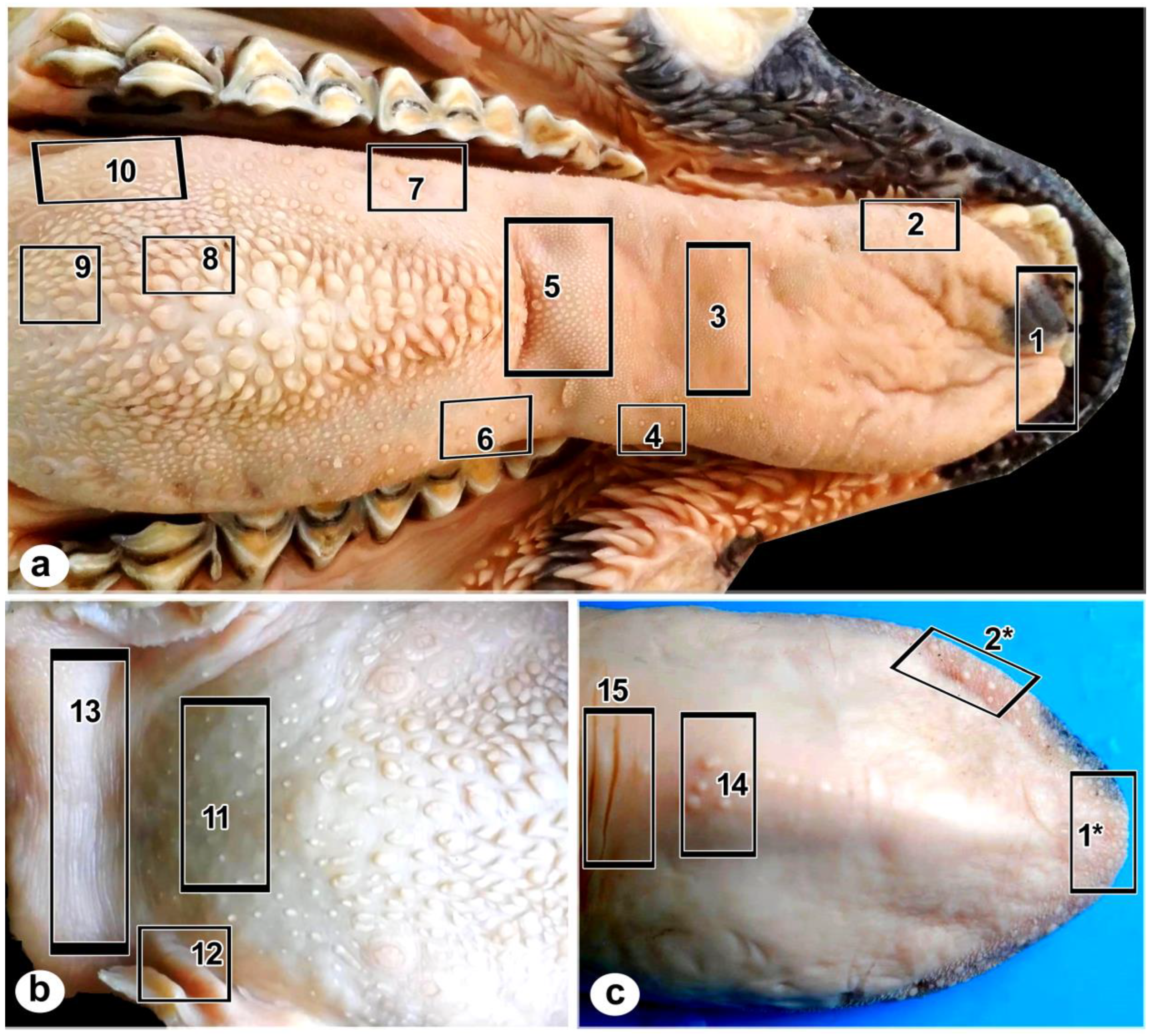
Figure 2.
Photograph of the tongue within the oral floor of Rahmani sheep. Lingual apex (A), body (B), and root (R) showing scattering of fungiform (Fup), filiform (Fp) papillae on the dorsal and lateral aspects of the tongue, dorsal median longitudinal groove (arrow). There are filiform papillae clusters in front of the transverse fossa (arrowhead), and the fungiform papillae become more distinct caudolaterally.Along with torus linguae (TL), there are conical (Cp), lenticular (Lp), two rows of vallate papillae (Vp) on each side, and buccal papillae (Bp).
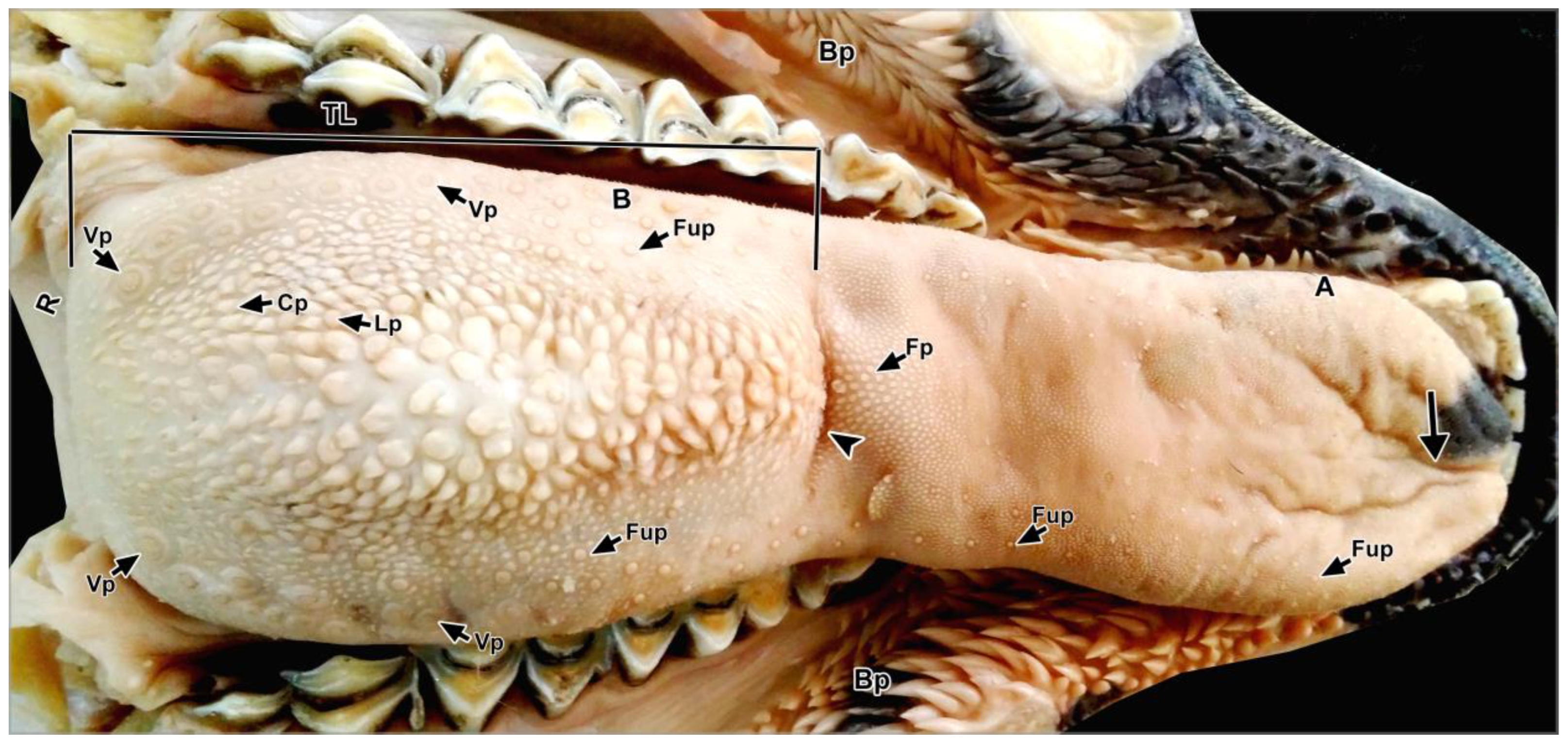
Figure 3.
Photographs of the tongue of Rahmani sheep. (a) The lingual apex’s ventral surface (VS) shows a V-shaped strip of fungiform (Fup) and filiform papillae (Fp) at the apical part of the lingual apex, as well as conical papillae (arrowheads) in front of the frenulum linguae (FL).(b) Dorsal view of the torus linguae (TL) and lingual root (R) showing a few fungiform papillae (Fup) and glossopalatine folds (arrows).
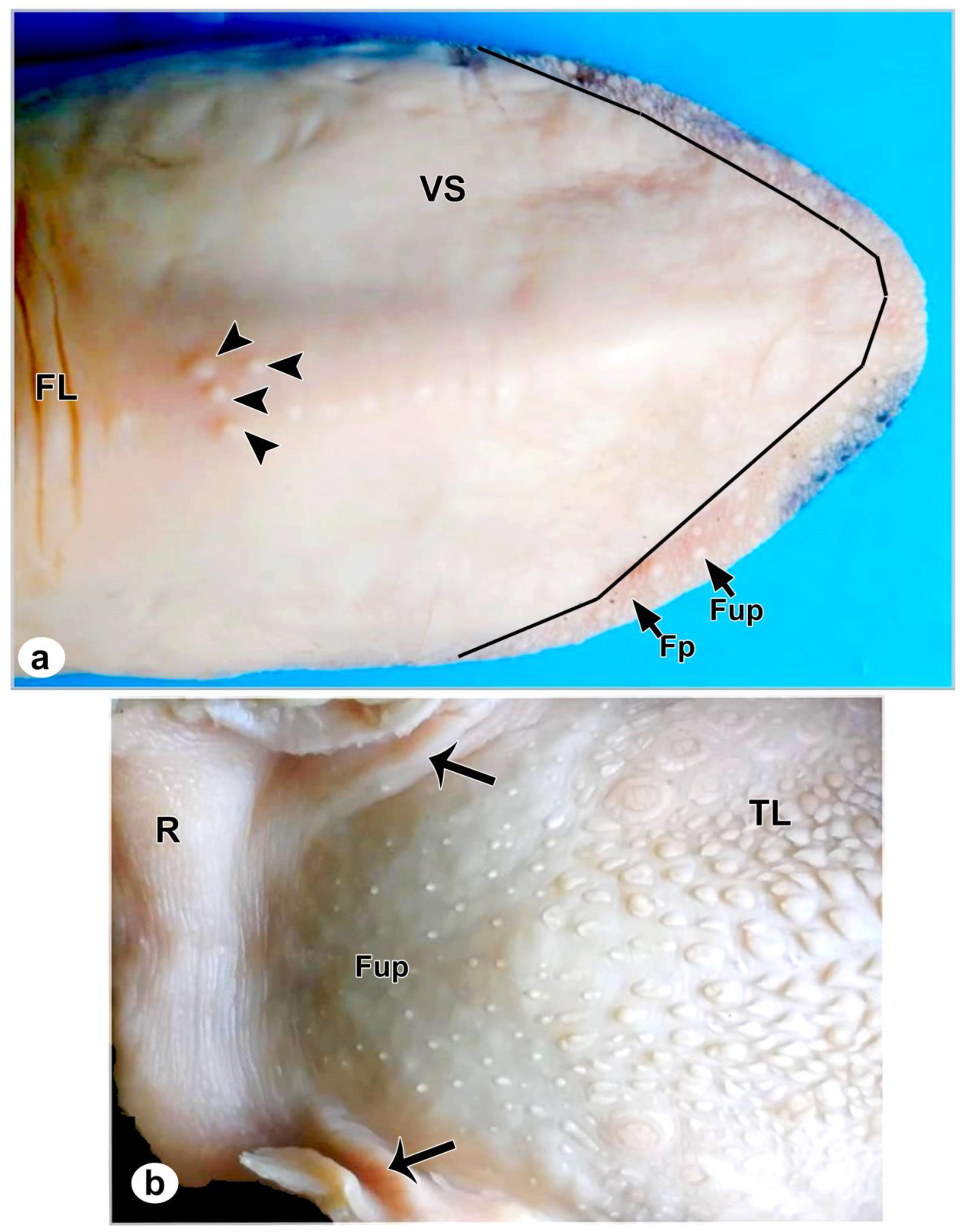
Figure 4.
Micrographs (a-d) of the dorsal, (e) of the ventral, and (f, g) of the lateral aspects of the lingual apex of Rahmani sheep show: (a-d) Button-shaped with a central deep depression fungiform papillae (Fup) surrounded by a deep circular groove (barbed arrow) scattered within filiform papillae (Fp), the basal parts of the former papillae surrounded by a shallow groove (arrowhead), and the main filiform papillae (mFp) surrounded by various sizes of secondary papillae (sFp). (e-f) Button-shaped with shallow depression fungiform papillae (Fup), convex appearance fungiform papillae (*Fup), and filiform papillae (Fp), each main one (mFp) closely packed by small secondary papillae (sFp) and surrounded by a deep groove (arrowhead).
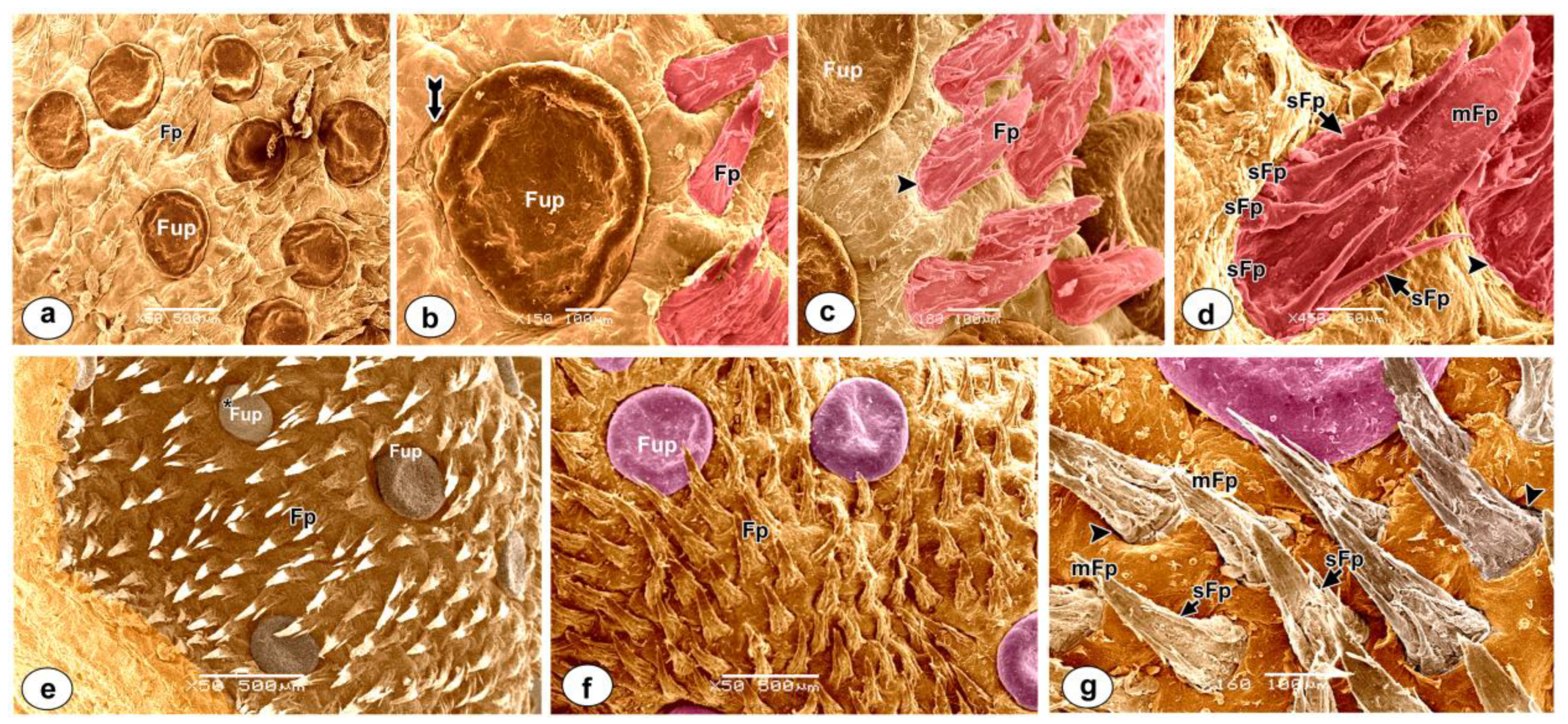
Figure 5.
Micrographs of the ventral surface of the lingual apex (a, b) and the frenulum linguae (c, d) of the tongue of Rahmani sheep demonstrating:(a,b) Three large conical papillae (Cp) in addition to small flattened papillae of various sizes (asterisks) are surrounded by longitudinal and transverse mucosal folds (arrowheads). (c, d) Nearly symmetrical transverse folds (TF) are separated by deep grooves (twisted arrows) covered by secretory granules (Sg).
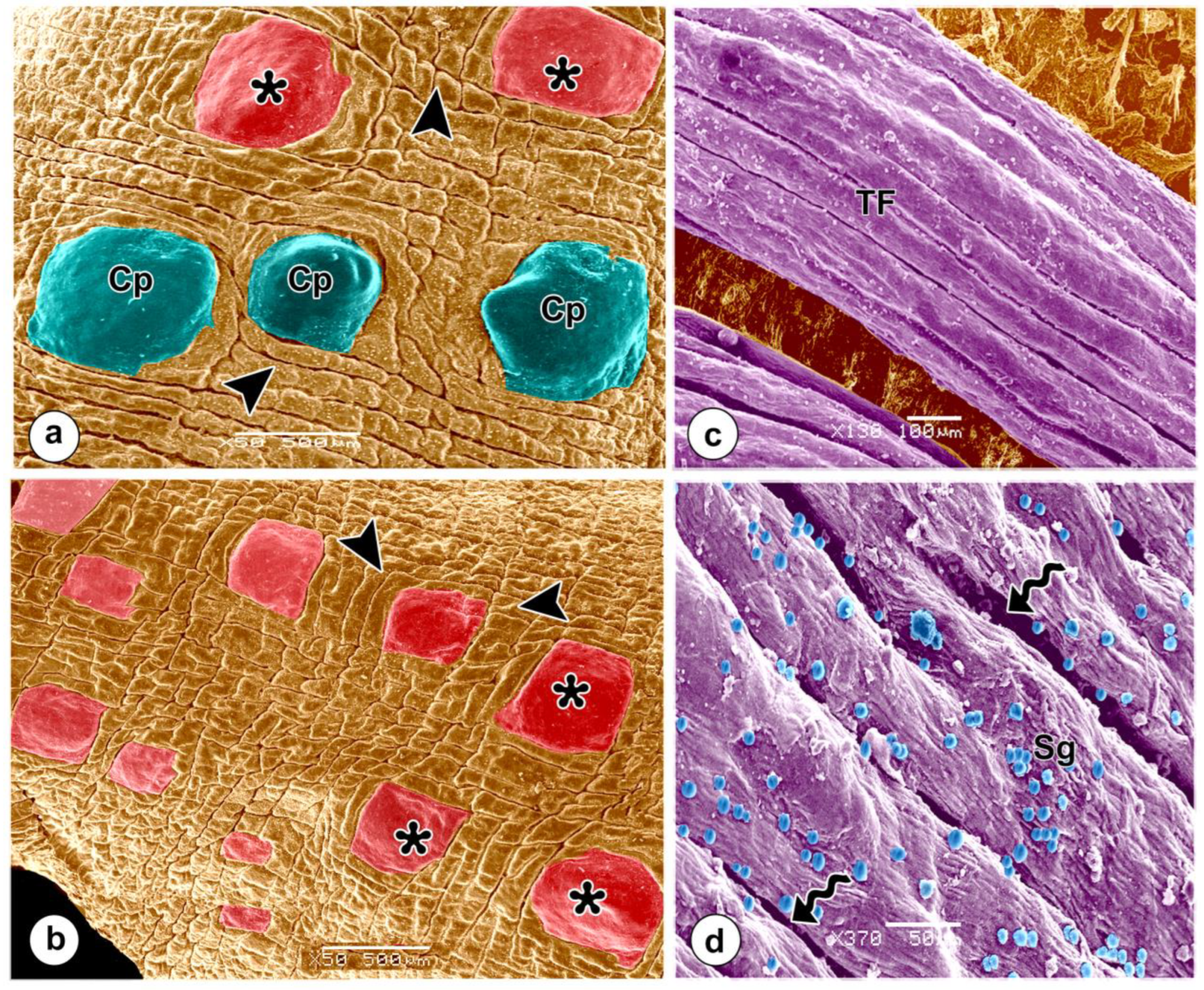
Figure 6.
Micrographs of the lingual apex showing: Median longitudinal groove (arrow) occupied by fungiform (Fup) and filiform (Fp) papillae, main filiform papillae with double process (mFp2), with three processes (mFp3), with five processes (mFp5), keratin (K), openings of the anterior lingual glands (arrowheads) guarded by spiral mucosal fold (short arrow), secretory granules (Sg).
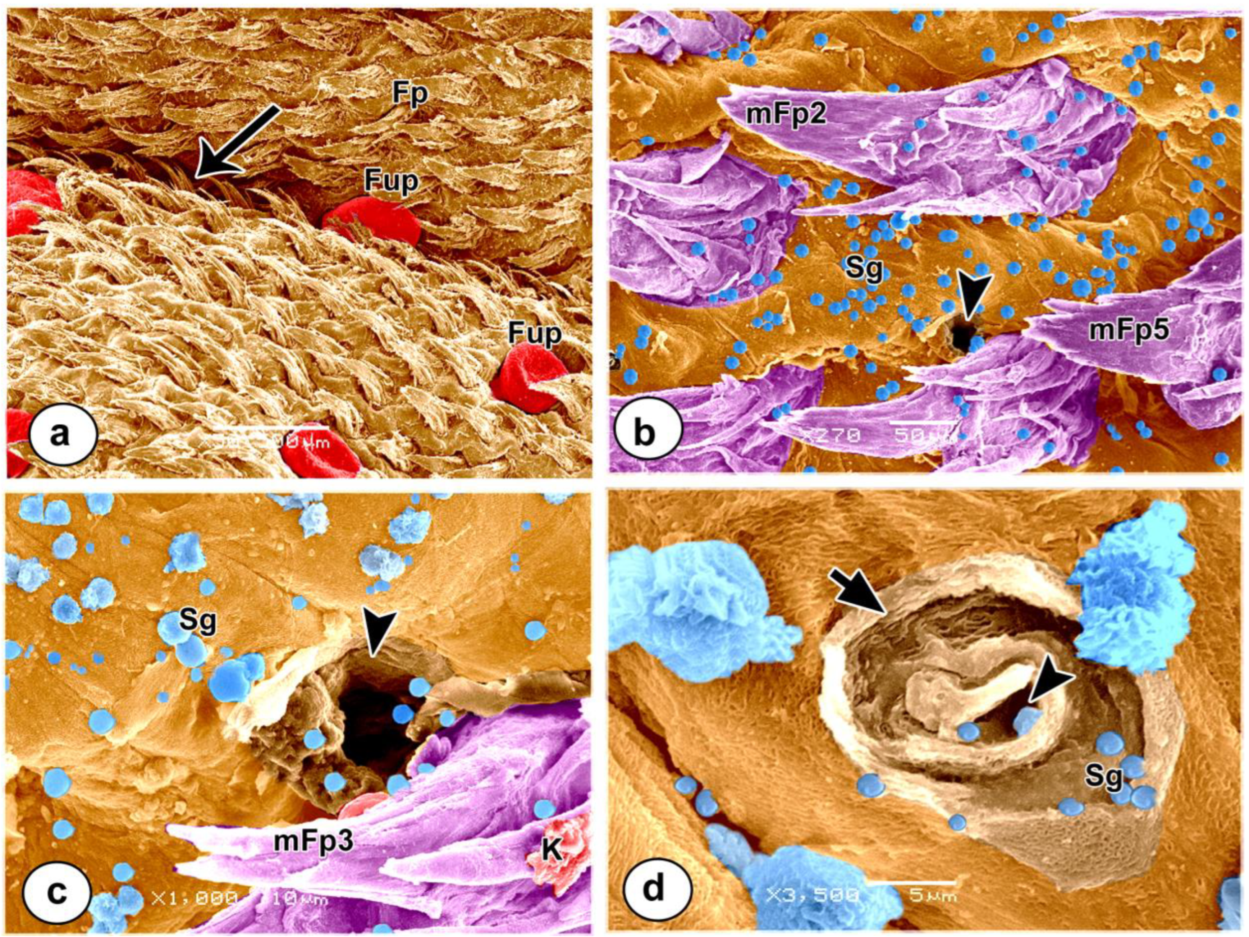
Figure 7.
Micrographs of the anterior dorsum of the lingual body showing:(a) A fungiform papilla with a distinct neck (Fup) was surrounded by filiform papillae (Fp) and had 5-6 taste buds’ pores (arrowhead).(b, c) High magnification view of taste buds’ pores (arrowhead) guarded by overlapped keratinized epithelial layers (short arrows) (d, e) Filiform papillae (Fp) were composed of a main filiform papilla (mFp) enclosing secondary (sFp) and tertiary (tFp) papillae of varying sizes.(f, g) Rounded-shaped anterior lingual gland opening (arrow) within overlapping keratinized layers (asterisks) at the base of the filiform papillae.
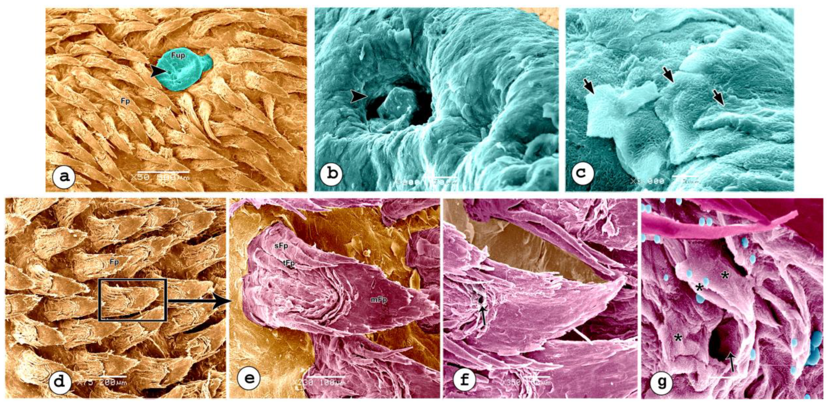
Figure 8.
Micrographs of the transverse fossa area of the tongue of a Rahmani sheep, demonstrating:(a, b) Transverse fossa (arrowhead) guarded by short straight conical papillae with rounded apices rostrally (asterisks) and long curved conical papillae with slightly pointed apices caudally (arrows), their bases encircled by a shallow groove (twisted arrow). (c) Cut-section of conical papilla (arrow) showing keratinized stratified squamous epithelium (Ep), core of connective tissue (Co). (d, e) A filiform papilla with a common basal part bifurcated into small papillae (barbed arrow) was highly keratinized by several rows of keratin (K). (f) Opening of lingual glands (short arrow).

Figure 9.
Micrographs of the lateral aspect and (d, e) dorsum of the torus linguae of the tongue of Rahmani sheep showing (a-c) Conical papillae with wide base and pointed apices (CP), and fungiform papilla (Fup) with distinct neck (arrow).(d, e) conical papillae (Cp*) of various sizes, nearly rounded lenticular (rLP), and tongue-shaped lenticular papillae (LP). (f-h) Circumvallate papillae (cv) of various shapes surrounded by papillary groove (g) and flat broad undulating annular rods (asterisks), mucosal ridge (twisted arrow) between two bodies of vallate papillae, stratified squamous epithelium covered by secretory granules (Sg), (i) high magnification view of the surface of the circumvallate papillae. Take note of the large and small stubby microplicae (arrowheads).
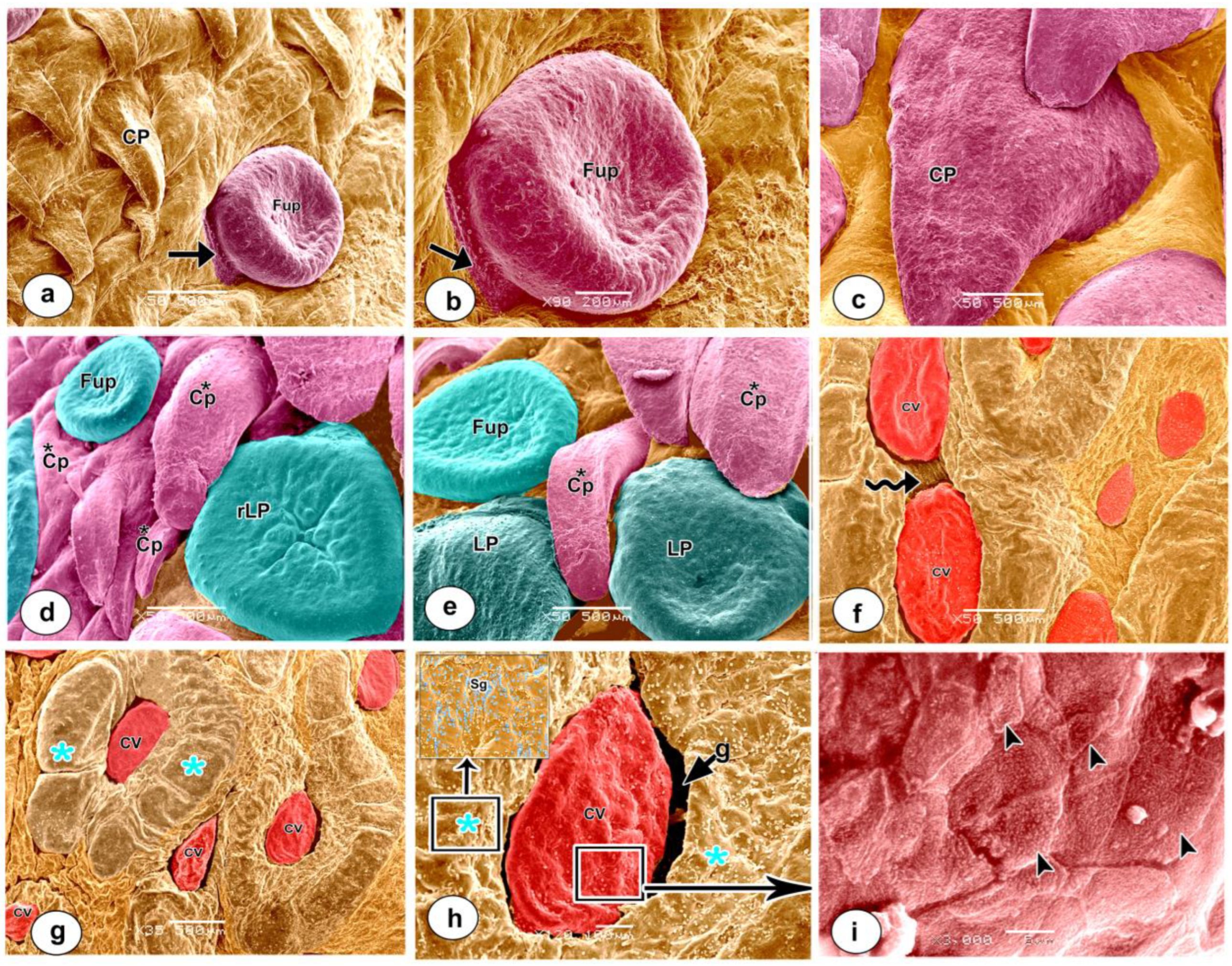
Figure 10.
Micrographs of the glossopalatine arch of the tongue of a Rahmani sheep, showing corrugated mucosal folds (arrow) separated by a groove (arrowhead), and flattened conical papillae surrounded by a shallow groove (CCp).
Figure 10.
Micrographs of the glossopalatine arch of the tongue of a Rahmani sheep, showing corrugated mucosal folds (arrow) separated by a groove (arrowhead), and flattened conical papillae surrounded by a shallow groove (CCp).
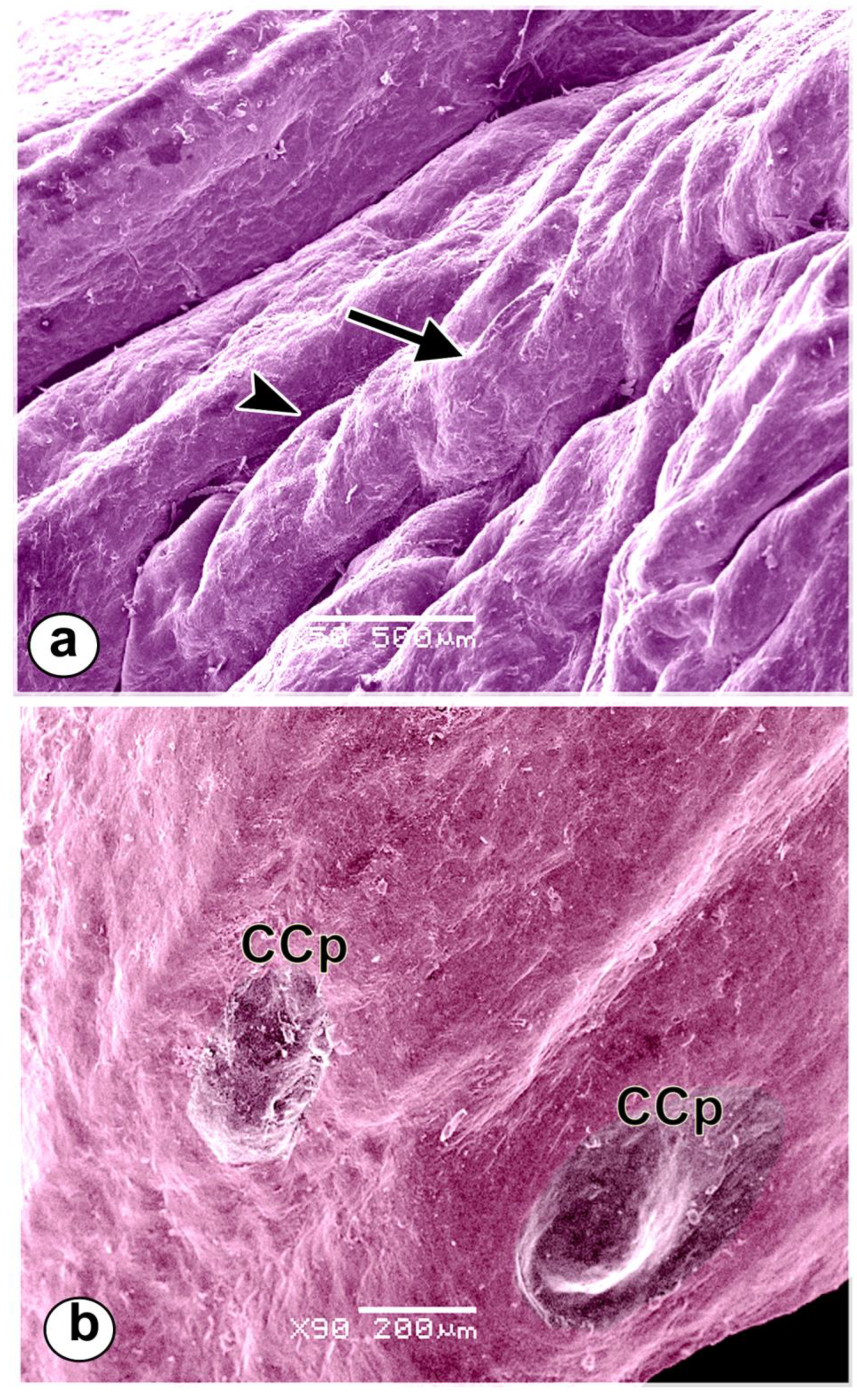
Figure 11.
Micrographs of the lingual root of a Rahmani sheep. (a) Several openings of the posterior lingual glands (Lsg) and a flattened conical papilla surrounded by a shallow groove (CCp). (b, c) A rounded-shaped opening of the posterior lingual glands (Lsg) within a rounded depression with secretory granules (Sg). (d, e) Volcanic crater-shaped openings of the posterior lingual glands (Lsg) with excessive secretory granules (Sg), desquamated keratinized cells around the openings of the glands (arrowheads).
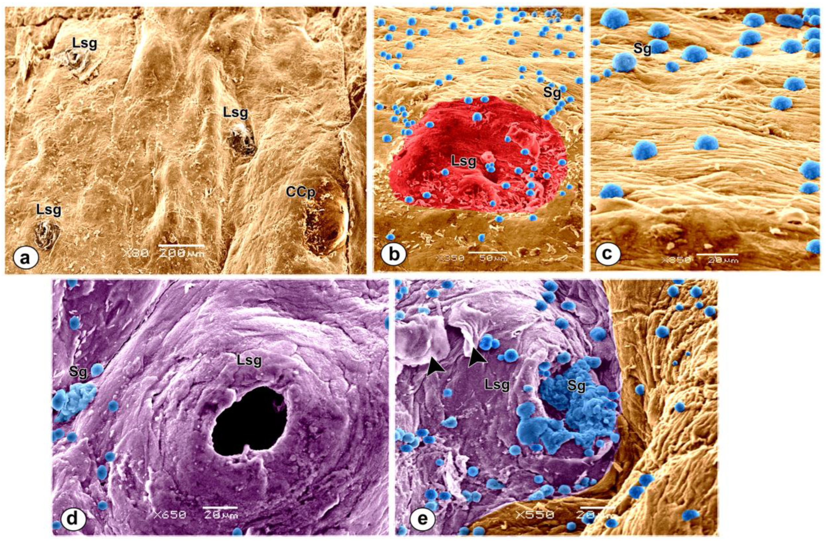

Table 1.
morphometrical data of the tongue of Rahmani sheep.
| Dimensions | Mean | SE |
|---|---|---|
| Total length of the tongue | 147.66 | 2.14 |
|
Apex: - Length Thickness at tip Thickness at frenulum linguae Width at tip Width in front frenulum linguae |
41.09 5.675 21.015 20.85 26.56 |
1.89 0.50 2.24 1.25 3.22 |
|
Body: - Length Width of body: - At transverse groove In front glossopalatine arch Thickness of body: - At level of torus Linguae At highest point of torus Linguae At palatoglossal fold |
85.01 32.13 36.5 34.865 48.55 26.9 |
0.08 0.61 1.62 6.03 10.53 3.62 |
|
Torus linguae: - Length Width Distance bet torus linguae & tip of tongue |
54.71 34.55 69.95 |
9.32 3.56 4 |
| Length of transverse groove Length of dorsal longitudinal groove |
20.33 34 |
1.78 2.52 |
|
Root: - Length width |
21.56 31.7 |
0.10 4.31 |
Disclaimer/Publisher’s Note: The statements, opinions and data contained in all publications are solely those of the individual author(s) and contributor(s) and not of MDPI and/or the editor(s). MDPI and/or the editor(s) disclaim responsibility for any injury to people or property resulting from any ideas, methods, instructions or products referred to in the content. |
© 2023 by the authors. Licensee MDPI, Basel, Switzerland. This article is an open access article distributed under the terms and conditions of the Creative Commons Attribution (CC BY) license (http://creativecommons.org/licenses/by/4.0/).
Copyright: This open access article is published under a Creative Commons CC BY 4.0 license, which permit the free download, distribution, and reuse, provided that the author and preprint are cited in any reuse.


