
Submitted:
07 June 2023
Posted:
07 June 2023
You are already at the latest version
Abstract
Present study reports the green synthesis of pectin fabricated silver-based nanocomposite (Pectin-AgNPs) with Carpesiumnepalense leaves extract and evaluated their bactericidal kinetics, in-vivo hepatoprotective and cytotoxic potentials with their possible mechanisms. GC/MS and LC/MS analysis indicated the presence of different new phytochemicals constituents in the plant extract. The physicochemical characterization of Pectin-AgNPs by UV/Vis, SEM, DLS, FTIR and EDX techniques showed the spherical and uniform size range synthesis of nanocomposite i.e. 50-110 nm. The highly significant (P<0.005) antibacterial activity was found against all tested four bacterial strains with the ZIs of 24.8 to 27.2 mm. Significant damage in bacterial cell membrane was also observed in AFM study after treated with Pectin-AgNPs. At the doses of 0.05 mg/kg, nanocomposite showed highly significant (P<0.005)hepatoprotective activity in biochemical and histopathology analysis as compared to CCL4 administered control group. The ameliorative effects of Pectin-AgNPs were observed in PCR analysis on both GAPDH and PPARs genes expression and its potential to restore gene alterations caused by CCl4 intoxication.Pectin-AgNPs produced cytotoxic activity against HeLa cell lines at higher doses with the LC50 of 223.7 µg/mL. The current findings demonstrate positive attributes of pectin fabricated AgNPs as a promising antibacterial, hepatoprotective and cytotoxic agent.
Keywords:
Green Synthesis
; Pectin functionalized silver nanoparticles
; Antibacterial activity
; Hepatoprotective
1. Introduction
Nanotechnology is an emerging field and occurred as a scientific revolution in the pharmaceutical industry due to their reduced particle size and more surface area for targeted response in a wide range of diseases. Today, nanoparticles (NPs) have been used as fluorescent labels, diagnostic agents, transfection labels, antimicrobial and different therapeutic agents [1,2]. Specifically, silver nanoparticles (AgNPs) have considerably unique biological and physicochemical characteristics with relatively high surface area. AgNPs have been attracted globally as a potent antimicrobial agent due to their variable size ranges i.e. 1-100 nm3 [3]. At present, it is utilized in various fields of applications as biosensors, conductive nanofluids and antimicrobial agents [4]. The ecofriendly and cost-effective method for the synthesis of NPs is extensively focused due to the harmful and high-cost materials involved in other conventional methods. However, green synthesis is a highly cost and time-effective, sustainable and biologically safest method [5].
Nanocomposites of inorganic metals with different polymers are gaining attraction and scientific importance. It has been reported that the metallic NPs incorporation in polymer matrix significantly improved the performance of nanocomposite at a lower cost [6]. Metallic NPs have been synthesized after conjugation with various polymers like graphene oxide, chitosan, carboxymethyl cellulose (CMC), gelatin, lactic acid, guar gum and polyethersulphoneand many others [7,8,9]. These nanocomposites are used in several applications, including aerospace, automotive, packaging, defense, electronics, semiconductors, energy, medical and healthcare, coatings and sports [10,11]. In recent times, pectin is attaining immense interest for researchers to synthesized multifunctional biopolymericnanocomposites due to its greater biocompatibility and toxic-free nature compared to other biopolymer. It is a naturally occurring polysaccharide and gelling is the typical characteristic of pectin that grabs an important distinct position among other natural polymers [12].
Carpesiumnepalense (C. nepalense) belongs to the genus Carpesium family Asteraceaedistributed in Azad Kashmir Pakistan. Traditionally this plant is reported primarily for hepatitis anti-inflammatory, hepatitis detoxifying anti-scabies and pustule sores [13]. In Pakistan, this plant is domestically used in the treatment of liver inflammation and skin infections. However, to date, no scientific report has been reported related to its antimicrobial and hepatoprotective potentials.
The liver being the major organ in human diverse fundamental functions including regulate storage, metabolism andwaste excretion, detoxification of exogenous and endogenous substances and maintain homeostasis. Due to the hepatotoxic nature of xenobiotics, the liver is the primary target for the number of xenobiotics and toxicants [14]. The detoxification process of xenobiotics, induction of free radicals, oxidative stress and inflammation in the liver causes significant liver damages [15]. Despite enormous advancements in present days for hepatoprotection, liver diseases remain the leading morbidity among the top adverse diseases. Unfortunately, there is no completely effective drugwhich protects the liver or regenerates liver cells. The synthetic drugs for different liver diseases are quite selective, expensive with broader adverse effects [16]. Therefore, researchers greatly focus on a search for a safe and effective alternatives for the cure of liver diseases. Alternative green synthesized nanomedicines are now considered as an ideal and unique substitute for conventional synthetic drugs. The liver is the major organ for carbon tetrachloride (CCl4) metabolism via monooxygenase (P450 2E1) in mitochondria, which results in a free radical generation. This free radical’s further causes phospholipid cell membrane damage through fatty acids peroxidation [17].
Previously few studies reported the synthesis of pectin functionalized AgNPs through the chemical reduction method [18,19]. In addition, Zhang et al. also described the hepatoprotective activity of green synthesized NPs using Rhizophoraapiculata [20]. However, according to our literature survey, there is no study reported the antibacterial, liver protective and cytotoxic potentials of green synthesized pectin functionalized silver NPs using C. nepalenseethanolic leaves extract. Therefore a current study was designed to investigate the antibacterial, hepatoprotective and cytotoxic activities ofgreen synthesized Pectin-AgNPs using C. nepalenseextract with their possible mechanisms for activities. Further, the plant extract was also examined phytochemically for the presence of pharmacologically active components using LC/MS and GC/MS techniques.
2. Materials and Methods
2.1. Plant Material and Extraction
Fresh leaves of Carpesium nepalense (L.) were received in the month of September-2020 from the region of South Waziristan, Khyber Pakhtunkhwa (KPK)-Pakistan according to therelevant national, institutional and international guidelines and recommended legislation. The collected plant was identified by the Pharmacognosist of the Faculty of Pharmacy, Federal Urdu University of Arts Science and Technology (FUUAST) and the voucher specimen number of a plant (RAW 101021C) was submitted at the herbarium library of FUUAST. Leaves were air-dried and crushed to small particles. Thecrushed powder was macerated with 60% ethanol (Sigma Aldrich, USA) for a period of 15 days and then filtered using Whatman filter paper. Furtherafter filtration, the extract was concentrated using a rotary evaporator (Buchi, Switzerland) at 40 °C and obtained 143 g (12%) brownish C. nepalenseethanolic leaves extract (CNEE). The obtained extract was stored at a temperature of 4 °C for further studies [21,22].
2.2. Gas Chromatography Mass Spectrometry(GC/MS) Analysis of CNEE
CNEE was evaluated on a gas chromatograph (USB-393752, Agilent, USA) usinga 5% phenylmethylsiloxaneHHP-5MS capillary column (30m × 0.25mm × 0.25μm) along withan FID detector. The instrumentwas set according to the specified conditions as reported in our previous study [23]. Briefly, helium (99.999%) was used as a mobile gas at a 1 mL/min flow rate, and a 2 μL injection volume was employed. However, the ion-source temperature was 200 °C, the injector was maintained at 250 °C temperature while the temperature of the oven was programmed from 110 °C and ending at 280 °C. Mass spectra were obtained at 70 eV; with 0.5 s scan interval and 45 to 450 Da fragments. The total running time of GC/MS analysis was 40 min. The detected compounds in the CNEE via GC/MS analysis were authenticated by the comparison of their retention times with the authorized reported spectral data in Wiley and NIST library.
2.3. Liquid Chromatography Mass Spectrometry (LC/MS) Analysis of CNEE
The bioactive compounds of CNEE were further evaluatedvia LC/MS analysis. The analysis was performed on LC/MS (Q-TOF 6530, Agilent, USA) with positive ionization and HPLC inlet modes with MS2 spectrum type. The reverse-phase column was used in the HPLC system with the specifications of RSLC, 120 OA, C18, 50 mm x 2.1 mm, 2.2 µm (Thermoscientific). Analytical grade methanol (Merck, Germany) was used as mobile phase and the LC/MS condition was operatedat the temperature of 280 °C, while the flow rate of the sample was adjusted at 8 μL/min. A volume of 2 μL sample was injected whereas the mass range was maintained from 50 to 1000 m/z [24]. The obtained spectrum from the CNEE analysis was authenticated with Wiley and NIST library for the identification of differentphytochemical constituents.
2.4. Green Synthesis of Silver Nanoparticles
Analytical grade chemicals were obtained from Sigma-Aldrich, USA for the synthesis of AgNPs. AgNPs were green synthesized by adding dropwiseCNEE (10 mg/mL) as reducing agents to AgNO3 (1 mmol/L) aqueous solution in the ratio of 9:1. At ambient temperature, the mixture was continuously stirred and left for a day in a dark place to avoid AgNPs photodegradation. Then, the obtained suspension was centrifuged for 40 min at the speed of 10,000 rpm. After 40 min, the supernatant was drawn and washed with distilled water and analyzed on a UV spectrophotometer for the confirmation of AgNPs synthesis. The change in color from silver to brown also indicated the synthesis of AgNPs. Finally, the green synthesized AgNPs were dried at 65 °C in a hot air oven for 6 h [25].
2.5. Synthesis of Pectin Functionalized Silver Nanoparticles (Pectin-AgNPs)
Pectin-AgNPs along with CNEE were synthesized using the method described by Hileuskaya et al. with some modifications[26]. In this synthesis scheme, high-methoxyl (PectHM, Sigma, 81.2% degree of esterification, Mν = 141•103)pectin served as stabilizer and reducer along with CNEE during the synthesis ofPectin-AgNPs. Initially, the reaction was startedby adding dropwise solutions of Pectin (5 mg/mL) and SAEE (10 mg/mL) into an aqueous solution of AgNO3 (1 mmol/L). The obtained suspension was centrifuged for 30 minat 10,000 rpm, and the supernatant was drawn, washed with distilled water, and assessed spectrophotometrically for the successful synthesis ofPectin-AgNPs. The color changed from silver to brownish-black also confirmed the synthesis of pectin-based AgNPs. Finally, the synthesized nanocomposite was dried at 65 °C using a hot air oven for 12 h to obtain purified Pectin-AgNPs. However, the value of the degree of substitution (DS) for pectin in Pectin-AgNPs was estimated from the potentiometric titration method [27].
2.6. Characterization of AgNPs and Pectin-AgNPs
The pH of the solution was measured before and after the reduction process (addition of extract and pectin), using the digital pH meter (Benchtop). The optical properties of synthesized NPs were evaluated using a UV visible spectrophotometer (UV-1710, Shimadzu, Japan) at the wavelength range of 300–700 nm. A volume of 250 µL methanolic solution was placed on Greiner 96 flat polystyrene plates for the determination of maximum absorbance. The surface morphology, physical characteristics and the deposition of pectin on AgNPs were assessed via electron microscope (SEM) (4380B, Joel, Japan). The size and zeta potential of synthesized Pectin-AgNPs were analyzed using the dynamic light scattering (DLS) technique, along with a particle size analyzer (Brookhaven Cooperation, NY, USA). However, the functional group's present in nanocompositeand interaction of pectin with AgNPs were evaluated using Fourier transform infrared spectroscope (FTIR) (IR-100 Shimadzu, Japan) at 0.5 cm-1 resolution with the operational range of 500–4000 cm-1while the sample was taken as potassium bromide (KBr) powder discs. The mixture of KBr powder and sample was used to prepare KBr discs using a hydraulic press. Further, the energy dispersion x-ray spectroscope (EDX) (JSM 6380, Joel, Japan) was used from 0 and 10 kV for the detection and measure the amount of carbon and silver in AgNPs and Pectin-AgNPs.The practical yield of nanocomposites was measured using a PerkinElmer Optima, ICP-OES analyzer (8300, Shelton, USA) [28].
2.7. Antibacterial Activity
2.7.1. Zones of Inhibitions (ZIs)
Different highly resistant pathogenic bacterial isolates of Bacillus subtilis (LT 2352), Klebsiella pneumonia (LT 4524), methicillin-resistance Staphylococcus aureus (MRSA)[LT 2416] and Vibrio cholerae[LT 3131] were obtained from Hamdard University pathological laboratory Karachi-Pakistan. The antibacterial potentialof C. nepalense ethanolic extract (CNEE), pectin, AgNPs, Pectin-AgNPs and standard commercially available antibiotics were assessed using the well-reputed Oxford cup diffusion method [4]. All clinical strains were diluted to 106cfu/mL as McFarland turbidity standard using nutrient broth while the turbidity of prepared dilutions was measured using the UV visible spectrophotometer (UV-1710, Shimadzu, Japan). Each tested bacterial strain was spread on a plate filled with nutrient agar then five Oxford diffusion cups were placed on each plate after perforation. Then, a volume of 0.05 mL solutions of each tested antibacterial agent with the concentrations of 10,000 μg/mL of CNEE; 1000 μg/mL of pectin, 50 μg/mL of AgNPs, and Pectin-AgNPs while 600 μg/mL (Eq. to standard 30 μg disc) of each tested antibiotic i.e. vancomycin and cefoxitin(Oxoid, UK) were poured into five different Oxford diffusion cups of each plate. Plates were incubated for 24 h at 37 ± 2°C after 30 min of diffusion. The obtained zones of inhibitions (ZIs) were recorded in millimeters (mm) using a digital Vernier caliper. The ZIs of each bacterial strain were determined in triplicates.
2.7.2. Minimum Inhibitory Concentrations (MICs) and Minimum Bactericidal Concentrations (MBCs)
The MIC values of CNEE, pectin, AgNPs and Pectin-AgNPs were investigated using the broth dilution method [29,30]. The serial dilutions of CNEE (from 20,000 to 1000 µg/mL), pectin, AgNPs and Pectin-AgNPs (512 to 1 µg/mL) were prepared in nutrient broth for the determination of MICs values. All tested bacterial strains concentrations were adjusted to 106 CFU/mL. After incubation for 24 h at 37 ± 2°C, optical densities (ODs) of each strain were determinedusing an ELISA reader (Infinite 300; USA) at 600 nm. In addition, the value of MBC was determined by plating the already incubated bacterial strain on different plates containing nutrient agar. After incubation for 24 h at 37 °C, viable bacterial cell colonies were counted in each plate [31]. Both MICs and MBCs were determined three times and presented as their mean value with SD.
2.7.3. Time Killing Kinetics
Time killing kinetics assay of Pectin-AgNPs was performed according to the method reported in a previous study [29]. The assay was performed at the Pectin-AgNPs concentrations equal to the MBC of each tested bacterial strain. Before the addition of nanocomposite, the bacterial cells were grown to logarithmic phase for 6 h in freshly prepared nutrient broth equal to the standard concentration of 1×10⁸cfu/mL. Then, the culture was incubated at 37 °C in a Benchtop incubator shaker (Amerex Instruments, USA), and a sample was drawn after every 5 min interval. Viable cell counts were measured by spreading a drawn sample onto the nutrient agar plates. Then, plates were incubated for 48 h at 37 °C, and viable bacterial colonies were counted. The bacterial killing kinetic curve was constructed between the viability of cells in cfu/mL and time in min.
2.7.4. Bacterial Killing Mechanism
AFM study was performed to assess the morphological alterations in cells of tested bacterial strains. A volume of 1 mL gelatin (10%) was used for the preparation of mica slides. Then, tested clinical isolates of different bacterial strains i.e. B. subtilis, K. pneumonia, MRSA and V. cholera were harvested using microtiter and placed on prepared mica slides. A similar procedure was used to prepare negative and positive control samples. After the inoculation of tested isolates, the prepared slides were air-dried at room temperature. All mica slides were observed underAFM for the detection of any cellular changes in bacterial strains after exposure to synthesized nanocomposites [8].
2.8. Hepatoprotective Activity
2.8.1. Animals
The hepatoprotectivepotential of green synthesized AgNPs and Pectin-AgNPs was evaluated on one hundred and twenty healthy white Wistar albino rats which were purchased from the animal house of Dow University and Health Science (DUHS), Karachi-Pakistan with an average body weight of 195 ± 10 g. The consent form was filled by the in-charge of DUHS animal house to use these rats for the present study. Both control and tested animals were placed in a controlled environment i.e. 25 °C ± 2 °C and 45–55% humidity in a 12 h light and dark cycle and provided standard food to all studied animals. Rats were divided into 11 groups and each group consists of 10 rats. All tested solutions were prepared in normal saline and were administered once daily via the oral route. Animals of Group I was considered as naïve group (received no treatment) while remaining groups animals received 1 mL/kg body weight CCl4 (Merck, Germany) intraperitoneally in olive oil (30% v/v) for the induction of hepatic toxicity. However, the animals of Group II were considered as a positive control (administered CCl4 only) while III and IV groups animals were treated with plant extract in two different doses i.e. 125 and 250 mg/kg. At the doses of 0.025 and 0.05 mg/kg, pectin was given to V and VI groups. At the same doses of 0.025 and 0.05 mg/kg, AgNPs were administered to VII and VIII groups while Pectin-AgNPs were given to IX and X groups. However, Group XI was taken Silymarin (Sigma Aldrich, USA) at a dose of 100 mg/kg as standard treatment [20]. Treatment with different doses was initiated after three days from the induction of hepatotoxicity and continued for 14 days period.At the end of the treatment period, blood samples were collected after6 h fasting to avoid lipemia [32]. Animalswere euthanized using the cervical dislocation method while before euthanasia each animal was sedated with an intravenous administration of medetomidine (Sigma Aldrich, USA) with the dose of 0.5 mg/kg [33]. Animal handling was performed following the National Advisory Committee for Laboratory Animal Research (NACLAR) guidelines and in compliance with the ARRIVE guidelines.Moreover, approval for the study was obtained by an animal ethical committee of the Pakistan Council of Scientific and Industrial Research (PCSIR), Karachi-Pakistan (04/07/KP/PCSIR).
2.8.2. Clinical Examinations and Body Weight
Initially, animals were evaluated for generalized well-being. During dosing of each nanocomposite, vital signs or any sign of toxicity were observed twice daily. The effects of a particular treatment on animal general health, skin, hairs and behavior were monitored. At initial and after 14 days, animal body weight was also noted.
2.8.3. Biochemical Analysis
Ten millimeter blood samples were drawn from the femoral artery of each rat and mixed with 20 mg/mL ofethylenediaminetetraacetic acid (EDTA) to avoid sample coagulation. Biochemical analysis was performed on each blood sample to evaluate the hepatoprotective effect of each synthesized nanocomposite. The important liver biomarkers including lactate dehydrogenase (LDH), aspartate transaminase (AST), alanine transaminase (ALT), alkaline phosphatase (ALP), total protein (TP), and direct bilirubin(DB) were quantified according to the recommended standard protocols by manufacturers of diagnostic kits as previously reported [34]. The biochemical analysis on each sample was performed on UV visible spectrophotometer (UV-1710, Shimadzu, Japan).
2.8.4. Histopathological Examination
The liver of each treated animal was harvested after the treatment period. Tissues of the liver were fixed in (10%) formalin and incubated for 24 h. Tissue dehydration and cleaning were performed using ethanol (90%) and xylene solution (1%) respectively. Further, the tissue was embedded in paraffin wax using a tissue embedding instrument while the tissues handling wasperformed using standard techniques recommended for histopathologicalstudies. Then, 3 to 5 µm tissue sections were sliced using a microtome (5062, Mainz-CUT, Germany) and stained with hematoxylin and eosin (H&E) stain according to the standard protocol for staining. Treated tissues were observed under a smart microscope (Zeiss AxioLab-5, Germany).Alterations in tissueswere recorded as none (--), very mild (˗), mild (+), moderate (++) and severe (+++) [35].
2.8.5. Real Time Polymerase Chain Reaction (RT-PCR) Assay
The regulation of peroxisome proliferator-activated receptor- delta (PPAR-δ) gene expression was also evaluated to investigate the mechanism of the hepatoprotective potential of Pectin-AgNPs. Liver tissues were harvested and pulverized into small pieces for the extraction of total RNA using the Trizol reagent method [36]. The extracted RNA was quantified for their purity determination in a ratio of 260/280 by a micro-volume Colibri spectrophotometer (Titertek Berthold). An amount of 1 µg RNA was transcribed reversely into complementary DNA viaan optimal cDNA synthesiskit. The PPAR-δ primer sequences were as follows; reverse: GCAAAGATGGCCTCATGCA, forward: GCCAAGAACATCCCCAACTTC. However, GAPDH, reverse: GAGGGCCTCTCTCTTGCTCT, forward: AACTCCATTCCTCCACCTT was used as an internal control for RT-PCR. The cycle of PCR was performed into following sequences; 1 cycle for 5 min at 95 ºC for initial denaturation then 35 cycles were performed for 30 sec for RNA denaturation, annealing and extension at 95, 60 and 72 ºC respectively. The obtained results of PCR assay were analyzed using the commonly used 2˗ΔΔCT method for gene expression [37].
2.9. Cytotoxicity Evaluation
The cytotoxic potential of Pectin-AgNPs was evaluated using cultured HeLa cell line (ATCC, Virginia, USA) using MTT (3 (4,5-dimethyl thiazolyl-2) 2,5-diphenyltetrazolium bromide) assay. The medium containing AgNPs and Pectin-AgNPs with different concentrations (25–500 μg/mL) were added individually in adherent culture replacement and incubated for 24 h at 37 °C while doxorubicin (50 mg) was used as a standard. Then, the treated HeLacells were washed with phosphate-buffered saline (PBS) and incubated in an MTT reagent (1 mg/mL) for 30 min. The cellular proliferation and viability were determined using contrast microscopy and UV spectrophotometer at the wavelength of 570 nm[4]. Cellular toxicity was performed in triplicates for each test solution while the percentage cell growth inhibition was calculated using the formula as given below:
Percentage cell inhibition = (100 – [(At – Ab)/ (AcAb)])×100
Whereas,
At= Absorbance of test solution
Ab = Absorbance of blank
Ac= Absorbance of control solution.
2.10. Statistical Analysis
All the findings obtained from this study are presented as their standard mean with their S.D values. However, inferential analyses were performed such as one-way ANOVA followed by Tukey post hoc test for evaluating the significant differences in antibacterial, hepatoprotective and cytotoxic potentials among different test solutions using SPSS software (version 23). P<0.05 and P<0.005 were designated as significant and highly significant results respectively. Furthermore, the regression analysis and correlation coefficient were used to analyze the correlations nature among different tested solutions in bactericidal killing kinetics assay.
3. Results and Discussion
3.1. GC/MS and LC/MS Analysis of CNEE
The crude leaves extract of C. nepalense was evaluated phytochemically for the presence of important bioactive compounds and also obtain a complete metabolite profile of plant extract. The GC/MS analysis (Figure 1) revealed the presence of various lower and high molecular weight compounds such as oleic acid, palmitic acid,arachidic acid,glycine, n-[(3α,5β,7α,12α)-24-oxo-3,7,12-tris[(trimethylsilyl)oxy]cholan-24-yl]-, methyl ester, pregnan-18-oic acid, benzofuran, 2,3-dihydro, tyrosol, α-tocopherol, cetrimide,2-decenal, (Z)-,2,4-decadienal, (E,E), 2H-indeno[1,2-b]furan-2-one, 3,3a,4,5,6,7,8,8b-octahydro-8,8-dimethyl, l-(+)-ascorbic acid 2,6-dihexadecanoate,benzeneethanol, 4-hydroxy-,DL-tyrosine, tyrosine,tyrosol, neocurdione, barrigenol R1,digitoxin,24,25-dihydroxyvitamin D3,dipalmitin, and lupenyl acetate. C. nepalense crude leaves extract was further subjected to LC/MS analysis as shown in Figure 2. The library of LC/MS of C. nepalense revealed several meaningful bioactive compounds and peptides like Vit-C, his-his-lys and many others. In addition,ethanolic extract showed a high content of flavanol and flavonol compounds. Different types of phenolic acids were also detected. These all compounds were useful in the synthesis of AgNPs and also possess significant pharmacological activities [38,39,40,41].
3.2. Synthesis and Characterization of Pectin-AgNPs Nanocomposite
The synthesis of stable AgNPs and Pectin-AgNPs nanocomposites were completed after 40 min of centrifugation at 12000 rpm. The synthesis was confirmed using a UV-Vis spectrophotometer with the maximum wavelengths at 410 nm for AgNPs while the maximum absorbance for Pectin-AgNPs was found at 418 nm (Figure 3). The multiple surface plasmon resonance (SPR) bands of Pectin-AgNPs were obtained at different wavelengths in the UV-Vis spectrum, indicated the mixture of polymer with AgNPs and C. nepalance extract. However, strong SPR was found in the region of 400–420 nm in the spectrums of both green synthesized AgNPs and Pectin-AgNPs corresponding to the presence of silver ions [25]. Tummalapalliet al. and Zahranet al. also reported SPR bands at closer wavelengths in UV/Vis spectrums of pectin fabricated AgNPs i.e. 420 nm and 418 nm, respectively [42,43]. The internal structure, surface morphology and facial appearance of AgNPs and Pectin-AgNPs were evaluated by scanning electron microscopy (SEM) technique.More agglomeration was clearly observed in SEM image of green synthesized Pectin-AgNPs nanocomposite as compared to AgNPs (Figure 4). The size distributions of AgNPs and Pectin-AgNPs using the DLS technique are given in Figure 5. Monodispersion was observed in both AgNPs and Pectin-AgNPs while the mean size range of Pectin-AgNPs was found in the range of 50-110 nm, which is slightly greater than AgNPs size range i.e. 10-50 nm. This increase in size after pectin functionalization might be due to the non-specific binding and aggregation between pectin and AgNPs. Similar size ranges were observed in our previous studies for the green synthesized AgNPs using S. aromaticum and S. cumini plants extracts [4,44]. However, Su et al. and Hileuskayaet al. observed similar monodispersionin pectin containing AgNPs [12,26].The mean zeta potential of AgNPs and Pectin-AgNPs was measured for the assessment of synthesized nanocomposite stability. It was reported that nanocomposites with the zeta potential range from -25 mV to +25 mV have a high degree of stability [45]. In Figure 6, AgNPs and Pectin-AgNPs mean zeta potentials were found to be -20.1 mV and -24.4 mV respectively indicating the high stability and dispersity of synthesized nanocomposite. In addition, a high ratio of negative charges supports good colloidal nature, high dispersity, and long-term stability of green synthesized pectin decorated nanocomposite[46]. In the FTIR analysis, green synthesized AgNPs showing a broad absorption peak at 3412/cm indicating the presence of O–H groups in C. nepalance leaves extract, while the absorption peaks were obtained at 476/cm in both AgNPs and Pectin-AgNPs corresponded to the presence of silver ions as shown in Figure 7 [3,47]. However, the appearance of some additional peaks and changes in the intensity of certain peaks in the Pectin-AgNPs spectrum compared to AgNPs, suggested the interaction of functional groups between pectin and Ag+ ions.At 2922/cm, peak corresponded to the stretching mode of C–H bond while C–O, and C–O–H bonds from hydrocarbon chains were observed at 1647/cm, and 1382/cm,, respectively [44]. In Figure 8, the absorption peaks observed at 3 KeV in EDX spectrum in Pectin-AgNPs confirmed the presence of silver ions with the atomic percentages of 24.71% [48]. The final yields of green synthesized Pectin-AgNPs was 47.6% respectively.
3.3. Antibacterial Activity
3.3.1. Zones of Inhibitions, MICs and MBCs
The antibacterial potential of green synthesized AgNPs and Pectin-AgNPs was evaluated against different clinical isolates i.e. B. subtilis, K. pneumonia, MRSA and V. cholerae using the Oxford cup diffusion method. The ZIs produced by CNEE, Pectin, AgNPs and Pectin-AgNPs against tested isolates are presented in Table 1, while the obtained ZIs are also shown in Figure 9. The obtained findings demonstrated that the Pectin-AgNPs nanocomposite produced antibacterial activity in a highly significant (P<0.005) manner in comparison to reference antibiotics and control against all four tested bacterial strains in the range of 4.1 ± 0.15 to 27.2 ± 3.84 mm. Amongst all tested isolates, the synthesized nanocomposite of Pectin-AgNPs exhibited maximum antibacterial activity against B. subtilis and K. pneumonia with the obtained ZIs of 27.2 ± 3.84 mm and 26.1 ± 2.18 mm, respectively.
The MIC of Pectin-AgNPs was found at 64 μg/mL against B. subtilis while the growth of remaining all three bacterial strains was inhibited at 128 μg/mL as shown in Table 2. However, the MBC values of Pectin-AgNPs was 128 μg/mL against all bacterial strains,which indicated that synthesized nanocomposite exhibited higher antibacterial activity than other tested antibacterial agents i.e. CNEE, pectin, AgNPs and cefoxitin. These low MIC/MBC values with larger ZIs for Pectin-AgNPs indicated its remarkable antibacterial potential. Su et al. and Pallaviciniet al. have been reported much identical augmented antibacterial responses of pectin decorated green synthesized silver NPs in previous studies [12,18]. However, the prominent antibacterial potential of pectin was also reported by Ciriminnaet al., in a previous study [49]. In the present study, the antibacterial activity of AgNPs has been enhanced after fabrication with pectin due to the synergistic antibacterial activity of AgNPs with pectin as similar synergistic antibacterial activity of silver NPswas observed with chitosan and CMC in our previous studies [4,8]. This synergistic response for antibacterial activity was observed might be due to the coordination of Ag-O bonds between AgNPs and COO− moieties of pectin, increasing the Ag+ ions release ability of AgNPs into the aqueous dispersion [19,26].
3.3.2. Time Killing Kinetics
After 2 h exposure to the Pectin-AgNPs at MBC, the growth profile of each bacterial strain at different time intervals are given in Figure 10. Interestingly, Pectin-AgNPs remarkably produced a significant drop in viable cells ofGram-ve strains compared to Gram+vewithin 20 min exposure time. However, after 2 h exposure with synthesized nanocomposite, all tested strains produced a stationary growth phase. Furthermore, the correlation coefficient test results showed the linear relationship (R2 = 0.792) among viable cell counts of both Gram+ve and Gram-vebacterial strains at different time intervals, while regression analysis indicated that the increasing exposure time of Pectin-AgNPs significantly (R2 = -0.893) decreases the viable cell counts of all tested bacterial strains.
3.3.3. Bacterial Killing Mechanism
The obtained findings of the antibacterial study demonstrated that Pectin-AgNPs produced significant antibacterial activity against both Gram-positive and Gram-negative bacterial strains. Therefore, an AFM analysis was performed on both strains for the evaluation of the bacterial killing mechanism of the green synthesized Pectin-AgNPs nanocomposite. The cells of treated bacterial strains were magnified and captured their images in rainbow mode to observe their surface, shape and surface biofilm. AFM images (Figure 11), clearly indicated that Pectin-AgNPs produced remarkable changes in cells of all treated bacterial strains. Significant damage was observed in the cellular membrane of all bacterial strains while the drainage of cytoplasmic organelles was witnessed in MRSA and V. choleraestrains after exposure with Pectin-AgNPs. The findings of the AFM study suggested that the synthesized Pectin-AgNPs have significant interaction with different constituents present on the bacterial cell membrane particularly lipopolysaccharides. This binding affinity of Pectin-AgNPs with bacterial lipopolysaccharide resulted in the alteration of the bacterial cell membrane.
3.4. Hepatoprotective Activity
3.4.1. Biochemical Analysis
CCl4 is a commonly used chemical in animal model studies as hepatotoxin for the assessment of the liver injury. The reasons behind the utilization of CCl4 induced hepatopathies are; reduction in the activity of antioxidant enzymes, lipid peroxidation and the free radicals generation [35,50]. The creations of antioxidant effect or reduction in free radicals generation are effective ways for the management of hepatopathies[51]. However, an increase in the levels of serum transaminases is an indication of the structural damage in the liver [52]. Similarly, an increase in circulating liver enzymes levels resulting in the leakage of serum enzymes leads to lipid peroxidation [53].Among the biochemical analyses for the evaluation of hepatoprotective activity, LDH is a very important and prominent indicator for the diagnosis of different tissue injuries [54]. Table 3 presents the effects of CNEE, pectin, AgNPs and Pectin-AgNPson LDH, AST, ALT, ALP, TP and DB at different doses in comparison to control and standard drug (silymarin). The green synthesized and fabricated Pectin-AgNPs produced highly significant decrease (p ≤ 0.005)in LDH (0.025 mg/kg, 231.7 ± 21.76 IU/dL; 0.05 mg/kg, 221.5 ± 18.22 IU/dL), AST (0.025 mg/kg, 108.0 ± 4.02 IU/L; 0.05 mg/kg, 76.5 ± 2.05 IU/L), ALT (0.025 mg/kg, 96.5 ± 6.53 IU/L; 0.05 mg/kg, 74.2 ± 2.34 IU/L), ALP (0.025 mg/kg, 95.3 ± 4.55 IU/L; 0.05 mg/kg, 82.3 ± 2.64 IU/L), and DB (0.025 mg/kg, 0.20 ± 0.02 mg/dL; 0.05 mg/kg, 0.15 ± 0.02 mg/dL) in dose-dependent manner as compared to control group. The obtained results also showed highly significant increase (p ≤ 0.005)in TP (0.025 mg/kg, 5.5 ± 0.46 g/dL; 0.05 mg/kg, 7.8 ± 1.03 g/dL) level at both doses of Pectin-AgNPs. The achieved findings are much comparable with standard silymarin. Similar hepatoprotective potential was reported by Zhang et al., and Kumar et al., on AgNPs synthesized by Rhizophoraapiculata and Punicagranatum respectively [20,55]. However, Vasilenkoet al., reported the significant hepatoprotective potential of pectin in a previous study while different phenolic acids and flavonoids constituents in CNEE may also produce alteration in enzymatic levels [56,57].
3.4.2. Histopathological Examination
The biochemical findings of the present study were further confirmed via microscopic investigations of histopathology. The significant histological alterations were seen in different groups of animals with respect to treatment. Histopathological sections of liver tissue of the naïve group indicated normal hepatic structure with distinct sinusoidal spaces and hepatocytes (Figure 12). Significant hydropic degeneration, endothelium disruption, lymphocytic infiltration, lymphoid aggregate in portal vein, cytolysis, congestion and glucagon depletion were observed in the CCl4 intoxicated (control) group. Cytoplasmic vacuolization and tissue necrosis were also seen around the central vein in control group animals. Dilation was also observed in the sinusoidal spaces. These all histopathological changes in the CCl4 intoxicated group were brought back to normal hepatic morphology in the Pectin-AgNPs treated group, indicating significant protection of the liver. These all histopathological observations are also given in Table 4. Zhang et al. also reported similar histological findings of rat’s liver after the exposure with green synthesized silver NPs [20].
3.4.3. RT-PCR Assay
Several studies demonstrated that malonylation of the glycolytic enzyme particularly GAPDH, has a significant impact on the production of the pro-inflammatory cytokine, by modulating both its RNA-binding capacity and enzymatic activity [58,59,60]. Hence, GAPDH was used as an internal control for the RT-PCR assay.However, it was also reported that PPARs have anti-inflammatory potentials in a wide range of pathological conditions [61]. Figure 13 showed the effect of synthesized nanocomposites on PPARs genes expression at different concentrations. In CCl4 treated group (positive control), PPAR-δ gene expression was significantly decreased (p<0.05) compared to the control group. However, after exposure with Pectin-AgNPs,the PPARs gene was significantly up-regulated (p<0.05) in a dose-dependent manner (Figure 13). The ameliorative effects of Pectin-AgNPs were observed on both GAPDH and PPARs genes expression and its potential to restore gene alterations caused by CCl4 intoxication. Similarly,our previous study reported down and up-regulation of PPAR-δ gene levels following isoniazid-induced hepatotoxicity in positive control and Ajugaparviflora treated groups respectively [62].PPAR-δ up-regulation in Pectin-AgNPs treated group, suggested that the effect of liver protection may produce due to the elimination of damaged hepatocytes through apoptosis phenomena [63].
3.5. Cytotoxic Activity
Previously many research studies reported the green synthesis of silver NPs and their conjugation with different types of biopolymers, while according to our scientific knowledge there is no single research reported on cytotoxic activity of pectin conjugated green synthesized AgNPs using CNEE, particularly concerning cell apoptosis. Hence, the present study reported the cytotoxic potential of AgNPs and Pectin-AgNPs on pre-treated HeLa cells line using MTT assay and the obtained findings are given in Figure 14. At the dose of 100 µg/mL, Pectin-AgNPs produced the highest viability (91.2%) of HeLa cells while the highly significant reduction was observed in the percentage of living cells at higher concentrations i.e. (300 µg/mL, 52.4%; 400 µg/mL, 35.5%;500 µg/mL, 17.2%) as compared to control in dose-dependent manner. However, the obtained LC50 of Pectin-AgNPs was 223.7 µg/mL, which was several times higher than the concentrations of synthesized nanocomposite used in the present study for the evaluation of various therapeutic activities.
4. Conclusion
On light of current study findings, it is concluded that the pectin decorated silver-based nanocomposite can be easily synthesized using Carpesiumnepalense extract. This is a simple, green, reliable and economical biological process that could promote the large-scale industrial production of Pectin-AgNPs without using any harmful capping, reducing and dispersing agent. The green synthesized nanocomposite of Pectin-AgNPs was found to be of uniform shape and size with optimum physicochemical characteristics. The present study also revealed highly significant antibacterial and hepatoprotective potentials of Pectin-AgNPs along with their appropriate mechanisms. In addition, synthesized nanocomposite also produced low cytotoxic potential in MTT assay against HeLa cell line compared to AgNPs alone. Therefore, due to the stable nature and potential antibacterial and hepatoprotective activities, it is suggested that C. nepalense synthesized Pectin decorated AgNPs may be utilized as a medicinal agent in different biomedical applications.
Author Contributions
Conceptualization, Zeba Gul Burki; Data curation, Muhammad Asif Asghar; Formal analysis, Muhammad Arif Asghar; Funding acquisition, Riaz Ullah; Investigation, Samiullah Burki and Muhammad Asif Asghar; Methodology, Riaz Ullah; Resources, Imdad Ali and Riaz Ullah; Software, Imdad Ali and Ibrahim Javed; Validation, Muhammad Arif Asghar, Zeba Gul Burki and Ibrahim Javed; Writing – original draft, Muhammad Asif Asghar. All authors have read and agreed to the published version of the manuscript.
Funding
This research work was supported by researchers supporting project Number RSP2023R110 at King Saud University Riyadh Saudi Arabia.
Acknowledgement
Authors wish to thanks researchers supporting project Number RSP2023R110 at King Saud University Riyadh Saudi Arabia for financial support.
Conflict of Interest
All authors declare that thay have no conflict of interest.
References
- Khawaja Heena, Zahir Erum, Asghar Muhammad Asif, Rafique Kashif, and Asghar Muhammad Arif, Synthesis and Application of Covalently Grafted Magnetic Graphene Oxide Carboxymethyl Cellulose Nanocomposite for the Removal of Atrazine From an Aqueous Phase. Journal of Macromolecular Science, Part B, 2021: p. 1-20.
- Khawaja Heena, Zahir Erum, Asghar Muhammad Asif, Asghar Muhammad Arif, and Daniel Asher Benjamin, A sustainable nanocomposite, graphene oxide bi-functionalized with chitosan and magnetic nanoparticles for enhanced removal of Sudan dyes. Journal of Dispersion Science and Technology, 2021: p. 1-13.
- Asghar Muhammad Asif, Zahir Erum, Shahid Syed Muhammad, Khan Muhammad Naseem, Asghar Muhammad Arif, Iqbal Javed, and Walker Gavin, Iron, copper and silver nanoparticles: Green synthesis using green and black tea leaves extracts and evaluation of antibacterial, antifungal and aflatoxin B1 adsorption activity. Lwt, 2018. 90: p. 98-107.
- Asghar Muhammad Arif, Yousuf Rabia Ismail, Shoaib Muhammad Harris, and Asghar Muhammad Asif, Antibacterial, anticoagulant and cytotoxic evaluation of biocompatible nanocomposite of chitosan loaded green synthesized bioinspired silver nanoparticles. International Journal of Biological Macromolecules, 2020. 160: p. 934-943.
- Asghar Muhammad Asif and Asghar Muhammad Arif, Green synthesized and characterized copper nanoparticles using various new plants extracts aggravate microbial cell membrane damage after interaction with lipopolysaccharide. International Journal of Biological Macromolecules, 2020. 160: p. 1168-1176.
- Arif Asghar Muhammad, Ismail Yousuf Rabia, Harris Shoaib Muhammad, and Mumtaz Nazish, A Review on Toxicity and Challenges in Transferability of Surface-functionalized Metallic Nanoparticles from Animal Models to Humans. BIO Integration, 2021.
- Khawaja Heena, Zahir Erum, Asghar Muhammad Asif, and Asghar Muhammad Arif, Graphene oxide decorated with cellulose and copper nanoparticle as an efficient adsorbent for the removal of malachite green. International Journal of Biological Macromolecules, 2021. 167: p. 23-34.
- Asghar Muhammad Arif, Yousuf Rabia Ismail, Shoaib Muhammad Harris, Asghar Muhammad Asif, Zehravi Mehrukh, Rehman Ahad Abdul, Imtiaz Muhammad Suleman, and Khan Kamran, Green Synthesis and Characterization of Carboxymethyl Cellulose Fabricated Silver-Based Nanocomposite for Various Therapeutic Applications. International Journal of Nanomedicine, 2021. 16: p. 5371.
- Khawaja Heena, Zahir Erum, Asghar Muhammad Asif, and Asghar Muhammad Arif, Graphene oxide, chitosan and silver nanocomposite as a highly effective antibacterial agent against pathogenic strains. Colloids and Surfaces A: Physicochemical and Engineering Aspects, 2018. 555: p. 246-255.
- Karimi-Maleh Hassan, Ranjbari Sara, Tanhaei Bahareh, Ayati Ali, Orooji Yasin, Alizadeh Marzieh, Karimi Fatemeh, Salmanpour Sadegh, Rouhi Jalal, and Sillanpää Mika, Novel 1-butyl-3-methylimidazolium bromide impregnated chitosan hydrogel beads nanostructure as an efficient nanobio-adsorbent for cationic dye removal: Kinetic study. Environmental Research, 2021. 195: p. 110809.
- Makvandi Pooyan, Ghomi Matineh, Padil Vinod VT, Shalchy Faezeh, Ashrafizadeh Milad, Askarinejad Sina, Pourreza Nahid, Zarrabi Ali, Nazarzadeh Zare Ehsan, and Kooti Mohamad, Biofabricated Nanostructures and Their Composites in Regenerative Medicine. ACS Applied Nano Materials, 2020. 3(7): p. 6210-6238.
- Su Dong-lin, Li Pei-jun, Ning Meng, Li Gao-yang, and Shan Yang, Microwave assisted green synthesis of pectin based silver nanoparticles and their antibacterial and antifungal activities. Materials Letters, 2019. 244: p. 35-38.
- Yu Yi-Fan, Li Yue-Qian, Wang Ru-Yue, Liu Xu, Cui Wen-Bo, Chen Xiao-Han, Zhao Cheng-Mu, Qi Feng-Ming, Zhang Zhan-Xin, and Fei Dong-Qing, A new highly oxygenated germacranolide from Carpesium nepalense var. lanatum (CB Clarke) Kitam. Natural product research, 2020: p. 1-8.
- Rusyn Ivan, Arzuaga Xabier, Cattley Russell C, Corton J Christopher, Ferguson Stephen S, Godoy Patricio, Guyton Kathryn Z, Kaplowitz Neil, Khetani Salman R, and Roberts Ruth, Key Characteristics of Human Hepatotoxicants as a Basis for Identification and Characterization of the Causes of Liver Toxicity. Hepatology, 2021.
- Gupta PK, Target Organ Toxicity, in Problem Solving Questions in Toxicology:. 2020, Springer. p. 83-117.
- Jin Yuanyuan, Wang Haixia, Yi Ke, Lv Shixian, Hu Hanze, Li Mingqiang, and Tao Yu, Applications of Nanobiomaterials in the Therapy and Imaging of Acute Liver Failure. Nano-Micro Letters, 2021. 13(1): p. 1-36.
- Recknagel Richard O, Glende Eric A, and Britton Robert S, Free radical damage and lipid peroxidation, in Hepatotoxicology. 2020, CRC press. p. 401-436.
- Pallavicini Piersandro, Arciola CR, Bertoglio Federico, Curtosi S, Dacarro Giacomo, D'Agostino A, Ferrari F, Merli D, Milanese C, and Rossi S, Silver nanoparticles synthesized and coated with pectin: An ideal compromise for anti-bacterial and anti-biofilm action combined with wound-healing properties. Journal of colloid and interface science, 2017. 498: p. 271-281. [CrossRef]
- Shankar Shiv, Tanomrod Nattareya, Rawdkuen Saroat, and Rhim Jong-Whan, Preparation of pectin/silver nanoparticles composite films with UV-light barrier and properties. International Journal of Biological Macromolecules, 2016. 92: p. 842-849.
- Zhang Hongru, Jacob Joe Antony, Jiang Ziyu, Xu Senlei, Sun Ke, Zhong Zehao, Varadharaju Nithya, and Shanmugam Achiraman, Hepatoprotective effect of silver nanoparticles synthesized using aqueous leaf extract of Rhizophora apiculata. International Journal of Nanomedicine, 2019. 14: p. 3517.
- Shafiq Yousra, Naqvi Syed Baqir Shyum, Rizwani Ghazala H, Asghar Muhammad Arif, Bushra Rabia, Ghayas Sana, Rehman Ahad Abdul, and Asghar Muhammad Asif, A mechanistic study on the inhibition of bacterial growth and inflammation by Nerium oleander extract with comprehensive in vivo safety profile. BMC complementary medicine and therapies, 2021. 21(1): p. 1-19.
- Mumtaz NAZISH, Naqvi SYED BAQIR SHYUM, Asghar MUHAMMAD ARIF, and Asghar MUHAMMAD ASIF, Assessment of antimicrobial activity of Sphaeranthus indicus L. against highly resistant pathogens and its comparison with three different antibiotics. J Dis Glob Health, 2017. 10: p. 67-73.
- Burki Samiullah, Burki Zeba Gul, Jahan Noor, Muhammad Shafi, Mohani Nadeem, Siddiqui Faheem Ahmed, and Owais Farah, GC-MS profiling, FTIR, metal analysis, antibacterial and anticancer potential of Monotheca buxifolia (Falc.) leaves. Pakistan journal of pharmaceutical sciences, 2019. 32.
- Burki Samiullah, Burki Zeba Gul, Shah Zafar Ali, Imran Muhammad, and Khan Muhammad, Phytochemical screening, antioxidant and in vivo neuropharmacological effect of Monotheca buxifolia (Falc.) barks extract. Pakistan journal of pharmaceutical sciences, 2018. 31.
- Asghar Muhammad Arif, Yousuf Rabia Ismail, Shoaib Muhammad Harris, Asghar Muhammad Asif, Ansar Sabah, Zehravi Mehrukh, and Rehman Ahad Abdul, Synergistic Nanocomposites of different antibiotics coupled with green synthesized chitosan-based silver nanoparticles: characterization, antibacterial, in vivo toxicological and biodistribution studies. International Journal of Nanomedicine, 2020. 15: p. 7841.
- Hileuskaya Kseniya, Ladutska Alena, Kulikouskaya Viktoryia, Kraskouski Aliaksandr, Novik Galina, Kozerozhets Irina, Kozlovskiy Artem, and Agabekov Vladimir, ‘Green’approach for obtaining stable pectin-capped silver nanoparticles: Physico-chemical characterization and antibacterial activity. Colloids and Surfaces A: Physicochemical and Engineering Aspects, 2020. 585: p. 124141.
- Eyler RW, Klug ED, and Diephuis Floyd, Determination of degree of substitution of sodium carboxymethylcellulose. Analytical chemistry, 1947. 19(1): p. 24-27. [CrossRef]
- Asghar Muhammad Asif, Asghar Muhammad Arif, Rehman Ahad A, Khan Kamran, Zehravi Mehrukh, Ali Shoukat, and Ahmed Aftab, Synthesis and characterization of graphene oxide nanoparticles and their antimicrobial and adsorption activity against aspergillus and aflatoxins. Lat Am J Pharm, 2019. 38: p. 1036-44.
- Shafiq Yousra, Naqvi Syed Baqir Shyum, Rizwani Ghazala H, Abbas Tanveer, Sharif Huma, Ali Huma, Asghar Muhammad Arif, Bushra Rabia, Zafar Farya, and Abdein Saima, Assessment of killing kinetics assay and bactericidal mechanism of crude methanolic bark extract of Casuarina equisetifolia. Pak J Pharm Sci, 2018. 31(5): p. 2143-8.
- Shafiq Yousra, Asghar Muhammad Arif, Ali Huma, Abedin Saima, Rehman Ahad Abdul, and Anser Humaira, In vitro assessment of antimicrobial potential of Ethanolic and aqueous extract of Phlomis Umbrosa against some highly resistant pathogens. Annals of Jinnah Sindh Medical University, 2020. 6(1): p. 3-9.
- Mumtaz Nazish, Asghar Muhammad Arif, Naqvi Syed Baqir Shyum, Asghar Muhammad Asif, Raza Muhammad Liaquat, and Rehman Ahad Abdul, Time kill assay and bactericidal mechanism of action of ethanolic flowers extract of Sphaeranthus indicus. RADS Journal of Pharmacy and Pharmaceutical Sciences, 2019. 7(1): p. 27-33.
- Rehman Ahad Abdul, Riaz Azra, Asghar Muhammad Arif, Raza Muhammad Liaquat, Ahmed Shadab, and Khan Kamran, In vivo assessment of anticoagulant and antiplatelet effects of Syzygium cumini leaves extract in rabbits. BMC complementary and alternative medicine, 2019. 19(1): p. 1-8.
- Close Bryony, Banister Keith, Baumans Vera, Bernoth Eva-Maria, Bromage Niall, Bunyan John, Erhardt Wolff, Flecknell Paul, Gregory Neville, and Hackbarth Hansjoachim, Recommendations for euthanasia of experimental animals: Part 1. Laboratory animals, 1996. 30(4): p. 293-316.
- Zakaria ZA, Rofiee MS, Somchit MN, Zuraini A, Sulaiman MR, Teh LK, Salleh MZ, and Long K, Hepatoprotective activity of dried-and fermented-processed virgin coconut oil. Evidence-Based Complementary and Alternative Medicine, 2011. 2011. [CrossRef]
- Burki Samiullah, Burki Zeba Gul, Asghar Muhammad Arif, Ali Imdad, and Zafar Saba, Phytochemical, acute toxicity and renal protective appraisal of Ajuga parviflora hydromethanolic leaf extract against CCl4 induced renal injury in rats. BMC complementary medicine and therapies, 2021. 21(1): p. 1-14.
- Chen Hailong, Wu Rui, Xing Yuan, Du Quanli, Xue Zerun, Xi Yanli, Yang Yujie, Deng Yangni, Han Yuewen, and Li Kaixin, Influence of different inactivation methods on severe acute respiratory syndrome coronavirus 2 RNA copy number. Journal of clinical microbiology, 2020. 58(8): p. e00958-20.
- Javanmard Shaghayegh Haghjooy, Vaseghi Golnaz, Ghasemi Ahmad, Rafiee Laleh, Ferns Gordon A, Esfahani Hajar Naji, and Nedaeinia Reza, Therapeutic inhibition of microRNA-21 (miR-21) using locked-nucleic acid (LNA)-anti-miR and its effects on the biological behaviors of melanoma cancer cells in preclinical studies. Cancer cell international, 2020. 20(1): p. 1-12.
- Keskes Henda, Belhadj Sahla, Jlail Lobna, El Feki Abdelfattah, Damak Mohamed, Sayadi Sami, and Allouche Noureddine, LC-MS–MS and GC-MS analyses of biologically active extracts and fractions from Tunisian Juniperus phoenice leaves. Pharmaceutical Biology, 2017. 55(1): p. 88-95.
- Hajji Mohamed, Jarraya Raoudha, Lassoued Imen, Masmoudi Ons, Damak Mohamed, and Nasri Moncef, GC/MS and LC/MS analysis, and antioxidant and antimicrobial activities of various solvent extracts from Mirabilis jalapa tubers. Process Biochemistry, 2010. 45(9): p. 1486-1493.
- Roy Nayan, Mondal Samiran, Laskar Rajibul A, Basu Saswati, Mandal Debabrata, and Begum Naznin Ara, Biogenic synthesis of Au and Ag nanoparticles by Indian propolis and its constituents. Colloids and Surfaces B: Biointerfaces, 2010. 76(1): p. 317-325.
- Reda May, Ashames Akram, Edis Zehra, Bloukh Samir, Bhandare Richie, and Abu Sara Hamed, Green synthesis of potent antimicrobial silver nanoparticles using different plant extracts and their mixtures. Processes, 2019. 7(8): p. 510.
- Tummalapalli Mythili, Deopura BL, Alam MS, and Gupta Bhuvanesh, Facile and green synthesis of silver nanoparticles using oxidized pectin. Materials science and engineering: C, 2015. 50: p. 31-36.
- Zahran MK, Ahmed Hanan B, and El-Rafie MH, Facile size-regulated synthesis of silver nanoparticles using pectin. Carbohydrate polymers, 2014. 111: p. 971-978. [CrossRef]
- Asghar Muhammad Asif, Zahir Erum, Asghar Muhammad Arif, Iqbal Javed, and Rehman Ahad Abdul, Facile, one-pot biosynthesis and characterization of iron, copper and silver nanoparticles using Syzygium cumini leaf extract: As an effective antimicrobial and aflatoxin B1 adsorption agents. PloS one, 2020. 15(7): p. e0234964.
- Sun Di, Kang Shifei, Liu Chenglu, Lu Qijie, Cui Lifeng, and Hu Bing, Effect of zeta potential and particle size on the stability of SiO2 nanospheres as carrier for ultrasound imaging contrast agents. Int. J. Electrochem. Sci, 2016. 11(10): p. 8520-8529.
- Jyoti Kumari, Baunthiyal Mamta, and Singh Ajeet, Characterization of silver nanoparticles synthesized using Urtica dioica Linn. leaves and their synergistic effects with antibiotics. Journal of Radiation Research and Applied Sciences, 2016. 9(3): p. 217-227.
- Asghar Muhammad Arif, Yousuf Rabia Ismail, Shoaib Muhammad Harris, and Asghar Muhammad Asif, Antibacterial, anticoagulant and cytotoxic evaluation of biocompatible nanocomposite of chitosan loaded green synthesized bioinspired silver nanoparticles. International Journal of Biological Macromolecules, 2020.
- Cakić Milorad, Glišić Slobodan, Nikolić Goran, Nikolić Goran M, Cakić Katarina, and Cvetinov Miroslav, Synthesis, characterization and antimicrobial activity of dextran sulphate stabilized silver nanoparticles. Journal of Molecular Structure, 2016. 1110: p. 156-161.
- Ciriminna Rosaria, Fidalgo Alexandra, Meneguzzo Francesco, Presentato Alessandro, Scurria Antonino, Nuzzo Domenico, Alduina Rosa, Ilharco Laura M, and Pagliaro Mario, Pectin: A long-neglected broad-spectrum antibacterial. ChemMedChem, 2020. 15(23): p. 2228-2235.
- Ighodaro Osasenaga Macdonald and Akinloye Oluseyi Adeboye, Sapium ellipticum (Hochst) Pax leaf extract: antioxidant potential in CCl4-induced oxidative stress model. Bulletin of Faculty of Pharmacy, Cairo University, 2018. 56(1): p. 54-59.
- Al-Dbass Abeer M, Al-Daihan Sooad K, and Bhat Ramesa Shafi, Agaricus blazei Murill as an efficient hepatoprotective and antioxidant agent against CCl4-induced liver injury in rats. Saudi journal of biological sciences, 2012. 19(3): p. 303-309.
- Tian Zhiqiang, Liu Hong, Su Xiaofang, Fang Zheng, Dong Zhitao, Yu Changchun, and Luo Kunlun, Role of elevated liver transaminase levels in the diagnosis of liver injury after blunt abdominal trauma. Experimental and therapeutic medicine, 2012. 4(2): p. 255-260.
- Suja SR, Latha PG, Pushpangadan P, and Rajasekharan S, Assessment of hepatoprotective and free radical scavenging effects of Rhinacanthus nasuta (Linn.) Kurz in Wistar rats. Journal of Natural Remedies, 2004. 4(1): p. 66-72.
- Neal Jeremy L, Lowe Nancy K, and Corwin Elizabeth J, Serum lactate dehydrogenase profile as a retrospective indicator of uterine preparedness for labor: a prospective, observational study. BMC pregnancy and childbirth, 2013. 13(1): p. 1-8.
- Kumar Manoj, Ranjan Rakesh, Kumar Amar, Sinha Manoranjan Prasad, Srivastava Rohit, Subarna Sweta, and Kumar Mandal Samir, Hepatoprotective activity of Silver Nanoparticles synthesized using aqueous leaf extract of Punica granatum against induced hepatotoxicity in rats. Nova Biologica Reperta, 2021. 7(4): p. 381-389.
- Zhang Jian-Ping, Wang Guo-Wei, Tian Xin-Hui, Yang Yong-Xun, Liu Qing-Xin, Chen Li-Ping, Li Hui-Liang, and Zhang Wei-Dong, The genus Carpesium: A review of its ethnopharmacology, phytochemistry and pharmacology. Journal of ethnopharmacology, 2015. 163: p. 173-191.
- Vasilenko Yu K, Moskalenko SV, Kaisheva N Sh, Frolova LM, Shcherbak SN, and Mokin Yu N, Extraction of pectins and investigation of their physicochemical and hepatoprotective properties. Pharmaceutical chemistry journal, 1997. 31(6): p. 303-305.
- Galván-Peña Silvia, Carroll Richard G, Newman Carla, Hinchy Elizabeth C, Palsson-McDermott Eva, Robinson Elektra K, Covarrubias Sergio, Nadin Alan, James Andrew M, and Haneklaus Moritz, Malonylation of GAPDH is an inflammatory signal in macrophages. Nature communications, 2019. 10(1): p. 1-11.
- Millet Patrick, Vachharajani Vidula, McPhail Linda, Yoza Barbara, and McCall Charles E, GAPDH binding to TNF-α mRNA contributes to posttranscriptional repression in monocytes: a novel mechanism of communication between inflammation and metabolism. The Journal of Immunology, 2016. 196(6): p. 2541-2551.
- Teja Kavalipurapu Venkata, Ramesh Sindhu, and Priya Vishnu, Regulation of matrix metalloproteinase-3 gene expression in inflammation: A molecular study. Journal of conservative dentistry: JCD, 2018. 21(6): p. 592.
- Varga Tamas, Czimmerer Zsolt, and Nagy Laszlo, PPARs are a unique set of fatty acid regulated transcription factors controlling both lipid metabolism and inflammation. Biochimica et Biophysica Acta (BBA)-Molecular Basis of Disease, 2011. 1812(8): p. 1007-1022.
- Burki Samiullah, Burki Zeba Gul, Ahmed Ijaz, Jahan Noor, Owais Farah, Tahir Nasim, and Khan Muhammad, GC/MS assisted phytochemical analysis of Ajuga parviflora leaves extract along with anti-hepatotoxic effect against anti-tubercular drug induced liver toxicity in rat. Pakistan journal of pharmaceutical sciences, 2020. 33.
- Wu Liwei, Guo Chuanyong, and Wu Jianye, Therapeutic potential of PPARγ natural agonists in liver diseases. Journal of cellular and molecular medicine, 2020. 24(5): p. 2736-2748.
Figure 1.
Gas chromatography mass spectrometry (GC/MS) chromatogram of CNEE.) chromatogram of CNEE.
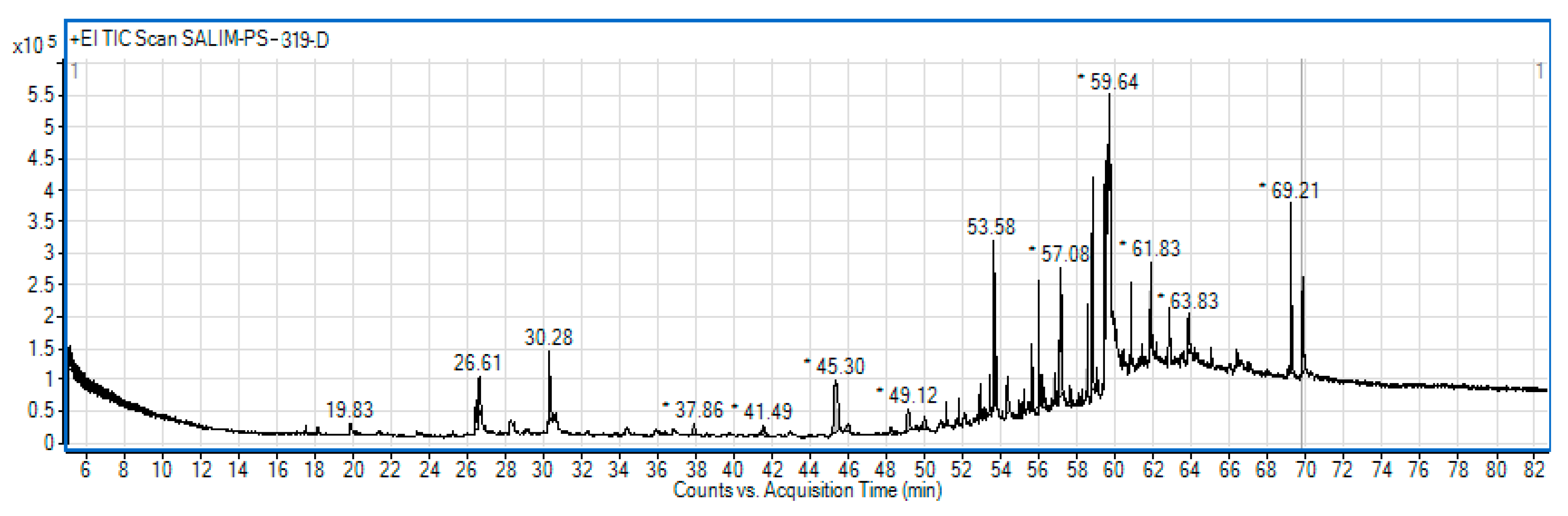
Figure 2.
Liquid chromatography mass spectrometry (LC/MS) chromatogram of CNEE.
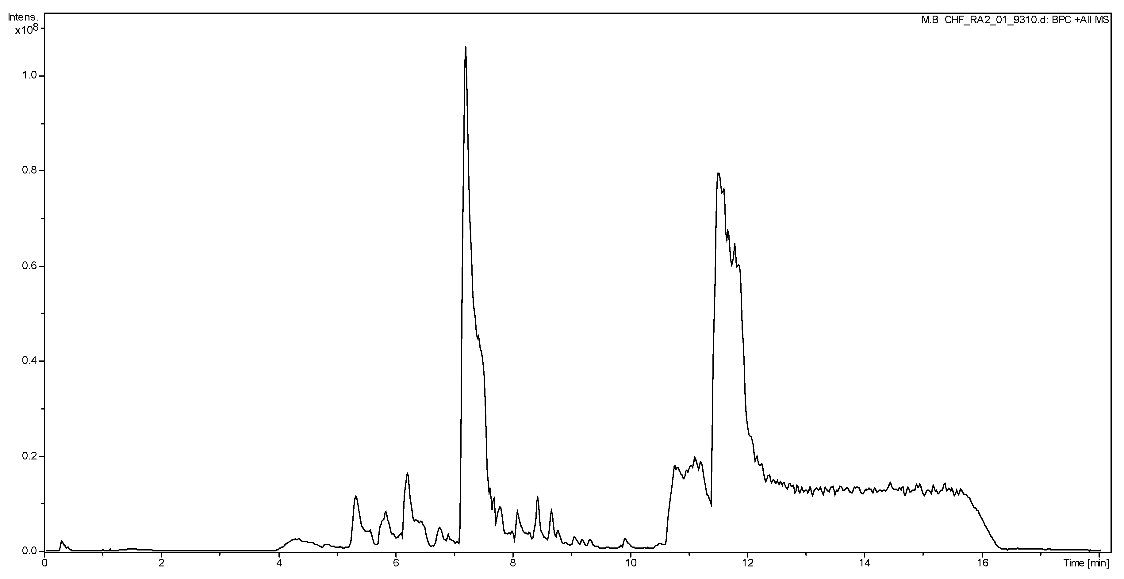
Figure 3.
UV/visible spectra of Pectin, AgNO3, CNEE, AgNPs and Pectin-AgNPs.
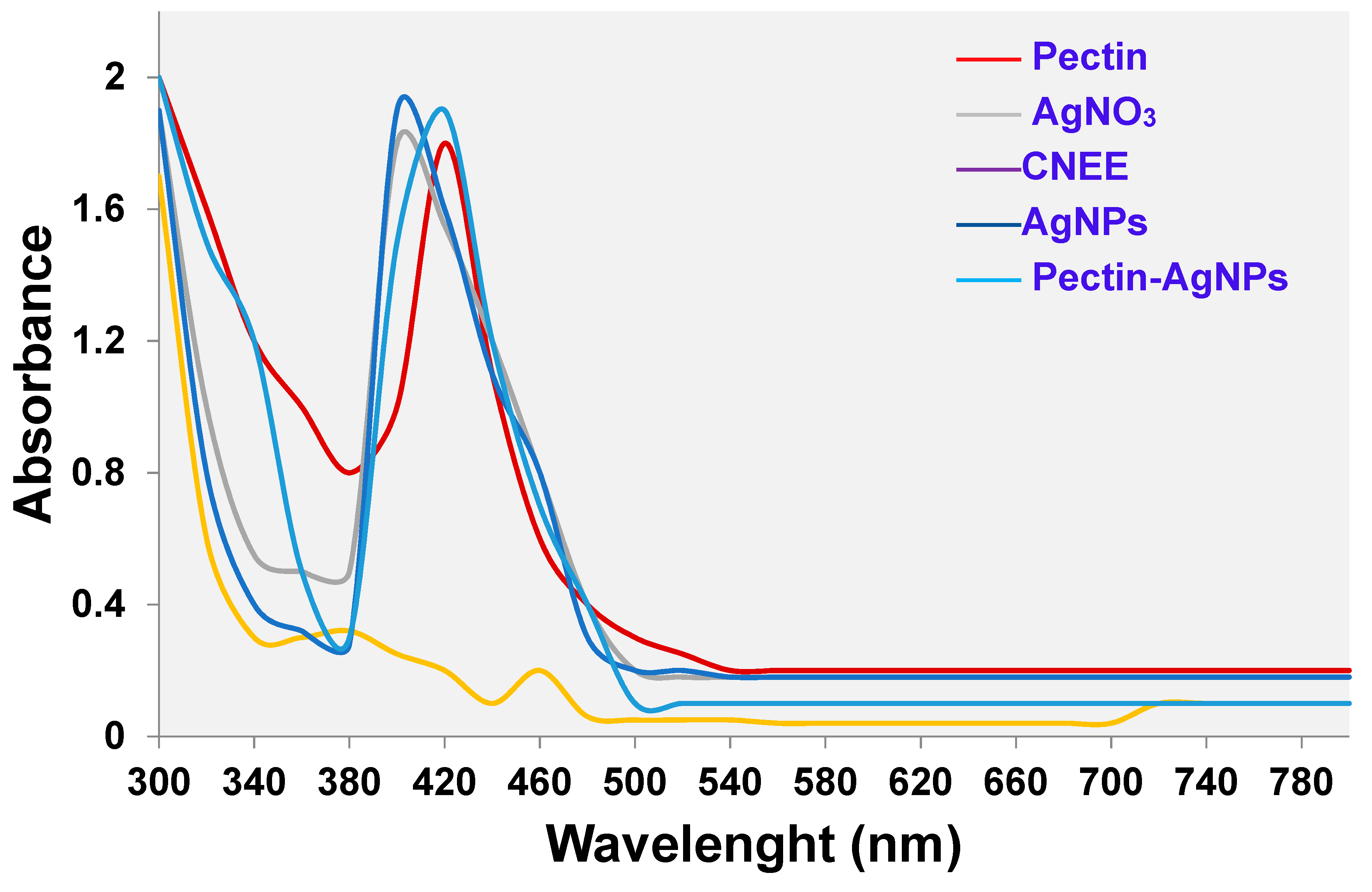
Figure 4.
Atomic foce microscopy (AFM) images of (A) AgNPs (B) Pectin-AgNPs.
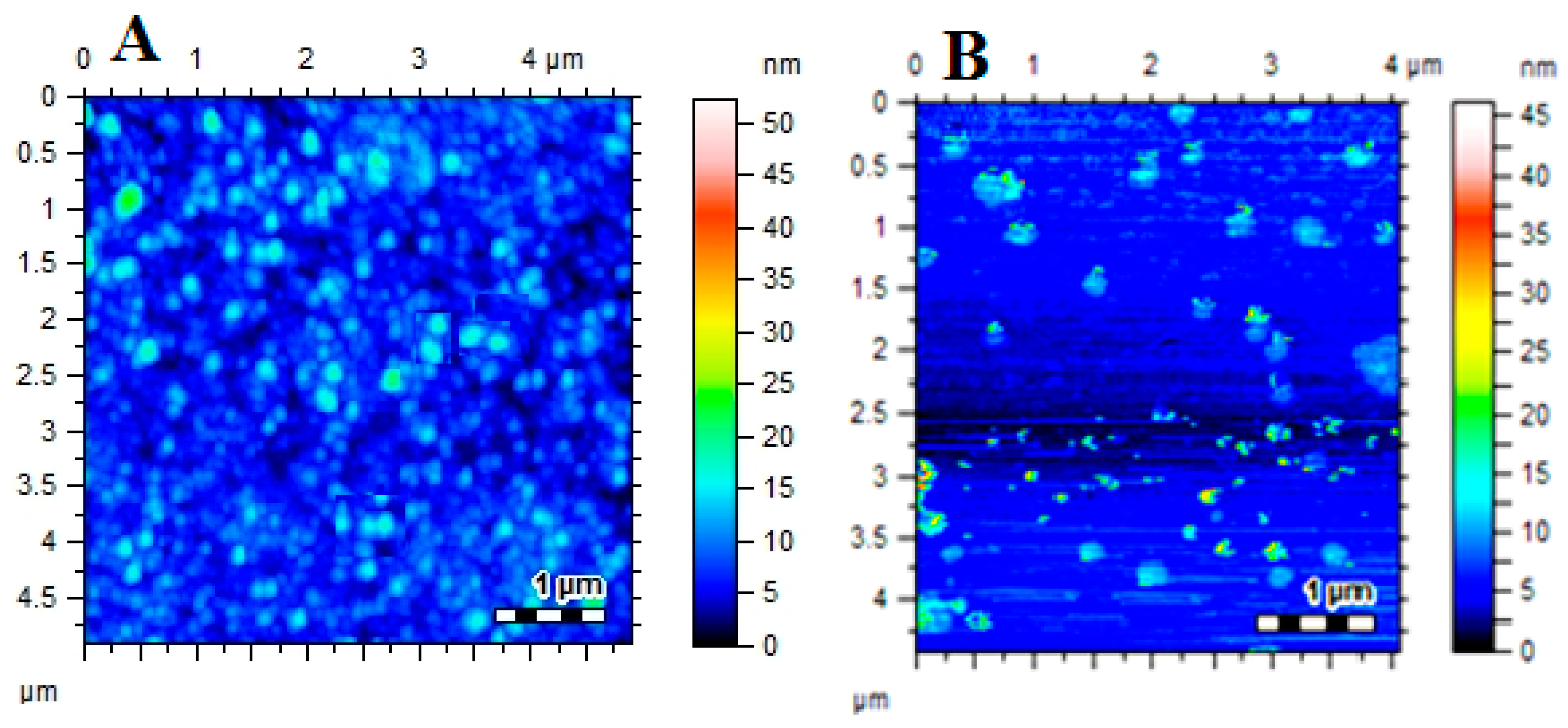
Figure 5.
Particle size distribution of (A) AgNPs and (B) Pectin-AgNPs. All experiments were performed three times and reported as mean.
Figure 5.
Particle size distribution of (A) AgNPs and (B) Pectin-AgNPs. All experiments were performed three times and reported as mean.
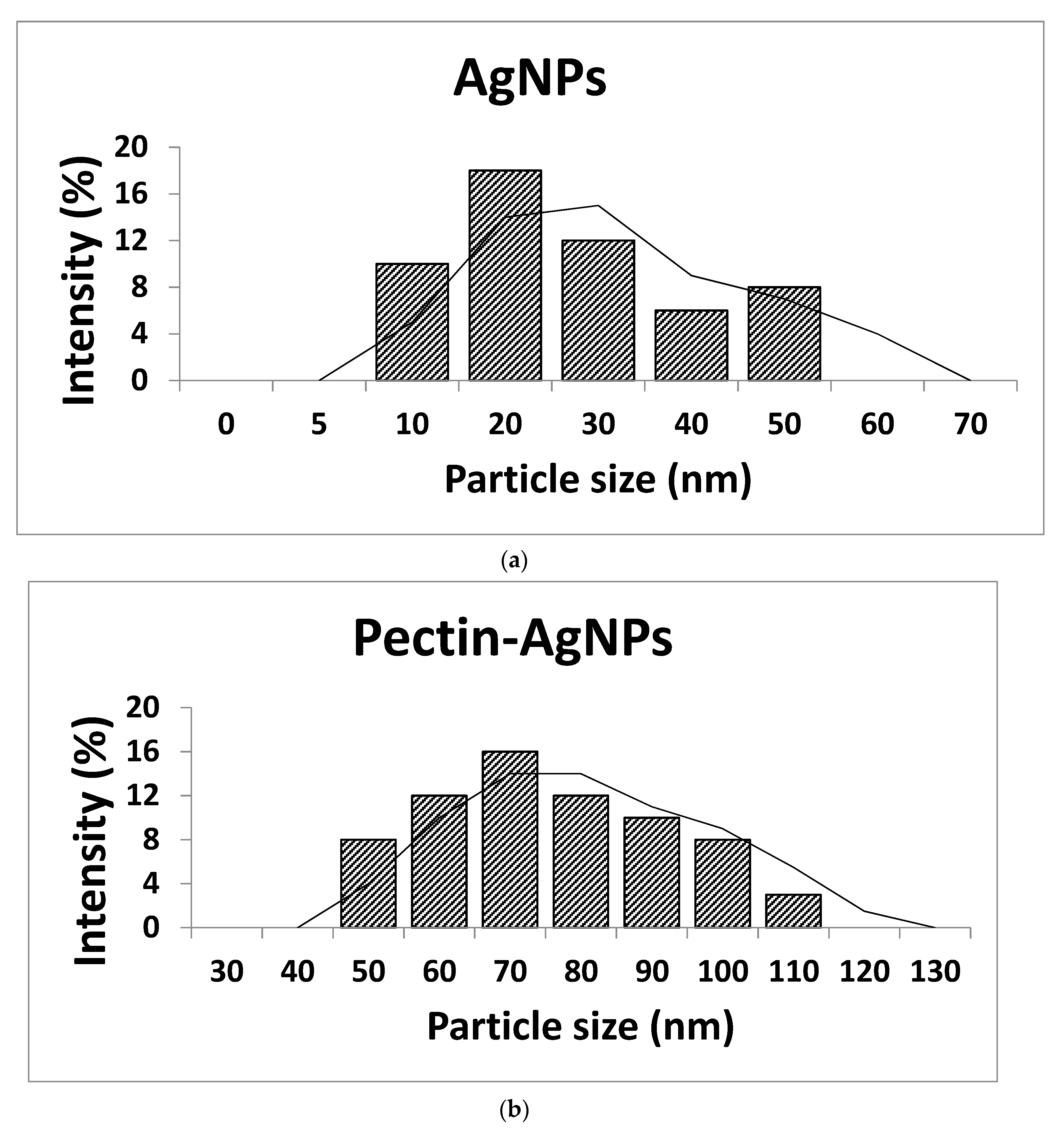
Figure 6.
Mean zeta potential of (A) AgNPs and (B) Pectin-AgNPs. All experiments were performed three times and reported as mean.
Figure 6.
Mean zeta potential of (A) AgNPs and (B) Pectin-AgNPs. All experiments were performed three times and reported as mean.
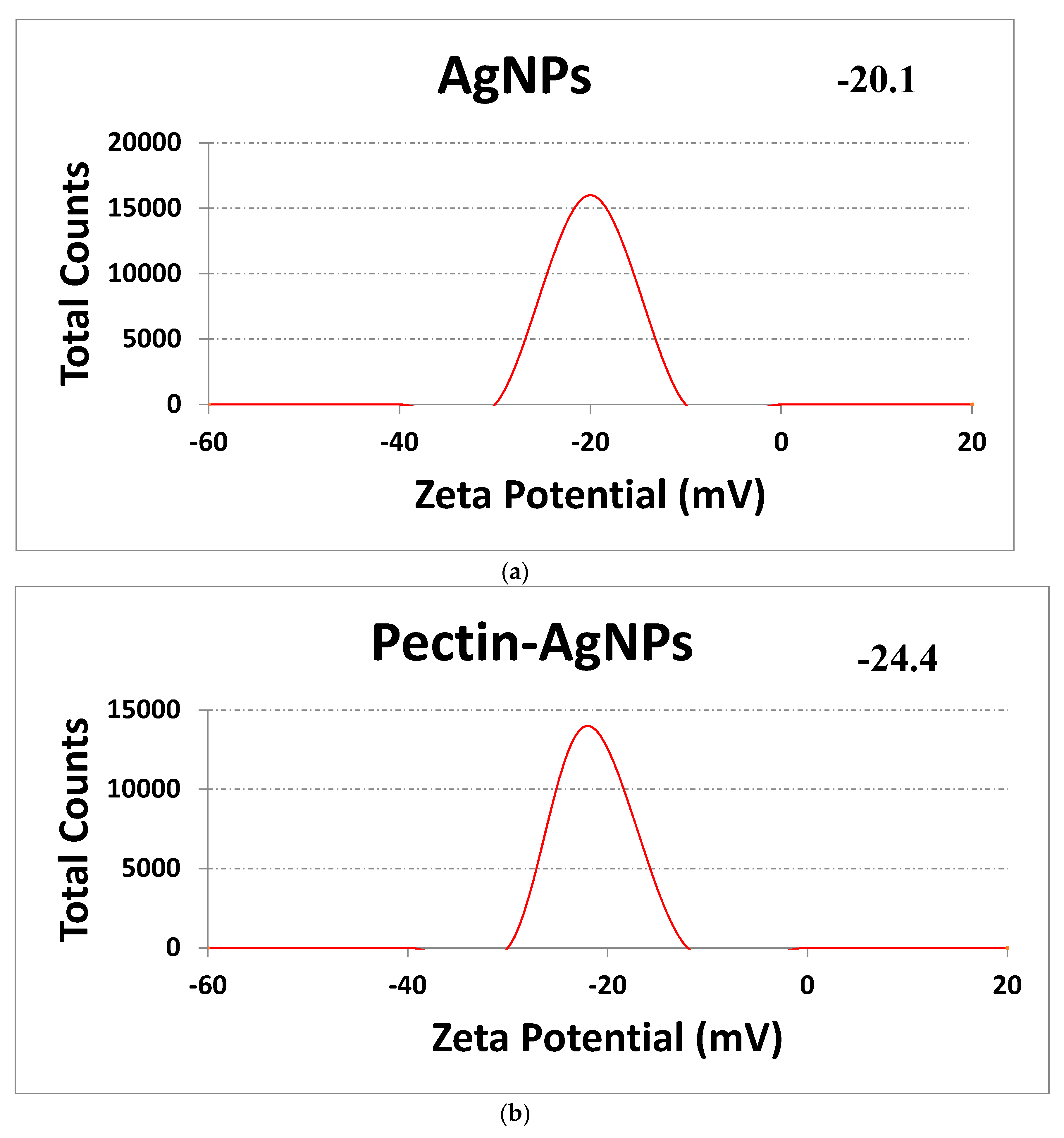
Figure 7.
Fourier transforms infrared (FTIR) spectra of (A) AgNPs and (B) Pectin-AgNPs.
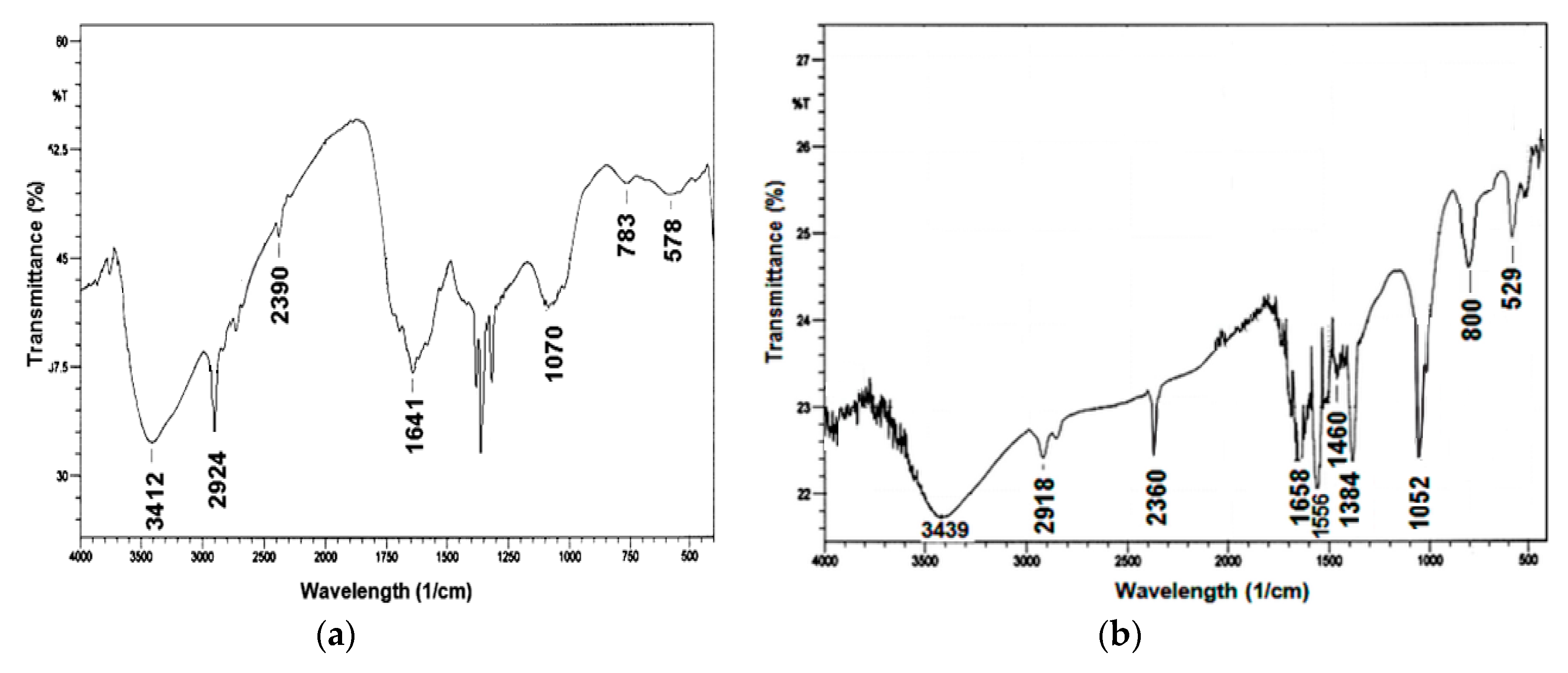
Figure 8.
Energy Dispersive X-Ray (EDX) spectra of Pectin-AgNPs.
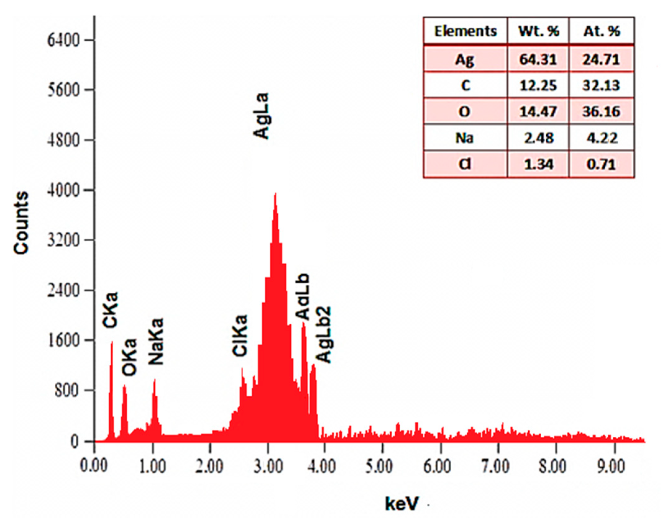
Figure 9.
Antibacterial activities of different antimicrobial agents against (A) Bacillus subtilis (B) Klebsiella pneumonia, (C)methicillin-resistance Staphylococcus aureusand (D)Vibrio cholera.
Figure 9.
Antibacterial activities of different antimicrobial agents against (A) Bacillus subtilis (B) Klebsiella pneumonia, (C)methicillin-resistance Staphylococcus aureusand (D)Vibrio cholera.
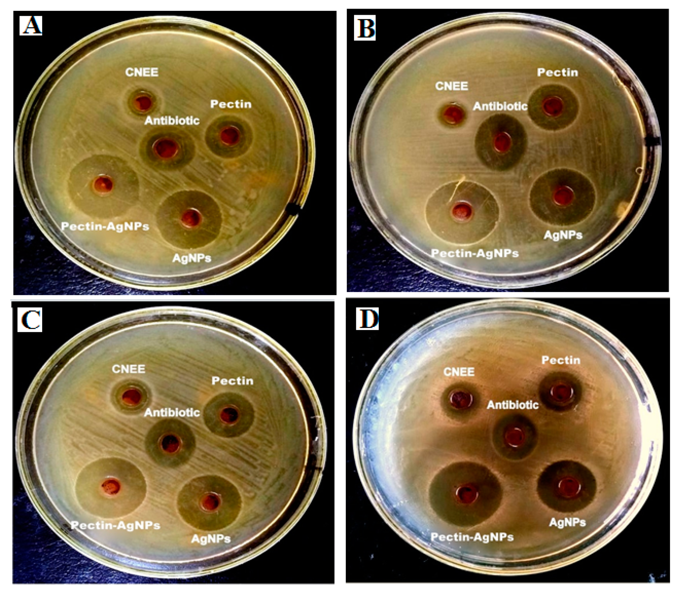
Figure 10.
Time killing kinetics of Pectin-AgNPs nanocomposite against different bacterial isolates. All experiments were performed in triplicates. Linear relationship (R2= 0.792) among viable cells counts of different bacterial strains at different time intervals while the viable cells counts of all bacterial strains significantly (R2 = −0.893) decreases with increasing exposure time of Pectin-AgNPs.
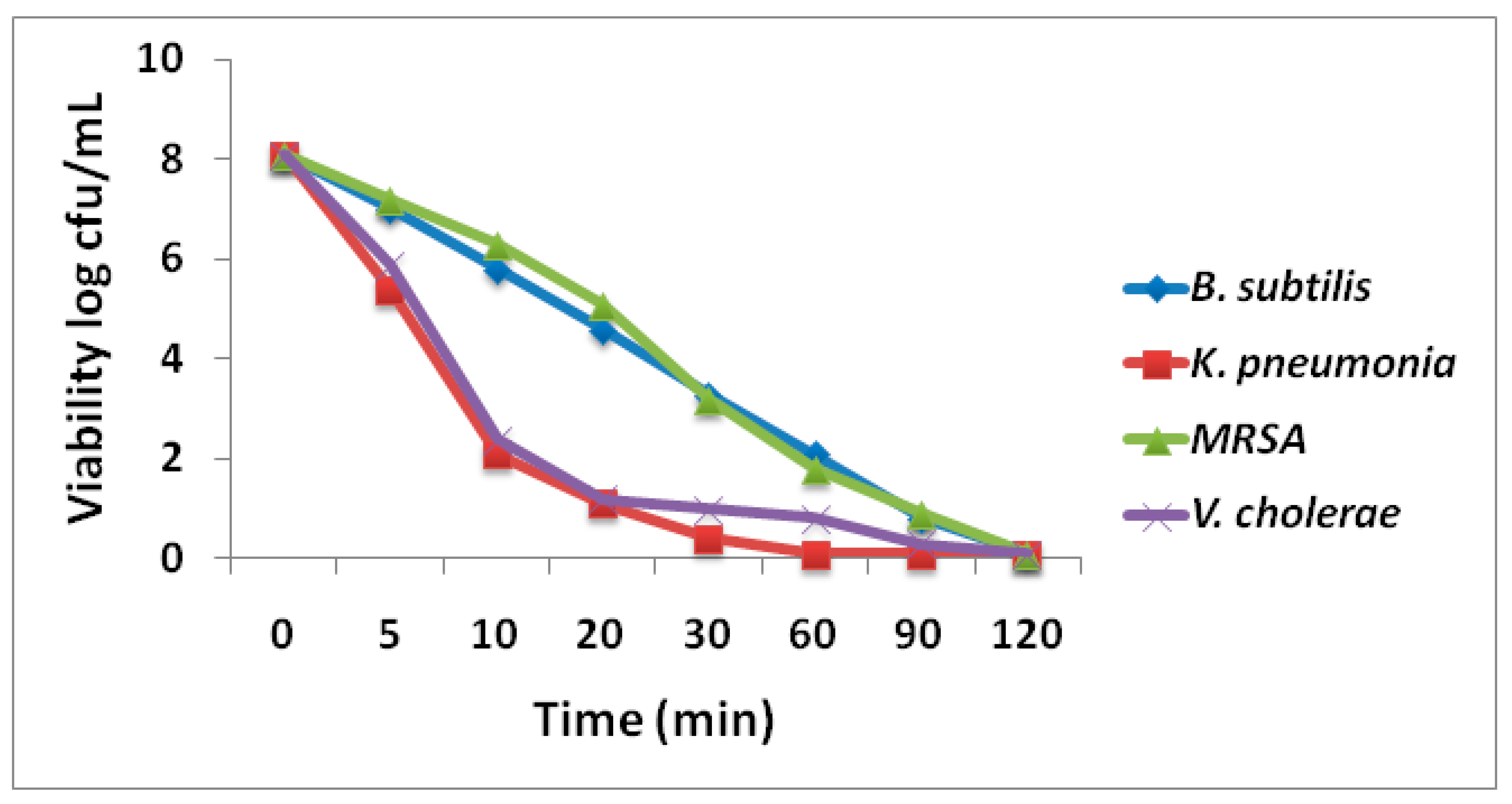
Figure 11.
Atomic force microscopic (AFM) images of A) Bacillus subtilis (B) Klebsiella pneumonia, (C) methicillin-resistance Staphylococcus aureusand (D)Vibrio cholera after treated with CMC-AgNPs.
Figure 11.
Atomic force microscopic (AFM) images of A) Bacillus subtilis (B) Klebsiella pneumonia, (C) methicillin-resistance Staphylococcus aureusand (D)Vibrio cholera after treated with CMC-AgNPs.
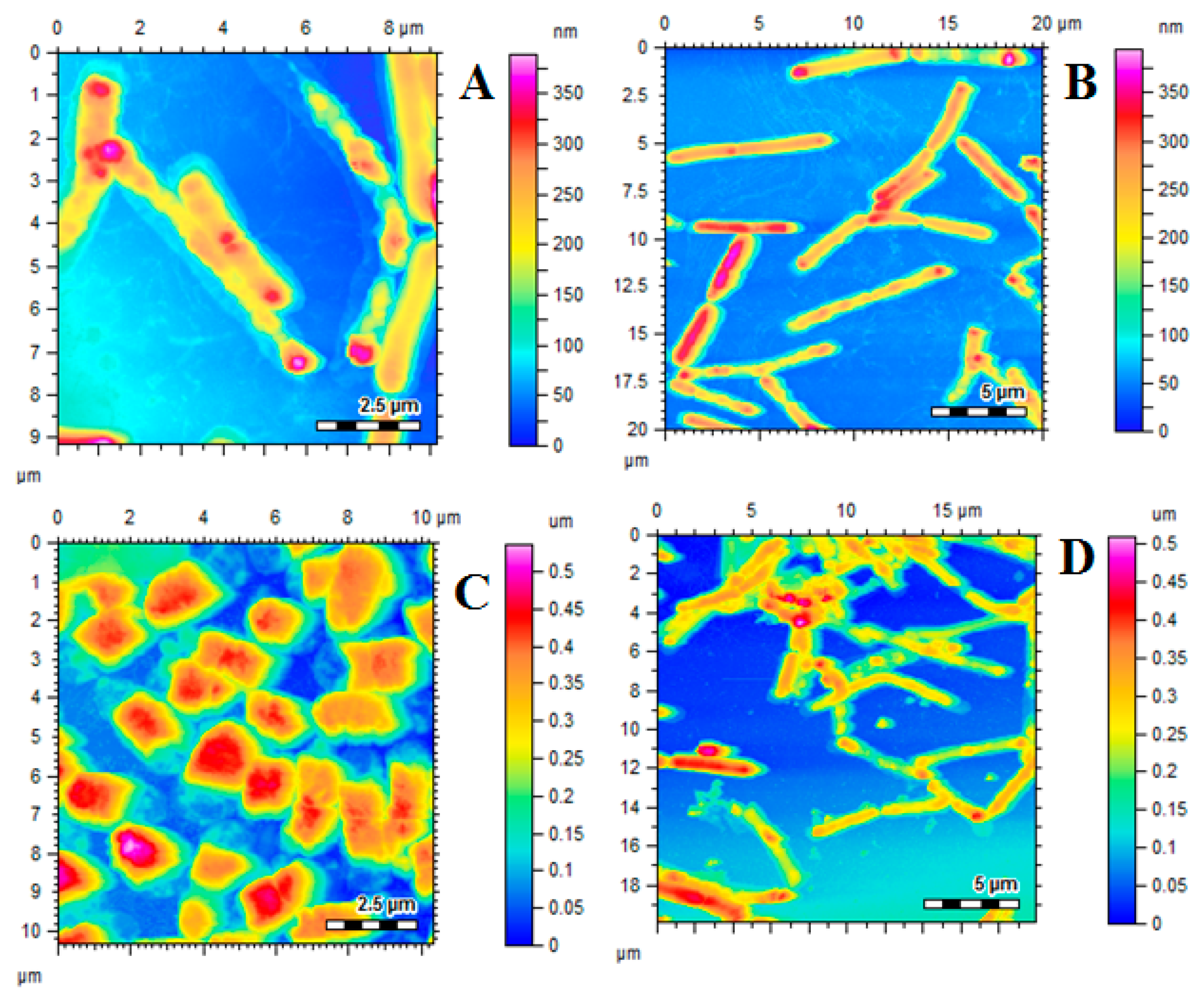
Figure 12.
Histopathology of liver tissue of treated rats at 40× magnification. I = Naïve; II = Positive control; III = Pectin-AgNPs(0.025 mg/kg); IV = Pectin-AgNPs (0.05 mg/kg)and V = Standard (Silymarin).
Figure 12.
Histopathology of liver tissue of treated rats at 40× magnification. I = Naïve; II = Positive control; III = Pectin-AgNPs(0.025 mg/kg); IV = Pectin-AgNPs (0.05 mg/kg)and V = Standard (Silymarin).
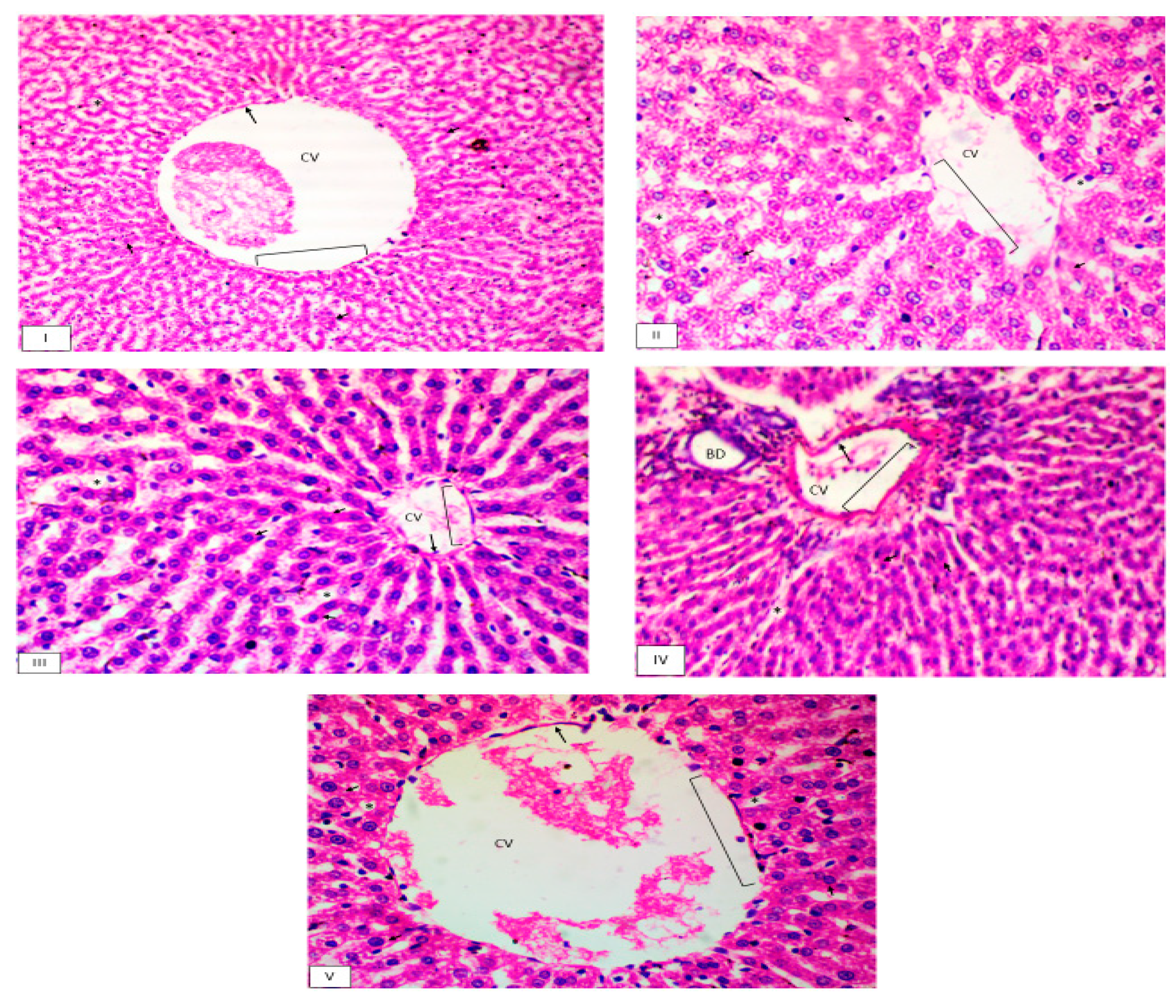
Figure 13.
Polymerase chain reaction (PCR) analysis of genes expression in treated rats.
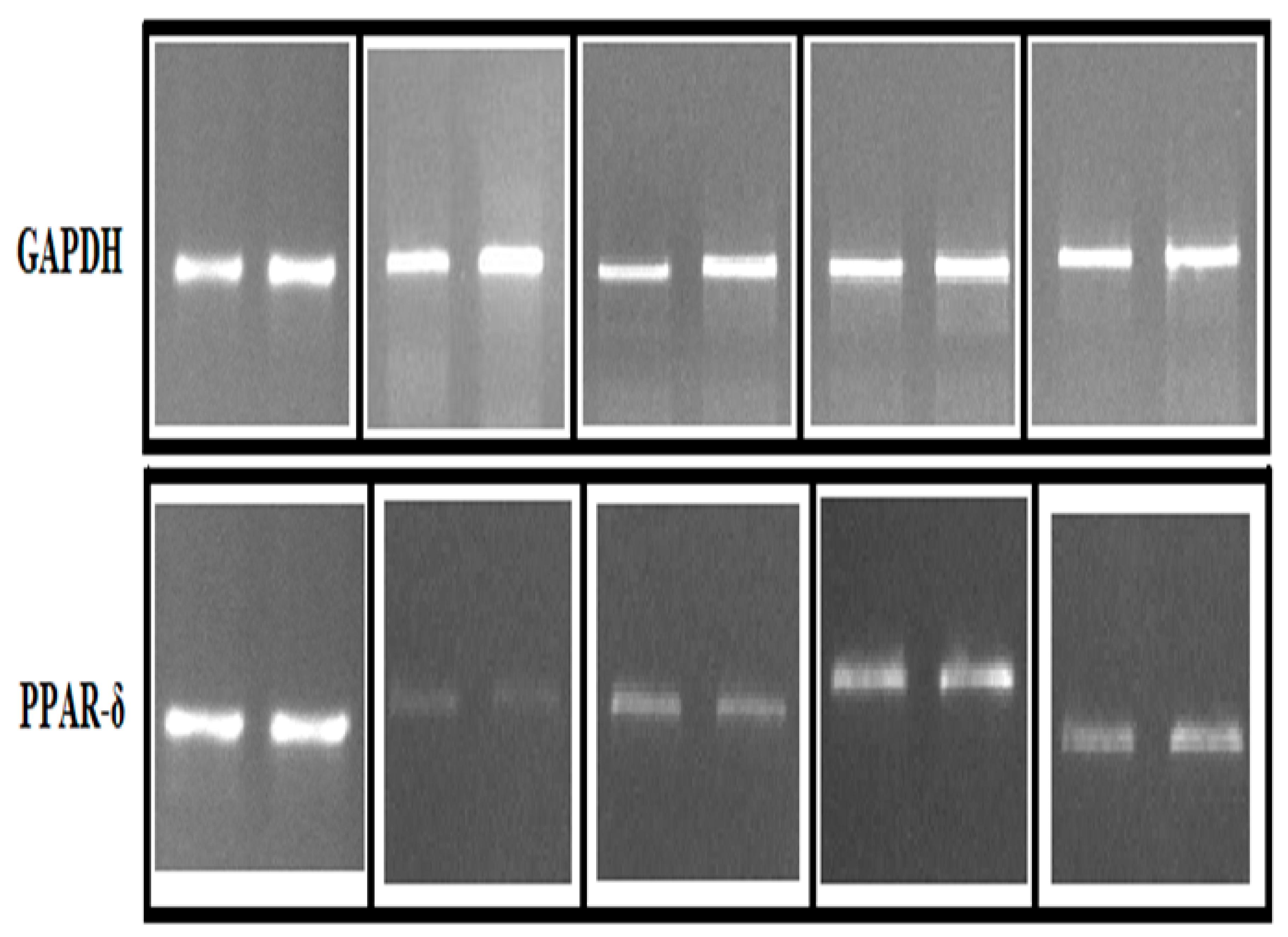
Figure 14.
Percentage viability of HeLA cells treated with different concentrations of AgNPs and Pectin-AgNPs compared with control and standard doxorubicin at 50 mg standard dose. Data is represented as average values ± SEM. *p ≤ 0.05 significant and **p ≤ 0.005 highly significant as compared to control.
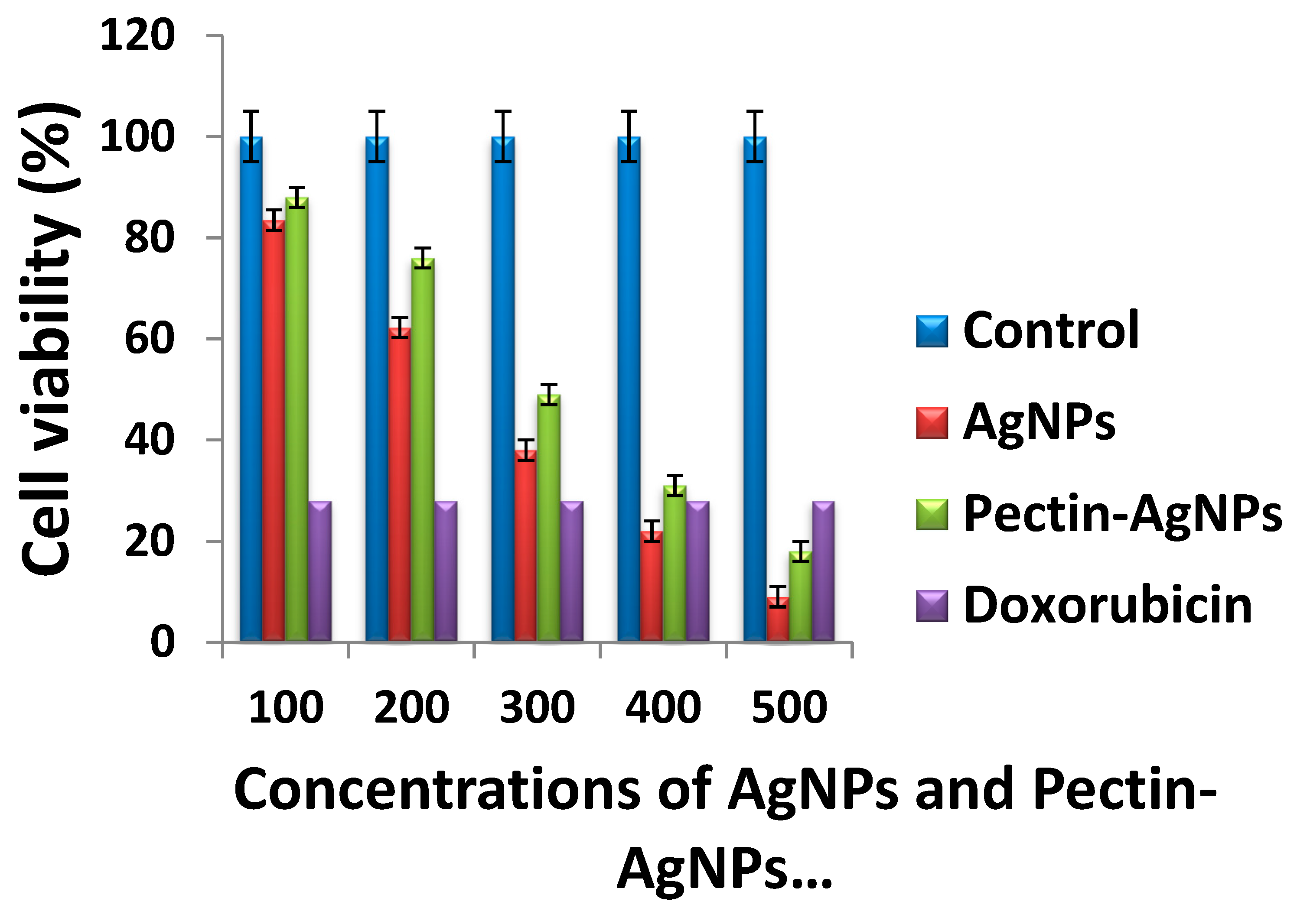

Table 1.
Zone of inhibitions (ZIs) of different antibacterial agents against different standard isolates.
Table 1.
Zone of inhibitions (ZIs) of different antibacterial agents against different standard isolates.
| Antimicrobial agents | Zone of Inhibition (mm ± S.D) | |||
|---|---|---|---|---|
| B. subtilis | K. pneumonia | MRSA | V. cholerae | |
| Control | 0.00 ± 0.00 | 0.00 ± 0.00 | 0.00 ± 0.00 | 0.00 ± 0.00 |
| CNEE | 6.9 ± 0.41 | 4.1 ± 0.15 | 8.3 ± 0.82 | 7.2 ± 0.26 |
| Pectin | 11.9 ± 1.38 | 13.8 ± 1.24* | 12.4 ± 1.33* | 10.3 ± 0.84 |
| AgNPs | 18.7 ± 2.18* | 17.3 ± 1.04* | 17.6 ± 1.83* | 15.2 ± 1.51* |
| Pectin-AgNPs | 27.2 ± 3.84** | 26.1 ± 2.18** | 24.8 ± 3.71** | 25.2 ± 2.46** |
| Cefoxitin | 13.7 ± 0.86* | 14.8 ± 1.01* | 13.1 ± 0.53* | 12.5 ± 0.64* |
S.D = Standard deviation, CNEE = Carpesium nepalense ethanolic extract. All experiments were performed in triplicates and reported as mean ± SD. *p ≤ 0.05 significant as compared to control, **p ≤ 0.005 highly significant as compared to control.
Table 2.
Minimum inhibitory concentrations (MICs) and minimum bactericidal concentrations (MBCs) of different antibacterial agents against different standard isolates.
Table 2.
Minimum inhibitory concentrations (MICs) and minimum bactericidal concentrations (MBCs) of different antibacterial agents against different standard isolates.
| Isolates | Antimicrobial agents | |||||||
|---|---|---|---|---|---|---|---|---|
| CNEE | Pectin | AgNPs | Pectin-AgNPs | |||||
| MIC | MBC | MIC | MBC | MIC | MBC | MIC | MBC | |
| B. subtilis | 5500 ± 271.3 | 6500 ± 226.1 | 256 ± 35.2 | 512 ± 81.2 | 128 ± 27.4 | 256 ± 56.2 | 64 ± 10.3 | 128 ± 14.9 |
| K .pneumonia | 7500 ± 364.2 | 8500 ± 421.2 | 512 ± 73.1 | 512 ± 57.5 | 256 ± 21.3 | 256 ± 42.5 | 128 ± 9.2 | 128 ± 16.4 |
| MRSA | 7500 ± 393.5 | 8500 ± 463.8 | 512 ± 84.6 | 512 ± 77.4 | 256 ± 18.2 | 256 ± 61.7 | 128 ± 9.1 | 128 ± 10.5 |
| V. cholera | 8500 ± 336.4 | 9500 ± 392.3 | 512 ± 52.4 | 512 ± 62.8 | 256 ± 31.8 | 256 ± 40.0 | 128 ± 11.5 | 128 ± 13.1 |
CNEE = Carpesium nepalense ethanolic extract, MIC = Minimum inhibitory concentrations, MBC = Minimum bactericidal concentrations. All experiments were performed in triplicates and reported as mean ± SD.
Table 3.
In vivo assessments of Naïve, Control, Carpesium nepalense ethanolic extract, Pectin, AgNPs, Pectin-AgNPs and standard on different biochemical in CCl4 induced hepatotoxic rats
Table 3.
In vivo assessments of Naïve, Control, Carpesium nepalense ethanolic extract, Pectin, AgNPs, Pectin-AgNPs and standard on different biochemical in CCl4 induced hepatotoxic rats
| Groups | Doses (mg/kg) |
CCl4 induced hepatotoxicity | |||||
|---|---|---|---|---|---|---|---|
| LDH (IU/dL) |
AST (IU/L) |
ALT (IU/L) |
ALP (IU/L) |
TP (g/dL) |
DB (mg/dL) |
||
| Naïve1 | –– | 210 ± 12.86 | 69.1 ± 1.54 | 62.0 ± 2.16 | 91.36 ± 2.49 | 6.5 ± 0.23 | 0.12 ± 0.05 |
| Positive control | –– | 322.4 ± 25.64 | 228.2 ± 5.22 | 195.0 ± 4.03 | 233.4 ± 6.85 | 3.0 ± 0.28 | 0.79 ± 0.08 |
| CNEE | 125 | 287.5 ± 18.53 | 184.6 ± 8.12 | 172.4 ± 5.75 | 198.2 ± 4.40 | 3.9 ± 0.27 | 0.67 ± 0.12 |
| 250 | 271.4 ± 21.65 | 175.1 ± 6.58 | 160.2 ± 7.52 | 184.0 ± 5.62 | 4.1 ± 0.64 | 0.61 ± 0.16 | |
| Pectin | 0.025 | 291.6 ± 31.57 | 193.4 ± 7.15 | 184.1 ± 6.36 | 212.6 ± 5.84 | 3.5 ± 0.40 | 0.71 ± 0.10 |
| 0.05 | 286.4 ± 28.14 | 182.5 ± 5.67 | 175.8 ± 6.06 | 201.9 ± 4.95 | 3.8 ± 0.51 | 0.68 ± 0.12 | |
| AgNPs | 0.025 | 255.3 ± 21.90* | 156.0 ± 7.03* | 134.0 ± 3.58* | 158.2 ± 3.27* | 4.1 ± 0.72* | 0.45 ± 0.09* |
| 0.05 | 238.7 ± 26.52* | 121.1 ± 8.94* | 110.2 ± 4.04* | 121.5 ± 4.18* | 4.8 ± 0.78* | 0.32 ± 0.08* | |
| Pectin-AgNPs | 0.025 | 231.7 ± 21.76** | 108.0 ± 4.0** | 96.5 ± 6.53** | 96.5 ± 4.55** | 5.5 ± 0.46** | 0.20 ± 0.02** |
| 0.05 | 221.5 ± 18.22** | 76.5 ± 2.0** | 74.2 ± 2.34** | 82.3 ± 2.64** | 7.8 ± 1.03** | 0.15 ± 0.02** | |
| Silymarin | 100 | 235.6 ± 24.64** | 93.5 ± 4.2** | 82.2 ± 2.09** | 98.2 ± 4.02** | 6.1 ± 0.33** | 0.32 ± 0.06* |
n=10, Average values ± SD; *p ≤ 0.05 significant as compared to control; **p ≤ 0.005 highly significant as compared to control LDH = Lactate dehydrogenase; AST = Aspartate transaminase; ALT = Alanine transaminase; ALP = Alkaline phosphatase;TP = Total protein; DB = Direct bilirubin; CNEE = Carpesium nepalense ethanolic extract.
Table 4.
Effect of different test solutions on liver of CCl4 induced hepatotoxic rats after 14 days of treatment.
Table 4.
Effect of different test solutions on liver of CCl4 induced hepatotoxic rats after 14 days of treatment.
| Histopathological findings | Naïve | Positive control | Pectin-AgNPs (0.025 mg/kg) |
Pectin-AgNPs (0.05 mg/kg) |
Standard (Silymarin) |
|---|---|---|---|---|---|
| Hydropic degeneration | ˗ | ++ | ˗ | ˗ | ˗ ˗ |
| Endothelium disruption | ˗ | +++ | + | ˗ | ˗ |
| Lymphocytic infiltration | ˗ | ++ | + | ˗ | ˗ |
| Lymphoid aggregate in portal vein | ˗ | +++ | + | ˗ | ˗ ˗ |
| Cytolysis | ˗ | + | ˗ | ˗ | ˗ ˗ |
| Congestion | ˗ | ++ | + | ˗ | + |
| Glucagon depletion | ˗ | ++ | + | ˗ | ˗ |
(˗ ˗) none, (˗) very low, (+) mild, (++) moderate, (+++) severely damaged.
Disclaimer/Publisher’s Note: The statements, opinions and data contained in all publications are solely those of the individual author(s) and contributor(s) and not of MDPI and/or the editor(s). MDPI and/or the editor(s) disclaim responsibility for any injury to people or property resulting from any ideas, methods, instructions or products referred to in the content. |
© 2023 by the authors. Licensee MDPI, Basel, Switzerland. This article is an open access article distributed under the terms and conditions of the Creative Commons Attribution (CC BY) license (http://creativecommons.org/licenses/by/4.0/).
Copyright: This open access article is published under a Creative Commons CC BY 4.0 license, which permit the free download, distribution, and reuse, provided that the author and preprint are cited in any reuse.


