Submitted:
08 May 2023
Posted:
09 May 2023
You are already at the latest version
Abstract
The awareness of the existence in plants of bioactive compounds namely phytochemicals (PHYs), having health properties is progressively expanding. Therefore, their massive introduction in the normal diet, in food supplements, and their use as natural therapeutics to treat several diseases are increasingly emphasized by several sectors. Particularly, most PHYs possessing antifungal, antiviral, anti-inflammatory, antibacterial, antiulcer, anti-cholesterol, hypoglycemic, immunomodulatory, and antioxidant properties have been isolated from plants. Additionally, their secondary modification with new functionalities, to further improve their intrinsic beneficial effects, is extensively investigated. Unfortunately, although the idea of exploiting PHYs as therapeutics is amazing, its realization is far from simple, and the possibility of exploiting them as effective orally administrable drugs is almost utopic. Most PHYs are insoluble in water and, when introduced orally, they scarcely reach the site of action in therapeutic concentrations. Degradation by enzymatic and microbial digestion, occurring in the mouth, stomach, and intestine, as well as fast metabolism and rapid excretion via the kidney, biliary, or lung, strongly limit their in vivo activity. To overcome these drawbacks, several nanotechnological approaches have been used and many PHYs-loaded delivery systems with dimensions of nanometers have been developed. This paper, also by reporting various recent case studies, reviews the foremost nano-suspension and nano-emulsion-based techniques developed for formulating the most relevant PHYs in more bioavailable nanoparticles (NPs), suitable or promising for clinical application. Also, the acute and chronic toxic effects due to the exposure to NPs reported so far, the possible nanotoxicity which could derive by their massive employment, as well as the ongoing actions to improve the knowledge in the field were discussed. The state of the art concerning the actual clinical application of both PHYs and the nanotechnologically engineered PHYs was also reviewed.
Keywords:
Nanotechnology application
; nano-suspension techniques
; nano-emulsion techniques
; bioactive constituents of plant
; phytochemicals (PHYs)
; poor water solubility
; drug delivery systems (DDSs)
; toxicological risks of NPs
; clinical application of NPs
1. Introduction
Epidemiological studies have evidenced that the ingestion of some foods, including edible plants, is associated to the onset of healthy effects. As an example, the consumption of red wine is related to a reduction in mortality by cardiovascular events triggered by atherothrombosis, due to its capability of decreasing the progression of atherosclerotic lesions [1]. Additionally, green tea has proved to have protective effects on cardiovascular diseases [1]. The bioactive chemical compounds responsible of these benefits are known as phytochemicals (PHYs). Precisely, PHYs are defined as bioactive chemical compounds findable in plants, such as fruits, vegetables, grains, and other plant-derived foods that may supply health benefits, beyond basic nutrition and could help to reduce the risk of major chronic diseases [2]. PHYs are generally produced by plants to help themselves resist fungi, bacteria, and plant virus infections, and also to hamper their consumption by insects and other animals [3]. In the years, humans have used PHYs both as poisons and as traditional medicine [3]. Nowadays, the recognition that plants, can be a source of compounds, having health properties, is increasingly expanding worldwide, and both the food market and the sector of natural compounds are involved [1]. Often PHYs are regarded only as research compounds, rather than molecules which could have actual clinical application as possible therapeutics, because proofs of their possible health effects have not been proven yet [4]. However, experts in the field incessantly emphasize their extensive introduction in the normal diet, in food supplements, as well as their use as natural therapeutics to treat several diseases. As of the beginning of January 2022, a total of 130 thousand PHYs have been found [5]., and others will be discovered in the next years. Several PHYs owing many beneficial effects, including antifungal, antiviral, anti-inflammatory, antibacterial, antiulcer, anti-cholesterol, hypoglycaemic, immunomodulatory, and antioxidant activities have been isolated from plants, such as vegetables, herbs, fruits, legumes, oils, spices, nuts, and whole grains [6,7,8,9,10,11]. Anyway, in Western societies, the industry dealing with natural products and plants progressively searches for the discovery of new PHYs, to be used as possible health promoters safer than synthetic compounds [1]. Parallelly, experts in the field, including scientists, engineers, and technologists, incessantly investigate the possibilities of chemical modification of known PHYs, by the introduction of new functionalities, such as antioxidant, anti-free radicals, and anticancer moieties, to boost up their intrinsic activities. Unfortunately, although the idea of exploiting the strong potential of PHYs for health purposes is brilliant, its realization is far from simple [12]. Additionally, thinking of exploiting extracted PHYs as effective orally administrable health promoters is almost utopic. Solubility in an aqueous medium, permeability across the membrane of epithelial cells, and molecular interactions in the fluids of the gastrointestinal tract (GIT) are key factors that greatly affect the possible beneficial effects of PHYs, by influencing their route to the bloodstream and their final distribution to the targets. In this regard, most PHYs are insoluble in water and, in whatever form they are introduced orally, they hardly will reach the target in therapeutic concentrations. Moreover, early degradation by chemical, enzymatic and microbial digestion, occurring in the mouth, stomach, small and large intestine [12], as well as fast metabolism and rapid excretion via the kidney, biliary, or lung further limit their activity in vivo [13]. Collectively, PHYs generally achieve only nano/picomolar concentrations in cells and tissues, which are doses insignificant for producing a health promotion response. Figure 1 shows the main possible drawbacks related to most PHYs and the events that limit their in vivo beneficial effects after oral administration.
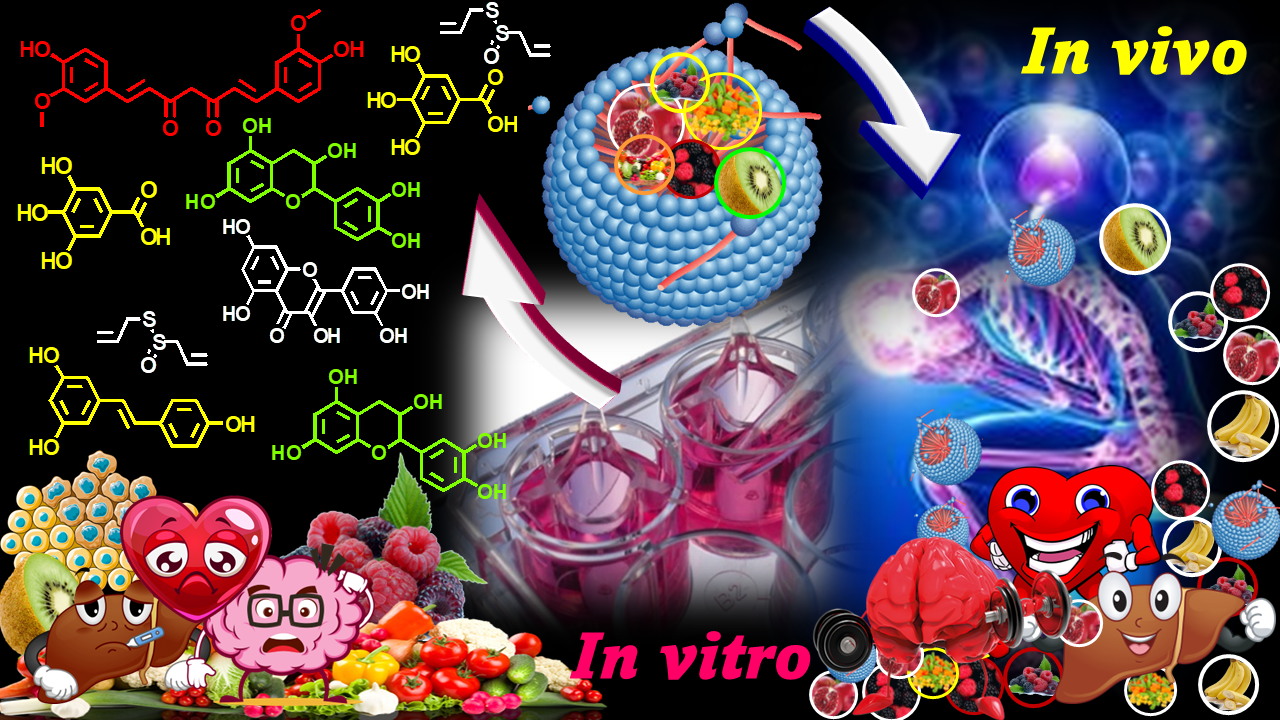
Additionally, concerning PHYs recognized for having demonstrated beneficial properties in vitro, we must bear in mind that, since when ingested they can be altered by microbial fermentation, also their biological properties may be distorted, and paradoxically converted in toxic effects. In this regard, important methods are necessary to judge safety and efficacy of these products, including potential risks and gaps. Anyway, to overcome the afore-mentioned problems and to allow the exploitation of PHYs as health enhancers, researchers increasingly resort to nanotechnology and nanostructures with dimensions of nanometers (nm). In the years, several PHYs-enriched nanomaterials have been engineered to overcome the poor solubility, permeability, and negative pharmacokinetics of PHYs, and different nanosized delivery systems (DSs)to transport therapeutic concentrations of PHYs to the targets have been developed [12,14].
The following Figure 2 provides an idea of the growing scientific interest in PHYs for healthy purposes and of the consequent application of nanotechnologies for solving their physicochemical issues, during the last 15 years. After having carried out a survey both in Pubmed and in Scopus datasets, we have reported the results obtained in Scopus because they included a greater number of papers, not considered in Pubmed. Particularly, the bar graph in Figure 2 was obtained by carrying out a survey about the number of works published year by year from 2008 so far (excluding the current year), making a search by “nanomaterials OR nanoparticles OR nano-formulation AND phytochemicals” keywords.
While the interest in the topic was low up to the year 2015 (less than 100 papers), in the following years, it has grown exponentially, as established by the several papers which have been published. This review, also by reporting several recent case studies, provides an up-to-date overview of the nanotechnological approaches based on nano-suspension (NS) and nano-emulsion (NE) techniques developed so far for formulating the most relevant PHYs in more bioavailable NPs promising for clinical applications by oral administration. We also paid attention to the pending issue relating to the possible toxic effects of NPs on humans, animals, and the environment in a scenario of limited knowledges, and we briefly reported the ongoing actions to improve the expertise in the field and/or to promote the development of increasingly safe nanomaterials.
The last section of the present paper gives a glance to the actual clinical applicability of PHYs and of nanotechnologically engineered PHYs, by reviewing those which are already clinically approved or are currently in advanced clinical trial. Originally, most information provided here has been also graphically presented and statistically analyzed.
2. Methodology for Literature Search and Study Selection
The literature search was performed using PubMed, and Scopus database considering the period January 2008 up now using first the same keywords used for detecting the publications taken into account in Figure 2 (nanomaterials OR nanoparticles OR nano-formulation AND phytochemicals). Secondly, using nano-suspensions or nano-emulsions in place of nanomaterials, nanoparticles, nano-formulation, we have limited our investigation only to publications concerning these two techniques. Among results, we selected studies, which contained information specifically suitable for the sections which we have decided to report in the present review, Studies published in English language only were included in this review. Conference abstracts, patents and unpublished results were excluded.
3. Phytochemicals (PHYs)
3.1. Phytochemicals and Nutraceuticals: Not Quite the Same
Both PHYs, also known as phytonutrients, and nutraceuticals are bioactive compounds which could be found in edible products, possessing beneficial properties capable of enhancing humans’ health, and which could be commonly ingested with the diet. Anyway, while nutraceuticals are essential nutrients for human life necessarily present in edible products, which can derive from animals, plants and fungi, PHYs are non-nutrients, which exclusively originate form plants [15]. Differently from the major nutrients like vitamins, whose reduced intake leads to the onset of serious deficiencies, PHYs are powerful health-boosting nutrient-like substances, whose lower intake does not cause defects [16]. However, these compounds can be invaluable to humans’ health, and a diet rich in PHYs is strongly connected with better health [16]. They could improve the ability to detox, as well as could boost the immune system. They may also help protect against age-related diseases, such as diabetes, heart disease, and osteoporosis [16]. PHYs contained in food, when ingested and metabolized, provide benefits. In vitro studies have demonstrated that these plants constituents are multifunctional compounds, with healthy properties like those of conventional drugs and can be considered “pharmaceutical-grade compounds”. Although, as above-mentioned, also nutraceuticals could be plants-derived bioactive compounds, we focused our work on PHYs.
3.2. Phytochemicals: An Overview
It has been reported that scientists have already identified over 5,000 different classes of PHYs. Anyway, many more exist undiscovered, as well as much more has to be learned about their potential benefits [17]. Although PHYs can derive from both edible and non-edible plants [18], all plant-based foods including fruits, vegetables, nuts, and herbs contain them. Since processing methods may lower the PHYs content of some foods [19], fresh, whole foods have to be preferred to get the most benefit from PHYs. Additionally, although generally PHYs confer to the plant-derived food containing them a particular bright color, also foods without these characteristics can contain these healthy promoters as well (Table 1). So, if green, purple, red, blue, or yellow vegetables and fruits are containers of colored PHYs [17,20,21], also not brightly colored potatoes, cauliflower and nuts such as almonds, cashews, and hazelnuts contain several PHYs (Table 1). Both tea and dark chocolate, even if not brightly colored, yet they are packed with these health-boosting plant compounds [17,22,23,24]. Moreover, essential oils (EOs) including pine needles, cedar, and lavender are used as health-promoters, due to their PHYs content (Table 1) [25,26,27].
Note that, we have not reported the beneficial properties of each plant, but a series of health promoting activities attributed to all plants, because each plant, being a source of multiple phytochemicals, each having multiple pharmacological activities, rationally, can possess all the reported properties. Anyway, we have examined the point more in depth in Table 2, where the different beneficial activities were reported for each class of phytocompounds or even for the single phytocompound.
3.2.1. Specific Sources and Benefits of Main Types of PHYs
The most common PHYs comprise polyphenols, including four principal classes of compounds: phenolic acids (caffeic acid, ellagic acid, gallic acid, tannic acid), stilbenes, lignans and flavonoids, such as catechins. Flavonoids in turn include, among others, the subgroups of iso-flavonoids, pro-anthocyanidins, comprising procyanidins, anthocyanidins, including cyanidins, and anthocyanins, which are anthocyanidins with sugar groups. Also, PHYs encompass carotenoids, coumarins, indoles, organosulfur compounds, isothiocyanates, saponins, tannins, phenylpropanoids, anthraquinones, ginsenosides, terpenoids etc. [38]. In the following Table 2 we have reported the most relevant PHYs, their source and the associated healthy effects [3,39].
As above-mentioned, the known PHYs are thousands. Following, some examples of the most common [1,15].
Polyphenols
Figure 3.
Structures of some plants derived polyphenols. ROS = reactive oxygen species.
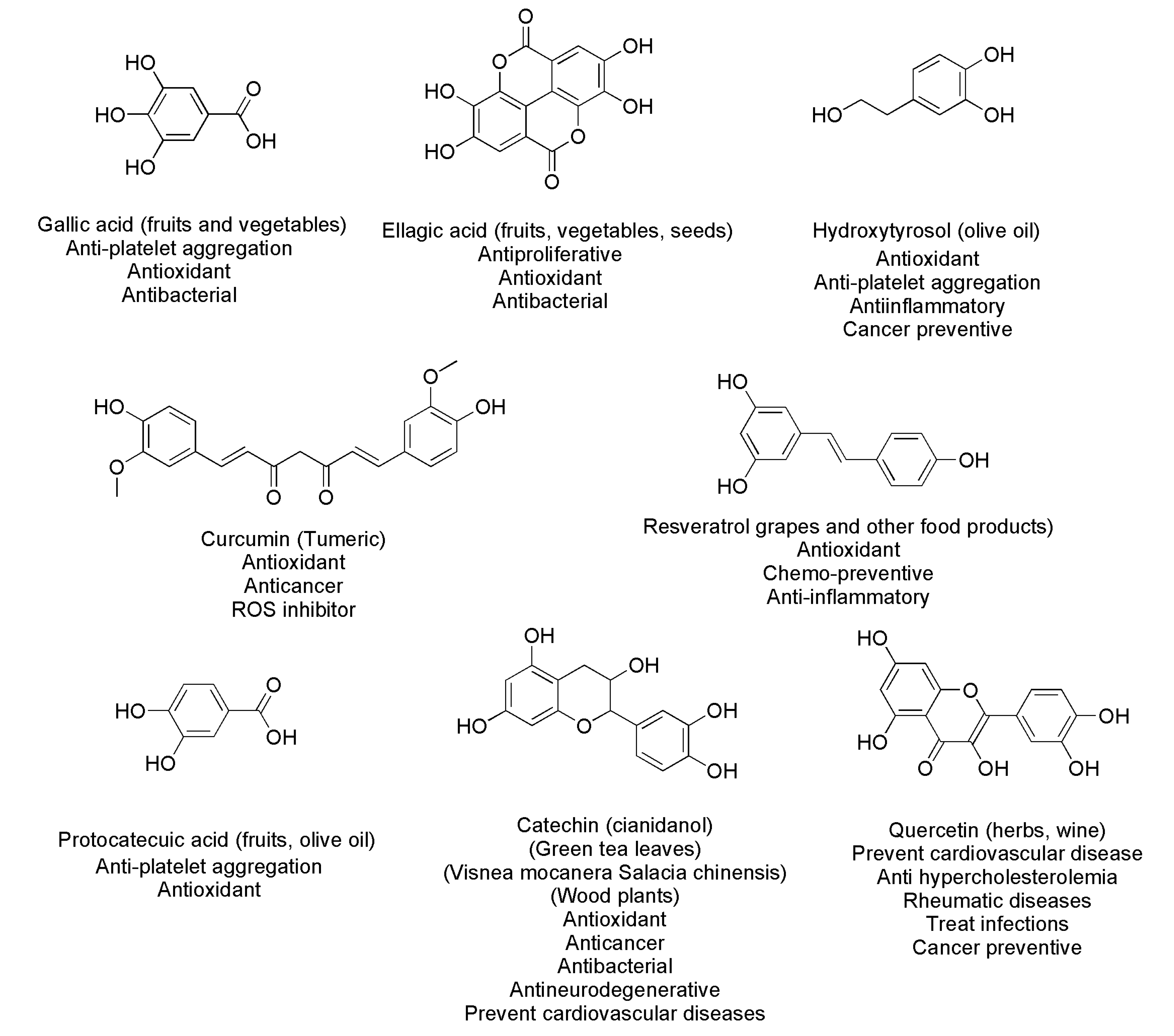
Among polyphenols, gallic acid (GA), ellagic acid (EA), and other natural products such as propolis extracts, which are composed of phenolic acids, flavonoids, terpenes, and essential oils, can treat infections sustained by bacteria of several species, including those of sporogenic type. Particularly, GA showed antibacterial activity against Paenibacillus larvae, Staphylococcus aureus [3], Escherichia coli, Pseudomonas fragi, Pseudomonas fluorescens, Pseudomonas putida, Pseudomonas spp. P304, Plesiomonas schigelloides, and Schigella flexneri B [3]. Both in in vitro studies and in animal models, GA and EA have shown activities against several degenerative diseases, such as cardiovascular, cancer, and central-nervous-system-disabling disorders, including Parkinson’s disease, Alzheimer, multiple sclerosis, and amyotrophic lateral sclerosis [47,48,49,50,51,52]. Furthermore, hydroxytyrosol, a polyphenol extracted from extra-virgin olive oil has demonstrated antioxidant, anti-inflammatory and cancer preventive activities. Curcumin from Curcuma longa L. rhizome, GA, protocatechuic acid, quercetin, and resveratrol (RES), which are contained in many fruits and vegetables, are capable of inhibiting platelet aggregation and reactive oxygen species (ROS) activity induced by thrombin, collagen, or other agonists [3,15]. Particularly, RES is a phytoalexin derived from grapes and other food products has antioxidant and potential chemo-preventive activities and anti-inflammatory effects. Quercetin is found in many foods and herbs, including wine successful in the treatment or in the prevention of diverse conditions including cardiovascular disease, hypercholesterolemia, rheumatic diseases, infections and cancer. Quercetin as a nutritional supplement is well tolerated and has not been linked to serum enzyme elevations or to episodes of clinically apparent liver injury. Also, curcumin (from Turmeric), catechins (from tea leaves, Visnea mocanera Salacia chinensis, wood plants), also known as cianidanol, and RES (from grapes) possess antioxidant effects and can help in preventing and treating cancer, cardiovascular illness, neuronal degenerative diseases, diabetes, and infections. Most of them are inhibitors of platelet aggregation and prevent OS triggered by ROS [15]. As above reported, polyphenols encompass flavonoids which in turn comprise anthocyanins, whose name comes from the Greek words for ‘flower’ and ‘blue.’ Anthocyanins give fruits and vegetables their vivid red, blue, and purple colors. The main sources of anthocyanins are red, blue, and purple vegetables, including berries, cherries, currants, grapes, plums, purple potatoes and red cabbage [21]. Scientists have discovered over 700 different types of anthocyanins [16]. In vitro and in vivo studies on healthy properties of anthocyanins established they can decrease serious health risks, eliminate FR, help in controlling weight, in preventing heart disease, and increase insulin sensitivity. Additionally, they reduce inflammation, diabetic complications, protect DNA and the brain. Anthocyanins also boost other PHYs and the activity of other phytonutrients [53,54,55].
Tannins
Tannins confer to edible plants astringent flavors and bitter tasting, but also a variety of health benefits, such as antioxidant effects [56]. Additionally, they fight parasites, microbes, and other pathogens, lower blood pressure, inflammation, serious health risks, and regulate the immune system [57]. Many different foods could be the sources of tannins, such as tart fruits like cranberries, currants, blackberries, sweeter fruits like apples, grapes, peaches, strawberries, and nuts, such as almonds, hazelnuts, pecans, pistachios, and walnuts. Additional sources of tannins could be barley, beans, lentils, and rice.
Tea is a common way by which people consume tannins, along with cacao beans and dark chocolate [57]. Lastly, antiparasitic herbs, including black walnut, sagebrush, garlic, oregano, Tribulus terrestris L., Mimosa pudica, neem (Azadirachta indica), grapefruit seed, vidanga (Embelia ribes Burm F.), carnations etc. contain tannins. Among tannins, ellagitannins (ETs) have shown activities against several degenerative diseases, such as cardiovascular, cancer, and central-nervous-system-disabling disorders, including Parkinson’s disease, Alzheimer, multiple sclerosis, and amyotrophic lateral sclerosis. Moreover, ETs are source of GA and EA that, as above introduced, have shown similar activities, both in vitro studies and animal models [47,48,49,50,51,52].
Lutein and Zeaxanthin
Lutein and zeaxanthin can give foods a rich orange or yellow color, are potent antioxidants, and are mainly known for their role in eye health. These compounds absorb 40-90% of blue light and reduce OS, thus helping to protect retina from damage [58]. The eye functions resulted ameliorated, thus allowing to see with more clarity and to be less bothered by glaring lights [58]. Additionally, lutein and zeaxanthin may also enhance memory and brain function [59], as well as improve the body’s use of insulin, and skin health [60], while they lower blood pressure, reduce inflammation, and support heart health [16]. According to their orange and yellow colors, the possible sources are egg yolks, orange peppers, oranges, pumpkins, and yellow corn [60]. However, whopping dose of lutein and zeaxanthin is contained also in green foods. Kale, parsley, romaine lettuce, and spinach are excellent sources of lutein and zeaxanthin, as well as pistachios and olive oil [60].
Sulforaphane
Sulforaphane is a potent antioxidant and is highly researched for its potential health benefits. In vivo and in vitro studies suggest that sulforaphane acts as an anti-inflammatory agent and reduces serious health issues. Additionally, this compound could potentially trigger unhealthy cells to die [16]. Also, sulforaphane may boost the ability to fight pathogens, increases detoxification and liver function, lowers autism symptoms, protects eyes, and reduces depression and anxiety [61]. Cruciferous vegetables including cabbage, kale, broccoli sprouts, and kohlrabi are great sources of sulforaphane [61], which need activation by mustard.
Eugenol
Eugenol may act as a strong anti-inflammatory and antioxidant compound and helps eliminate parasites [62,63]. Animal, test-tube, and human cell studies established that eugenol is effective against fungi, inhibits serious health concerns, protects the brain, the liver, reduces bacterial biofilm, as well as supports heart and stomach health [64]. The best source of eugenol is clove. Also, eugenol can be found in multiple herbs and spices such as cinnamon, cumin, nutmeg, and is present in coffee, mung beans, and soybeans, as well as in bananas, melons, strawberries, and tomatoes [46].
Terpenoids
Terpenoids represent one of the largest groups of natural products, mainly extracted from plants, which account for more than 40,000 compounds, but new terpenoid compounds are discovered incessantly every year. They possess anticancer effects against several tumors, including breast, mammary, skin, lung, forestomach, colon, pancreas, and prostate carcinomas. Additionally, most triterpenoids suppress cancer cells without exerting toxicity towards normal cells [3]. Terpenoids, which are found mainly in spices and di-and tri-terpenoids extracted from different typologies of Salvia, such as ursolic and oleanolic acids have shown antibacterial, hypoglycemic, and anticancer properties [10,11].
3.2.2. Let's Eat in Colors!
Since PHYs confer food particular colors, colored foods surely contain PHYs, and possess the same benefits of PHYs having the same color. So, by eating foods from all the different color groups, a wide range of different PHYs can be assumed with a consequent broad spectrum of benefits [65]. The following Table 3 reports the general subdivision of plants-related foods in the different color groups.
3.2.3. Phytochemicals: A Plethora of Benefits in Vitro Against Poor Findings in Vivo
The clinical development of PHYs as orally administrable therapeutics, is strongly restrained by several physicochemical and pharmacokinetic limitations, mainly including low water solubility, poor bioavailability and deficient targeting [66]. The dissolution in the intestinal fluids of a poor water-soluble compound is very slow, as well as its GIT permeation, and its systemic concentration will hardly be enough for having a significant therapeutic response [1]. Poor water solubility means a low absorption rate at gut level, low bioavailability, and insufficient blood and tissue concentration. Consequently, while tested in vitro, PHYs show a plethora of beneficial effects and high activity, they are often several times less effective when assayed in vivo. Figure 4 reports the main factors that can influence the water solubility of a bioactive compound.
Particularly, unsymmetrical small-size particles with high surface area dissolve better and more quickly. Higher temperatures promote dissolution, while high molecular weight (MW) lowers compounds’ solubility. Since it is known that branched polymers are more soluble than linear ones with equal MW, major amount of branching in carbon chains favors solubility. In fact, branched-chain molecules have minor volume/dimension ratio in solution and higher dissolution rate. Molecules arranged in amorphous forms own higher aqueous solubility than the crystalline ones, as well as different polymorphs have different solubility. Additionally, since ionized forms have higher solubility in water and weak acids or weak bases ionize in solution on the base of the pH of medium, pH of solutions can strongly affect the compounds’ solubility. An approach to improve the water dispersibility of bioactive compounds could consist of formulating them in colloidal suspensions or emulsions, but when their solubility is excessively low, very high concentrations of surfactants, stabilizers, polymers, osmotic agents, organic solvents, complexing agent, would be required, which may trigger unpleasant side effects, including GIT irritation, in a future oral administration [67,68]. In this context, efficient and low-cost solubilizing methods, which minimize or even avoid the use of harmful excipients, organic solvents, co-solvents, emulsifiers, or other additives are necessary. Currently, nanotechnology is extensively exploited to improve PHYs solubility, to help their formulation and to enhance their absorption and bioavailability, with the final goal to solve the gap between the exciting and very promising results obtained in vitro and the unsatisfying results obtained in in vivo studies [69,70,71,72].
3.2.4. Improving Solubility of Bioactive Compounds
Bioactive compounds’ solubility can be improved by two modalities, which are both based on the reduction of particles size to micrometer or nanometer dimensions.
Particle Engineering Techniques (PETs)
Particle engineering techniques consist of old and novel methods to improve the solubility and bioavailability of a compound. By using PETs, the physicochemical, micromeritic and biopharmaceutical properties of a compound are changed mainly reducing its particles size [73]. In addition to wet-milling, dry-milling, high-pressure homogenization (HPH), ultra-high-pressure homogenization (UHPH), the novel PETs comprise the supercritical fluid technologies and the cryogenic technologies [73,74,75]. The techniques belonging to these PETs are reported in Figure 5.
Formulation Approaches (FAs)
FAs aim to obtain solid, lipid or amorphous nano-formulations from colloidal dispersions, in turn prepared using mixtures of water/oil phases, stabilizers, solvents/co-solvents, by using PETs, such as spray-drying (SD), milling, and other techniques reported in Figure 5. Table 4 collects the most commonly used techniques with the related advantages and disadvantages.
Except for SD, SFD, SFL and TFF, all the techniques in Table 4 have the disadvantages of retaining a lot of residual solvent and of allowing low percentages of EE%. Additionally, except for cryogenic methods, they could cause reduction in the biological activity of bioactive compounds by thermal degradations or other undesirable events. All are multi-stage processes, frequently requiring an additional micronization step by air jet milling to obtain the needed particle size and size distribution, which could cause occasional crystallographic defects in the products [15]. On the contrary, although high costly, the SFE method is a single-step process requiring shorter operation time. In the SFE method, the residual solvent content can be monitored, and micronized dry powders with controllable particle size, morphology, and crystallinity can be achieved [15].
4. Nanotechnology
Currently, nanotechnology is the most promising science, engineering, and technology conducted at the nanoscale (1-100 nm), used to improve the bioavailability of bioactive compounds.
4.1. Advantages of Nanotechnology Application
By using nanotechnology and NPs, in addition to improve the solubility of bioactive compounds, their delivery, and cell uptake, it is possible to protect them from early degradation and fast metabolism. A typical PHY engineered by nanotechnology is EA, a polyphenol found in fruits and vegetables, whose several healthy properties are unfortunately associated with very poor solubility and numerous pharmacokinetic drawbacks. Several studies exist on the adoption of appropriate nanomaterial-based devices to enhance EA solubility, its hydrophilic-lipophilic balance (HLB), and GIT absorbability, as well as to protect it from early metabolism [77,78,79,80,81,82,83]. In particular, an EA high-water solubility was achieved using cyclodextrins [81,82], pectin [83], and polyester-based dendrimers [83].
Furthermore, by formulating PHYs using NPs, it can be realized their controlled and targeted release, which is essential for having an effective administration. A controlled delivery results in a higher concentration at the target, thus allowing to reduce the overall administered dose and consequently the systemic toxicity [1]. Both internal and external factors can control the specific release of bioactive compounds, including pH, temperature, ultrasound or magnetic fields applications, light incidence, type and physicochemical features of NPs, as well as the chemical structure and the physicochemical features of bioactive compounds themselves [1].
Stimuli-sensitive nano-capsules loaded with a bioactive paclitaxel derivative and possessing an oil core were shown to improve the anticancer effects of the encapsulated compound following the oral administration, due to targeted delivery and a controlled long-term release [84]. The improved effects allowed to decrease the dosage and the administration frequency, thus improving the patient compliance [84]. The layer-by-layer self-assembly of pH-sensitive building blocks proved to be a promising approach to obtain biomaterials with customized properties, which were successfully applied as stimuli-responsive nanocarriers [15]. Starting from biocompatible pH-dependent polyelectrolytes, nontoxic nanocarriers with high permeability were designed [84].
In addition, the encapsulation of bioactive compounds in properly functionalized NPs can allow an increased cellular uptake and a slower drug release, thus improving the drug bioactivity, and contributing to a sustained therapy [15]. The effects of phospholipid composition on the pharmacokinetics and biodistribution of epirubicin-loaded liposomes were investigated, demonstrating a significantly prolonged circulating time, reduced clearance and reduced heart toxicity [85]. Furthermore, carrying bioactive compounds in NPs can favor their distribution in specific brain areas, thus providing more valuable benefits in neuro-regenerative treatments, while minimizing their accumulation in the systemic circulation and related toxic side effects [86]. Collectively, nanotechnology provides nano-formulation techniques, which, by using NS and NE approaches, and/or different types of nanomaterials enhance the solubility of bioactive compounds, PHYs included (Figure 6).
Due to the incessantly increasing interest in nanotechnology for manipulating poorly soluble bioactive compounds including PHYs and engineering them in more soluble and bioavailable dosage forms potentially suitable for clinical applications, the numbers of studies in the field are enormously improved in the last decade and especially in the past five years (Figure 2). In this regard, we though that a single paper cannot be sufficient to review all type of nano-formulation approaches recently developed to formulate PHYs. On the other hand, according to a recent research paper, up to year 2019, while liposomes were the most studied NPs for nano formulating PHYs, NEs were the nanotechnological approach less considered, and NSs were even not reported [87]. Additionally, since for their production, GRAS ingredients are usually employed, in addition to liposomes, NSs and NEs can be considered the less toxic and the most suitable tactics for clinical applications purposes. Accordingly, with the aim of emphasizing the potential of these promising nano-formulation techniques and stimulating scientists to further study and use them, we decided to focus the present review specifically on NSs- and NEs-based techniques.
4.2. Nanosuspension and Nanoemulsion Approaches
4.2.1. Nanosuspension Techniques
These techniques are suitable to improve solubility and bioavailability of both hydrophilic and lipophilic bioactive compounds. A nanosuspension consists of an aqueous colloidal dispersion of NPs, stabilized by surfactants, co-surfactants, and polymers [15].
Drug-loaded NPs achieved by this technique usually possess high dispersibility and solubility, and can allow a sustained, controlled, and targeted delivery of the loaded drug, as well as are endowed with improved stability and therapeutic effects [88]. NS technique encompasses both conventional and combined approaches. The conventional approaches to prepare NSs consist of the bottom-up (B-U) and top-down (T-D) methods as reported in Figure 7 and Table 5.
Particularly, the T-D techniques are referred to as physical methods, which start from particles with large dimension and reduce their size to nanoscale dimensions by media milling technique, HPH, UHPH, or supercritical fluid processes. On the contrary, the B-U methods are referred to as chemical and physical methods, which start subjecting the atomic bioactive compound to precipitation, melt emulsification, coacervation, inclusion complexation, supercritical fluid extraction, or liquid antisolvent precipitation, causing self-association and self-organization, forming nanosized materials [15,88]. Differently, the combined approaches mix both a B-U phase, such as precipitation with a subsequent T-D approach, such as HPH. They consist of Nanoedge™ Technique (Baxter Healthcare), H 69 Technology (SmartCrystal® technology group), H 42 Technology (SmartCrystal® technology), H 96 Technology (SmartCrystal®, Abbott/Soliqs, Ludwigshafen, Germany) and Combination Technology (CTNO) as reported in Table 5.
Particularly, Nanoedge™ Technique combines the microprecipitation of the PHY in water and the homogenization techniques giving a better particle size distribution and better stability. Usually, the precipitation is performed using water-miscible solvents including methanol, ethanol, and isopropanol and leads to the obtainment of an amorphous precipitate [15]. In the Nanoedge™ technology, an evaporation step is included to yield solvent-free starting material, which is further processed by HPH, using piston-gap homogenizers, or by sonication. The homogenization phase allows to achieve in short time nanosized particles (80–700 nm), endowed with great stability and impedes the further crystal growth [15]. H 69 Technology makes part of the SmartCrystal® technology group and is like the Nanoedge™ approach, except for an immediate treatment of the micro-precipitate with cavitation, particle collision, and shear forces. Highly stable drug nanocrystals in the range of 20–900 nm can be obtained [15].
H 42 Technology belongs to the platform of SmartCrystal® technology as well. In this case, the B-U step which consists of the PHYs precipitation by SD carried out in aqueous media containing surfactants, is followed by the usual HPH phase (T-D phase), for further particle size reduction. Solvent-free dry intermediates, and small drug nanocrystals, are obtainable after a reduced number of HPH cycles (170–600 nm) and short processing times. Unfortunately, since high temperatures are necessary during SD, this technique is unsuitable for processing thermolabile compounds [15].
Differently, H 96 Technology (SmartCrystal®, Abbott/Soliqs, Ludwigshafen, Germany) involves a B-U pre-treatment step by FD, followed by the usual T-D step for particle size reduction through HPH. In this case, H 96 technology is suitable to process thermolabile or expensive drugs, due to the low temperatures and the high yields of the FD [15]. Finally, combination technology (CTNO), without using organic solvents, combines two T-D approaches. Particularly, a low-energy pearl milling phase of an aqueous macro-suspensions that provide particles of 600–1500 nm, is associated with the usual HPH phase, thus obtaining particles with a size of 250–600 nm. This approach permits a limited risk of crystal growth during storage, providing NPs with enhanced physical stability. In addition, to allow reducing processing times and costs, CTNO is suitable for scaling up [89].
Nano-suspensions-based Phytochemicals Delivery Systems
Table 6 reports some examples of PHYs nano-formulated by both conventional and combined nanosuspension techniques.
Considering the most recent case studies reported in Table 6, the poorly water-soluble flavonoid extracted from licorice root, namely isoliquiritigenin, effective against several forms of cancer, was nano-formulated by Qiao and colleagues using a T-D approach [96]. Hydroxypropyl cellulose-SSL (HPC-SSL) and polyinylpyrrolidone-K30 (PVP-K30) were used as stabilizers and particles with mean sizes of 238 nm and 354 nm respectively, were achieved. Both NSs showed a lamelliform or ellipse shape, higher dissolution rate of isoliquiritigenin, improved cytotoxicity and enhanced cellular uptake [96]. Additionally, while the developed NSs caused an apoptosis rate 7.5-10-fold higher than that caused by the not formulated isoliquiritigenin, toxicity on normal cells (HELF) was lower [96].
Also, celastrol (CSL), that is one of the main components of Tripterygium wilfordii Hook f., having significant antitumor activity, but poor solubility, low oral bioavailability and systemic toxicity, was nano-formulated by Huang et al. by way of a B-U technique [97]. Particularly, through an antisolvent precipitation method with poloxamer 188 (P-188) as stabilizer, CSL nanosuspensions (CSL-NSs) were prepared having nanosized spherical-shaped particles, with high EE (98%) and DL (87%). Upon its nano-formulation, CSL dissolution in vitro was greatly enhanced, and its cumulative drug release reached approximately 69.20% within 48 h [97]. Additionally, in in vivo experiments, CSL-NSs (3 mg/kg, i.g.) displayed a significantly enhanced tumor inhibition rate (TIR) in comparison with that of CSL suspension when administered orally [97].
SC-CO2 extracts obtained from the de-oiled C. longa Linn (turmeric) rhizome was converted to NPs, by a B-U nano-suspension technique, performing a supercritical fluid expansion method using SC-CO2 [98]. The production of particles was based on the expansion of the supercritical solution and provided nanosized almost spherical particles with significantly improved dissolubility [98].
The poor aqueous solubility and low oral bioavailability of narigenin (NRG) was addressed by preparing NRG nanosuspensions (NRG-NS) using polyvinylpyrrolidone (PVP K-90) and Tween 80 as stabilizers via an antisolvent sono-precipitation method [99]. Optimized conditions provided NRG-NSs with smallest particle size of 117 nm and zeta potential of −15 mV into an amorphous form possessing higher absorption in GIT, as well as improved dissolution rate and oral bioavailability [99].
More recently, NRG was nano-suspended by a precipitation-ultrasonication method using different surfactants and polymers such as sodium cholate (SC), sodium lauryl sulphate (SLS), polyethylene glycol 4000 (PEG), polysorbate 80 (Tween® 80), poloxomer-188 and D-α-tocopherol polyethylene glycol 1000 succinate (TPGS or Vitamin E-TPGS) [100].
The best nano-formulation showed small particles of 118 nm, increased drug dissolution rate in simulated gastric fluid pH 1.2 (SGF) and phosphate buffer pH 6.8 (PB), an improved pharmacokinetic profile compared to pure NRG and was stable over a period of six months [100].
As a continuation of their previous research, Rajamani et al. prepared NRG-loaded NSs using TPGS to evaluate the ability of the TPGS-coated NRG-NS to reverse the drug-resistance in human breast adenocarcinoma MCF-7 cells and animal models [101]. The treatment with NRG-NS significantly increased intracellular ROS level, mitochondrial membrane potential, caspase-3 activity, lipid peroxidation status (TBARS) and decreased GSH levels when compared to free NRG treatment in MCF-7 cells, while exhibited dose-dependent in vitro antitumor activity on DLA cells [101]. Additionally, a significant increase in the life span, associated to a decrease in the cancer cell number and tumor weight were noted in mice [101].
By a precipitation-combined ultrasonication method, glaucocalyxin A (GLA), which is a PHY component with multiple pharmacological activities affected by poor solubility, has been formulated in NSs [102]. The GLA-NSs were obtained as spherical particles with a smooth surface, small size (143 nm), and DL% of about 9%. In contrast to the free drug solution, GLA NSs showed higher in vitro antitumor activity against HepG2 cells (IC50 value of 1.793 vs. 2.884 µg/mL at 24 h, p < 0.01), and better anticancer efficacy on H22 bearing mice (54.11% vs. 36.02% tumor inhibition rate) [102].
Also, sucrose ester (SE)-stabilized oleanolic acid (OA) NSs (SEOA NSs) for enhanced delivery were prepared via organic solvent evaporation methods, achieving spherical SEOA NS particles (~100 nm in diameter) which were stable over a month at 4°C. The best performant SEOA 4121 NS showed a great increase in saturation solubility (1.89 mg/mL vs. 3.43 µg/mL), dissolution rate, cytotoxicity, bio-efficacy and bioavailability [103].
NSs-based formulations of the flavonoid-rich fraction of P. guajava L. extracts with enhanced antihyperglycemic activity and best physical parameters, were prepared using PVA, through the nanoprecipitation method and tested in vivo on type 2 diabetes in high-fat diet-fed, streptozotocin-induced diabetic animals [104]. Upon oral administration, the developed NSs restored the normal level of blood glucose in the first hour and showed beneficial effects on various hepatic and renal parameters [104]. Additionally, NSs enhanced the absorption, decreased the metabolism, and improved the stability of flavonoids [104].
Nigella sativa L.-based NSs were prepared and their composition, as well as their bioactivities in terms of antioxidant, antidiabetic, antibacterial, and hemolytic activities were investigated and compared with those of the not formulated ethanolic extract [105]. The results revealed that the NSs of N. sativa seeds showed a total phenolic and a total flavonoid content higher than those of the ethanolic seed extract. NSs showed antioxidant and antidiabetic activity, as well as biofilm inhibition activity against Escherichia coli higher than those of both the extract and ciprofloxacin. Additionally, the study showed that NSs had enhanced the bioavailability of bioactive plant compounds as compared to the ethanolic extract [105].
4.2.2. Emulsion-Based Techniques
As suspension methods, emulsions techniques can be used to reduce particles size of both hydrophilic and hydrophobic bioactive compounds, thus improving their solubility and bioavailability, and obtaining orally administrable formulations suitable for pharmaceutical applications [1,106]. Emulsions technology involves the encapsulation of bioactive compounds in small droplets mixing an aqueous phase (w) with an oil one (o) and obtaining either water in oil (w/o), oil in water (o/w), or bi-continuous colloidal dispersions, which are stabilized using specific additives, such as generally-regarded-as-safe (GRAS) pharmaceutical surfactants, co-surfactants, and emulsifiers (Table 7) [1,107].
Emulsions encompass micro-emulsions (micro-sized droplets, not considered in this paper), NEs (100-500 nm droplets), self-emulsifying drug delivery systems (SEDDSs), which in turn include self-nanoemulsifying drug delivery systems (SNEDDSs) and self-micro-emulsifying drug delivery systems (SMEDDSs), so classified based on the dimensions of their NPs. Additionally, self-double-emulsifying drug delivery systems (SDEDDSs) represent a further evolution of conventional SEDDSs (Table 7) [1].
By NE techniques, it is possible to obtain PHYs-based formulations characterized by particles of 100–500 nm, endowed with improved solubility, stability, bioavailability, and extended half-life. Upon the use of suitable additives (5-10%), isotropic, transparent, and kinetically stable suspension are achievable [108,109]. NEs are generally prepared using either low energy techniques (LET), not involving mechanical devices, or high energy techniques (HET), needing the use of mechanical devices and strong agitation [107].
Among NEs, SEDDSs are anhydrous nano-dispersions obtained by spray drying or freeze drying a mixture of an oil phase, surfactants, co-surfactants/co-solvents, and a lipophilic bioactive compound. SEDDSs are particularly suitable for orally delivering lipophilic bioactive compounds because they spontaneously arrange in colloidal emulsions when mixed with water or with fluids in GIT, after ingestion of capsules filled with the SEDDSs [1,110]. SEDDSs include SNEDDSs (droplets size < 50 nm), SMEDDSs (droplets size of 100–200 nm), and SDEDDSs. The latter can form water-in-oil-in-water (w/o/w) or oil-water-in-oil double emulsions in GIT fluids, thus representing novel self-emulsifying formulations, expressive of a further evolution of conventional SEDDSs [111].
High Energy and Low Energy Methods
The high energy methods involve the use of a mechanical device such as high-pressure valve homogenizers, microfluidizers and ultra-sonicators, while the low-energy methods, use the energy input deriving from the chemical potential of the components used to from NEs. In the latter case, the NE forms at the oil and water phase interface, by gentle mixing of the components, and its formation depends on factors such as temperature, composition, and compounds solubility. Table 8 reports the main adopted high energy and low energy methods and their recent applications in nano formulating PHYs.
Novel Nano-emulsion Preparation Techniques
New emulsification approaches are being increasingly developed to increase the range of materials which can be formulated, the available operating conditions, and to simultaneously lower the production costs. Water-in-oil NEs can be prepared by condensing water vapor on subcooled oil-surfactant solution. The NEs formed using the condensation approach have dimensions of about 100 nm. Particularly, an oil bath is placed in a humid environment with appropriate concentration of surfactant, and on decreasing the temperature below the dew point, water condensation is induced on the oil surface resulting in NE formation. The process is simple, rapid, scalable, and energy efficient with potential application in processed foods [122]. Additionally, pickering emulsions (PNEs) are NEs stabilized by solid particles (for example colloidal silica) which adsorb onto the interface between the water and oil phases [123]. Typically, the emulsions are either water-in-oil or oil-in-water emulsions, but other more complex systems such as water-in-water, oil-in-oil, water-in-oil-in-water, and oil-in-water-in-oil also exist. The use of PNEs overcomes the problems associated with surfactant desorption and Ostwald ripening. Low energy approach cannot be used to produce PNEs, while the high energy approach prevents adsorption of the particles on the droplets. The vapor condensation method used for PNEs preparation is a single step process and has several advantages over the conventional techniques such as the use of low concentration of NPs [124].
NE-based Phytochemicals Delivery Systems
NEs-based DSs have been exploited for formulating herbal drugs, whole plant extracts, a single PHY or mixtures, which are poorly insoluble, unstable in highly acidic pH and undergo liver metabolism if orally administered as free. Interestingly, NE-based drug delivery systems (DDSs) allow to minimize the side effects due to the possible drug accumulation in the non-targeted areas; so that their oral administration is authorized also in pediatric and geriatric individuals [125]. As examples, NE techniques were used to nano-formulate turmeric, curcumin (diferuloylmethane), and di-benzoyl-methane (a structural analog of curcumin). Tannins, stilbenes, and flavonoids, which demonstrated in vitro antioxidant effects, have been encapsulated in nano drops by NE methods [1]. Also, bioactive lipids and carotenoids were formulated as NEs by observing, respectively, more stability against autoxidation and increased bio-accessibility [1].
In this regard, curcumin-loaded lipid NEs (CmLN) functionalized with a nona-arginine peptide (R9-CmLN) have been prepared by Simion and colleagues, using triacylglycerol as the oil phase and Tween-20 as emulsifier [125]. When used in therapeutically relevant concentration, R9-CmLN demonstrated low hemolytic activity, low cytotoxicity, and anti-inflammatory effects. Additionally, in vivo biodistribution studies in mice revealed high accumulation of R9-CmLN in the liver and the lungs, suggesting their potential therapeutic applications in different inflammatory pathologies localized in such organs [125].
The limited efficacy of curcumin due to its low oral bioavailability was overcome by developing SNEDDS by the group of Nazari-Vanani [126]. An optimal formula for a SNEDDS comprised ethyl oleate/Tween 80/PEG 600 (50/40/10 w/w) which formed 11.2-nm uniform droplets by mild agitation. In in vivo experiments in rats orally administered with the SNEDDS, the curcumin C max was increased of 3.95 times, while its bioavailability was enhanced by 194.2%, compared to the curcumin suspension in water [126]. In another study, to enhance the bioavailability of curcumin and its impact on the levels of docosahexaenoic acid (DHA), which is an important long chain omega-3 polyunsaturated fatty acid (PUFA), Sugasini and Lokesh developed a curcumin-loaded NE using a phospholipid core material (Lipoid™) [127]. Particularly, curcumin was dissolved in coconut oil, sunflower oil, or linseed oil, and the NEs were achieved after mixing with Lipoid™ using HPH. Experiments in rats demonstrated high levels of curcumin in serum liver, heart and brain, and a significant increase in DHA levels of serum and lipid tissue [127].
Moreover, curcumin-loaded NPs were prepared by a particular emulsion technique referred to as emulsion–diffusion–evaporation method [128]. Briefly, curcumin was dissolved in acetone and ultrasonicated, stirred for 1 h at 55 °C, and finally heated in an oven up to the complete evaporation of the organic solvent, achieving 32 nm-sized NPs [128].
Experiments carried out on diabetic rats evidenced a significant reduction of the blood glucose level, while an increasing of that of insulin, in the group treated with the developed curcumin-enriched NPs [128].
Aiming at developing an effective anticancer agent against oral squamous cell carcinoma (OSCC), curcumin was formulated as curcumin-loaded lipid NEs (CUR-NEs), obtaining 100 nm-sized particles. In in vitro investigations on OSCC HSC-3 cells, CUR-NEs exhibited significant cytotoxic effects on OSCC cells in a dose-dependent manner, compared with the control [129].
Hu and co-authors manufactured a SDEDDS loaded with both epigallocatechin-3-gallate (EGCG) and α-lipoic acid (ALA) (EA-SDEDDS), having improved photo-stability respect to free EGCG and equal antioxidant activity respect to a solution of EGCG and ALA [130]. Particularly, a modified two-step method was used and optimized to prepare EA-SDEDDS. In the first step, a primary emulsification was achieved by adding the aqueous phase containing EGCG to the oily one consisting of macadamia oil, cetostearyl alcohol, ALA (6g/L) and polyglycerol polyricinoleate (PGPR) as a hydrophobic emulsifier, using an overhead stirrer. In the second step, the primary emulsion was furtherly mixed with different types of hydrophilic emulsifiers (S721, P10, L23. and S40) [130].
In a further study, the in vivo poor antioxidant activity of EGCG was significantly increased when it was formulated as NE by Koutelidakis et al. [131]. Particularly, in a typical experiment w/o, o/w and double emulsions were prepared and administered to mice by gavage. After 2-hour administrations the total antioxidant capacity (TAC) was measured with Ferric-Reducing Antioxidant Power (FRAP) and Oxygen Radical Absorbance Capacity (ORAC) assays in plasma and some tissues (especially colon, jejunum, heart, spleen). While no toxic effects were observed, the EGCG emulsion II (o/w), which contained 10% olive oil and 0.23 mg/mL esterified EGCG in fatty phase, exerted an antioxidant effect in mice plasma remarkably higher than that of the aqueous solution of EGCG. Additionally, in several tissues of mice administered with emulsion II were observed values of TAC higher than those observed in animals treated with emulsions I and III [131].
Table 9 reports some examples of plants-derived bioactive compounds nano-formulated by NE techniques obtaining emulsion-based DSs.
Naringenin and hesperetin are citrus flavonoids possessing well-documented protective effects on cardiovascular system. Unfortunately, their poor water solubility, affecting their bioavailability, strongly restricts their therapeutic use. To address these issues, they were recently encapsulated into lipid NEs (LNEs) achieving flavonoids loaded LNEs which showed nanosized particles of 190-200 nm (naringenin) and 193-218 nm (hesperetin), negative zeta potential, an EE over 80%, good in vitro stability and steady release of the cargo. Additionally, while the LNEs did not exhibit in vitro cytotoxicity, and did not provoke lysis of mouse erythrocytes, they exerted significant anti-inflammatory effects [139].
The potential of NE techniques as enhancers of drugs solubility, oral bioavailability and stability, was demonstrated by Yin et al when they prepared NEs-based baicalein DSs [140]. Particularly, baicalein (BCL) possessing important pharmacological activities but poor solubility and low stability in the GIT, was formulated using a NE technique, in which a HPH process was exploited to minimize the quantity of surfactants [140]. BCL-loaded NEs were achieved which demonstrated ~90 nm-sized particles, EE > 99%, and oral bioavailability of BCL 525% higher than that of BCL suspensions. Additionally, BCL-loaded NEs exhibited excellent intestinal permeability and transcellular transport ability, while cytotoxicity was acceptably low for oral purposes [140].
Liang and co-workers used NE techniques to formulate imperatorin, having antitumor, antibacterial, anti-inflammatory, anticoagulant activities, and myocardial hypertrophy inhibitory effects [141]. An optimized preparation required 1.39 g of egg lecithin, 0.21 g of poloxamer 188, and 10.6% soybean oil, as stabilizers and oily phase respectively, thus providing imperatorin-loaded spheres, showing round globules of relatively uniform shape and sizes within 200 nm. The imperatorin-loaded lipid NPs allowed a significantly enhanced bioavailability of imperatorin and inhibited MDA-MB-231 cell proliferation, thus resulting promising for the treatment of late-stage breast cancer [141].
The PHY constituents of Pandanus conoideus Lamk (red fruit) are endowed with significant antitumor activity against breast cancer but are poorly adsorbable in GIT. To address this issue, Satria and co-workers prepared SNEDDS-type formulations (particles size 193 nm) of the Pandanus conoideus Lamk (red fruit)’s red oil extract [142]. Once tested in vitro against MCF-7 breast cancer cell lines, the red oil-loaded SNEDDS formulations showed good cytotoxic activity, higher than that demonstrated by the not formulated P. conoideus extract at the equivalent dose [142].
More recently the red oil (P. conoideus) previously reported was formulated by the NE technique in the form of a conventional NE, as a cream NE and as a NE gel, intended for skin application. The NEs were prepared employing sucrose palmitate as emulsifying agent and a brute force method, using Ultra-Turrax homogenizer as a high-speed mixer [143].
The red fruit oil-based conventional NE showed pseudoplastic flow properties, spherical shape, and an average particle size of 103 nm. The cream NE demonstrated plastic flow properties, and an average particle size of 392 nm, while that in the form of gel revealed plastic flow properties, and an average particle size of 144 nm. In antioxidant experiments using 2.2-diphenyl-1-picrylhydrazyl (DPPH) assay, the cream and gel nano-suspensions showed IC50 values of 6.14 and 48.85, respectively [143].
Likewise, herbal drugs such as Plantago lanceolate [144], ethyl acetate extracts of bay leaves (Eugenia polyantha Wight) [145], myricetin [146,147], quercetin [148], and baicalin [149], were also developed in SNEDDS formulations (Table 9) to increase their solubility, permeability, bioavailability, and pharmacological effects.
Particularly, the low oral bioavailability of myricitrin was improved preparing myricitrin (M)-loaded SMEDDS consisting of an oil phase (ethyl oleate), a surfactant (Cremophor EL35) and a co-surfactant (dimethyl carbinol) [147]. The prepared M-SMEDDS exhibited stable physicochemical properties, small droplets (22 nm), negative zeta potential (−23 mV) and high EE (92.7%) [147]. The in vitro release study showed that the release of myricitrin from M-SMEDDS was significantly higher than that from a free myricitrin solution, while the oral bioavailability of M-SMEDDS was 2.47-fold higher than that of the free drug [147].
Some natural isothiocyanates (ITCs), including sulforaphane (SFN) isolated from broccoli in 1992, allyl isothiocyanate (AITC) abundant in mustard, horseradish and wasabi, benzyl isothiocyanate (BITC) from garden cress, and phenethyl isothiocyanate (PEITC) from watercress demonstrated a plethora of pharmacological effects [150]. Currently, some of them are under Phase I and II clinical trials to assess their safety, tolerance, pharmacokinetics, and therapeutic benefit in the context of different types of cancer, diabetes, kidney disease, skin disorder, blood/vascular disease, asthma and autism [150].
Unfortunately, the results of these clinical trials only partially confirmed the promising beneficial potential demonstrated during in vitro studies, due to their instability, low bioavailability, and, concerning cancer, because of the influence of the complex tumor microenvironment [150].
In the last five years, Encinas-Basurto et al., encapsulated AITC into poly (lactic-glycolic acid) nanoparticles (PLGA NPs) to extend its shelf life and enhance its antiproliferative properties, using an emulsion-solvent evaporation method [151]. The obtained AITC PLGA NPs had a particle size of about 200 nm, polydispersity >2%, and negative zeta-potentials (−8.0mV). These NPs demonstrated reduced degradation, volatility and an extended shelf life when compared with free AITC [151]. In vitro experiments on cancerous HeLa and MDA-MB-231 cells showed that the sustained release of AITC from polymeric NPs resulted in a significant toxicity towards tumor cells. Subsequently, the same group modified the surface of AITC-loaded PLGA NPs using a specific antibody to target the Epidermal Growth Factor (EGF) receptor overexpressed on the epithelial squamous carcinoma cells [152]. AITC-loaded PLGA NPs showed more effective anti-cancer properties when compared with free AITC. The attachment of the anti-EGFR antibody on the NPs’ surfaces further enhanced their cytotoxicity towards the tumor cells, and reduced toxicity against normal cells [152].
Kumar et al. encapsulated BITC in a NE through ultrasonication using Tween 80 or decyl-β-d-glucopyranoside as stabilizers [153]. The average size of NE particles was about 32 nm and EE was 99%. The nano-DD showed good stability at pH 5, 7 and 9, while in highly acidic or basic conditions (pH 2 and 12) aggregation occurred, probably due to ineffective electrostatic repulsion and hydrolysis [150].
In a recent research, Uppal et al. developed cerium oxide NPs (CONPs)- based DDSs using the ultrasonic nano-emulsification method [154]. The synthesized NPs (size ≤ 5 nm) were then loaded with BITC. The average particle size of BITC-loaded CONPs was about 5 nm, while zeta-potential value was about −15 mV. The formulation achieved showed high EE and good DL, while demonstrated to significantly inhibit the viability in MDA-MB-231 cells. The same group has also produced a new BITC-based NE system employing the heating stirring-sonication method and rhamnolipid as biosurfactant [155]. The optimized NE exhibited good long-term stability, very high EE, sustained release of BITC and increased cytotoxicity against MDA-MB-231 cells as compared to BITC alone [155]. Collectively, from data in literature, it results that NE techniques are the main method used for the nano-formulation of BITC providing BITC-loaded NPs with very high EE%, enhanced absorption and bioavailability, prolonged shelf-life, sustained release of BITC, as well as improved anti-cancer activity against several cancer cell lines [155].
Oil in water (o/w) NEs composed by Satureja Montana essential oil (SEO), owing high content of active PHYs and several biological effects, were prepared using Tween 20 or Tween 80 as emulsifiers [156]. The achieved SEO-NEs were analyzed in terms of hydrodynamic diameter, zeta-potential and polydispersity index, which confirmed the formation of homogeneous in size stable NEs. Microbiologic experiments carried out on Gram-positive and Gram-negative clinical isolates established that the NE-based formulation preserved and improved the antimicrobial activity of pristine SEO [156].
More recently, Rinaldi and colleagues formulated SEO, preparing and optimizing o/w NEs composed of SEO and Tween-80 and achieving 112 nm-sized NPs [157].
Minimal inhibitory concentrations (MICs) and minimal bactericidal concentrations (MBCs) evaluated by the microdilution method, showed that the SEO-based NEs exhibited higher inhibitory effects against planktonic E. coli than SEO alone. Additionally, SEO formulations enabled an efficient reduction of the biofilm produced by the strong producer strains at sub-MIC concentrations. On these results, SEO-based NEs could be promising to ensure food safety quality, and to counteract the antibiotic resistance of poultry associated E. coli, if applied/aerosolized in poultry farms [157].
A SNEDDS for RES capable to exert anti-fatigue activity was developed by Yen et al. to improve RES bioavailability and was evaluated for its anti-fatigue activity in rats. The optimized SNEDDS was composed of Capryol 90, Cremophor EL, and Tween 20 and showed nanosized particle of approximately 41 nm. Such RES-SNEDDS not only enhanced the oral bioavailability of RES upon administration in rats, but also exerted improved anti-fatigue pharmacological effect [158].
More recently, RES-loaded SMEDDS with particles in the range 22-26 nm have been developed with (SMEDDS-1) and without inhibitory excipients (SMEDDS-2) to increase RES oral bioavailability, by inhibiting intestinal metabolism [159]. Results demonstrated that while similar physicochemical properties between inhibitory SMEDDS-1 and non-inhibitory SMEDDS-2 were observed, bioavailability of RES was increased up to 76.1% in SMEDDS-1 [159].
Astaxanthin and α-tocopherol, which showed activity in wound healing in diabetics, were recently formulated with κ-carrageenan to obtain bicomponent NEs (AS-TP@KCNEs) [160]. In vitro and in vivo experiments on diabetic mice demonstrated that AS-TP@KCNEs were biocompatible and possessed healing properties that accelerated wound closure and exhibited better control of hyperglycemia, thus reversing the diabetes mellitus complications [160].
Spices have been known to exert numerous functions useful against different diseases along with strong anti-cancer potential. Particularly, clove and turmeric are spices with strong anti-cancer potential.
In this context, Nirmala et al. developed an oil-based NE of cloves (Syzygium aromaticum) buds and tested its anti-cancer efficacy against thyroid cancer cells (HTh-7). The cloves loaded NE showed anti-proliferative effects against thyroid cancer cells, with apoptosis seen as the mode of cell death [161].
More recently, to improve the medicinal properties of Syzygium Aromaticum L, the S. Aromaticum L. bud EO was nano-formulated using the ultra-sonication NE technique obtaining a nanosized DSs (SABE-NE) with 131 nm-sized particles [162]. While the produced SABE-NE induced apoptosis response and significant cellular death in HT-29 cancer cells, HFF normal cells indicated confined cytotoxic impacts. Moreover, in vivo tests on mice livers demonstrated the cytoprotective properties of SABE-NE [162].
Since kaempferol (KPF) has been reported to induce glioma cell death, Colombo et al. prepared NEs containing KPF with and without chitosan to investigate their potential for KPF brain delivery following intranasal administration, and to evaluate their antitumor activity against glioma cells [163]. KPF-loaded NE (KPF-NE) and KPF-loaded mucoadhesive NE (KPF-MNE) were prepared by HPH technique which demonstrated significantly higher permeation capability across the mucosa in ex vivo diffusion studies [163]. Both types of KPF-NE were safe for the nasal mucosa and able to preserve KPF antioxidant capability. Additionally, KPF-MNE enhanced significantly the amount of drug into rat’s brain following intranasal administration, and reduced C6 glioma cell viability through induction of apoptosis to a greater extent than either free KPF or KPF-NE [163].
Other case studies have been reported in literature and Table 9 on the formulation of β-carotene, astaxanthin, curcumin, ginger EO and capsaicin by NE technology, using both HPH, SE, US or MF methods and obtaining nanosized droplets with significantly improved physicochemical and biological properties [164,165,166,167,168,169,170,171,172].
NSs- and NEs-based Phytochemicals Formulations: The State of The Art in Graphs
In this following section we have provided some graphical interpretation of information reported in the previous Sections 3.2.1. and 3.2.2. Particularly, Figure 8 gives us a scenario concerning the main NSs- and NEs-based PHYs-loaded formulations developed in the last recent years grouped according to their PHY content.
The results evidence that the NE-based technologies are the most adopted and studied (75.6%), respect to the NS-based ones (24.4%). Secondly, concerning the type of plants-derived bioactive compounds used to prepare NE-based formulations finalized to treat human diseases, EOs and curcumin are the most chosen (11.5 and 10.3% respectively), followed by carotenoids (7.7%). Differently, concerning NS-based formulations here considered, naringenin (3.8%), quercetin (2.6%) and resveratrol (2.6%) are the PHYs most used, while no NS-based formulation of EOs has been recently developed.
Figure 9 shows that the B-U methods (63.2%) are the preferred ones to develop NSs containing PHYs, followed by the CONI (2.6%) and by the T-D ones (only 1.1%).
Within the three main types of NEs reported, the most developed in literature are the conventional ones (65.2%), SNEDDSs (including SMEDDSs, SNEDDSs and SDEDDSs) are the 32.6%, while ME are only 2.2% (Figure 10). Figure 11 shows the frequency with which HET are employed to develop NEs vs that of LET, thus evidencing that the first are preferred to the second ones (64.3 vs. 35.7%).
Finally, within the HET, US and SE are the most applied (28.2%), followed by HPH (25.6%), while the frequency of utilization of MF (10.3%), PIT (5.1%) and PIC (2.6%) was remarkably lower (Figure 12).
5. Nanomaterials and Nanoparticles: What We Know and What We Should Know
The use of nanotechnology in products regulated by the FDA, such as foods, cosmetics, medical devices and drugs has been enduring for several decades. According to the FDA’s Center for Drug Evaluation and Research (CDER), drug products containing nanomaterials are very different from conventional ones and should have a particular attention. Since the early 1970s, there has been an incessant increase in the number of approved drug products containing nanomaterials, and more than 60 applications have been approved so far, but interest continues to rise [173].
Together with liposomes and nanocrystals, NEs are among the most common types of drug products containing nanomaterials being approved, due to the use of GRAS material for their development. Drug products containing nanomaterials are unique and possess nonpareil chemical, physical, or biological properties different from those owned by traditional drugs [173]. Importantly, the presence of nanomaterials in a drug formulation may positively or negatively impact the quality, safety, or efficacy of the product, mainly because drug products containing nanomaterials may follow a different pathway in the body compared to that of a not nano-sized drug [173] (Table 10). After a drug product formulated as NPs enters the bloodstream, it could interact with specialized immune cells called macrophages, which can engulf and transport it to the target site, such as that where bacteria, fungi, viruses or tumor cell reside. Differently, these areas are typically difficult to reach for a not nano-formulated drug. Also, a drug formulated as NPs often has a special coating that can prevent it from the immune cells attack, thus having the possibility to circulate in the bloodstream for prolonged time, and to reach untouched the tumor tissues or infected areas. The ability to target areas of the body and to bypass others possessed by nano-formulated drugs can significantly reduce the risk of side effects, such as toxicity to nontarget organs, and potentially increase the effectiveness of the treatment. For these reasons, nanomaterials are most frequently used to formulate drugs intended for the treatment of cancer or infections [173].
5.1. Ongoing Actions to Address Challenges Related to Nanotechnology
With the aim to inform agency guidance and regulatory review, the Office of Testing and Research (OTR) in CDER’s Office of Pharmaceutical Quality has been conducting research to better understand the manufacturing and quality issues associated with drug products containing nanomaterials. Particularly, OTR is establishing clear standards to pave the way for approval of future generics containing nanomaterials. Currently, studies are focused on identifying the critical processes and the material properties that can impact the quality of the drug products containing nanomaterials. Figure 13 puts in evidence some of factors which could impact the quality of nanomaterials-based drug formulation within the context of efficacy and safety. Obviously, manufacturers should select and implement the right quality control measures so that any variability can be captured and accounted for.
To reduce variations in product quality, OTR encourages the use of advanced manufacturing techniques. In this context, OTR has been collaborating with scientists at the University of Connecticut (grant numbers HHSF223201310117C, HHSF223201610121C and 1U01FD005773-01) to develop a platform for the continuous manufacturing of nanomaterials, which should allow better control over the manufacturing and quality of the process, and which should potentially lead to higher quality products. In nanomaterial formulations, excipients play a more significant role than in traditional ones, but their characterization inside complex matrices has been only recently applied and their critical attributes are not yet well understood. Regarding this, OTR collaborates with CDER’s Office of Generic Drugs to determine if current characterization tools and standards for excipients are sufficient to support generic product development, or if different ones are needed. This research is funded in part by the Generic Drug User Fee Amendments (known as GDUFA II). Currently, very few nanomaterial-containing drug products have generic versions on the market. Additionally, to better evaluate the nanoproduct quality, safety and efficacy, OTR research also focuses on determining by in vivo and in vitro advanced analytical experiments, how the drug is released from the nano carriers, and on establishing the relationship between the in vivo and the in vitro measurements.
5.2. Providing Nanotechnology Guidance and Information
The Nanotechnology Risk Assessment Working Group (NRAWG) is an organization that works to assess the potential impact of nanotechnology on pharmaceuticals. It aims at developing standards for nanomaterials used in drug development and at facilitating the advancement of the nanotechnology. Promisingly, the working group established that in most cases the current evaluation practices are adequate to evaluate medicines that include nanomaterials. On the other hand, the CDER has worked over the past several years to understand which are the properties of nanomaterials when they are used in drug products, to inform and ensure the development of a regulatory framework that appropriately could assesses the impact of the unique physical properties of NPs on the safety and efficacy of nanomedicines. Recently, CDER issued a draft guidance for industry titled “Drug Products, including Biological Products, that Contain Nanomaterials” [174]. CDER projects involved research on the nanomaterial characterization and safety assessment in drug products, aimed at identifying the limitations of current test methods to assess the quality and safety of NPs-based therapeutics, and at evaluating the influence of nanotechnology application on the product characteristics, including stability and content uniformity. In this context, several peer-reviewed research articles which inform the scientific community on findings and advancement have been reported in literature [175,176,177,178,179,180].
5.2.1. Safety of Nanocarriers
From years, we are assisting to an exponential growth of nanotechnology. In nanomedicine, NPs are used as pharmaceutical drug carriers with applications in both diagnostics and therapy. These NPs, including polymeric NPs, nano-emulsions, liposomes and solid NPs, are suggested to have potential clinical applications [166]. However, their clinical applicability depends on different parameters such as their physical and chemical properties, drug loading efficiency, drug release and most importantly the low or no toxicity of the carrier itself [181]. Nanocarriers have unique properties very different from those of small drug molecules, such as nano-size, high surface to volume ratio, and are capable to infuse efficiently from the intestinal barrier to circulation. Currently, the toxic impact of NPs properties is not totally cleared [182]. The actual safety of nanomaterials-based drug formulations should not be underestimated, and more studies focusing on the risks associated to an extensive use of NPs and nanotechnology are necessary. In fact, despite our increased exposure to NPs, information regarding NPs’ safety is limited and the research on safe NPs and/or on safety of NPs lags behind that on the possible application of NPs [183]. As represented in Figure 14, reporting the number of scientific papers published from the year 2000 so far (except the ongoing year 2023) reporting on NPs and those on safe NPs, it is evident that, while the research on NPs is enormous (787,017 papers), that focused on the development of safe NPs as well as the studies concerning their toxicity are dramatically limited (10942 papers, 72-fold lower).
Despite the unequivocal advantage of using NPs for clinical application, some studies have suggested that NPs can be toxic. NPs could display molecular toxicity, cell toxicity, tissue toxicity or immunological toxicity. Exposure to NPs may be through lungs, injection, ingestion or skin absorption, while the organs distribution includes liver, spleen and kidney. The brain is suggested as a potential target for NP distribution, however direct evidence is still lacking [181]. NPs may enter cell by endocytosis and exert toxic effects, causing mitochondrial defunction, OS, inflammation, and DNA damage, both in animals, and humans [184,185]. Size, surface modification, surface charge, composition, shape, and aggregation state of NPs are key factors in dictating NPs distribution in different organ systems, following their exposure, and in dictating for their possible toxicity [181]. Increasing proofs have demonstrated nanotoxicity by the induction of autophagy [186,187]. Additionally, the possible penetration of NPs into the skin, which could lead to damage to epidermal cells or their possible accumulation in secondary organs following biodistribution are also of great concern [188,189]. These studies have demonstrated the ability of NPs to accumulate in cells and to induce organ-specific toxicity, and due to the ever-increasing human exposure to NPs, the design of progressively safer nanomaterials and the development of strict guidelines for their development with regards to toxicity testing are urgent [181]. On the other hand, there are also growing reports on the safety evaluation of nanocarriers, and there is a scenario of rising findings establishing that certain bioactive compound loaded on nanocarriers are efficient and safe thus being usable in medicine. In this regard, it has been demonstrated that after ingestion of biodegradable polymers chitosan-sodium alginate-oleic acid-based NPs loaded with lutein (LNCs) with dose of 10 mg/kg body weight, no mortality and no morphological and clinical changes in rats were revealed [182]. Table 11 reports some examples of nano-formulations that demonstrated to be safe in in vivo or in vitro experiments.
However, while a vast array of nanocarrier is under development, many of which are undergoing advanced clinical trials, relatively few have achieved full translation to clinical practice. This slow uptake may be due, in part, to the need for a more rigorous demonstration of safety in these new nanotechnologies. Following, we have provided a Table (Table 12) in which the main studies on acute and chronic toxicity of the most used NPs have been included.
As mentioned above, the toxic effects of NPs are dependent on their size, shape, their chemical composition, their extent of agglomeration, crystallinity, composition, and particle size distribution [181]. Also, like all drugs, the dissolution rate of NPs can affect their acute toxicity. Unlike polymeric NPs or lipid-based NPs, inorganic NPs such as metal oxide NPs including zinc, iron, and silver oxide NPs, are believed to dissolve after exposure, and it is supposed that the release of free ions associated with these inorganic NPs contributes to their toxicity. In this regard, studies have demonstrated that insoluble NPs of cerium oxide, titanium dioxide and zirconium dioxide exhibited no toxicity when used in human mesothelioma cells at concentrations up to 30 µg/mL [196]. In contrast, soluble NPs of iron oxide and zinc oxide were toxic at similar concentrations after three days of exposure [196]. This study is supported by several others works on chronic toxicity of metal NPs suggesting that the solubility of inorganic NPs is a key property in assessing their toxicity by ROS formation and inactivation of enzymes [208,209]. A study focused on the evaluation of toxicity of different metal oxide particles (CuO, TiO2, ZnO, CuZnFe2O4, Fe3O4, Fe2O3) and that of carbon NPs and multiwalled carbon nanotubes (MWCNT), demonstrated that when tested on the human lung epithelial cell line A549, cytotoxicity, DNA damage and oxidative lesions were determined mainly for CuO NPs [197]. Importantly, acute toxicity assessment of NPs is not sufficient to evaluate their safety, because exposure to NPs is a continuous daily process, such as the exposure of workers in the manufacturing sector, or the exposure through daily applied cosmetics. Further, the dissolution or degradation of NPs may take a significant amount of time, possibly much longer than the elimination of the therapeutic they are carrying, while the products of degradation of NPs may themselves be toxic [181]. Finally, the biodistribution and accumulation of NPs may change over time [182]. Therefore, studies regarding the chronic exposure outcomes of NPs are necessary, which should include studies assessing the chronic use of NPs in humans both in clinical and industry settings, and their bioaccumulation in the environment. Chronic exposure conditions need to be studied differently from acute exposure, since they could involve several steps that cannot be simulated in a single-step acute toxicological exposure [181]. Chronic studies of NP toxicity should try to cover all the toxicology observations (hematology, analysis of organs and tissues, genetic analysis). Further, to enhance the rigor of the data, chronic studies should use proper advanced analysis and an efficient number of animals. In some cases, chronic studies should be performed over the lifetime of the animals (typically 2 years for rodents) [203]. In addition, in in vivo chronic toxicity studies, route of exposure, dose, frequency, duration of exposure, animal age and sex need to be considered, along with understanding the physicochemical properties of the NPs involved, including their composition, size, shape, charge, aggregation status and degradation [181].
Titanium Dioxide: The European Case
Related to the initial work of CDER, which focused on evaluating the role of zinc oxide and titanium dioxide (TiO2) NPs in sun creams, studies were carried out to examine if TiO2 NPs could penetrate normal skin [210]. The in vivo study using a pig model demonstrated that the TiO2 NPs did not penetrate the dermis [210]. Anyway, additional investigations carried out in the subsequent years have shown that TiO2 NPs can penetrate the protective barriers of the human body and accumulate in the liver, lungs, digestive system with carcinogenic and genotoxic effects [211].
As a consequence, the use of TiO2 as a food coloring in food, food supplements, and feed was banned starting from 2020 in France and subsequently in all Europe starting from mid-2022, on a proposal from the European Commission approved on 8 October 2021 by the European Food Safety Authority (EFSA) committee [212]. Anyway, concerning far pharmaceutic products, TiO2 is still frequently used in solid oral pharmaceutical forms, such as tablets (coating), soft and hard capsules, pastes, gels for oromucosal and sublingual use, as well as for semi-solid cutaneous and vaginal formulations [213].
On request of the EU Commission, the European Medicine Agency (EMA) recently evaluated the possible impact of the removal of TiO2 from the list of the authorized food additives on medicinal products. The final feedback from EMA established that no single material has been identified that could provide the same combination properties that are unique to TiO2 (opacity, enhancing contrast, protection from UV light and finish/smoothness of the resulting product). Possible alternatives could be calcium carbonate, talc, and starch, but disadvantages have been identified with these alternatives [213].
Collectively, the rapid feasibility of replacing TiO2 was not confirmed, mainly because each medicinal product containing TiO2 need an individual review and assessment, which will require investigation of alternatives, product reformulation, generation of new data related to manufacture, dissolution and stability etc. and potential new clinical data, which subsequently will have to be assessed by the national authorities and EMA [213].
The direct and indirect impacts on medicines for human and veterinary use are expected to be aggravated in a scenario where only Europe would be the only region globally to ban TiO2 as excipient in medicines. The pharmaceutical industry should develop new drug formulations potentially for Europe only. Additionally, considering the scale of the use of TiO2, the time and costs involved in the reformulation, and the volume of products impacted, the replacement of TiO2 will almost certainly cause significant medicines shortages and discontinuations of medicines from the EU/European Economic Area (EEA) market with major implications for patients and animals. Collectively, it will not be possible to carry out the work for all products simultaneously and prioritization of products reformulation will be necessary. Taking the times estimated by the pharmaceutical industries, the Quality Working Party (QWP) committee concluded that a transition period of 10 years or even longer would be required for the phasing out of TiO2 in medicines [213].
5.2.2. Strategies to Reduce the Toxicity of Nanoparticles
It has been reported that NSs, NEs and SLNPs are the fewer toxic ones being formulated using food grade ingredients that have been generally recognized as safe (GRAS) by the FDA, such as lipids, proteins, polysaccharides and surfactants [214]. Anyway, efforts are being made to limit the toxicity of nanomaterial. Table 13 collects the main strategies that have been developed to prepare safer NPs.
The most used materials for coating NPs surface include polyethylene glycol (PEG), polyvinylpyrrolidone (PVP), polyvinyl alcohol (PVA), poly(N–isopropylacrylamide) (PNIPAM), zwitterionic polymers such as poly(carboxybetaine) (PCB), poly(sulfo-betaine) (PSB), phosphorylcholine-based copolymers and polysaccharides such as dextran and chitosan [227,228,229,230,231]. In this context, single-walled carbon nanotubes (SWCNTs) and MWCNTs may induce inflammation, fibrosis and promote cancer progression, due to their surface chemistry, length, and aggregation state [232,233]. By the surface coating strategy, using a nonionic triblock copolymer (PF108), Wang et al. improved their dispersion state, reduced their agglomeration, cellular uptake and pro-fibrogenic effects [216]. Particularly, experiments in vitro on bronchial epithelial BEAS-2B cells and phagocytic THP-1 cells and in vivo using mice lungs, demonstrated a decreased toxicity due to a decrease in pro-inflammatory cytokine (IL-1), to a low deposition in the lung, and to a high protection against pulmonary fibrosis. Also, Mutlu et al. demonstrated that CNTs coated with PF108 protected against lung toxicity and were cleared from the lungs after 90 days compared to non-coated CNTs, which aggregated and induced granulomatous lung inflammation and fibrosis [217]. More recently, Rosso et al., by coating previously synthetized ligand-free carbynoid-encapsulated gold nanocomposites (Au@Carbynoid NCs) with pluronic-F127 copolymer (PF127) achieved a fully biocompatible colloidal solution of Au@Carbynoid/Copolymer nanocomposite (NC), as confirmed by cytotoxicity studies on human skin fibroblasts [218]. Due to the stability of the colloidal dispersions of Au@Carbynoid NCs functionalized with PF127 and their biocompatibility, the green carbynoid based NCs are promising as drug-carrier in biological applications. Doping technique, specifically used for reducing toxicity of inorganic NPs, alters the crystal structure of materials through the addition of impurities to improve chemical and physical properties [234,235,236]. Possible dopants include aluminum, titanium and iron, while FSP is a well-established technique used in NP doping which uses a rapid combustion method. ZnO NPs have wide applications in cosmetics, such as sun creams and electronics, but ZnO-induced pulmonary inflammations have been reported in humans. George et al. synthesized Fe-doped ZnO NPs by FSP and assessed their cytotoxicity in vitro using RAW 264.7 and BEAS-2B mammalian cells. Results demonstrated a decreased ZnO dissolution, which correlated to a reduced in vitro cytotoxicity [237]. Subsequently, also in vivo studies showed the reduced toxicity of Fe-doped ZnO NPs in zebrafish embryos and rodent lungs [238]. Among surface modification to reduce NPs toxicity, alteration of charge density and hydrophobicity have been reported to improve the efficacy of some NPs in biomedical applications and their targeted drug delivery ability [181]. Alteration of surface chemistry properties of NPs can be achieved by covalent binding of functional groups such as anionic, nonionic and cationic groups onto their surface. In this context, Li et al. synthesized and assessed the toxicity of CNTs functionalized with anionic, nonionic and cationic surface groups in vitro and in vivo. CNTs with the anionic groups (carboxylate and polyethylene glycol) displayed the lowest pro-fibrogenic effects and uptake in THP-1 and BEAS-2B cells [181]. Surfaces of iron oxide NPs, whose toxicity is attributed to the release of hydroxyl radicals were modified by functionalization with organic compounds such as aldehyde, carboxyl and amino groups, thus resulting in their stabilization, in a decreased toxicity, and in an increased biological compatibility [223,224,225,226].
6. Phytochemicals-Loaded Nanomedicines: Where Are We and Where Are We Going?
The following Table 14 and Table 15 give us the current scenario of the actual clinical application of PHYs (Table 14) and of nanotechnological PHYs (Table 15) developed so far.
In total (not formulated PHYs (Table 14) and nano-formulated ones (Table 15) the types of PHYs clinically applied are 23, but only 5 (21.7%) are currently administered to humans in form of NPs (camptothecin, curcumin, irinotecan, paclitaxel and vincristine) (Figure 15).
All of these five are finalized to counteract tumors, and while vincristine and irinotecan are administered only as NPs (Table 15), the others are dispensed also as not nanotechnological engineered drugs (Table 14 and Table 15). While for vincristine, curcumin and irinotecan only one nano-formulation exists and irinotecan-based NPs and vincristine-based NPs are already approved and marketed, for camptothecin and paclitaxel 5 and 13 different nano-formulations exist respectively, and 3 out 13 (23.0%) paclitaxel-based nano-formulations are already marketed (Table 15, Figure 16), while those camptothecin-based are yet in clinical trial.
Curiously, among not nano-formulated PHYs only paclitaxel is currently clinically approved (Table 14). Figure 17 shows the clinical status of not formulated PHYs (Figure 17a) and of those nano-formulated (Figure 17b).
Collectively, while only one out of 21 (4.8%) not formulated PHYs is currently approved, the PHY based NPs clinically approved are 5 out of 21 (23.8%).
Phase 1 and phase 2 clinical trials are currently ongoing, to assess the feasibility of combination therapies using associations of curcumin with 5-fluoro uracile (Phase 1) and taxotene (Phase 2) to counteract multi drug resistant metastatic colon cancer and resistant metastatic prostate cancer [239].
7. Conclusions
With this review we have offered to the scientific community a complete scenario concerning the most relevant PHYs, their sources and their pharmacological properties. By reporting several case studies, the main NS and NE techniques, in the past and recently developed to nano-formulate different types of PHYs have been reviewed. Originally, the most information provided in the present paper has been organized in reader-friendly tables, has been graphically presented and also statistically analyzed.We have realized that, the interest in the use of nanotechnology to solve the solubility and bioavailability issues of pharmacologically active molecules (phytocompounds in the present case), is growing so rapidly, that the review of only two of the least exploited nanotechnological approaches among those which have been reported so far, that is NSs and NEs, has led to a very notable and important work. We have decided to deal with these nanoformulation techniques, precisely because, in our opinion, they are underestimated despite their great potential and the limited toxicity, due to the use of GRAS ingredients compared, for example, to the use of inorganic and organic NPs. Anyway, based on the number of case studies found in the literature, we conclude that, between the NSs and NEs techniques, those based on NEs may be the most advantageous, as they are capable of obtaining nanoformulations made of smaller particles than NSs, thus providing nanomaterials with increased solubility, bioavailability and pharmacological efficacy, as well as reduced toxicity. We also think that among NEs techniques reported here, those preparing SNEDDSs (including SMEDDSs, SNEDDSs and SDEDDSs) are remarkably ingenious as they leads to the obtainment of solid nanoformulations, therefore more stable, than only once ingested by exploiting biological fluids and peristalting movements of stomach provides nanoemulsions readily and easily absorbable, avoiding phenomena of early degradation by enzymatic and microbial digestion, occurring in the mouth. We are confident that the present review will inspire the interest of a large audience of experts in the sector of plants-derived products and nanomaterials and will encourage an increasing research work to improve the current results, and to solve the pending issue regarding the possible toxic effect of NPs, thus increasing the number of PHYs-based nanoformulations in clinical trials and then clinically approved.
Author Contributions
Conceptualization, S.A.; investigation, resources, data curation, writing—original draft preparation, S.A.; writing—review and editing, S.A. and G.Z.; visualization, supervision, project administration, S.A. All authors have read and agreed to the published version of the manuscript.
Institutional Review Board Statement
Not applicable.
Informed Consent Statement
Not applicable.
Conflicts of Interest
The authors declare no conflict of interest.
References
- Alfei, S.; Schito, A.M.; Zuccari, G. Nanotechnological Manipulation of Nutraceuticals and Phytochemicals for Healthy Purposes: Established Advantages vs. Still Undefined Risks. Polymers 2021, 13, 2262. [Google Scholar] [CrossRef] [PubMed]
- Jimenez-Garcia, S.N.; Vazquez-Cruz, M.A.; Garcia-Mier, L.; Contreras-Medina, L.M.; Guevara-González, R.G.; Garcia-Trejo, J.F.; Feregrino-Perez, A.A. Phytochemical and Pharmacological Properties of Secondary Metabolites in Berries. Therapeutic Foods 2018, 397–427. [Google Scholar] [CrossRef]
- Phytochemical. Available online at https://en.wikipedia.org/wiki/Phytochemical. Accessed on 06 April 2023.
- Higdon, J.; Drake, V.J. Evidence-Based Approach to Phytochemicals and Other Dietary Factors; Thieme, 2012; ISBN 978-3-13-169712-7.
- Rutz, A.; Sorokina, M.; Galgonek, J.; Mietchen, D.; Willighagen, E.; Gaudry, A.; Graham, J.G.; Stephan, R.; Page, R.; Vondrášek, J.; et al. The LOTUS Initiative for Open Knowledge Management in Natural Products Research. eLife 2022, 11, e70780. [Google Scholar] [CrossRef] [PubMed]
- Yu, M.; Gouvinhas, I.; Rocha, J; et al. Phytochemical and antioxidant analysis of medicinal and food plants towards bioactive food and pharmaceutical resources. Sci Rep 2021, 11, 10041. [Google Scholar] [CrossRef] [PubMed]
- BenSaad, L.A.; Kim, K.H.; Quah, C.C.; Kim, W.R.; Shahimi, M. Anti-Inflammatory Potential of Ellagic Acid, Gallic Acid and Punicalagin A&B Isolated from Punica Granatum. BMC Complementary and Alternative Medicine 2017, 17, 47. [Google Scholar] [CrossRef]
- Hsieh, Y.-S.; Yang, S.-F.; Sethi, G.; Hu, D.-N. Natural Bioactives in Cancer Treatment and Prevention. BioMed Research International 2015, 2015, 182835. [Google Scholar] [CrossRef]
- Imm, B.-Y.; Kim, C.H.; Imm, J.-Y. Effects of Partial Substitution of Lean Meat with Pork Backfat or Canola Oil on Sensory Properties of Korean Traditional Meat Patties (Tteokgalbi). Food Sci. Anim. Resour. 2014, 34, 496–499. [Google Scholar] [CrossRef]
- Bisio, A.; Milella, L.; Parricchi, A.; Alfei, S.; Vignola, L.; De Mieri, M.; Schito, A.; Hamburger, M.; De Tommasi, N. Biological activity of constituents of Salvia chamaedryoides. Plant. Med. 2016, 81, S1–S381. [Google Scholar] [CrossRef]
- Bisio, A.; De Mieri, M.; Milella, L.; Schito, A.M.; Parricchi, A.; Russo, D.; Alfei, S.; Lapillo, M.; Tuccinardi, T.; Hamburger, M.; et al. Antibacterial and Hypoglycemic Diterpenoids from Salvia chamaedryoides. J. Nat. Prod. 2017, 80, 503–514. [Google Scholar] [CrossRef]
- Ting, Y.; Jiang, Y.; Ho, C.-T.; Huang, Q. Common delivery systems for enhancing in vivo bioavailability and biological efficacy of nutraceuticals. J. Funct. Foods 2014, 7, 112–128. [Google Scholar] [CrossRef]
- Xiao, J.; Cao, Y.; Huang, Q. Edible Nanoencapsulation Vehicles for Oral Delivery of Phytochemicals: A Perspective Paper. J. Agric. Food Chem. 2017, 65, 6727–6735. [Google Scholar] [CrossRef] [PubMed]
- Zuccari, G.; Alfei, S.; Zorzoli, A.; Marimpietri, D.; Turrini, F.; Baldassari, S.; Marchitto, L.; Caviglioli, G. Increased Water-Solubility and Maintained Antioxidant Power of Resveratrol by Its Encapsulation in Vitamin E TPGS Micelles: A Potential Nutritional Supplement for Chronic Liver Disease. Pharmaceutics 2021, 13, 1128. [Google Scholar] [CrossRef] [PubMed]
- Alfei, S. Nanotechnology Applications to Improve Solubility of Bioactive Constituents of Foods for Health-Promoting Purposes. In Nanofood Engineering, 1st ed.; Hebbar, U., Ranjan, S., Dasgupta, N., Mishra, R.K., Eds.; Springer Nature Switzerland AG: Cham, Switzerland, 2020; Volume 1, pp. 189–258. [Google Scholar]
- Phytonutrients: What Are They & Why Are They Important? . Available online at Phytonutrients: What Are They & Why Are They Important? - Cancer Nutrition Consortium. Accessed on 27 April 2023.
- Liu, R.H. Health-promoting components of fruits and vegetables in the diet. Advances in nutrition (Bethesda, Md.) 2013, 4, 384S–92S. [Google Scholar] [CrossRef] [PubMed]
- Westergaard, D.; Li, J.; Jensen, K.; Kouskoumvekaki, I.; Panagiotou, G. Exploring Mechanisms of Diet-Colon Cancer Associations through Candidate Molecular Interaction Networks. BMC Genomics 2014, 15, 380. [Google Scholar] [CrossRef]
- Palermo, M.; Pellegrini, N.; Fogliano, V. The effect of cooking on the phytochemical content of vegetables. Journal of the science of food and agriculture 2014, 94, 1057–1070. [Google Scholar] [CrossRef]
- Budisan, L.; Gulei, D.; Zanoaga, O.M.; Irimie, A.I.; Sergiu, C.; Braicu, C.; Gherman, C.D. Berindan-Neagoe, I. Dietary Intervention by Phytochemicals and Their Role in Modulating Coding and Non-Coding Genes in Cancer. International journal of molecular sciences 2017, 18. [Google Scholar] [CrossRef]
- Khoo, H.E.; Azlan, A.; Tang, S.T.; Lim, S.M. Anthocyanidins and anthocyanins: colored pigments as food, pharmaceutical ingredients, and the potential health benefits. Food & nutrition research 2017, 61. [Google Scholar] [CrossRef]
- Chen, C.Y.; Blumberg, J.B. Phytochemical composition of nuts. Asia Pacific journal of clinical nutrition 2008, 17 Suppl 1, 329–332. [Google Scholar]
- Peluso, I.; Serafini, M. Antioxidants from black and green tea: from dietary modulation of oxidative stress to pharmacological mechanisms. British journal of pharmacology 2017, 174, 1195–1208. [Google Scholar] [CrossRef]
- Magrone, T.; Russo, M.A.; Jirillo, E. Cocoa and Dark Chocolate Polyphenols: From Biology to Clinical Applications. Frontiers in immunology 2017, 8, 677. [Google Scholar] [CrossRef]
- Tümen, İ.; Akkol, E.K.; Taştan, H.; Süntar, I.; Kurtca, M. Research on the antioxidant, wound healing, and anti-inflammatory activities and the phytochemical composition of maritime pine (Pinus pinaster Ait). Journal of ethnopharmacology 2018, 211, 235–246. [Google Scholar] [CrossRef] [PubMed]
- Saab, A.M.; Gambari, R.; Sacchetti, G.; Guerrini, A.; Lampronti, I.; Tacchini, M.; El Samrani, A.; Medawar, S.; Makhlouf, H.; Tannoury, M.; Abboud, J.; Diab-Assaf, M.; Kijjoa, A.; Tundis, R.; Aoun, J.; Efferth, T. Phytochemical and pharmacological properties of essential oils from Cedrus species. Natural product research 2018, 32, 1415–1427. [Google Scholar] [CrossRef] [PubMed]
- Malcolm, B.J.; Tallian, K. Essential oil of lavender in anxiety disorders: Ready for prime time? The mental health clinician 2018, 7, 147–155. [Google Scholar] [CrossRef] [PubMed]
- Gupta, C.; Prakash, D. Phytonutrients as therapeutic agents. Journal of complementary & integrative medicine 2014, 11, 151–169. [Google Scholar] [CrossRef]
- Upadhyay, S.; Dixit, M. Role of Polyphenols and Other Phytochemicals on Molecular Signaling. Oxidative medicine and cellular longevity 2015, 504253. [Google Scholar] [CrossRef] [PubMed]
- Yin, J.; Xing, H.; Ye, J. Efficacy of berberine in patients with type 2 diabetes mellitus. Metabolism: clinical and experimental 2008, 57, 712–717. [Google Scholar] [CrossRef]
- Russo, M.; Spagnuolo, C.; Tedesco, I.; Russo, G.L. Phytochemicals in cancer prevention and therapy: truth or dare? Toxins 2010, 2, 517–551. [Google Scholar] [CrossRef]
- Xiao, S.; Tian, Z.; Wang, Y.; Si, L.; Zhang, L.; Zhou, D. Recent progress in the antiviral activity and mechanism study of pentacyclic triterpenoids and their derivatives. Medicinal research reviews 2018, 38, 951–976. [Google Scholar] [CrossRef]
- Barbieri, R.; Coppo, E.; Marchese, A.; Daglia, M.; Sobarzo-Sánchez, E.; Nabavi, S.F.; Nabavi, S.M. Phytochemicals for human disease: An update on plant-derived compounds antibacterial activity. Microbiological research 2017, 196, 44–68. [Google Scholar] [CrossRef]
- Ahmad, Z.; Hassan, S.S.; Azim, S. A Therapeutic Connection between Dietary Phytochemicals and ATP Synthase. Current medicinal chemistry 2017, 24, 3894–3906. [Google Scholar] [CrossRef]
- Serafini, M.; Peluso, I. Functional Foods for Health: The Interrelated Antioxidant and Anti-Inflammatory Role of Fruits, Vegetables, Herbs, Spices and Cocoa in Humans. Current pharmaceutical design 2016, 22, 6701–6715. [Google Scholar] [CrossRef] [PubMed]
- Zhu, F.; Du, B.; Xu, B. Anti-inflammatory effects of phytochemicals from fruits, vegetables, and food legumes: A review. Critical reviews in food science and nutrition 2018, 58, 1260–1270. [Google Scholar] [CrossRef] [PubMed]
- Lushchak, V.I. Glutathione homeostasis and functions: potential targets for medical interventions. Journal of amino acids, 7368. [Google Scholar] [CrossRef]
- Jianbo, X.; Bai, W. Bioactive phytochemicals. Critical Reviews in Food Science and Nutrition 2019, 59(6), 827–829. [Google Scholar] [CrossRef]
- Prakash, B. Functional and Preservative Properties of Phytochemicals; Elsevier Science & Technology, 2020; ISBN 978-0-12-818593-3.
- Food Source of Carotenoids and Why they are so Important. Available online at https://nutritionyoucanuse.com/food-sources-of-carotenoids. Accessed on 28 April 2023.
- List of phytochemicals in food. Available online at https://en.wikipedia.org/wiki/List_of_phytochemicals_in_food. Accessed on 28 April 2023.
- A list of food high in phytosterols. Available online at https://lazyplant.com/foods-with-phytosterols/. Accessed on 28 April 2023.
- Gutiérrez-Grijalva, E.P. , López-Martínez, L.X., Contreras-Angulo, L.A., Elizalde-Romero, C.A., Heredia, J.B. (2020). Plant Alkaloids: Structures and Bioactive Properties. In: Swamy, M. (eds) Plant-derived Bioactives. Springer, Singapore. [CrossRef]
- Domínguez-Perles, R.; Abellán, Á.; León, D.; Ferreres, F.; Guy, A.; Oger, C.; Galano, J.M.; Durand, T.; Gil-Izquierdo, Á. Sorting out the phytoprostane and phytofuran profile in vegetable oils. Food research international (Ottawa, Ont.), 2018, 107, 619–628. [Google Scholar] [CrossRef]
- Which foods are high in fiber? Available online at https://www.healthline.com/nutrition/high-fiber-foods-chart#food-chart. Accessed on 28 April 2023.
- Ulanowska, M.; Olas, B. Biological Properties and Prospects for the Application of Eugenol—A Review. Int. J. Mol. Sci. 2021, 22, 3671. [Google Scholar] [CrossRef]
- Hseu, Y.-C.; Chou, C.-W.; Kumar, K.S.; Fu, K.-T.; Wang, H.-M.; Hsu, L.-S.; Kuo, Y.-H.; Wu, C.-R.; Chen, S.-C.; Yang, H.-L. Ellagic acid protects human keratinocyte (HaCaT) cells against UVA-induced oxidative stress and apoptosis through the upregulation of the HO-1 and Nrf-2 antioxidant genes. Food Chem. Toxicol. 2012, 50, 1245–1255. [Google Scholar] [CrossRef]
- V, A.; Kp, M. Ellagic Acid as a Potential Anti-Cancer Drug. Int. J. Radiol. Radiat. Ther. 2017, 3, 1–3. [Google Scholar] [CrossRef]
- Larrosa, M.; Conesa, M.T.G.; Espín, J.C.; Tomás-Barberán, F.A. Ellagitannins, ellagic acid and vascular health. Mol. Asp. Med. 2010, 31, 513–539. [Google Scholar] [CrossRef]
- Nejad, K.H.; Dianat, M.; Sarkaki, A. Ellagic acid improves electrocardiogram waves and blood pressure against global cerebral ischemia rat experimental models. Electron. Phys. 2015, 7, 1153–1162. [Google Scholar]
- Hoseinynejad, K.; Gharib-Naseri, M.K.; Sarkaki, A.; Dianat, M.; Badavi, M.; Farbood, Y. Effects of ellagic acid pretreatment on renal functions disturbances induced by global cerebral ischemic-reperfusion in rat. Iran J. Basic Med. Sci. 2017, 20, 75–82. [Google Scholar]
- Firdaus, F.; Zafeer, M.F.; Waseem, M.; Anis, E.; Hossain, M.M.; Afzal, M. Ellagic acid mitigates arsenic-trioxide-induced mitochondrial dysfunction and cytotoxicity in SH-SY5Y cells. J. Biochem. Mol. Toxicol. 2018, 32, e22024. [Google Scholar] [CrossRef] [PubMed]
- Bakuradze, T.; Tausend, A.; Galan, J.; Groh, I.A.M.; Berry, D.; Tur, J.A.; Marko, D.; Richling, E. Antioxidative activity and health benefits of anthocyanin-rich fruit juice in healthy volunteers. Free radical research 2019, 53, 1045–1055. [Google Scholar] [CrossRef] [PubMed]
- Qi, Q.; Chu, M.; Yu, X.; Xie, Y.; Li, Y.; Du, Y.; Liu, X.; Zhang, Z.-F.; Shi, J.; Yan, N. Anthocyanins and Proanthocyanidins: Chemical Structures, Food Sources, Bioactivities, and Product Development. Food Reviews International 2022. [Google Scholar] [CrossRef]
- Aboonabi, A.; Aboonabi, A. Anthocyanins reduce inflammation and improve glucose and lipid metabolism associated with inhibiting nuclear factor-kappaB activation and increasing PPAR-γ gene expression in metabolic syndrome subjects. Free radical biology & medicine 2020, 150, 30–39. [Google Scholar] [CrossRef]
- Huang, Q.; Liu, X.; Zhao, G.; Hu, T.; Wang, Y. Potential and Challenges of Tannins as an Alternative to In-Feed Antibiotics for Farm Animal Production. Animal Nutrition 2018, 4, 137–150. [Google Scholar] [CrossRef] [PubMed]
- Smeriglio, A.; Barreca, D.; Bellocco, E.; Trombetta, D. Proanthocyanidins and Hydrolysable Tannins: Occurrence, Dietary Intake and Pharmacological Effects. British Journal of Pharmacology 2017, 174, 1244–1262. [Google Scholar] [CrossRef]
- Mrowicka, M.; Mrowicki, J.; Kucharska, E.; Majsterek, I. Lutein and Zeaxanthin and Their Roles in Age-Related Macular Degeneration—Neurodegenerative Disease. Nutrients 2022, 14, 827. [Google Scholar] [CrossRef]
- Zuniga, K.E.; Bishop, N.J.; Turner, A.S. Dietary Lutein and Zeaxanthin Are Associated with Working Memory in an Older Population. Public Health Nutrition 2021, 24, 1708–1715. [Google Scholar] [CrossRef]
- Healthline. Nutrition. Lutein and Zeaxanthin: Benefits, Dosage and Food Sources. Available online at . Accessed on 06 April 2023.
- Healthline. Nutrition. Sulforaphane: Benefits, Dosage and Food Sources. Available online at https://www.healthline.com/nutrition/sulforaphane. Accessed on 06 April 2023.
- Barboza, J.N.; da Silva Maia Bezerra Filho, C.; Silva, R.O.; Medeiros, J.V.R.; de Sousa, D.P. An Overview on the Anti-inflammatory Potential and Antioxidant Profile of Eugenol. Oxidative medicine and cellular longevity, 3957. [Google Scholar] [CrossRef]
- Palmer-Young, E.C.; Calhoun, A.C.; Mirzayeva, A.; Sadd, B.M. Effects of the Floral Phytochemical Eugenol on Parasite Evolution and Bumble Bee Infection and Preference. Scientific Reports 2018, 8, 2074. [Google Scholar] [CrossRef]
- Nisar, M.F.; Khadim, M.; Rafiq, M.; Chen, J.; Yang, Y.; Wan, C.C. Pharmacological Properties and Health Benefits of Eugenol: A Comprehensive Review. Oxidative Medicine and Cellular Longevity 2021, 2021, 2497354. [Google Scholar] [CrossRef]
- Minich, D.M. A Review of the Science of Colorful, Plant-Based Food and Practical Strategies for "Eating the Rainbow". J. Nutr. Metab. 2019, 2, 2125070. [Google Scholar] [CrossRef] [PubMed]
- Ricardo Lagoa, João Silva, Joaquim Rui Rodrigues, Anupam Bishayee, Advances in phytochemical delivery systems for improved anticancer activity, Biotechnology Advances, Volume 38, 2020, 107382. [CrossRef]
- Sivakumar, M.; Tang, S.Y.; Tan, K.W. Cavitation technology–A greener processing technique for the generation of pharmaceutical nanoemulsions. Ultrason. Sonochem. 2014, 21, 2069–2083. [Google Scholar] [CrossRef] [PubMed]
- Wang, S.; Su, R.; Nie, S.; Sun, M.; Zhang, J.; Wu, D.; Moustaid-Moussa, N. Application of nanotechnology in improving bioavailability and bioactivity of diet-derived phytochemicals. J. Nutr. Biochem. 2014, 25, 363–376. [Google Scholar] [CrossRef] [PubMed]
- Alfei, S. , Taptue, G.B., Catena, S. et al. Synthesis of Water-soluble, Polyester-based Dendrimer Prodrugs for Exploiting Therapeutic Properties of Two Triterpenoid Acids. Chin. J. Polym. Sci. 2018, 36, 999–1010. [Google Scholar] [CrossRef]
- Schito, A.M.; Schito, G.C.; Alfei, S. Synthesis and Antibacterial Activity of Cationic Amino Acid-Conjugated Dendrimers Loaded with a Mixture of Two Triterpenoid Acids. Polymers 2021, 13, 521. [Google Scholar] [CrossRef] [PubMed]
- Alfei, S.; Schito, A.M.; Zuccari, G. Considerable Improvement of Ursolic Acid Water Solubility by Its Encapsulation in Dendrimer Nanoparticles: Design, Synthesis and Physicochemical Characterization. Nanomaterials 2021, 11, 2196. [Google Scholar] [CrossRef] [PubMed]
- Schito, A.M.; Caviglia, D.; Piatti, G.; Zorzoli, A.; Marimpietri, D.; Zuccari, G.; Schito, G.C.; Alfei, S. Efficacy of Ursolic Acid-Enriched Water-Soluble and Not Cytotoxic Nanoparticles against Enterococci. Pharmaceutics 2021, 13, 1976. [Google Scholar] [CrossRef]
- Kale, B.B.; Aloorkar, N.H.; Deshmukh, S.M.; Sulake, S.P.; Humbe, P.V.; Mane, P.P. Recent advancements in particle engi-neering techniques for pharmaceutical applications. Indo. Am. J. Pharm. Res. 2014, 4, 2027–2049. [Google Scholar]
- Koshy, P.; Pacharane, S.; Chaudhry, A.; Jadhav, K.; Kadam, V. Drug particle engineering of poorly water-soluble drugs. Der. Pharm. Lett. 2010, 2, 65–76. [Google Scholar]
- Morales, J.O.; Watts, A.B.; McConville, J.T. Mechanical particle-size reduction techniques. In Formulating Poorly Water-Soluble Drugs; Williams, R.O., III, Watts, A.B., Miller, D.A., Eds.; Springer: New York, NY, USA, 2016; pp. 133–170. ISBN 9781461411444. [Google Scholar]
- Yang, W.; Owens, D.E.; Williams, R.O. Pharmaceutical Cryogenic Technologies. In: Williams III, R., Watts, A., Miller, D. (eds) Formulating Poorly Water-Soluble Drugs. AAPS Advances in the Pharmaceutical Sciences Series, vol 3. Springer, New York, NY., pp. 443–500. [CrossRef]
- Zuccari, G.; Baldassari, S.; Ailuno, G.; Turrini, F.; Alfei, S.; Caviglioli, G. Formulation Strategies to Improve Oral Bioavailability of Ellagic Acid. Appl. Sci. 2020, 10, 3353. [Google Scholar] [CrossRef]
- Avachat, A.M.; Patel, V.G. Self nanoemulsifying drug delivery system of stabilized ellagic acid–phospholipid complex with improved dissolution and permeability. Saudi Pharm. J. 2015, 23, 276–289. [Google Scholar] [CrossRef] [PubMed]
- Madrigal-Carballo, S.; Lim, S.; Rodriguez, G.; Vila, A.O.; Krueger, C.G.; Gunasekaran, S.; Reed, J.D. Biopolymer coating of soybean lecithin liposomes via layer-by-layer self-assembly as novel delivery system for ellagic acid. J. Funct. Foods 2010, 2, 99–106. [Google Scholar] [CrossRef]
- Hajipour, H.; Hamishehkar, H.; Rahmati-yamchi, M.; Shanehbandi, D.; Nazari Soltan Ahmad, S.; Hasani, A. Enhanced Anti-Cancer Capability of Ellagic Acid Using Solid Lipid Nanoparticles (SLNs). International Journal of Cancer Management 2018, 11, e9402. [Google Scholar] [CrossRef]
- Bulani, V.D.; Kothavade, P.S.; Kundaikar, H.; Gawali, N.B.; Chowdhury, A.A.; Degani, M.S.; Juvekar, A.R. Inclusion complex of ellagic acid with β-cyclodextrin: Characterization and in vitro anti-inflammatory evaluation. J. Mol. Struct. 2016, 1105, 308–315. [Google Scholar] [CrossRef]
- Mady, F.; Ibrahim, S.R.-M. Cyclodextrin-based nanosponge for improvement of solubility and oral bioavailability of Ellagic acid. Pak. J. Pharm. Sci. 2018, 31, 2069–2076. [Google Scholar] [PubMed]
- Alfei, S.; Turrini, F.; Catena, S.; Zunin, P.; Parodi, B.; Zuccari, G.; Pittaluga, A.M.; Boggia, R. Preparation of ellagic acid micro and nano formulations with amazingly increased water solubility by its entrapment in pectin or non-PAMAM dendrimers suitable for clinical applications. New J. Chem. 2019, 43, 2438–2448. [Google Scholar] [CrossRef]
- Liu, Y.; Li, K.; Liu, B.; Feng, S.-S. A strategy for precision engineering of nanoparticles of biodegradable copolymers for quantitative control of targeted drug delivery. Biomaterials 2010, 31, 9145–9155. [Google Scholar] [CrossRef]
- Sha, X.; Guo, J.; Chen, Y.; Fang, X. Effect of phospholipid composition on pharmacokinetics and biodistribution of epirubicin liposomes. J. Liposome Res. 2011, 22, 80–88. [Google Scholar] [CrossRef]
- Angelova, A.; Angelov, B.; Drechsler, M.; Lesieur, S. Neurotrophin delivery using nanotechnology. Drug Discov. Today 2013, 18, 1263–1271. [Google Scholar] [CrossRef]
- Han, H.S.; Koo, S.Y.; Choi, K.Y. Emerging Nanoformulation Strategies for Phytocompounds and Applications from Drug Delivery to Phototherapy to Imaging. Bioactive Materials 2022, 14, 182–205. [Google Scholar] [CrossRef]
- Wu, W.; Zu, Y.; Wang, L.; Wang, L.; Wang, H.; Li, Y.; Wu, M.; Zhao, X.; Fu, Y. Preparation, Characterization and Antitumor Activity Evaluation of Apigenin Nanoparticles by the Liquid Antisolvent Precipitation Technique. Drug Delivery 2017, 24, 1713–1720. [Google Scholar] [CrossRef] [PubMed]
- Al Shaal, L.; Müller, R.H.; Shegokar, R. SmartCrystal Combination Technology–Scale up from Lab to Pilot Scale and Long Term Stability. Die Pharm. Int. J. Pharm. Sci. 2010, 65, 877–884. [Google Scholar]
- De Paz, E. ; Martín, Á; Estrella, A.; Rodríguez-Rojo, S.; Matias, A.A.; Duarte, C.M.; Cocero, M.J. Formulation of β-carotene by precipitation from pressurized ethyl acetate-on-water emulsions for application as natural colorant. Food Hydrocoll.
- Lai, F.; Franceschini, I.; Corrias, F.; Sala, M.C.; Cilurzo, F.; Sinico, C.; Pini, E. Maltodextrin Fast Dissolving Films for Quercetin Nanocrystal Delivery. A Feasibility Study. Carbohydrate Polymers 2015, 121, 217–223. [Google Scholar] [CrossRef] [PubMed]
- Karadag, A.; Ozcelik, B.; Huang, Q. Quercetin Nanosuspensions Produced by High-Pressure Homogenization. J. Agric. Food Chem. 2014, 62, 1852–1859. [Google Scholar] [CrossRef] [PubMed]
- Campardelli, R.; Reverchon, E. α-Tocopherol nanosuspensions produced using a supercritical assisted process. J. Food Eng. 2015, 149, 131–136. [Google Scholar] [CrossRef]
- Zhang, X.; Xia, Q.; Gu, N. Preparation of All-Trans Retinoic Acid Nanosuspensions Using a Modified Precipitation Method. Drug Development and Industrial Pharmacy 2006, 32, 857–863. [Google Scholar] [CrossRef]
- Sinha, B.; Müller, R.H.; Möschwitzer, J.P. Precipitation followed by high pressure homogenization as a combinative approach to prepare drug nanocrystals. In Tag der Pharmazie; Abstract V2, Booklet Page 6; FU Berlin: Berlin, Germany, 2012. [Google Scholar]
- Qiao, F.Z. , Yue; Mai, Yaping; Guo, Jueshuo; Dong, Luning; Zhang, Wannian; Yang, Jianhong Isoliquiritigenin Nanosuspension Enhances Cytostatic Effects in A549 Lung Cancer Cells. Planta Med 2020, 86, 538–547. [Google Scholar] [CrossRef]
- Huang, T.; Wang, Y.; Shen, Y.; Ao, H.; Guo, Y.; Han, M.; Wang, X. Preparation of High Drug-Loading Celastrol Nanosuspensions and Their Anti-Breast Cancer Activities in Vitro and in Vivo. Scientific Reports 2020, 10, 8851. [Google Scholar] [CrossRef]
- Momenkiaei, F.; Raofie, F. Preparation of Curcuma Longa L. Extract Nanoparticles Using Supercritical Solution Expansion. Journal of Pharmaceutical Sciences 2019, 108, 1581–1589. [Google Scholar] [CrossRef]
- Gera, S.; Talluri, S.; Rangaraj, N.; Sampathi, S. Formulation and Evaluation of Naringenin Nanosuspensions for Bioavailability Enhancement. AAPS PharmSciTech 2017, 18, 3151–3162. [Google Scholar] [CrossRef]
- Singh, M.K.; Pooja, D.; Ravuri, H.G.; Gunukula, A.; Kulhari, H.; Sistla, R. Fabrication of Surfactant-Stabilized Nanosuspension of Naringenin to Surpass Its Poor Physiochemical Properties and Low Oral Bioavailability. Phytomedicine 2018, 40, 48–54. [Google Scholar] [CrossRef] [PubMed]
- Rajamani, S.; Radhakrishnan, A.; Sengodan, T.; Thangavelu, S. Augmented Anticancer Activity of Naringenin-Loaded TPGS Polymeric Nanosuspension for Drug Resistive MCF-7 Human Breast Cancer Cells. Drug Dev. Ind. Pharm. 2018, 44, 1752–1761. [Google Scholar] [CrossRef] [PubMed]
- Han, M.; Li, Z.; Guo, Y.; Zhang, J.; Wang, X. A Nanoparticulate Drug-Delivery System for Glaucocalyxin A: Formulation, Characterization, Increased in Vitro, and Vivo Antitumor Activity. Drug Delivery 2016, 23, 2457–2463. [Google Scholar] [CrossRef] [PubMed]
- Li, W.; Das, S.; Ng, K.; Heng, P.W.S. Formulation, Biological and Pharmacokinetic Studies of Sucrose Ester-Stabilized Nanosuspensions of Oleanolic Acid. Pharmaceutical Research 2011, 28, 2020–2033. [Google Scholar] [CrossRef] [PubMed]
- Gaur, P.K. Nanosuspension of Flavonoid-Rich Fraction from Psidium Guajava Linn for Improved Type 2-Diabetes Potential. Journal of Drug Delivery Science and Technology 2021, 62. [Google Scholar] [CrossRef]
- Ali, T.; Hussain, F.; Naeem, M.; Khan, A.; Al-Harrasi, A. Nanotechnology Approach for Exploring the Enhanced Bioactivities and Biochemical Characterization of Freshly Prepared Nigella Sativa L. Nanosuspensions and Their Phytochemical Profile. Frontiers in Bioengineering and Biotechnology 2022, 10. [Google Scholar] [CrossRef] [PubMed]
- S. M, S.; Naveen, N.R.; Rao, G.S.N.K.; Gopan, G.; Chopra, H.; Park, M.N.; Alshahrani, M.M.; Jose, J.; Emran, T.B.; Kim, B. A Spotlight on Alkaloid Nanoformulations for the Treatment of Lung Cancer. Frontiers in Oncology 2022, 12. [Google Scholar] [CrossRef]
- Lu, H.; Zhang, S.; Wang, J.; Chen, Q. A Review on Polymer and Lipid-Based Nanocarriers and Its Application to Nano-Pharmaceutical and Food-Based Systems. Frontiers in Nutrition 2021, 8. [Google Scholar] [CrossRef]
- Chime, S.A.; Kenechukwu, F.C.; Attama, A.A. Nanoemulsions–Advances in Formulation, Characterization and Applications in Drug Delivery; IntechOpen: London, UK, 2014; ISBN 9789535116288. [Google Scholar]
- Odriozola-Serrano, I.; Oms-Oliu, G.; Martã n-Belloso, O. Nanoemulsion-Based Delivery Systems to Improve Functionality of Lipophilic Components. Front. Nutr. 2014, 1, 24. [Google Scholar] [CrossRef]
- Lee, D.R.; Ho, M.J.; Choi, Y.W.; Kang, M.J. A Polyvinylpyrrolidone-Based Supersaturable Self-Emulsifying Drug Delivery System for Enhanced Dissolution of Cyclosporine A. Polymers 2017, 9, 124. [Google Scholar] [CrossRef]
- Singh, B.; Beg, S.; Khurana, R.K.; Sandhu, P.S.; Kaur, R.; Katare, O.P. Recent advances in self-emulsifying drug delivery systems (SEDDS). Crit. Rev. Ther. Drug Carr. Syst. 2014, 31, 121–185. [Google Scholar] [CrossRef] [PubMed]
- Ruiz-Montañez, G.; Ragazzo-Sánchez, J.A.; Picart-Palmade, L.; Calderón-Santoyo, M.; Chevalier-Lucia, D. Optimization of Nanoemulsions Processed by High-Pressure Homogenization to Protect a Bioactive Extract of Jackfruit (Artocarpus Heterophyllus Lam). Innovative Food Science and Emerging Technologies 2017, 40, 35–41. [Google Scholar] [CrossRef]
- Shariffa, Y.N.; Tan, T.B.; Uthumporn, U.; Abas, F.; Mirhosseini, H.; Nehdi, I.A.; Wang, Y.-H.; Tan, C.P. Producing a Lycopene Nanodispersion: Formulation Development and the Effects of High Pressure Homogenization. Food Research International 2017, 101, 165–172. [Google Scholar] [CrossRef] [PubMed]
- Tabilo-Munizaga, G.; Villalobos-Carvajal, R.; Herrera-Lavados, C.; Moreno-Osorio, L.; Jarpa-Parra, M.; Pérez-Won, M. Physicochemical Properties of High-Pressure Treated Lentil Protein-Based Nanoemulsions. LWT 2019, 101, 590–598. [Google Scholar] [CrossRef]
- Luo, X.; Zhou, Y.; Bai, L.; Liu, F.; Deng, Y.; McClements, D.J. Fabrication of β-Carotene Nanoemulsion-Based Delivery Systems Using Dual-Channel Microfluidization: Physical and Chemical Stability. Journal of Colloid and Interface Science 2017, 490, 328–335. [Google Scholar] [CrossRef] [PubMed]
- Aswathanarayan, J.B.; Vittal, R.R. Nanoemulsions and Their Potential Applications in Food Industry. Frontiers in Sustainable Food Systems 2019, 3. [Google Scholar] [CrossRef]
- Gahruie, H.H.; Ziaee, E.; Eskandari, M.H.; Hosseini, S.M.H. Characterization of Basil Seed Gum-Based Edible Films Incorporated with Zataria Multiflora Essential Oil Nanoemulsion. Carbohydrate Polymers 2017, 166, 93–103. [Google Scholar] [CrossRef]
- Kumar, R.; Kaur, K.; Uppal, S.; Mehta, S.K. Ultrasound Processed Nanoemulsion: A Comparative Approach between Resveratrol and Resveratrol Cyclodextrin Inclusion Complex to Study Its Binding Interactions, Antioxidant Activity and UV Light Stability. Ultrasonics Sonochemistry 2017, 37, 478–489. [Google Scholar] [CrossRef]
- Chuesiang, P.; Siripatrawan, U.; Sanguandeekul, R.; Mcclements, D.J.; McLandsborough, L. Antimicrobial Activity of PIT-Fabricated Cinnamon Oil Nanoemulsions: Effect of Surfactant Concentration on Morphology of Foodborne Pathogens. Food Control 2019. [Google Scholar] [CrossRef]
- García-Melero, J.; López-Mitjavila, J.-J.; García-Celma, M.J.; Rodríguez-Abreu, C.; Grijalvo, S. Rosmarinic Acid-Loaded Polymeric Nanoparticles Prepared by Low-Energy Nano-Emulsion Templating: Formulation, Biophysical Characterization, and In Vitro Studies. Materials 2022, 15. [Google Scholar] [CrossRef]
- Liew, S.N.; Utra, U.; Alias, A.K.; Tan, T.B.; Tan, C.P.; Yussof, N.S. Physical, Morphological and Antibacterial Properties of Lime Essential Oil Nanoemulsions Prepared via Spontaneous Emulsification Method. LWT 2020, 128, 109388. [Google Scholar] [CrossRef]
- Guha, I.F.; Anand, S.; Varanasi, K.K. Creating Nanoscale Emulsions Using Condensation. Nature Communications 2017, 8, 1371. [Google Scholar] [CrossRef] [PubMed]
- Pickering emulsions. Available online at https://en.wikipedia.org/wiki/Pickering_emulsion. Accessed on 06 April 2023.
- Kang, D.J.; Bararnia, H.; Anand, S. Synthesizing Pickering Nanoemulsions by Vapor Condensation. ACS Appl. Mater. Interfaces 2018, 10, 21746–21754. [Google Scholar] [CrossRef] [PubMed]
- Simion, V.; Stan, D.; Constantinescu, C.A.; Deleanu, M.; Dragan, E.; Tucureanu, M.M.; Gan, A.M.; Butoi, E.; Constantin, A.; Manduteanu, I.; et al. Conjugation of curcumin-loaded lipid nanoemulsions with cell-penetrating peptides increases their cellular uptake and enhances the anti-inflammatory effects in endothelial cells. J. Pharm. Pharmacol. 2016, 68, 195–207. [Google Scholar] [CrossRef] [PubMed]
- Nazari-Vanani, R.; Moezi, L.; Heli, H. In Vivo Evaluation of a Self-Nanoemulsifying Drug Delivery System for Curcumin. Biomedicine & Pharmacotherapy 2017, 88, 715–720. [Google Scholar] [CrossRef]
- Sugasini, D.; Lokesh, B.R. Curcumin and Linseed Oil Co-Delivered in Phospholipid Nanoemulsions Enhances the Levels of Docosahexaenoic Acid in Serum and Tissue Lipids of Rats. Prostaglandins, Leukotrienes and Essential Fatty Acids 2017, 119, 45–52. [Google Scholar] [CrossRef]
- 128. Gouda,W.; Hafiz, N.A.; Mageed, L.; Alazzouni, A.S.; Khalil,W.K.; Afify, M.; Abdelmaksoud, M.D. Effects of nano-curcumin on gene expression of insulin and insulin receptor. Bull. Natl. Res. Cent.
- Liu, W.; Wang, J.; Zhang, C.; Bao, Z.; Wu, L. Curcumin Nanoemulsions Inhibit Oral Squamous Cell Carcinoma Cell Proliferation by PI3K/Akt/MTOR Suppression and MiR-199a Upregulation: A Preliminary Study. Oral Diseases 2022, n/a. [Google Scholar] [CrossRef]
- Hu, C.; Zhao, G.; Xia, Q.; Sun, R. Development and characterization of a self-double-emulsifying drug delivery system containing both epigallocatechin-3-gallate and α-lipoic acid. J. Mater. Sci. 2015, 50, 6567–6577. [Google Scholar] [CrossRef]
- Koutelidakis, A.E.; Argyri, K.; Sevastou, Z.; Lamprinaki, D.; Panagopoulou, E.; Paximada, E.; Sali, A.; Papalazarou, V.; Mallouchos, A.; Evageliou, V.; et al. Bioactivity of Epigallocatechin Gallate Nanoemulsions Evaluated in Mice Model. Journal of Medicinal Food 2017, 20, 923–931. [Google Scholar] [CrossRef]
- Chow, P.Y.; Gue, S.Z.; Leow, S.K.; Goh, L.B. Solid self-microemulsifying system (S-SMECS) for enhanced bioavailability and pigmentation of highly lipophilic bioactive carotenoid. Powder Technol. 2015, 274, 199–204. [Google Scholar] [CrossRef]
- Yoo, J.H.; Shanmugam, S.; Thapa, P.; Lee, E.-S.; Balakrishnan, P.; Baskaran, R.; Yoon, S.-K.; Choi, H.-G.; Yong, C.S.; Yoo, B.K.; et al. Novel self-nanoemulsifying drug delivery system for enhanced solubility and dissolution of lutein. Arch. Pharm. Res. 2010, 33, 417–426. [Google Scholar] [CrossRef] [PubMed]
- Shanmugam, S.; Baskaran, R.; Balakrishnan, P.; Thapa, P.; Yong, C.S.; Yoo, B.K. Solid self-nanoemulsifying drug delivery system (S-SNEDDS) containing phosphatidylcholine for enhanced bioavailability of highly lipophilic bioactive carotenoid lutein. Eur. J. Pharm. Biopharm. 2011, 79, 250–257. [Google Scholar] [CrossRef] [PubMed]
- Li, Y.; Zheng, J.; Xiao, H.; Mcclements, D. Nanoemulsion-Based Delivery Systems for Poorly Water-Soluble Bioactive Compounds: Influence of Formulation Parameters on Polymethoxyflavone Crystallization. Food Hydrocoll. 2012, 272, 517–528. [Google Scholar] [CrossRef] [PubMed]
- Qian, C.; Decker, E.; Xiao, H.; McClements, D.J. Nanoemulsion delivery systems: Influence of carrier oil on β-carotene bioaccessibility. Food Chem. 2012, 135, 1440–1447. [Google Scholar] [CrossRef] [PubMed]
- Ha, T.V.A.; Kim, S.; Choi, Y.; Kwak, H.-S.; Lee, S.J.; Wen, J.; Oey, I.; Ko, S. Antioxidant activity and bioaccessibility of size-different nanoemulsions for lycopene-enriched tomato extract. Food Chem. 2015, 178, 115–121. [Google Scholar] [CrossRef] [PubMed]
- Tran, T.H.; Guo, Y.; Song, D.; Bruno, R.; Lu, X. Quercetin-Containing Self-Nanoemulsifying Drug Delivery System for Improving Oral Bioavailability. J. Pharm. Sci. 2014, 103, 840–852. [Google Scholar] [CrossRef]
- Fuior, E.V.; Deleanu, M.; Constantinescu, C.A.; Rebleanu, D.; Voicu, G.; Simionescu, M.; Calin, M. Functional Role of VCAM-1 Targeted Flavonoid-Loaded Lipid Nanoemulsions in Reducing Endothelium Inflammation. Pharmaceutics 2019, 11, 391. [Google Scholar] [CrossRef]
- Yin, J.; Xiang, C.; Wang, P.; Yin, Y.; Hou, Y. Biocompatible nanoemulsions based on hemp oil and less surfactants for oral delivery of baicalein with enhanced bioavailability. Int J Nanomedicine. 2017, 12, 2923–2931. [Google Scholar] [CrossRef]
- Liang, X.; Chen, X.; Zhao, G.; Tang, T.; Dong, W.; Wang, C.; Zhang, J.; Liao, Z. Preparation, Characterization, and Pharmacokinetic Evaluation of Imperatorin Lipid Microspheres and Their Effect on the Proliferation of MDA-MB-231 Cells. Pharmaceutics 2018, 10, 236. [Google Scholar] [CrossRef]
- Setiawan, S.D.; Ramadhani, C.C.; Veronika, A.; Ningrum, A.D.K.; Nugroho, B.H.; Syukri, Y. Study of Self Nano-Emulsifying Drug Delivery System (SNEDDS) Loaded Red Fruit Oil (Pandanus Conoideus Lamk.) As an Eliminated Cancer Cell MCF-7. Journal of drug delivery 2018, 8, 229–232. [Google Scholar]
- JUFRI, M.; ISWANDANA, R.; WARDANI, D.A.; MALIK, S.F. FORMULATION OF RED FRUIT OIL NANOEMULSION USING SUCROSE PALMITATE. International Journal of Applied Pharmaceutics 2022, 14, 175–180. [Google Scholar] [CrossRef]
- Kalantari, A.; Kósa, D.; Nemes, D.; Ujhelyi, Z.; Fehér, P.; Vecsernyés, M.; Váradi, J.; Fenyvesi, F.; Kuki, Á.; Gonda, S.; Vasas, G.; Gesztelyi, R.; Salimi, A.; Bácskay, I. Self-Nanoemulsifying Drug Delivery Systems Containing Plantago lanceolata—An Assessment of Their Antioxidant and Antiinflammatory Effects. Molecules 2017, 22, 1773. [Google Scholar] [CrossRef] [PubMed]
- Prihapsara, F.; Harini, M.; Widiyani, T.; Artanti, A.N.; Ani, I.L. Antidiabetic Activity of Self Nanoemulsifying Drug Delivery System from Bay Leaves (Eugenia Polyantha Wight) Ethyl Acetate Fraction. IOP Conference Series: Materials Science and Engineering 2017, 176, 012004. [Google Scholar] [CrossRef]
- Qian, J.; Meng, H.; Xin, L.; Xia, M.; Shen, H.; Li, G.; Xie, Y. Self-Nanoemulsifying Drug Delivery Systems of Myricetin: Formulation Development, Characterization, and in Vitro and in Vivo Evaluation. Colloids and Surfaces B: Biointerfaces 2017, 160, 101–109. [Google Scholar] [CrossRef] [PubMed]
- Man, N.; Wang, Q.; Li, H.; Adu-Frimpong, M.; Sun, C.; Zhang, K.; Yang, Q.; Wei, Q.; Ji, H.; Toreniyazov, E.; et al. Improved oral bioavailability of myricitrin by liquid self-microemulsifying drug delivery systems. J. Drug Deliv. Sci. Technol. 2019, 52, 597–606. [Google Scholar] [CrossRef]
- Ahmad, N.A. , Rizwan; Naqvi, Atta Abbas; Alam, Md Aftab; Abdur Rub, Rehan; Ahmad, Farhan Jalees Enhancement of Quercetin Oral Bioavailability by Self-Nanoemulsifying Drug Delivery System and Their Quantification Through Ultra High Performance Liquid Chromatography and Mass Spectrometry in Cerebral Ischemia. Drug Res (Stuttg) 2017, 67, 564–575. [Google Scholar] [CrossRef]
- Jakab, G.; Fülöp, V.; Bozó, T.; Balogh, E.; Kellermayer, M.; Antal, I. Optimization of Quality Attributes and Atomic Force Microscopy Imaging of Reconstituted Nanodroplets in Baicalin Loaded Self-Nanoemulsifying Formulations. Pharmaceutics 2018, 10, 275. [Google Scholar] [CrossRef]
- Wang, Q.; Bao, Y. Nanodelivery of Natural Isothiocyanates as a Cancer Therapeutic. Free Radical Biology and Medicine 2021, 167, 125–140. [Google Scholar] [CrossRef]
- Encinas-Basurto, D.; Ibarra, J.; Juárez, J.; Burboa, M.G.; Barbosa, S.; Taboada, P.; Troncoso-Rojas, R.; Valdez, M.A. Poly(Lactic-Co-Glycolic Acid) Nanoparticles for Sustained Release of Allyl Isothiocyanate: Characterization, in Vitro Release and Biological Activity. Journal of Microencapsulation 2017, 34, 231–242. [Google Scholar] [CrossRef]
- Encinas-Basurto, D.; Juarez, J.; Valdez, A.M.; Burboa, G.M.; Barbosa, S.; Taboada, P. Targeted Drug Delivery Via Human Epidermal Growth Factor Receptor for Sustained Release of Allyl Isothiocyanate. Current Topics in Medicinal Chemistry 2018, 18, 1252–1260. [Google Scholar] [CrossRef]
- Kumar, R.; Kaur, K.; Pandey, S.K.; Kumar, R.; Uppal, S.; Mehta, S.K. Fabrication of Benzylisothiocynate Encapsulated Nanoemulsion through Ultrasonication: Augmentation of Anticancer and Antimicrobial Attributes. Journal of Molecular Liquids 2018, 263, 324–333. [Google Scholar] [CrossRef]
- Uppal, S.; Aashima; Kumar, R. ; Sareen, S.; Kaur, K.; Mehta, S.K. Biofabrication of Cerium Oxide Nanoparticles Using Emulsification for an Efficient Delivery of Benzyl Isothiocyanate. Applied Surface Science 2020, 510, 145011. [Google Scholar] [CrossRef]
- Uppal, S.; Sharma, P.; Kumar, R.; Kaur, K.; Bhatia, A.; Mehta, S.K. Effect of Benzyl Isothiocyanate Encapsulated Biocompatible Nanoemulsion Prepared via Ultrasonication on Microbial Strains and Breast Cancer Cell Line MDA MB 231. Colloids and Surfaces A: Physicochemical and Engineering Aspects 2020, 596, 124732. [Google Scholar] [CrossRef]
- Maccelli, A.; Vitanza, L.; Imbriano, A.; Fraschetti, C.; Filippi, A.; Goldoni, P.; Maurizi, L.; Ammendolia, M.G.; Crestoni, M.E.; Fornarini, S.; Menghini, L.; Carafa, M.; Marianecci, C.; Longhi, C.; Rinaldi, F. Satureja montana L. Essential Oils: Chemical Profiles/Phytochemical Screening, Antimicrobial Activity and O/W NanoEmulsion Formulations. Pharmaceutics 2020, 12, 7. [Google Scholar] [CrossRef] [PubMed]
- Rinaldi, F.; Maurizi, L.; Conte, A.L.; Marazzato, M.; Maccelli, A.; Crestoni, M.E.; Hanieh, P.N.; Forte, J.; Conte, M.P.; Zagaglia, C.; et al. Nanoemulsions of Satureja Montana Essential Oil: Antimicrobial and Antibiofilm Activity against Avian Escherichia Coli Strains. Pharmaceutics 2021, 13, 1–22. [Google Scholar] [CrossRef] [PubMed]
- Yen, C.C.; Chang, C.W.; Hsu, M.C.; Wu, Y.T. Self-nanoemulsifying drug delivery system for resveratrol: Enhanced oral bioavailability and reduced physical fatigue in rats. Int. J. Mol. Sci. 2017, 18, 1853. [Google Scholar] [CrossRef] [PubMed]
- Yang, F.F.; Zhou, J.; Hu, X.; Cong, Z.Q.; Liu, C.Y.; Pan, R.L.; Chang, Q.; Liu, X.M.; Liao, Y.H. Improving oral bioavailability of resveratrol by a udp-glucuronosyltransferase inhibitory excipient-based self-microemulsion. Eur. J. Pharm. Sci. 2018, 114, 303–309. [Google Scholar] [CrossRef]
- Shanmugapriya, K.; Kim, H.; Kang, H.W. A new alternative insight of nanoemulsion conjugated with k-carrageenan for wound healing study in diabetic mice: In vitro and in vivo evaluation. Eur. J. Pharm. Sci. 2019, 133, 236–250. [Google Scholar] [CrossRef]
- Nirmala, M.J.; Durai, L.; Gopakumar, V.; Nagarajan, R. Anticancer and antibacterial effects of a clove bud essential oil-based nanoscale emulsion system. Int. J. Nanomed. 2019, 14, 6439–6450. [Google Scholar] [CrossRef]
- Abadi, A.V.M.; Karimi, E.; Oskoueian, E.; Mohammad, G.R.K.S.; Shafaei, N. Chemical investigation and screening of anti-cancer potential of Syzygium aromaticum L. bud (clove) essential oil nanoemulsion. 3 Biotech 2022, 12, 49.
- Colombo, M.; Melchiades, G.L.; Figueiró, F.; Battastini, A.M.O.; Teixeira, H.F.; Koester, L.S. Validation of an HPLC-UV method for analysis of Kaempferol-loaded nanoemulsion and its application to in vitro and in vivo tests. J. Pharm. Biomed. Anal. 2017, 145, 831–837. [Google Scholar] [CrossRef]
- Colombo, M.; Figueiró, F.; Dias, A. de F.; Teixeira, H.F.; Battastini, A.M.O.; Koester, L.S. Kaempferol-Loaded Mucoadhesive Nanoemulsion for Intranasal Administration Reduces Glioma Growth in Vitro. International Journal of Pharmaceutics 2018, 543, 214–223. [Google Scholar] [CrossRef] [PubMed]
- Chen, J.; Li, F.; Li, Z.; McClements, D.J.; Xiao, H. Encapsulation of Carotenoids in Emulsion-Based Delivery Systems: Enhancement of β-Carotene Water-Dispersibility and Chemical Stability. Food Hydrocolloids 2017, 69, 49–55. [Google Scholar] [CrossRef]
- Medeiros, A.K. de O.C.; Gomes, C. de C.; Amaral, M.L.Q. de A.; Medeiros, L.D.G. de; Medeiros, I.; Porto, D.L.; Aragão, C.F.S.; Maciel, B.L.L.; Morais, A.H. de A.; Passos, T.S. Nanoencapsulation Improved Water Solubility and Color Stability of Carotenoids Extracted from Cantaloupe Melon (Cucumis Melo L.). Food Chemistry 2019, 270, 562–572. [Google Scholar] [CrossRef] [PubMed]
- Alarcón-Alarcón, C.; Inostroza-Riquelme, M.; Torres-Gallegos, C.; Araya, C.; Miranda, M.; Sánchez-Caamaño, J.C.; Moreno-Villoslada, I.; Oyarzun-Ampuero, F.A. Protection of Astaxanthin from Photodegradation by Its Inclusion in Hierarchically Assembled Nano and Microstructures with Potential as Food. Food Hydrocolloids 2018, 83, 36–44. [Google Scholar] [CrossRef]
- Silva, H.D.; Poejo, J.; Pinheiro, A.C.; Donsì, F.; Serra, A.T.; Duarte, C.M.M.; Ferrari, G.; Cerqueira, M.A.; Vicente, A.A. Evaluating the Behaviour of Curcumin Nanoemulsions and Multilayer Nanoemulsions during Dynamic in Vitro Digestion. Journal of Functional Foods 2018, 48, 605–613. [Google Scholar] [CrossRef]
- Artiga-Artigas, M.; Lanjari-Pérez, Y.; Martín-Belloso, O. Curcumin-Loaded Nanoemulsions Stability as Affected by the Nature and Concentration of Surfactant. Food Chemistry 2018, 266, 466–474. [Google Scholar] [CrossRef] [PubMed]
- Abdou, E.S.; Galhoum, G.F.; Mohamed, E.N. Curcumin Loaded Nanoemulsions/Pectin Coatings for Refrigerated Chicken Fillets. Food Hydrocolloids 2018. [Google Scholar] [CrossRef]
- Noori, S.; Zeynali, F.; Almasi, H. Antimicrobial and Antioxidant Efficiency of Nanoemulsion-Based Edible Coating Containing Ginger (Zingiber Officinale) Essential Oil and Its Effect on Safety and Quality Attributes of Chicken Breast Fillets. Food Control 2018, 84, 312–320. [Google Scholar] [CrossRef]
- Akbas, E.; Soyler, B.; Oztop, M.H. Formation of Capsaicin Loaded Nanoemulsions with High Pressure Homogenization and Ultrasonication. LWT 2018. [Google Scholar] [CrossRef]
- Advancing the Science of Nanotechnology in Drug Development. Available online at https://www.fda.gov/drugs/news-events-human-drugs/advancing-science-nanotechnology-drug-development. Accessed on. 28 April.
- Drug Products, including Biological Products, that Contain Nanomaterials. Available online at https://www.fda.gov/media/109910/download (Accessed on ). 06 April.
- D'Mello, S. R. , Cruz, C. N., Chen, M. L., Kapoor, M., Lee, S. L., & Tyner, K. M. (2017). The evolving landscape of drug products containing nanomaterials in the United States. M. ( 12(6), 523–529. [CrossRef]
- Cruz, C. N. , Tyner, K. M., Velazquez, L., Hyams, K. C., Jacobs, A., Shaw, A. B., Jiang, W., Lionberger, R., Hinderling, P., Kong, Y., Brown, P. C., Ghosh, T., Strasinger, C., Suarez-Sharp, S., Henry, D., Van Uitert, M., Sadrieh, N., & Morefield, E. (2013). CDER risk assessment exercise to evaluate potential risks from the use of nanomaterials in drug products. ( 15(3), 623–628. [CrossRef] [PubMed]
- Tyner, K. M. , Zou, P., Yang, X., Zhang, H., Cruz, C. N., & Lee, S. L. (2015). Product quality for nanomaterials: current U.S. experience and perspective. Wiley interdisciplinary reviews. Nanomedicine and nanobiotechnology. [CrossRef]
- Chen, M. L. , John, M., Lee, S. L., & Tyner, K. M. (2017). Development Considerations for Nanocrystal Drug Products. M. ( 19(3), 642–651. [CrossRef]
- Kapoor, M. , Lee, S. L., & Tyner, K. M. (2017). Liposomal Drug Product Development and Quality: Current US Experience and Perspective. The AAPS journal. [CrossRef]
- Zou, P. , Tyner, K., Raw, A., & Lee, S. (2017). Physicochemical Characterization of Iron Carbohydrate Colloid Drug Products. ( 19(5), 1359–1376. [CrossRef] [PubMed]
- Najahi-Missaoui, W.; Arnold, R.D.; Cummings, B.S. Safe Nanoparticles: Are We There Yet? Int. J. Mol. Sci. 2021, 22, 385. [Google Scholar] [CrossRef] [PubMed]
- Toragall, V.; Jayapala, N.; P, M.S.; Vallikanan, B. Biodegradable Chitosan-Sodium Alginate-Oleic Acid Nanocarrier Promotes Bioavailability and Target Delivery of Lutein in Rat Model with No Toxicity. Food Chemistry 2020, 330, 127195. [Google Scholar] [CrossRef] [PubMed]
- Missaoui, W.N.; Arnold, R.D.; Cummings, B.S. Toxicological status of nanoparticles: What we know and what we don’t know. Chem. Biol. Interact. 2018, 295, 1–12. [Google Scholar] [CrossRef]
- Freyre-Fonseca, V.; Delgado-Buenrostro, N.L.; Gutiérrez-Cirlos, E.B.; Calderón-Torres, C.M.; Cabellos-Avelar, T.; Sánchez-Pérez, Y.; Pinzón, E.; Torres, I.; Molina-Jijón, E.; Zazueta, C.; et al. Titanium Dioxide Nanoparticles Impair Lung Mitochondrial Function. Toxicology Letters 2011, 202, 111–119. [Google Scholar] [CrossRef]
- Yu, Y.; Duan, J.; Yu, Y.; Li, Y.; Sun, Z. Silica nanoparticle-induced blockage of autophagy leads to autophagic cell death in HepG2 cells. J Biomed Nanotechnol. 2017, 13, 485–99. [Google Scholar] [CrossRef]
- Wu, L.; Zhang, Y.; Zhang, C.; Cui, X.; Zhai, S.; Liu, Y.; Li, C.; Zhu, H.; Qu, G.; Jiang, G.; et al. Tuning Cell Autophagy by Diversifying Carbon Nanotube Surface Chemistry. ACS Nano 2014, 8, 2087–2099. [Google Scholar] [CrossRef]
- Feng, S.; Wang, L.; Shao, P.; Sun, P.; Yang, C.S. A Review on Chemical and Physical Modifications of Phytosterols and Their Influence on Bioavailability and Safety. Critical Reviews in Food Science and Nutrition 2022, 62, 5638–5657. [Google Scholar] [CrossRef]
- Prow, T.W.; Grice, J.E.; Lin, L.L.; Faye, R.; Butler, M.; Becker, W.; Wurm, E.M.T.; Yoong, C.; Robertson, T.A.; Soyer, H.P.; et al. Nanoparticles and Microparticles for Skin Drug Delivery. Advanced Drug Delivery Reviews 2011, 63, 470–491. [Google Scholar] [CrossRef] [PubMed]
- Roberts, M.S.; Mohammed, Y.; Pastore, M.N.; Namjoshi, S.; Yousef, S.; Alinaghi, A.; Haridass, I.N.; Abd, E.; Leite-Silva, V.R.; Benson, H.A.E.; et al. Topical and Cutaneous Delivery Using Nanosystems. Journal of Controlled Release 2017, 247, 86–105. [Google Scholar] [CrossRef] [PubMed]
- Din, F.U.; Choi, J.Y.; Kim, D.W.; Mustapha, O.; Kim, D.S.; Thapa, R.K.; Ku, S.K.; Youn, Y.S.; Oh, K.T.; Yong, C.S.; et al. Irinotecan-Encapsulated Double-Reverse Thermosensitive Nanocarrier System for Rectal Administration. Drug Deliv 2017, 24, 502–510. [Google Scholar] [CrossRef] [PubMed]
- Yoshimura, K.; Aoki, H.; Teruyama, C.; Iijima, M.; Tsutsumi, H.; Kuroda, S.; Hamano, K. A Novel Hybrid Drug Delivery System for Treatment of Aortic Aneurysms. International Journal of Molecular Sciences 2020, 21. [Google Scholar] [CrossRef] [PubMed]
- Sim, T.; Han, S.M.; Lim, C.; Won, W.R.; Lee, E.S.; Youn, Y.S.; Oh, K.T. A PH-Sensitive Polymer for Cancer Targeting Prepared by One-Step Modulation of Functional Side Groups. Macromolecular Research 2019, 27, 795–802. [Google Scholar] [CrossRef]
- Cui, M.; Zhang, M.; Liu, K. Colon-Targeted Drug Delivery of Polysaccharide-Based Nanocarriers for Synergistic Treatment of Inflammatory Bowel Disease: A Review. Carbohydrate Polymers 2021, 272, 118530. [Google Scholar] [CrossRef] [PubMed]
- Sim, T.; Lim, C.; Hoang, N.H.; Kim, J.E.; Lee, E.S.; Youn, Y.S.; Oh, K.T. Synergistic Photodynamic Therapeutic Effect of Indole-3-Acetic Acid Using a PH Sensitive Nano-Carrier Based on Poly(Aspartic Acid-Graft-Imidazole)-Poly(Ethylene Glycol). J. Mater. Chem. B 2017, 5, 8498–8505. [Google Scholar] [CrossRef]
- Manjili, H.K.; Ghasemi, P.; Malvandi, H.; Mousavi, M.S.; Attari, E.; Danafar, H. Pharmacokinetics and in vivo delivery of curcumin by copolymeric mPEG-PCL micelles. European Journal of Pharmaceutics and Biopharmaceutics 2017, 116, 17–30. [Google Scholar] [CrossRef]
- Brunner, T.J.; Wick, P.; Manser, P.; Spohn, P.; Grass, R.N.; Limbach, L.K.; Bruinink, A.; Stark, W.J. In vitro cytotoxicity of oxide nanoparticles: Comparison to asbestos, silica, and the effect of particle solubility. Environ. Sci. Technol. 2006, 40, 4374–4381. [Google Scholar] [CrossRef]
- Karlsson, H.L.; Cronholm, P.; Gustafsson, J.; Moller, L. Copper oxide nanoparticles are highly toxic: A comparison between metal oxide nanoparticles and carbon nanotubes. Chem. Res. Toxicol. 2008, 21, 1726–1732. [Google Scholar] [CrossRef]
- Sayes, C.M.; Gobin, A.M.; Ausman, K.D.; Mendez, J.; West, J.L.; Colvin, V.L. Nano-C60 cytotoxicity is due to lipid peroxidation. Biomaterials 2005, 26, 7587–7595. [Google Scholar] [CrossRef] [PubMed]
- Long, T.C.; Saleh, N.; Tilton, R.D.; Lowry, G.V.; Veronesi, B. Titanium dioxide (P25) produces reactive oxygen species in immortalized brain microglia (BV2): Implications for nanoparticle neurotoxicity. Environ. Sci. Technol. 2006, 40, 4346–4352. [Google Scholar] [CrossRef] [PubMed]
- Poland, C.A.; Duffin, R.; Kinloch, I.; Maynard, A.; Wallace, W.A.; Seaton, A.; Stone, V.; Brown, S.; Macnee, W.; Donaldson, K. Carbon nanotubes introduced into the abdominal cavity of mice show asbestos-like pathogenicity in a pilot study. Nat. Nanotechnol. 2008, 3, 423–428. [Google Scholar] [CrossRef] [PubMed]
- Kane, A.B.; Hurt, R.H. Nanotoxicology: The asbestos analogy revisited. Nat. Nanotechnol. 2008, 3, 378–379. [Google Scholar] [CrossRef] [PubMed]
- Lu, X.; Zhu, Y.; Bai, R.; Wu, Z.; Qian, W.; Yang, L.; Cai, R.; Yan, H.; Li, T.; Pandey, V.; et al. Long-term pulmonary exposure to multi-walled carbon nanotubes promotes breast cancer metastatic cascades. Nat. Nanotechnol. 2019, 14, 719–727. [Google Scholar] [CrossRef] [PubMed]
- Mohammadpour, R.; Dobrovolskaia, M.A.; Cheney, D.L.; Greish, K.F.; Ghandehari, H. Subchronic and chronic toxicity evaluation of inorganic nanoparticles for delivery applications. Adv. Drug Deliv. Rev. 2019, 144, 112–132. [Google Scholar] [CrossRef] [PubMed]
- Yousef, M.I.; Mutar, T.F.; Kamel, M.A.E. Hepato-renal toxicity of oral sub-chronic exposure to aluminum oxide and/or zinc oxide nanoparticles in rats. Toxicol. Rep. 2019, 6, 336–346. [Google Scholar] [CrossRef]
- Pacheco, A.; Martins, A.; Guilhermino, L. Toxicological interactions induced by chronic exposure to gold nanoparticles and microplastics mixtures in Daphnia magna. Sci. Total Environ. 2018, 628–629, 474–483.
- Li, J.; Schiavo, S.; Xiangli, D.; Rametta, G.; Miglietta, M.L.; Oliviero, M.; Changwen, W.; Manzo, S. Early ecotoxic effects of ZnO nanoparticle chronic exposure in Mytilus galloprovincialis revealed by transcription of apoptosis and antioxidant-related genes. Ecotoxicology 2018, 27, 369–384. [Google Scholar] [CrossRef]
- Oberdörster, G.; Ferin, J.; Lehnert, B.E. Correlation between particle size, in vivo particle persistence, and lung injury. Environ. Health Perspect. 1994, 102 (Suppl. 5), 173–179. [Google Scholar]
- Franklin, N.M.; Rogers, N.J.; Apte, S.C.; Batley, G.E.; Gadd, G.E.; Casey, P.S. Comparative toxicity of nanoparticulate ZnO, bulk ZnO, and ZnCl2 to a freshwater microalga (Pseudokirchneriella subcapitata): The importance of particle solubility. Environ. Sci. Technol. 2007, 41, 8484–8490. [Google Scholar] [CrossRef]
- Xia, T.; Kovochich, M.; Liong, M.; Madler, L.; Gilbert, B.; Shi, H.; Yeh, J.I.; Zink, J.I.; Nel, A.E. Comparison of the mechanism of toxicity of zinc oxide and cerium oxide nanoparticles based on dissolution and oxidative stress properties. ACS Nano 2008, 2, 2121–2134. [Google Scholar] [CrossRef] [PubMed]
- Sadrieh, N.; Wokovich, A.M.; Gopee, N.V.; Zheng, J.; Haines, D.; Parmiter, D.; Siitonen, P.H.; Cozart, C.R.; Patri, A.K.; McNeil, S.E.; et al. Lack of Significant Dermal Penetration of Titanium Dioxide from Sunscreen Formulations Containing Nano- and Submicron-Size TiO2 Particles. Toxicological Sciences 2010, 115, 156–166. [Google Scholar] [CrossRef] [PubMed]
- Bevacqua, E.; Occhiuzzi, M.A.; Grande, F.; Tucci, P. TiO2-NPs toxicity and safety: an update of the findings published over the last six years. Mini reviews in medicinal chemistry, 2174. [Google Scholar] [CrossRef]
- Boutillier, S.; Fourmentin, S.; Laperche, B. History of titanium dioxide regulation as a food additive: a review. Environ Chem Lett 2022, 20, 1017–1033. [Google Scholar] [CrossRef]
- European Medicine Agency (EMA). Available online at https://www.ema.europa.eu/en/documents/report/final-feedback-european-medicine-agency-ema-eu-commission-request-evaluate-impact-removal-titanium_en.pdf. Accessed on 06 April 2023.
- McClements, D.J. Emulsion design to improve the delivery of functional lipophilic components. Annu. Rev. Food Sci. Technol. 2010, 1, 241–269. [Google Scholar] [CrossRef] [PubMed]
- Maier, M.A.; Jayaraman, M.; Matsuda, S.; Liu, J.; Barros, S.; Querbes, W.; Tam, Y.K.; Ansell, S.M.; Kumar, V.; Qin, J.; et al. Biodegradable lipids enabling rapidly eliminated lipid nanoparticles for systemic delivery of RNAi therapeutics. Mol. Ther. 2013, 21, 1570–1578. [Google Scholar] [CrossRef] [PubMed]
- Wang, X.; Xia, T.; Duch, M.C.; Ji, Z.; Zhang, H.; Li, R.; Sun, B.; Lin, S.; Meng, H.; Liao, Y.P.; et al. Pluronic F108 coating decreases the lung fibrosis potential of multiwall carbon nanotubes by reducing lysosomal injury. Nano Lett. 2012, 12, 3050–3061. [Google Scholar] [CrossRef] [PubMed]
- Mutlu, G.M.; Budinger, G.R.; Green, A.A.; Urich, D.; Soberanes, S.; Chiarella, S.E.; Alheid, G.F.; McCrimmon, D.R.; Szleifer, I.; Hersam, M.C. Biocompatible nanoscale dispersion of single-walled carbon nanotubes minimizes in vivo pulmonary toxicity. Nano Lett. 2010, 10, 1664–1670. [Google Scholar] [CrossRef]
- Del Rosso, T.; Louro, S.R.W.; Deepak, F.L.; Romani, E.C.; Zaman, Q.; Tahir; Pandoli, O. ; Cremona, M.; Freire Junior, F.L.; De Beule, P.A.A.; et al. Biocompatible Au@Carbynoid/Pluronic-F127 Nanocomposites Synthesized by Pulsed Laser Ablation Assisted CO2 Recycling. Applied Surface Science 2018, 441, 347–355. [Google Scholar] [CrossRef]
- Yuan, Y.; Ding, J.; Xu, J.; Deng, J.; Guo, J. TiO2 nanoparticles co-doped with silver and nitrogen for antibacterial application. J. Nanosci. Nanotechnol. 2010, 10, 4868–4874. [Google Scholar] [CrossRef]
- Teoh, W.Y.; Amal, R.; Madler, L. Flame spray pyrolysis: An enabling technology for nanoparticles design and fabrication. Nanoscale 2010, 2, 1324–1347. [Google Scholar] [CrossRef]
- Li, R.; Wang, X.; Ji, Z.; Sun, B.; Zhang, H.; Chang, C.H.; Lin, S.; Meng, H.; Liao, Y.P.; Wang, M.; et al. Surface charge and cellular processing of covalently functionalized multiwall carbon nanotubes determine pulmonary toxicity. ACS Nano 2013, 7, 2352–2368. [Google Scholar] [CrossRef] [PubMed]
- Goodman, C.M.; McCusker, C.D.; Yilmaz, T.; Rotello, V.M. Toxicity of gold nanoparticles functionalized with cationic and anionic side chains. Bioconjug. Chem. 2004, 15, 897–900. [Google Scholar] [CrossRef] [PubMed]
- Wu,W. ;Wu, Z.; Yu, T., Jiang, C., Eds.; Kim,W.S. Recent progress on magnetic iron oxide nanoparticles: Synthesis, surface functional strategies and biomedical applications. Sci. Technol. Adv. Mater 2015, 16, 023501. [Google Scholar]
- Thomas, R.; Park, I.K.; Jeong, Y.Y. Magnetic iron oxide nanoparticles for multimodal imaging and therapy of cancer. Int. J. Mol. Sci. 2013, 14, 15910–15930. [Google Scholar] [CrossRef] [PubMed]
- 225. Wu,W.; He, Q.; Jiang, C. Magnetic iron oxide nanoparticles: Synthesis and surface functionalization strategies. Nanoscale Res. Lett.
- Cardoso, V.F.; Francesko, A.; Ribeiro, C.; Banobre-Lopez, M.; Martins, P.; Lanceros-Mendez, S. Advances in Magnetic Nanoparticles for Biomedical Applications. Adv. Health Mater 2018, 7. [Google Scholar] [CrossRef] [PubMed]
- Schubert, J.; Chanana, M. Coating Matters: Review on Colloidal Stability of Nanoparticles with Biocompatible Coatings in Biological Media, Living Cells and Organisms. Curr. Med. Chem. 2018, 25, 4553–4586. [Google Scholar] [CrossRef] [PubMed]
- Wang, X.; Xia, T.; Ntim, S.A.; Ji, Z.; Lin, S.; Meng, H.; Chung, C.H.; George, S.; Zhang, H.; Wang, M.; et al. Dispersal state of multiwalled carbon nanotubes elicits profibrogenic cellular responses that correlate with fibrogenesis biomarkers and fibrosis in the murine lung. ACS Nano 2011, 5, 9772–9787. [Google Scholar] [CrossRef]
- Schlenoff, J.B. Zwitteration: Coating surfaces with zwitterionic functionality to reduce nonspecific adsorption. Langmuir 2014, 30, 9625–9636. [Google Scholar] [CrossRef]
- Pombo García, K.; Zarschler, K.; Barbaro, L.; Barreto, J.A.; O’Malley, W.; Spiccia, L.; Stephan, H.; Graham, B. Zwitterionic-coated “stealth” nanoparticles for biomedical applications: Recent advances in countering biomolecular corona formation and uptake by the mononuclear phagocyte system. Small 2014, 10, 2516–2529. [Google Scholar] [CrossRef]
- Shen, C.R.; Wu, S.T.; Tsai, Z.T.; Wang, J.J.; Yen, T.C.; Tsai, J.S.; Shih, M.F.; Liu, C.L. Characterization of quaternized chitosantem. ionic-coated “stealth” nanoparticles forel potential magnetic resonance imaging contrast agent for cell tracking. Polym. Int.
- Tang, S.; Tang, Y.; Zhong, L.; Murat, K.; Asan, G.; Yu, J.; Jian, R.; Wang, C.; Zhou, P. Short- and long-term toxicities of multi-walled carbon nanotubes in vivo and in vitro. J. Appl. Toxicol. 2012, 32, 900–912. [Google Scholar] [CrossRef]
- Boyles, M.S.; Young, L.; Brown, D.M.; MacCalman, L.; Cowie, H.; Moisala, A.; Smail, F.; Smith, P.J.; Proudfoot, L.; Windle, A.H.; et al. Multi-walled carbon nanotube induced frustrated phagocytosis, cytotoxicity and pro-inflammatory conditions in macrophages are length dependent and greater than that of asbestos. Toxicol. Vitro 2015, 29, 1513–1528. [Google Scholar] [CrossRef]
- Rao, G.T.; Babu, B.; Stella, R.J.; Manjari, V.P.; Ravikumar, R.V. Spectral investigations on undoped and Cu(2)(+) doped ZnO-CdS composite nanopowders. Spectrochim. Acta A Mol. Biomol. Spectrosc 2015, 139, 86–93. [Google Scholar] [PubMed]
- Adeleye, A.S.; Pokhrel, S.; Madler, L.; Keller, A.A. Influence of nanoparticle doping on the colloidal stability and toxicity of copper oxide nanoparticles in synthetic and natural waters. Water Res. 2018, 132, 12–22. [Google Scholar] [CrossRef] [PubMed]
- Ahmad, J.; Siddiqui, M.A.; Akhtar, M.J.; Alhadlaq, H.A.; Alshamsan, A.; Khan, S.T.; Wahab, R.; Al-Khedhairy, A.A.; Al-Salim, A.; Musarrat, J.; et al. Copper doping enhanced the oxidative stress-mediated cytotoxicity of TiO2 nanoparticles in A549 cells. Hum. Exp. Toxicol. 2018, 37, 496–507. [Google Scholar] [CrossRef] [PubMed]
- George, S.; Pokhrel, S.; Xia, T.; Gilbert, B.; Ji, Z.; Schowalter, M.; Rosenauer, A.; Damoiseaux, R.; Bradley, K.A.; Madler, L.; et al. Use of a rapid cytotoxicity screening approach to engineer a safer zinc oxide nanoparticle through iron doping. ACS Nano 2010, 4, 15–29. [Google Scholar] [CrossRef]
- Xia, T.; Zhao, Y.; Sager, T.; George, S.; Pokhrel, S.; Li, N.; Schoenfeld, D.; Meng, H.; Lin, S.; Wang, X.; et al. Decreased dissolution of ZnO by iron doping yields nanoparticles with reduced toxicity in the rodent lung and zebrafish embryos. ACS Nano 2011, 5, 1223–1235. [Google Scholar] [CrossRef]
- More, M.P.; Pardeshi, S.R.; Pardeshi, C.V.; Sonawane, G.A.; Shinde, M.N.; Deshmukh, P.K.; Naik, J.B.; Kulkarni, A.D. Recent Advances in Phytochemical-Based Nano-Formulation for Drug-Resistant Cancer. Medicine in Drug Discovery 2021, 10, 100082. [Google Scholar] [CrossRef]
Figure 1.
Main possible drawbacks related to most PHYs and events that limit their beneficial effects after oral administration. * Chemical, enzymatic or microbial.
Figure 1.
Main possible drawbacks related to most PHYs and events that limit their beneficial effects after oral administration. * Chemical, enzymatic or microbial.
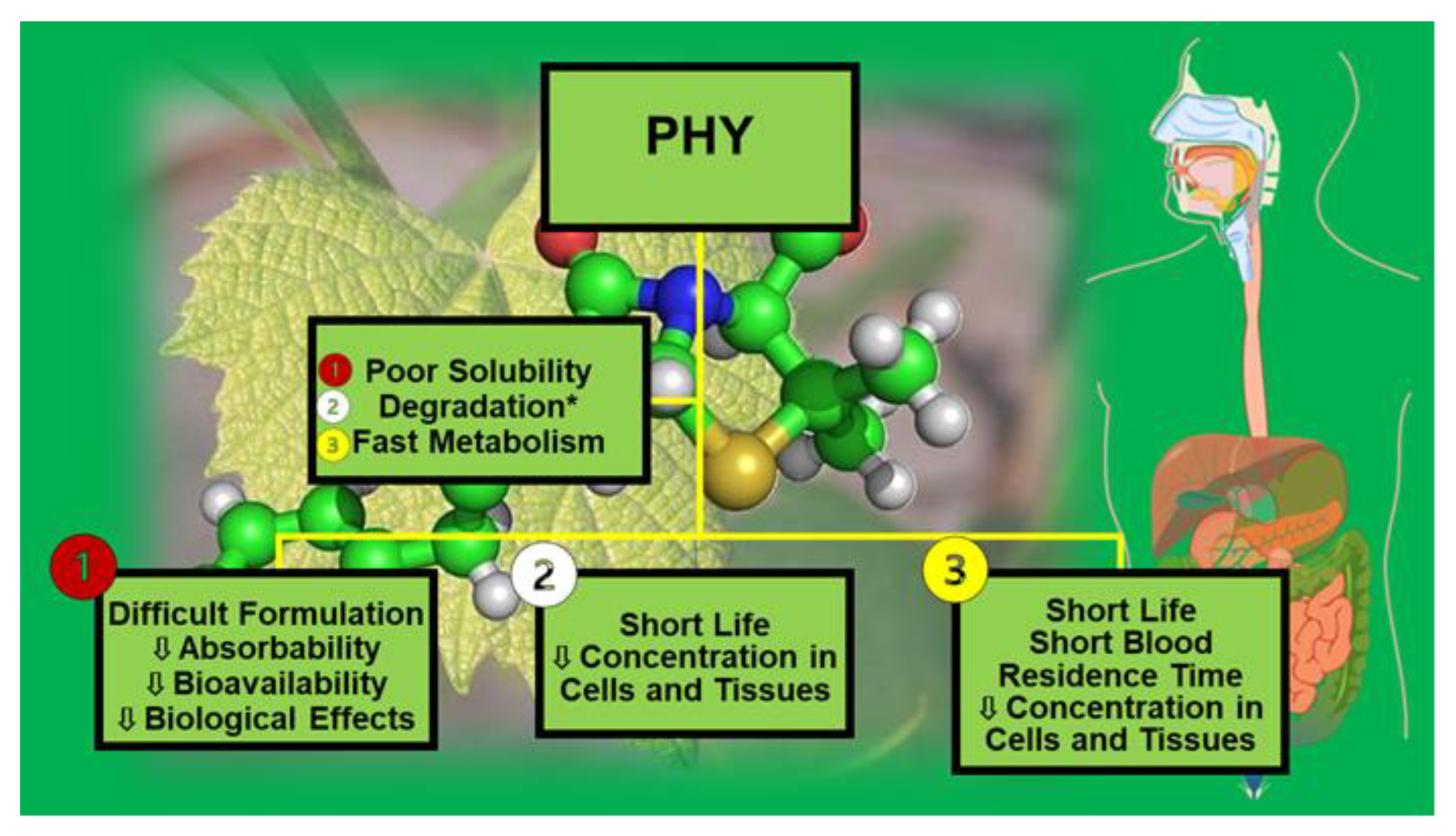
Figure 2.
Number of publications per year over the last 15 years according to Scopus. * The research was carried out on 05 April 2023 by the following keywords: nanomaterials OR nanoparticles OR nano-formulation AND phytochemicals.
Figure 2.
Number of publications per year over the last 15 years according to Scopus. * The research was carried out on 05 April 2023 by the following keywords: nanomaterials OR nanoparticles OR nano-formulation AND phytochemicals.
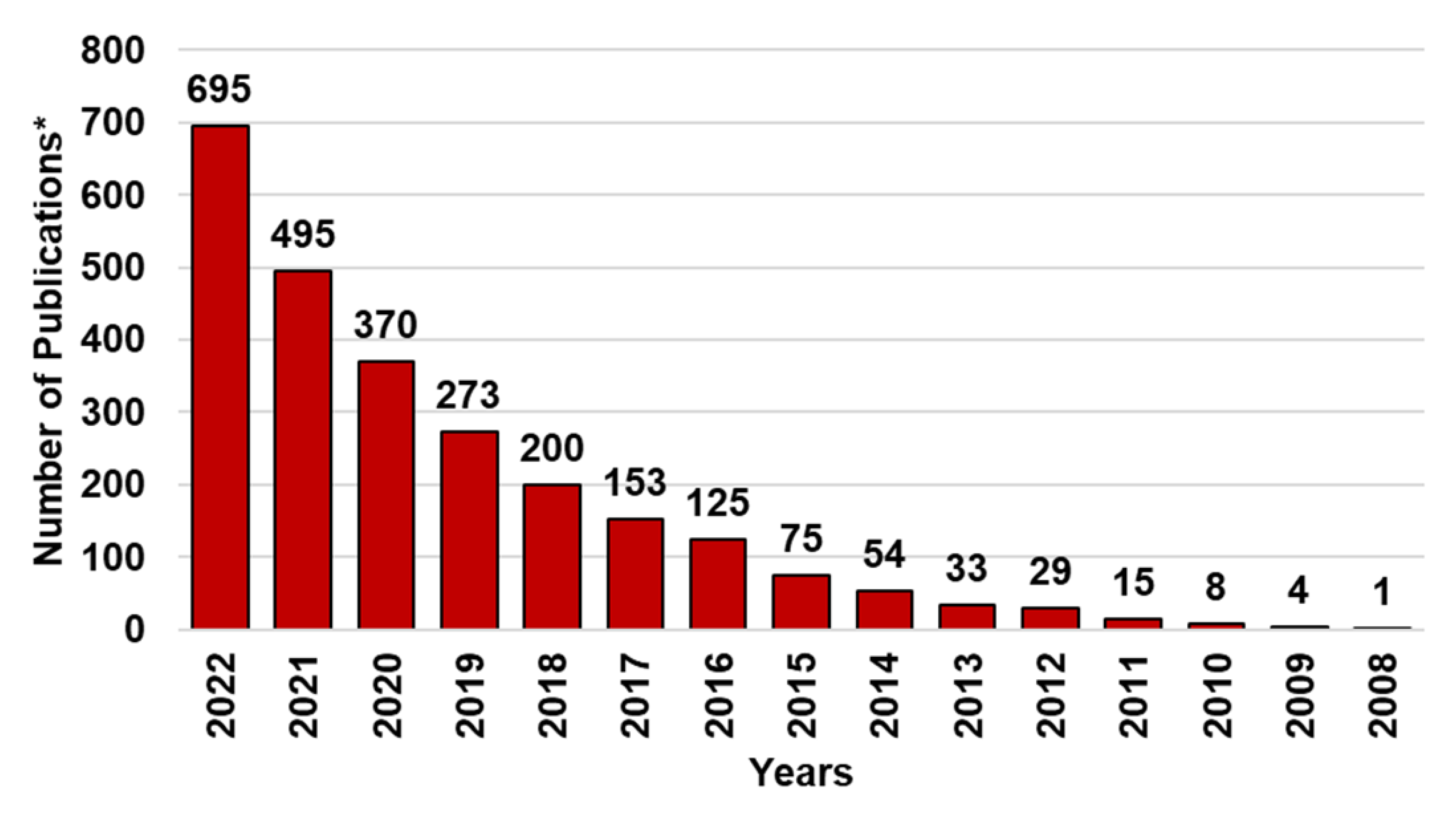
Figure 4.
Main factors that can influence the water solubility of bioactive compounds (BACs).
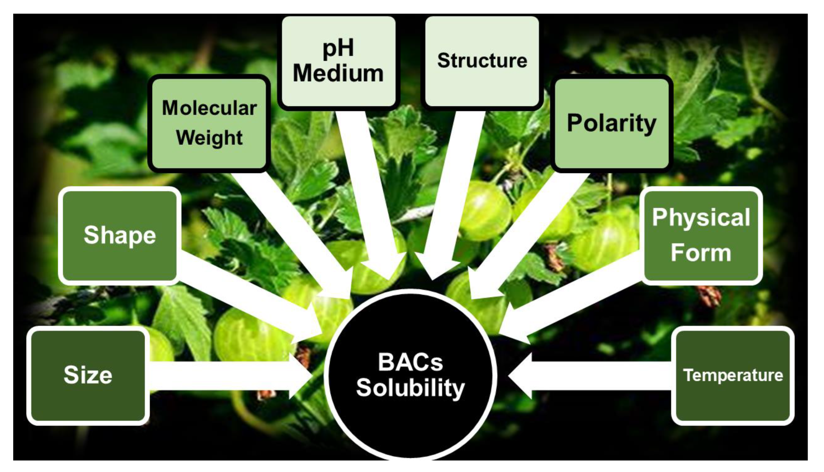
Figure 5.
Supercritical fluid technologies and cryogenic technologies.
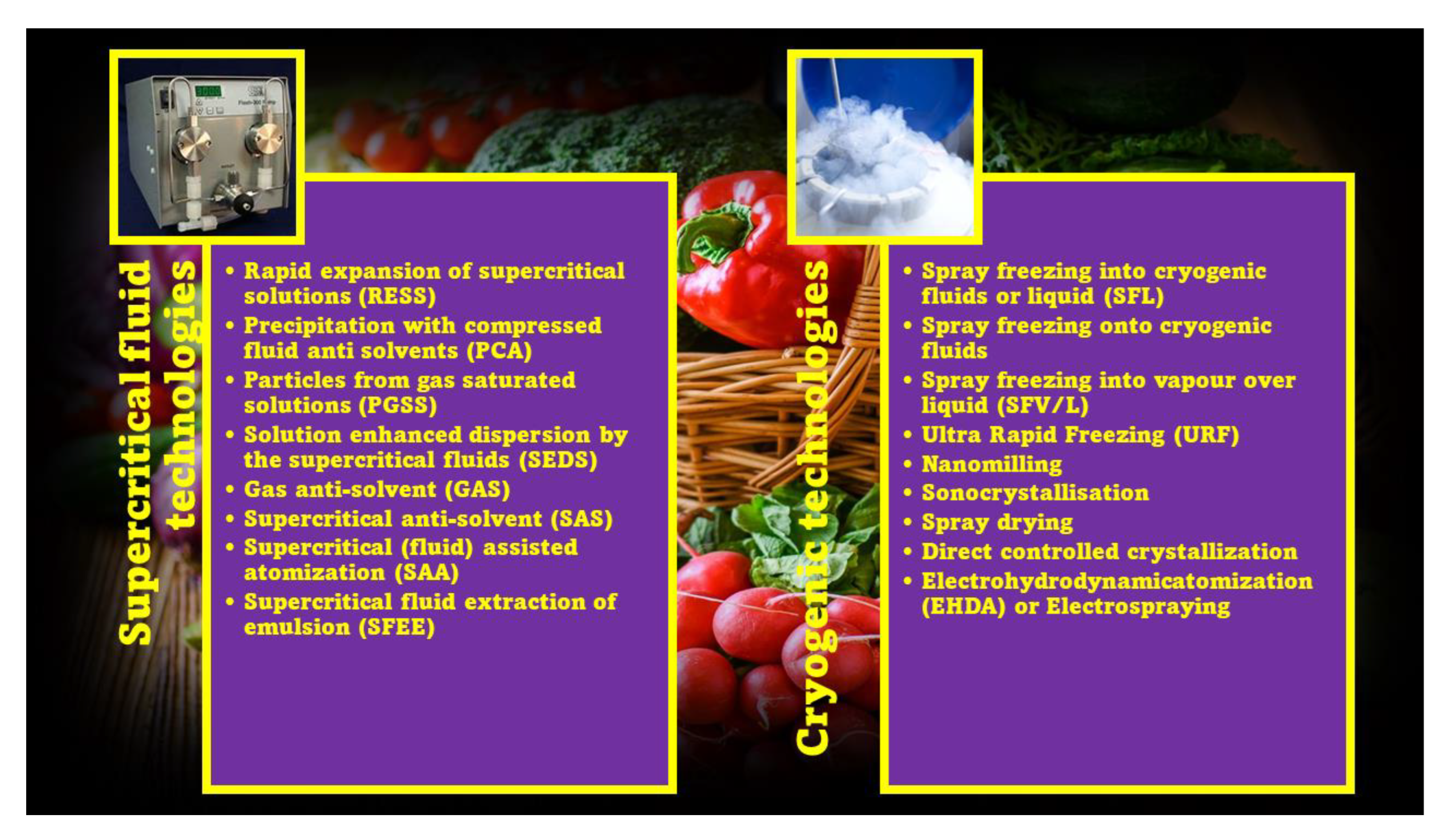
Figure 6.
Main nano-formulation techniques.
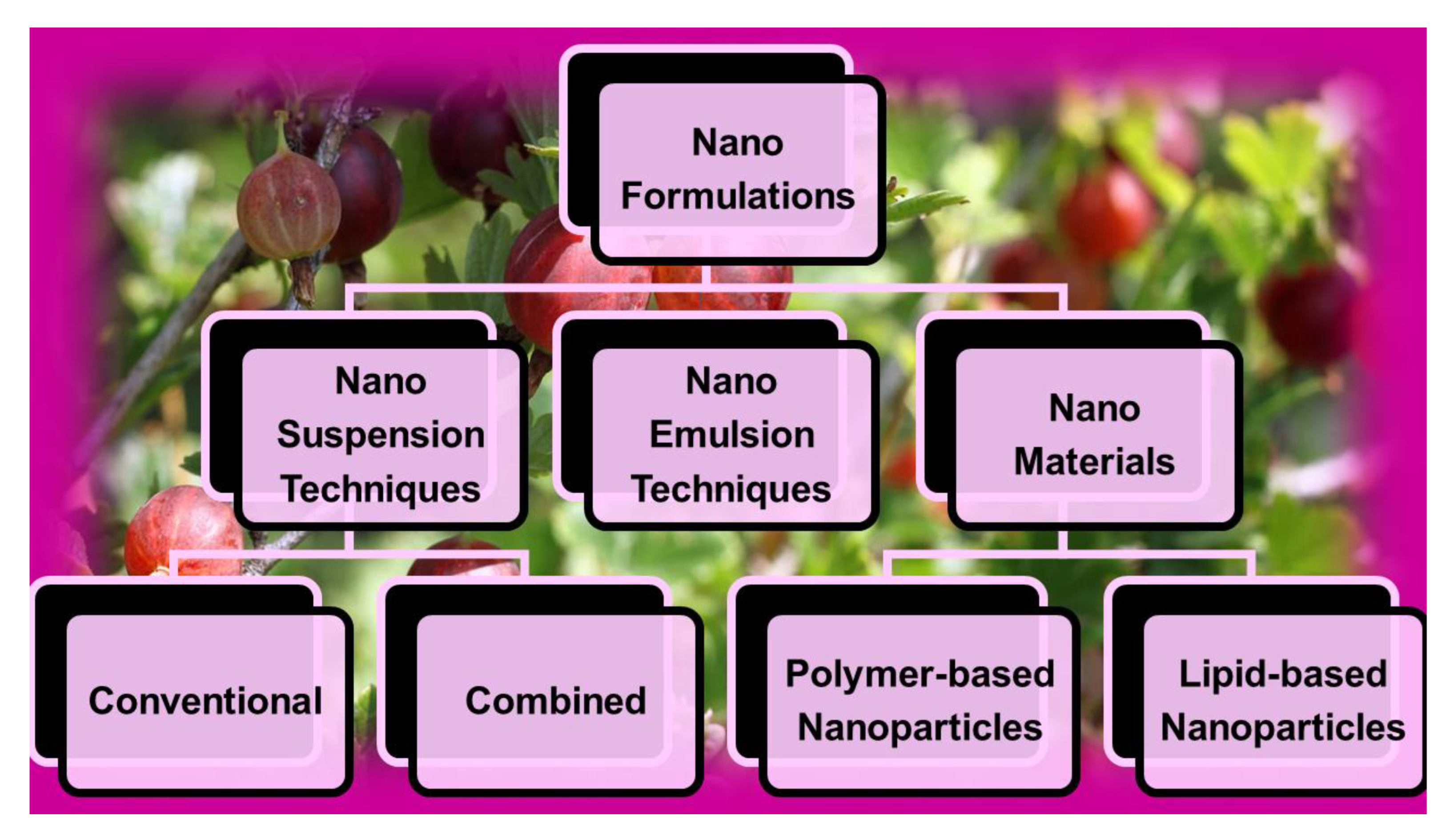
Figure 7.
Bottom-up (B-U) and top-down (T-D) methods.
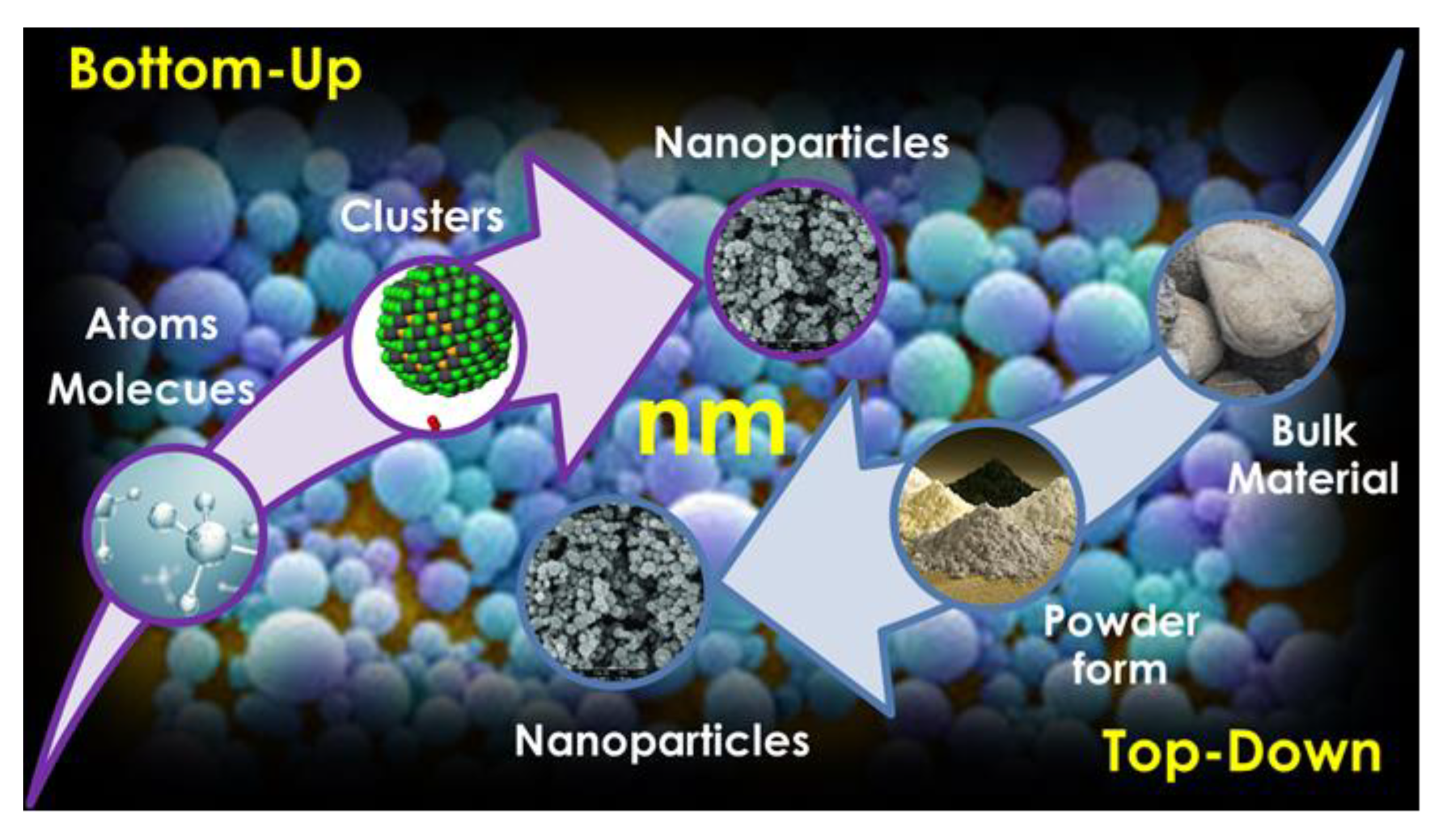
Figure 8.
Main NSs and NEs-based PHY formulations described in the last recent years. * Including also other carotenoids; ATRA = all trans retinoic acids; RES = resveratrol; ** including also Curcuma Longa extracts; OA = oleanolic acid; EGCG = epigallocatechin-3-gallate; *** red fruits; # plants extract; AITC = isothiocyanate; BITC = benzyl isothiocyanate.
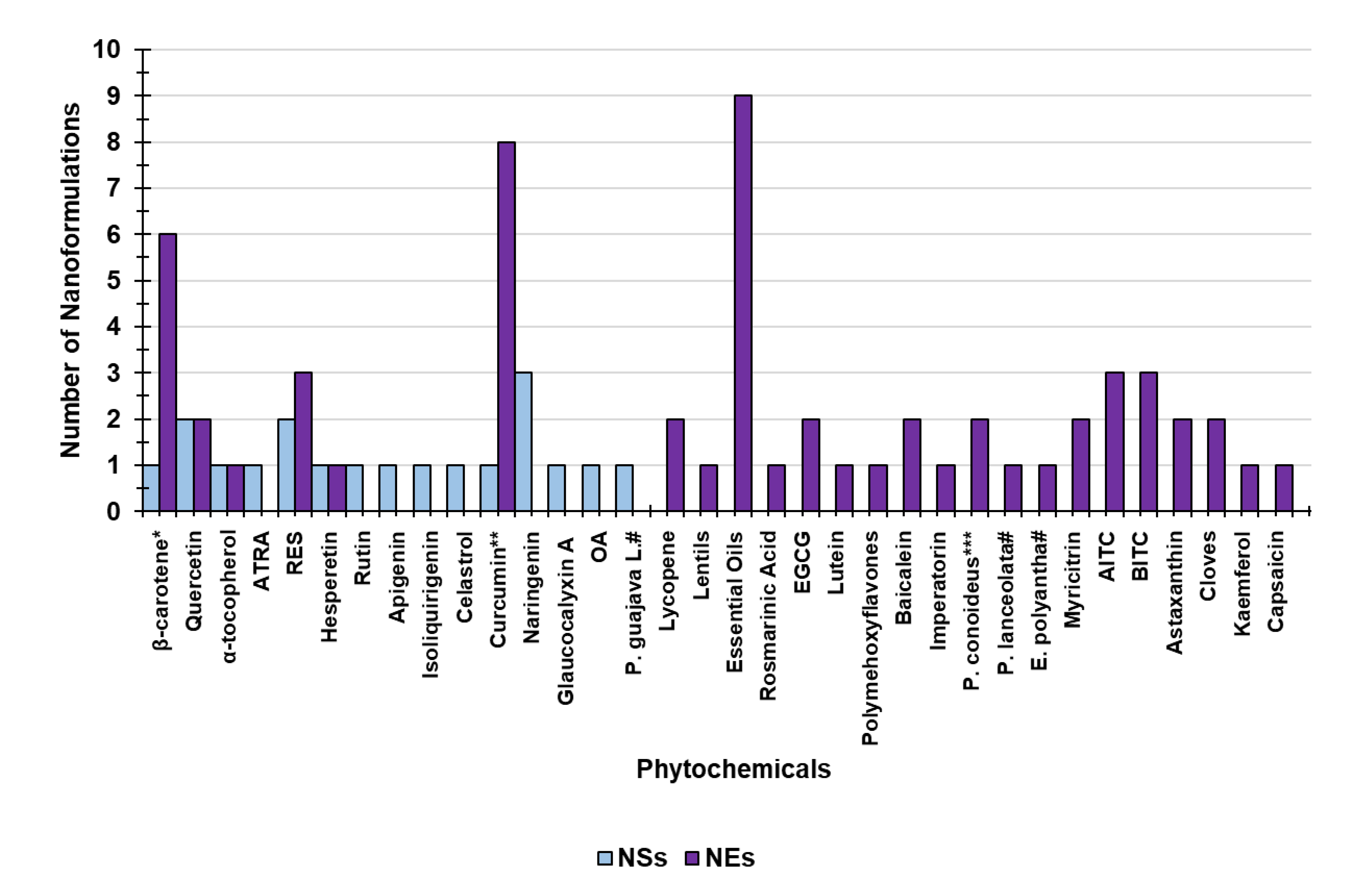
Figure 9.
Main NSs methods.
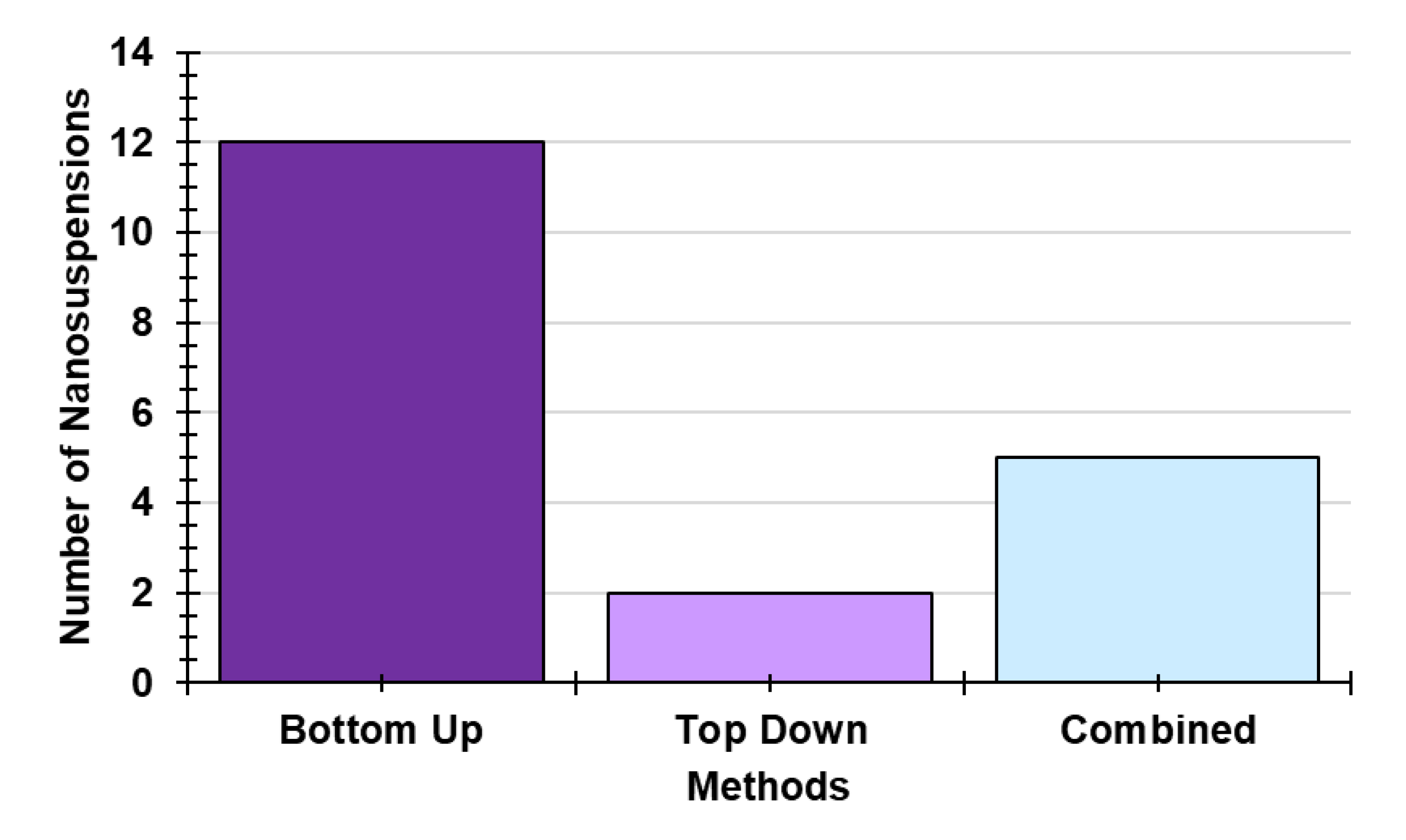
Figure 10.
Main types of NEs.
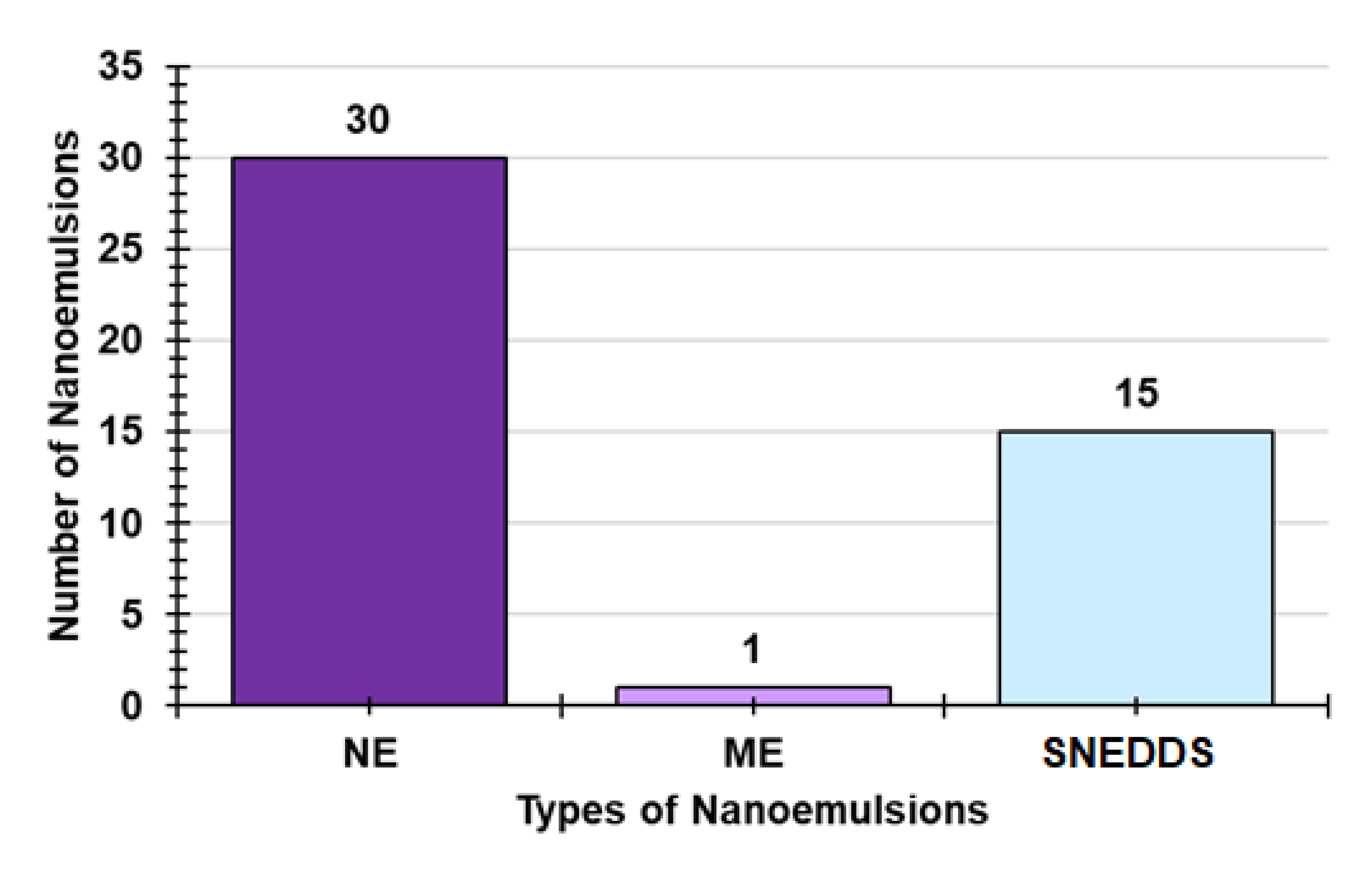
Figure 11.
Main types of methods to prepare NEs.
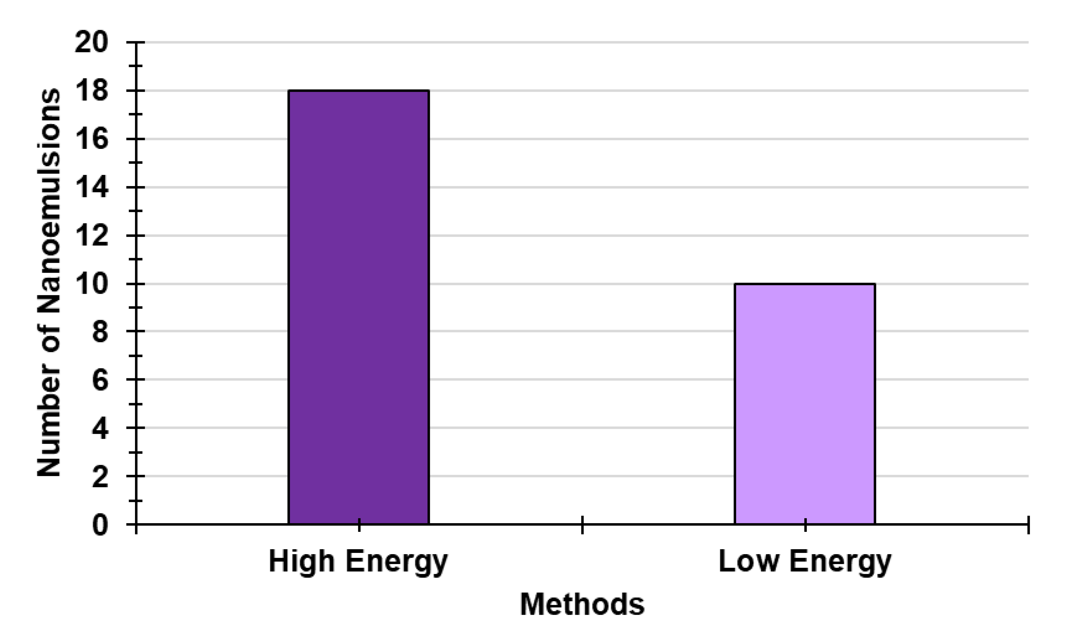
Figure 12.
Main techniques of HET (purple) and of LET (light purple).
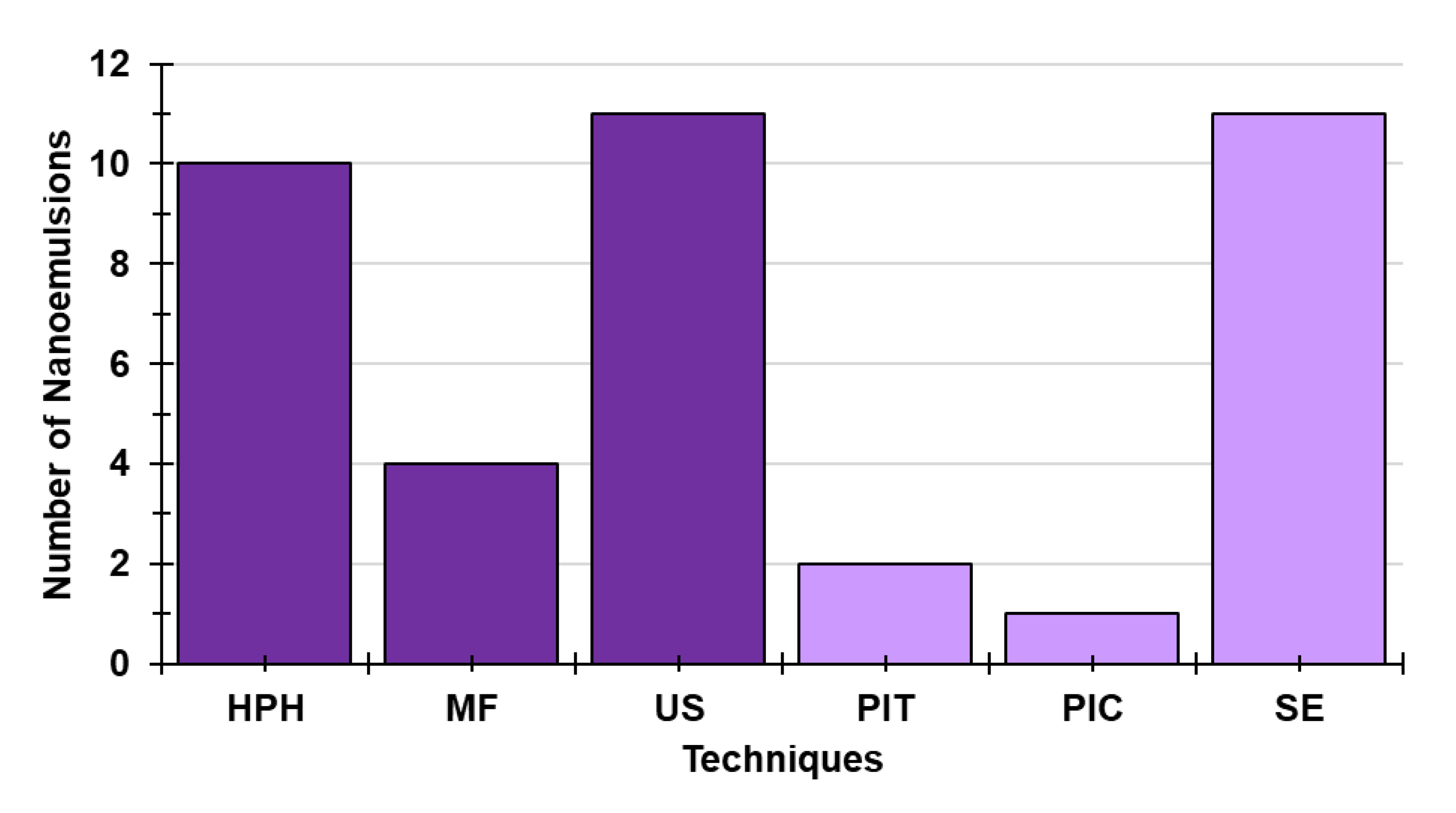
Figure 13.
Main factors that could impact the quality of nanomaterials-based drug formulation within the context of efficacy and safety. NMs = nanomaterials; MP = manufacturing process.
Figure 13.
Main factors that could impact the quality of nanomaterials-based drug formulation within the context of efficacy and safety. NMs = nanomaterials; MP = manufacturing process.
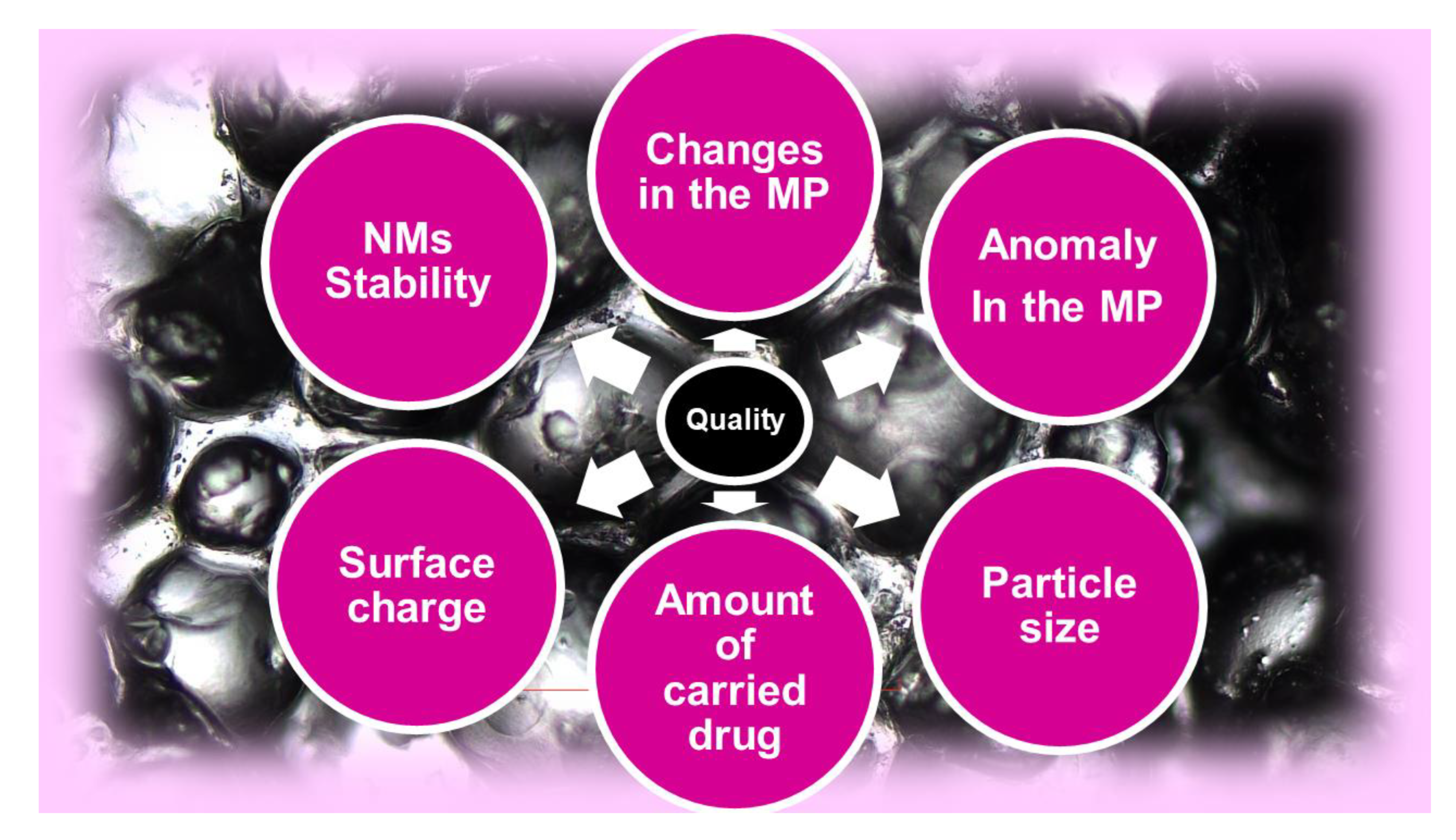
Figure 14.
Papers were identified in Scopus database from the year 2000 until 2022 using the key words nanoparticles (fuchsia) and safe nanoparticles (blue).
Figure 14.
Papers were identified in Scopus database from the year 2000 until 2022 using the key words nanoparticles (fuchsia) and safe nanoparticles (blue).
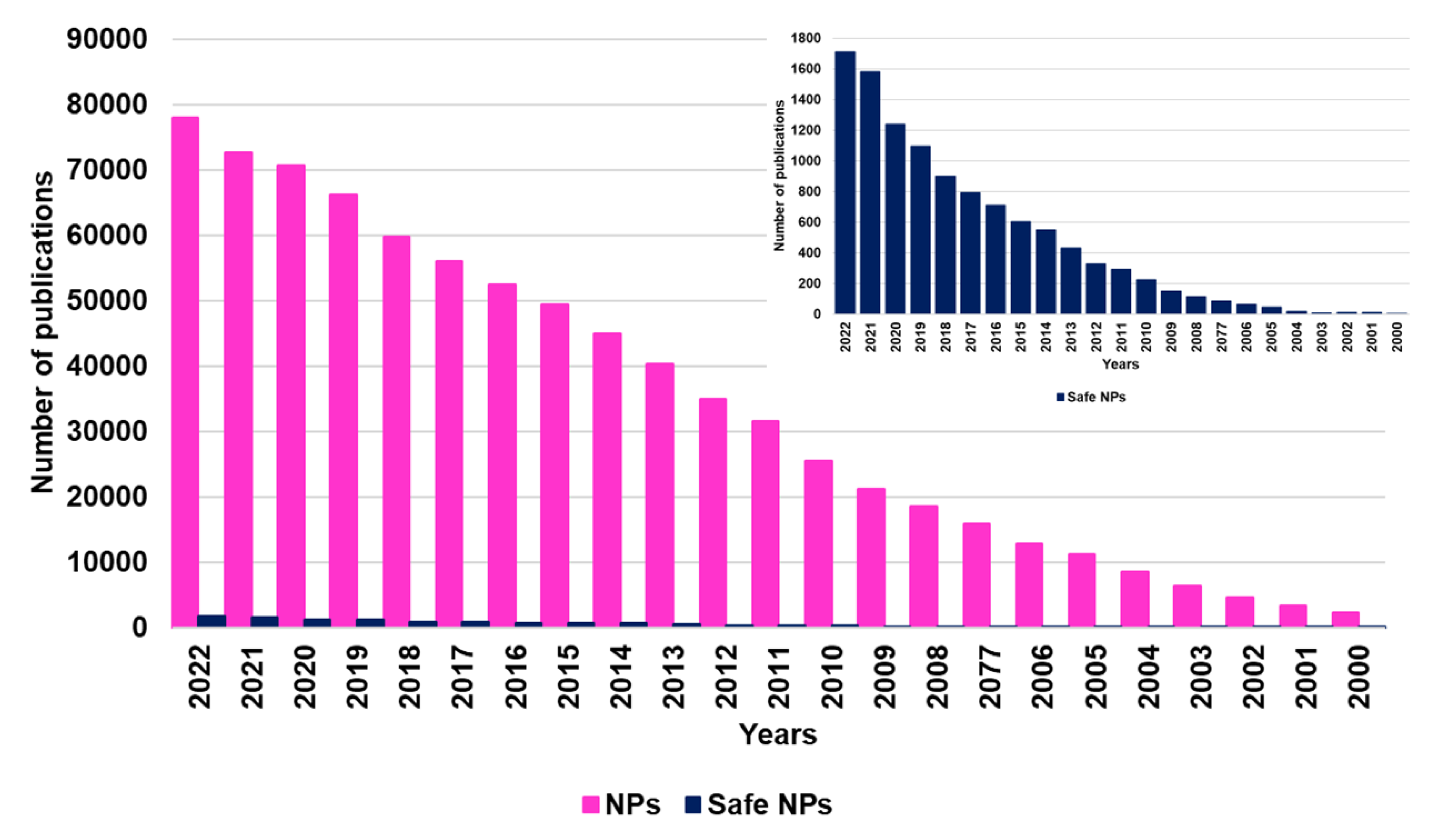
Figure 15.
PHYs clinically applied.
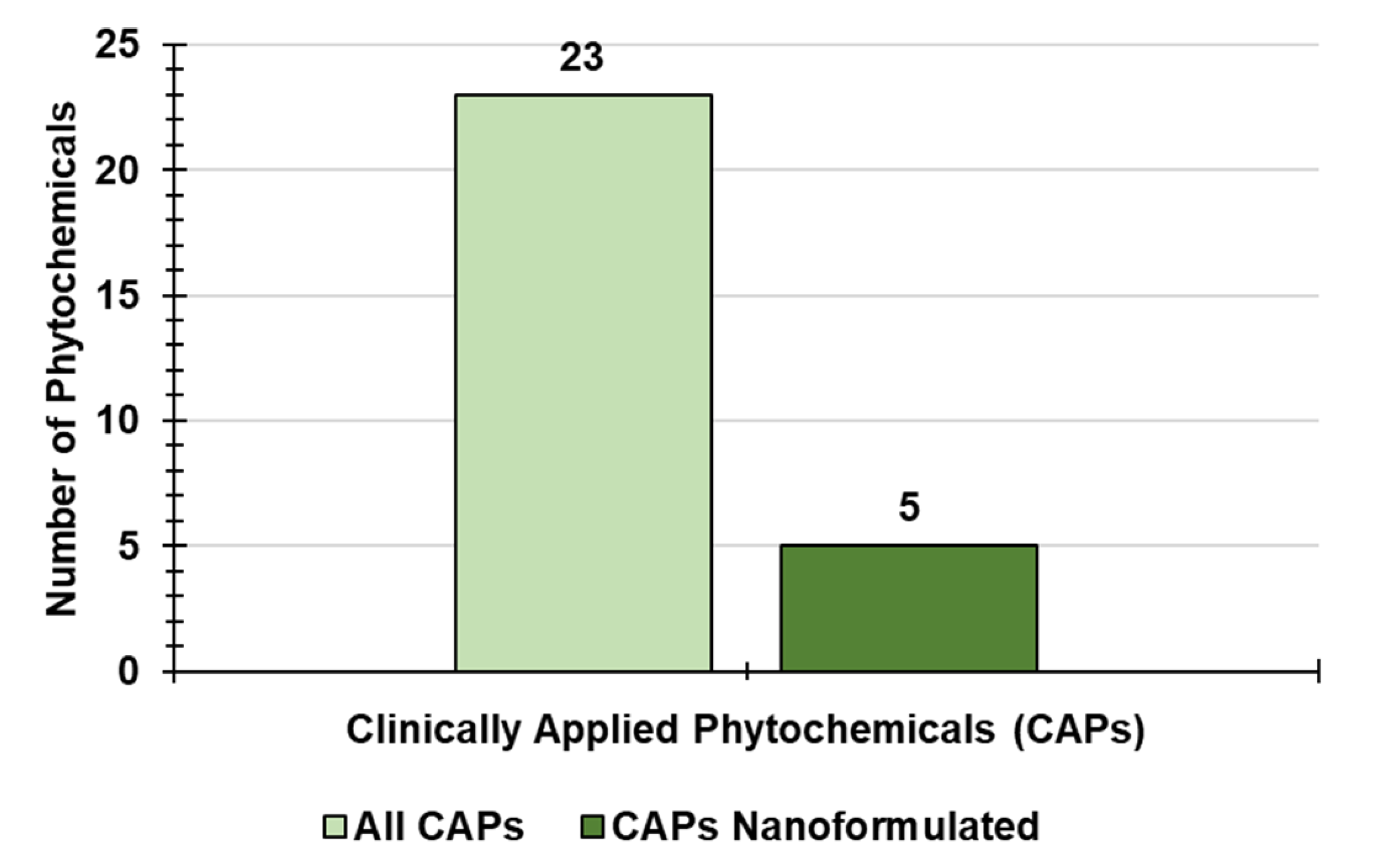
Figure 16.
Number of nano-formulations existing for type of PHY clinically applied.
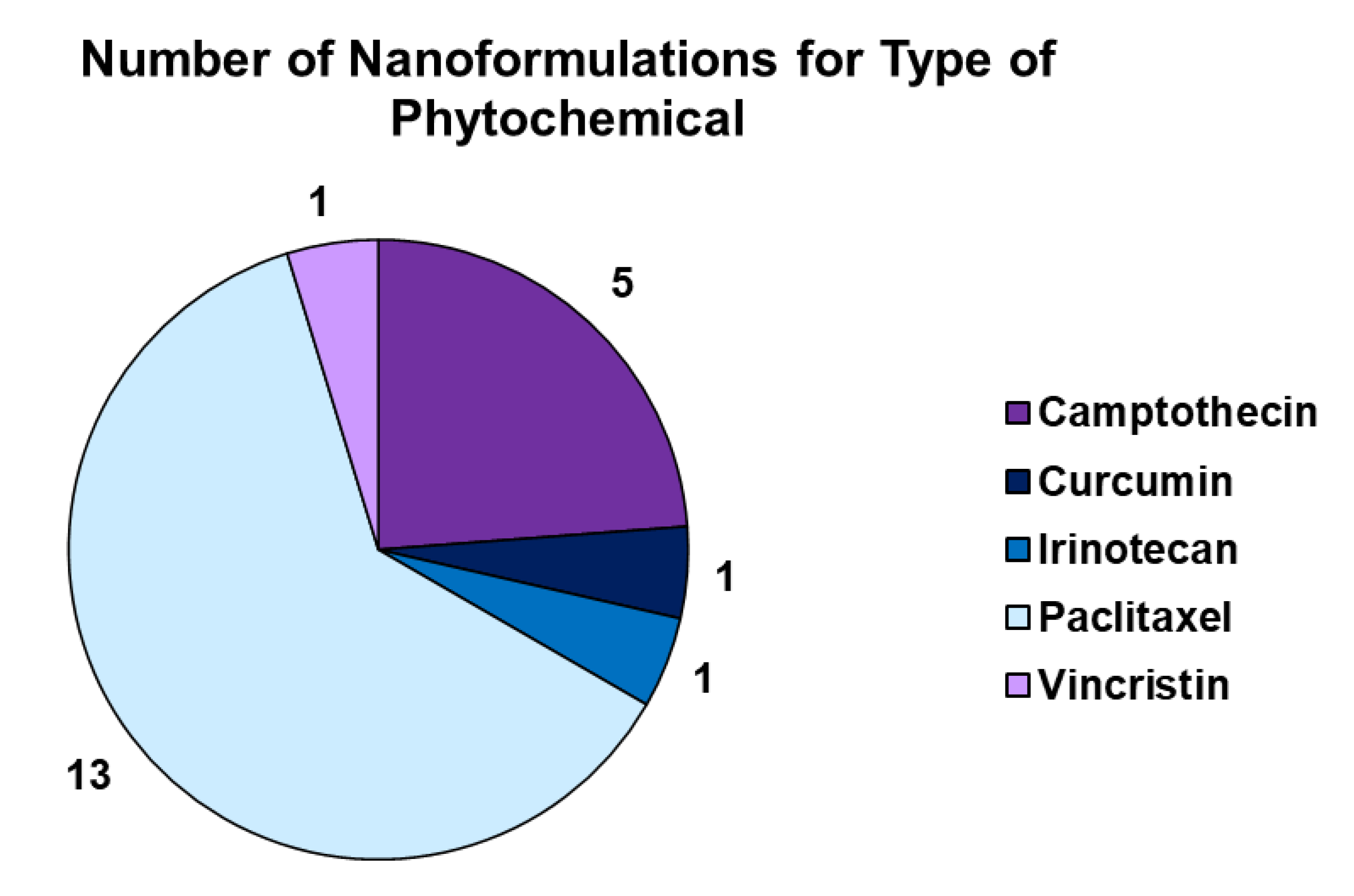
Figure 17.
Clinical status of not formulated PHYs (a) and of PHY-based nano-formulations (b).
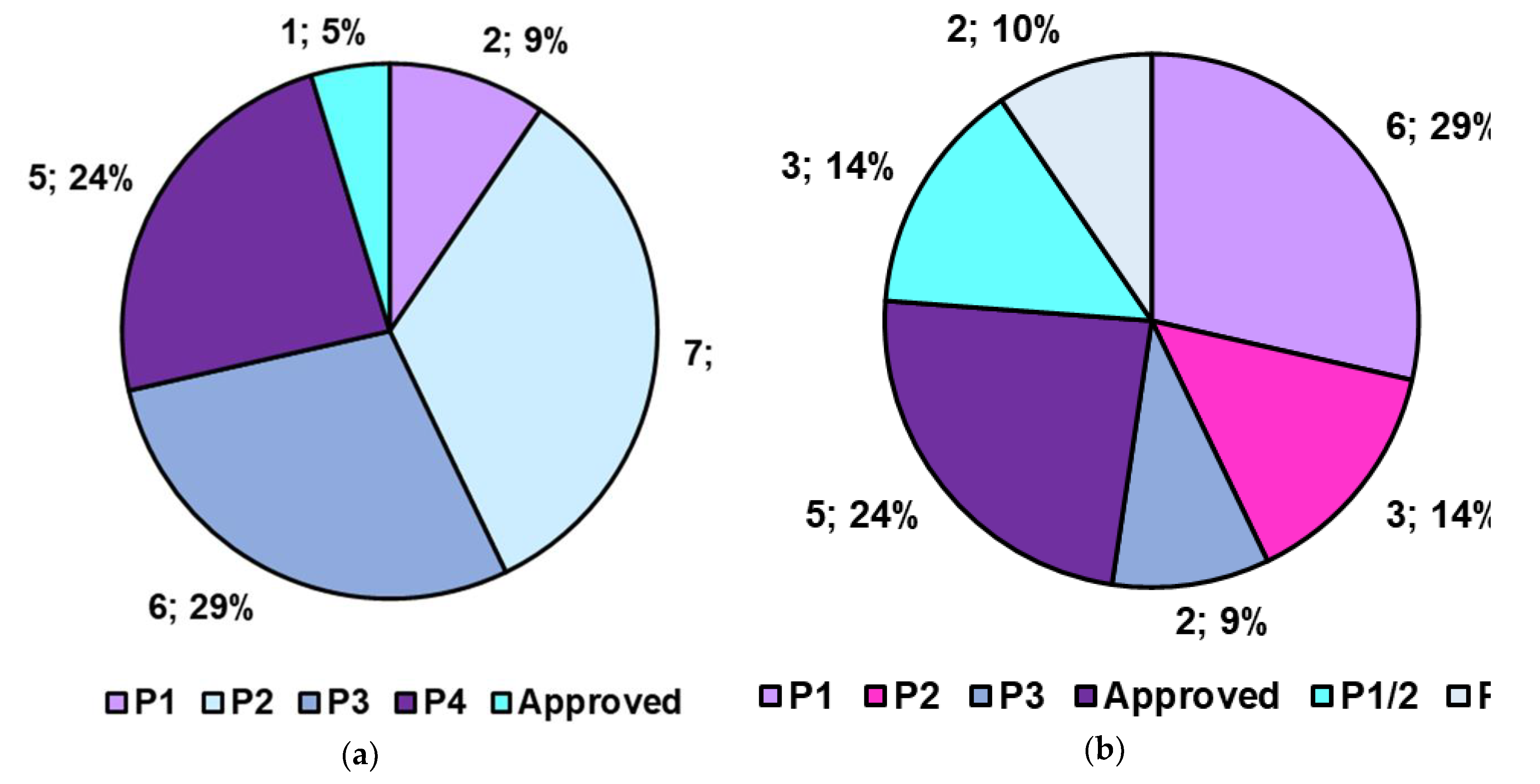

Table 1.
Sources of PHYs and their main benefits.
| Edible Plants | Essential Oils | Benefits | |
|---|---|---|---|
| Brightly Colored | Not Brightly Colored | ||
| Berries | Potatoes Almonds Pecans Pistachios Cauliflower Walnuts Cashews Hazelnuts Tea Dark chocolate Cacao beans Barley Beans Lentils Rice Coffee Mung beans Soybeans Cloves Cinnamon Cumin Nutmeg |
Pine needles Cedar Lavender |
Boost the immune system [28] Combat OS and FR [29] ⇓ Blood sugar levels [29,30] ⇓ Blood pressure [28] ⇓ Diabetes risk [29,31] ⇓ Serious health issues [29,31] Prevent chronic disease [28,29] Protect from pathogens [32,33,34] Protect brain and liver [28] ⇓ Cholesterol [28] ⇓ Inflammation [29,35,36] Support detoxification [29,37] Ward off osteoporosis [28] |
| Cranberries | |||
| Blackberries | |||
| Strawberries | |||
| Cherries | |||
| Currants | |||
| Grapes | |||
| Plums | |||
| Purple potatoes | |||
| Red Cabbage | |||
| Cabbage | |||
| Kohlrabi | |||
| Broccoli sprouts | |||
| Apples | |||
| Bananas | |||
| Peaches | |||
| Antiparasitic herbs | |||
| Egg yolks | |||
| Orange peppers | |||
| Oranges | |||
| Pumpkins | |||
| Yellow corn | |||
| Kale | |||
| Parsley | |||
| Romain lettuce | |||
| Spinach | |||
| Olive oil | |||
| Melons | |||
OS = Oxidative stress; FR = free radicals; ⇓ reduces, lowers.
Table 2.
Most relevant types of PHYs, their own source, and the associated healthy effects.
| PHYs | Sources | Health benefits |
|---|---|---|
| Carotenoids [40,41] | Carrots, tomatoes, parsley Orange and green leafy vegetables Chenopods, fenugreek Spinach, cabbage, radish, turnips |
Antioxidant Protect against uterine, prostate, colorectal, lung, and digestive tract cancers |
| Phytosterols [41,42] | Vegetables, nuts, fruits, seeds | Suppress the growth of diverse tumour cell lines |
| Limonoids [41] | Citrus fruits | Inhibit phase I enzymes and induce phase II detoxification enzymes in liver Provide protection to lung tissue, detoxify enzymes |
| Curcuminoids [41] | Turmeric, curry powder, mango, ginger | Analgesic, anti-inflammatory, anti-cancer, antioxidative, anti-depressive Against hay fever, depression, ⇓ cholesterol and itching risk |
| Indole compounds [41] (Indole-3-carbinol) |
Cabbage, cauliflower, broccoli, kale Brussels sprouts |
Strong antioxidant, DNA protector, chemo-preventive, anti-cancer, ⇑ heart health |
| Alkaloids [43] | Plants (also animals and bacteria) | Antimalarial, antiasthma, anticancer, cholinomimetics, vasodilatory, antiarrhythmic Analgesic, antibacterial, antihyperglycemic, psychotropic, stimulant |
| Phytoprostanes [44] Phytofurans [44] |
Almonds, vegetal oils, olives, algaePassion fruit, nut kernels, rice | Immunomodulators, anti-inflammatory, anti-tumours |
| Polyphenols [41] | Fruits, vegetables, cereals, beverages, legumes Chocolates, oilseeds | Action against free radicals, anti-inflammatory, anti-allergenic Inhibition of platelet aggregation, against hepatotoxins |
| Flavonoids * [41] | Fruits, vegetables, cereals, beverages, legumes Chocolates, oilseeds | Action against free radicals, anti-inflammatory, anti-allergenic Inhibition of platelet aggregation, against hepatotoxins |
| Iso-flavonoids ** [41] | Fruits, vegetables, cereals, beverages, legumes Chocolates, oil seeds | Action against free radicals, anti-inflammatory, anti-allergenic Inhibition of platelet aggregation, against hepatotoxins |
| Anthocyanidins ** [41] Anthocyanins ** [41] |
Fruits, vegetables, cereals, beverages, legumes Chocolates, oilseeds | Action against free radicals, anti-inflammatory, anti-allergenic Inhibition of platelet aggregation, against hepatotoxins |
| Glucosinolates [41] | Cruciferous vegetables | Protection against cancer of colon, rectum, stomach |
| Phytoestrogens [41] | Legumes, berries, whole grains, cereals Red wine, peanuts, red grapes |
Protection against bone loss, heart disease, cardiovascular diseases Protection against breast and uterine cancers |
| Terpenoids [41] Isoprenoids [41] |
Mosses, liverworts, algae, lichens, mushrooms | Antimicrobial, antiparasitic, antiviral, antiallergic, anti-inflammatory, chemotherapeutic Antihyperglycemic, antispasmodic |
| Fibers [45] | Fruits and vegetables (green leafy), oats | ⇓ Blood cholesterol, ⇓ cardiovascular disease |
| Polysaccharides [41] | Fruits and vegetables | Antimicrobial, antiparasitic, antiviral, antiallergic, anti-inflammatory ⇓ Serum, ⇑ defence mechanisms |
| Saponins [41] | Oats, leaves, flowers, green fruits of tomato | Protection against pathogens, antimicrobial, anti-inflammatory, antiulcer agent |
| Tannins [41] | Cranberries, currants, blackberries, apples Grapes, peaches, strawberries, almonds Hazelnuts, pecans, pistachios, walnuts, barley Beans, lentils, rice, tea, cacao beans Dark chocolate, antiparasitic herbs |
Antioxidant, fight pathogens, ⇓ blood pressure, ⇓ inflammation, ⇓ serious health risks Regulate the immune system |
| Lutein [41] Zeaxanthin [41] |
Egg yolks, orange peppers, oranges, pumpkins Yellow corn, kale, parsley, romaine lettuce Spinach, pistachios, olive oil | Protect retina from damage, ⇑ eye function, ⇑ memory and brain function Promote the body’s use of insulin, ⇑ skin health, ⇓ blood pressure, ⇓ inflammation Support heart health |
| Eugenol [46] | Cloves, cinnamon, cumin, nutmeg, coffee Mung beans, soybeans, bananas, melons Strawberries, tomatoes |
Anti-inflammatory, antioxidant, eliminate parasites, fights fungi Inhibits serious health concerns, protects the brain and the liver, ⇓ bacterial biofilm Supports heart and stomach health |
⇑ Improved, higher; ⇓ reduced, lower; * are a subgroup of polyphenols; ** are a subgroup of flavonoids.
Table 3.
General classification of the different color groups *.
| Color Group | Foods | PHYs | Properties | Ref |
|---|---|---|---|---|
| Green | Asparagus, avocados, Celery Cucumbers, Green beans, green peppers, kale, kiwi Spinach, zucchini |
EGCG, glucosinolates, indolesisoflavones Isothiocyanates, lutein and zeaxanthin Sulforaphane |
Promote wound healing and healthy gums Support arteries, blood cells, eyes, liver, and lungs |
[16,65] |
| Purple | Black beans, blackberries Eggplants, elderberries, plums Purple cabbage, purple grapes, raisins |
Anthocyanins, flavonoids, phenols, tannins, RES | Protect against serious health issues Support arteries, bones, brain, cognition, healthy aging, and heart |
[16,65] |
| Red | Cherries, cranberries, kidney beans Red beans. strawberries, tomatoes watermelon |
Anthocyanins, ellagic acid, eugenol, hesperidin Lycopene, tannins, quercetin |
Protect against heart disease and other serious health issues Support prostate, urinary tract, and DNA health |
[16,65] |
| Yellow | Apricots, cantaloupe, carrots, grapefruit, Yellow pears, yellow peppers Yellow winter squash |
α-Carotene, β-carotene, β-cryptoxanthin, lutein Zeaxanthin, hesperidin |
Boost the immune system, support heart and vision health | [16,65] |
| White | Apples, cauliflower Great northern beans Mushrooms, onions |
Allicin, ECGC, glucosinolates, indoles, tannins Quercetin |
Protect against heart disease and other serious health issues Support arteries, bones, and circulation |
[16] |
* The present Table has been constructed by authors exploiting information found in literature [16].
Table 4.
Advantages and disadvantages of the most used techniques and approaches for enhancing the solubility of bioactive compounds. *.
Table 4.
Advantages and disadvantages of the most used techniques and approaches for enhancing the solubility of bioactive compounds. *.
| Methods | Advantages | Disadvantages | Ref. |
|---|---|---|---|
| Emulsion solvent extraction Double-emulsion solvent extraction |
Low-cost | ⇑Residual solvent ⇓Encapsulation efficiency (EE) Thermal degradation Multi-steps processes Micronization step needed |
[15] |
| SD | Improved bioavailability Taste masking Modified release achievable Aseptic manufacturing Fine powders Improved stability |
High overhead Possible thermal degradation Not for every compound Not widely available Multi-steps processes Micronization step needed |
[15] |
| Liquid anti-solvent technique | Low-cost | ⇑Residual solvent ⇓ EE Thermal degradation Multi-steps processes Micronization step needed |
[15] |
| Spray freeze drying (SFD)* | Higher rate of freezing Independent control over particle size |
⇓ Biological activity Possible protein denaturation Excipients required |
[15] |
| Spray freezing into liquid (SFL)* | Higher level of biological activity High degree of atomization Ultra-rapid freezing (URF) Formation of amorphous highly porous NPs |
Possible ⇑ viscosity of the feed liquid Limited applications ⇑ Cost equipment Time and energy-intensive |
[15] |
| Thin film freezing (TFF) ** | High-yield products Flexibility on processable drugs Large-scale production Simple, efficient, robust process ⇑ stability of the protein product |
⇑-Cost equipment | [15] |
| Supercritical fluid extraction (SFE) method | Single-step process Controllable particle size Controllable morphology Controllable crystallinity Monitorable residual solvent |
⇑-Cost equipment | [15] |
| Solvent evaporation technique | Low-cost | ⇑Residual solvent ⇓EE Thermal degradation Multi-steps processes Micronization step needed |
[15] |
Table 5.
Suspension-based nano-formulation techniques.
| Nano-suspension techniques | ||
|---|---|---|
| Conventional techniques | Combined techniques (CTNI) | |
| Bottom-up (B-U) | Top-down (T-D) | Nanoedge™ Technique (Baxter Healthcare) H 69 Technology (SmartCrystal® technology group) H 42 Technology (SmartCrystal® technology) H 96 Technology (SmartCrystal®, Abbott/Soliqs, Ludwigshafen, Germany) Combination Technology (CTNO) |
Table 6.
Examples of plant derived bioactive compounds nano-formulated by nanosuspension conventional and combined techniques.
Table 6.
Examples of plant derived bioactive compounds nano-formulated by nanosuspension conventional and combined techniques.
| PHY | NPs/Mechanical Method (MM)/ Combined techniques (CTNI) |
Particle size | Characteristics |
|---|---|---|---|
| β-carotene [90] (B-U) |
n-Octenyl succinate-modified starch (NPs) | 300-600-nm-sized particles | ⇑ Dispersibility ⇑Coloring strength, ⇑ Bioavailability |
| Quercetin [91] (B-U) |
Maltodextrin (NPs) | 753-nm-sized particles | Water-re-dispersible ⇑ RSA ⇑ ORAC |
| Quercetin [92] (T-D) |
HPH (MM) + spray-drying (MM) | 400-nm-sized particles | ⇑ Antioxidant ⇑ Anti-inflammatory ⇑ Anticancer properties ⇑ Water solubility ⇑ Oral bioavailability |
| α-Tocopherol [93] (B-U) |
Supercritical assisted process | 150-nm-sized particles | ⇑ Solubility ⇑ Bioavailability |
| All-trans retinoic acid (ATRA) [94] | Nanoedge™ Technique | 155-nm-sized particles | Orally administrable 30′ Operation time |
| Resveratrol (RES) [95] | H 69 Technology | 150 nm-sized NPs | Orally administrable 10 cycles HPH/1200 bar |
| RES [15] | H 42 Technology | 200 nm-sized NPs | Orally administrable 1 HPH cycle at 1500 bar |
| Hesperidin [15] * | CTNO | 599 nm-sized NPs | ⇑ Solubility Long-term stability Orally administrable Topically applicable Five homogenization cycles (1000 bar) |
| Rutin [15] ** | CTNO | 600 nm-sized NPs | ⇑ Solubility Orally administrable Topically applicable 1 Cycle of HPH at 100 bar |
| Apigenin [15] | CTNO | 275 nm-sized NPs | 1 Cycle HPH at 300 bar |
| Isoliquiritigenin [96] T-D |
Wet media milling (MM) | 238 nm-sized NPs (HPC-SSL) 354 nm-sized NPs (PVP-K30) |
⇑ Solubility ⇑ Cytotoxicity ⇑ Cellular up-take ⇑ Apoptosis induction ⇓ Toxicity |
| Celastrol [97] B-U |
Antisolvent precipitation method | 148 nm-sized NPs | Stable in plasma Orally administrable ⇑ EE% ⇑ DL% ⇑ Solubility (vitro) ⇑ Cytotoxicity (vivo) ⇑ Cumulative release (48h) |
|
Curcuma Longa L. Extracts [98] B-U |
Supercritical fluid expansion | 47 nm-sized NPs | ⇑ Solubility |
| Naringenin (NRG) [99] B-U |
Antisolvent sonoprecipitation | 117 nm-sized NPs | ⇑ Absorption in GIT ⇑ Dissolution ⇑ Oral bioavailability |
| NRG [100] B-U |
Precipitation-ultrasonication | 118 nm-sized NPs | ⇑ Drug dissolution ⇑ Pharmacokinetic profile ⇑ Stability |
| NRG [101] B-U |
HPH | 81 nm-sized NPs | ⇑ Intracellular ROS level ⇑ Mitochondrial membrane potential ⇑ Caspase-3 activity ⇑ Lipid peroxidation status ⇓ GSH levels ⇑ Antitumor activity on DLA cells ⇑ Life span ⇓ Cancer cell number ⇓ Tumor weight |
| Glaucocalyxin A [102] B-U |
Precipitation-ultrasonication | 143 nm-sized NPs | ⇑In vitro antitumor activity ⇑ In vivo anticancer efficacy |
| Oleanolic acid (OA) [103] B-U |
Organic solvent evaporation | 100 nm-sized NPs | ⇑ Stability ⇑ Saturation solubility ⇑ Dissolution rate ⇑Cytotoxicity ⇑ Bio-efficacy ⇑ Bioavailability |
|
P. guajava L. extracts [104] B-U |
Nanoprecipitation | 241-327 nm-sized NPs | ⇑ Antihyperglycemic activity ⇑ Physical parameters ⇑ Hepatic parameters ⇑ Renal parameters ⇑ Absorption ⇓ Metabolism ⇑ Stability |
|
Nigella sativa L. [105]B-U |
Nanoprecipitation | N.R. | ⇑ Total phenolic content ⇑ Total flavonoid contents ⇑ Antioxidant activity ⇑ Antidiabetic activity ⇑ Antibiofilm activity ⇑ Bioavailability |
RSA = radical scavenging activity; ORAC = oxygen radical absorbance capacity; * Hesperidin nanocrystals can be found in the Platinum Rare cosmetic product (La Prairie, Volketswil, Switzerland); ** Rutin nanocrystals are in a cosmetic product launched by Juvena, St. Margrethen, Switzerland [15]; DL = drug loading content; GSH = glutathione reductase; DLA = Dalton lymphoma ascites; ⇑ improved, higher, high; ⇓ decreased, lower.
Table 7.
Main types of NEs and common oily phases.
| Type of NEs | Oily Phase | Advantages | Drawbacks | Ref. | |
|---|---|---|---|---|---|
| NEs o/w or w/o (100-500 nm) |
Captex 355 Captex 8000 Witepsol Myritol 318 Isopropyl myristate Capryol 90Sefsol-218 Triacetin Isopropyl myristate Castor oil Olive oil |
PHYs protection Controlled release Sustained release ⇑ DL% |
No high-melting PHYs 5-10% additives* required |
[1] | |
| SEDDSs o/w ** | ⇑ Oral bioavailability Possibility of easy scale-up ⇑ DL% Allow delivering peptides/lipids without the risk of digestion |
5-10% additives* required For low therapeutic dose PHYs Many parameters The physicochemical properties of PHYs can influence the efficiency of oral absorption and the performances of SEDDSs. |
[1] | ||
| SNEDDSs ** (<50 nm) |
SMEDDSs ** (100–200 nm) |
||||
| SDEDDSs ** (w/o/w) or (o/w/oil) | |||||
SNEDDSs = self-nanoemulsifying drug delivery systems; SMEDDSs = self-micro-emulsifying drug delivery systems; SDEDDSs = self-double-emulsifying drug delivery systems; * surfactants, co-surfactants, and stabilizers; ⇑ high, higher; ** anhydrous systems.
Table 8.
Main adopted high and low energy methods and their recent applications in nano-formulating PHYs.
Table 8.
Main adopted high and low energy methods and their recent applications in nano-formulating PHYs.
| Technique | Method | PHYs | Activity | Particle size (nm) | Ref. |
|---|---|---|---|---|---|
| HET | High pressure homogenization (HPH) | Jackfruit pulp extract* rich in carotenoids | Antioxidant | 166 | [112] |
| Lycopene | Radicals scavenging activity | 92.6 | [113] | ||
| Lentil | ⇓ Blood pressure ⇓ Bad cholesterol (LDL) ⇑ Good cholesterol (HDL) ⇓ Heart disease risk |
149 | [114] | ||
| Microfluidization (MF) | β-carotene | Antioxidant | 140-160 | [115,116] | |
| Ultrasonication (US) | Essential oils (EOs)(Zataria multiflora) | Antibacterial | 91-211 | [117] | |
| RES | ⇓ Diseases by oxidative stress | 20.4 **; 24.5 *** | [118] | ||
| LET | Phase Inversion Temperature (PIT) | Cinnamon EO | Antioxidant Antimicrobial |
101 | [119] |
| Phase Inversion Composition (PIC) | Rosmarinic acid (RA) | Antiviral Anti-inflammatory Antioxidant |
70-100 | [120] | |
| Spontaneous Emulsification (SE) | Key lime (Citrus aurantifolia) EO | Antibacterial | 21 | [121] | |
| Kaffir lime (Citrus hystrix) EO | 28 | ||||
| Calamansi lime (Citrofortunella microcarpa) EO | 60 |
* Rich in carotenoids; ** NE; *** NE-cyclodextrin inclusion complex.
Table 9.
Emulsion-based formulation for delivering PHYs.
| PHY | Emulsion type/Method | Additives | Results | Ref. |
|---|---|---|---|---|
| Curcumin | NE | Tween-20 | ⇓ Toxicity ⇑ Bioavailability ⇑ Bioactivity ⇑ Anti-inflammatory |
[125] |
| o/w SNEDDS Mild agitation |
Tween 80 PEG 600 |
⇑ Oral bioavailability ⇑ C max |
[126] | |
| NE/HPH | PEG (3%) | ⇑ Oral bioavailability ⇓ DHA levels |
[127] | |
| Emulsion–diffusion evaporation |
N.R. | ⇓ Blood glucose levels, ⇑ Insulin | [128] | |
| Interfacial prepolymer deposition and SE | Lipoid 100 | Inhibition of OSCC cell ⇓ PI3K/Akt/mTOR ⇑ miR-199a |
[129] | |
| EGCG (E) + ALA (A) | SDEDDS | PGPR S721, P10, L23. and S40 |
⇑Photo-stability Antioxidant ⇑EE |
[130] |
| EGCG | NE (o/w) | BC WPI |
No toxicity ⇑ Antioxidant |
[131] |
| Carotenoids (Paprika Oleoresin) |
SMEDDS | Tween 80 | ⇑ Solubility | [132] |
| Lutein | SMEDDS | Tween 80 Labrasol TranscutolHP/Lutro-E400 1 |
⇑ Solubility ⇑ Bioavailability |
[133,134] |
| Polymethoxyflavones (PMFs) |
NE | Tween 20 Tween 85 |
⇑ Dissolution rate | [135] |
| β-Carotene | o/w NE | Tween 20 | ⇑ Emulsion stability ⇑ Solubility ⇑ Bioaccessibility |
[136] |
| Lycopene | Microemulsion (ME) | ESE 3GIO SML |
⇑ Solubility | [137] |
| Quercetin | SNEDDS | Tween 80PEG 400 | ⇑ Solubility | [138] |
| Naringenin Hesperetin |
NE | Glycerin | ⇑ Solubility ⇑ In vitro stability No cytotoxic (vitro) No hemolytic (vivo) Anti-inflammatory |
[139] |
| Baicalein | NE/HPH | PEGM Sodium oleate Hoechst 33258 3,3-DODOXAP |
⇑ Oral bioavailability ⇑GIT permeability ⇑Transcellular transport ability ⇓ Cytotoxicity |
[140] |
| Imperatorin | NE/HSS NE/HPH |
Polaxamer 188 | ⇑ Bioavailability Antiploriferative (MDA-MB-231) |
[141] |
| Pandanus conoideus Lamk (red fruit) | SNEDDS | PPG Tween 20 |
⇑ Cytotoxic activity | [142] |
| Pandanus conoideus Lamk (red fruit) | NE/high-speed mixer | PPG SEPIGEL 305™ |
Antioxidant | [143] |
| Plantago lanceolata L. | SNEDDS | Labrasol or Kolliphor RH 40 Transcutol HP | ⇑ Solubility ⇑ Permeability ⇑ Bioavailability ⇑ Pharmacological effects |
[144] |
| Bay Leaves extracts (Eugenia polyantha Wight) |
Tween 80 PEG 400 |
[145] | ||
| Myricitrin | Capryol 90 Cremophor RH 40 PEG 400 Cremophor EL Transcutol HP |
[146] | ||
| Myricitrin | Cremophor EL35 Dimethyl carbinol |
[147] | ||
| Quercetin | PEG 200 Tween 40 Tween 60 Tween 80 PEG 400 Transcutol HP |
[148] | ||
| Baicalin | Peceol® (14.3%, w/w)Kolliphor® EL (57.1%, w/w) Transcutol® P (28.6%, w/w) |
[149] | ||
| AITC | Emultion solvent evaporation | Polyvinyl alcohol (PVA) (3%) | ⇓ Degradation ⇓ Volatility ⇑ Shelf life Sustained release ⇑ Toxicity on tumor |
[150,151] |
| ⇑⇑ Anti-cancer activity ⇓ toxicity |
[152] | |||
| BITC | US | Tween 80 Decyl-β-d-glucopyranoside |
⇑ EE% | [153] |
| BITC | US | Tween 80 | ⇑ EE% Good DL% MDA-MB-231 cells inhibition |
[154] |
| Heating stirring-sonication | Good long-term stability ⇑⇑⇑ EE Sustained release ⇑ Cytotoxicity (MDA-MB-231) |
[155] | ||
| SEO | o/w NEs | Tween 20 Tween 80 |
⇑ Antibacterial effects | [156] |
| o/w NEs Votexed/sonication |
Tween 80 | ⇑ Antibacterial effects ⇓ Biofilm formation |
[157] | |
| RES | SNEDDS | Capryol 90 Cremophor EL Tween 20 |
⇑ Oral bioavailability ⇑ Anti-fatigue effect |
[158] |
| SMEDDS | Labrafil Labrasol RH40 |
⇑ Oral bioavailability | [159] | |
| Astaxanthin + α-tocopherol |
κ-carrageenan o/w NE/SE | Span® 80 (1%) PEG (1%) Tween® 20 (1%) |
⇓ Toxicity ⇓ Hyperglycemia ⇓ Diabetes Complications ⇑ Wound healing |
[160] |
| κ-carrageenan o/w NE Ultrasonication |
None | |||
| Cloves | SMEDDS | Tween 20 Tween 80 |
⇑ Anticancer effects ⇑ Antibacterial effects |
[161] |
| Cloves | US | Tween 20 Tween 80 PEG |
Cytoprotective effects ⇑ Anticancer effects ⇓ Toxicity |
[162] |
| Kaempferol | HPH | Egg-lecithin Tween 80 |
⇑ Permeation Safe Antioxidant capability ⇑ Drug into rat’s brain ⇓ C6 cell viability |
[163,164] |
| β-Carotene |
o/w NE/US and MF | Casein | ⇑ Water dispersibility ⇑ Chemical stability |
[165] |
| o/w NE/HPH | Porcine gelatin | ⇑ Water dispersibility ⇑ Stability ⇑ Dispersibility in foods |
[166] | |
| Astaxanthin | o/w NE/SE | Lecithin | ⇑ Stability ⇓Photodegradation |
[167] |
| Curcumin | o/w NE/HPH | SDS | ⇑ Bioavailability ⇑ Antioxidant effects ⇑ Lipids digestion |
[168] |
| o/w NE/MF | Lecithin Tween 20 SMP |
⇑ Stability ⇑ Antioxidant effects |
[169] | |
| o/w NE/SE | Tween 80 | ⇑ Antimicrobial effects | [170] | |
| Ginger EO | o/w NE/US | Tween 80 | ⇑ Antimicrobial effects ⇑ Antioxidant effects |
[171] |
| Capsaicin | o/w NE/HPH+US | Tween 80 | ⇑ Antimicrobial effects ⇑ Physical properties |
[172] |
1 co-surfactant; OSCC = oral squamous cell carcinoma; EGCG = epigallocatechin-3-gallate; ALA = α-lipoic acid; EE = entrapment efficiency; 3GIO = tri-glycerol monooleate; SML = sucrose monooleate; PEG = polyethylene glycol; PGPR = polyglycerol polyricinoleate; BC = bacterial cellulose; WPI = whey protein isolate; ESE = ethoxylated sorbitan esters; PEGM = poly(ethylene glycol) monooleate; 3,3-DODOXAP = 3,3-dioctadecyloxacarbocyanine perchlorate; DHA = docosahexaenoic acid; HSS = high-speed shearing; SEO = Satureja Montana essential oil; PPG = polypropylene glycol; MDA-MB-231 = cell model of late-stage breast cancer; RES = resveratrol; SDS = sodium dodecyl sulphate; SMP = sucrose palmitate; ⇑ = increasing, improvement, higher, up-regulated; ⇓ reduced, lower.
Table 10.
Post-administration events and related advantages concerning nanomaterials-based and not nanomaterials-based drug formulations (NDF NNDF).
Table 10.
Post-administration events and related advantages concerning nanomaterials-based and not nanomaterials-based drug formulations (NDF NNDF).
| Post-administration event | NNDF | NDF | Result | Advantages | Ref. |
|---|---|---|---|---|---|
| Interaction with specialized immune cells | NO | SI | Easier and faster macrophage-mediated targeting |
⇓ Risk of side effects ⇓ Toxicity to nontarget organs ⇑ Effectiveness |
[173] |
| Possible presence of a special coating | NO | SI | Capability to by-pass immune cells attack | ||
| SI | Prolonged residence time in bloodstream |
Table 11.
Some examples of nano-formulations that demonstrated to be safe in in vivo or in vitro experiments.
Table 11.
Some examples of nano-formulations that demonstrated to be safe in in vivo or in vitro experiments.
| Formulation | Cargo | Administration route | Animal model Disease |
Tests | Results | Ref. |
|---|---|---|---|---|---|---|
| SLN | Irinotecan | Rectal | Mice Cancer |
Gel properties Pharmacokinetics Morphology Anticancer activity Immunohistopathology |
Easily administered to the anus Rapid and Strong gelling No damage to the rat rectum No body weight loss |
[190] |
| Liposome | SP60015 (JNK inhibitor) Pitavastatin | Intravenous (iliac vein) | Male miceAneurysm | Binding in vitro Binding in vivo Charging capacity Recharging capacity Drugs release |
Good drug transport Targeted drug release Repeatable drug release Safe |
[191] |
| [P(bAsp-co-APIA)-PEG] | Docetaxel | N.A. | N.A. Cancer RA |
DL% EE% Drug release |
Biodegradable Biocompatible ⇓ Toxicity pH sensitive |
[192] |
| PS-NPs | Dictyophora indusiata | Gavage | Male miceIBD Colitis |
Disease activity index Hystological analysis Myeloperoxidase activity Goblet cells Mucous thicknessNitrogen oxide Cytokines Proteins |
Effect against colitis Ameliorated intestinal injury ⇓ oxidative stress ⇓ pro-inflammatory cytokine ⇓ Inflammation ⇑ mucins ⇑ Tight junction proteins (TJs) Restored intestinal microbiome ⇓ Harmful bacterial flora ⇑ Beneficial bacterial flora |
[193] |
| [P(Asp-g-Im)-PEG] | Indole-3-acetic acid | Subcutaneous injection | Female nude miceSkin cancer | DL% EE% Morphology Drug release Cells viability Haemolysis Anticancer activity |
⇓ Systemic toxicity (physiologic pH) ⇑ Antitumor efficacy ⇑ Accumulation in cancer cells ⇑ Release in cancer cells |
[194] |
| mPEG-PCL micelles | Curcumin | Intravenous injection | Rats */Mice ** Breast cancer |
DL% EE% Size Drug release Haemolysis In vivo organ toxicity In vivo anticancer effects |
⇓ Systemic toxicity (physiologic pH) ⇑ Antitumor efficacy ⇑ Accumulation in cancer cells ⇑ Circulation time No mortality No organs toxicity No organs degeneration No necrosis No neutrophils No activation of immune response |
[195] |
SLNs = solid lipid NPs; JNK = c-Jun N-terminal kinase, a proinflammatory signaling molecule; [P(b-Asp-co-APIA)-PEG] = pH sensitive poly{(benzyl-L-aspartate)-co-[N-(3-aminopropyl) imidazole-L-aspartamide]}-poly(ethylene glycol); N.A. = not applicable; IBD = inflammatory bowel disease; RA = rheumatoid arthritis; N.A. = not applicable; PS-NPs = polysaccharide-based nanocarriers; [P(Asp-g-Im)-PEG] = Poly(aspartic acid-graft-imidazole)-poly(ethylene glycol); mPEG-PCL = mono methoxy poly (ethylene glycol)-poly (e-caprolactone) di-block copolymers; * mortality and in vivo toxicity (kidney, liver, heart, spleen); ** antitumor activity.
Table 12.
Acute and chronic toxicity of NPs.
| Acute toxicity | |||
|---|---|---|---|
| NPs Type | Animal/Cells | Toxicity | Ref. |
| Fe2O3 NPs** ZnO NPs** |
Human mesothelioma cells Rodents’ mesothelioma cells |
⇓ Overall cell culture activity ⇓⇓⇓ DNA content |
[196] |
| CuO, TiO2 ZnO CuZnFe2O4, |
A549 cells | Cytotoxicity DNA damage ⇑ OS by ROS Oxidative lesions |
[197] |
| Nano-C60 fullerene aggregate | Human dermal fibroblasts Human liver carcinoma cells (HepG2) Neuronal human astrocytes |
⇓ Normal cellular function Lipid peroxidation ⇑ ROS Membrane damage |
[198] |
| TiO2 NPs | Brain microglia (BV2) | ⇑ ROS Neurotoxicity |
[199] |
| Chronic toxicity | |||
| NPs Type | Animal/Cells | Toxicity | Ref. |
| CNTs | Mice * | Asbestos-like pathogenicity | [200] |
| [201] | |||
| MWCNTs | Female mice | Breast cancer metastasis | [202] |
| Inorganic NPs | Rodents Non rodents |
Genotoxicity Carcinogenesis Embryotoxicity |
[203] |
| Al2O3 NPs ZnO NPs |
Rats# | Hepato-renal toxicities ⇓ Hepatic expression of mtTFA and PGC-1α proteins |
[204] |
| Au NPs | Daphnia magna | Mortality ⇓ Reproductive development ⇓ Reproductive fitness ⇓ Total eggs offspring Aborted eggs |
[205] |
| ZnO NPs | Mytilus galloprovincialis | ⇓ Transcription of key genes involved in DNA damage/repair, antioxidation and apoptosis | [206] |
| TiO2 NPs | Rats | Inflammation Lung injury ⇓ Alveolar macrophage function |
[207] |
| ZnO NPs** ZnCl2 NPs** |
P. subcapitata | Cytotoxicity | [208] |
| ZnO NPs | RAW 264.7 cells BEAS-2B cells |
Cytotoxicity OS |
[209] |
** Soluble NPs; CNTs = Carbon nanotubes; * Abdominal cavity; MWCNTs = Multi-walled CNTs; # oral administration.
Table 13.
The main strategies developed to prepare safer NPs.
| Strategy | Features | Type of NPs | Results | Ref |
|---|---|---|---|---|
| Use of next-generation lipids* | ⇑ High potency Biodegradability |
SLNPs | Rapid elimination from plasma ⇑ Tolerability in preclinical studies ⇑ In vivo potency |
[215] |
| Surface coating strategies** | Biocompatibility ⇑ Colloidal stability ⇓ Degradation Faster excretion ⇓ Accumulation Reversible coating Altered dispersion state |
Polymeric SLNPs Inorganic NPs. |
⇑ Dispersion state ⇓ Agglomeration ⇓ Cellular up-take ⇓ Pro-fibrogenic effects |
[216] |
| ⇓ Lung toxicity | [217] | |||
| AuC/PF127 NCs | ⇑ Stability ⇑ Biocompatibility Photodynamic therapeutic |
[218] | ||
| Doping | Altered density of surface reactive chemicals ⇓ Binding energy of metal ions to oxygen ⇓ NPs dissolution ⇓ Toxic ions release ⇓ ROS generation |
Inorganic NPs. | ⇓ Dissolution ⇓ Toxicity |
[219] |
| ⇓ Dissolution ⇓ Toxicity |
[220] | |||
| Surface chemistry properties modifications | Altered charge density Altered hydrophobicity |
CNTs | ⇓ Pro-fibrogenic effects ⇓ Uptake in THP-1 and BEAS-2B cells |
[221] |
| AuNPs | ⇓ Toxicity ⇓ Uptake in cells |
[222] | ||
| Fe2O3 NPs | ⇑ Stability ⇓ Toxicity ⇑ Biocompatibility |
[223,224,225,226] |
* The surface of NPs can be covered with various substances such as polymers in single- or multi-layers that can be either complete or incomplete; ⇓ reduced, decreased; ⇑ increased, higher, improved, high; AuC/PF127 NCs = carbynoid-encapsulated gold nanocomposites (NCs) functionalized with pluronic-F127 (PF127).
Table 14.
Clinical status and biological activity of PHYs currently applied on humans.
| Compound | Source | Activity | Status* |
|---|---|---|---|
| Artemisinin | Artemisia annua | Anticancer | Phase 3 |
| Ursolic acid | Fruits (waxes of apples, pears) | Antioxidant | Phase 2 |
| Thymoquinone | Herbs Spices |
Hepatoprotective Antioxidant Anticancer |
Phase 2 |
| Sulforaphane | Brassica vegetables | Anticancer Antioxidant Antimicrobial Anti-inflammatory |
Phase 2 |
| PEITC | Watercress | Anticancer (lung, oral) | Phase 2 |
| Not specified ITC | Cruciferous vegetables | Bladder cancer | Phase 1 |
| Sinomenine | Roots of Sinomenium acutum | Anti-inflammatory Anti-rheumatic |
Phase 3 |
| Silibinin | Milk thistle Coffee |
Hepatoprotective Anticancer |
Phase 4 |
| Catechin | Green tea Beans |
Antioxidant | Phase 4 |
| Salvianolic acid B | Red sage | Antioxidant Angiogenetic |
Phase 2 |
| RES | Grapes Blueberries Raspberries Mulberries |
Antioxidant Anti-inflammatory Cardioprotective Anti-carcinogenic |
Phase 4 |
| Quercetin | Fruits Red onions Kale |
Anti-inflammatory Anticancer |
Phase 3 |
| Paclitaxel | Bark of Pacific yew tree | Mitotic inhibitor in cancer Chemotherapy |
Approved (Taxol®) FDA (1998) |
| Genistein | Plants (lupins, fava beans, soybeans) |
Anticancer Anti-inflammatory |
Phase 3 |
| Lycopene | Tomato | Antioxidant Anticancer |
Phase 4 |
| EGCG | Green tea White tea, Black tea |
Antioxidant, Chemo-preventive |
Phase 3 |
| Epicatechin | Woody plants | Antioxidant | Phase 2 |
| Caffeic acid | Coffee Eucalyptus |
Anticancer Antioxidant Anti-inflammatory |
Phase 3 |
| Camptothecin | Stem wood of the Chinese tree Camptotheca acuminate |
Anticancer | Phase 1 |
| Combretastatin | Bark of Combretum caffrum |
Anticancer | Phase 2 |
| Curcumin | Tumeric | Inhibition of tumor cell proliferation Anti-inflammatory |
Phase 4 |
* Clinical status was obtained by http://clinicaltrials.gov (top clinical status in drugs of intervention category). Accessed on 06 April 2023.
Table 15.
PHYs-based nano-formulations currently applied on humans.
| Compound | Formulation type | Indication | Status* |
|---|---|---|---|
| Camptothecin | PEG–drug conjugate | Gastric cancer | Phase 2 |
| Polyglutamic acid–drug conjugate | Colon cancer Ovarian cancer |
Phase1/2 | |
| Cyclodextrin NP | Solid tumors, renal cell carcinoma, rectal cancer, non-small-cell lung cancer |
Phase1/2 | |
| HPMA–drug conjugate | Solid tumors | Phase 1 | |
| Fleximer–drug conjugate | Gastric cancer Lung cancer |
Phase 1 | |
| Curcumin | Liposome | Advanced cancer | Phase 1 |
| Irinotecan | Liposome | Metastatic pancreatic cancer | Onivyde® Approved: FDA (2015) |
| Paclitaxel | NPs albumin-bound | Breast cancer Non-small cell lung cancer Pancreatic cancer |
Abraxane® Approved: FDA (2005, 2012, 2013) |
| Polymeric micelle | Ovarian cancer | Phase 1 | |
| Polymeric NPs | Peritoneal neoplasms | Phase 1 | |
| Liposome | Ovarian cancer Breast cancer Lung cancer |
Phase 1/2 | |
| Liposome | Triple-negative breast cancer | Phase 2 | |
| Liposome | Solid tumors Gastric cancer Metastatic breast cancer |
Phase 2 | |
| PEG-PAA polymeric micelle | Gastric cancer Breast cancer |
Phase 2/3 | |
| DHA–drug conjugate | Melanoma Liver cancer Adenocarcinoma Kidney cancer Non-small-cell lung cancer |
Phase 2/3 | |
| Polyglutamic acid–drug conjugate | Lung cancer Ovarian cancer |
Phase 3 | |
| Polymeric micelle | Advanced breast cancer | Phase 3 | |
| Micelle | Ovarian cancer Primary peritoneal cancer |
Apealea® Approved: EMA (2018) |
|
| PEG-PLA polymeric micelle | Breast cancer Lung cancer |
Genexol-PM® Approved: marketed in Korea and Europe (2007) |
|
| HPMA–drug conjugate | Solid tumor | Phase 1 | |
| Vincristine | Liposome | Acute lymphoid leukemia | Marqibo® Approved: FDA (2012) |
* Clinical status was obtained by http://clinicaltrials.gov (top clinical status in drugs of intervention category). Accessed on 06 April 2023.
Disclaimer/Publisher’s Note: The statements, opinions and data contained in all publications are solely those of the individual author(s) and contributor(s) and not of MDPI and/or the editor(s). MDPI and/or the editor(s) disclaim responsibility for any injury to people or property resulting from any ideas, methods, instructions or products referred to in the content. |
© 2023 by the authors. Licensee MDPI, Basel, Switzerland. This article is an open access article distributed under the terms and conditions of the Creative Commons Attribution (CC BY) license (http://creativecommons.org/licenses/by/4.0/).
Copyright: This open access article is published under a Creative Commons CC BY 4.0 license, which permit the free download, distribution, and reuse, provided that the author and preprint are cited in any reuse.


