
Preprint
Article

This version is not peer-reviewed.

High Vulnerability of Brandt’s Voles to Heatwaves: 39°C as a Threshold for Male Reproductive Dysfunction

Submitted:

15 June 2026

Posted:

16 June 2026

You are already at the latest version

Abstract
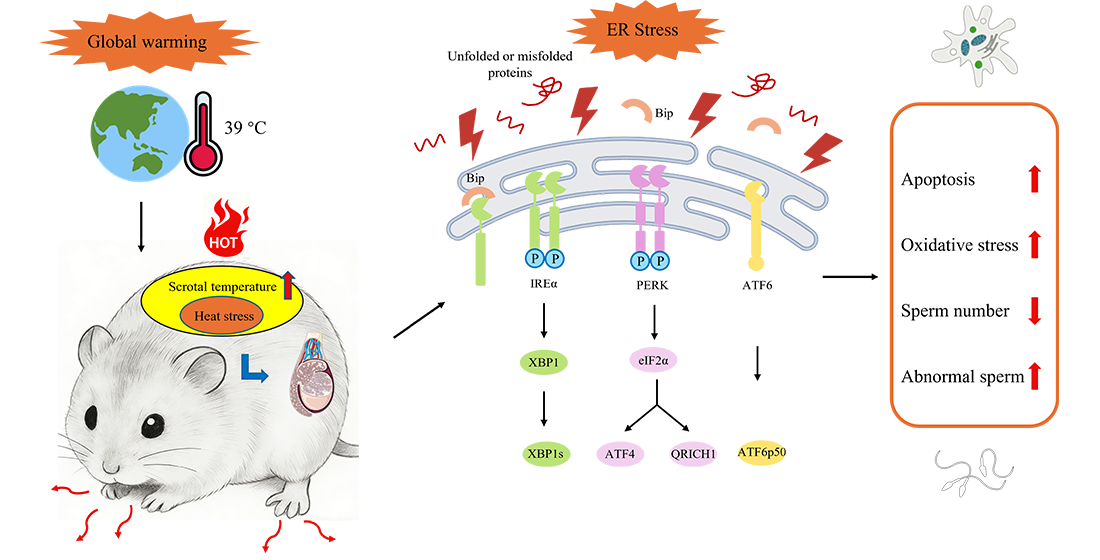
Rising global temperatures pose a serious threat to wild small mammal’s population persistence. In this study, we investigated the molecular mechanisms underlying heat-induced testicular impairment in Brandt’s vole (Lasiopodomys brandtii), a dominant small mammal species of the Eurasian temperate steppe. Adult males were subjected to short term heat exposure at 37°C, 39°C, and 41°C. Heat stress at temperatures ≥39°C significantly reduced the testicular index and caused histopathological damage. Integrated transcriptomic and data-independent acquisition proteomic analyses revealed significant enrichment of pathways related to endoplasmic reticulum protein processing and barrier function. Further molecular validation demonstrated robust activation of the unfolded protein response, indicated by increased expression of ATF4, ATF6B, phosphorylated eIF2α, and XBP1. Together, these results identify endoplasmic reticulum stress as a key mediator of heat-induced testicular injury and highlight that 39°C represents a critical reproductive threshold for Brandt’s voles, even following short-term exposure.
Keywords: 
;  ;  ;  

1. Introduction

Rising global temperatures pose a significant threat to wild animal health and population persistence [1,2]. In male mammals, spermatogenesis is highly temperature sensitive and requires scrotum maintained approximately 2–4°C below core body temperature. Scrotal temperature is easily elevated by the environment, which can disrupt spermatogenic homeostasis, resulting in impaired sperm quality and male infertility [3]. Brandt’s vole (Lasiopodomys brandtii) is a representative small mammal species of the Eurasian temperate steppe. It dominates the typical steppes of Inner Mongolia (China), Mongolia, and the Trans-Baikal region of Russia, where its population dynamics exert substantial influences on grassland structure and ecosystem functioning [4]. In the temperate steppe of Inner Mongolia, regional warming has accelerated to approximately 0.37°C per decade, which is three times the global average [5,6]. Extreme temperatures reached 42.3°C in 2021, as recorded by the Inner Mongolia Meteorological Observatory. In reality, ambient temperature is measured inside a shaded instrument shelter (Stevenson screen) at a standard height above the ground. However, the actual temperature within a few centimeters of the ground is significantly higher. It is precisely this near-surface microclimate temperature that influences Brandt’s vole. Climate change has been closely associated with changes in the population dynamics and geographical distribution of prairie-dwelling small mammals, including Brandt’s vole, with these changes being heavily driven by reproductive capacity [7,8,9]. Due to its high sensitivity to thermal stress, Brandt’s vole provides an ideal model for investigating how climate warming affects wild small mammals.
Endoplasmic reticulum (ER) stress signaling plays a critical role in spermatogenesis, sperm maturation, and fertilization [10]. Although heat stress is widely recognized to impair male reproductive function, the molecular mechanisms linking heat stress, ER stress, and reproductive dysfunction in wild small mammals remain poorly understood. Here, we hypothesize that climate warming impairs male reproductive function in Brandt’s voles via ER stress. Using integrated transcriptomic and proteomic analyses, we aim to elucidate the molecular basis of heat-induced testicular dysfunction in Brandt’s voles.

2. Materials and Methods

2.1. Experimental Animals

Adult male Brandt’s voles (body weight: 45 ± 5.5 g; age: 9–10 weeks) were utilized for this study. These animals were the offspring of a wild-captured population originally sourced from the grasslands of Xilinhot, Inner Mongolia, China (43° 02’–44° 52’ N, 115° 13’–117° 06’ E). Voles were housed individually in a controlled environment maintained at a temperature of 22 ± 2°C and relative humidity of 35%–50%, under a 12:12 h light/dark cycle. Animals were provided ad libitum access to standard laboratory chow and water, with daily monitoring for health status and sanitary cage maintenance. All experimental procedures were approved by the Institutional Animal Care and Use Committee of Zhengzhou University (Approval No. ZZUIRB2023-008) and were conducted in strict accordance with the Guide for the Care and Use of Laboratory Animals of China.

2.2. Heat Stress Treatment and Sample Collection

Male Brandt’s voles were randomly assigned to four experimental groups (n=12 per group): a control group (Ctrl) maintained at room temperature, and three heat stress groups subjected to temperatures of 37°C (C37), 39°C (C39), and 41°C (C41), respectively. Prior to heat exposure, all animals were anesthetized via intraperitoneal injection of 5 mg/kg Xylazine (J329BA0031, Sangon Biotech, Shanghai, China). Based on an established protocol [11]. Heat stress was induced by immersing the lower abdomen and scrotum in a water bath at the corresponding temperature for 30 min. Following treatment, animals were allowed to recover at room temperature for 24 h. The animals were subsequently euthanized, and the testes were immediately dissected and weighed. For histological analysis, intact testes were fixed in a specialized testis fixative (Servicebio, Wuhan, China). For molecular analysis, testicular tissues were snap-frozen in dry ice and stored at -80 °C until RNA and protein extraction.

2.3. Histological Analysis

Following fixation, testes were dehydrated and embedded in paraffin. Six micrometer-thick sections were prepared using a microtome (Thermo Fisher Scientific, San Jose, CA, USA) and subjected to standard hematoxylin and eosin (H&E) staining protocols. Slides were then scanned and documented using an Olympus VS200 SLIDEVIEW imaging system to evaluate histological alterations.

2.4. Biochemical Analysis of Oxidative Stress

Testicular tissues were homogenized in an ice-water bath using 0.9% NaCl at a mass-to-volume ratio (w/v) of 1:5 to 1:10. The resulting homogenates were centrifuged at 8000 × g for 10 min at 4°C, and the supernatants were collected for subsequent biochemical analysis. The activities of catalase (CAT) and superoxide dismutase (SOD), as well as the concentrations of malondialdehyde (MDA) and reduced glutathione (GSH), were quantified using assay kits (Solarbio Science & Technology Co., Ltd., Beijing, China; Cat. Nos. BC4780, BC5160, BC0020, and BC1170).

2.5. Transmission Electron Microscopy

Testicular tissues were primarily fixed in 0.1 M cacodylate buffer (pH 7.4) containing 2.5% glutaraldehyde and 1.4% sucrose at 37°C for 1.5 h. Samples underwent post-fixation in 1% osmium tetroxide and were subsequently dehydrated and embedded in Epon-812 resin. Ultra-thin sections (90 nm) were prepared using an ultramicrotome and double-stained with uranyl acetate and lead citrate. Testicular ultrastructure was visualized using a TEM (JEM-1400, JEOL Ltd., Tokyo, Japan) operating at an acceleration voltage of 80 kV.

2.6. Quantitative Real-Time PCR Analysis

Total RNA was isolated from testicular tissue using the Trizol reagent (Solarbio Science & Technology Co., Ltd., China; Cat. No. P1011). RNA concentration and purity were assessed using a NanoDrop spectrophotometer. First-strand cDNA was synthesized using the HiScript® III All-in-One RT SuperMix Perfect kit (Vazyme Biotech Co., Ltd., China; Cat. No. R333-01). Quantitative real-time PCR was performed using the SYBR green method on an Applied Biosystems 7500 real-time PCR system. Species-specific primers for Brandt’s voles were synthesized by Sangon Biotech (Shanghai, China), and all primer sequences are provided in supplementary Table S1.

2.7. RNA Sequencing and Data Analysis

Transcriptome sequencing was performed by Feisha Biotechnology (Wuhan, China). Raw sequence data were quality-filtered using fastp (v1.0.1) to remove low-quality reads and adapter sequences. High-quality clean reads were assembled de novo using Trinity (v2.5.1). Temporal or pattern-based gene expression profiles were analyzed using the Mfuzz R package (v2.60.0) with default fuzzy c-means clustering parameters. Differentially expressed genes (DEGs) were identified using a threshold of fold change ≥ 2 and P-adj ≤ 0.01. The False Discovery Rate (FDR) was applied to correct for multiple testing. Functional annotation of DEGs was conducted against multiple databases, including the NCBI non-redundant (NR) protein sequences, Gene Ontology (GO), Kyoto Encyclopedia of Genes and Genomes (KEGG), evolutionary genealogy of genes: Non-supervised Orthologous Groups (eggNOG), Swiss-Prot, and Pfam.

2.8. Liquid Chromatography Tandem Mass Spectrometry and Data Analysis

Testicular protein expression profiles were characterized using data-independent acquisition (DIA) mass spectrometry performed with Orbitrap Astral mass spectrometer (Thermo Scientific) provided by SanshuBio Co. Ltd. (Shanghai, China). Samples were analyzed in DIA mode. Raw MS data were processed using DIA-NN (v1.9). Protein identifications were filtered at a 1% FDR at both the precursor and protein levels. Differentially expressed proteins (DEPs) were functionally annotated against GO and KEGG databases using eggNOG-mapper.

2.9. Statistical Analysis

Data are presented as the mean ± SEM. Statistical evaluations were conducted using GraphPad Prism (version 9.1.0, GraphPad Software, San Diego, CA, USA). For comparisons between two groups, an unpaired Student’s t-test was employed. A value of P < 0.05 was defined as the threshold for statistical significance.

3. Results

3.1. Heat Stress Impaired Testicular Histological Structure

As illustrated in Figure 1 and 2A. Following heat exposure at 37°C, 39°C, and 41°C, a significant reduction in both testicular size and index was observed (P < 0.05). Morphological assessment of spermatozoa revealed normal structures in the Ctrl group, whereas heat-stressed groups exhibited marked sperm abnormalities, including absent tail, multi-headed, and coiled-tail phenotypes (Figure 2B). Histological examination of the Ctrl group revealed a healthy testicular architecture characterized by well-organized spermatogonia layers and abundant luminal spermatozoa. In contrast, the heat stressed groups displayed progressive morphological deterioration, including vascular congestion, irregular seminiferous tubule contours, and a disrupted spermatogenic epithelium (Figure 2C). Furthermore, ultrastructural analysis via TEM demonstrated that heat stress induced significantly impaired mitochondrial integrity and pronounced swelling of the ER (Figure 2D).

3.2. Heat Stress Induced Oxidant Stress in Testis

The effects of heat stress on the antioxidant capacity and lipid peroxidation of testes are summarized in Figure 3. Compared to the Ctrl group, C39 and C41 groups showed significantly reduced GSH content (P < 0.05), whereas no significant alterations were observed in the C37 group (Figure 3A). Conversely, SOD activity was upregulated in the C39 and C41 groups (P < 0.05) but remained unchanged under the 37°C treatment (Figure 3C). Interestingly, MDA levels showed no fluctuation across all heat-stressed groups (P > 0.05; Figure 3B). Regarding CAT activity, only the C41 group showed a reduction (P < 0.05) (Figure 3D).

3.3. Heat Stress Disturbed the Barrier Function in Testis

Compared to the Ctrl group, claudin 1 (Cldn1) mRNA abundance remained unchanged in the C37 and C39 groups, but was significantly upregulated in C41 group (P < 0.05; Figure 4A). In contrast, heat stress induced a robust inhibitory effect on the expression of occludin (Ocln) and tight junction protein 1 (Tjp1) (P < 0.05; Figure 4B,C).

3.4. Heat Stress Induced ER Stress in Testis

The expression profiles of key ER stress markers were evaluated at both the transcriptional (Figure 5) and translational (Figure 6) levels. Compared to the Ctrl group, heat stress resulted in a downregulation of DNA-damage inducible transcript 3 (Ddit3) and glutamine-rich 1 (Qrich1) mRNA abundance (P < 0.05; Figure 5A,B). Notably, the mRNA levels of activating transcription factor 4 (Atf4) and X-box binding protein 1 (Xbp1) were significantly upregulated only in the C41 group, whereas no significant changes were observed at C37 and C39 group (Figure 5C,D).
To validate these findings, the protein abundance was analyzed via western blotting (Figure 6A). Consistent with the transcriptional data for the C41 group, the protein expression of ATF4, ATF6B, α-subunit of eukaryotic initiation factor 2 (eIF2α), and XBP1 was elevated (P < 0.05; Figure 6B–E). Similar to the mRNA patterns, heat stress at 37°C and 39°C did not induce a significant increase in this protein abundance.
Figure 6. Analysis of ER stress-related protein expression in testicular tissue of Brandt’s voles. Representative Western blot images of ATF4, ATF6B, eIF2α and XBP1(A). Quantitative analysis of relative protein levels for ATF4 (B), ATF6B (C), eIF2α (D) and XBP1 (E) in the testicular tissue of Brandt’s voles. Data are expressed as mean ± SEM (n = 6 per group). ns means no significance, * means P < 0.05, ** means P < 0.01.
Preprints 218750 g006

3.5. Results of RNA-Sequencing Data Filtering

Following stringent quality control, the proportion of high-quality filtered reads ranged from 99.92% to 99.93% (supplementary Table S2). The sequencing depth yielded between 57.02M and 91.85M clean reads per sample, with a minimum Q30 score of 92.97%. A total of 673474 transcripts were identified, and the average mapping rate of reads to the reference genome was 86.66% (supplementary Tables S3 and S4).

3.6. Functional Annotation

350367 unigenes were successfully annotated in at least one database (supplementary Table S5). Specifically, 63463 unigenes were identified in the NCBI NR database, followed by 60107 in Swiss-Prot, and 59484 in Pfam. Furthermore, functional classification and pathway analysis assigned 57631 unigenes to GO terms, 43964 to eggNOG clusters, and 40680 to KEGG pathways. Specialized annotations included 12637 unigenes in TMHMM, 9130 in SignalP, and 3271 in the CAZy database.
Table S5. Unigenes annotations.
Databases Annotation Number of Unigenes
Nr 63463
Swiss-Prot 60107
KEGG 40680
Gene_Ontology 57631
eggNOG 43964
Pfam 59484
CAZy 3271
SignaIP 9130
TMHMM 12637

3.7. Identification of Differentially Expressed Genes

In the C37 group, 1344 DEGs were identified, comprising 645 upregulated and 699 downregulated genes. The C39 group exhibited the highest transcriptional response with 3248 DEGs (1876 upregulated; 1372 downregulated), while the C41 group contained 1580 DEGs (763 upregulated; 817 downregulated) (supplementary Figure S1A). Overlap analysis via a Venn diagram revealed a core set of 340 common DEGs consistently altered across all three heat treatments (supplementary Figure S1B). The distribution and significance of these expression profiles were further visualized through volcano plots for each pairwise comparison (supplementary Figure S1C–E).

3.8. DEGs Cluster Analysis Based on Temperature Course

As shown in Figure 7, these clusters, ranging in size from 6,919 to 11,893 genes. Cluster 1 (6,919 genes) remained at basal levels at 37°C but showed a transient peak at 39°C before declining at 41°C. In contrast, genes in clusters 2, 4, and 5 exhibited V-shaped trajectories, characterized by a sharp downregulation at 37°C or 39°C followed by a robust induction at 41°C. Cluster 3 genes demonstrated a progressive, temperature-dependent upregulation throughout the 37-41°C. Conversely, cluster 6 genes were consistently repressed across all thermal treatments. Clusters 7 and 9 displayed inverted V-shaped profiles, with marked transcriptional enhancement at 37°C or 39°C followed by a subsequent reduction. Finally, cluster 8 was characterized by an initial upregulation at 37°C followed by a decrease at 39°C.

3.9. GO and KEGG Enrichment Analysis of DEGs

GO analysis revealed that 833, 1,734, and 995 DEGs were categorized into 54, 53, and 52 functional subcategories in the C37 (supplementary Figure S2A), C39 (supplementary Figure S2B), and C41 (supplementary Figure S2C) groups, respectively. Predominantly enriched terms included “response to stimulus”, “membrane part”, and “antioxidant activity”. KEGG pathway mapping further identified 1311, 2412, and 1536 DEGs associated with diverse biological functions. These were classified into six primary KEGG categories, with the highest DEG distribution observed in environmental information processing, organismal systems, and human diseases (supplementary Figure S3).

3.10. Validation of DEGs with qRT-PCR

To verify the accuracy of the RNA-sequencing findings, 15 representative DEGs involved in ER stress, inflammation, protein quality control, and metabolic regulation were selected for qRT-PCR validation (supplementary Figure S4). Consistent with the transcriptomic profiles, the mRNA expression levels of CCAAT enhancer binding protein delta (Cebpd), Suppressor of cytokine signaling 3 (Socs3), Heat shock protein beta-1 (Hspb1), Activating transcription factor 3 (Atf3), Membrane associated ring-CH-type finger 10 (Marchf10), Spermatogenesis associated 21 (Spata21), Meiosis specific nuclear structural 1 (Mns1), SEC11 homolog A (Sec11a), Coiled-coil domain containing 33 (Ccdc33), Prostaglandin-endoperoxide synthase 2 (Ptgs2), IQ motif and ubiquitin domain containing gene (Iqub), and Uromodulin (Umod) were significantly upregulated across the heat-stress cohorts. Conversely, Patatin-like phospholipase domain-containing 3 (Pnpla3), GLI Family Zinc Finger 1 (Gli1), and Aquaporin 2 (Aqp2) exhibited marked downregulation following thermal exposure. Although the magnitude of fold changes varied slightly between the two platforms, the directional trends of gene expression were highly concordant. These results provide robust validation of the transcriptomic data and confirm the reliability of the sequencing-based expression profiles.

3.11. Protein Identification and Quantification

A total of 5,808 proteins were high-confidently identified across three biological replicates (supplementary Table S6).
Table S6. Overview of protein and peptide identification and quantification.
Sample Identified Peptides Quantified Peptides Identified Protein Quantified Protein
Ctrl_1 67725 51407 5808 5298
Ctrl_2 67725 55594 5808 5465
Ctrl_3 67725 54412 5808 5446
C39_1 67725 54421 5808 5394
C39_2 67725 55591 5808 5531
C39_3 67725 55773 5808 5518
Principal component analysis (PCA) revealed that the first two components accounted for 56.3% of the total variance, with the PC1 clearly separating the Ctrl and C39 group (supplementary Figure S5).
Hierarchical clustering analysis further confirmed these results (Figure 8).
Differentially expressed proteins (DEPs) were identified using a fold change ≥ 2.0 and Padj < 0.01. A total of 394 DEPs were identified between the two groups (supplementary Figure S6). Among these, 233 proteins were upregulated and 161 proteins were downregulated.

3.12. Functional Categorization Analysis

The DEPs were predominantly localized to protoplasm, cytoplasm, cytoplasmic granule lumen, cytoplasmic granule membrane, cytoplasmic granule (supplementary Figure S7).
Within the Biological process category, DEPs were significantly enriched in reproduction. Cellular component analysis revealed high enrichment in the nuclear chromosome, golgi membrane, and ubiquitin ligase complexes. In the molecular function category, proteins associated with nucleotide binding and transcription cis-regulatory region binding predominated (supplementary Figure S8).
KEGG pathway mapping further contextualized the proteomic response to heat stress (supplementary Figure S9A,B). Significant enrichment was observed in the MAPK signaling pathway and tight junctions. Notably, stratified analysis of directional expression revealed that Heat Stress-Up DEPs were primarily associated with protein processing in the ER, neurotrophin signaling pathway, and Salmonella infection pathways. Conversely, Heat Stress-Down DEPs were predominantly enriched in pathways related to aldosterone-regulated sodium reabsorption and various cancer-related signaling modules (supplementary Figure S9C).

4. Discussion

As a representative steppe rodent, Brandt’s vole provides an ideal model for examining how climate warming compromises the fertility of wild small mammals [12]. We identified ER stress as a central mediator of heat-induced testicular dysfunction in Brandt’s voles. These findings establish a mechanistic framework linking climate warming to male reproductive vulnerability in wild small mammals. Spermatogenesis is strictly temperature dependent, and germ cells with high mitotic activity are particularly vulnerable to heat stress [13]. Consistent with previous studies [14,15,16], we observed that in Brandt’s voles, a single 30-min heat exposure at 39°C or 41°C was sufficient to markedly reduce the testicular index, induce structural sperm abnormalities, and cause widespread degeneration of the seminiferous epithelium (Figure 1 and Figure 2A–C). Heat stress is known to exacerbate ROS production, leading to oxidative stress when antioxidant defenses are overwhelmed. Although MDA levels did not increase significantly, the increasing trend across heat-stressed groups indicates an early shift toward oxidative damage (Figure 3B). Heat exposure also caused a marked depletion of GSH, reflecting its rapid consumption in ROS scavenging and impaired redox homeostasis [17]. In parallel, SOD activity increased, whereas CAT activity declined, indicating a disruption of the antioxidant cascade (Figure 3C,D). As SOD converts superoxide radicals into H₂O₂, reduced CAT activity would limit H₂O₂ detoxification, favoring its accumulation. Given that H₂O₂ is a potent inducer of testicular cell apoptosis under heat stress, this imbalance likely underlies the reduced testicular index and histopathological damage observed at 39°C and 41°C [18]. The blood–testis barrier (BTB), formed by specialized junctions between Sertoli cells, is essential for maintaining the microenvironment required for spermatogenesis. In Brandt’s voles, heat stress markedly suppressed the expression of Ocln and Tjp1, consistent with Sertoli cell dysfunction and BTB disruption reported in other rodent models (Figure 4B,C) [19]. Notably, Cldn1 expression was significantly upregulated (Figure 4A). We therefore propose that Cldn1 upregulation in Brandt’s voles reflects a cytoprotective but ultimately insufficient response to counteract BTB breakdown under heat stress.
To comprehensively characterize the testicular response to heat stress, we integrated transcriptomic and proteomic analyses. Transcriptome profiling revealed extensive heat-induced gene reprogramming, with functional enrichment predominantly associated with the ER and protein proteostasis pathways, including folding, sorting, and degradation (supplementary Figure S2). These transcriptional signatures were supported by ultrastructural evidence of ER swelling, indicating pronounced ER perturbation following heat exposure. Given the cellular heterogeneity of the testis, DIA-based quantitative proteomics was employed to resolve the executive molecular responses, focusing on the C39 group, which exhibited the most pronounced transcriptional shift (supplementary Figure S1A). Proteomic analysis identified differentially expressed proteins enriched within the endomembrane system and inner cell membranes, with functional annotations highlighting roles in reproduction and stress adaptation (supplementary Figures S8 and S9). The strong consistency among transcriptomic, proteomic, and ultrastructural evidence identifies ER stress as the central mediator of heat-induced testicular dysfunction in Brandt’s voles, providing a mechanistic framework for the reproductive impairment observed under heat stress.
The ER is central to cellular proteostasis, maintaining an oxidative environment essential for protein folding and post-translational modification. Heat-induced ROS impair ER folding capacity, while the accumulation of misfolded proteins further amplifies ROS generation, establishing a self-reinforcing cycle of cellular stress [20,21]. To counteract this stress, cells activate the UPR, a conserved signaling network that initially limits protein synthesis and enhances chaperone expression (Figure 6) [22]. The downregulation of Qrich1 and pro-apoptotic factor Ddit3 validates the activation of early protective mechanisms of the UPR (Figure 5A,B) [23,24]. This pattern suggests that germ cell loss in heat-stressed Brandt’s voles may proceed via alternative pathways, such as IRE1-dependent RNA decay or mitochondria-mediated apoptosis [25,26].
This study demonstrates that heat exposure severely disrupts testicular function in Brandt’s voles, leading to seminiferous degeneration and impaired sperm quality. Integrative omics analyses and protein abundance analyses reveal that oxidative stress–driven activation of ER stress is a central mechanism underlying this pathology. These findings highlight that 39°C represents a critical threshold for the reproductive vulnerability of Brandt’s voles even following short-time exposure.

Supplementary Materials

The following supporting information can be downloaded at the website of this paper posted on Preprints.org.

Author Contributions

Kang Lou: Validation, Formal analysis, Writing - Original Draft. Jiaxue Jin: Writing, Formal analysis. Yankai Yang: Validation, Formal analysis, Writing. Xiaomeng Zhao: Formal analysis. Lijuan Zhao: Writing - Review & Editing. Zhiguang Chang: Writing - Review & Editing. Zhenlong Wang: Review & Editing, Conceptualization. Senlin Li: Supervision, Funding acquisition, Review & Editing. All authors have read and agreed to the published version of the manuscript.

Funding

This work was supported by the fellowship of China Postdoctoral Science Foundation (2022M712898); Postdoctoral Science Foundation of Henan Province (308009); Natural Science Foundation of Henan Province (242300420169; 262300422174); National Natural Science Foundation of China (32301302).

Institutional Review Board Statement

All experimental procedures were approved by the Institutional Animal Care and Use Committee of Zhengzhou University (Approval No. ZZUIRB2023-008) and were conducted in strict accordance with the Guide for the Care and Use of Laboratory Animals of China.

Data Availability Statement

The mass spectrometry proteomics data have been deposited to the ProteomeXchange Consortium (https://proteomecentral.proteomexchange.org) via the iProX partner repository with the dataset identifier PXD073388. Reviewers can access the data using the following link: https://www.iprox.cn/page/PSV023.html;?url=17691574457356iDQ; Password: 6fvL. The transcriptomic raw reads have been deposited in the NCBI Sequence Read Archive (SRA) under BioProject accession number PRJNA1399431. These data are currently private for peer review. Reviewers can access the data via the following link: https://dataview.ncbi.nlm.nih.gov/object/PRJNA1399431?reviewer=kgqqjjc63p3n0rhvnsk17h9j7c. All data will be released to the public upon the formal publication of this study.

Conflicts of Interest

The authors declare no conflict of interest.

Abbreviations

Aqp2, aquaporin 2; Atf3, activating transcription factor 3; Atf4, activating transcription factor 4; ATF6B, activating transcription factor 6B; BTB, blood-testis barrier; CAT, catalase; Ccdc33, coiled-coil domain containing 33; Cebpd, CCAAT enhancer binding protein delta; Cldn1, claudin-1; Ctrl, control; C37, 37°C; C39, 39°C; C41, 41°C; Ddit3, DNA damage inducible transcript 3; DEPs, differentially expressed proteins; DIA, data-independent acquisition; eggNOG, evolutionary genealogy of genes: non-supervised orthologous groups; ER, endoplasmic reticulum; Gli1, gli family zinc finger 1; GO, gene ontology; GSH, reduced glutathione; H&E, hematoxylin and eosin; H2O2, hydrogen peroxide; Hspb1, heat shock protein beta-1; Iqub, iq motif and ubiquitin domain containing gene; KEGG, Kyoto Encyclopedia of Genes and Genomes; LC-MS/MS, liquid chromatography tandem mass spectrometry; Marchf10, membrane associated ring-CH-type finger 10; MDA, malondialdehyde; Mns1, meiosis specific nuclear structural 1; Ocln, occludin; O 2   , superoxide anion; PCA, principal component analysis; PC1, primary component; Pnpla3, patatin-like phospholipase domain-containing 3; p-eIF2α, phosphorylated eukaryotic initiation factor 2α; Ptgs2, prostaglandin-endoperoxide synthase 2; Qrich1, glutamine rich 1; ROS, reactive oxygen species; SEC11A, sec11 homolog A; Socs3, suppressor of cytokine signaling 3; SOD, superoxide dismutase; Spata21, spermatogenesis associated 21; TEM, transmission electron microscopy; Tjp1, tight junction protein 1; Umod, uromodulin; UPR, unfolded protein response; Xpb1, x-box binding protein 1.

References

  1. Matthews HD, Wynes S. Current global efforts are insufficient to limit warming to 1.5°C. Science 2022; 376:1404-1409. [CrossRef]
  2. Alexeeva N, Erbajeva M, Khenzykhenova F. Lasiopodomys brandti in Pleistocene of Transbaiklia and adjacent territories: distribution area, evolutionary development in context of global and regional events. quat. int. 2015; 355:11-17. [CrossRef]
  3. De Toni L, Finocchi F, Jawich K, Ferlin A. Global warming and testis function: A challenging crosstalk in an equally challenging environmental scenario. Front. Cell Dev. Biol. 2023; 10:1104326. [CrossRef]
  4. Jiang G, Liu J, Xu L, Yu G, He H, Zhang Z. Climate warming increases biodiversity of small rodents by favoring rare or less abundant species in a grassland ecosystem. Integr. Zool. 2013; 8:162-174. [CrossRef]
  5. Ma L, Li H, Liu T, Liang L. Abrupt temperature change and a warming hiatus from 1951 to 2014 in Inner Mongolia, China. J. Arid Land 2019; 11:192-207. [CrossRef]
  6. Hu Q, Pan F, Pan X, Zhang D, Li Q, Pan Z, Wei Y. Spatial analysis of climate change in Inner Mongolia during 1961–2012, China. Appl. Geogr. 2015; 60:254-260. [CrossRef]
  7. Shahat AM, Rizzoto G, Kastelic J. Amelioration of heat stress-induced damage to testes and sperm quality. Theriogenology 2020; 158:84-96. [CrossRef]
  8. Myers P, Lundrigan BL, Hoffman SM, Haraminac AP, Seto SH. Climate-induced changes in the small mammal communities of the Northern Great Lakes Region. Glob. Chang. Biol. 2009; 15:1434-1454. [CrossRef]
  9. Jiang G, Zhao T, Liu J, Xu L, Yu G, He H, Krebs C, Zhang Z. Effects of ENSO-linked climate and vegetation on population dynamics of sympatric rodent species in semiarid grasslands of Inner Mongolia, China. Can. J. Zool. 2011; 89:678-691. [CrossRef]
  10. Karna KK, Shin YS, Choi BR, Kim HK, Park JK. The role of endoplasmic reticulum stress response in male reproductive physiology and pathology: a review. World J. Men's Health 2019; 38:484. [CrossRef]
  11. Bai L, Zhang Y, Zheng C, Xu S, He Y, Yu G, Huang D, Huang Y, Li M, Xu C. Tanshinone IIA protects mouse testes from heat stress injury by inhibiting apoptosis and TGFβ1/Smad2/Smad3 signaling pathway. Cell Stress Chaperones 2023; 28:749-759. [CrossRef]
  12. Bai D, Wan X, Li G, Wan X, Guo Y, Shi D, Zhang Z. Factors influencing range contraction of a rodent herbivore in a steppe grassland over the past decades. Ecology and evolution 2022; 12:2045-7758.
  13. Aldahhan RA, Stanton PG. Heat stress response of somatic cells in the testis. Molecular and cellular endocrinology 2021; 527:111216. [CrossRef]
  14. Abd El-Emam MM, Ray MN, Ozono M, Kogure K. Heat stress disrupts spermatogenesis via modulation of sperm-specific calcium channels in rats. Journal of Thermal Biology 2023; 112:0306-4565.
  15. Teixeira MB, Ferreira JCP, Codognoto VM, Rossi ES, Pupulim AGR, de Carvalho JC, Rattes PZ, Oba E, Navolar FMN, Di Santis GW. Heat stress induced by testicular insulation for 24 or 48 h rapidly impairs epididymal sperm quality and reduces spermatogenesis in rams. Small Ruminant Research 2025; 243:0921-4488. [CrossRef]
  16. Rizzoto G, Boe-Hansen G, Klein C, Thundathil J, Kastelic J. Acute mild heat stress alters gene expression in testes and reduces sperm quality in mice. Theriogenology 2020; 158:375-381. [CrossRef]
  17. Tang Z, Yang Y, Wu Z, Ji Y. Heat stress-induced intestinal barrier impairment: current insights into the aspects of oxidative stress and endoplasmic reticulum stress. Journal of Agricultural and Food Chemistry 2023; 71:0021-8561. [CrossRef]
  18. Xiang J, Wan C, Guo R, Guo D. Is hydrogen peroxide a suitable apoptosis inducer for all cell types? BioMed research international 2016; 2016:2314-6141. [CrossRef]
  19. Zhao Z, Cai Y, Lin X, Liu N, Qin Y, Wu Y. The Role of Heat-Induced Stress Granules in the Blood–Testis Barrier of Mice. Int. J. Mol. Sci. 2024; 25:3637. [CrossRef]
  20. Li X, Zhu J, Lin Q, Yu M, Lu J, Feng J, Hu C. Effects of curcumin on mitochondrial function, endoplasmic reticulum stress, and mitochondria-associated endoplasmic reticulum membranes in the jejunum of oxidative stress piglets. J. Agric. Food Chem. 2022; 70:8974-8985. [CrossRef]
  21. Fani G, La Torre CE, Cascella R, Cecchi C, Vendruscolo M, Chiti F. Misfolded protein oligomers induce an increase of intracellular Ca2+ causing an escalation of reactive oxidative species. Cellular and Molecular Life Sciences 2022; 79:500. [CrossRef]
  22. Casas-Martinez JC, Xia Q, Li P, Borja-Gonzalez M, Miranda-Vizuete A, McDermott E, Dockery P, Quinlan LR, Goljanek-Whysall K, Samali A, McDonagh B. Adaptive ER stress promotes mitochondrial remodelling and longevity through PERK-dependent MERCS assembly. Cell Death & Differentiation 2026; 33:732-747. [CrossRef]
  23. You K, Wang L, Chou C-H, Liu K, Nakata T, Jaiswal A, Yao J, Lefkovith A, Omar A, Perrigoue JG. QRICH1 dictates the outcome of ER stress through transcriptional control of proteostasis. Science 2021; 371:eabb6896. [CrossRef]
  24. Huang L, Cheng Y, Pan X, Li H, Liu J, Zhao G, Huang L, Wang L, Song C, Song P. DDIT3-Mediated Endoplasmic Reticulum Stress Drives Manganese-Induced Apoptosis in Bovine Testicular Leydig Cells. Cell Stress and Chaperones 2025:1355-8145.
  25. Cairrao F, Santos CC, Le Thomas A, Marsters S, Ashkenazi A, Domingos PM. Pumilio protects Xbp1 mRNA from regulated Ire1-dependent decay. Nature Communications 2022; 13:2041-1723. [CrossRef]
  26. Bartoszewska S, Sławski J, Collawn JF, Bartoszewski R. Dual RNase activity of IRE1 as a target for anticancer therapies. Journal of Cell Communication and Signaling 2023; 17:1145-1161. [CrossRef]
Figure 1. Testicular index. Data are expressed as mean ± SEM (n = 7 per group). * Means P < 0.05.
Preprints 218750 g001
Figure 2. Histological evaluation of testes from heat stressed. (A) Testicular morphology; (B) Sperm morphology (#: decapitated spermatozoa, ※: coiled-tail, ★: multiple heads); (C) Histological structure of testicular tissue (dotted arrow: a normal seminiferous tubule with spermatozoa in the lumen, #: interstitial vascular congestion, *: collapse and vacuolization of the seminiferous epithelium, arrow: germ cell depletion); (D) The ultrastructure for endoplasmic reticulum (*) and mitochondria (▲).
Preprints 218750 g002
Figure 3. The concentration of GSH (A), MDA (B) and activities of SOD (C), CAT (D) in the testicular tissue of Brandt’s voles. Data are expressed as mean ± SEM (n = 9 per group). Absence of marks or ns means no significance, * means P < 0.05, ** means P < 0.01, ****means P < 0.0001.
Preprints 218750 g003
Figure 4. mRNA levels for Cldn1(A), Ocln(B), Tjp1(C) in the testicular tissue of Brandt’s voles. Data are expressed as mean ± SEM (n = 8 per group). ns means no significance, * means P < 0.05, ** means P < 0.01, ***means P < 0.001, ****means P < 0.0001.
Preprints 218750 g004
Figure 5. mRNA levels for Ddit3 (A), Qrich1 (B), Atf4 (C), Xbp1 (D) in the testicular tissue of Brandt’s voles. Data are expressed as mean ± SEM (n = 8 per group). Absence of marks or ns means no significance, * means P < 0.05, ** means P < 0.01.
Preprints 218750 g005
Figure 7. Clusters of DEGs in Ctrl and different exposed temperature.
Preprints 218750 g007
Figure 8. Heatmap illustrating the differentially expressed proteins (DEPs) between the Ctrl and C39 groups.
Preprints 218750 g008
Disclaimer/Publisher’s Note: The statements, opinions and data contained in all publications are solely those of the individual author(s) and contributor(s) and not of MDPI and/or the editor(s). MDPI and/or the editor(s) disclaim responsibility for any injury to people or property resulting from any ideas, methods, instructions or products referred to in the content.
Copyright: This open access article is published under a Creative Commons CC BY 4.0 license, which permit the free download, distribution, and reuse, provided that the author and preprint are cited in any reuse.
Prerpints.org logo

Preprints.org is a free preprint server supported by MDPI in Basel, Switzerland.

Subscribe

© 2026 MDPI (Basel, Switzerland) unless otherwise stated

Accessibility

Disclaimer

Terms of Use

Privacy Policy

Privacy Settings