Submitted:
20 May 2026
Posted:
21 May 2026
You are already at the latest version
Abstract
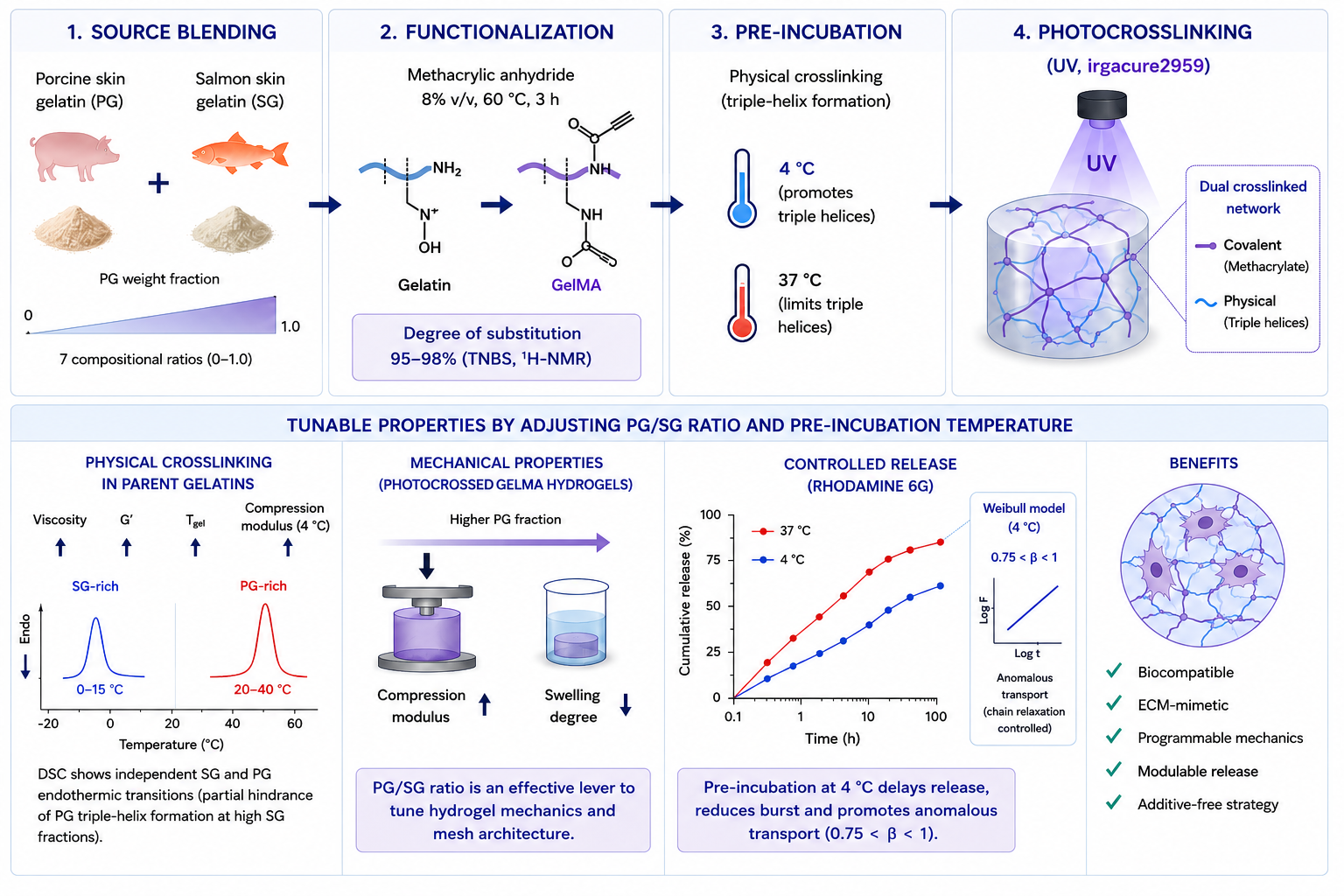
Modulating the physical crosslink architecture of gelatin methacryloyl (GelMA) hydro-gels without altering total polymer concentration or introducing exogenous components remains a central challenge in biomaterial design. Here we report a source blending strategy in which porcine skin gelatin (PG) and salmon skin gelatin (SG), two gelatins with markedly different proline and hydroxyproline contents, are combined across seven compositional ratios (PG weight fractions 0–1.0) and functionalized to GelMA under standardized conditions (8% v/v methacrylic anhydride, 60 °C, 3 h). Near-complete de-grees of substitution (95–98%) were achieved across all formulations, confirmed by both TNBS and ¹H-NMR. In the parent gelatin mixtures, increasing PG fraction progressively increased viscosity, elastic modulus (G′), gelation temperature (Tgel), and compression modulus at 4 °C, with DSC revealing independent SG (0–15 °C) and PG (20–40 °C) en-dothermic transitions that suggest partial hindrance of PG triple-helix formation by high SG fractions. These composition-dependent trends were preserved after functionalization to GelMA, albeit with attenuated physical crosslinking due to steric impairment by the methacrylate groups. Photocrosslinked GelMA hydrogels fabricated after pre-incubation at 4 °C exhibited systematically higher compression moduli and lower swelling degrees with increasing PG content, demonstrating that the PG/SG ratio is an effective lever for independently tuning hydrogel mechanics and mesh architecture. In vitro release assays using Rhodamine 6G confirmed that pre-incubation at 4°C prior photocrosslinking serves as a effective way to modulate transport kinetics in SG-PG GelMA hydrogels. This strategy delayed characteristic release times and constrained Weibull shape parameters to the anomalous-transport regime (0.75< β< 1), where diffusion is governed by network chain relaxation. This thermally induced structural restriction was most pronounced in the 0.4SG:0.6PG formulation, where lower SG content permitted unhindered triple-helix formation as corroborated by DSC and compression studies. Ultimately, adjusting the pre-incubation temperature and gelatin source combination provides a straightforward, processing additive free strategy to achieve programmable release profiles via controlled matrix tortuosity.
Keywords:
GelMA
; gelatin hydrogels
; salmon gelatin
; porcine gelatin
; mechanical properties
; degree of substitution
; controlled release
; tissue engineering
1. Introduction
Gelatin is a versatile hydrocolloid derived from the partial hydrolysis and thermal denaturation of collagen [1], obtainable from bovine, porcine, fish, and poultry sources, each yielding materials with distinct physicochemical properties depending on the animal source and extraction conditions [2]. Two principal types are recognized: type A, produced by acid hydrolysis (isoelectric point pH 6–9, predominantly from porcine skin [3]), and type B, produced by alkaline hydrolysis (isoelectric point pH 5-6 [4,5]). At the molecular scale, gelatin comprises α-, β-, and γ-chains [4,6], where α-chains adopt a polyproline II conformation [7,8], sustained by the repetitive Gly-X-Y tripeptide sequence, with proline (Pro) and hydroxyproline (Hyp) most frequently occupying the X and Y positions [9]. This sequence governs the thermoreversible formation of collagen-like triple-helix structures upon cooling, whose extent and thermal stability are directly proportional to total Pro + Hyp content [10,11]. Cold-water fish gelatins, including those derived from salmon skin, contain markedly lower Pro + Hyp levels than mammalian gelatins, resulting in lower Bloom values, reduced gel strength, and melting points that fall well below physiological temperature [11,12,13]. These properties, however, confer distinct processing advantages: salmon skin gelatin remains in the sol state at 5 °C [14,15,16], making it particularly attractive for biofabrication approaches that require low-viscosity inks at near-ambient temperatures. Moreover, its use offers eco-friendly valorization of a major aquaculture by-product and avoids the religious and ethical restrictions associated with mammalian-derived gelatins[10,17,18,19].
Interest in gelatin for biomedical applications has grown considerably, driven by its excellent biocompatibility, biodegradability, and intrinsic cell-adhesion motifs that recapitulate key features of the native extracellular matrix (ECM) [20,21,22]. Nevertheless, its low melting temperature and susceptibility to enzymatic degradation under physiological conditions limit direct use in tissue engineering scaffolds [23,24]. Chemical modification to gelatin methacryloyl (GelMA) overcomes these limitations by introducing photocrosslinkable methacrylate groups that enable the formation of covalently stabilized hydrogel networks with independently tunable stiffness, porosity, and degradation rate [25,26,27]. The resulting hydrogels retain the ECM-mimetic properties of native gelatin, supporting cell adhesion, proliferation, and spreading, and are compatible with a wide range of fabrication strategies including bioprinting and microfabrication [28]. Mechanical and structural properties can be further modulated by adjusting the degree of substitution (DS), polymer concentration, and photocrosslinking conditions [29,30,31], though high GelMA concentrations increase network stiffness at the expense of porosity and cell infiltration [32]. Strategies to decouple these trade-offs include the incorporation of nanomaterials [25,32], optimization of the functionalization protocol [33], and the induction of physical crosslinks through temperature-driven triple-helix formation prior to photopolymerization [34,35]. This last strategy is particularly relevant when working with gelatin mixtures, as the source-dependent differences in Pro + Hyp content create compositionally programmable physical crosslink densities. Geonzon et al. [36] demonstrated that blending porcine skin gelatin with cold-water fish gelatin reinforces gelation temperature and complex modulus even at low porcine fractions, with the extent of reinforcement and the resulting network architecture, ranging from intertwined hybrid triple-helices to isolated, separate polymer networks, governed by the cooling rate applied during gelation [36]. This thermally programmable interspecies co-aggregation mechanism offers a compelling strategy for modulating the physical crosslink network in GelMA hydrogels without altering total polymer concentration or introducing exogenous materials.
Despite the promise of GelMA, its production still lacks standardization: reported synthesis protocols vary widely in reaction pH, temperature, and duration [37,38,39,40,41,42], and these variables have a significant impact on the degree of substitution, photopolymerization efficiency, and the resulting mechanical performance [43]. Thorough characterization of GelMA after synthesis is therefore essential before any inference about hydrogel behavior can be drawn.
From a sourcing perspective, salmon skin represents an abundant and underutilized co-product of the aquaculture industry, offering collagen contents among the highest reported for fish-derived by-products [44]. The highly controlled feeding and husbandry protocols of intensive salmon farming further reduce the compositional variability that typically complicates batch-to-batch reproducibility in gelatin extraction, making salmon an attractive and sustainable raw material for biomedical-grade GelMA.
Building on this context, the present work comprehensively investigates the gelation behavior and physicochemical properties of hydrogels derived from mixtures of well-characterized porcine and salmon gelatins across a range of compositional ratios, and their corresponding GelMA derivatives. We demonstrate how the PG-SG ratio can be exploited as a tunable structural parameter, independently of total polymer concentration or chemical crosslink density, to produce GelMA hydrogels with systematically varied triple-helix content, mechanical stiffness, swelling behavior, and controlled-release kinetics. The approach offers a simple, cost-effective route to structurally modulated hydrogels based on naturally derived materials.
2. Materials and Methods
2.1. Materials
Atlantic salmon (Salmo salar) skins were provided by AquaChile, Cardonal Plant, Puerto Montt, Chile. Sodium hydroxide (≥99.0%), glacial acetic acid, phosphate-buffered saline (PBS) 10X, chlorohydric acid (HCl), Sodium Dodecyl Sulfate (SDS), and deuterium oxide (≥99.9% deuteration) were purchased from Merck (Germany). Gelatin from porcine skin (gel strength ~300 g Bloom, Type A), methacrylic anhydride (≥94%), 2-Hydroxy-4′-(2-hydroxyethoxy)-2-methylpropiophenone (IRGACURE 2959), glycine, serine, 2,4,6-Trinitrobenzenesulphonic acid (TNBS), sodium bicarbonate, and rhodamine 6G were purchased from Sigma Aldrich (USA).
2.2. Salmon Gelatin Extraction
Salmon gelatin was extracted following Zhou & Regenstein [45] with modifications as described in Padilla et al. [46]. Briefly, salmon skins were cleaned and cut into ~3 cm² pieces, subjected to two pretreatments in 0.1M NaOH at 10 °C for 1h each, then 0.05M acetic acid under similar conditions, with tap water washes between steps. Gelatin was extracted at pH 4.0 (acetic acid) at 60 °C for 4h, vacuum filtered, and dried at 60 °C for 72h. The resulting gelatin was ground and stored until use.
2.3. Gelatin Composition
Composition was determined by proximate analysis and amino acid profiling according to AOAC methods [47]. Moisture content was measured gravimetrically after drying the samples at 105 °C for 24h. Crude fat content was determined using Soxhlet extraction, and total protein content was evaluated via the Kjeldahl method, utilizing a nitrogen-to-protein conversion factor of %N × 5.55. Ash content was measured by calcination in a muffle furnace at 550 °C, while the non-nitrogenous fraction was estimated by difference [17]. For amino acid profiling, key residues (Gly, Pro, and Hyp) were quantified using high-performance liquid chromatography (HPLC)[48]. Samples were subjected to acid hydrolysis using 6N HCl at 110 °C for 24h, followed by pre-column derivatization with phenylisothiocyanate. Separation was performed on a Luna RP18 column (Phenomenex, USA) utilizing an HPLC system equipped with a Waters 600 controller and a Waters 996 photodiode array detector (DAD) set at 254 nm. All proximate and amino acid values were expressed as g/100 g of sample.
2.4. Production of Gelatin Mixtures
Dry salmon (SG) and porcine (PG) gelatins were mixed in different weight proportions. PG weight fractions: 0, 0.1, 0.2, 0.4, 0.6, 0.8, and 1.0, yielding seven samples.
2.5. Gelatin Suspensions Functionalization
SG-PG mixtures were suspended at 10% w/v in PBS 1X (pH 7.4) and functionalized with methacrylic anhydride (MA) at 8% v/v, pH ~4, 60 °C for 3 h. The reaction was stopped by dilution, with three times the initial PBS 1X volume. Suspensions were vacuum filtered (22 µm, Whatman) and diafiltered in a TFF system SartoFlow® Smart (Sartorius, Germany) with a 10 kDa cutoff Sartocon Hydrosart® cassette at 20% crossflow rate and 1.5 bar, using 60 °C distilled water, until conductivity stabilized at ~100 µS. GelMA suspensions were freeze-dried and stored at 4 °C.
2.6. Degree of Substitution by TNBS
The TNBS assay was performed as described by Claaßen et al. [49], with minor modifications. Samples were dissolved at a concentration of 20mg/mL in a sodium bicarbonate solution (4% w/v, pH 6.0). Aliquots of 25µL were subsequently mixed with 25 µL of the sodium bicarbonate buffer and 25 µL of a 0.1% (v/v) TNBS solution. The reaction mixture was incubated at 37°C for 2.5h protected from the light. After incubation, the reaction was stopped by adding 25µL of a 10% (w/v) SDS solution and 25µL of 1M HCl. The absorbance of the resulting solution was measured at 335 nm using a microplate reader (Spark, Tecan Männedorf, Switzerland). A glycine standard curve (12.5 - 100 µg/mL) was used. Measurements were performed in triplicate. The DS of amine groups in GelMA samples was determined in respect to an average value of amine groups of the corresponding unmodified gelatin as follows:
2.7. Degree of Substitution by ¹H-NMR
Samples at 6mg/mL in D2O were analyzed on a Bruker Avance II+ 500 MHz spectrometer (B500, Bruker, Karlsruhe, Germany). All measurements were performed at 25 °C and 16 scans were acquired. Data was processed with MestReNova14 (Mestrelab, Spain). Spectra was normalized to the phenylalanine signal (6.9–7.5 ppm). The lysine methylene peak (2.95-3.05 ppm) was integrated, using manual integration of peaks of interest with automatic lineal correction. Measurements were performed one time, to confirm TNBS results. The DS was calculated as described by Hoch et al. [50]:
2.8. Rheological Characterization
Gelatin mixtures and GelMA samples prepared at 7% w/v in PBS 1X pH 7.4 were characterized using a Discovery HR-2 rheometer (TA Instruments, USA). After the determination of the linear viscoelastic region (LVR), oscillatory measurements during cooling (40 to -5 °C, 3 °C/min) at 1% deformation and 1Hz werw performed. A 50 mm parallel plate geometry with a 300µm gap were used, as well as and a solvent trap to prevent water evaporation during the analysis. Gelation temperature (Tgel) determined as the elastic modulus (G’) and loss modulus (G’’) crossover. Viscosity was measured at 800 s⁻¹ using a 40 mm, 0.5° cone geometry (50 µm gap). Measurements were performed at least in triplicate. The viscosity of the suspensions was determined using the same temperature range and rate as oscillatory measurements, with a shear rate of 800 s⁻¹. A 40 mm diameter and 0.5º angle cone geometry with a 50µm gap were used.
2.9. Mechanical Characterization of Gelatin Mixtures
Gelatin mixtures at 7% w/v PBS 1X pH 7.4 were loaded on a 40 mm parallel plate (2 mm gap) preheated to 37 °C using a Discovery HR-30 rheometer (TA Instruments, USA), conditioned at 4 °C for 480 s, then subjected to unconfined linear compression at 10 µm/s at 4 °C. Compression modulus was calculated from the initial linear region up to 4% strain. Measurements were performed at least in triplicate.
2.10. Thermal Properties by DSC
Thermal transitions were determined with a DSC-1 (Mettler Toledo) calibrated with indium (Tm = 156.6 °C, ΔH = 28.4 J/g). Approximately 90µL of a 10% w/v pH 7.4 suspension was loaded into 100µL aluminum pans. The thermal program was as follows: heating to 40 °C at 10 °C/min (hold 3 min); cooling to -15 °C at 3 °C/min (hold 3 min); heating to 40 °C at 3 °C/min. ΔHm was estimated from the main endotherm. Measurements were performed at least in triplicate.
2.11. GelMA Hydrogel Fabrication
GelMA suspensions were prepared at 7% w/v in PBS 1X with 0.3% w/v of IRGACURE 2959 and adjusted to pH 7.4. The samples were maintained at 37˚C for at least one hour and immediately poured into PDMS molds (10mm diameter, 2mm height) for crosslinking or poured into PDMS molds, preincubated at 4˚C for 1h (FOC 215E, VELP Scientifica, Usmate, Italy) and then immediately crosslinked. Crosslinking was achieved by exposing samples to UV light (365 nm, 12.0 W/cm2, Cool Cure 395, Lesco UV, American Ultraviolet, Paramount, CA, USA) at 4 cm for 2 min.
2.12. Mechanical Characterization of GelMA Hydrogels
Hydrogels were tested using a TA.XT2 Plus texture analyzer (Stable Micro Systems, Surrey, UK) in an unconfined compression setting. Hydrogels were compressed 1mm at a compression rate of 0.2mm/s using a 1cm diameter cylindrical probe at room temperature. The compression modulus was calculated from the initial slope of the stress/strain curve that included data up to 5% strain. Measurements were performed at least in triplicate.
2.13. Swelling
The swelling degree was quantified by first freezing the hydrogels through a stepwise cooling protocol (4 °C for 2h, -20 °C for 24h, and -80 °C for 24h) and subsequent freeze-drying for 24 h. Upon recording the dry weight, the samples were rehydrated in 1X PBS at 37 °C for 24 h. After reaching equilibrium, the hydrogels were removed from the medium, gently blotted to remove excess surface moisture, and re-weighed to obtain the swollen mass. The final swelling ratio was calculated as follows:
2.14. Controlled Release Assays
Rhodamine 6G (R6G) was used as a model molecule, as it has been previously employed in controlled release assays from hydrogels [51]. Hydrogels were prepared as in Section 2.11, cooled through a stepwise cooling protocol (4 °C for 2h, -20 °C for 24h, and -80 °C for 24h), freeze-dried, and rehydrated in PBS 1X for 24 h at 37 °C. After rehydration, hydrogels were immersed in R6G loading solution (0.5 mg/mL in PBS 1X,) for 72 h at 37 °C. The release at 37 °C was monitored in 2.5 mL PBS 1X by withdrawing 100 µL aliquots at 10, 20, 30, 50, 110, 170, 230, 350 min, replacing with an equal volume of fresh PBS 1X to maintain sink conditions. R6G was quantified spectrophotometrically at 525 nm (Infinite 200 Pro, Tecan). Cumulative released mass was calculated as:
2.15. Weibull Kinetic Modeling
To characterize the release kinetics of R6G from the GelMA hydrogels, the Weibull model [52] was fitted to the cumulative release data normalized to the release value at 350 min, which was assumed to represent the plateau (maximum) release since no significant increase was observed at longer times. The Weibull equation corresponds to:
2.16. Statistical Analysis
Data obtained was summarized in tables by calculating the media average and standard deviation. The number of replicates for each experiment is depicted on each figure description. For inferential statistics analysis, specific analysis between two experimental groups was applied using a Mann-Whitney or Unpaired t-tests. All statistical analysis was performed with a confidence level of 95%, using the software Graph Pad Prism V.10.
3. Results and Discussion
3.1. Gelatin Characterization
Both porcine and salmon gelatins exhibited high purity, with protein contents exceeding 90% in both cases (Table S1). Salmon gelatin (SG) showed slightly lower protein and higher ash content compared to commercial porcine gelatin (PG) (Table S1). Consistent with previous reports [46], SG contained significantly lower concentrations of proline (Pro) and hydroxyproline (Hyp) compared to PG (Table S2), reflecting the well-established compositional differences between cold-water fish and mammalian gelatins.
3.2. Gelatin Mixtures Characterization
Gelatin mixture samples were prepared and characterized at 7% w/v, a concentration chosen for its comparability to standard Bloom value determinations [18]. Increasing PG proportions in gelatin mixtures increased viscosity across all evaluated temperature ranges (Figure 1A), accompanied by increases in elastic modulus (G’), compression modulus at 4 °C (Figure 1B, 2A, and S1), and in the gelation temperature (Tgel) (Figure 1C, and S1). This suggests that PG effectively reinforces the gelatin mixture molecular structure even at low contents, as evidenced by G′, Tgel, and compression modulus of the 0.9SG0.1PG sample. Interestingly, this reinforcement is not linear (Figure 1C), suggesting that molecular interactions or entanglements may be occurring.
Thermal properties determined by DSC revealed no unique transition temperature at the tested cooling rate; instead, separate transitions from SG (0 - 15 °C) and PG (20 - 40 °C) were identified (Figure 2B and Table 1), suggesting a low inter-species chain interaction, where it is probable that SG concentrations may be hindering PG triple-helix formation. Since our DSC data show separate endothermic transitions for PG and SG under the tested conditions, we cannot confirm the presence of interspecies chain aggregation. This contrasts with the work of Geonzon et al. [36], who showed a single endothermic transition dependent on the relative fractions of fish scale and porcine gelatins using micro-DSC. However, in that study both gelatins had similar molecular weights, whereas our SG has a lower molecular weight than PG [46], potentially influencing the extent of SG-PG interspecies chain interactions. Overall, under the cooling conditions used (3 °C/min) in this study, it is possible to obtain hydrogels with mixed and separate PG and SG triple-helix structures.
3.3. GelMA Suspensions Characterization
After functionalization, DS was determined by TNBS and 1H-NMR, with both methods yielding very similar results and near-complete functionalization (95–98%) across all samples (Table 2). Rheological characterization showed similar trends to gelatin mixtures: increasing PG weight fraction increased viscosity, G′ at 4 °C, and Tgel of GelMA samples (Figure 3A-C and Figure S2). However, functionalization decreased overall viscosity, G′ at 4 °C, and Tgel compared to original gelatin mixtures (Figure 1 and Figure 3), attributed to steric impairment due to the presence of methacryloyl groups and decreased molecular weight during the functionalization reaction [38,41,46]. DSC showed similar trends, showing an overall lower triple-helix amounts due to chain destructuring (Table 3 and Figure 4A). For example, in the case of the 0.8SG0.2PG GelMA sample, the transition related to porcine gelatin cannot be detected. Another evidence of this destructuring can be identified in the 0.2SG0.8PG GelMA sample, were no significant differences in rheological behavior or triple helix content were observed compared to porcine GelMA (Table 3 and Figure 4A), suggesting that the maximum triple helix content was reached in the evaluated system.
Pre-incubation at 4 °C for 1 h prior to photocrosslinking produced hydrogels with significantly increased compression modulus with increasing PG content (Figure 4B). Although no differences were found in rheological or thermal characterization between 0.2SG0.8PG GelMA and porcine GelMA, a higher compression modulus was observed in the latter, potentially due to the contribution of salmon GelMA triple helices. The presence of PG in the GelMA hydrogels also decreased swelling degree (Figure 4C), particularly at higher PG contents. Overall, under the cooling conditions used (3 °C/min) in this study, GelMA hydrogels with differential triple-helix content, mechanical properties, and swelling degree can be obtained by modulating the SG-PG ratio: higher PG content yields hydrogels with higher compression modulus and lower swelling, suggesting potential for modulating drug release profiles, as further explored in Section 3.4.
3.4. Rhodamine 6G Loading and Release
Rhodamine 6G (R6G) was used as a model molecule to evaluate loading and release capacity of the GelMA hydrogels, as it has been previously employed in analogous systems [51]. Similar cumulative release profiles were obtained for all GelMA formulations (Figure 5). The pre-incubation temperature prior to photocrosslinking markedly modulated R6G delivery: hydrogels pre-incubated at 4 °C exhibited slower cumulative R6G release compared to those pre-incubated at 37 °C (Figure 5).
The Weibull model was selected to describe the release kinetics of the different GelMA formulations, yielding an excellent fit across all datasets (R20.995) (Table 4). The model parameters considered most critical for our samples are α and β. The parameter α represents the time required to reach a cumulative molecule release fraction of 63.2%, a value derived from the mathematical structure of the exponential Weibull distribution. Meanwhile, β defines the physical transport mechanism of the model molecule and dictates the geometric shape of the release curve [52,53]. This model showed an increase in α values when physical crosslinking was promoted, indicating an overall slower release kinetics and greater resistance to the diffusion process, likely arising from physical or chemical barriers such as matrix tortuosity or polymer-molecule interactions [52,53].
The values obtained for β ranged between 0.75<β<1, suggesting that the transport mechanism within these hydrogels is not purely dominated by Fickian diffusion, instead it corresponds to an anomalous transport mechanism that combines Fickian diffusion with structural relaxation or changes in the polymer network [54,55]. This behavior reflects the dual network architecture of GelMA hydrogels, in which covalent crosslinks established during photopolymerization coexist with physically associated triple-helix domains derived from the gelatin chains
Taken together, these results demonstrate that the pre-incubation temperature, and thus the extent of physical crosslinking prior to photopolymerization, is a simple and effective lever for tuning the drug release profile of SG-PG GelMA hydrogels. While the overall differences among different SG-PG proportions remained relatively subtle, the 0.4SG0.6PG formulation exhibited a higher increase in β at 4 °C, with a more pronounced disparity between its 37 °C and 4 °C kinetic profiles. Given that higher SG contents appeared to hinder the assembly of PG triple-helical structures, as corroborated by our DSC and compression tests, the 0.4SG0.6PG formulation stands out as the most promising candidate and an interesting starting point for further optimization.
4. Conclusions
This study demonstrates that blending porcine skin gelatin (PG) and salmon skin gelatin (SG) at controlled compositional ratios constitutes an interesting additive free strategy for engineering GelMA hydrogels with tunable physical crosslinkuing density, mechanical stiffness, swelling behavior, and compound release kinetics, without modifying total polymer concentration or the degree of chemical functionalization. Gelatin source composition determines the physical crosslink landscape. SG, with its lower Pro + Hyp content, remains largely in the sol state at biofabrication relevant temperatures and contributes minimal physical crosslinking, while porcine gelatin forms thermally stable triple helices that reinforce the network in a dose dependent, non-linear fashion. DSC confirmed that under the tested cooling conditions, PG and SG chains form largely independent helical populations, with high SG fractions partially hindering PG triple-helix nucleation, behavior distinct from co-aggregation systems where both gelatins share similar molecular weights. Gelatin mixtures functionalizationn attenuates their ability to form physical crosslinks, reducing viscosity, G′, and Tgel in all samples relative to parent gelatins, consistent with steric impairment of triple-helix formation. However, compositional trends were preserved after functionalization, validating source ratio as a reliable pre-functionalization control parameter. The pre-incubation temperature prior to photocrosslinking acts as a direct lever for modulating molecule delivery in SG-PG GelMA hydrogels. Promoting physical crosslinking via low temperature pre-incubation prolonged the characteristic release time, probably by introducing structural barriers like matrix tortuosity. Modeled release profiles confirmed, an anomalous transport governed by network chain relaxation, which reflects a dual network architecture of the hydrogels, covalent crosslinks with physically associated triple-helix domains. Future work should address cell compatibility across the formulation space, explore slower cooling rates to evaluate the promotion of interspecies triple-helix formation, and validate release behavior with biologically relevant small molecules.
Supplementary Materials
The following supporting information can be downloaded at the website of this paper posted on Preprints.org.
Author Contributions
C.P. and J.E. conceived and designed the study. C.P., V.C., E.G., and F.K. performed experiments and analyzed data. C.P. and J.E. drafted and revised the manuscript. All authors have read and agreed to the published version of the manuscript.
Funding
This research was funded by FONDECYT REGULAR 1230645 grant from ANID.
Data Availability Statement
Data will be available upon request.
Conflicts of Interest
The authors declare no conflicts of interest.
References
- Rather, J.A.; Akhter, N.; Ashraf, Q.S.; Mir, S.A.; Makroo, H.A.; Majid, D.; Barba, F.J.; Khaneghah, A.M.; Dar, B.N. A Comprehensive Review on Gelatin: Understanding Impact of the Sources, Extraction Methods, and Modifications on Potential Packaging Applications. Food Packag. Shelf Life. 2022, 34. [Google Scholar] [CrossRef]
- Samatra, M.Y.; Noor, N.Q.I.M.; Razali, U.H.M.; Bakar, J.; Shaarani, S.M. Bovidae-Based Gelatin: Extractions Method, Physicochemical and Functional Properties, Applications, and Future Trends. Compr. Rev. Food Sci. Food Saf. 2022, 21, 3153–3176. [Google Scholar] [CrossRef]
- Alipal, J.; Mohd Pu’ad, N.A.S.; Lee, T.C.; Nayan, N.H.M.; Sahari, N.; Basri, H.; Idris, M.I.; Abdullah, H.Z. A Review of Gelatin: Properties, Sources, Process, Applications, and Commercialisation. In Proceedings of the Materials Today: Proceedings; Elsevier Ltd., 2019; Vol. 42, pp. 240–250. [Google Scholar]
- Karim, A.A.; Bhat, R. Fish Gelatin: Properties, Challenges, and Prospects as an Alternative to Mammalian Gelatins. Food Hydrocoll. 2009, 23, 563–576. [Google Scholar] [CrossRef]
- Gómez-Guillén, M.C.; Giménez, B.; López-Caballero, M.E.; Montero, M.P. Functional and Bioactive Properties of Collagen and Gelatin from Alternative Sources: A Review. Food Hydrocoll. 2011, 25, 1813–1827. [Google Scholar] [CrossRef]
- Papon, P.; Leblond, J.M.P.H.E. Gelation and Transitions in Biopolymers. In The Physics of Phase Transitions. Advanced Texts in Physics.; Springer: Berlin, Heidelberg, 2002. [Google Scholar]
- Gornall, J.L.; Terentjev, E.M. Concentration-Temperature Superposition of Helix Folding Rates in Gelatin. Phys. Rev. Lett. 2007, 99, 028304. [Google Scholar] [CrossRef]
- Ahmad, M.I.; Li, Y.; Pan, J.; Liu, F.; Dai, H.; Fu, Y.; Huang, T.; Farooq, S.; Zhang, H. Collagen and Gelatin: Structure, Properties, and Applications in Food Industry. Int. J. Biol. Macromol. 2024, 254. [Google Scholar] [CrossRef]
- Bella, J.; Brodsky, B.; Berman, H.M. Hydration Structure of a Collagen Peptide. Structure 1995, 3, 893–906. [Google Scholar] [CrossRef]
- Joly-Duhamel, C.; Hellio, D.; Djabourov, M. All Gelatin Networks: 1. Biodiversity and Physical Chemistry. Langmuir 2002, 18, 7208–7217. [Google Scholar] [CrossRef]
- Guerrero, P.; Zugasti, I.; Etxabide, A.; Bao, H.N.D.; Si, T.T.; Peñalba, M.; de la Caba, K. Effect of Fructose and Ascorbic Acid on the Performance of Cross-Linked Fish Gelatin Films. Polymers . 2020, 12. [Google Scholar] [CrossRef] [PubMed]
- Karayannakidis, P.D.; Zotos, A. Physicochemical Properties of Yellowfin Tuna (Thunnus Albacares) Skin Gelatin and Its Modification by the Addition of Various Coenhancers. J. Food Process. Preserv. 2015, 39, 530–538-. [Google Scholar] [CrossRef]
- Yang, H.; Wang, H.; Huang, M.; Cao, G.; Tao, F.; Shen, Q.; Zhou, G.; Yang, H. Repurposing Fish Waste into Gelatin as a Potential Alternative for Mammalian Sources: A Review. Compr. Rev. Food Sci. Food Saf. 2022, 21, 942–963. [Google Scholar] [CrossRef]
- Diaz, P.; López, D.; Matiacevich, S.; Osorio, F.; Enrione, J. State Diagram of Salmon (Salmo Salar) Gelatin Films. J. Sci. Food Agric. 2011, 91, 2558–2565. [Google Scholar] [CrossRef]
- Enrione, J.I.; Sáez, C.; López, D.; Skurtys, O.; Acevedo, C.; Osorio, F.; Macnaughtan, W.; Hill, S. Structural Relaxation of Salmon Gelatin Films in the Glassy State. Food Bioproc. Tech. 2012, 5, 2446–2453. [Google Scholar] [CrossRef]
- Char, C.; Padilla, C.; Campos, V.; Pepczynska, M.; Calderón, P.D. Characterization and Testing of a Novel Sprayable Crosslinked Edible Coating Based on Salmon Gelatin. Coatings 2019, 9, 595. [Google Scholar] [CrossRef]
- Díaz-Calderón, P.; Flores, E.; González-Muñoz, A.; Pepczynska, M.; Quero, F.; Enrione, J. Influence of Extraction Variables on the Structure and Physical Properties of Salmon Gelatin. Food Hydrocoll. 2017, 71, 118–128. [Google Scholar] [CrossRef]
- Díaz-Calderón, P.; Caballero, L.; Melo, F.; Enrione, J. Molecular Configuration of Gelatin-Water Suspensions at Low Concentration. Food Hydrocoll. 2014, 39, 171–179. [Google Scholar] [CrossRef]
- Yoon, H.J.; Shin, S.R.; Cha, J.M.; Lee, S.; Kim, J.; Do, T.; Song, H.; Bae, H. Cold Water Fish Gelatin Methacryloyl Hydrogel for Tissue Engineering Application. PLoS ONE 2016, 11, e0163902. [Google Scholar] [CrossRef] [PubMed]
- Im, G.B.; Lin, R.Z. Bioengineering for Vascularization: Trends and Directions of Photocrosslinkable Gelatin Methacrylate Hydrogels. Front. Bioeng. Biotechnol. 2022, 10. [Google Scholar] [CrossRef] [PubMed]
- Jia, X.; Fan, X.; Chen, C.; Lu, Q.; Zhou, H.; Zhao, Y.; Wang, X.; Han, S.; Ouyang, L.; Yan, H.; et al. Chemical and Structural Engineering of Gelatin-Based Delivery Systems for Therapeutic Applications: A Review. Biomacromolecules 2024, 25, 564–589. [Google Scholar] [CrossRef] [PubMed]
- Su, K.; Wang, C. Recent Advances in the Use of Gelatin in Biomedical Research. Biotechnol. Lett. 2015, 37, 2139–2145. [Google Scholar] [CrossRef]
- Stubbe, B.; Mignon, A.; Van Damme, L.; Claes, K.; Hoeksema, H.; Monstrey, S.; Van Vlierberghe, S.; Dubruel, P. Photo-Crosslinked Gelatin-Based Hydrogel Films to Support Wound Healing. Macromol. Biosci. 2021, 21. [Google Scholar] [CrossRef]
- Tadsen, M.; Friedrich, R.P.; Riedel, S.; Alexiou, C.; Mayr, S.G. Contact Guidance by Microstructured Gelatin Hydrogels for Prospective Tissue Engineering Applications. ACS Appl. Mater. Interfaces 2019, 11, 7450–7458. [Google Scholar] [CrossRef]
- Herrera-Ruiz, A.; Tovar, B.B.; García, R.G.; Tamez, M.F.L.; Mamidi, N. Nanomaterials-Incorporated Chemically Modified Gelatin Methacryloyl-Based Biomedical Composites: A Novel Approach for Bone Tissue Engineering. Pharmaceutics 2022, 14. [Google Scholar] [CrossRef] [PubMed]
- Rajabi, N.; Rezaei, A.; Kharaziha, M.; Bakhsheshi-Rad, H.R.; Luo, H.; Ramakrishna, S.; Berto, F. Recent Advances on Bioprinted Gelatin Methacrylate-Based Hydrogels for Tissue Repair. Tissue Eng. Part A 2021, 27, 679–702. [Google Scholar] [CrossRef] [PubMed]
- Shukla, P.; Mitruka, M.; Pati, F. The Effect of the Synthetic Route on the Biophysiochemical Properties of Methacrylated Gelatin (GelMA) Based Hydrogel for Development of GelMA-Based Bioinks for 3D Bioprinting Applications. Materialia (Oxf) . 2022, 25. [Google Scholar] [CrossRef]
- Yue, K.; Trujillo-de Santiago, G.; Alvarez, M.M.; Tamayol, A.; Annabi, N.; Khademhosseini, A. Synthesis, Properties, and Biomedical Applications of Gelatin Methacryloyl (GelMA) Hydrogels. Biomaterials 2015, 73, 254–271. [Google Scholar] [CrossRef] [PubMed]
- Li, X.; Sun, Q.; Li, Q.; Kawazoe, N.; Chen, G. Functional Hydrogels with Tunable Structures and Properties for Tissue Engineering Applications. Front. Chem. 2018, 6, 1–20. [Google Scholar] [CrossRef]
- Sun, M.; Sun, X.; Wang, Z.; Guo, S.; Yu, G.; Yang, H. Synthesis and Properties of Gelatin Methacryloyl (GelMA) Hydrogels and Their Recent Applications in Load-Bearing Tissue. Polymers . 2018, 10. [Google Scholar] [CrossRef]
- Dong, Z.; Yuan, Q.; Huang, K.; Xu, W.; Liu, G.; Gu, Z. Gelatin Methacryloyl (GelMA)-Based Biomaterials for Bone Regeneration. RSC Adv. 2019, 9, 17737–17744. [Google Scholar] [CrossRef]
- Sakr, M.A.; Sakthivel, K.; Hossain, T.; Shin, S.R.; Siddiqua, S.; Kim, J.; Kim, K. Recent Trends in Gelatin Methacryloyl Nanocomposite Hydrogels for Tissue Engineering. J. Biomed. Mater. Res. A 2022, 110, 708–724. [Google Scholar] [CrossRef]
- Lee, B.H.; Lum, N.; Seow, L.Y.; Lim, P.Q.; Tan, L.P. Synthesis and Characterization of Types A and B Gelatin Methacryloyl for Bioink Applications. Materials 2016, 9, 797. [Google Scholar] [CrossRef]
- Van Den Bulcke, A.I.; Bogdanov, B.; De Rooze, N.; Schacht, E.H.; Cornelissen, M.; Berghmans, H. Structural and Rheological Properties of Methacrylamide Modified Gelatin Hydrogels. Biomacromolecules 2000, 1, 31–38. [Google Scholar] [CrossRef]
- Park, H.E.; Gasek, N.; Hwang, J.; Weiss, D.J.; Lee, P.C. Effect of Temperature on Gelation and Cross-Linking of Gelatin Methacryloyl for Biomedical Applications. Phys. Fluids 2020, 32, 033102. [Google Scholar] [CrossRef]
- Geonzon, L.C.; Takagi, H.; Hayano, Y.; Draget, K.I.; Nordgård, C.T.; Matsukawa, S. Elucidating the Rheological and Thermal Properties of Mixed Fish and Pork Skin Gelatin Gels: Effects of Cooling Conditions and Incubation Times. Food Hydrocoll. 2024, 156. [Google Scholar] [CrossRef]
- Hoch, E.; Hirth, T.; Tovar, G.E.M.; Borchers, K. Chemical Tailoring of Gelatin to Adjust Its Chemical and Physical Properties for Functional Bioprinting. J. Mater. Chem. B 2013, 1, 5675–5685. [Google Scholar] [CrossRef]
- Rizwan, M.; Peh, G.S.L.; Ang, H.P.; Lwin, N.C.; Adnan, K.; Mehta, J.S.; Tan, W.S.; Yim, E.K.F. Sequentially-Crosslinked Bioactive Hydrogels as Nano-Patterned Substrates with Customizable Stiffness and Degradation for Corneal Tissue Engineering Applications. Biomaterials 2017, 120, 139–154. [Google Scholar] [CrossRef]
- Young, A.T.; White, O.C.; Daniele, M.A. Rheological Properties of Coordinated Physical Gelation and Chemical Crosslinking in Gelatin Methacryloyl (GelMA) Hydrogels. Macromol. Biosci. 2020, 20, 1–15. [Google Scholar] [CrossRef] [PubMed]
- Nichol, J.W.; Koshy, S.T.; Bae, H.; Hwang, C.M.; Yamanlar, S.; Khademhosseini, A. Biomaterials Cell-Laden Microengineered Gelatin Methacrylate Hydrogels. Biomaterials 2010, 31, 5536–5544. [Google Scholar] [CrossRef] [PubMed]
- Sewald, L.; Claaßen, C.; Götz, T.; Claaßen, M.H.; Truffault, V.; Tovar, G.E.M.; Southan, A.; Borchers, K. Beyond the Modification Degree: Impact of Raw Material on Physicochemical Properties of Gelatin Type A and Type B Methacryloyls. Macromol. Biosci. 2018, 18, 1800168. [Google Scholar] [CrossRef] [PubMed]
- Pepelanova, I.; Kruppa, K.; Scheper, T.; Lavrentieva, A. Gelatin-Methacryloyl (GelMA) Hydrogels with Defined Degree of Functionalization as a Versatile Toolkit for 3D Cell Culture and Extrusion Bioprinting. Bioengineering 2018, 5. [Google Scholar] [CrossRef]
- Chen, S.; Wang, Y.; Lai, J.; Tan, S.; Wang, M. Structure and Properties of Gelatin Methacryloyl (GelMA) Synthesized in Different Reaction Systems. Biomacromolecules 2023, 24, 2928–2941. [Google Scholar] [CrossRef] [PubMed]
- Aspevik, T.; Pedersen, M.; Veiseth-Kent, E.; Albrektsen, S. A Comparative Study of Collagen Proteins in Skin, Bones, and Guts of Atlantic Salmon (Salmo Salar L.) and Atlantic Cod (Gadus Morhua). J. Aquat. Food Product. Technol. 2024, 33, 683–696. [Google Scholar] [CrossRef]
- Zhou, P.; Regenstein, J.M. Effects of Alkaline and Acid Pretreatments on Alaska Pollock Skin Gelatin Extraction. J. Food Sci. 2005, 70, c392–c396. [Google Scholar] [CrossRef]
- Padilla, C.; Quero, F.; Pępczyńska, M.; Díaz-Calderon, P.; Acevedo, J.P.; Byres, N.; Blaker, J.J.; MacNaughtan, W.; Williams, H.E.L.; Enrione, J. Understanding the Molecular Conformation and Viscoelasticity of Low Sol-Gel Transition Temperature Gelatin Methacryloyl Suspensions. Int. J. Mol. Sci. 2023, 24, 7489. [Google Scholar] [CrossRef]
- AOAC Official Methods of Analysis of Association of Official Analytical Chemists; Official Methods of Analysis, 19th ed.; AOAC International: Gaithersburg, MD., USA., 2012; ISBN 9780935584837.
- Rebane, R.; Herodes, K. A Sensitive Method for Free Amino Acids Analysis by Liquid Chromatography with Ultraviolet and Mass Spectrometric Detection Using Precolumn Derivatization with Diethyl Ethoxymethylenemalonate: Application to the Honey Analysis. Anal. Chim. Acta 2010, 672, 79–84. [Google Scholar] [CrossRef] [PubMed]
- Claaßen, C.; Claaßen, M.H.; Truffault, V.; Sewald, L.; Tovar, G.E.M.; Borchers, K.; Southan, A. Quantification of Substitution of Gelatin Methacryloyl: Best Practice and Current Pitfalls. Biomacromolecules 2018, 19, 42–52. [Google Scholar] [CrossRef]
- Hoch, E.; Schuh, C.; Hirth, T.; Tovar, E.M.; Borchers, K. Stiff Gelatin Hydrogels Can Be Photo-Chemically Synthesized from Low Viscous Gelatin Solutions Using Molecularly Functionalized Gelatin with a High Degree of Methacrylation. J. Mater. Sci. Mater. Med. 2012, 23, 2607–2617. [Google Scholar] [CrossRef]
- Ilgin, P.; Ozay, H.; Ozay, O. A New Dual Stimuli Responsive Hydrogel: Modeling Approaches for the Prediction of Drug Loading and Release Profile. Eur. Polym. J. 2019, 113, 244–253. [Google Scholar] [CrossRef]
- Dash, S.; Murthy, P.N.; Nath, L.; Chowdhury, P. Kinetic Modelling on Drug Release from Controlled Drug Delivery Systems. Acta Pol. Pharm. 2010, 67, 217–223. [Google Scholar]
- Costa, P.; Manuel, J.; Lobo, S. Modeling and Comparison of Dissolution Profiles. Eur. J. Pharm. Sci. 2001, 13, 123–133. [Google Scholar] [CrossRef]
- Papadopoulou, V.; Kosmidis, K.; Vlachou, M.; Macheras, P. On the Use of the Weibull Function for the Discernment of Drug Release Mechanisms. Int. J. Pharm. 2006, 309, 44–50. [Google Scholar] [CrossRef] [PubMed]
- Zheng, T.; Doyle, P.S. Injectable Sustained-Release Hydrogel for High-Concentration Antibody Delivery. RSC Pharm. 2024, 2, 186–196. [Google Scholar] [CrossRef]
Figure 1.
Rheological characterization of gelatin suspensions at 7% w/v pH 7.4 from SG-PG mixtures. A. Representative viscosity cooling ramps. B. Elastic modulus (G′) at 4 °C obtained from oscillation cooling ramp measurements. C. Gelation temperature (Tgel) determined as the G′ and G″ crossover. Each measurement was done at least in triplicate. *p<0.05, ns: not significant.

Figure 2.
Mechanical and thermal characterization of gelatin suspensions from SG-PG mixtures. A. Compression modulus at 7% w/v pH 7.4 after gelation at 4 °C. B. Representative endotherms of samples at 10% w/v pH 7.4. Each measurement was done at least in triplicate *p<0.05, **p<0.005, ns: not significant.

Figure 3.
Rheological characterization of GelMA suspensions at 7% w/v pH 7.4 obtained from SG-PG mixtures. A. Representative viscosity cooling ramps. B. Elastic modulus (G′) at 4 °C obtained from oscillation cooling ramp measurements. C. Gelation temperature (Tgel) determined as the G’and G’’ crossover. Each measurement was done at least in triplicate. *p<0.05, **p<0.005, ***p<0.0005, ****p<0.0001, ns: not significant.

Figure 4.
Thermal, mechanical, and structural characterization of GelMA samples and hydrogels obtained from SG-PG mixtures. A. Representative DSC endotherms of samples at 10% w/v pH 7.4. B. Compression modulus and C. Swelling of hydrogels photocrosslinked at 7% w/v pH 7.4 after 1h pre-incubation at 4 °C. Each measurement was done at least in triplicate. **p<0.005, ****p<0.0001, ns: not significant.

Figure 5.
Release profiles of Rhodamine 6G (R6G) from GelMA hydrogels submerged in PBS 1X. Hydrogels were produced by pre-incubation at 37 °C (closed circles) or 4 °C (open circles) prior to photocrosslinking. Values represent the mean of 6 hydrogels per sample.
Figure 5.
Release profiles of Rhodamine 6G (R6G) from GelMA hydrogels submerged in PBS 1X. Hydrogels were produced by pre-incubation at 37 °C (closed circles) or 4 °C (open circles) prior to photocrosslinking. Values represent the mean of 6 hydrogels per sample.


Table 1.
Melting enthalpy (∆Hm) values, determined by the integration of the endotherms derived from salmon gelatin (SG endo) and porcine gelatin (PG endo) in SG-PG mixtures at 10%w/v pH 7.4. Different lower-case letters indicate significant differences in a specific column.
Table 1.
Melting enthalpy (∆Hm) values, determined by the integration of the endotherms derived from salmon gelatin (SG endo) and porcine gelatin (PG endo) in SG-PG mixtures at 10%w/v pH 7.4. Different lower-case letters indicate significant differences in a specific column.
| Gelatin sample | ΔHₘ (Jg-1) | |
|---|---|---|
| SG endo | PG endo | |
| SG | 4.00 ± 0.14a | - |
| 0.9SG0.1PG | 3.12 ± 0.23b | 0.01 ± 0.03a |
| 0.8SG0.2PG | 2.20 ± 0.42c | 0.20 ± 0.15b |
| 0.6SG0.4PG | 0.67 ± 0.42d | 2.23 ± 0.19c |
| 0.4SG0.6PG | 0.41 ± 0.28d | 4.42 ± 0.31d |
| 0.2SG0.8PG | 0.25 ± 0.15d | 7.03 ± 0.29e |
| PG | - | 8.98 ± 0.73f |
Table 2.
Degree of substitution (DS) of GelMA samples obtained by the TNBS method and 1H-NMR. TNBS measurements were done in triplicate. Different lower-case letters indicate significant differences in a specific column.
Table 2.
Degree of substitution (DS) of GelMA samples obtained by the TNBS method and 1H-NMR. TNBS measurements were done in triplicate. Different lower-case letters indicate significant differences in a specific column.
| GelMA sample | GelMA DS (%) | |
|---|---|---|
| TNBS | 1H-NMR | |
| SG | 95.7 ± 2.1a | 98.2 |
| 0.8SG0.2PG | 96.5 ± 1.7a | 95.2 |
| 0.6SG0.4PG | 96.8 ± 1.2a | 98.3 |
| 0.4SG0.6PG | 96.0 ± 3.0a | 95.0 |
| 0.2SG0.8PG | 95.9 ± 1.0a | 95.1 |
| PG | 96.8 ± 1.2a | 97.8 |
Table 3.
Melting enthalpy (∆Hm) values, determined by the integration of the endotherms derived from salmon gelatin (SG endo) and porcine gelatin (PG endo) in GelMA samples (obtained using different SG-PG mixtures) at 10%w/v pH 7.4. Different lower-case letters indicate significant differences in a specific column.
| GelMA sample | ΔHₘ (Jg-1) | |
|---|---|---|
| SG endo | PG endo | |
| SG | 2.98 ± 0.32a | - |
| 0.8SG0.2PG | 1.42 ± 0.33b | ND |
| 0.6SG0.4PG | 0.76 ± 0.62c | 0.72 ± 0.32a |
| 0.4SG0.6PG | 0.26 ± 0.11c | 1.75 ± 0.63b |
| 0.2SG0.8PG | 0.26 ± 0.14c | 4.12 ± 0.40c |
| PG | - | 4.91 ± 0.89c |
Table 4.
Weibull kinetic parameters (, obtained from release profile assays (R6G release from GelMA hydrogels). R2: coefficient of determination that determines fit accuracy.
Table 4.
Weibull kinetic parameters (, obtained from release profile assays (R6G release from GelMA hydrogels). R2: coefficient of determination that determines fit accuracy.
| GelMA sample | Pre-incubation temperature (°C) |
(min) | R2 | |
|---|---|---|---|---|
| 0.8SG0.2PG | 37 | 52.3 ± 12.0 | 0.873 ± 0.03 | 0.998 |
| 4 | 75.8 ± 14.0 | 0.849 ± 0.04 | 0.997 | |
| 0.6SG0.4PG | 37 | 60.3 ± 8.8 | 0.900 ± 0.08 | 0.998 |
| 4 | 77.3 ± 6.3 | 0.860 ± 0.05 | 0.995 | |
| 0.4SG0.6PG | 37 | 47.2 ± 9.4 | 0.844 ± 0.06 | 0.997 |
| 4 | 87.3 ± 9.8 | 0.791 ± 0.05 | 0.995 | |
| 0.2SG0.8PG | 37 | 58.3 ± 8.6 | 0.880 ± 0.03 | 0.998 |
| 4 | 76.4 ± 8.7 | 0.871 ± 0.07 | 0.998 |
Disclaimer/Publisher’s Note: The statements, opinions and data contained in all publications are solely those of the individual author(s) and contributor(s) and not of MDPI and/or the editor(s). MDPI and/or the editor(s) disclaim responsibility for any injury to people or property resulting from any ideas, methods, instructions or products referred to in the content. |
© 2026 by the authors. Licensee MDPI, Basel, Switzerland. This article is an open access article distributed under the terms and conditions of the Creative Commons Attribution (CC BY) license (http://creativecommons.org/licenses/by/4.0/).
Copyright: This open access article is published under a Creative Commons CC BY 4.0 license, which permit the free download, distribution, and reuse, provided that the author and preprint are cited in any reuse.


