
Preprint
Article

This version is not peer-reviewed.

Temporal Patterns of Natural Infection of Grapevine Pruning Wounds by Trunk Disease Pathogens: A Two-Year Multi-Site Field Study

Submitted:

05 May 2026

Posted:

06 May 2026

You are already at the latest version

Abstract
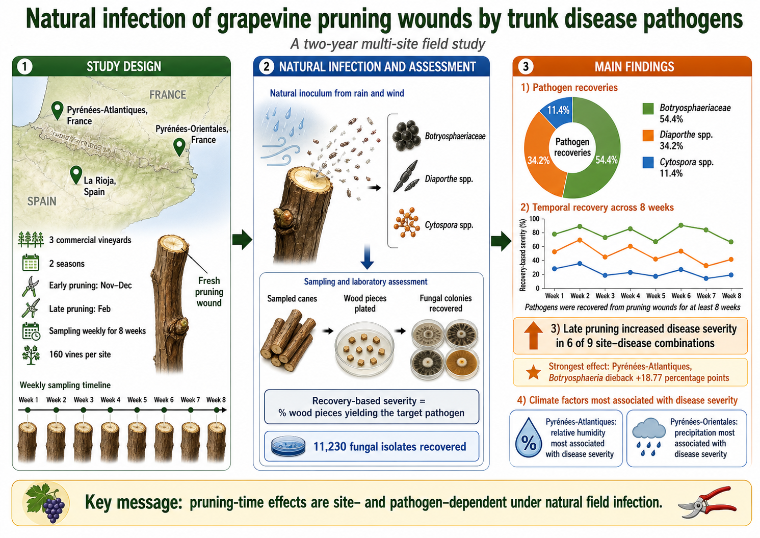
Grapevine trunk diseases (GTDs) are major constraints to vineyard longevity and productivity worldwide, and pruning wounds are recognized as key infection courts for their causal fungi. However, the dynamics of natural infection after pruning under field conditions remain insufficiently defined. This study evaluated natural infection of grapevine pruning wounds by GTD pathogens in three commercial vineyards in Spain and France over two growing seasons. At each site, vines were pruned in the dormant season either early (November-December) or late (February), and wounds were sampled weekly for 8 weeks. Disease severity was quantified using the percentage of wood pieces yielding GTD pathogens after isolation. A total of 11,230 fungal isolates were recovered, of which Botryosphaeriaceae accounted for 54.4%, followed by Diaporthe spp. (34.2%) and Cytospora spp. (11.4%). Disease severity varied significantly over time in all site-disease combinations, and temporal trajectories differed with pruning time and season. Late pruning resulted in significantly greater disease severity than early pruning in 6 of 9 site-disease combinations. The strongest effect was observed in Pyrénées-Atlantiques for Botryosphaeria dieback, where late pruning increased severity by 18.77 %; Cytospora canker at the same site increased by 7.24 %. Climatic analyses revealed site-specific associations, with relative humidity most strongly associated with disease severity in Pyrénées-Atlantiques and precipitation in Pyrénées-Orientales. These results indicate that GTD pathogens can be recovered from pruning wounds for at least 8 weeks after pruning and that the effect of pruning time depends strongly on vineyard and pathogen group.
Keywords: 
;  ;  ;  ;  ;  ;  

Introduction

Grapevine trunk diseases (GTDs) represent a complex of fungal diseases that threaten viticulture worldwide, causing progressive vine decline, reduced productivity, and premature vineyard death (Gramaje et al., 2018). These diseases are caused by diverse fungal taxa, including members of Botryosphaeriaceae associated with Botryosphaeria dieback (Úrbez-Torres, 2011), Diaporthe spp. associated with Phomopsis dieback (Úrbez-Torres et al., 2013), Cytospora spp. associated with Cytospora canker (Lawrence et al., 2017), Diatrypaceae associated with Eutypa dieback (Trouillas et al., 2010), and esca-associated fungi (Mugnai et al., 1999), among others. GTDs are characterized by long latency periods, multiple infection pathways, and complex within-vine progression patterns that make them particularly challenging to manage (Gramaje et al., 2018).
Pruning wounds are widely recognized as the primary infection portals for most GTD pathogens (Ojeda et al., 2026). During dormant-season pruning, fresh wounds expose vascular tissues to fungal propagules dispersed by rain splash, wind, and arthropods (Moyo et al., 2014; Ji et al., 2024). The duration of wound susceptibility and the factors influencing infection risk have been subjects of considerable research interest, as understanding these dynamics is critical for developing effective disease management strategies (Rosace et al., 2023).
Previous research on pruning wound susceptibility has relied heavily on artificial inoculation studies, in which wounds are experimentally challenged with known inoculum doses. Across studies, susceptibility typically declines with wound age, but reported “meaningful risk windows” range from ~2 weeks to >2 months, and in some cases low-level susceptibility has been detected up to ~4 months under artificial inoculation (Eskalen et al., 2007; Elena and Luque, 2016; Díaz and Latorre, 2022; Sosnowski et al., 2023). A quantitative meta-analysis highlighted this variance, with susceptibility spanning less than one week to more than eight weeks depending on study context (Rosace et al., 2023). Moreover, these studies typically target specific pathogens individually, whereas natural infection involves simultaneous exposure to mixed inoculum from multiple GTD agents.
Several interacting factors likely contribute to this variability. (i) Pathogen biology: GTD causal agents differ in their ability to colonize aging wounds . (ii) Climate and wound healing: temperature and moisture conditions influence wound desiccation and callusing, as well as the balance between host defenses and pathogen growth; warmer conditions may accelerate healing, but may also coincide with spore release depending on region. (iii) Inoculum pressure and methodology: high-dose artificial inoculations can detect residual susceptibility that may be less relevant under natural infection, whereas high natural disease pressure can mask treatment effects and complicate interpretation. (iv) Assessment approach: outcomes differ when relying on culture-based recovery versus symptom incidence, and when measuring establishment versus downstream necrosis.
Conflicting recommendations on early, standard, or late pruning largely reflect genuine regional differences in inoculum phenology, rainfall-driven dispersal, and temperature-dependent wound healing (Serra et al., 2008; Úrbez-Torres and Gubler, 2011; Luque et al., 2014; Martínez-Diz et al., 2020; Sosnowski et al., 2023). In some regions, delaying pruning can reduce infection risk because the high-inoculum period precedes late-winter pruning and/or warmer conditions shorten susceptibility (Úrbez-Torres and Gubler, 2011; Sosnowski et al., 2023). In other regions, pathogen detection and infection pressure may remain high later in winter, making early pruning preferable or making timing-based decisions alone insufficient (Luque et al., 2014; Martínez-Diz et al., 2020). Moreover, pathogen groups differ: patterns observed for Botryosphaeriaceae may not transfer directly to esca-associated pathogens, and year-to-year variability can be large (Serra et al., 2008).
Although controlled inoculation experiments have provided valuable mechanistic insights into pruning-wound infection biology, they may not fully reflect natural infection processes. Artificial inoculation typically involves high inoculum concentrations applied directly to wounds, potentially overestimating infection risk compared to natural exposure where spore arrival is stochastic and dependent on environmental conditions (Carter, 1991; Pearson, 1980). Furthermore, controlled studies often focus on single pathogens, missing the multi-pathogen competition and synergistic interactions that occur under field conditions. Consequently, the temporal dynamics of pruning wound susceptibility to natural, mixed-inoculum infection remain poorly characterized, limiting our ability to develop evidence-based pruning timing recommendations applicable across diverse vineyard environments.
The objectives of this study were to: (1) characterize temporal patterns of natural infection of grapevine pruning wounds by GTD pathogens; (2) compare disease severity between early pruning (November-December) and late pruning (February); (3) characterize the prevalence and distribution of GTD pathogens across contrasting vineyard environments; and (4) identify climatic variables associated with disease severity under field conditions. By conducting this study under natural infection conditions across multiple sites and years, we aimed to provide ecologically relevant data that better reflect the complexity of GTD epidemiology in commercial vineyards.
Materials and Methods
Experimental sites and design. This study was conducted over two consecutive growing seasons (2024-2025 and 2025-2026; hereafter referred to as Season 1 and Season 2) in three commercial vineyards located in Spain and France. Vineyard 1 was located in Logroño (La Rioja, Spain; 42.4666°N, 2.2927°W). The vineyard was planted in 1991 with Vitis vinifera cv. Garnacha Tinta grafted onto 110 Richter rootstock, with a planting density of 4,000 vines/ha. Vines were trained using a double cordon Royat system and spur-pruned. Approximately 50% of vines showed dead arms and spurs in recent years, and tiger-stripe foliar symptoms of esca were observed at low incidence (<5%). Vineyard 2 was located in Madiran (Pyrénées-Atlantiques, France; 43.5431°N, 0.1562°W). The vineyard was planted in 1996 with V. vinifera cv. Tannat grafted onto 3309 rootstock, with a planting density of 5,000 vines/ha. Vines were trained using a single Guyot system and cane-pruned. Approximately 15% of vines exhibited dead arms and spurs in recent years. Vineyard 3 was located in Le Roussillon (Pyrénées-Orientales, France; 42.802473°N, 2.840338°W). The vineyard was planted in 2009 with V. vinifera cv. Grenache noir grafted onto 110 Richter rootstock, with a planting density of 4,000 vines/ha. Vines were trained using a double cordon Royat system and spur-pruned. Approximately 10% of vines showed tiger-stripe foliar symptoms of esca in recent years.
At each site, 160 vines were randomly selected and arranged across four rows. Two pruning times were evaluated: early dormant-season pruning (November-December, depending on site) and late dormant-season pruning (February). For each pruning time, 80 vines were randomly assigned to eight groups of 10 vines each, with each group designated for sampling at a specific week during the eight-week monitoring period. This design ensured that each vine was sampled only once, avoiding repeated wounding stress on the same plant. Pruning was performed to three buds using secateurs in all vineyards, leaving a 5- to 7-cm stub above the uppermost bud as described by Martínez-Diz et al. (2021). Pruning secateurs were disinfected with 70% ethanol before and after every pruning cut. No wound protectants or fungicides were applied to pruning wounds, allowing natural infection to occur. Pruning was conducted on commercial dates specific to each vineyard and season: in La Rioja, early pruning was performed on 4 November 2024 and 3 November 2025, and late pruning on 3 February 2025 and 2 February 2026; in the Pyrénées-Atlantiques, early pruning was performed on 4 December 2024 and 3 December 2025, and late pruning on 7 February 2025 and 7 February 2026; and in the Pyrénées-Orientales, early pruning was performed on 13 November 2024 and 18 November 2025, and late pruning on 12 February 2025 and 18 February 2026. At the time of pruning, 5-cm wood segments were collected from three canes per plant, preserved, and transported to the laboratory to assess the presence of wood-inhabiting fungal pathogens in one-year-old shoots.
Following pruning, wounds were monitored weekly for eight consecutive weeks (Week 1 through Week 8 post-pruning). At each sampling time, three canes per plant were harvested from the designated group of 10 plants (30 canes total per site × season × pruning time × week combination). Cane segments were collected from the portion immediately above the second bud (about 10-cm-long pieces). This sampling scheme resulted in a total of 2,880 cane samples across the entire study (3 sites × 2 seasons × 2 pruning times × 8 weeks × 30 canes per sampling). Harvested canes were stored at 4°C prior to laboratory assessment.
Fungal recovery and identification. In the laboratory, canes were surface-disinfected by immersion in 70% ethanol for 30 s, followed by 1% sodium hypochlorite for 2 min, and then rinsed twice with sterile distilled water. The bark was removed aseptically, and the wood was sectioned into small pieces (approximately 5 mm × 5 mm). Ten wood pieces per cane were plated onto two plates of potato dextrose agar (PDA) amended with streptomycin sulfate (100 mg/L) to inhibit bacterial growth (5 wood pieces per plate). Plates were incubated at 25°C in darkness and examined daily for fungal growth over a 3-week period.
Emerging fungal colonies were subcultured onto fresh PDA to obtain pure cultures. Morphological characteristics were recorded, and representative isolates from each morphotype were selected for molecular identification. Fungal DNA was extracted from fresh mycelium after 3 weeks of incubation on PDA using the E.Z.N.A. Plant Miniprep Kit (Omega Bio-Tek, Doraville, GA, USA) following the manufacturer’s instructions. Initially, fungal identity was determined by sequencing the internal transcribed spacer (ITS) region using primers ITS1-F and and ITS4 (Gardes and Bruns, 1993;White et al., 1990). For specific fungal groups, additional informative loci were sequenced (Gramaje et al., 2025). For Botryosphaeriaceae and Cytospora spp., primers EF1-728F and EF1-986R (Carbone and Kohn, 1999) were used to amplify part of the translation elongation factor 1-alpha gene (tef1). The beta-tubulin (tub2) region was amplified using primers Bt2a and Bt2b (Glass and Donaldson, 1995) for Diaporthe spp. All PCR products were visualized on 1% agarose gels (agarose D-1 Low EEO, Conda Laboratories) and sequenced in both directions by Eurofins GATC Biotech (Cologne, Germany). Sequences were compared against the NCBI GenBank database using BLASTn, and species identifications were confirmed based on ≥98% sequence similarity to reference strains.
Disease severity was calculated as the percentage of infected wood pieces yielding each GTD pathogen at each sampling time: (number of wood pieces yielding the GTD pathogen / total number of wood pieces plated) × 100. The experimental unit for disease severity was the vine. Severity was first calculated for each cane (10 wood pieces per cane) and then averaged across the three canes sampled per plant, resulting in one plant-level severity value. Consequently, each sampling time yielded 10 replicate plant-level disease severity values per pathogen (one per plant). These plant-level values were used as the response variable in all statistical analyses. Disease incidence was calculated as the percentage of pruning wounds (vines) showing any disease infection (severity > 0) for each pathogen at each site and season: (number of vines with severity > 0 / total number of vines assessed) × 100. This metric complemented mean severity by indicating the proportion of wounds successfully colonized by each pathogen, regardless of the degree of colonization.
Climatic data and vineyard management. Standard cultural practices were used in the vineyards during the growing season, and management of both powdery and downy mildews was performed using only wettable sulphur and copper compounds applied at label dosages and following Integrated Pest Management (IPM) guidelines. Each vineyard was located less than 2 km from an automatic weather station, and its climatic data was considered to be representative of the environmental conditions at each site.
Climatic variables recorded daily included maximum temperature (Tmax, °C), mean temperature (Tmean, °C), minimum temperature (Tmin, °C), relative humidity (HR, %), precipitation (P, mm), and cumulative precipitation (Pcum, mm). Weekly averages were calculated for temperature and relative humidity variables, while precipitation was summed weekly. These data were used to explore potential relationships between environmental conditions and disease severity.
To account for the delay between infection, colonization, and pathogen recovery, lagged climatic variables were created. Specifically, 1-week lag (lag-1) and 2-week lag (lag-2) variables were generated, representing climatic conditions from one and two weeks prior to disease assessment, respectively. This approach allowed us to evaluate whether environmental conditions preceding fungal colonization were more strongly associated with disease severity than concurrent conditions.
Statistical analysis. Disease severity data were analyzed separately for each site-disease combination using three-way fixed-effects linear models. The primary model for each combination was specified as:
Severity ~ Pruning Time × Season × Week
where Pruning Time (Early vs. Late), Season (Season 1 vs. Season 2), and Week (1-8) were included as fixed effects. Week was treated as a categorical rather than continuous variable to allow detection of non-linear temporal patterns in disease severity. Because each vine was sampled only once and assigned to a single weekly sampling group, no repeated-measures structure or random plant effect was included. Models were fitted in R v4.5.3 (R Core Team, 2026) using the lm() function. Because the design was fully balanced and residual diagnostics indicated no major departures from normality or homoscedasticity, fixed-effects linear models were considered appropriate for inference on disease severity. Model assumptions were evaluated by visual inspection of residual and normal Q-Q plots.
For each site-disease combination, Type II ANOVA was performed using the car package (version 3.1-2) to test the significance of the three main effects (Pruning Time, Season, and Week), the three two-way interactions (Pruning Time × Season, Pruning Time × Week, and Season × Week), and the three-way interaction (Pruning Time × Season × Week).
Estimated marginal means (EMMs) for Early and Late pruning were calculated from the fitted models using the emmeans package (version 1.10.0), averaging predictions across all weeks and seasons. Differences between pruning times were expressed as mean differences (Late - Early pruning) and summarized with 95% confidence intervals. Cohen’s d was calculated as a standardized effect size using the pooled residual standard deviation from the fitted model. Effect sizes were interpreted using conventional thresholds: |d| < 0.2, negligible; 0.2-0.5, small; 0.5-0.8, medium; 0.8-1.2, large; and >1.2, very large.
For graphical presentation, observed means and standard errors were calculated for each Week × Pruning Time × Season combination from vine-level data. Statistical inference for temporal effects was based on the corresponding Type II ANOVA results from the fixed-effects models.
As a separate exploratory analysis, associations between climatic variables and disease severity were examined using Spearman rank correlations. Climatic variables included maximum temperature (Tmax), mean temperature (Tmean), minimum temperature (Tmin), relative humidity (HR), and precipitation (P), each evaluated for the current week, 1-week lag, and 2-week lag. To ensure independence of observations, disease severity was aggregated to weekly means for each unique Site × Season × Pruning Time × Week × Disease combination. This aggregation yielded up to 32 independent observations per site–disease combination (2 seasons × 2 pruning times × 8 weeks). P-values were adjusted for multiple testing within each site-disease combination using the Benjamini-Hochberg procedure to control the false discovery rate at α = 0.05. All statistical analyses were conducted in R v4.5.3 (R Core Team, 2026), and significance was declared at α = 0.05.

Results

Fungal Recovery and Identification. Over the 2-year survey, 11,230 fungal isolates were recovered from isolated wood pieces. Based on colony morphology and DNA sequencing, isolates were assigned to three major GTDs: Phomopsis dieback (Diaporthe spp.), Botryosphaeria dieback (Botryosphaeriaceae spp.), and Cytospora canker (Cytospora spp.). Botryosphaeriaceae accounted for the largest proportion of recoveries (6,107 isolates; 54.4%), followed by Diaporthe spp. (3,839; 34.2%) and Cytospora spp. (1,284; 11.4%).
Isolate composition differed among vineyards. In La Rioja, 3,394 isolates were recovered, mainly Diaporthe spp. (1,662; 49.0%) and Botryosphaeriaceae (1,471; 43.3%), whereas Cytospora spp. were less frequent (261; 7.7%) and were not detected in the early-pruning period of Season 1. In Pyrénées-Atlantiques, 3,078 isolates were obtained, with Botryosphaeriaceae predominating (1,655; 53.8%), followed by Diaporthe spp. (833; 27.1%) and Cytospora spp. (590; 19.2%). In Pyrénées-Orientales, the highest number of isolates was recovered (4,758), again dominated by Botryosphaeriaceae (2,981; 62.7%), followed by Diaporthe spp. (1,344; 28.2%) and Cytospora spp. (433; 9.1%).
Across sites, isolate recovery was generally greater after late pruning than after early pruning (February vs. November-December totals: 1,890 vs. 1,504 in La Rioja; 2,312 vs. 766 in Pyrénées-Atlantiques; 2,673 vs. 2,085 in Pyrénées-Orientales). Within Botryosphaeria dieback, species composition varied among vineyards. In La Rioja, Botryosphaeriaceae isolates were identified as Diplodia seriata (63%), Neofusicoccum parvum (18%), N. luteum (11%), and Botryosphaeria dothidea (8%). In Pyrénées-Atlantiques, isolates were assigned to D. seriata (83%), N. parvum (14%), and B. dothidea (3%). In Pyrénées-Orientales, isolates belonged to D. seriata (77%) and B. dothidea (23%). Within Phomopsis dieback, most isolates were identified as Diaporthe ampelina (92% in La Rioja, 79% in Pyrénées-Atlantiques, and 81% in Pyrénées-Orientales). Within Cytospora canker, most isolates were identified as Cytospora viticola (96% in La Rioja, 90% in Pyrénées-Atlantiques, and 97% in Pyrénées-Orientales). The remaining isolates were assigned only to Diaporthe spp. or Cytospora spp., and additional loci would be required for reliable species-level identification.
Other trunk-disease fungi were occasionally isolated, but at low incidence, precluding meaningful statistical analysis. In La Rioja, 106 isolates of Phaeomoniella chlamydospora, 26 of Phaeoacremonium minimum, and 27 of Eutypa lata were recovered. In Pyrénées-Atlantiques, 31 isolates of P. chlamydospora, 41 of E. lata, and 12 of Eutypella vitis were obtained. In Pyrénées-Orientales, 61 isolates of P. chlamydospora were recovered. Notably, among wood fragments collected from one-year-old pruning shoots, only fungi associated with Botryosphaeria dieback were recovered, and severity in these tissues was low, ranging from 0.6% in La Rioja to 1.4% in Pyrénées-Orientales.
Disease Incidence and Severity. Disease incidence and severity differed among sites, diseases, and seasons (Table 1). In La Rioja, Phomopsis dieback showed the highest mean severity (17.5% in Season 1; 17.1% in Season 2) and was nearly ubiquitous (100.0% and 98.8% incidence, respectively). Botryosphaeria dieback was similarly widespread (98.8-99.4%) with comparable mean severities (15.5% in Season 1; 15.2% in Season 2). Cytospora canker remained low in severity (1.4% in Season 1; 4.1% in Season 2), but incidence increased from 27.5% in Season 1 to 82.5% in Season 2.
In Pyrénées-Atlantiques, Botryosphaeria dieback was the dominant disease in Season 1 (mean severity 19.2%; incidence 88.1%). Phomopsis dieback showed the largest seasonal increase, from 1.7% severity and 18.1% incidence in Season 1 to 15.9% severity and 97.5% incidence in Season 2. Cytospora canker increased from 2.5% severity and 26.2% incidence in Season 1 to 9.9% severity and 90.0% incidence in Season 2.
Pyrénées-Orientales showed the highest overall disease severity. Botryosphaeria dieback reached mean severities of 35.0% in Season 1 and 27.2% in Season 2, with incidences of 99.4% and 100.0%, respectively. Phomopsis dieback increased from 7.7% severity and 73.1% incidence in Season 1 to 20.4% severity and 100.0% incidence in Season 2. Cytospora canker remained comparatively low in both seasons (1.2% in Season 1 and 7.8% in Season 2).
Temporal Dynamics of Disease Severity. The three-way fixed-effects linear models revealed significant temporal variation in disease severity across all nine site-disease combinations (Week main effect: F = 10.46-76.13, p < 0.001 in all cases; Table 2). Temporal patterns also varied with pruning time and season, as indicated by significant Pruning Time × Week interactions in all nine combinations (F = 11.80-45.80, p ≤ 0.044), significant Season × Week interactions in all nine combinations (F = 6.89-50.00, p < 0.001), and significant Pruning Time × Season × Week interactions in all nine combinations (Table 2).
Weekly trajectories differed among vineyards and diseases (Figs. 1-3). In La Rioja, temporal variation was moderate for Botryosphaeria dieback and Phomopsis dieback, whereas Cytospora canker remained low overall except in late-pruned vines during Season 1 (Figure 1). In Pyrénées-Atlantiques, Botryosphaeria dieback showed the clearest separation between pruning times, particularly in Season 1, with consistently higher severity in late-pruned vines across most sampling dates (Figure 2). In Pyrénées-Orientales, temporal patterns were more variable, with pronounced mid-season peaks for Botryosphaeria dieback and Phomopsis dieback and shifts in peak timing between pruning times and seasons (Figure 3). For example, Botryosphaeria dieback reached maximum severity later in late-pruned than in early-pruned vines during Season 2, whereas in Season 1 both pruning times converged at similar severity around Week 4.
Across sites, temporal differences between pruning times were expressed not only as changes in magnitude but also as differences in trajectory shape, including earlier or later peaks, transient convergence between pruning treatments, and season-specific reversals in relative severity. These patterns were also evident in the supplementary heatmaps, which highlighted the week-specific distribution of disease severity within each site, season, and pruning treatment (Supplementary Figs. S1-S3). Overall, disease severity changed substantially during the 8 weeks after pruning, and both the shape and timing of these temporal patterns depended on vineyard, season, and pruning time.
Pruning Time Effects. Late pruning significantly increased recovery-based disease severity in 6 of 9 site-disease combinations (Figure 4; Table 3). Mean differences between late and early pruning, based on estimated marginal means averaged across weeks and seasons, ranged from 0.58 to 18.77 percentage points, and Cohen’s d values ranged from 0.09 to 1.71.
The strongest pruning-time effect was observed in Pyrénées-Atlantiques for Botryosphaeria dieback, where late pruning increased severity by 18.77 % (16.35 to 21.19; p < 0.001; Cohen’s d = 1.715). At the same site, late pruning also increased severity of Cytospora canker by 7.24 % (95% CI, 5.54 to 8.94; p < 0.001) and Phomopsis dieback by 6.52 % (95% CI, 4.19 to 8.85; p < 0.001).
In La Rioja, significant but smaller increases were detected for Botryosphaeria dieback (3.26 %; , 1.33 to 5.19; p < 0.001) and Phomopsis dieback (4.10 %; , 1.86 to 6.34; p < 0.001), whereas Cytospora canker was not significantly affected by pruning time. In Pyrénées-Orientales, a significant increase was detected only for Botryosphaeria dieback (10.27 %; , 6.49 to 14.04; p < 0.001), whereas differences for Phomopsis dieback and Cytospora canker were not significant.
Overall, late pruning was frequently associated with greater recovery-based disease severity, although the magnitude and significance of this effect varied among vineyards and pathogen groups.
Climatic Drivers of Disease Severity. Exploratory Spearman correlation analyses revealed site-specific associations between climatic variables and recovery-based disease severity (Figs. 5-7; Supplementary Table S1). Across all sites and diseases, 14 correlations remained significant after Benjamini-Hochberg correction. Because these analyses were based on aggregated weekly means, they should be interpreted cautiously.
At La Rioja, only one correlation remained significant after correction: Botryosphaeria dieback severity was positively associated with maximum temperature lagged by 1 week (ρ = 1.000, p < 0.001; Figure 5). No robust climatic associations were detected for Cytospora canker or Phomopsis dieback at this site.
Figure 5. Spearman rank correlations between climatic variables and recovery-based disease severity at La Rioja. Correlations were calculated using weekly mean severity aggregated by Site × Season × Pruning Time × Week × Disease. P-values were adjusted using the Benjamini-Hochberg procedure within each site–disease combination. Tmax = maximum temperature; Tmean = mean temperature; Tmin = minimum temperature; HR = relative humidity; P = precipitation.
Preprints 211964 g005
Figure 6. Spearman rank correlations between climatic variables and recovery-based disease severity at Pyrénées-Atlantiques. Correlations were calculated using weekly mean severity aggregated by Site × Season × Pruning Time × Week × Disease. P-values were adjusted using the Benjamini-Hochberg procedure within each site–disease combination. Tmax = maximum temperature; Tmean = mean temperature; Tmin = minimum temperature; HR = relative humidity; P = precipitation.
Preprints 211964 g006
Figure 7. Spearman rank correlations between climatic variables and recovery-based disease severity at Pyrénées-Orientales. Correlations were calculated using weekly mean severity aggregated by Site × Season × Pruning Time × Week × Disease. P-values were adjusted using the Benjamini-Hochberg procedure within each site–disease combination. Tmax = maximum temperature; Tmean = mean temperature; Tmin = minimum temperature; HR = relative humidity; P = precipitation.
Preprints 211964 g007
In Pyrénées-Atlantiques, the strongest associations involved relative humidity. Cytospora canker severity was negatively correlated with relative humidity at the current week and at both lag periods (ρ = −0.638 to −0.565), and Phomopsis dieback showed a similar pattern (ρ = −0.556 to −0.545). Positive associations with temperature were also detected, although these were weaker. Botryosphaeria dieback severity was positively associated with current-week maximum temperature, and Phomopsis dieback was positively associated with mean temperature. However, no significant associations with precipitation remained after correction.
In Pyrénées-Orientales, precipitation was the main climatic variable associated with disease severity. Cytospora canker severity was positively correlated with precipitation at the current week and at both lag periods (ρ = 0.561-0.643), and Phomopsis dieback severity was positively associated with precipitation at the 2-week lag (ρ = 0.651). No significant climatic associations were detected for Botryosphaeria dieback after correction.
Overall, the climatic analyses indicate that associations between weather conditions and recovery-based disease severity were site-dependent, with relative humidity most strongly associated with disease severity in Pyrénées-Atlantiques and precipitation in Pyrénées-Orientales.

Discussion

This study extends the still limited body of field-based work on natural infection of grapevine pruning wounds by GTD fungi by assessing three diseases across three commercial vineyards, two growing seasons, two pruning times, and eight consecutive weeks after pruning. Most previous studies on pruning-wound susceptibility have relied on artificial inoculation, have focused on single pathogens, or have been conducted in a single region (Eskalen et al. 2007; Elena and Luque 2016; Úrbez-Torres and Gubler 2011; Díaz and Latorre 2022; Sosnowski et al. 2023). Building on this, our design integrated natural inoculum pressure, mixed pathogen communities, and local environmental variability under commercial vineyard conditions. Additonally, our results complement previous natural infection studies in Europe (Luque et al. 2014; Martínez-Diz et al. 2020) by providing weekly resolution during the first 8 weeks after pruning and by simultaneously examining Botryosphaeriaceae, Diaporthe, and Cytospora in contrasting viticultural environments.
A central result was that recovery-based disease severity changed significantly over time in all site-disease combinations, but the temporal trajectories were not uniform. Rather than showing a simple monotonic decline, disease severity often displayed intermediate peaks, shifts in peak timing, temporary convergence between pruning treatments, or season-specific reversals in the relative severity of early- and late-pruned wounds. These findings reinforce the general view that pruning wounds remain vulnerable for weeks to months after pruning (Eskalen et al. 2007; Serra et al. 2008; Elena and Luque 2016; Díaz and Latorre 2022; Rosace et al. 2023; Sosnowski et al. 2023), but they also show that under natural infection the temporal expression of this risk window is more irregular than the smoother decline curves often observed in artificially pathogen inoculation studies.
Importantly, these nonmonotonic temporal patterns should not be interpreted as evidence that pruning wound receptivity increased with wound age. Artificial inoculation studies consistently indicate that wound susceptibility generally declines over time as healing progresses, likely through anatomical and biochemical processes such as tylosis formation, suberization, and lignification (Biggs 1990; Munkvold and Marois 1995; Sun et al. 2006; Sosnowski et al. 2023). Chapuis et al. (1998), working in south-western France, likewise showed that pruning wounds inoculated with E. lata were most susceptible after early pruning and that susceptibility declined as wounds aged, with a shorter susceptible period after later pruning. However, those studies quantify infection success after application of a defined inoculum dose to wounds of known age, thereby isolating the effect of wound age on host receptivity. Under natural infection, by contrast, wounds remain continuously exposed to fluctuating inoculum pressure and variable weather conditions throughout the post-pruning period. Thus, the weekly recovery-based severity observed here reflects the net outcome of at least three interacting processes: progressive wound healing, temporal variation in inoculum availability, and weather-dependent opportunities for dispersal, infection, and early colonization. Intermediate peaks or later increases in pathogen recovery are therefore biologically plausible even if intrinsic wound receptivity declines over time. In this sense, our findings do not contradict artificial inoculation studies, but rather complement them by showing how declining wound receptivity is modulated in the field by changing infection pressure and environmental conditions. The observations of Chapuis et al. (1998) also suggest an additional possibility, namely that natural wound colonizers may contribute to these dynamics by competing with pathogens, further decoupling field recovery patterns from the simpler decline expected from wound age alone.
The effect of pruning time was one of the clearest outcomes of the study. Late pruning increased recovery-based disease severity in six of nine site-disease combinations, with the strongest effect observed for Botryosphaeria dieback in Pyrénées-Atlantiques. This result agrees with field-based studies indicating that late pruning can increase natural wound colonization in some regions (van Niekerk et al. 2011; Luque et al. 2014; Martínez-Diz et al. 2020; Mutawila et al. 2016), but contrasts with studies from other viticultural areas where delayed pruning reduced infection risk or shortened the effective susceptibility period (Moller and Kasimatis 1981; Petzoldt et al. 1981; Munkvold and Marois 1995; Úrbez-Torres and Gubler 2011; Sosnowski et al. 2023). Taken together, these comparisons confirm that the epidemiological value of early versus late pruning is not universal. Its effect depends on how pruning timing overlaps with local periods of inoculum availability and with weather conditions favoring dispersal, germination, and wound colonization.
In our study, the largest pruning-time effects occurred in Pyrénées-Atlantiques, suggesting that late pruning in this Atlantic environment exposed fresh wounds to particularly favorable infection conditions. This interpretation is biologically consistent with aerobiological work showing that spore dispersal of GTD fungi is often driven by rain, humidity, and temperature, but that the strength and timing of these drivers vary by region and pathogen group (van Niekerk et al. 2010; Úrbez-Torres et al. 2010; Valencia et al. 2015; González-Domínguez et al. 2020; Ji et al. 2024). Thus, the higher recovery-based severity observed after late pruning at this site likely reflects not pruning date , but the coincidence of fresh wounds with more favorable infection conditions, including higher inoculum availability and a more infection-conducive environment. This interpretation also helps explain why pruning-time effects were smaller and less consistent in La Rioja and more selective in Pyrénées-Orientales.
Botryosphaeriaceae were the dominant pathogen group recovered in this study, accounting for more than half of all isolates, and D. seriata was the most frequently identified species. This pattern agrees with previous surveys and natural infection studies in Mediterranean and Atlantic vineyards, where Botryosphaeriaceae, and especially D. seriata, are recurrent and often dominant wound colonizers (Úrbez-Torres 2011; Rolshausen et al. 2010; Luque et al. 2014; Leal et al. 2024). Diaporthe was also highly prevalent, confirming that Phomopsis dieback should not be regarded as a minor component of the trunk disease complex in these vineyards. By contrast, Cytospora represented a smaller overall proportion of isolates but showed pronounced variation among sites and seasons. These differences in pathogen composition indicate that pruning wounds are exposed not to a single risk, but to a local pathogen assemblage whose structure may differ substantially among vineyards and may influence both colonization dynamics and the apparent effect of pruning date.
The three diseases also differed in their response to pruning time. Botryosphaeria dieback showed the strongest and most consistent increase after late pruning, whereas Phomopsis dieback and Cytospora canker were more variable among sites and seasons. This result fits well with experimental evidence showing that Botryosphaeriaceae are highly efficient colonizers of fresh pruning wounds and can establish rapidly under favorable conditions (Rolshausen et al. 2010; Úrbez-Torres and Gubler 2011; Elena and Luque 2016). Our data therefore support a broader epidemiological conclusion: pruning date does not affect all GTDs equally. Management decisions based on pruning timing alone may therefore be more effective against some pathogen groups than against others, especially where Botryosphaeriaceae dominate the wound-colonizing community.
The climatic analyses further emphasized this context dependence. Associations between weather variables and recovery-based disease severity were clearly site specific. In Pyrénées-Atlantiques, the clearest associations involved relative humidity and temperature, whereas in Pyrénées-Orientales precipitation was the main correlate. In La Rioja, climatic associations were weak, and the only significant correlation after correction was based on very few independent observations, so it should not be overinterpreted. These results should be regarded as observational rather than mechanistic, but they are consistent with the broader literature showing that weather may influence GTD infection through several linked processes, including inoculum production, spore dispersal, wound wetness, and subsequent colonization (van Niekerk et al. 2010; Úrbez-Torres et al. 2010; Valencia et al. 2015; González-Domínguez et al. 2020). In this context, Ji et al. (2024) are especially informative because their quantitative synthesis showed that rainfall is a good predictor of spore trapping for fungi associated with Botryosphaeria dieback and Eutypa dieback, but a much weaker predictor for ascomycetes associated with the esca complex. This supports a cautious interpretation of our climatic correlations: a significant association with rainfall or relative humidity may reflect dispersal, wound wetness, colonization success, or several of these processes acting together. It also reinforces that the climatic drivers of infection risk are pathogen dependent rather than universal.
The response variable used here also deserves careful interpretation. Recovery-based disease severity, defined as the percentage of wood pieces yielding the target pathogen, reflects colonization success at the time of sampling rather than final canker development, lesion length, or long-term symptom expression. It is therefore best interpreted as a measure of early infection or colonization intensity, not of eventual severity. This distinction is important because culture-based recovery may underestimate latent or low-biomass infections relative to molecular detection methods (Pouzoulet et al. 2017). Even so, recovery-based severity remains a useful epidemiological metric because it captures biologically meaningful differences in the extent to which pathogens become established in wounded wood under field conditions.
Several limitations should be acknowledged. First, the study covered two seasons and three vineyards, and the sites differed not only in climate but also in cultivar, rootstock, vine age, and training system. Therefore, although the within-site comparison of pruning times is robust, cross-site contrasts must be interpreted with caution. Second, we did not directly quantify airborne inoculum, so we cannot separate the relative contribution of spore availability from wound receptivity. Third, we did not directly assess wound-healing dynamics, nor did we use molecular methods that might detect low-abundance or non-culturable infections. Future studies combining spore trapping, molecular pathogen quantification, and direct assessment of wound healing would help disentangle these mechanisms more clearly and improve interpretation of climate-driven risk.
Despite these limitations, the practical implications are clear. A single recommendation on pruning date is unlikely to be valid across viticultural regions or pathogen groups. In our study, late pruning frequently increased recovery-based disease severity, particularly for Botryosphaeria dieback in the Atlantic site, indicating that delayed pruning may increase GTD risk where humid post-pruning conditions favor infection. Pruning timing should therefore be viewed as one component of an integrated strategy that also includes sanitation and protection of pruning wounds (Gramaje et al. 2018). More broadly, our results support moving away from generalized pruning recommendations toward region-specific GTD management based on local pathogen pressure, local weather patterns after pruning, and the dominant diseases present in each vineyard.

Acknowledgments

We thank the vineyard owners and managers for providing access to field sites and for their cooperation throughout the study. We acknowledge the technical assistance of laboratory staff in fungal isolation and molecular identification. This research was supported by project EFA 033/01 - VITRES, which was co-financed at 65% by the European Regional Development Fund (ERDF) through the Interreg V-A Spain-France-Andorra Programme (POCTEFA 2021–2027). K. Ashley gratefully acknowledges financial support for this project by the Fulbright U.S. Scholar Program, which is sponsored by the U.S. Department of State and The Spain Fulbright Commission.

Conflicts of Interest

The authors declare no conflict of interest.

References

  1. Biggs, A.R. Managing wound-associated diseases by understanding wound healing in the bark of woody plants. J. Arboric. 1990, 16, 108–112. [Google Scholar] [CrossRef]
  2. Carbone, I.; Kohn, L. M. A method for designing primer sets for speciation studies in filamentous ascomycetes. Mycologia 1999, 91, 553–556. [Google Scholar] [CrossRef]
  3. Carter, M. V. The Status of Eutypa lata as a Pathogen. In Monogr. Phytopathol. Pap. No. 32.; CAB International Mycological Institute: Wallingford, Oxfordshire, U.K, 1991. [Google Scholar]
  4. Chapuis, L.; Richard, L.; Dubos, B. Variation in susceptibility of grapevine pruning wound to infection by Eutypa lata in south-western France. Plant Pathol. 1998, 47, 463–472. [Google Scholar] [CrossRef]
  5. Díaz, G. A.; Latorre, B. A. Duration of the susceptibility of pruning wounds of different ages to infections by Phaeomoniella chlamydospora on grapevine cv. Cabernet Sauvignon in central Chile. Front. Fungal Biol. 2022, 3, 1026516. [Google Scholar] [CrossRef]
  6. Elena, G.; Luque, J. Seasonal susceptibility of grapevine pruning wounds and cane colonization in Catalonia, Spain, following artificial infection with Diplodia seriata and Phaeomoniella chlamydospora. Plant Dis. 2016, 100, 1651–1659. [Google Scholar] [CrossRef] [PubMed]
  7. Eskalen, A.; Feliciano, A. J.; Gubler, W. D. Susceptibility of grapevine pruning wounds and symptom development in response to infection by Phaeoacremonium aleophilum and Phaeomoniella chlamydospora. Plant Dis. 2007, 91, 1100–1104. [Google Scholar] [CrossRef]
  8. Gardes, M.; Bruns, T. D. ITS primers with enhanced specificity for basidiomycetes: application to the identification of mycorrhizae and rusts. Mol. Ecol. 1993, 2, 113–118. [Google Scholar] [CrossRef]
  9. Glass, N. L.; Donaldson, G. C. Development of primer sets designed for use with the PCR to amplify conserved genes from filamentous ascomycetes. Appl. Environ. Microbiol. 1995, 61, 1323–1330. [Google Scholar] [CrossRef]
  10. González-Domínguez, E.; Berlanas, C.; Gramaje, D.; Armengol, J.; Rossi, V.; Berbegal, M. Temporal dispersal patterns of Phaeomoniella chlamydospora, causal agent of Petri disease and esca, in vineyards. Phytopathology 2020, 110, 1216–1225. [Google Scholar] [CrossRef]
  11. Gramaje, D.; Mostert, L.; Trouillas, F. P.; Úrbez-Torres, J. R.; Alves, A. The most informative loci to identify trunk disease pathogens associated with grapevine and perennial fruit and nut crops. Phytopathol. Mediterr. 2025, 64, 631–636. [Google Scholar] [CrossRef]
  12. Gramaje, D.; Úrbez-Torres, J. R.; Sosnowski, M. R. Managing grapevine trunk diseases with respect to etiology and epidemiology: current strategies and future prospects. Plant Dis. 2018, 102, 12–39. [Google Scholar] [CrossRef] [PubMed]
  13. Ji, T.; Altieri, V.; Salotti, I.; Li, M.; Rossi, V. Role of rain in the spore dispersal of fungal pathogens associated with grapevine trunk diseases. Plant Dis. 2024, 108, 1041–1052. [Google Scholar] [CrossRef]
  14. Lawrence, D. P.; Travadon, R.; Pouzoulet, J.; Rolshausen, P. E.; Wilcox, W. F.; Baumgartner, K. Characterization of Cytospora isolates from wood cankers of declining grapevine in North America, with the descriptions of two new Cytospora species. Plant Pathol. 2017, 66, 713–725. [Google Scholar] [CrossRef]
  15. Leal, C.; Bujanda, R.; López-Manzanares, B.; Ojeda, S.; Berbegal, M.; Villa-Llop, A.; Santesteban, L. G.; Palacios, J.; Gramaje, D. Evaluating treatments for the protection of grapevine pruning wounds from natural infection by trunk disease fungi. Plant Dis. 2024, 108, 3052–3062. [Google Scholar] [CrossRef]
  16. Luque, J.; Elena, G.; Garcia-Figueres, F.; Reyes, J.; Barrios, G.; Legorburu, F. J. Natural infections of pruning wounds by fungal trunk pathogens in mature grapevines in Catalonia (northeast Spain). Aust. J. Grape Wine Res. 2014, 20, 134–143. [Google Scholar] [CrossRef]
  17. Martínez-Diz, M. P.; Díaz-Losada, E.; Díaz-Fernández, A.; Bouzas-Cid, Y.; Gramaje, D. Protection of grapevine pruning wounds against Phaeomoniella chlamydospora and Diplodia seriata by commercial biological and chemical methods. Crop Prot. 2021, 143, 105465. [Google Scholar] [CrossRef]
  18. Martínez-Diz, M. P.; Eichmeier, A.; Spetik, M.; Bujanda, R.; Díaz-Fernández, A.; Díaz-Losada, E.; Gramaje, D. Grapevine pruning time affects natural wound colonization by wood-invading fungi. Fungal Ecol. 2020, 48, 100994. [Google Scholar] [CrossRef]
  19. Moller, W.J.; Kasimatis, A.N. Further evidence that Eutypa armeniacae—not Phomopsis viticola—incites dead arm symptoms on grape. Plant Dis. 1981, 65, 429–431. [Google Scholar] [CrossRef]
  20. Moyo, P.; Allsopp, E.; Roets, F.; Mostert, L.; Halleen, F. Arthropods vector grapevine trunk disease pathogens. Phytopathology 2014, 104, 1063–1069. [Google Scholar] [CrossRef] [PubMed]
  21. Mugnai, L.; Graniti, A.; Surico, G. Esca (black measles) and brown wood-streaking: two old and elusive diseases of grapevines. Plant Dis. 1999, 83, 404–416. [Google Scholar] [CrossRef]
  22. Munkvold, G.P.; Marois, J.J. Factors associated with variation in susceptibility of grapevine pruning wounds to infection by Eutypa lata. Phytopathology 1995, 85, 249–256. [Google Scholar] [CrossRef]
  23. Mutawila, C.; Halleen, F.; Mostert, L. Optimisation of time of application of Trichoderma biocontrol agents for protection of grapevine pruning wounds. Aust. J. Grape Wine Res. 2016, 22, 279–287. [Google Scholar] [CrossRef]
  24. Ojeda, S.; Leal, C.; Diaz-Losada, E.; Gramaje, D. Pruning practices and grapevine trunk diseases: A critical analysis of infection dynamics, management strategies, and research gaps. submitted to preprint.org. 2026.
  25. Pearson, R. C. Discharge of ascospores of Eutypa armeniacae in New York. Plant Dis. 1980, 64, 171–174. [Google Scholar] [CrossRef]
  26. Petzoldt, C.H.; Moller, W.J.; Sall, M.A. Eutypa dieback of grapevine: seasonal differences in infection and duration of susceptibility of pruning wounds. Phytopathology 1981, 71, 540–543. [Google Scholar] [CrossRef]
  27. Pouzoulet, J.; Rolshausen, P. E.; Schiavon, M.; Bol, S.; Travadon, R.; Lawrence, D. P.; Baumgartner, K.; Ashworth, V. E.; Comont, G.; Corio-Costet, M.-F.; Pierron, R. J. G.; Besson, X.; Jacques, A. A method to detect and quantify Eutypa lata and Diplodia seriata-complex DNA in grapevine pruning wounds. Plant Dis. 2017, 101, 1470–1480. [Google Scholar] [CrossRef] [PubMed]
  28. R Core Team. R: A language and environment for statistical computing; R Foundation for Statistical Computing: Vienna, Austria, 2026. [Google Scholar]
  29. Rolshausen, P. E.; Úrbez-Torres, J. R.; Rooney-Latham, S.; Eskalen, A.; Smith, R. J.; Gubler, W. D. Evaluation of pruning wound susceptibility and protection against fungi associated with grapevine trunk diseases. Am. J. Enol. Vitic. 2010, 61, 113–119. [Google Scholar] [CrossRef]
  30. Rosace, M. C.; Legler, S. E.; Salotti, I.; Rossi, V. Susceptibility of pruning wounds to grapevine trunk diseases: a quantitative analysis of literature data. Front. Plant Sci. 2023, 14, 1063932. [Google Scholar] [CrossRef]
  31. Serra, S.; Mannoni, M.; Ligios, V. Studies on the susceptibility of pruning wounds to infection by fungi involved in grapevine wood diseases in Italy. Phytopathol. Mediterr. 2008, 47, 234–246. [Google Scholar]
  32. Sosnowski, M. R.; Ayres, M. R.; Billones-Baaijens, R.; Savocchia, S.; Scott, E. S. Susceptibility of pruning wounds to grapevine trunk disease pathogens Eutypa lata and Diplodia seriata in three climatic conditions in Australia. Fungal Ecol. 2023, 64, 101260. [Google Scholar] [CrossRef]
  33. Sun, Q.; Rost, T.L.; Matthews, M.A. Pruning-induced tylose development in stems. 2006. [Google Scholar]
  34. of current-year shoots of Vitis vinifera (Vitaceae). Am. J. Bot. 93, 1567–1576. [CrossRef]
  35. Trouillas, F. P.; Úrbez-Torres, J. R.; Gubler, W. D. Diversity of diatrypaceous fungi associated with grapevine canker diseases in California. Mycologia 2010, 102, 319–336. [Google Scholar] [CrossRef]
  36. Úrbez-Torres, J. R. The status of Botryosphaeriaceae species infecting grapevines. Phytopathol. Mediterr. 2011, 50, S5–S45. [Google Scholar]
  37. Úrbez-Torres, J. R.; Battany, M.; Bettiga, L. J.; Gispert, C.; McGourty, G.; Roncoroni, J.; Smith, R. J.; Verdegaal, P.; Gubler, W. D. Botryosphaeriaceae species spore-trapping studies in California vineyards. Plant Dis. 2010, 94, 717–724. [Google Scholar] [CrossRef]
  38. Úrbez-Torres, J. R.; Gubler, W. D. Susceptibility of grapevine pruning wounds to infection by Lasiodiplodia theobromae and Neofusicoccum parvum. Plant Pathol. 2011, 60, 261–270. [Google Scholar] [CrossRef]
  39. Úrbez-Torres, J. R.; Peduto, F.; Smith, R. J.; Gubler, W. D. Phomopsis dieback: a grapevine trunk disease caused by Phomopsis viticola in California. Plant Dis. 2013, 97, 1571–1579. [Google Scholar] [CrossRef]
  40. Valencia, D.; Torres, C.; Camps, R.; López, E.; Celis-Diez, J. L.; Besoain, X. Dissemination of Botryosphaeriaceae conidia in vineyards in the semiarid Mediterranean climate of the Valparaíso region of Chile. Phytopathol. Mediterr. 2015, 54, 394–402. [Google Scholar]
  41. van Niekerk, J. M.; Calitz, F. J.; Halleen, F.; Fourie, P. H. Temporal spore dispersal patterns of grapevine trunk pathogens in South Africa. Eur. J. Plant Pathol. 2010, 127, 375–390. [Google Scholar] [CrossRef]
  42. van Niekerk, J.M.; Calitz, F.J.; Halleen, F.; Fourie, P.H. Temporal susceptibility of grapevine pruning wounds to trunk pathogen infection in South African grapevines. Phytopathol. Mediterr. 2011, 50, 139–150. [Google Scholar]
  43. White, T. J.; Bruns, T.; Lee, S.; Taylor, J. Amplification and direct sequencing of fungal ribosomal RNA genes for phylogenetics. Pages 315-322 in: PCR Protocols: A Guide to Methods and Applications; Innis, M. A., Gelfand, D. H., Sninsky, J. J., White, T. J., Eds.; Academic Press: San Diego, CA, U.S.A, 1990. [Google Scholar]
Figure 1. Temporal patterns of recovery-based disease severity at La Rioja over the 8-week post-pruning period. Each panel shows one disease in one season. Points represent observed weekly means for 10 vines per pruning treatment, and error bars indicate ± SE. Disease severity was measured as the percentage of wood pieces yielding the target pathogen upon re-isolation.
Preprints 211964 g001
Figure 2. Temporal patterns of recovery-based disease severity at Pyrénées-Atlantiques over the 8-week post-pruning period. Each panel shows one disease in one season. Points represent observed weekly means for 10 vines per pruning treatment, and error bars indicate ± SE. Disease severity was measured as the percentage of wood pieces yielding the target pathogen upon re-isolation.
Preprints 211964 g002
Figure 3. Temporal patterns of recovery-based disease severity at Pyrénées-Orientales over the 8-week post-pruning period. Each panel shows one disease in one season. Points represent observed weekly means for 10 vines per pruning treatment, and error bars indicate ± SE. Disease severity was measured as the percentage of wood pieces yielding the target pathogen upon re-isolation.
Preprints 211964 g003
Figure 4. Effect of pruning time on recovery-based disease severity across site-disease combinations. Points represent mean differences in disease severity (Late - Early) derived from estimated marginal means averaged across weeks and seasons; error bars indicate 95% confidence intervals. Positive values indicate higher recovery-based disease severity following late pruning. Significance corresponds to the Type II ANOVA test for the main effect of Pruning Time in each model.
Preprints 211964 g004
Table 1. Disease incidence and mean recovery-based disease severity for three grapevine trunk diseases at three vineyard sites across two growing seasons. Disease severity was measured as the percentage of wood pieces yielding the target pathogen upon re-isolation.
Preprints 211964 i001
Table 2. Type II ANOVA results from three-way fixed-effects linear models testing the effects of pruning time, season, and week on disease severity for each site-disease combination. Models were fitted as Severity ~ Pruning Time × Season × Week.
Preprints 211964 i002
Table 3. Comparison of recovery-based disease severity between early and late pruning across site–disease combinations. Estimated marginal means (EMMs) were obtained from three-way fixed-effects linear models and averaged across weeks and seasons. Mean differences are presented as Late - Early pruning with 95% confidence intervals. Positive values indicate greater severity after late pruning. P-values correspond to the Type II ANOVA test for the main effect of Pruning Time, and Cohen’s d is provided as a standardized measure of effect size.
Preprints 211964 i003
Disclaimer/Publisher’s Note: The statements, opinions and data contained in all publications are solely those of the individual author(s) and contributor(s) and not of MDPI and/or the editor(s). MDPI and/or the editor(s) disclaim responsibility for any injury to people or property resulting from any ideas, methods, instructions or products referred to in the content.
Copyright: This open access article is published under a Creative Commons CC BY 4.0 license, which permit the free download, distribution, and reuse, provided that the author and preprint are cited in any reuse.
Prerpints.org logo

Preprints.org is a free preprint server supported by MDPI in Basel, Switzerland.

Subscribe

Disclaimer

Terms of Use

Privacy Policy

Privacy Settings

© 2026 MDPI (Basel, Switzerland) unless otherwise stated