Submitted:
24 April 2026
Posted:
27 April 2026
You are already at the latest version
Abstract
One-dimensional fibrous scaffolds with tunable bioactivity offer promise for bone tissue regeneration, yet optimal calcium phosphate phases for enhancing osteogenic perfor-mance remain underexplored. This study aimed to evaluate the impact of monetite, brushite, and cerium-doped phosphates deposition on electrospun nylon nanofibres func-tionalized via matrix-assisted pulsed laser evaporation (MAPLE). Six nylon fibre composi-tions were synthesized, coated with three calcium phosphate phases, calcined at varying temperatures (500–800 °C) before laser deposition. Physicochemical properties were as-sessed using energy-dispersive X-ray spectroscopy (EDX), scanning electron microscopy (SEM), and fibre diameter measurements. Biocompatibility assays following MC3T3 pre-osteoblast seeding and incubation evaluated biological performance. EDX confirmed homogeneous phase deposition; SEM showed phase- and temperature-dependent mor-phology, with monetite yielding uniform granular structures and cerium-doped phos-phate at 800 °C forming dense aggregates. Brushite-coated fibres exhibited superior preos-teoblast metabolic activity versus monetite variants, indicating phase-specific stimulation of bone cells growth. These phosphate-functionalized nylon fibres retain structural integ-rity, hierarchical porosity, and enhanced bioactivity, providing a versatile electrospin-ning-MAPLE platform for customizable bone grafts with clinical potential.
Keywords:
nanofibres
; nylon
; calcium phosphates
; MAPLE deposition
; electrospinning
; bone tissue engineering
; bioactive coatings
; scaffolds
; composite materials
1. Introduction
Bone defects resulting from trauma, disease, or surgical resection represent a significant clinical challenge [1], with more than two million bone graft procedures performed annually worldwide [2]. While autologous and allogeneic bone grafts remain the gold standard, they are constrained by donor site morbidity, limited tissue availability, immunogenic responses, and disease transmission risk [3]. Consequently, engineered biomaterials designed to recapitulate not only the hierarchical structure but also the dynamic bioactivity of native bone are urgently needed to address this unmet clinical demand [4]. The extracellular matrix (ECM) of bone is fundamentally a composite structure combining a collagenous organic phase with an inorganic mineral phase composed predominantly of calcium phosphate apatite [5]. This hierarchical organization, spanning scales from nanometers to millimeters, provides mechanical resilience, load transfer, and cell-instructive biochemical cues [6]. Scaffolds designed for bone tissue engineering must therefore reproduce this multiscale architecture, maintain interconnected porosity [7] for nutrient diffusion and vascular ingrowth [8], and present osteoconductive surfaces [9] capable of supporting progenitor cell recruitment and differentiation [10].
Electrospun nanofibres have emerged as a powerful platform for recreating the nano- to micro-structure of the ECM [11]. They offer precise control over fibre diameter [12] in the submicron regime, very high surface area-to-volume ratio for protein adsorption and cell interaction [6], tunable pore size and orientation through adjustment of process parameters and collector design [13], and compatibility with a wide range of natural and synthetic polymers that can be blended, functionalized, or loaded with bioactive agents [14].
Natural polymers such as collagen [15] and gelatine provide inherent cell-recognition motifs [16] but often display limited mechanical strength and rapid, difficult-to-control degradation [17]. In contrast, synthetic polymers afford superior mechanical tunability and degradation control, yet they are typically bioinert and require additional strategies to introduce osteogenic signalling [14]. Nylon (polyamide) represents a compelling compromise [18]. Its repeating amide bonds structurally resemble peptide linkages found in proteins [19], which supports good biocompatibility and relatively low immunogenicity, while its mechanical robustness and thermal stability enable the reproducible fabrication of defect-free nanofibres across broad electrospinning windows [20]. Nylon has already been used in sutures, catheters [21] and preliminary bone tissue engineering constructs [12], but systematic studies addressing how different bioactive mineral coatings modulate its performance as a bone scaffold are still scarce [12].
Calcium phosphates (CaP) are the most physiologically relevant inorganic phases for bone regeneration [22], as their composition closely mirrors the mineral content of native bone [23]. Among them, monetite (CaHPO₄, anhydrous dicalcium phosphate), brushite (CaHPO₄·2H₂O, hydrated dicalcium phosphate) and related orthophosphate (Ca₃(PO₄)₂) and pyrophosphate (Ca₂P₂O₇) phases exhibit distinct solubility, resorption and ion-release profiles, which translate into different osteogenic responses [24]. Monetite possesses higher solubility than hydroxyapatite, enabling sustained calcium and phosphate release and relatively fast but controlled resorption [25]. Brushite resorbs even more rapidly, but it can transform in vivo into less soluble apatite, altering its long-term behaviour [26]. Rare-earth-doped CaP, such as cerium-substituted phosphate [11], retain the osteoconductivity of CaP while introducing additional antioxidant [27] and antimicrobial functionalities arising from the redox activity of cerium ions [28]. These compositional levers make CaP phases attractive candidates for tailoring the biological performance of otherwise bioinert nanofibrous scaffolds [11].
To exploit these phases effectively, deposition techniques must integrate minerals onto delicate polymer fibres without damaging their morphology [29]. Matrix-assisted pulsed laser evaporation (MAPLE) [30] has recently gained attention as a gentle, highly adaptable method for this purpose [31]. Unlike conventional pulsed laser deposition, which directly ablates a solid target and can induce significant thermal and mechanical stress on heat-sensitive substrates, MAPLE employs a frozen target in which the solute of interest is dispersed in a volatile solvent [32]. Upon laser irradiation, the solvent predominantly absorbs the energy [33], undergoes rapid sublimation and entrains the solute species toward the substrate in a plume, where they condense as a thin coating [34]. This indirect transfer mechanism minimizes substrate heating, preserves polymer integrity, and allows deposition on three-dimensional architectures such as electrospun mats, including their internal fibre junctions and shadowed regions [28]. Furthermore, MAPLE readily accommodates multicomponent targets , enabling deposition of doped CaP phases or composite organic–inorganic layers with controlled thickness and composition [35].
In this context, the present study systematically evaluates how the controlled deposition of monetite, brushite and cerium-doped calcium phosphate phases onto electrospun nylon nanofibres via MAPLE affects scaffold architecture, compositional uniformity and early osteogenic performance. By combining structurally robust, ECM-mimetic nylon nanofibres with phase-specific CaP coatings produced by a gentle laser-based method, this work aims to establish a tunable platform for designing next-generation fibrous bone grafts tailored to different clinical requirements.
2. Materials and Methods
2.1. Materials
Nylon 66 (Mw ≈ 15,000–30,000 Da) was purchased from Sigma-Aldrich (St. Louis, MO, USA). Formic acid (99.5 %) and the inorganic precursors calcium nitrate tetrahydrate (Ca(NO₃)₂·4H₂O), diammonium hydrogen phosphate ((NH₄)₂HPO₄), calcium carbonate (CaCO₃), phosphoric acid (H₃PO₄, 85 %) and cerium ammonium nitrate ((NH4)2Ce(NO3)6) were obtained from Merck or Sigma-Aldrich at analytical grade. Dimethyl sulfoxide (DMSO, (CH3)2SO) was used for MAPLE target preparation, and all media components and solvents for cell culture (α-MEM, fetal bovine serum, penicillin–streptomycin, PBS, trypsin–EDTA) were supplied by standard commercial vendors and handled according to the manufacturers’ instructions.
2.2. Synthesis of Calcium Phosphates
Brushite (CaHPO₄·2H₂O), monetite (CaHPO₄) and cerium-doped phosphate powders used in this study were synthesized and fully characterized in our previous works [11,23], where their phase purity, crystallinity and morphology were confirmed by XRD, FTIR and SEM/EDS. Briefly, monetite and brushite were obtained by aqueous coprecipitation routes optimized to yield phase-pure dicalcium phosphates with controlled particle size, while cerium-doped phosphate was prepared by co-precipitation in basic medium followed by calcination at 500 °C or 800 °C to give the CP-Ce-500 and CP-Ce-800 powders [11], respectively. In the present work, these previously validated CaP phases were used as targets for MAPLE deposition onto electrospun nylon scaffolds without further modification.
2.3. Nylon Nanofibre Synthesis via Electrospinning
Nylon solutions (15 wt% and 25 wt%) were prepared by dissolving nylon 66 in formic acid at room temperature under magnetic stirring for 48 h. For the 15 % solution, complete dissolution was achieved; whereas the 25 % solution required extended stirring and mild warming to reach homogeneity.
Electrospinning was performed using a custom-built apparatus with the following standard configuration: a high-voltage power supply (0–30 kV), a syringe pump delivering polymer solution at controlled flow rates, and a grounded aluminium foil collector.
The 15 % nylon solution was electrospun using a 20-gauge needle with applied voltage 20–30 kV, a needle–collector distance of 19–31 cm and a feed rate of 0.1–0.5 mL/h; under these conditions, droplet formation and beading defects were frequently observed on the collector, indicating suboptimal jet stability. In contrast, the 25 % solution electrospun at 30 kV, 0.1 mL/h feed rate and 20 cm working distance produced uniform, bead-free nanofibres, and was therefore selected as the base formulation for subsequent CaP MAPLE coatings.
2.4. MAPLE Deposition of Calcium Phosphate Coatings
MAPLE targets were prepared by freezing the CaP suspensions in dedicated copper holders using liquid-nitrogen (77 K), as shown in Figure 1. For each composition, 0.06 g of ultradispersed CaP powder was suspended in 6 mL DMSO, corresponding to a concentration of 10 g/L, and rapidly frozen to obtain a solid target.
Four distinct targets were prepared in this way: monetite, brushite, and cerium-doped phosphate, calcined at 500 °C, and 800 °C [11]. Deposition was carried out in a stainless steel reaction chamber using a KrF excimer laser (λ = 248 nm, τFWHM ≈ 20 ns), operated at a fluence of 300 mJ/cm2 and a repetition rate of 20 Hz. The laser beam was focused on the rotating frozen target and 100,000 pulses were applied for each deposition, with all parameters kept constant to allow direct comparison between CaP phases. To ensure a uniform energy distribution across the laser spot, a laser beam homogeniser was used. During the MAPLE experiments, the target (maintained frozen by the cooling system) and the electrospun nylon substrates were placed 5 cm apart, in parallel configuration, inside a high-vacuum chamber evacuated to 5×10⁻⁵ mbar. Under these conditions, the ejected CaP species condensed uniformly onto the fibres, forming thin, conformal coatings. The obtained samples are described in Table 1.
2.5. Characterization Methods
2.5.1. Scanning Electron Microscopy and Energy Dispersive X-Ray Spectroscopy
The morphology of the electrospun and MAPLE-coated scaffolds, as well as of the CaP powders, was examined using a FEI Quanta Inspect F50 scanning electron microscope (Thermo Fisher Scientific, Waltham, MA, USA). Samples were mounted on aluminium stubs with carbon adhesive and sputter-coated with a 5–10 nm gold layer to reduce charging under the electron beam. Images were acquired at accelerating voltages of 10–20 kV over a wide range of magnifications to evaluate fibre continuity, coating morphology and pore architecture.
Elemental composition was assessed by energy-dispersive X-ray spectroscopy (EDS) using the detector integrated in the same equipment. Semi-quantitative analysis of peak intensities was used to compare relative CaP loading between coatings.
2.5.2. Fourier-Transform Infrared Spectroscopy
Chemical structure of the scaffolds was analysed by Fourier-transform infrared (FTIR) spectroscopy using a Thermo Scientific Nicolet iS50 spectrometer equipped with an attenuated total reflectance (ATR) module. Spectra were collected at room temperature in the 4000–400 cm⁻¹ range, averaging 32 scans at 4 cm⁻¹ resolution. The data were used to identify nylon amide bands and phosphate- and hydroxyl-related vibrations specific to monetite, brushite and cerium-doped phosphate.
2.5.3. Fibre Diameter Measurements
Fibre diameters were determined from representative scanning electron microscopy (SEM) images using Fiji (ImageJ) software [36]. For each scaffold composition, at least 50 individual fibres were randomly selected and measured. The resulting values were used to calculate mean diameter, standard deviation and diameter histograms to describe size distribution.
2.5.4. Fibre Orientation Analysis
Fibre orientation was quantified on low-magnification SEM micrographs using the OrientationJ plugin [37] implemented in Fiji. Images were converted to 8-bit grayscale and minimally filtered to enhance contrast without altering fibre geometry. OrientationJ’s structure tensor analysis [38] was applied to compute the local orientation of fibres over the whole image and to generate orientation histograms expressed as frequency versus angle (−90 ° to 90 °). The mean preferred direction angle and standard deviation were extracted to compare anisotropy between uncoated and MAPLE-coated scaffolds. Experimental data was subsequently smoothed using a Savitzky–Golay filter (second-order polynomial) to reduce high-frequency noise while preserving peak shape. Curves were plotted in Origin 8.5.
2.5.5. MC3T3-E1 Cell Culture
MC3T3-E1 preosteoblasts (ATCC, USA) [40,41] were cultured in Alpha Minimum Essential Medium (α-MEM, Gibco, Thermo Fisher, USA) supplemented with 10 % foetal bovine serum, 1% L-glutamine and 1 % penicillin–streptomycin under standard conditions (37 °C, 5 % CO₂, 90 % humidity). The scaffolds were thermally sterilized at 120 °C for 1 h before cell seeding. Cells were seeded at a density of 40,000 cells/100 µL/sample in 12-well plates and incubated for 1 h to allow initial adhesion, after which 1 mL of fresh culture medium was added to each well. Constructs were then incubated for 2 days under standard conditions, after which cell viability assays were performed and selected samples were prepared for morphological investigations using SEM.
2.5.6. MTT Viability Assay
Cell viability was assessed after 2 days of direct contact using the MTT tetrazolium salt assay [42]. Following the incubation period, the culture medium was removed and replaced with an MTT solution prepared by diluting a 5 mg/mL MTT stock in PBS to 10 % (v/v) in complete α-MEM; samples were then incubated for 2 h under standard culture conditions. In this assay, metabolically active cells reduce MTT to insoluble formazan crystals, and the amount of formazan formed is proportional to cell viability [42]. After incubation, the MTT-containing medium was discarded and the formazan crystals were solubilized in DMSO.
The resulting solutions were analysed spectrophotometrically at 570 nm, and cell viability for each scaffold was calculated relative to the negative control, consisting of cells cultured on tissue-culture plastic in complete medium, which was set to 100 %. Data were expressed as mean ± standard error of the mean (SEM), and statistical significance was evaluated using the Student t-test, with *p < 0.05, **p < 0.01 and ***p < 0.001 considered significant.
2.5.7. Cell Fixation for SEM
For SEM, the culture medium was removed, samples were gently rinsed with PBS and fixed in 2.5 % glutaraldehyde for 1 h at room temperature. After fixation, specimens were washed with PBS and dehydrated in graded ethanol solutions (70–100 %, 30 min at each step), followed by incubation in increasing hexamethyldisilazane (HDMS):ethanol mixtures (50:50, 75:25) and finally 100 % HMDS. Dried samples were subsequently mounted and sputter-coated prior to electron microscopy imaging.
3. Results
3.1. Nylon Nanofibre and MAPLE Deposition Characterization
3.1.1. Scanning Electron Microscopy and X-Ray Energy Dispersive Spectroscopy
Electrospinning of 25 % nylon solutions yielded uniform, non-woven fibre mats. SEM analysis of the 15 % formulation revealed fibres with diameters ranging from 39 nm to 98 nm (mean ± SD: 68 ± 18 nm, n = 50). The 25 % solution produced fibres with slightly larger diameters (mean ± SD: 75 ± 22 nm, n = 50) due to higher polymer concentration. Both solutions exhibited smooth fibre surfaces, with random fibre orientation characteristic of conventional electrospinning geometry.
SEM analysis of the uncoated nylon scaffold (Ny_25%) in Figure 2. revealed a highly entangled mat of smooth nanofibres with a mean diameter of nm (range 31–147 nm), with only occasional thicker strands and no evidence of beading, confirming stable electrospinning conditions and preservation of high porosity. At higher magnification, individual fibres displayed well-defined contours and a consistent circular cross-section within this nanometric range, indicative of a mechanically robust network suitable as a base substrate for subsequent mineral functionalization. The corresponding EDS spectrum showed only the signals associated with the polymer and mounting system (C, N and O from nylon, Al from the sample holder and Au from the sputter-coating layer), with no detectable Ca or P, confirming that Ny_25% is a purely organic reference scaffold without intrinsic mineral content.
In the case of the Ny_monetite sample, the nanofibrous architecture remained clearly visible, but SEM micrographs (Figure 3) revealed that the nylon fibres were covered by a granular coating consisting of sub-micrometric particles tightly attached to the surface. These particles formed a fine, almost conformal layer along the fibre length without generating large bridges between adjacent fibres, so that the open pore structure of the electrospun mat was largely preserved while surface roughness was markedly increased. EDS spectra exhibited pronounced Ca and P peaks superimposed on the characteristic C, N, O, Al and Au signals, demonstrating successful deposition of a CaP phase over the whole analysed area and confirming that MAPLE deposition efficiently mineralized the nylon scaffold with monetite.
SEM micrographs of Ny_brushite (Figure 3) showed a distinct coating morphology compared with monetite, with the nylon fibres partially covered by thicker, plate-like mineral formations that overlapped and occasionally formed small clusters along the filaments. While the overall fibrous structure and inter-fibre spacing were retained, local regions appeared more heavily mineralized, suggesting that brushite tends to form sheet-like deposits that enhance surface roughness but may slightly narrow pore openings compared with the finer monetite particles. The EDS spectrum again revealed clear Ca and P signals in addition to the polymer- and substrate-derived elements, confirming uniform CaP deposition; the similar elemental composition but different microstructure relative to Ny_monetite indicates that the observed morphological differences arise from the intrinsic crystallization behaviour of the brushite phase rather than from differences in overall mineral loading.
For Ny_CP_Ce_500, SEM imaging (Figure 4) indicated that the cerium-doped phosphate deposited from the 500 °C calcined powder formed a finely textured coating, with discrete mineral particles scattered over the nanofibre surfaces and only limited coalescence into larger aggregates. The fibres remained individually discernible and the network kept its open, highly porous architecture, suggesting that moderate calcination temperature favors the retention of nanoscale particles that primarily modify local surface topography and chemistry without masking the underlying nylon scaffold. EDS analysis showed Ca and P peaks together with the characteristic C, O, Al and Au signals, and a low-intensity contribution assigned to the Ce dopant, confirming successful deposition of a cerium-containing CaP layer over the fibres while maintaining a relatively thin and dispersed coating.
In contrast, the Ny_CP_Ce_800 sample (Figure 4) displayed a markedly different morphology, with SEM revealing large, petal-like mineral clusters anchored to the nanofibre network and composed of densely packed particles that coalesced during calcination at 800 °C. Although individual nylon fibres could still be recognized, these compact aggregates locally spanned multiple filaments and partially obstructed the pores, indicating that higher calcination temperature promotes sintering and thickening of the cerium-doped phosphate coating, which may alter cell accessibility. The corresponding EDS spectrum again contained intense Ca and P peaks together with C, O, Al and Au, confirming that the aggregates consist of CaP; the similar elemental composition but significantly coarser microstructure compared with Ny_CP_Ce_500 highlights the strong influence of thermal history on coating morphology and suggests a trade-off between mineral continuity and preservation of fine porosity.
3.1.2. Fourier-Transform Infrared Spectroscopy
FTIR analysis (Figure 5) of the electrospun nylon scaffold (Ny_25%) displayed the characteristic polyamide-6,6 bands: N–H stretching at 3300–3500 cm⁻¹, CH₂ stretching in the range 2930–2850 cm⁻¹, amide I and II at ≈1640 and ≈1540 cm⁻¹, and amide III vibrations between 1260 and 1170 cm⁻¹, confirming preservation of the nylon backbone after processing. Upon MAPLE deposition of monetite (Ny_monetite), additional phosphate bands corresponding to [PO₄]3‒ groups emerged in the 1200–900 cm⁻¹ region, together with a prominent [PO₄]3‒band at ~1070 cm⁻¹ and a P–OH vibration at ~880 cm⁻¹, matching the reference monetite spectrum and indicating formation of anhydrous dicalcium phosphate (DCPA, CaHPO₄). Brushite-coated fibres (Ny_brushite) exhibited a broad O–H stretching envelope between 3600 and 3100 cm⁻¹ and a strong H–O–H bending mode near 1640 cm⁻¹, together with characteristic [PO₄]3‒ vibrations, consistent with hydrated dicalcium phosphate dihydrate (DCPD, CaHPO₄·2H₂O), as characterized in previous work [24]. In contrast, Ny_CP_Ce_500 and Ny_CP_Ce_800 samples showed the typical apatite [PO₄]3‒ bands at ≈1040–1090, 960 and 600/560 cm⁻¹, confirming successful deposition of cerium-doped phosphate, without detectable degradation of the nylon substrate [11].
3.1.3. Electrospun Fibre Diameter
Uncoated Ny_25% fibres exhibited a relatively narrow diameter distribution mainly in the 40–80 nm range, with a modest tail toward larger values, as shown in Figure 6, confirming that the electrospinning conditions yield a uniform nanofibrous scaffold prior to mineralization.
The fibre diameter histograms of the MAPLE-coated scaffolds (Figure 6) show that mineral functionalization modulates but does not radically alter these nanoscale dimensions. Ny_monetite exhibits the narrowest distribution, centred around 55–60 nm, indicating that the fine granular monetite layer preserves the original nylon diameter and even slightly reduces variability. Ny_brushite and Ny_CP_Ce_500 shift the distribution toward larger values, with most fibres in the 70–100 nm range, consistent with the formation of thicker phosphate coatings on the polymer core. Ny_CP_Ce_800 displays the largest mean diameter, approaching 100 nm and extending beyond 130 nm, which correlates with the presence of coarser aggregates along the fibres.
3.1.4. Electrospun Fibre Orientation
Fibre orientation analysis (Figure 7) showed that all samples exhibited a pronounced preferential alignment rather than a fully random distribution, with major peaks centred close to 90 ° (horizontal) and a secondary component near 0 °, indicative of a cross-aligned nanofibre network. The alignment remained moderate, and MAPLE coating with different CaP phases did not significantly disrupt this anisotropy, as Ny_monetite, Ny_brushite, Ny_CP_Ce_500 and Ny_CP_Ce_800 all followed the same bimodal trend observed for Ny_25%. This preserved directional organization is expected to contribute to anisotropic mechanical behaviour and may provide contact guidance cues for adherent cells along the predominant fibre direction.
3.2. Cell Viabilty Evaluation
3.2.1. MTT Cell Viability Assay
MTT assays after 48 h revealed that all MAPLE-functionalized scaffolds maintained and enhanced MC3T3-E1 metabolic activity compared to the negative control, confirming good cytocompatibility of the coatings. Uncoated nylon (Ny_25%) showed a response similar to the negative control, with a cellular viability of about 108.4±2.4% (not significant compared to negative control). Brushite-coated fibres showed the highest viability, reaching 178.5±1.7 % of the negative control (p<0.001), which indicates a pronounced stimulatory effect on preosteoblast metabolism. Monetite and cerium-doped phosphate coatings also increased cell viability above the negative control (p<0.05), but to a lesser extent, suggesting that CaP chemistry and microstructure modulate the early osteogenic response. The lower, but still statistically significant, cellular metabolic values measured for Ny_CP_Ce_500 (119±0.6%, p=0.04) and Ny_CP_Ce_800 (134.3±1%, p=0.003) in comparison to the negative control, are consistent with their finer or more aggregated morphologies and indicate that the contribution of cerium does not translate into an acute boost of osteoblastic metabolism at 48 h.
Figure 8.
Cell viability of MC3T3-E1 pre-osteoblasts after 48 h culture on uncoated nylon nanofibres (Ny_25%) and MAPLE-coated scaffolds (Ny_monetite, Ny_brushite, Ny_CP_Ce_500, Ny_CP_Ce_800), expressed as percentage relative to the tissue-culture plastic negative control (mean ± SD, n = 3; *p < 0.05, **p < 0.01, ***p < 0.001 vs. negative control).

3.2.2. Cell Distribution via SEM Imaging
For the uncoated nylon scaffold (Ny_25%), SEM imaging of MC3T3-E1 (Figure 9) pre-osteoblasts after 48 h revealed a continuous but relatively sparse cellular layer, with cells displaying mainly flattened morphologies and limited spreading compared to the mineral-coated samples. At low magnification, cells were distributed across the fibrous mat, but numerous bare regions of exposed fibres remained visible, indicating that the pristine nylon surface anchored cells at multiple contact points to the nanofibres, with short filopodia bridging neighbouring strands and following the underlying fibre orientation.
On monetite-functionalized fibres (Ny_monetite), SEM images (Figure 10a) revealed dense cellular coverage after 48 h, with pre-osteoblasts forming an almost continuous layer that closely followed the underlying nanofibre topography. At higher magnification, cells exhibited extensive spreading and numerous filopodial extensions bridging adjacent fibres, suggesting strong focal adhesion and intimate contact with the monetite particles, in line with the elevated MTT metabolic activity measured for this composition. Ny_brushite scaffolds also supported robust MC3T3-E1 attachment, with SEM micrographs (Figure 10b) showing confluent cell monolayer regions interspersed with small pores corresponding to the underlying brushite-coated fibre bundles.
On Ny_CP_Ce_500, SEM micrographs (Figure 11a) revealed a continuous but somewhat more heterogeneous cellular distribution, with regions of well-spread cells interspersed with sparsely populated areas where the cerium-doped phosphate-coated fibres remained visible. At high magnification, adherent cells extended filopodia that anchored to individual fibres and appeared to probe the nanoscale phosphate particles, suggesting that this coating maintains good cytocompatibility, in agreement with the viability values. In contrast, Ny_CP_Ce_800 samples displayed more discontinuous cell coverage, with SEM images (Figure 11b) revealing clusters of pre-osteoblasts separated by regions dominated by large apatitic aggregates and exposed fibres. Higher-magnification images showed cells that were still able to attach and spread but often wrapped around or perched on top of the coarse mineral clusters, indicating that excessive aggregate size and surface roughness may hinder uniform colonization.
4. Discussion
Electrospun nylon nanofibres functionalized with bioactive CaP phases via MAPLE represent a versatile platform for bone tissue engineering. Systematic variation of phosphate chemistry (monetite, brushite, cerium-doped phosphate) and processing parameters revealed brushite-coated fibres to have the most bone cell stimulating properties, enhancing pre-osteoblasts metabolism to about 178 % of control levels. The gentle MAPLE deposition process preserved fibre diameter and three-dimensional architecture while enabling mineral deposition across the nanofibre network.
The distinct morphologies observed for brushite, monetite, and cerium-doped phosphate reflect differences in particle size, crystallinity, and interaction with the polymeric substrate. Monetite fine, uniform granular coating provides maximal surface area for cellular interaction while minimizing aggregate bridging between adjacent fibres, preserving three-dimensional porosity. Brushite sheet-like morphology, while promoting roughness, may create regions of local fibre fusion that reduce pore accessibility. Cerium-doped phosphate phase-dependent morphology [11] illustrates how thermal processing modulates material behaviour: the moderate temperature (500 °C) preserves fine particle size and dispersed distribution, while aggressive calcination (800 °C) promotes sintering and coalescence, potentially limiting cellular accessibility to mineral surfaces.
The reduced measured performances of cerium-doped phases at 48 h suggests that additional functionalities induced by Ce ions do not necessary improve the bone cells response, as compared with monetite. Given the thinness of the deposited layer and the low overall cerium content, these coatings are not expected to induce cytotoxic effects based on concentration alone, but extended culture periods, antimicrobial evaluation and in vivo implantation studies will be necessary to fully evaluate cerium-doped phosphate utility [43].
The preservation of fibre diameter and structural integrity following MAPLE deposition validates this approach as a gentle functionalization method compatible with nanofibrous substrates. Unlike conventional pulsed laser deposition [44] or thermal spraying techniques that can alter or destroy the underlying fibre architecture [29], MAPLE deposition process and room-temperature substrate exposure maintain scaffold porosity and mechanical continuity [35]. Similar behaviour has been reported for MAPLE coatings on flexible polymers and three-dimensional architectures, where mineral, polymeric or hybrid films [28] are deposited without compromising substrate morphology or mechanics [34]. In the presented system, this compatibility enables a modular design in which electrospun nylon mats are first optimized solely for structural performance [18] and subsequently functionalized with diverse bioactive CaP [45] or composite phases [14], thereby reducing process complexity and expanding the accessible material design space.
The developed phosphate-coated nylon fibre scaffolds address several critical design criteria for bone regeneration [46]: micro-architecture mimicking native ECM porosity, bioactivity through CaP deposition, mechanical resilience from nylon inherent strength, and customizability through modular MAPLE deposition. The hierarchical fibre organization combined with nanoscale mineral coatings creates a multi-scale architecture conducive to osteogenic differentiation [3] and mineralization. Importantly, although calcium phosphate ceramics are intrinsically brittle, confining them to thin, conformal layers on a ductile fibrous substrate mitigates this limitation, allowing the scaffold to retain overall flexibility while still presenting a bioactive mineral interface to cells.
Future investigations should include: extended cell culture assays (14, 21 days) to evaluate proliferation and osteogenic marker expression (alkaline phosphatase, osteocalcin) [47]; incorporation of osteoinductive factors (bone morphogenetic proteins, growth factors) via dual MAPLE deposition or subsequent chemical functionalization; optimization of coating thickness and porosity through laser parameter variation; and manufacturing scale-up and regulatory pathway assessment for clinical translation.
These findings establish a rational design framework for customizable, bioactive fibrous scaffolds poised for clinical translation in bone regeneration and other hard tissue engineering applications.
5. Conclusions
This study demonstrates that combining electrospinning with matrix-assisted pulsed laser evaporation enables precise, phase-specific mineral functionalization of nylon nanofibres without compromising their nanoscale architecture. Monetite, brushite and cerium-doped phosphate could all be deposited as conformal coatings, yet their morphology and biological performance diverged markedly, underscoring the importance of calcium phosphate chemistry in scaffold design. Brushite-coated fibres were particularly effective, generating a homogeneous granular layer that preserved high porosity and produced the highest early preosteoblast metabolism, highlighting this phase as a strong candidate for promoting rapid osteogenic activation at the implant interface. Monetite coatings enhanced surface roughness and supported robust cell adhesion but showed comparatively lower metabolic stimulation, suggesting that their lower solubility and tendency to form thinner structures may moderate short-term bone cells signalling. Cerium-doped phosphate provided a route to introduce rare-earth functionality; however, the strong sintering and aggregate growth observed at 800 °C indicate that careful control of calcination is required to balance the well-known antibacterial or antioxidant benefits against potential reductions in accessible surface area.
From a processing standpoint, the results confirm that MAPLE can uniformly deposit inorganic phases into nanofibrous scaffolds while maintaining fibre diameter, orientation and mechanical continuity, which are often compromised by conventional high-energy coating methods. This compatibility opens the possibility of decoupling scaffold fabrication from surface functionalization [48]: electrospinning can be optimized solely for structural and mechanical performance, after which MAPLE can independently tune the mineral phase, loading and distribution. Such modularity is particularly attractive for clinical translation, where different calcium phosphate chemistries, dopants or drug payloads may be required for distinct indications or patient populations [49].
The phosphate-coated nylon scaffolds developed here meet several central criteria for next-generation bone graft substitutes: they mimic the fibrous architecture of the native extracellular matrix, incorporate bioactive mineral phases capable of delivering osteoconductive and potentially osteoinductive cues, and rely on a processable synthetic backbone. Together, these attributes create a multi-scale environment that can support cell attachment, guide tissue organization and accommodate physiological loading. Looking ahead, extending in vitro assays to longer time points and relevant osteogenic markers, followed by in vivo evaluation in critical-size defect models, will be essential to confirm whether the early advantages observed for monetite translate into superior bone formation and integration. Parallel optimization of MAPLE, coating thickness and dopant content, could ultimately position this platform as a flexible manufacturing route for mineralized fibrous scaffolds aimed at repairing challenging defects in craniofacial, and dental applications.
Author Contributions
Conceptualization, C.B.; methodology, C.B., G.P.-P. and R.-C.P.; software, A.T., E.L. and D.-D.C.; validation, E.L. and D.-D.C.; formal analysis, A.T:; investigation, G.P.-P., R.-C.P. and C.B.; resources, G.P.-P., R.-C.P. and C.B.; data curation, A.T.; writing—original draft preparation, A.T. and C.B.; writing—review and editing, A.T., C.B. and D.-D.C.; visualization, A.T.; supervision, C.B. and E.L.; project administration, C.B.; funding acquisition, C.B. All authors have read and agreed to the published version of the manuscript.
Funding
This research received no external funding.
Data Availability Statement
Data is contained within the article.
Acknowledgments
The authors are grateful to the Romanian Government for providing access to the research infrastructure of the National Center for Micro and Nanomaterials through the National Program titled “Installations and Strategic Objectives of National Interest”.
Conflicts of Interest
The authors declare no conflicts of interest.
Abbreviations
The following abbreviations are used in this manuscript:
| MAPLE | Matrix-Assisted Pulsed Laser Evaporation |
| CaP | Calcium Phosphate |
| SEM | Scanning Electron Microscopy |
| EDS | Energy-dispersive X-ray Spectroscopy |
| FTIR | Fourier Transform Infrared Spectroscopy |
| PBS | Phosphate-buffered Saline |
| MTT | (3-(4,5-Dimethylthiazol-2-yl)-2,5-Diphenyltetrazolium Bromide) |
| HDMS | Hexamethyldisilazane |
| DMSO | Dimethyl sulfoxide |
References
- Sun, J.; Chen, C.; Zhang, B.; Yao, C.; Zhang, Y. Advances in 3D-printed scaffold technologies for bone defect repair: materials, biomechanics, and clinical prospects. Biomed. Eng. OnLine 2025, vol. 24(no. 1), 51. [Google Scholar] [CrossRef]
- Baniameri, S.; et al. Tissue Engineering 3D-Printed Scaffold Using Allograft/Alginate/Gelatin Hydrogels Coated With Platelet-Rich Fibrin or Adipose Stromal Vascular Fraction Induces Osteogenesis In Vitro. J. Cell. Physiol. vol. 240(no. 1), e31497, 2025. [CrossRef]
- Arciero; Buonvino, S.; Melino, S. Slow H2S-Releasing Donors and 3D Printable Arrays Cellular Models in Osteo-Differentiation of Mesenchymal Stem Cells for Personalized Therapies. Biomolecules 2024, vol. 14(no. 11), 1380. [Google Scholar] [CrossRef]
- Laubach, M. Lost in translation: the lack of agreement between surgeons and scientists regarding biomaterials research and innovation for treating bone defects. BMC Med. 2024, vol. 22(no. 1), 517. [Google Scholar] [CrossRef] [PubMed]
- Han, K.; Sathiyaseelan, A.; Wang, M.-H. Wolfiporia extensa Extract-Loaded Carboxymethyl Cellulose-Stabilized Cerium Oxide-Doped Hydroxyapatite Nanocomposites as Advanced Antioxidant Platform for Bone Tissue Regeneration. Biomacromolecules 2025, vol. 26(no. 7), 4069–4083. [Google Scholar] [CrossRef] [PubMed]
- Wang, C.; et al. Multi-scale hierarchical scaffolds with aligned micro-fibers for promoting cell alignment. Biomed. Mater. 2021, vol. 16(no. 4), 045047. [Google Scholar] [CrossRef] [PubMed]
- Morales, M. A.; Ruiz-Salgado, S.; Agustín-Serrano, R.; Zenteno-Mateo, B.; Rodríguez-Mora, J. I. Design and mathematical modeling of polymeric phases to obtain controlled microporosity materials by 3D printing. Prog. Addit. Manuf. 2023, vol. 8(no. 6), 1701–1710. [Google Scholar] [CrossRef]
- Liu, J.; Yang, L.; Liu, K.; Gao, F. Hydrogel scaffolds in bone regeneration: Their promising roles in angiogenesis. Front. Pharmacol. 2023, vol. 14. [Google Scholar] [CrossRef]
- Nguyen, M. N.-H.; et al. Fabrication of 3-Dimensional-Printed Bilayered Scaffold Carboxymethyl Chitosan/Oxidized Xanthan Gum, Biphasic Calcium Phosphate for Osteochondral Regeneration. Biomater. Res. 2025, vol. 29, 0186. [Google Scholar] [CrossRef]
- Yu, S.; Castellani, R.; Pereira, A.; Valette, R.; Budtova, T. Additive-free 3D-printed nanostructured carboxymethyl cellulose aerogels. Int. J. Biol. Macromol. 2025, vol. 300, 140277. [Google Scholar] [CrossRef]
- Plocon, C. Development and Characterization of Electrospun Composites Built on Polycaprolactone and Cerium-Containing Phases. Int. J. Mol. Sci. 2023, vol. 24(no. 18), 14201. [Google Scholar] [CrossRef]
- Fayemi, E.; Letsholo, P. B.; Shoyiga, H. O.; Ahia, C. C.; Meyer, E. L. Investigation of the effects of concentration and voltage on the physicochemical properties of Nylon 6 nanofiber membrane. Sci. Rep. 2025, vol. 15(no. 1), 10865. [Google Scholar] [CrossRef]
- Li, T.; et al. Parametric modeling and simulation analysis of porous materials prepared by 3D printing technology. Int. J. Simul. Multidiscip. Des. Optim. vol. 16(p. 3), 2025. [CrossRef]
- Oruc, M. E.; Evcimen Duygulu, N.; Onder, B.; Yelkenci, A.; Ustündag, C. B.; Ciftci, F. Nano-Hydroxyapatite/Poly(methyl methacrylate) Composite Bone Scaffold: Surfactant Surface Effects. Polymers 2025, vol. 17(no. 9), 1148. [Google Scholar] [CrossRef] [PubMed]
- Tharakan, S. 3D Printed Osteoblast–Alginate/Collagen Hydrogels Promote Survival, Proliferation and Mineralization at Low Doses of Strontium Calcium Polyphosphate. Pharmaceutics 2022, vol. 15(no. 1), 11. [Google Scholar] [CrossRef]
- Ameli, S.; Nourani, M.; Bakhshi, N.; Salemi, B.; Assadpour, E.; Jafari, S. M. Alginate-gelatin composite hydrogels for encapsulating Aloe vera extract; optimization, characterization, and release kinetics. Carbohydr. Polym. Technol. Appl. 2025, vol. 9, 100717. [Google Scholar] [CrossRef]
- Ding, W.; Ge, Y.; Zhang, T.; Zhang, C.; Yin, X. Advanced construction strategies to obtain nanocomposite hydrogels for bone repair and regeneration. NPG Asia Mater. 2024, vol. 16(no. 1), 14. [Google Scholar] [CrossRef]
- Duan, G.; Ding, M.; Yang, X.; Wu, Q.; Jiang, S. Optimizations of Electrospun Nylon Fibers Toward Potential Application in Artificial Heart Valve. J. Appl. Polym. Sci. vol. 142(no. 25), e57053, 2025. [CrossRef]
- Holec, P.; Jirkovec, R.; Kalous, T.; Baťka, O.; Brožek, J.; Chvojka, J. The Potential for the Direct and Alternating Current-Driven Electrospinning of Polyamides. Nanomaterials 2022, vol. 12(no. 4), 665. [Google Scholar] [CrossRef]
- Foroozandeh, et al. Electrospun nylon 6/hyaluronic acid/chitosan bioactive nanofibrous composite as a potential antibacterial wound dressing. J. Biomed. Mater. Res. B Appl. Biomater. 2024, vol. 112(no. 1), e35370. [Google Scholar] [CrossRef]
- Ragab, H.; Mettwally, B. S.; Mubarak, M. F.; Al-Ghamdi, A.; Hemdan, M. Eco-friendly Electrospinning of Recycled Nylon 6,12 Waste for High-Performance Nonwoven Nanofibers in Sustainable Textile Applications. J. Inorg. Organomet. Polym. Mater. 2024, vol. 34(no. 4), 1491–1505. [Google Scholar] [CrossRef]
- Khademi; Khandan, A.; Iranmanesh, P.; Heydari, M. Development of a 3D Bioprinted Alginate-Gelatin Hydrogel Scaffold Loaded with Calcium Phosphates for Dental Pulp Tissue Regeneration. Iran. J. Chem. Chem. Eng. 2025, vol. 44(no. 1), 1–16. [Google Scholar] [CrossRef]
- Takabatake, K. Effect of Scaffold Geometrical Structure on Macrophage Polarization during Bone Regeneration Using Honeycomb Tricalcium Phosphate. Materials 2024, vol. 17(no. 16), 4108. [Google Scholar] [CrossRef] [PubMed]
- Trifan, et al. Effect of Phosphate Phase Incorporation on 3D-Printed Hydrogel Scaffolds: Towards Customizable Bone Graft Materials. Gels 2025, vol. 11(no. 8), 665. [Google Scholar] [CrossRef] [PubMed]
- Boanini, E.; Pagani, S.; Tschon, M.; Rubini, K.; Fini, M.; Bigi, A. Monetite vs. Brushite: Different Influences on Bone Cell Response Modulated by Strontium Functionalization. J. Funct. Biomater. 2022, vol. 13(no. 2, Art. no. 2). [Google Scholar] [CrossRef] [PubMed]
- Issa, K.; et al. “Brushite: Synthesis, Properties, and Biomedical Applications,” in Crystallization and Applications; IntechOpen, 2022. [Google Scholar] [CrossRef]
- Ciobanu, S. Novel Dextran Coated Cerium Doped Hydroxyapatite Thin Films. Polymers 2022, vol. 14(no. 9), 1826. [Google Scholar] [CrossRef]
- Gradișteanu-Pircalabioru, G.; et al. Enhancing orthopaedic implant efficacy: the development of cerium-doped bioactive glass and polyvinylpyrrolidone composite coatings via MAPLE technique. Biomed. Mater. 2024, vol. 20(no. 1), 015019. [Google Scholar] [CrossRef]
- Drevet, R.; Fauré, J.; Benhayoune, H. Bioactive Calcium Phosphate Coatings for Bone Implant Applications: A Review. Coatings 2023, vol. 13(no. 6), 1091. [Google Scholar] [CrossRef]
- Prodana, M. In-Depth Characterization of Two Bioactive Coatings Obtained Using MAPLE on TiTaZrAg. Materials 2024, vol. 17(no. 12), 2989. [Google Scholar] [CrossRef]
- Caricato, P. MAPLE deposition of nanomaterials. Appl. Surf. Sci. 2014, vol. 302, 92–98. [Google Scholar] [CrossRef]
- Niemczyk. Solvent/non-solvent-based approach in MAPLE deposition of EVA coatings. Appl. Surf. Sci. 2023, vol. 615, 156336. [Google Scholar] [CrossRef]
- Dinu, M.; Bita, B.; Parau, A. C.; Ristoscu, C.; Negut, I. Bioactive Glass and Melittin Thin Films Deposited by MAPLE for Titanium Implant Functionalization. Materials 2025, vol. 18(no. 10), 2410. [Google Scholar] [CrossRef] [PubMed]
- Alfe, M.; Minopoli, G.; Tartaglia, M.; Gargiulo, V.; Ausanio, G. Biocompatible Hybrid Graphenic Thin Coatings on Flexible Substrates through Matrix-Assisted Pulsed Laser Evaporation (MAPLE). ACS Appl. Mater. Interfaces 2024, vol. 16(no. 30), 38956–38967. [Google Scholar] [CrossRef] [PubMed]
- Negut, et al. Multifunctional Bioactive Coatings Based on Bioglass, Vitamin D3, and Melittin Deposited via MAPLE for Enhanced Osseointegration, Antibacterial Activity, and Corrosion Resistance of Titanium Implants. Adv. Mater. Interfaces 2025, vol. 12(no. 22), e00505. [Google Scholar] [CrossRef]
- Schindelin, J. Fiji: an open-source platform for biological-image analysis. Nat. Methods 2012, vol. 9(no. 7), 676–682. [Google Scholar] [CrossRef]
- BIG > OrientationJ. Available online: https://bigwww.epfl.ch/demo/orientation/ (accessed on Feb. 24 2026).
- Satheesan, S.; Gehrig, J.; Thomas, L. S. V. Virtual Orientation Tools (VOTj): Fiji plugins for object centering and alignment. MicroPublication Biol. vol. 2024. [CrossRef]
- Illustrae - Professional Scientific Illustration Platform. Available online: https://illustrae.co/dashboard (accessed on Feb. 18 2026).
- RRID | Resource Report ( RRID: RRID:CVCL_0409). Available online: https://rrid.site/data/record/SCR_013869-1/RRID:CVCL_0409/resolver?q=CVCL_0409&i=rrid:cvcl_0409-0 (accessed on Feb. 26 2026).
- MC3T3-E1 Subclone 4 - CRL-2593 | ATCC. Available online: https://www.atcc.org https://www.atcc.org/products/crl-2593 (accessed on Feb. 26 2026).
- Ghasemi, M.; Turnbull, T.; Sebastian, S.; Kempson, I. The MTT Assay: Utility, Limitations, Pitfalls, and Interpretation in Bulk and Single-Cell Analysis. Int. J. Mol. Sci. 2021, vol. 22(no. 23), 12827. [Google Scholar] [CrossRef]
- Kokubo, T.; Kushitani, H.; Sakka, S.; Kitsugi, T.; Yamamuro, T. Solutions able to reproduce in vivo surface-structure changes in bioactive glass-ceramic A-W3. J. Biomed. Mater. Res. 1990, vol. 24(no. 6), 721–734. [Google Scholar] [CrossRef]
- Trandaș, A.-N.; et al. Properties of europium and silver doped bioglass thin films obtained by two deposition methods: Biointerfaces for bioinert implants. Mater. Chem. Phys. 2023, vol. 309, 128396. [Google Scholar] [CrossRef]
- Chen, X.; Li, H.; Ma, Y.; Jiang, Y. Calcium Phosphate-Based Nanomaterials: Preparation, Multifunction, and Application for Bone Tissue Engineering. Molecules 2023, vol. 28(no. 12), 4790. [Google Scholar] [CrossRef]
- Liu, X.-L.; et al. Nacre-mimetic cerium-doped nano-hydroxyapatite/chitosan layered composite scaffolds regulate bone regeneration via OPG/RANKL signaling pathway. J. Nanobiotechnology 2023, vol. 21(no. 1), 259. [Google Scholar] [CrossRef]
- Yokoi, T. Synthesis of Octacalcium Phosphate Containing Glutarate Ions with a High Incorporation Fraction. Materials 2023, vol. 16(no. 1, Art. no. 1). [Google Scholar] [CrossRef]
- Ng, P.; et al. Fabrication of Antibacterial, Osteo-Inductor 3D Printed Aerogel-Based Scaffolds by Incorporation of Drug Laden Hollow Mesoporous Silica Microparticles into the Self-Assembled Silk Fibroin Biopolymer. Macromol. Biosci. 2022, vol. 22(no. 4), e2100442. [Google Scholar] [CrossRef]
- Wassif, R. K.; Shamma, R. N.; El-Hoffy, N. M.; El-Kayal, M. Recent Advances in the Local Drug Delivery Systems for Diabetic Wound Healing: A Comprehensive Review. AAPS PharmSciTech 2025, vol. 26(no. 6), 177. [Google Scholar] [CrossRef]
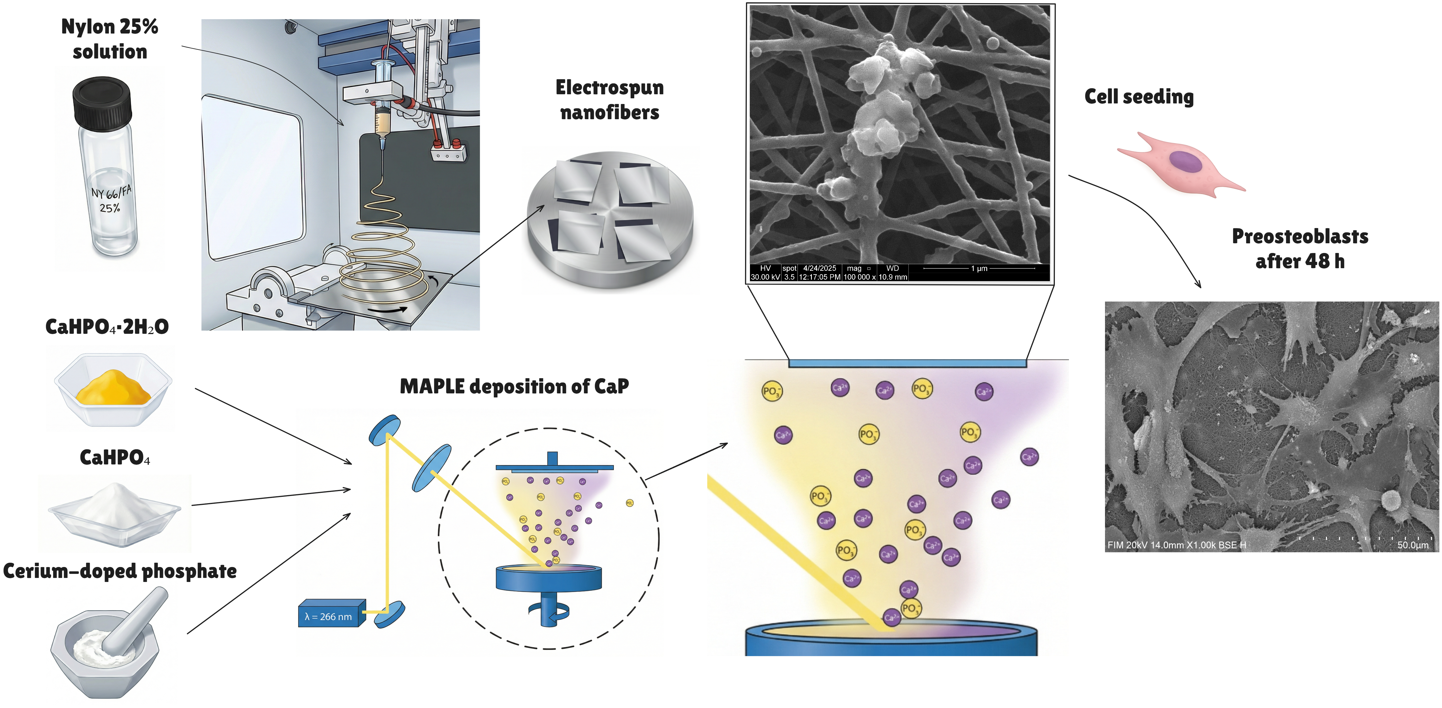
Figure 1.
Graphical workflow of scaffold fabrication and testing (made using Illustrae [39]).
Figure 1.
Graphical workflow of scaffold fabrication and testing (made using Illustrae [39]).

Figure 2.
SEM and EDS characterization of the uncoated nylon scaffold (Ny_25%). Top: EDS spectrum showing the elemental composition of the sample. Bottom: low- and high-magnification SEM micrographs of the electrospun Ny_25% mat, revealing a uniform, bead-free nanofibrous network with fibre diameters predominantly in the 40–80 nm range.

Figure 3.
Representative SEM and EDS characterization of CaP-coated nylon scaffolds. Top row, from left to right: high-magnification SEM image of Ny_monetite, low-magnification SEM image of the same sample, and corresponding EDS spectrum. Bottom row, from left to right: high-magnification SEM image of Ny_brushite, low-magnification SEM image, and corresponding EDS spectrum.

Figure 4.
Representative SEM and EDS characterization of cerium-doped phosphate-coated nylon scaffolds. Top row, from left to right: high-magnification SEM image of Ny_CP_Ce_500, low-magnification SEM image of the same sample, and corresponding EDS spectrum. Bottom row, from left to right: high-magnification SEM image of Ny_CP_Ce_800, low-magnification SEM image, and corresponding EDS spectrum.

Figure 5.
FTIR spectra of the electrospun and MAPLE coated samples.

Figure 6.
Fibre diameter distribution histograms for MAPLE-coated nylon scaffolds, with fitted Gaussian curves.
Figure 6.
Fibre diameter distribution histograms for MAPLE-coated nylon scaffolds, with fitted Gaussian curves.


Figure 7.
Distribution of the electrospun fibre samples based on orientation degree, using OrientationJ plugin on lower magnification SEM micrographs.
Figure 7.
Distribution of the electrospun fibre samples based on orientation degree, using OrientationJ plugin on lower magnification SEM micrographs.

Figure 9.
SEM micrographs of MC3T3-E1 preosteoblasts cultured for 48 h on the Ny_25% scaffold, shown at low, intermediate and high magnification to visualize overall coverage, cell spreading and interactions with the underlying nanofibres.
Figure 9.
SEM micrographs of MC3T3-E1 preosteoblasts cultured for 48 h on the Ny_25% scaffold, shown at low, intermediate and high magnification to visualize overall coverage, cell spreading and interactions with the underlying nanofibres.

Figure 10.
SEM micrographs of MC3T3-E1 preosteoblasts cultured for 48 h on the: a) Ny_monetite and b) Ny_brushite scaffolds, at different magnification orders.
Figure 10.
SEM micrographs of MC3T3-E1 preosteoblasts cultured for 48 h on the: a) Ny_monetite and b) Ny_brushite scaffolds, at different magnification orders.

Figure 11.
SEM micrographs of MC3T3-E1 preosteoblasts cultured for 48 h on the: a) Ny_CP_Ce_500 and b) Ny_CP_Ce_800 scaffolds, each at various magnification orders.
Figure 11.
SEM micrographs of MC3T3-E1 preosteoblasts cultured for 48 h on the: a) Ny_CP_Ce_500 and b) Ny_CP_Ce_800 scaffolds, each at various magnification orders.


Table 1.
Sample abbreviations and descriptions.
| Sample | Description |
| Ny_25% | Electrospun nylon scaffold (25 wt% solution), uncoated |
| Ny_monetite | Ny_25% scaffold coated by MAPLE with monetite (CaHPO₄) |
| Ny_brushite | Ny_25% scaffold coated by MAPLE with brushite (CaHPO₄·2H₂O) |
| Ny_CP_Ce_500 | Ny_25% scaffold coated by MAPLE with cerium-doped phosphate calcined at 500 °C |
| Ny_CP_Ce_800 | Ny_25% scaffold coated by MAPLE with cerium-doped phosphate calcined at 800 °C |
Disclaimer/Publisher’s Note: The statements, opinions and data contained in all publications are solely those of the individual author(s) and contributor(s) and not of MDPI and/or the editor(s). MDPI and/or the editor(s) disclaim responsibility for any injury to people or property resulting from any ideas, methods, instructions or products referred to in the content. |
© 2026 by the authors. Licensee MDPI, Basel, Switzerland. This article is an open access article distributed under the terms and conditions of the Creative Commons Attribution (CC BY) license.
Copyright: This open access article is published under a Creative Commons CC BY 4.0 license, which permit the free download, distribution, and reuse, provided that the author and preprint are cited in any reuse.


