Submitted:
12 April 2026
Posted:
14 April 2026
You are already at the latest version
Abstract
Ginger (Zingiber officinale roscoe) is a high-value horticultural crop widely cultivated for its culinary and medicinal applications, yet its production is increasingly constrained by soil-borne diseases. Among these, Fusarium yellows, caused by Fusarium oxysporum f. sp. zingiberi (Foz), is one of the most damaging constraints in ginger-growing regions around the world, leading to progressive yellowing, vascular blockage, and decline in rhizome quality. Members of the Fusarium oxysporum species complex are known to include both pathogenic and non-pathogenic lineages that often co-occur within the same host and environment, complicating disease diagnosis and epidemiological understanding. In this study, we examined Fusarium-like isolates recovered from both symptomatic and symptomless ginger plants within Eumundi, the major ginger production region in Australia, to investigate their genetic diversity, effector gene content, and pathogenic potential. Comparative analyses revealed two genetically and functionally distinct groups: a clonal Foz lineage consistently associated with Fusarium yellows symptoms and characterised by a conserved set of Secreted In Xylem (SIX) effector genes (SIX7, SIX9, SIX10, and SIX12), and a diverse set of F. oxysporum isolates lacking these effectors. The conserved presence and co-localisation of SIX7, SIX10, and SIX12 within a 5 kb region on a 1.4 Mb contig in the Foz lineage is consistent with the retention of a stable lineage-specific effector module, likely associated with accessory genomic regions that may contribute to host specificity and pathogenicity in the Fusarium oxysporum species complex. Patho-genicity assays confirmed that only the Foz lineage induced disease, whereas non-Foz isolates were asymptomatic despite limited colonisation of host tissues. These findings highlight the coexistence of pathogenic and endophytic Fusarium lineages within ginger production systems and support the use of effector-based markers for improved detection and disease management.
Keywords:
Fusarium oxysporum f. sp. zingiberi
; Ginger (Zingiber officinale roscoe)
; Secreted In Xylem effectors
; effector gene evolution
; accessory chromosomes
; plant–fungal interactions
; endophytes
; Fusarium yellows
; comparative fungal genomics
1. Introduction
Ginger (Zingiber officinale Roscoe) is a perennial, herbaceous plant grown as an annual crop and cultivated for its pungent and aromatic underground rhizomes. Ginger has been cultivated for thousands of years as food and medicine [1,2]. Traditionally, ginger has been used as a medicinal remedy for infections, nausea, inflammation, digestive and circulatory diseases [3,4]. Ginger is traditionally cultivated for domestic consumption in many cultures as a spice or vegetable and is also widely used in confectionery and beverages. It is grown across tropical and subtropical regions, with a reported global production of over 4 million tons per annum [5]. Ginger is believed to have originated in Southeast Asia.
The most significant ginger rhizome diseases are caused by Fusarium oxysporum f. sp. zingiberi (Foz), Pythium species such as P. myriotylum, bacteria such as Ralstonia solanacearum or R. sequeirae, and root-knot nematodes (Meloidogyne incognita and M. javanica) [6,7,8,9]. Fusarium yellows, the name of the disease caused by Foz, is characterized by the appearance of bright yellow leaves among the dark green foliage of the ginger crop. Generally, the leaves at the base of the tiller turn yellow first, with the discolouration progressing up the tiller within a few days. The yellow leaves turn brown and papery and newly emerging tillers may be stunted and discoloured. At harvest, the infected rhizomes will have extensive areas of dry, brown rot, rendering the rhizomes unmarketable [10]. The later disease stages are associated with the browning and rotting of the rhizomes which is indicative of the colonisation and proliferation of the fungus. Once present in the soil, Foz can persist in the soil for a long time, by surviving as resting spores in the absence of ginger hosts [11]. A deeper understanding of the infection cycle with respect to the host and rotational cropping would be valuable to the ginger industry.
Fusarium yellows has been detected in many countries, including United States [12], China [13] and Australia [9]. In Australia, commercial ginger production relies heavily on two cultivars, cv. Canton and cv. Queensland, both of which have been found to be susceptible to Foz [9]. This lack of disease resistant genotypes underscores a critical need for targeted research to understand the pathogenicity of Foz on ginger and in turn contribute towards better management of this disease.
Although Fusarium oxysporum is widely known as a plant pathogen, many strains also exist as non-pathogenic endophytes that colonise plants without causing disease symptoms. These endophytic F. oxysporum strains have been reported in many plant species and can contribute to plant health by promoting growth and inducing resistance against pathogens [14,15,16]. Endophytic F. oxysporum strains often share close genetic relatedness with pathogenic forms and can colonise plant host tissues asymptomatically [17,18]. Some F. oxysporum endophytes may carry an effector gene repertoire, including the pathogenicity-associated Secreted In Xylem (SIX) genes [19]. While some of these effector genes have been implicated in disease symptom development and pathogenic interactions, the role of these effector genes during asymptomatic and endophytic interactions is yet to be understood. Furthermore, some endophytic strains can serve as biocontrol agents by directly or indirectly acting against pathogens through antagonistic mechanisms or competition for space and nutrients [20]. Therefore, they may represent a valuable resource for biocontrol strategies against Foz in managing Fusarium yellows.
The link between SIX effector gene profiles, pathogenicity, and genetic divergence in Fusarium oxysporum associated with ginger remains unclear, limiting our understanding of pathogen emergence and the development of reliable diagnostics. In this study, we isolated a group of Fusarium oxysporum-like isolates from ginger plants that were either healthy or showing Fusarium yellow symptoms. Screening using SIX effector gene markers showed the presence of four SIX genes consistently present in one group of isolates associated with pathogenicity on ginger. In contrast, non-pathogenic isolates lacked most of these SIX genes, and caused no visible symptoms on ginger plants. These isolates were also genetically divergent from the Foz clade, suggesting that Foz in Australia likely arose from a single clonal introduction event. The unique sequence variation observed in Foz SIX effector genes may provide a useful basis for developing diagnostic markers for detecting Fusarium yellows in the field.
Despite reports of Fusarium oxysporum associated with ginger, the genetic basis of variation in pathogenicity remains unclear, particularly in relation to accessory genome structure and effector gene organisation. Comparative genomic analysis is therefore needed to resolve variation in these regions and to clarify how ginger-associated Fusarium oxysporum isolates relate to diversity within the broader Fusarium oxysporum species complex. We addressed this by combining multilocus phylogenetic analysis with comparative genomics of isolates representing different host-specialisation lineages. In particular, we investigated the organisation and conservation of the SIX7–SIX10–SIX12 effector region to assess differences in virulence-associated gene content between pathogenic lineages.
2. Materials and Methods
2.1. Fungal Isolates
Fifty-two Fusarium oxysporum strains were isolated from ginger plants symptomatic or asymptomatic for yellows in Southeast Queensland, Australia (Table S1). Monoconidial isolates were subsequently reisolated using hyphal tips of single germinated conidia.
2.2. Genes and PCR Primers
A set of universal primers for detecting SIX1-14 genes were firstly used to detect SIX genes in these isolates [19]. Full-length SIX7, SIX9, SIX10 and SIX12 gene sequences were retrieved from the existing Foz genome assemblies from NCBI under Bioproject PRJNA846078 [21] and then used to develop primers to amplify near-full length copies these genes from Foz (Table S2).
Translation elongation factor 1α (TEF-1α) and DNA-directed RNA polymerase II second largest subunit (RPB2) sequences from 150 Fusarium genome sequences were retrieved from NCBI and used as a backbone dataset for phylogenetic placement of our isolates. For SRA (NCBI, Sequence Read Archive) accessions where assemblies were not available, genome sequence data were assembled using SPAdes (v3.15.3) [22] prior to the sequence retrieval of these two genes. The backbone collection of SIX7, SIX9, SIX10, and SIX12 genes were retrieved from the NCBI non-redundant database by performing tBLASTn searches using the amino acid sequences of these SIX genes from Fusarium oxysporum f. sp. lycopersici as query.
2.3. DNA Extraction and PCR
DNA was extracted from the mycelia of these isolates using a microwave method [23]. TEF-1α and RPB2 genes were PCR-amplified using primers EF1/EF2 [24], and RPB2-5F2 [25] / 11aR [26], respectively (Table S2). Some of the RPB2 genes were difficult to amplify and instead were amplified as two overlapping fragments (860 bp + 917 bp) using primer pairs 5F2/7cR and 7cF/11aR (Table S2) [25,26]. A set of universal primers were used to detect the presence of SIX1-14 genes in the F. oxsporum isolates obtained in this study [19]. Foz-specifc primers were then developed to amplify and retrieve full-length SIX7, SIX9, SIX10 and SIX12 genes using gene templates retrieved from Foz genome assemblies available in NCBI (VPRI4420, 44278, 44279, 44280) and Foz Pin2.2 long-read genome assembly (GCA_051624415.1). PCR products of the correct size were confirmed on a 1 % agarose gel and then purified using a GeneJET PCR purification kit (Thermo Fisher Scientific, Waltham, MA, USA), and Sanger-sequenced at the Australian Genome Research Facility, Melbourne, Australia.
2.4. Phylogenetic Analysis
Geneious Prime v 2023.0.1 (Biomatter Pty Ltd, Auckland, New Zealand) was used for the phylogenetic reconstruction of the isolates. Firstly, multiple sequence alignment was performed using MAFFT v 7.490 [27]. The subsequent alignment was edited manually to minimized gaps. Multi-gene sequences were then concatenated and aligned to derive a consensus alignment sequence, which was then used as an input in MrBayes v 3.2.6 to construct phylogenetic trees using the Bayesian inference method [28]. The running parameters used the GTR-G-I model of substitution with two independent analyses on four Markov chain Monte Carlo (MCMC) chains for 2 000 000 generations. A burn-in rate of 25 % was used to sample every 1000 generations. Fusarium verticillioides (7600) was used as an outgroup to anchor the phylogenetic tree. The SIX gene phylogenies were unrooted. The resulting consensus phylogenetic trees were visualised with branch support indicated by posterior probability values at each node, and branch lengths scaled to represent the expected number of substitutions per site.
2.5. Plant Materials
Ginger plantlets of cultivar ‘Canton’ were produced by in vitro tissue culture [29] and then grown in a greenhouse to maintain a disease-free state at the Maroochydore Research Facility, Queensland Department of Primary Industries, Queensland, Australia. ‘Canton’ rhizomes, approximately 40g pieces, were planted into clean potting mix (Searles, Kilcoy, Queensland, Australia) in 5 L poly bags (company). A total of 90 plants were arranged on two benches in a 50% shade house. The bags were initially watered by hand and then subsequently watered daily by an automatic overhead misting system. Plants were treated with a slow-release fertilizer (Osmocote Exact High K 8-9 month®) and subsequently with potassium sulphate at tiller emergence and thereafter as required.
2.6. Preparation of Inoculum
For inoculation, 15 isolates were chosen to represent F. oxysporum obtained from rhizomes grown in major ginger-producing regions of Southeast Queensland, and from fields with a range of crop histories prior to the ginger crop from which the rhizomes were sourced (Table S1).
The inoculum consisted of F. oxysporum spores extracted according to a previously published method [10,30]. To generate the spore suspensions, flasks of potato dextrose broth were inoculated with 4-5 mycelial plugs from plates colonised by each isolate and were then incubated for 7 days at 26 °C with shaking at 120 rpm on a rotating platform (Ratek, Victoria, Australia). Additional inoculum in the form of F. oxysporum infested Japanese millet seed (Echinochloa esculenta) was prepared as per previously described [31].
2.7. Plant Inoculation
On the day of inoculation, the bags containing the germinated ginger plants were arranged into 16 groups of five plants, so that the groups had an even mix of plant height and number of tillers per plant. Each plant had at least one semi-mature tiller (approx. 700 mm tall) and at least one smaller tiller. Bags were arranged onto two nursery benches on the same side of the greenhouse. Those designated for mock inoculation and those for endophyte inoculation were placed on one bench and those designated for Foz inoculation on the other bench. For each inoculation, approximately 15-20 g of colonised millet and 50 mL of fungal spore suspension were added to the soil. The inoculum was then lightly covered with more potting mix. Plants were maintained in the shade house for 8 weeks.
Plants were monitored for external symptoms of disease until harvest at 8 weeks post inoculation. At harvest, external symptoms including the number of tillers, leaf discoloration and the height of the tallest tiller were recorded. Rhizomes were weighed and then split in half through the vertical axis. The cut surface was then rated on the percentage of the internal surface that was discoloured, as a measure of internal disease symptoms.
2.8. Reisolation of Fusarium oxysporum from Infected Plants
Four tissue pieces each from roots, rhizome, stem and leaves were collected from two plants from each treatment. The stem samples comprised of two pieces each taken at the rhizome shoulder and approximately 5 cm above the rhizome. The leaf pieces included the petiole and three pieces of the leaf blade transecting the central leaf rib. Tissue pieces were surface-sterilised with 70% ethanol for a minute, air-dried, and plated onto half strength potato dextrose agar (PDA) to detect the presence of colonies and spores typical of F. oxysporum.
A colony was considered to be F. oxysporum-like if it had abundant fluffy aerial and submerged mycelia and the colony colour varied from white to pale violet to dark magenta top on the media surface or underside of a PDA plate (Figure S1). Microconidia were produced in false heads on short monophialides and were generally abundant; oval, elliptical or reniform; single celled; 3.0 to 5.0 µm wide and 6.0 to 14.0 µm long. Macroconidia produced in some isolates; typically 3-septate; hyaline; slightly curved with an apical hook; approximately 3.0 to 5.0 µm wide and 20.0 to 30.0 µm long.
2.9. Statistical Analysis
Statistical analysis was conducted in Prism version 10.3.1. For rhizome discolourattion, rhizome weight and plant height, were analysed using one-way ANOVA, followed by Tukey’s post-hoc test was used to group means into homogeneous sets, with different letters above columns indicating significant differences at p ≤ 0.05. Plotted values represent means with 95% confidence intervals.
Using SPSS (IBM SPSS Statistics, v.29.0.1.0), a generalised linear model with a negative binomial distribution and log link was used to analyse the number of green tillers and yellow leaves at 8 weeks post inoculation across all strains. Pairwise comparisons were performed using estimated marginal means with Bonferroni adjustment for multiple testing.
2.10. Genomics Analysis
2.10.1. Genome Assemblies
To analyse the genomic structure around the SIX7-10-12 gene cluster, long read genome assemblies were retrieved from the public NCBI genome database. These included Fusarium oxysporum ff. spp. narcissi (GCA_045837865.1), lini (GCA_013423245.1), cepae (GCA_003615085.1), sesami (GCA_017979615.1), lycopersici 4287 (GCA_000149955.2) and zingiberi (GCA_051624415.1). F. oxysporum f. sp. cubense assembly FocCAV2318 (Subtropical Race 4 or STR4) was retrieved from Mycocosm (https://mycocosm.jgi.doe.gov/mycocosm/home). F. verticillioides 7600 (GCA_000149555.1) was used as an outgroup to anchor the phylogenetic tree. All seven genomes were assessed for completeness using BUSCO v 5.8.0 [32] with the ascomycota_odb12 lineage dataset (v.2026-03-20-145944), and AUGUSTUS [33] with Fusarium graminearum as the species training model for gene prediction within the BUSCO pipeline.
2.10.2. Structural Annotation of Protein-Coding Genes
Genome annotations were not available for the assemblies derived from F. oxysporum ff. spp. sesami, lini and narcissi. For these three assemblies, repeat sequences in the genome were modelled using RepeatModeller v 2.0.4 [34] and then soft-masked using RepeatMasker v 4.1.5 [35]. Structural annotation of protein-coding genes was performed using BRAKER3 v3.0.6 [36]. Proteins from lycopersici 4287 were mapped to the soft-masked genomes, and the fungal model was applied during the GeneMark training step. AGAT v1.4.0 was used to identify and remove overlapping gene models and then retain only the longest isoform of each gene (https://zenodo.org/records/11106497).
2.10.3. Collinearity Analysis
Collinearity at the chromosome level was investigated using the MCScanX function of OrthoVenn3 [37] and six long-read genome assemblies of Fusarium oxysporum formae speciales lycopersici, zingiberi, narcissi, lini, cepae, and sesami, while F. oxysporum f. sp. cubense CAV2318 was excluded due to its fragmented assembly. Genome-wide contig alignments were first generated using the NUCmer program from the MUMmer package (v4.01) [38], with F. oxysporum f. sp. lycopersici used as the reference to determine chromosome number and order across the remaining five genomes. Alignments were then ordered according to the F. oxysporum f. sp. lycopersici reference using a custom script and visualised on the interactive dot plot tool online (https://dot.sandbox.bio) with default settings. For synteny analysis, core chromosomes with unique aligned regions across the five genomes were arranged according to the F. oxysporum f. sp. lycopersici reference, and the three to seven largest unaligned sequences per genome were also included. Two large non-aligned chromosomes, CM023981 and CM023982, from F. oxysporum f. sp. lini were excluded from this analysis. Corresponding GFF annotations were reordered, concatenated, and converted to BED format using a script provided by OrthoVenn3. Gene orthology clustering and collinearity analysis were performed in OrthoVenn3 using an e-value cutoff of 1e−15 with all other parameters set to default. The resulting collinearity plot was interactively edited online, and the final SVG output was refined in Inkscape (v1.4.3).
2.10.4. Manual Curation of SIX Gene Annotations
Genomes and their gene models were loaded into Geneious Prime v 2026.02 and local BLASTn and tBLASTn searches using a set of SIX1-14 nucleotide or amino acid sequences were performed against all seven F. oxysporum assemblies to check their presence and absence in these genomes. For SIX7, SIX10 and SIX12, the coding domain sequence and their positions in each genome were carefully examined and then corrected using the F. oxysporum f. sp. lycopersici SIX gene models as a guide. SIX gene paralogues were assigned letters a, b, and c to differentiate their positions on the same or different chromosomes. The fixed gene annotations were exported from Geneious Prime in strict GFF3 format. All revised annotations were then processed with a custom script to correctly define gene–CDS relationships. The script parses each feature, assigns unique IDs to genes, mRNAs, exons, and CDSs, updates parent–child relationships, and ensures CDS phases are properly set. Features without a Name= tag are left unchanged, while annotations from other sources (e.g., GenBank, MAKER, Augustus) are preserved as provided. Gffread v 0.12.8 was then used to extract amino acid sequences from all seven genomes [39].
2.10.5. Orthologue Clustering and Evolutionary Analysis
Gene Orthology and phylogenetic analysis of the annotated genomes were performed using OrthoVenn3 [40]. Gene-coding proteins from the seven genomes were clustered using OrthoFinder v3.0.1 [41] with an e-value threshold of 0.01 and an inflation value of 1.50. The evolution model was JTT-CAT. divergence time of 5.34 million years ago (MYA) was estimated between F. oxysporum and F. verticillioides, using TimeTree5 [42]. Gene family expansion and contraction between genomes were calculated based on this calibrated evolutionary divergence time using CAFE5 [43].
2.10.6. Orthologous Analysis Across the SIX7-10-12 Gene Cluster
The SIX7, SIX10, and SIX12 reference genes from the Foz pin2.2 reference genome were used as anchors to identify corresponding orthogroups across the other six Fusarium assemblies using the orthogroups output file from OrthoFinder. Using the orthogroup assignments, all orthologues—including any duplicated copies—were retrieved for each genome. Each gene was then linked back to its genome-specific GFF annotation to extract genomic coordinates, scaffold, and strand information using a custom R script. The final output was compiled into a structured ortholog table, listing the reference gene, its orthogroup, the genome in which it was found, gene ID, locus number, scaffold, start and end positions, and strand. This approach ensures that both single-copy and duplicated orthologues are captured, providing a complete mapping of SIX genes across all seven genomes. Visualisation of the genomic organisation of these orthologues was performed in R using the tidyverse, ggplot2, cowplot, and rtracklayer packages [44]. Gene coordinates were arranged along each scaffold, with large intergenic gaps compressed to highlight clustered regions while preserving their relative order. Genes are displayed as arrows oriented according to their direction of transcription. Boxes are added manually to indicate the start and end positions of each scaffold (genomic sequence, not drawn to scale) to provide genomic context.
3. Results
3.1. Detection of SIX Genes in Fusarium oxysporum Isolates from Ginger
Universal SIX gene profiles performed on 56 Fusarium oxysporum isolates showed that 38 isolates carried four SIX genes, namely SIX7, SIX9, SIX10 and SIX12 (Table S3). These include five categorised strains that were previously isolated from ginger hosts symptomatic for Fusarium yellows (BRIP: 44963, 44967, 44969, 44971, 44977). Twelve endophytic isolates do not appear to any of these four SIX genes, whereas five of them (SP1.2, SP2, SP2.2, SP3.2 and SP4.2) only carry SIX9 and not the other three (Table S3).
3.2. Phylogenetic Placements of Fusarium oxysporum Isolates Within Fusarium oxysporum Species Complex
Phylogenetic analysis based on concatenated sequences of TEF1 and RPB2 resolved a distinct F. oxysporum f. sp. zingiberi clade that includes 32 Foz isolates and the Foz reference strains (Figure 1). This phylogroup appeared to contain Foz-exclusive isolates. No genetic variation or single-nucleotide polymorphisms (SNPs) were detected in this phylogroup, suggesting that the Foz population belonged to a clonal lineage in Australia.
A total of 17 F. oxysporum endophytes are phylogenetically positioned within a large clade, seperated from the Foz clade (Figure 1). Within this large clade, several distinct subclades were observed. Mei1.2, Mei3.2 and Temp4.2 were clustered with a Race 1 strain of F. oxysporum f. sp. cubense (NRRL36118) [45], multiple races of F. oxysporum f. sp. niveum (Race 1-3) [46], F. oxysporum f. sp. vasinfectum strains F17 and 25433 (both Race 7) [47,48], as well as LA1E (MDS-12, race 4-like) [49]. The second subclade comprised ginger endophytes SP1, SP1.2, SP2, SP2.2, and SP3.2, which clustered together with F. oxysporum ff. spp. ciceris (EthFoc127), lini (isolate 39), melonis (Fom005), and pisi (RBG6416). The basal cluster within this phylogroup contained the largest number of ginger endophytes, including Can2, Can5, Can6, Can7, Can8, Can11, SP4.2, and Temp3.2. These isolates clustered with F. oxysporum ff. spp. ciceris (EthFoc12), vasinfectum (LA3B, ME23, TF1), melonis (RBG7064), cubense (NRRL36113), raphanin (54005), and a F. oxysporum endophyte isolate from wheat (CS5870). Notably, Can5 resolved within a subclade that clustered with F. oxysporum f. sp. cubense isolate P41b, previously characterised as a Race 1 strain [45]. This suggests that Can5 is most likely a banana-infecting F. oxysporum strain.
A single ginger endophyte, Pin3-2, clustered with a F. oxysporum f. sp. cubense strain P20a from banana and a F. oxysporum f. sp. pisi strain RBG6477. The banana infecting strain was defined as a Race 1 strain, and was isolated from the Silk subgroup banana cultivar ‘Mazano’ [45]. This cluster is nested within a broader clade that includes the known biocontrol agent Fo47, as well as specialised forms that infect banana, tomato and other plant hosts (Figure 1). Interestingly, no other ginger endophytes were positioned within this clade. Ginger endophytes Pin2 and Endo 5.2 clustered together with a Fusarium foetens strain NRRL 38302, confirming their F. foetens identity. This represents the first ever report of F. foetens associated with ginger plants in Australia.
3.3. Sequence Analysis of the SIX Genes from the Foz Isolates
Full-length SIX7, SIX9, SIX10, and SIX12 amino acid sequences from F. oxysporum f. sp. lycopersici was used to perform a tBLASTn search of the respective SIX gene homologs in the Pac-bio derived assembly of Foz Pin2.2 (NCBI genome accession GCA_051624415.1). Single hits to a full-length copy of each SIX gene were identified in Pin2.2 suggesting that these four SIX genes are present as single copy orthologues in the genome of Foz Pin2.2.
Full-length sequences of SIX7, SIX9, SIX10, and SIX12 were obtained from 32 Foz isolates and were phylogenetically analysed using a background dataset of the corresponding SIX gene sequences retrieved from 150 NCBI genome sequences (Figure 1). No SNPs were identified across all four SIX gene sequences among the 32 isolates, supporting the observation that they likely originate from a single clonal lineage of Fusarium oxysporum in Australia.
Phylogenetic analysis of SIX7 revealed that Foz isolates clustered within a well-supported phylogroup comprising homologues from multiple Fusarium oxysporum formae speciales, including lini, canariensis, cepae, pisi, lycopersici, dianthi, and sesami (Figure 2A). This phylogroup was distinct from a sister clade containing F. oxysporum f. sp. cubense Race 1, Subtropical Race 4, and Tropical Race 4 isolates. (Figure 2A).
SIX9 sequences were resolved into several well-supported clades within Fusarium oxysporum species complex (Figure 2B). This indicates the presence of several distinct evolutionary lineages. Foz SIX9 sequences formed a subgroup with homologues from ff. spp. pisi, cepae, dianthi, and narcissi, within a larger clade that also included F. oxysporum f. sp. cubense. A subset of ginger endophytes (FoSP1.2, FoSP2.2, FoSP2, FoSP3.2, and FoSP4.2) formed a divergent lineage within this clade. A separate lineage comprised homologues from ff. spp. niveum, lycopersici, passiflorae, and albedinis.
For SIX10, Foz sequences were grouped with a clade containing homologues from F. oxysporum f. sp. canariensis, F. oxysporum f. sp. cubense Tropical Race 4, as well as ff. spp. dacroydes and palmarum, and Fusarium nirenbergiae (Figure 2C). A second well-defined clade contained homologues from ff. spp. lini, dianthi, narcissi, pisi, and cepae, while sequences from ff. spp. physali and lycopersici were more distantly related.
SIX12 formed a distinct clade comprising Foz sequences, within which homologues from F. oxysporum f. sp. canariensis grouped as a subclade (Figure 2D). In addition, a second, more divergent set of SIX12 homologues from F. oxysporum f. sp. canariensis formed an independent phylogroup, suggesting the presence of duplicated SIX12 copies in this forma specialis. To date, SIX12 appears to be detected only in ff. spp. zingiberi and canariensis.
Overall, the phylogenetic placement of Foz SIX genes indicates gene-specific evolutionary relationships within the Fusarium oxysporum species complex, with clustering patterns varying among genes and spanning multiple formae speciales. With the exception of SIX9, which is absent in F. oxysporum f. sp. canariensis, Foz shares closely related copies of the remaining three SIX genes (SIX7, SIX10, and SIX12) with this forma specialis.
3.4. Greenhouse Pathogenicity Testing Using Pathogenic and Endophytic Fusarium oxysporum Isolates
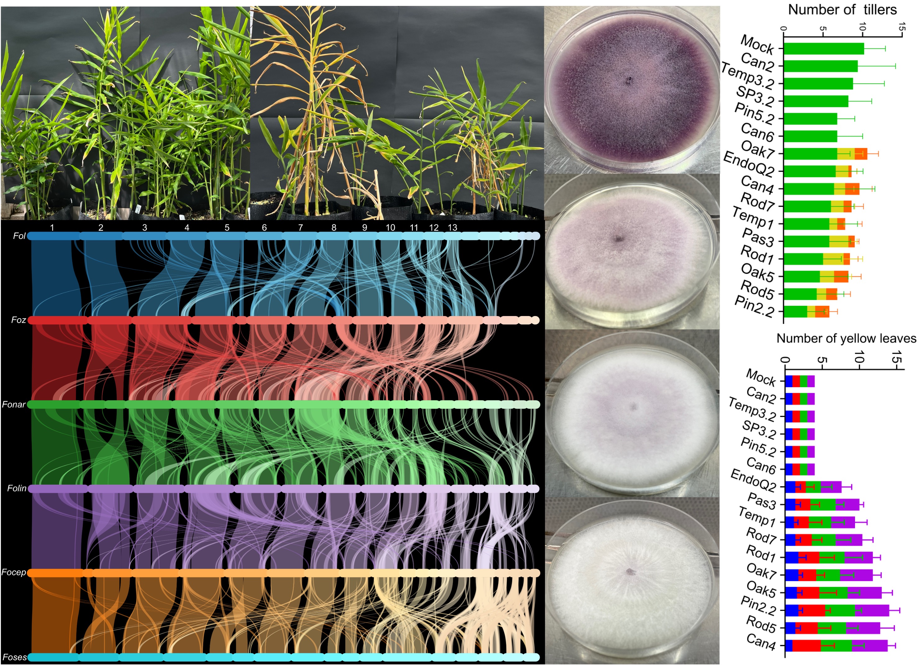
Formae speciales of these F. oxysporum isolates were further confirmed to be Fusarium oxysporum f. sp. zingiberi based on their ability to cause Fusarium yellows in ginger pot trials (Figure 3). Foz isolates Can4, EndoQ2, Oak5, Oak7, Pas3, Pin2.2, Rod1, Rod5, Rod7, and Temp1 all showed typical external symptoms of Fusarium yellows, including leaf and tiller yellowing (Figure 3). Disease severity appeared to vary amongst the treatment (isolates), with complete plant and tiller collapse observed in plants inoculated with Pin2.2 and Oak7. Disease expression was sporadic, with yellow tillers interspersed among healthy tillers within the same pot, which is a typical characteristic of Fusarium yellows (Figure 3). Internally, part or entire ginger rhizomes showed brown necrotic regions, indicative of the presence and proliferation of Foz in these issues.
Plants inoculated with the endophytic F. oxysporum strains (Can2, Can6, Pin5.2, Sp3.2, Temp3.2) remained healthy throughout the entire experiment. At harvest, these plants showed no visible external or internal symptoms of Fusarium yellows (Figure 3).
3.5. Plant Symptomatic Assessment and Comparisons Between Foz and Endophytes
Rhizome discolouration clearly distinguished the two treatment groups, with Foz causing 50–80% discolouration in individual rhizomes, whereas no visible symptoms were observed in endophyte-inoculated plants (Figure 4A). The ranking of Rhizome weight amongst the treatment groups is also consistent with their Foz susceptilblity, with pathogenic forms clearly associated with reductions in weights and non-pathogenic forms maintaining rhizome weight relative to the untreated controls (Figure 4B). Although statistically, there's a large overlap in the separation of means as the dispersion of the dataset is large, which indicates considerable variability in the rhizome weight. Similarly, plant height shows that plants inoculated with the endophytes and the water control were significantly taller than those inoculated with Pas3, Oak5, Pin2.2 at p = 0.05 (Figure 4C). The large overlap among mean plant height in the other Foz-inoculated plants reflects the sporadic occurrence of yellowed tillers among otherwise healthy tillers within individual pots.
Tiller number per treatment group indicates that tiller necrosis was associated with plant susceptibility to Foz, whereas endophyte-inoculated plants did not exhibit any yellowing symptoms (Figure 4D). Statistical modoelling using a generalised linear model with a negative bionoomial distribution showed a significant overall strain effect on the number of green tillers observed at 8 weeks post inoculation (Likelihood Ratio χ² = 36.37, df = 15, p = 0.002). Several strains were significantly different from the water control, including Oak5 (p = 0.019), Rod1 (p = 0.040), Pin2.2 (p < 0.001), and Rod5 (p = 0.008).
The number of yellow leaves were counted at each of 5 to 8 weeks post inoculation. The results show that the number of yellow leaves remained consistently low at all time points in the plants inoculated with the five endophytes and the water control (Figure 4E). In contrast, the number of yellow leaves increased from week 5 onwards, and by week 8 was significantly higher than in endophyte-inoculated plants across all 10 Foz-inoculated groups at p = 0.05 (Figure 4E). The generalised linear model indicated a strong overall strain effect (Likelihood Ratio χ² = 558.32, df = 15, p < 0.001). Pair-wise comparison against the water coonttrol indicated that all 10 Foz strains were significantly different (p < 0.001) when compared to the water control (Figure 2E).
3.6. Reisolation of Fusarium oxysporum from Ginger Tissues at Harvest
Tissue reisolation from the leaf, stem, rhizome and roots showed contrasting ability of pathogenic and non-pathogenic F. oxysporum to colonise ginger plants (Figure 4F). All tissues were negative for F. oxysporum in the water control and the endophyte Temp3.2 treated plants. Endophytes Pin5.2, Sp3.2, Can2, and Can6 treated plants showed limited presence of F. oxysporum in some of the reisolated tissues (Figure 4F). The presence of F. oxysorum was detected in all 4 tissue types in Sp3.2 and Can6 with an average range of 10 to 20 % reisolation frequency. The Foz isolates were recovered the most frequently in the roots and rhizomes of ginger plants, with 100% reisolation frequencies detected in the roots of Pin2.2 and Oak5, associating the presence of Foz with the rhizome discolouration observed in these plants (Figure 3, 4F). In the leaves, Foz was detected in 5 out of 10 Foz-inoculated treatment groups at approximately 20% reisolation frequency, suggesting that these isolates may have the ability to move up the aerial parts of the host plants. This is further supported by a high reisolation frequency of F. oxysporum detected in the stem of these plants. Foz was not detected in leaves of Pas3, Temp1, Rod5, Rod1, Rod7 inoculated plants, although their presence was detected in the stems of four of these treated groups (Figure 4F). This suggests that the aggressiveness of Foz isolates may vary and they may colonise the hosts and move up the aerial parts of the plants at different rates.
3.7. Comparative Genomics
3.7.1. Rational for Choosing Genomes for Comparative Analysis
Examination of SIX7, SIX10, and SIX12 showed that all three genes are co-located within a 5,182 bp region on contig 12 of the Foz Pin2.2 assembly. To investigate the structure of the virulence gene cluster containing these three SIX genes, and to explore the evolutionary relationships among Fusarium oxysporum strains harbouring this cluster, we selected seven genome assemblies. These assemblies are highly contiguous, and each has a BUSCO completeness score exceeding 95% (Table S4). These formae speciales were initially selected based on the presence of these SIX genes shown in Figure 2. Genome assemblies were then selected based on the presence of this gene cluster on a single contig. Annotations of all SIX7, SIX10, and SIX12 orthologues were manually inspected, corrected, and curated to account for any missing copies. The final curated protein-coding gene sets, excluding isoforms, were then used for the comparative analysis in Orthovenn3.
3.7.2. Genome Structure Analysis
Collinearity analysis using MCScanX, as implemented in OrthoVenn3, revealed both conserved and lineage-specific chromosomal features across the six Fusarium oxysporum formae speciales (Figure 5). Notably, chromosomes 3 and 6 of F. oxysporum f. sp. lycopersici lacked syntenic counterparts in all five other genomes, indicating that they are lineage-specific chromosomes. In contrast, eight core chromosomes showed strong and consistent synteny across all six genomes, indicating a conserved core genome structure. Chromosomes 11–13 of F. oxysporum f. sp. lycopersici exhibited limited collinearity, with fragmented or partial alignments across multiple contigs of the other genomes, suggesting reduced conservation and possible structural rearrangements. Toward the right side of the plot, lineage specific chromosomes 14 and 15 and other small scaffolds of F. oxysporum f. sp. lycopersici were not aligned to F. oxysporum f. sp. zingiberi, likely indicating divergent accessory genomic regions, as previously described for F. oxysporum f. sp. lycopersici [50]. In F. oxysporum f. sp. zingiberi, these small contigs also lacked detectable synteny with F. oxysporum f. sp. narcissi, whereas F. oxysporum ff. spp. narcissi and lini displayed only sparse and discontinuous syntenic relationships in these regions. In contrast, F. oxysporum ff. spp. cepae and sesami showed extensive multiple-to-multiple alignments among their unaligned contigs, indicative of highly repetitive or duplicated gene sequences shared between these genomes. Overall, syntenic relationships revealed variation in chromosomal conservation across the six Fusarium oxysporum formae speciales, with both conserved and lineage-specific genomic regions observed.
3.7.3. Orthologous Clustering Analysis
Protein clustering analysis using Orthovenn3 identified 20,094 conserved orthologous clusters across Fusarium oxysporum ff. spp. zingiberi (Pin2-2, GCA_051624415.1), lycopersici (Fol4287, GCA_000149955.2), cubense (CAV2318_1), narcissi (AJ275, GCA_045837865.1), lini (strain #39, GCA_013423245.1), cepae (FoC_Fus2, GCA_003615085.1), and sesami (MR4003, GCA_017979615.1). Fusarium verticillioides (7600, GCA_000149555.1) was included as the outgroup. A core set of 10,954 orthologous genes was identified (Figure 6A), of which 6,807 were single-copy across all eight genomes (Table S5). The seven F. oxysporum strains shared 682 orthologues exclusively.
Unique gene clusters were most abundant in forma specialis lycopersici (582 clusters / 1,862 genes), followed by formae speciales sesami (98/261), cepae (89/273), lini (86/220), narcissi (50/129), zingiberi (12/29), and cubense (8/20). GO term enrichment analysis indicated that F. oxysporum f. sp. zingiberi is enriched for oxidoreductase activity (3 genes, p = 0.0008). The top two most significant GO terms for the other formae speciales were: pathogenesis (2 genes, p = 4.66E−19) and metal ion binding (2 genes, p = 1.05E−11) for lini; regulation of transcription (2 genes, p = 1.51E−06) and mycotoxin catabolic process (4 genes, p = 0.0001) for narcissi; oxidoreductase activity (7 genes, p = 1.17E−11) and metal ion binding (6 genes, p = 1.27E−11) for cepae; and oxidoreductase activity (4 genes, p = 3.05E−104) and zinc ion binding (2 genes, p = 4E−19) for sesami. No significant GO enrichment was detected for F. oxysporum f. sp. cubense. Overall, oxidoreductase activity and metal ion binding are recurrently enriched among the unique gene clusters across multiple F. oxysporum formae speciales.
3.7.3. Gene Family Expansion and Contraction Analysis
Gene family evolution analysis using CAFE5 revealed distinct patterns of gene family expansion and contraction over an estimated 5.3 million years since the divergence of Fusarium oxysporum and Fusarium verticillioides (Figure 6B). Within F. oxysporum, an early divergence occurred at ~1.5 MYA, separating f. sp. cubense (CAV2318) from the remaining lineages. A subsequent split at ~1.2 MYA gave rise to f. sp. zingiberi (Pin2.2) and the lineage leading to the remaining formae speciales. These early-diverging lineages are characterised by pronounced gene family contraction, with 1,105 and 1,164 contracted families observed in f. sp. zingiberi and f. sp. cubense, respectively, alongside relatively limited expansion (56 and 78 gene families, respectively). The outgroup, F. verticillioides, exhibited moderate gene family dynamics, with 272 expansions and 74 contractions.
These patterns highlight the early divergence of the cubense strain CAV2318 from zingiberi Pin2.2, with both lineages being distinct from the clade giving rise to the remaining formae speciales. Within this latter lineage, two well-supported subgroups are apparent. The first comprises f. sp. lini and f. sp. sesami, both of which display relatively balanced gene family dynamics (lini: 200 expansions / 198 contractions; sesami: 179 expansions / 206 contractions). The second subgroup includes f. sp. lycopersici, which shows substantial gene family expansion (764 gained vs 337 lost), alongside the closely related f. sp. cepae and f. sp. narcissi, both of which exhibit a net loss of gene families (cepae: +125 expansions / −221 contractions; narcissi: +159 expansions / −369 contractions).
Overall, with the exception of lycopersici and lini, most formae speciales display a general trend toward gene family contraction, with the most extensive losses observed in the early-diverging cubense and zingiberi lineages.
3.7.4. Analysis of the SIX7, SIX10 and SIX12 Virulence Gene Cluster
Comparative analysis of gene collinearity across the SIX7, SIX10, and SIX12 region revealed both conservation and structural variation among Fusarium oxysporum genomes (Figure 7). F. oxysporum f. sp. zingiberi (Pin2.2) contains a complete SIX7-SIX10-SIX12 gene cluster. F. oxysporum f. sp. cubense Race 1 strain Cav2318, an earlier diverging lineage, only carries SIX7 and SIX10 together.
Genomes within this lineage can be divided into two distinct subclades (Figure 6). The first subclade, including F. oxysporum f. sp. lycopersici (4287), F. oxysporum f. sp. narcissi (AJ275) and F. oxysporum f. sp. cepae (Fus2), show strong conservation of the entire gene cluster, with maintained gene content, order, and orientation (Figure 7).
In contrast, the second subclade, represented by F. oxysporum f. sp. lini (strain 39) and F. oxysporum f. sp. sesami (MR4003), exhibits substantial structural variation (Figure 7). In lini strain 39, the cluster is duplicated and redistributed with one complete cluster retained, while a partial duplicate containing SIX7 and SIX10 is translocated within chromosome 12. Additional copies of SIX7 and SIX12 are located on different chromosomes, resulting in three copies of SIX7 and two copies of each SIX10 and SIX12 (Figure 7). Conversely, F. oxysporum f. sp. sesami MR4003 contains only SIX7 and SIX10 on a ~0.28 Mb contig, with no detectable SIX12. These genes are not arranged as a contiguous cluster, indicating a reduced and structurally fragmented gene complement.
Across all genomes analysed, SIX genes are frequently located near contig ends, suggesting an enrichment in telomeric or subtelomeric regions.
4. Discussion
Secreted In Xylem effectors are small, secreted proteins delivered into the plant vasculature during infection and are widely implicated in the pathogenicity of Fusarium oxysporum across diverse host species [51,52,53]. Screening for SIX genes in this study revealed that all pathogenic Foz isolates consistently carried the same combination of effectors, SIX7, SIX9, SIX10, and SIX12, whereas non-pathogenic endophytic isolates lacked these and all other SIX genes. This clear partitioning between pathogenic and non-pathogenic isolates aligns with previous studies demonstrating that SIX effector repertoires are strongly associated with host specificity and virulence in F. oxysporum [54,55,56].
4.1. Clonal Nature of the Australian Foz Population
Phylogenetic analysis based on the concatenated sequences of TEF1 and RPB2 genes grouped all F. oxysporum f. sp. zingiberi isolates collected in this study into a single, well-supported clade. This clade also included previously characterised reference Foz strains, providing strong molecular evidence that the isolates examined here belong to the same forma specialis. The clustering of all pathogenic isolates together, with the absence of sequence variation across the concatenated loci among the 35 Foz isolates, indicates a high degree of genetic uniformity and supports the inference that the Australian Foz population is largely clonal, consistent with earlier reports on the genetic homogeneity and vegetative compatibility among Foz isolates in ginger-growing regions in Australia [6,9,57]. Similar clonal structure is observed in other F. oxysporum lineages, such as the banana infecting Tropical Race 4 strains, which also exhibit very low genetic diversity (Figure 1) [58]. A clonal pathogen population can spread rapidly, but its uniform genetics may make it easier to detect. The conserved presence of these four SIX genes across pathogenic Foz isolates therefore reflects the clonal nature of the Australian Foz population and highlights their potential to be leveraged as robust molecular markers for identifying pathogenic strains from ginger.
4.2. Endophytic Strains that Colonise Ginger Hosts Asymptomatically
A diverse set of Fusarium oxysporum isolates recovered from asymptomatic ginger plants clustered across multiple lineages within the Fusarium oxysporum species complex (Figure 1). These isolates were phylogenetically associated with several known formae speciales, including F. oxysporum ff. spp. cubense, vasinfectatum, niveum, cepae, pisi, and lycopersici. Despite their placement within or adjacent to pathogenic lineages of other hosts, these isolates did not induce Fusarium yellows symptoms in ginger under glasshouse conditions. Notably, the majority of these isolates also lacked the core SIX effector genes detected in pathogenic Foz isolates, further distinguishing them from the pathogenic lineage identified in this study. Together, these patterns are consistent with the well-documented ecological continuum within F. oxysporum and related plant-associated fungi, where isolates can exist as pathogens or endophyte-like forms depending on host association and environmental context [59,60,61]. Non-pathogenic F. oxysporum strains have also been reported to occur alongside pathogenic lineages in soil and plant-associated environments, often without causing disease in non-host plants [62,63]. Accordingly, these isolates may represent either non-pathogenic lineages or members of host-specific formae speciales associated with other plant hosts that are unable to infect ginger, and may act as asymptomatic or transient colonisers within ginger-associated environments. However, their precise ecological status cannot be definitively resolved based on phylogenetic placement and SIX gene content alone.
4.3. Role of SIX Gene Effectors in Virulence and Host Specificity
SIX gene effectors are associated with pathogenicity in Fusarium oxysporum and may play roles in host–pathogen interactions during infection. While the exact functions of all SIX proteins are not fully understood, studies in tomato show that some effectors suppress plant immune responses and promote colonization [56,64]. Different SIX genes can also interact with specific plant resistance proteins, which can determine whether a plant variety is resistant to particular F. oxysporum strains [55,65].
Our phylogenetic trees showed that Foz SIX genes formed subgroups with the corresponding SIX effectors from specialised forms of F. oxysporum on other hosts. This indicates that even distantly related formae speciales may share similar effectors due to evolutionary constraints [65] or horizontal transfer [66]. These shared effectors may maintain structural functions needed for infection even if their sequences have evolved and diverged [67]. In F. oxysporum f. sp. lycopersici, chromosomal deletions encompassing the SIX7–SIX10–SIX12 cluster did not result in reduced pathogenicity on tomato, indicating that these genes are dispensable for Fol virulence on its host [68]. However, comparative genomic analyses have revealed highly conserved homologues of this cluster in other formae speciales, including F. oxysporum f. sp. physali, leading to the hypothesis that this genomic region may be undergoing adaptive diversification in response to alternative host environments [69]. Supporting this notion, recent functional evidence implicates SIX10 in pathogenicity in other host systems, such as strawberry, suggesting that its contribution to virulence may be context-dependent [70]. Together, the presence of SIX7, SIX10, and SIX12 in Foz may reflect a conserved but functionally flexible effector repertoire, potentially involved in host-specific interactions rather than core pathogenicity.
4.4. The Comparative Analysis of the SIX7-SIX10-SIX12 Gene Cluster in Fusarium oxysporum
Overall genome synteny analysis highlights patterns consistent with a conserved core genome structure alongside dynamic accessory regions contributing to genomic diversity and evolution within Fusarium oxysporum species complex [50]. The patterns of conservation and structural variation observed across the SIX7–SIX10–SIX12 region point to a highly dynamic evolutionary landscape within Fusarium oxysporum. Only two F. oxysporum f. sp. cubense genomes (Cav2318 Race 1 and 36102 Tropical Race 4) have been reported to contain both SIX7 and SIX10 together, and none include the full cluster with SIX12 [71]. The presence of a complete gene cluster in F. oxysporum f. sp. zingiberi (Pin2.2), but not in F. oxysporum f. sp. cubense Cav2318, suggests that SIX12 was likely acquired after divergence from a lineage containing only SIX7 and SIX10, consistent with the modular evolution of effector repertoires in this species complex [50,72].
The strong conservation of the full cluster in F. oxysporum ff. spp. lycopersici, narcissi, and cepae, despite differences in plant host range (dicots versus monocots), suggests that this gene set is maintained across divergent lineages, potentially reflecting a role in host–pathogen interactions rather than a universally required virulence function. This aligns with previous work showing that SIX genes can be maintained across lineages where they contribute to host colonisation and pathogenic fitness [56,73].
Among the closest related F. oxysporum f. sp. lini strain 39 and F. oxysporum f. sp. sesami MR4003 genomes, variation is observed in the SIX7–SIX10–SIX12 genomic region, indicating recent diversification of this accessory gene cluster. In F. oxysporum f. sp. lini strain 39, duplication and redistribution of these genes across multiple chromosomes are consistent with segmental duplication and rearrangement events. Segmental duplications have been shown to contribute to the expansion and structural evolution of accessory genomic regions in F. oxysporum [45]. These duplications may contribute to the expansion of effector-rich regions and the formation of novel gene combinations for virulence and host range. Conversely, the smaller gene complement observed in F. oxysporum f. sp. sesami MR4003, including the absence of SIX12, suggests an evolutionary trajectory characterised by gene loss within this cluster. In contrast to the expansion observed in lini, these differences suggest that closely related lineages can show divergent patterns of virulence gene content, potentially reflecting different selective pressures imposed by their respective hosts [74].
Finally, the localisation of SIX genes near telomeric or subtelomeric regions across genomes further supports their association with accessory genomic compartments. These regions are generally dynamic and enriched for structural variation, and are thought to facilitate effector evolution through recombination, duplication, and rearrangement [75,76]. Notably, recent work has shown that accessory regions can expand through repeated segmental duplications, highlighting their contribution to the evolution of genomic diversity in pathogenic F. oxysporum lineages [45].
4.5. Fusarium Foetens Associated with Ginger Hosts
The detection of F. foetens in the ginger in this study is unexpected as F. foetens has not previously been detected in ginger plants. F. foetens was originally described by Schroers et al. (2004), following its isolation from Begonia × hiemalis hybrids (Elatior begonias) showing symptoms of basal rot, vein yellowing and wilting in The Netherlands [77]. It was subsequently reported in other European countries [78], as well as in the United States [79] and Canada [80]. F. foetens can be distinguished genetically from members of its sister taxa, Fusarium oxysporum, by sequencing and concatenation of the TEF1-α and RPB2 genes (Figure 1) or TEF1-α, β-tubulin, and mtSSU rRNA genes [77]. Morphologically, F. foetens is characterised by occasional production of polyphialides, relatively long monophialides intermingled with shorter monophialides, distinct sporodochial conidiomata and a characteristic colony odour [77].
Since its initial taxonomic description, F. foetens has subsequently been detected in South Africa as a pathogen of rooibos [81]; and in China as a pathogen of potato [82], tobacco [83], lavender [84] and sweet potato [85]. It has also been detected in cassava in Brazil [86]. F. foetens was first detected in Australia in undisturbed soil [87,88], approximately 3000 km from the current detection in Queensland. While F. foetens was found to not cause symptoms in ginger, its presence in Queensland indicates that as a species it is likely to have a much more widespread distribution and possibly a wide pathogenic or endophytic potential.
5. Conclusions
In conclusion, this study demonstrates a clear genetic and functional separation between pathogenic Fusarium oxysporum f. sp. zingiberi and asymptomatic endophytic F. oxysporum isolates associated with ginger in Australia. The pathogenic Foz population was highly clonal and consistently characterised by a conserved set of effector genes comprising SIX7, SIX9, SIX10, and SIX12. In comparison, endophytic isolates lacked these core effectors, were phylogenetically distinct, and showed contrasting host–microbe interactions. Glasshouse pathogenicity assays confirmed that only Foz isolates induced Fusarium yellows, leading to severe rhizome necrosis, reduced plant vigour, and successful tissue colonisation, while endophytes remained asymptomatic or showed only weak colonisation. Comparative genomic analyses further revealed that the SIX7–SIX10–SIX12 cluster showed lineage-specific patterns of conservation, duplication, loss, and rearrangement across these Fusarium oxysporum strains, consistent with modular evolution of effector genes. Together, these findings support the observation tgat Foz represents a recently introduced, genetically uniform pathogen in Australia with a stable effector signature that can be exploited for diagnostics, while endophytic F. oxysporum represents a diverse and ecologically distinct component of the ginger microbiome with potential biocontrol relevance.
6. Patents
Not applicable.
Supplementary Materials
The following supporting information can be downloaded at: https://www.mdpi.com/article/doi/s1, Figure S1: title; Table S1: title; Video S1: title.
Author Contributions
E.A.B.A. and A.C. conceptualised the study. A.C., A.M. and S.H. developed the methodology. A.M. performed the experiments and data collection, including maintaining Foz culture collection, plant growth, inoculation, pathogen reisolation, plant harvesting, phenotypic data collection, DNA extraction, PCR, and sequencing of conserved and SIX gene products. S.H. provided supervision and resources, including plant materials and glasshouse facilities. J.V. contributed resources and critically reviewed the manuscript. D.M.G. assembled the genome for Fusarium oxysporum f. sp. zingiberi isolate Pin2.2 and performed data curation. D.P.L. contributed to resources, including the collection of field isolates and logistical support. A.C. provided supervision, performed data curation, and formal analysis including comparative genomics, SIX gene primer design, performed DNA extraction for long-read sequencing, compiled and analysed all datasets, and prepared all figures. A.C. produced the initial draft and the final version of the manuscript. E.A.B.A. acquired funding and provided the overall supervision of this project. All authors reviewed and approved the final manuscript.
Funding
This work was supported by AgriFutures Australia Project no. PRO-019158, Norma Joan Ross Bequest award 2025 to Elizabeth Aitken at the University of Queensland. Andrea Matthews was supported by a Ph.D. scholarship at the University of Queensland.
Institutional Review Board Statement
Not applicable.
Informed Consent Statement
Not applicable.
Data Availability Statement
All analysis output generated in this study are provided within the article and its Supplementary Material. The Pin2.2 genome assembly, and its annotation files are available at NCBI, under genome accession GCA_051624415.1. The raw Pac-bio and Illumina data are publicly available within the sequence read archive SRR31741969 , under Bioproject PRJNA1195658.
Acknowledgments
We thank Rob Abbas for their assistance in the collection of ginger materials from the field. This work is supported by Galaxy Australia, a service provided by Australian BioCommons and its partners. The service receives NCRIS funding through Bioplatforms Australia, as well as The University of Melbourne and Queensland Government RICF funding.:
Conflicts of Interest
The authors declare no conflicts of interest. The funders had no role in the design of the study; in the collection, analyses, or interpretation of data; in the writing of the manuscript; or in the decision to publish the results.
References
- Nair, K.P. The Agronomy and Economy of Ginger. 2013; pp. 225-292.
- Ravindran, P.N.; Babu, K.N.; Shiva, K. Botany and crop improvement of ginger. In Ginger: The Genus Zingiber, Ravindran, P.N., Babu, K.N., Eds.; 2016; pp. 15-85.
- Kizhakkayil, J.; Sasikumar, B. Diversity, characterization and utilization of ginger: a review. Plant genetic resources: characterization and utilization 2011, 9, 464-477. [CrossRef]
- Promdam, N.; Panichayupakaranant, P. [6]-Gingerol: a narrative review of its beneficial effect on human health. Food Chemistry Advances 2022, 1, 100043.
- FAO. FAOSTAT Statistical Database. Available online: https://www.fao.org/faostat/en/ (accessed on 20 October).
- Stirling, A. The causes of poor establishment of ginger (Zingiber officinale) in Queensland, Australia. Australasian Plant Pathology 2004, 33, 203-210.
- Stirling, G.; Turaganivalu, U.; Stirling, A.; Lomavatu, M.; Smith, M. Rhizome rot of ginger (Zingiber officinale) caused by Pythium myriotylum in Fiji and Australia. Australasian Plant Pathology 2009, 38, 453-460.
- Prameela, T.P.; Suseela Bhai, R. Bacterial wilt of ginger (Zingiber officinale Rosc.) incited by Ralstonia pseudosolanacearum-a review based on pathogen diversity, diagnostics and management. Journal of Plant Pathology 2020, 102, 709-719.
- Prasath, D.; Matthews, A.; O'Neill, W.T.; Aitken, E.A.B.; Chen, A. Fusarium Yellows of Ginger (Zingiber officinale Roscoe) Caused by Fusarium oxysporum f. sp. zingiberi Is Associated with Cultivar-Specific Expression of Defense-Responsive Genes. Pathogens 2023, 12. [CrossRef]
- Matthews, A.; Muthukumar, S.P.T.; Hamill, S.; Aitken, E.A.B.; Chen, A. Impact of inoculum density of Fusarium oxysporum f. sp. zingiberi on symptomatic appearances and yield of ginger (Zingiber officinale Roscoe). Access Microbiology 2023, 5. [CrossRef]
- Soesanto, L.; Fakhiroh, Z.; Suharti, W.S. Viability and virulence of Fusarium oxysporum f. sp. zingiberi isolates from Boyolali and temanggung preserved for 17 Years in Sterile Soils. Jurnal Fitopatologi Indonesia 2022, 18, 91-99.
- Chawla, S.; Rafie, R.; Likins, M.; Ren, S.; Ndegwa, E.; Mersha, Z. First Report of Fusarium Yellows and Rhizome Rot Caused by Fusarium oxysporum f. sp. zingiberi on Ginger in the Continental United States. Plant Dis 2021, 105, 3289-3289. [CrossRef]
- Li, Y.; Chi, L.D.; Mao, L.G.; Yan, D.D.; Wu, Z.F.; Ma, T.T.; Guo, M.X.; Wang, Q.X.; Ouyang, C.B.; Cao, A.C. First report of ginger rhizome rot caused by Fusarium oxysporum in China. Plant Dis 2014, 98, 282-282. [CrossRef]
- Waweru, B.; Turoop, L.; Kahangi, E.; Coyne, D.; Dubois, T. Non-pathogenic Fusarium oxysporum endophytes provide field control of nematodes, improving yield of banana (Musa sp.). Biological control 2014, 74, 82-88.
- Constantin, M.E.; De Lamo, F.J.; Vlieger, B.V.; Rep, M.; Takken, F.L. Endophyte-mediated resistance in tomato to Fusarium oxysporum is independent of ET, JA, and SA. Frontiers in plant science 2019, 10, 979.
- Thangavelu, R.; Jayanthi, A. RFLP analysis of rDNA-ITS regions of native non-pathogenic Fusarium oxysporum isolates and their field evaluation for the suppression of Fusarium wilt disease of banana. Australasian Plant Pathology 2009, 38, 13-21.
- Constantin, M.E.; Fokkens, L.; de Sain, M.; Takken, F.L.W.; Rep, M. Number of Candidate Effector Genes in Accessory Genomes Differentiates Pathogenic From Endophytic Fusarium oxysporum Strains. Frontiers in Plant Science 2021, Volume 12 - 2021. [CrossRef]
- Forsyth, L.M.; Smith, L.J.; Aitken, E.A.B. Identification and characterization of non-pathogenic Fusarium oxysporum capable of increasing and decreasing Fusarium wilt severity. Mycological Research 2006, 110, 929-935. [CrossRef]
- Czislowski, E.; Zeil-Rolfe, I.; Aitken, E.A. Effector profiles of endophytic Fusarium associated with asymptomatic banana (Musa sp.) hosts. International Journal of Molecular Sciences 2021, 22, 2508.
- Bubici, G.; Kaushal, M.; Prigigallo, M.I.; Gómez-Lama Cabanás, C.; Mercado-Blanco, J. Biological Control Agents Against Fusarium Wilt of Banana. Frontiers in Microbiology 2019, Volume 10 - 2019. [CrossRef]
- Achari, S.R.; Mann, R.C.; Sharma, M.; Edwards, J. Diagnosis of Fusarium oxysporum f. sp. ciceris causing Fusarium wilt of chickpea using loop-mediated isothermal amplification (LAMP) and conventional end-point PCR. Scientific Reports 2023, 13, 2640. [CrossRef]
- Bankevich, A.; Nurk, S.; Antipov, D.; Gurevich, A.A.; Dvorkin, M.; Kulikov, A.S.; Lesin, V.M.; Nikolenko, S.I.; Pham, S.; Prjibelski, A.D. SPAdes: a new genome assembly algorithm and its applications to single-cell sequencing. Journal of computational biology 2012, 19, 455-477.
- Ferreira, A.; Glass, V.; Louise, N. PCR from fungal spores after microwave treatment. Fungal Genetics Reports 1996, 43, 25-26.
- O’Donnell, K.; Kistler, H.C.; Cigelnik, E.; Ploetz, R.C. Multiple evolutionary origins of the fungus causing Panama disease of banana: concordant evidence from nuclear and mitochondrial gene genealogies. Proceedings of the National Academy of Sciences 1998, 95, 2044-2049.
- O’Donnell, K.; Rooney, A.P.; Proctor, R.H.; Brown, D.W.; McCormick, S.P.; Ward, T.J.; Frandsen, R.J.; Lysøe, E.; Rehner, S.A.; Aoki, T. Phylogenetic analyses of RPB1 and RPB2 support a middle Cretaceous origin for a clade comprising all agriculturally and medically important fusaria. Fungal Genetics and Biology 2013, 52, 20-31.
- Liu, Y.J.; Whelen, S.; Hall, B.D. Phylogenetic relationships among ascomycetes: evidence from an RNA polymerse II subunit. Molecular biology and evolution 1999, 16, 1799-1808.
- Katoh, K.; Standley, D.M. MAFFT multiple sequence alignment software version 7: improvements in performance and usability. Molecular biology and evolution 2013, 30, 772-780.
- Huelsenbeck, J.P.; Ronquist, F. MRBAYES: Bayesian inference of phylogenetic trees. Bioinformatics 2001, 17, 754-755.
- Smith, M.K.; Hamill, S.D. Field evaluation of micropropagated and conventionally propagated ginger in subtropical Queensland. Australian Journal of Experimental Agriculture 1996, 36, 347-354.
- Prasath, D.; Matthews, A.; O’Neill, W.T.; Aitken, E.A.B.; Chen, A. Fusarium Yellows of Ginger (Zingiber officinale Roscoe) Caused by Fusarium oxysporum f. sp. zingiberi Is Associated with Cultivar-Specific Expression of Defense-Responsive Genes. Pathogens 2023, 12, 141.
- Chen, A.; Sun, J.; Matthews, A.; Armas-Egas, L.; Chen, N.; Hamill, S.; Mintoff, S.; Tran-Nguyen, L.T.T.; Batley, J.; Aitken, E.A.B. Assessing Variations in Host Resistance to Fusarium oxysporum f sp. cubense Race 4 in Musa Species, With a Focus on the Subtropical Race 4. Frontiers in Microbiology 2019, Volume 10 - 2019. [CrossRef]
- Manni, M.; Berkeley, M.R.; Seppey, M.; Simão, F.A.; Zdobnov, E.M. BUSCO update: novel and streamlined workflows along with broader and deeper phylogenetic coverage for scoring of eukaryotic, prokaryotic, and viral genomes. Molecular biology and evolution 2021, 38, 4647-4654.
- Stanke, M.; Schöffmann, O.; Morgenstern, B.; Waack, S. Gene prediction in eukaryotes with a generalized hidden Markov model that uses hints from external sources. BMC bioinformatics 2006, 7, 62.
- Flynn, J.M.; Hubley, R.; Goubert, C.; Rosen, J.; Clark, A.G.; Feschotte, C.; Smit, A.F. RepeatModeler2 for automated genomic discovery of transposable element families. Proceedings of the National Academy of Sciences 2020, 117, 9451-9457.
- Chen, N. Using Repeat Masker to identify repetitive elements in genomic sequences. Current protocols in bioinformatics 2004, 5, 4.10. 11-14.10. 14.
- Gabriel, L.; Brůna, T.; Hoff, K.J.; Ebel, M.; Lomsadze, A.; Borodovsky, M.; Stanke, M. BRAKER3: Fully automated genome annotation using RNA-seq and protein evidence with GeneMark-ETP, AUGUSTUS, and TSEBRA. Genome research 2024, 34, 769-777.
- Wang, Y.; Tang, H.; DeBarry, J.D.; Tan, X.; Li, J.; Wang, X.; Lee, T.-h.; Jin, H.; Marler, B.; Guo, H. MCScanX: a toolkit for detection and evolutionary analysis of gene synteny and collinearity. Nucleic acids research 2012, 40, e49-e49.
- Marçais, G.; Delcher, A.L.; Phillippy, A.M.; Coston, R.; Salzberg, S.L.; Zimin, A. MUMmer4: A fast and versatile genome alignment system. PLoS computational biology 2018, 14, e1005944.
- Pertea, G.; Pertea, M. GFF utilities: GffRead and GffCompare. F1000Research 2020, 9, ISCB Comm J-304.
- Sun, J.; Lu, F.; Luo, Y.; Bie, L.; Xu, L.; Wang, Y. OrthoVenn3: an integrated platform for exploring and visualizing orthologous data across genomes. Nucleic acids research 2023, 51, W397-W403.
- Emms, D.M.; Kelly, S. OrthoFinder: phylogenetic orthology inference for comparative genomics. Genome biology 2019, 20, 238.
- Kumar, S.; Suleski, M.; Craig, J.M.; Kasprowicz, A.E.; Sanderford, M.; Li, M.; Stecher, G.; Hedges, S.B. TimeTree 5: an expanded resource for species divergence times. Molecular biology and evolution 2022, 39, msac174.
- Mendes, F.K.; Vanderpool, D.; Fulton, B.; Hahn, M.W. CAFE 5 models variation in evolutionary rates among gene families. Bioinformatics 2020, 36, 5516-5518.
- Team, R.C. RA language and environment for statistical computing, R Foundation for Statistical. Computing 2020.
- Van Westerhoven, A.C.; Aguilera-Galvez, C.; Nakasato-Tagami, G.; Shi-Kunne, X.; Martinez de la Parte, E.; Chavarro-Carrero, E.; Meijer, H.J.; Feurtey, A.; Maryani, N.; Ordóñez, N. Segmental duplications drive the evolution of accessory regions in a major crop pathogen. New Phytologist 2024, 242, 610-625.
- Zhou, X.; Everts, K.; Bruton, B. Race 3, a new and highly virulent race of Fusarium oxysporum f. sp. niveum causing Fusarium wilt in watermelon. Plant disease 2010, 94, 92-98.
- Yang, D.; Zhang, X.; Ming, Y.; Liu, C.; Zhang, X.; Liu, S.; Zhu, L. Characterization of the high-quality genome sequence and virulence factors of Fusarium oxysporum f. sp. vasinfectum race 7. Journal of Fungi 2024, 10, 242.
- Le, D.P.; Nguyen, C.P.; Kafle, D.; Scheikowski, L.; Montgomery, J.; Lambeth, E.; Thomas, A.; O’Keeffe, K.; Shakeshaft, B.; Young, A. Surveillance, diversity and vegetative compatibility groups of Fusarium oxysporum f. sp. vasinfectum collected in cotton fields in Australia (2017 to 2022). Pathogens 2022, 11, 1537.
- Dyer, D.R.; Newman, M.; Lawrence, K.S. Diversity and temporal distribution of Fusarium oxysporum f. sp. vasinfectum races and genotypes as influenced by Gossypium cultivar. Frontiers in Fungal Biology 2022, 3, 1022761.
- Ma, L.-J.; van der Does, H.C.; Borkovich, K.A.; Coleman, J.J.; Daboussi, M.-J.; Di Pietro, A.; Dufresne, M.; Freitag, M.; Grabherr, M.; Henrissat, B.; et al. Comparative genomics reveals mobile pathogenicity chromosomes in Fusarium. Nature 2010, 464, 367-373. [CrossRef]
- Gawehns, F.; Ma, L.; Bruning, O.; Houterman, P.M.; Boeren, S.; Cornelissen, B.J.; Rep, M.; Takken, F.L. The effector repertoire of Fusarium oxysporum determines the tomato xylem proteome composition following infection. Front Plant Sci 2015, 6, 967. [CrossRef]
- Gawehns, F.; Houterman, P.M.; Ichou, F.A.; Michielse, C.B.; Hijdra, M.; Cornelissen, B.J.; Rep, M.; Takken, F.L. The Fusarium oxysporum effector Six6 contributes to virulence and suppresses I-2-mediated cell death. Mol Plant Microbe Interact 2014, 27, 336-348. [CrossRef]
- An, B.; Hou, X.; Guo, Y.; Zhao, S.; Luo, H.; He, C.; Wang, Q. The effector SIX8 is required for virulence of Fusarium oxysporum f.sp. cubense tropical race 4 to Cavendish banana. Fungal Biol 2019, 123, 423-430. [CrossRef]
- Houterman, P.M.; Cornelissen, B.J.C.; Rep, M. Suppression of Plant Resistance Gene-Based Immunity by a Fungal Effector. PLOS Pathogens 2008, 4, e1000061. [CrossRef]
- Lievens, B.; Houterman, P.M.; Rep, M. Effector gene screening allows unambiguous identification of Fusarium oxysporum f. sp. lycopersici races and discrimination from other formae speciales. FEMS Microbiol Lett 2009, 300, 201-215. [CrossRef]
- Rep, M.; van der Does, H.C.; Meijer, M.; van Wijk, R.; Houterman, P.M.; Dekker, H.L.; de Koster, C.G.; Cornelissen, B.J. A small, cysteine-rich protein secreted by Fusarium oxysporum during colonization of xylem vessels is required for I-3-mediated resistance in tomato. Mol Microbiol 2004, 53, 1373-1383. [CrossRef]
- Pappalardo, L.; Smith, M.K.; Hamill, S.D.; Stirling, A.M.; McKay, D. DNA amplification fingerprinting analysis of genetic variation within Fusarium oxysporum f.sp. zingiberi. Australasian Plant Pathology 2009, 38, 51-54. [CrossRef]
- Zheng, S.-J.; García-Bastidas, F.A.; Li, X.; Zeng, L.; Bai, T.; Xu, S.; Yin, K.; Li, H.; Fu, G.; Yu, Y.; et al. New Geographical Insights of the Latest Expansion of Fusarium oxysporum f.sp. cubense Tropical Race 4 Into the Greater Mekong Subregion. Frontiers in Plant Science 2018, Volume 9 - 2018. [CrossRef]
- Schulz, B.; Boyle, C. The endophytic continuum. Mycological research 2005, 109, 661-686.
- Rodriguez, R.J.; White Jr, J.; Arnold, A.E.; Redman, a.R.a. Fungal endophytes: diversity and functional roles. New phytologist 2009, 182, 314-330.
- Hardoim, P.R.; Van Overbeek, L.S.; Berg, G.; Pirttilä, A.M.; Compant, S.; Campisano, A.; Döring, M.; Sessitsch, A. The hidden world within plants: ecological and evolutionary considerations for defining functioning of microbial endophytes. Microbiology and molecular biology reviews 2015, 79, 293-320.
- Yates, I.; Bacon, C.; Hinton, D. Effects of endophytic infection by Fusarium moniliforme on corn growth and cellular morphology. Plant disease 1997, 81, 723-728.
- Mousa, W.K.; Raizada, M.N. The diversity of anti-microbial secondary metabolites produced by fungal endophytes: an interdisciplinary perspective. Frontiers in microbiology 2013, 4, 65.
- Houterman, P.M.; Ma, L.; Van Ooijen, G.; De Vroomen, M.J.; Cornelissen, B.J.C.; Takken, F.L.W.; Rep, M. The effector protein Avr2 of the xylem-colonizing fungus Fusarium oxysporum activates the tomato resistance protein I-2 intracellularly. The Plant Journal 2009, 58, 970-978. [CrossRef]
- Simbaqueba, J.; Catanzariti, A.-M.; González, C.; Jones, D.A. Evidence for horizontal gene transfer and separation of effector recognition from effector function revealed by analysis of effector genes shared between cape gooseberry- and tomato-infecting formae speciales of Fusarium oxysporum. Molecular Plant Pathology 2018, 19, 2302-2318. [CrossRef]
- Czislowski, E.; Fraser-Smith, S.; Zander, M.; O'Neill, W.T.; Meldrum, R.A.; Tran-Nguyen, L.T.T.; Batley, J.; Aitken, E.A.B. Investigation of the diversity of effector genes in the banana pathogen, Fusarium oxysporum f. sp. cubense, reveals evidence of horizontal gene transfer. Mol Plant Pathol 2018, 19, 1155-1171. [CrossRef]
- Seong, K.; Krasileva, K.V. Prediction of effector protein structures from fungal phytopathogens enables evolutionary analyses. Nat Microbiol 2023, 8, 174-187. [CrossRef]
- Li, J.; Fokkens, L.; Conneely, L.J.; Rep, M. Partial pathogenicity chromosomes in Fusarium oxysporum are sufficient to cause disease and can be horizontally transferred. Environ Microbiol 2020, 22, 4985-5004. [CrossRef]
- Simbaqueba, J.; Rodríguez, E.A.; Burbano-David, D.; González, C.; Caro-Quintero, A. Putative Novel Effector Genes Revealed by the Genomic Analysis of the Phytopathogenic Fungus Fusarium oxysporum f. sp. physali (Foph) That Infects Cape Gooseberry Plants. Frontiers in Microbiology 2021, Volume 11 - 2020. [CrossRef]
- Yang, W.; Ma, T.; Liang, D.; Zhang, C. Involvement of the SIX10 Gene in the Pathogenicity of Fusarium oxysporum Formae Speciales in Strawberries. International Journal of Molecular Sciences 2025, 26, 1123.
- Sun, J.; Zhang, J.; Gardiner, D.M.; van Dam, P.; Fu, G.; Ferguson, B.J.; Aitken, E.A.; Chen, A. Long-read draft genome sequences of two Fusarium oxysporum f. sp. cubense isolates from banana (Musa spp.). Journal of Fungi 2025, 11, 421.
- van Dam, P.; Fokkens, L.; Schmidt, S.M.; Linmans, J.H.; Kistler, H.C.; Ma, L.J.; Rep, M. Effector profiles distinguish formae speciales of Fusarium oxysporum. Environmental Microbiology 2016, 18, 4087-4102.
- De Sain, M.; Rep, M. The role of pathogen-secreted proteins in fungal vascular wilt diseases. International Journal of Molecular Sciences 2015, 16, 23970-23993.
- van Dam, P.; Rep, M. The Distribution of Miniature Impala Elements and SIX Genes in the Fusarium Genus is Suggestive of Horizontal Gene Transfer. J Mol Evol 2017, 85, 14-25. [CrossRef]
- Croll, D.; McDonald, B.A. The accessory genome as a cradle for adaptive evolution in pathogens. PLoS pathogens 2012, 8, e1002608.
- Dong, S.; Raffaele, S.; Kamoun, S. The two-speed genomes of filamentous pathogens: waltz with plants. Current opinion in genetics & development 2015, 35, 57-65.
- Schroers, H.-J.; Baayen, R.; Meffert, J.; De Gruyter, J.; Hooftman, M.; O’Donnell, K. Fusarium foetens, a new species pathogenic to begonia elatior hybrids (Begonia× hiemalis) and the sister taxon of the Fusarium oxysporum species complex. Mycologia 2004, 96, 393-406.
- Saurat, C.; Fourrier, C.; Wilson, V.; Casset, C.; Ioos, R. First report of begonia elatior wilt disease caused by Fusarium foetens in France. Plant Disease 2013, 97, 144-144.
- Elmer, W.; Vossbrinck, C.; Geiser, D. First report of a wilt disease of Hiemalis begonias caused by Fusarium foetens in the United States. Plant Disease 2004, 88, 1287-1287.
- Tian, X.; Dixon, M.; Zheng, Y. First report of Hiemalis begonias wilt disease caused by Fusarium foetens in Canada. Plant Disease 2010, 94, 1261-1261.
- Lamprecht, S.; Tewoldemedhin, Y. Fusarium species associated with damping-off of rooibos seedlings and the potential of compost as soil amendment for disease suppression. South African Journal of Botany 2017, 110, 110-117.
- Liu, L.; Jin, X.; Lu, X.; Guo, L.; Lu, P.; Yu, H.; Lv, B. Mechanisms of surfactin from Bacillus subtilis SF1 against Fusarium foetens: a novel pathogen inducing potato wilt. Journal of Fungi 2023, 9, 367.
- Yi, B.; Ma, J.; Luo, L.; Ghani, M.I.; Siddique, J.A.; Tang, X.; Cernava, T.; Chen, X. First report of Fusarium foetens causing tobacco root and stem rots in Guizhou, China. Journal of Phytopathology 2025, 173, e70104.
- Wei, X.-J.; Jia, B.-G.; Wang, X.; Li, N.-Y.; Wang, S.-N.; Li, L.; Zhang, A.; Zhang, H.-T.; Wang, L.-P. First report of Fusarium foetens causing root rot on lavender (Lavandula angustifolia) in China. Journal of Plant Pathology 2023, 105, 1173-1174.
- Shang, M.; Zhang, X.; Zhang, X.; Yan, J.; Liu, Q.; Zhai, H.; Gao, S.; Zhao, N.; He, S.; Zhang, H. A chromosome-level genome assembly of Fusarium foetens that causes sweet potato root rot facilitates the identification of a key Fusarium-specific pathogenicity factor. Plant Communications 2025, 6.
- Ono, L.; Silva, J.; Soto, T.; Doná, S.; Iamanaka, B.; Fungaro, M.; Taniwaki, M. Fungal communities in Brazilian cassava tubers and food products. International journal of food microbiology 2023, 384, 109909.
- Summerell, B.A.; Leslie, J.F.; Liew, E.C.; Laurence, M.H.; Bullock, S.; Petrovic, T.; Bentley, A.R.; Howard, C.G.; Peterson, S.A.; Walsh, J.L. Fusarium species associated with plants in Australia. Fungal Diversity 2011, 46, 1-27.
- Laurence, M.H.; Burgess, L.W.; Summerell, B.A.; Liew, E.C. High levels of diversity in Fusarium oxysporum from non-cultivated ecosystems in Australia. Fungal biology 2012, 116, 289-297.
Figure 1.
Bayesian phylogenetic tree reconstructed using concatenated sequences of translation elongation factor 1 alpha (TEF1) and the second largest subunit of RNA polymerase II (RPB2). The endophytic and pathogenic Fusarium oxysporum isolates from ginger analysed in this study are highlighted in red. The F. oxysporum strain Fo47 with known biocontrol properties is highlighted in blue. The bar denotes a scale range of 0.004. Posterior probabilities are shown at each node. Fusarium verticillioides (Fv7600) was used as an outgroup to anchor the phylogenetic tree.

Figure 2.
Bayesian phylogenetic analysis on the SIX genes present in the Foz isolates. Single SIX gene phylogenies derived from sequences within the Fusarium oxysporum species complex include (A) SIX9, (B) SIX10, (C) SIX11, and (D) SIX12. The endophytic and pathogenic Fusarium oxysporum isolates from ginger analysed in this study are highlighted in blue and red, respectively. The scale bar indicates substitutions per site. Posterior probabilities are shown at each node. Trees are shown as unrooted. Foz phylogroups are shaded blue. Abbreviations included F. oxysporum f. sp. cubense (Foc), Fusarium oxysporum f. sp. niveum (Fon), F. oxysporum f. sp. sesami (Foses), F. oxysporum f. sp. cepae (Focep), F. oxysporum f. sp. lycopersici, F. oxysporum f. sp. lilii (Folil), F. oxysporum f. sp. lini (Folin), F. oxysporum f. sp. canariensis (Focan), F. oxysporum f. sp. dianthi (Fod), F. oxysporum f. sp. narcissi (Fonar), F. oxysporum f. sp. pisi (Fop), F. oxysporum f. sp. palmarum (Fopal), Fusarium oxysporum f. sp. physali (Fophy), and F. oxysporum f. sp. dacryoides (Fodac).

Figure 3.
Symptomatology of ginger plants challenged with Fusarium oxysporum f. sp. zingiberi or endophytes at 3 months post inoculation. Red arrows indicate yellow tillers on plants or necrotic lessions in the rhizomes of these plants.
Figure 3.
Symptomatology of ginger plants challenged with Fusarium oxysporum f. sp. zingiberi or endophytes at 3 months post inoculation. Red arrows indicate yellow tillers on plants or necrotic lessions in the rhizomes of these plants.

Figure 4.
Plant pathogenicity pot trial using pathogenic and non-pathogenic Fusarium oxysporum isolattes from ginger. Assessment of symptoms on plants inoculated with the five endophytes (Can2, Temp3.2, Sp3.2, Pin5.2, Can6), and the 10 Foz isolates. (A) Rhizome discolouration expressed as a percentage area of necrotic region over the entire rhizome. (B) Rhizome weight measured on the day of the assessment. (C) Plant height derived from the height of the tallest tiller. (D) The total number of tillers at harvest. (E) The number of yellow leaves at harvest. (F) Reisolation frequency of Fusarium oxysporum-like colonies from the roots, rhizome, stems and leaves of plants from each treatment group. Numbers in brackets indicate the number of tissues assessed for reisolation. (A-C) Matching superscript letters above data columns indicate that the group means are not significantly different from one another at p ≤ 0.05. Statistical significance relative to the water control for the number of green tillers (D) and yellow leaves (E) at 8 weeks post inoculation is indicated as follows: * p ≤ 0.05, ** p ≤ 0.01, and *** p ≤ 0.001.

Figure 5.
Chromosome synteny among genome assemblies of six Fusarium oxysporum formae speciales, including lycopersici (Fol), zingiberi (Foz), narcissi (Fonar), lini (Folin), cepae (Focep), and sesami (Foses). The 13 core chromosomes of Fol are shown as references. Conserved syntenic blocks are visualised across the chromosomes and/or contigs of each genome.

Figure 6.
Comparative genomic analysis of Fusarium oxysporum f. sp. zingiberi genome Pin2.2 and other F. oxysporum pathogen genomes carrying some or all of the SIX7, SIX10, and SIX12 genes. (A) Upset plot of orthologous clusters identified using OrthoVenn3, highlighting the top 30 conserved orthogroup clusters across genomes and genome pairwise comparisons. (B) Time-calibrated phylogenetic tree generated using CAFE5, showing evolutionary relationships among F. oxysporum genomes and associated gene family gains and losses over time. The timeline is calibrated using an estimated divergence of 5.3 MYA between F. verticillioides and F. oxysporum.

Figure 7.
Gene collinearity across the SIX7, SIX10, and SIX12 target region was evaluated by comparing gene content, order, and orientation within this genomic region across seven Fusarium oxysporum genome assemblies. Gene clusters are shown to scale according to the grey scale bar at the bottom. Contigs (red bars) are not drawn to scale, but their lengths are indicated at the end of each bar. Dashed lines indicate the positions of specific gene clusters on the contigs. Arrows represent the direction of transcription. Genes are colour-coded as SIX7 (blue), SIX10 (orange), SIX12 (purple), and HYP (hypothetical proteins, green). Light-coloured links indicate gene orthology across genomes as determined by OrthoFinder v3.

Disclaimer/Publisher’s Note: The statements, opinions and data contained in all publications are solely those of the individual author(s) and contributor(s) and not of MDPI and/or the editor(s). MDPI and/or the editor(s) disclaim responsibility for any injury to people or property resulting from any ideas, methods, instructions or products referred to in the content. |
© 2026 by the authors. Licensee MDPI, Basel, Switzerland. This article is an open access article distributed under the terms and conditions of the Creative Commons Attribution (CC BY) license.
Copyright: This open access article is published under a Creative Commons CC BY 4.0 license, which permit the free download, distribution, and reuse, provided that the author and preprint are cited in any reuse.


