Preprint
Article

This version is not peer-reviewed.

Antimicrobial Activity of Plant Extracts Against Multidrug-Resistant and High Biofilm-Producing Clinical Isolates of Shiga Toxin-Producing Escherichia coli

Submitted:

01 April 2026

Posted:

01 April 2026

You are already at the latest version

Abstract

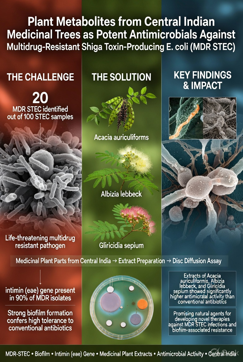
Introduction: The use of medicinal plants to cure human diseases is going on since the development of human civilization. Also, the discovery of antibiotics had profound impact to reduced death rates against various pathogens but due to rise of resistance against these antibiotics are serious threats for human health. Henceforth, the world is looking for alternative approach and the use of active plant metabolites are one of them. Multidrug resistant Shiga toxin producing Escherichia coli (MDR STEC) are life threatening microorganisms worldwide and their ability to produce aggregated biofilm makes them tolerant to many antibiotics used for treating STEC infections. In this study effect of plant metabolites were studied against MDR STEC samples. Methods: The different parts of 10 medicinal plants reported from central India were used in this study. Extract preparation and active fractions were used to test antimicrobial activity against MDR-STEC through measuring zone of inhibition. The biofilm structure was observed using electron microscopy. Results: Total 20 MDR STECs were identified out of 100 STEC samples. The intimin (eae) gene responsible for drug resistance was present in 18 (90%) MDR STEC samples. STEC were producing more aggregated biofilm layer as compare to sensitive E. coli. The plant extracts isolated from Acacia auriculiformis, Albizia lebbeck and Gliricidia sepium showed significantly high antimicrobial activity against MDR STEC as compared to various antibiotics. Conclusions: The study will be helpful to develop new or alternate antimicrobial agents and therapy against MDR STEC by using metabolites from medicinal plants.

Keywords: 
;  ;  ;  ;  ;  

1. Introduction

According to World Health Organization (WHO), more than 80% of the world's population relies on traditional medicine for their primary healthcare needs. Plants, as the source of medicine, have been playing an important role in the health services around the globe [1,2]. Use of herbal medicines worldwide represents a long history of human interactions with the environment [3]. Plants used in traditional medicine contain a wide range of ingredients that can be used to treat chronic as well as infectious diseases [4].
Microorganisms have potential to cause diseases. The human body is very prone to viral, bacterial and fungal infections [5]. The discovery of antibiotics in the early twentieth century provided an increasingly important tool to combat bacterial diseases [6]. But due to the indiscriminate use of commercial antimicrobial drugs commonly used in the treatment of infectious diseases multiple drug resistance (MDR) has been developed [1,7]. In addition to this problem, antibiotics are sometimes associated with adverse effects on the host including hypersensitivity, immune suppression and allergic reactions. This creates a need of new effective and safe antimicrobial therapeutic agents [8,9]. In this regard naturally available medicinal plants having active constituents with potent antimicrobial activity may provide the better treatment choice for drug resistant pathogens [10].
Antimicrobials of plant origin have enormous therapeutic potential [11]. They are effective in the treatment of infectious diseases while simultaneously mitigating many of the side effects that are often associated with synthetic antimicrobials [12]. The beneficial medicinal effects of plant materials typically result from the combinations of secondary products present in the plant. In plants, these compounds are mostly secondary metabolites such as alkaloids, steroids, tannins, and phenol compounds, flavonoids, steroids, resins fatty acids gums which are capable of producing definite physiological action on body [13].
Most E. coli bacteria are normal inhabitants of the intestinal tracts of humans and animals, and are non-pathogenic. However, some E. coli are pathogenic. Shiga toxin producing E. coli (STEC) is a type of pathogenic E. coli that, as the name implies, produces a potent toxin called Shiga toxin (Stx), also known as verotoxin or verocytotoxin. Stx causes blood vessel damage and plays a key role in other events that result in hemorrhagic colitis (bloody diarrhea) and a type of kidney failure called hemolytic uremic syndrome (HUS) in human patients [14]. Another virulence factor intimin encoded by eae gene responsible for adhesion and attachment to the surface of non-biotic and biotic. Intimin (eae gene) facilitate STEC to form biofilm for protection against harsh environment conditions as well as get resistance against antibiotics used to treat bacterial infection [15]. STEC, including E. coli O157:H7, is the number one cause of acute kidney failure in children [16]. Resistance to antimicrobials is a common issue worldwide and drug resistance in STEC infections is very common. For a better drug resistance free world, having good choice for treatment and to avoid human loss due to infection and drug resistance, people is working hard to get new antimicrobials to tackle the issue. Plant derived materials as a new antimicrobials and genetic typing of strains to analyze the resistance pattern and to stop the transmission of strains can be an option for fight against drug resistant STEC strains [17,18].
The present study address the role of few natural product easily available against clinical MDR STEC isolates, which is currently a significant public health problem worldwide and to assess the association of intimin (eae) gene with multidrug resistance.

2. Materials & Methods

2.1. Sample Collection & Isolation of STEC

A total of 100 STEC isolates were collected from stool samples of admitted patients in Bundelkhand Medical College Sagar and King George`s Medical University Lucknow (India) over a two-year period from 2015 to 2017. Each sample from the patients was transferred to the laboratory on the same day in transport media and cultured on Eosin Methylene Blue (EMB) agar medium (HiMedia, Mumbai, India) using a sterile glass spreader. The plates were then incubated at 37°C for overnight. After incubation the colonies showing metallic green shiny growth were picked and streaked on another EMB plates with the help of sterilized inoculating loop separately to get a single colony. Each colony was then streaked on different plates of Cefixime Tellurite-Sorbitol MacConkey (CT-SMAC) agar media (HiMedia, Mumbai,India) for confirmation of Escherichia coli O157 (E. coli O157) isolates. Further biochemical characterization was done by using certain biochemical test i.e. Methyl red, Voges Proskauer, indole, citrate utilization, lactose fermentation tests. Confirmation of STEC was done by presence of stx1 forward 5’-CGC TGA ATG TCA TTC GCT CTG C-3’, reverse 5’-CGT GGT ATA GCT ACT GTC ACC-3’ and stx2 forward 5’-ATC CTA TTC CCG GGA GTT TAC G-3’, reverse 5’-GCG TCA TCG TAT ACA CAG GAG C-3’ gene amplification by PCR using positive control (MTCC 9537).

2.2. Antibiotic Sensitivity Test

Antimicrobial susceptibility test of all isolates were performed on Muller Hinton Agar media by using Kirby -Bauer disc diffusion method [19] using Clinical Laboratory Standard Institute (CLSI 2016) guidelines. In brief, a diverse group of antibiotics used for the treatment of STEC associated infections were tested against each isolates e. g. Penicillin, Aminoglycosides, Fluroquinolones, Clavulanate (in combination), Carbapenems and Cephalosporins were used in 100µg/µl in concentration and purchased from HiMedia, Mumbai (India). Along with control strain E. coli (MTCC 9537). Sensitivity for antibiotics defined by minimum inhibitory concentration (MIC) dose required to inhibit bacterial growth. The isolates that were intermediate resistant (higher MIC than sensitive strain) or resistance (high MIC and most likelihood of therapeutic failure) against two or more antibiotics were denominated as multidrug resistant (MDR) isolates. A total of 20 MDR STEC strains were identified.

2.3. Biofilm Assay and Scanning Electron Microscopy

A static biofilm formation assay was performed in 96-well polystyrene microtiter plates for all the isolates along with negative (LB broth only) and positive (E. coli standard strains MTCC 9537) controls. Briefly, fresh grown STEC isolates were inoculated with 200µl Luria-Bertani (LB) broth at an initial turbidity of 0.5 at 600 nm and incubated at 37°C for overnight.
After incubation, supernatants were removed from each well and were washed thrice with PBS buffer (pH-7.2). Plates with STEC biofilms were then emptied, air-dried, and stained with 0.1% (w/v) crystal violet solution for 20 minutes. Plates were then washed by distilled sterile water for three times followed by air-drying of the wells. The dye bound biofilm was then dissolved in 100µl of 98% ethanol in to the well and mixed rigorously. All the dissolved biofilm containing ethanol was transferred into a fresh microtiter plate and optical density was measured by HTX Multimode Reader (BIOTEK, USA) at 570 nm. Biofilm assay was carried out three times in triplicates for each strain.
To determine the aggravation of biofilm of STEC isolates, Scanning Electron Microscopy (SEM) was done. For SEM analysis, STEC biofilm were grown on cover slip (18 mm) under 12-well cell culture polystyrene in 1 ml of LB broth at an initial turbidity of 0.5 at 600 nm of overnight fresh grown culture. After that the plate was incubated for overnight at 37°C. Supernatant was removed in the morning and the each well with cover slips was washed with PBS buffer (pH-7.2) for three times followed by dried in the air. Biofilm on cover slips was fixed with 0.25 % glutaraldehyde in PBS buffer (w/v, pH-7.2) for overnight. Cover slips were washed again three times with PBS buffer (pH-7.2). After dehydration of the biofilm with 30%, 50% 70%, 80%, 90% and absolute ethanol for 10 minutes at room temperature, samples were sputter coated with gold and viewed by SEM next day. SEM experiments were carried out in duplicate for each strain tested, and representative images of biofilm were selected. Standard strain E coli MTCC 9537 was taken as reference strain for SEM Microscopy.

2.4. Extraction of Plant Materials

Ethanolic extracts of fresh or dried plant (table 1) materials collected from the forest area in and around Sagar district, M.P. (India) was obtained by Soxhlet [20] extraction apparatus which was ran for 16 hrs. The antimicrobial activity of the each extracts was evaluated against MDR STEC isolates.

2.5. Susceptibility Test of Plant Extracts Against MDR STEC Isolates

Antibacterial susceptibility test of each plant extracts was performed against all MDR STEC isolates on Muller Hinton Agar (HiMedia, Mumbai) media by well diffusion and disc diffusion methods using CLSI-2016 [21] guidelines. For disc diffusion assay, one loop full of freshly grown culture of STEC was mixed in a tube containing 2 ml of sterile distilled water and OD was adjusted with 0.5 McFarland solutions. From this culture solution, 200µl of inoculum was spread evenly with a spreader on Muller Hinton Agar plates than antibiotic disc were placed using sterile forceps and kept for incubation for 16 hrs.
For well diffusion assay, 6 mm diameter wells were bored with sterile cork borer on the surface of the plates containing the above-said culture inoculum spread over the MHA plates. 100µg/µl reconstituted of each plant extracts were pipetted into each well. Inoculated plates with plant extracts were incubated at 370C at for overnight. The inhibition zone diameter was measured to the nearest millimeter (mm) by HiAntibiotic ZoneScale™- PW096 (HiMedia, Mumbai) for both tests. The test was performed in triplicate for each microorganism.

2.6. Intimin (eae) Genotype Detection

DNA was extracted from all the STEC strains by using QIAamp DNA mini kit (Qiagen, Germany) according to manufacturer instruction. Bacterial DNA amplification was done in 25µl reaction mixture containing 2.5µl of 10X Taq DNA polymerase buffer (containing 100 mM Tris with pH 9.0, 500 mM KCl, 15 mM MgCl2 and 1% Triton X-100), 2µl of 10 mM of dNTP mix, 0.9 U of Taq DNA polymerase (New England BioLab), 2µl each of 4 pM/µl of forward and reverse primers (eae F- 5’ TCA ATG CAG TTC CGT TAT CAG TT 3’, eae R- 5’GTA AAG TCC GTT ACC CCA ACCT G 3’) and 2µl of bacterial extracted DNA as template for PCR reaction. Amplification was done in thermal cycler (Applied Biosystems, USA) using following standardized conditions- Initial denaturation at 95°C for 5 minutes, Final denaturation at 95°C for 45 seconds, Annealing at 60°C for 30 seconds, initial extension at 72°C for 45 seconds and final extension at 72°C. The PCR was run for 35 cycles to amplify the STEC DNA and the amplified fragments were resolved by agarose (1.2%) gel electrophoresis, stained with ethidium bromide (0.5 µg/ml) and visualized by Gel documentation System (GelDoc-It, UVP,UK).

2.7. Statics

The results were compared with student t test and statically significance were shown (p values *>0.5, **>0.01, ***>0.001)

3. Results and Discussion

3.1. Characterization of MDR STEC

All the 100 clinical STEC isolates were tested for their antimicrobial susceptibility against varying spectrum of antibiotics commonly used for treatment of STEC infections using Kirby-Bauer disc diffusion method. Most of the isolates (80) were sensitive against different class of antibiotics used in this study. Resistant to Kanamycin, azithromycin, piperacillin, Ofloaxacin antibiotics were least common. The STEC isolates showed intermediate resistance to Cefazolin, Cefoxitin, Cefuroxime, Cefdinir, Cefaclor, Cefoperazone, Cefepime, Vancomycin, Methicillin, Ticarcillin, Ampicillin/Sulbactam, Ticarcillin/clavulanic acid, while resistant to antibiotics like, Cefpodoxime, Aztreonam, Penicillin G and Colistin observed (fig. 1). Further, we shorted 20 STEC isolates based on intermediate resistant or resistant to varying classes of antibiotics and denominated them as multidrug resistant (MDR) STEC isolates. All the MDR STEC isolates were further analyzed for biofilm aggregation/production. All the MDR STEC isolates were highly aggregated and producing high biofilm compared to sensitive isolates (fig. 2).

3.2. Antimicrobial Activity of Plant Extracts

Antimicrobial activity of the plant extracts were prepared by Soxhlet method and further tested against all the MDR STEC isolates. Different parts (table 1) of selected plants were used for making plant extracts. The antibacterial activity was performed by agar well diffusion and disc diffusion methods and measuring zone of inhibition (ZI). Results of antimicrobial susceptibility testing of plants showed higher frequency of sensitivity to MDR- STEC isolates as shown in table 2. The activity of Albizia lebbeck leaf extracts (12/20) was least frequent against different MDR STEC isolates. While extract from Gliricidia sepium (15/20), Millettia pinnata (16/20) A. lebbeck stem (17/20) and Terminalia arjuna (17/20) showed varied activity against MDR STECs. The maximum activity observed from extracts of Pithecellobium dulce and Pluchea lanceolate showing ZI against all the 20 MDR STEC. The average ZI were maximum in Acacia auriculaeformis and followed by G. sepium, Senna siamea and P. lanceolate. The minimum ZI was observed from T. arjuna extracts. When compared, A. auriculaeformis, Senna siamea, A. lebbeck & G. sepium showed significantly (p value >0.001, >0.05, >0.01) high antimicrobial activity compared to the antibiotics used (fig. 3). The plant extracts mostly from the stem and leaves reflected good antimicrobial activity.

3.3. Intimin (eae) Genotype Detection

We also analyze the prevalence of eae genes in all MDR STEC isolates. We found that the frequency of eae gene is more common in MDR STEC isolates. MDR- STEC isolates and data showed that almost 95% (19 Isolates) were positive for eae genes (fig. 4) and, are associated with resistant to varying spectrum of antibiotics. These isolates were also having the characteristics of highly aggravated production of biofilm compared to drug sensitive isolates, within hours after birth, bacteria colonize the gastrointestinal tract of infants and other mammals, beginning a lifelong symbiotic relationship where the host benefits from proper digestion and the bacteria are provided with an optimal, growth-supporting environment. However, this mutualism can be short lived. Consumption of contaminated food and water can introduce pathogenic bacteria to the gastrointestinal tract, causing a wide range of pathologies in animals and in humans. Enteric Escherichia coli are part of the normal intestinal flora in humans however certain pathotypes have acquired toxic traits during their evolution. These traits are encoded on genetic elements that are either fixed in their genomes or are mobile and can move between strains, creating new combinations of virulence factors. Strains acquiring highly virulent combinations have led to the evolution of six prevalent pathotypes characterized by their clinical outcomes in humans and they are listed as: Enteropathogenic E. coli (EPEC), Enterohemorrhagic E. coli (EHEC), Enteroinvasive E. coli (EIEC), Enteroaggregative E. coli (EAEC), Enterotoxigenic E. coli (ETEC), and Diffusely Adherent E. coli (DAEC). All EHEC produce Shiga toxins, originally named verocytotoxins, and are referred to as verocytotoxin-producing E. coli (VTEC) or more commonly, Shiga toxin-producing Escherichia coli (STEC).
In this study we observed the activity of plant extracts against the MDR STEC. The activity of different extracts was varied as variation in ZI against different isolates. Also the sensitivity of some plant extracts was more promising as compare to few other extracts. The variation in sensitivity and ZI depends on the part of plant used for extracts like Albizia lebbeck leaf vs. stem. This could be due to variation in compounds present in leaf and stem. Further, extracts showed least sensitivity against few isolates although having clear ZI in other isolates due to strain variation and needs further investigation. It is well known that the plant extracts can be a good source as antimicrobial agents [22] and exploitation of these could lead to a better world to cure infectious elements. The antimicrobial activity of plant extracts were dependent upon the plants material/parts used and its extraction methods [23].
In the present study, plant extracts were found to be more effective against MDR STEC isolates. There are numerous studies focusing on plenty of plant based compounds and reported very frequently about the activity of such compounds against various Gram-negative bacteria including STEC. Getting excellent results against clinical MDR STEC isolates of plant based compounds can pave the way to engineered new antibiotics and alternate therapy helpful to save millions of life around the world suffering from infectious pathogens. This study emphasized the role of plant extracts against MDR STEC isolates which were resistant to prescribed antibiotics used to treat STEC infections. The antibacterial effect of the plant extract justified its ethno-medicinal uses in traditional medicines and renders us to develop the efficient methodology and techniques to exploit them more rigorously to combat drug resistance in new modern world.
Presence of intimin (eae) gene in almost 95% of the isolates showed the relation between drug resistance and aggravated biofilm production. Microbial communities having the property of producing biofilm are responsible for higher drug and disinfectant resistance compare to non-producers. Plant associated material can be a good choice against biofilm producing pathogens. A study comprising the large sample size and advance molecular based techniques are required to further assess the biological active compounds against biofilm producers.

4. Conclusions

Plant extract are rich sources of useful chemical compounds and extensively used wordwide according to their need. In present study, it was observed that the plant extract obtained from various sources are active against multidrug resistant pathogenic bacteria and they can be used as a new compounds to treat patients after rigorous exploitation of pharmacology. The A. auriculaeformis, S. siamea, A. lebbeck and G. sepium are highly active against MDR STEC isolates, whereas T. arjuna has less activity among all the plant extracts used. The present work was planned to identify the active plant product as antimicrobial compound against MDR–STEC isolates. Moreover, a further study with having large group of microorganisms and elucidation of the structure and nature of pure chemical compounds and its activity spectrum against deadly microorganisms is required.

Author Contributions

All research done by the authors. SSG and RM had carried out experiments. RM interpreted data and conceptualize and proofread of manuscript.

Financial support

University Grants Commission Start-Up grant No F.30-56/2014 (BSR) to Dr Rajesh Mondal.

Declaration of Competing Interests

None.

Acknowledgments

Authors would like to thank Dr Sumit Rawat Microbiology Department Bundelkhand Medical Collage, Sagar (M.P.), Dr Sunita Singh, Department of Microbiology, King George`s Medical University, Lucknow (U.P.) for providing strains. Support from Soifisticated Instrument Centre SIC), Dr Harisingh Gour Viswavidyalaya, Sagar India is highly commendable..

References

  1. WHO Monographs on Selected Medicinal Plants- Volume 2 (1999), WHO, Genava.
  2. R.C. Agoha, Medicinal plants of Nigeria, Offsetdikker Jifaculteit waskunden, Natnurwenten schopp, pen, Netherlands, 1981, pp. 22-158.
  3. H.M. Burkill, The useful plants of West Tropical Africa vol 1, Families A-D, Royal Botanical Garden, Kew, 1984, pp. 441-415.
  4. H.M. Burkill, The useful plants of West Tropical Africa vol 3, Families J-L, Royal Botanical Garden, Kew, 1984, pp. 522.
  5. A. Sofowara, Medicinal plants and Traditional medicine in Africa. Spectrum Books Ltd, Ibadan, Nigeria, 1993, pp. 289.
  6. R. Taylor-Smith, Investigations on plants of West Africa, 111 Phytochemical Studies of some plants of Sierra Leone, Bull. Insst. France Afn noire A. 28 (1966) 528-541.
  7. H.O. Edeoga and A. Gomina, Nutritional values of some noncomventional leafy vegetables of Nigeria, J. Econ. Taxon. Bot. 24 (2000) 7-13.
  8. A.F. Hill, Economic Botany, A textbook of useful plants and plant products, 2nd ed., McGarw-Hill Book Company Inc, New York, 1952.
  9. D. Corogeanu, R. Willmes, M. Wolke, G. Plum, O. Utermohlen, M. Kronke, Therapeutic concentrations of antibiotics inhibit Shiga toxin release from enterohemorrhagic E. coli O104:H4 from the 2011 German outbreak, BMC Microbiol. 12 (2012) 160.
  10. Ahamad et al, Screening of some Indian medicinal plants for their antimi-crobial properties, Journal of Ethno pharmacology. 62 (1998) 183-193.
  11. J. Davis, Inactivation of the antibiotics and the dissemination of resistance genes, Science. 264 (1994) 375-382.
  12. S. Monroe and R. Polk, Antimicrobial use and bacterial resistance, Current Opinion Microbiology, 3 (2000) 496-501.
  13. G.E. Thomson, Further consideration of Asian Medicinal plants in treating common chronic disease in West, Journal of Medicinal Plants Research. 4 (2010) 125.
  14. S. Ethelberg, K.E. Olsen, F. Scheutz, C. Jensen, P, Schiellerup, J, Engberg et al, Virulence Factors for Hemolytic Uremic Syndrome, Denmark, Emerging Infectious Diseases. 10 (2004) 842-847. [CrossRef]
  15. M. Blanco, S. Schumacher, T. Tasara, C. Zweifel, J.E. Blanco, G. Dahbi, J. Blanco, R. Stephan, Serotypes, intimin variants and other virulence factors of eae positive Escherichia coli strains isolated from healthy cattle in Switzerland. Identification of a new intimin variant gene (eae-eta2), BMC Microbiology. 5 (2005) 23. [CrossRef]
  16. R. Wang, N. Kalchayanand, J.L. Bono, J.W. Schmidt, J.M. Bosilevac, Dual-serotype biofilm formation by shiga toxin-producing Escherichia coli O157:H7 and O26:H11 strains, Applied and environmental microbiology. 78 (2012) 6341–6344. [CrossRef]
  17. B.O. Obdoni, P.O.Ochuko, Phytochemical studies and comparative efficacy of the crude extracts of some Homostatic plants in Edo and Delta States of Nigeria, Global J. Pure Appl. Sci. 8b (2001) 203-208.
  18. T. Rahila, N. Rukhsandra, A.A. Zaidi, R. Shamishilia, Phytochemical Screening of medicinal plants belonging to Euphorbiaceae Pak, Vet. J. 14 (1994) 160-162.
  19. A.W. Bauer, D.M. Perry, W.M.M Kirby, Single-Disk Antibiotic-Sensitivity Testing of Staphylococci: An Analysis of Technique and Results, AMA Arch Intern Med. 104 (1959) 208–216. [CrossRef]
  20. D.S. Binnington, Improved soxhlet extraction apparatus, Fourth ed., Ind Eng Chem Anal Ed., 1932, pp. 125-126.
  21. Clinical Laboratory Standards Institute, Performance standards for antimicrobial disk susceptibility tests; approved standard—9th Ed. CLSI document M2-A9. 26:1. Clinical Laboratory Standards Institute, Wayne, PA, 2016.
  22. M.M. Cowan, Plant products as antimicrobial agents, Clin Microbiol Rev. 12 (1999) 564-82.
  23. J.Y. Lin, C.Y. Tang, Determination of total phenolic and flavonoid conents in selected fruits and vegetables, as well as their stimulatory effects on mouse splenocyte proliferation, Food Chem. 101 (2007) 140–147.
Figure 1. Antibiotic susceptibility test results of all STEC isolate. The graph represents percentage of isolates that are resistant to the antibiotics (commonly used to treat STEC infections) mentioned in the graph. S- Sulbactam, CA- Clavulanic Acid.
Preprints 206127 g001
Figure 2. Scanning Electron Microscopy (SEM) images (a) The image shows the biofilm formed by a normal E. coli strains whereas (b) is the image of aggregated biofilm formed by STEC isolates indicating that certain STEC isolated have a tendency to form aggregated biofilm.
Preprints 206127 g002
Figure 3. Comparison of Anti-MDR STEC activity of antibiotics with plant extracts. The graph compares the zone of inhibition of various antibiotics with the best three plant extracts. The graph shows that activity of these three plant extract is statistically higher than commonly used antibiotics used for the treatment of STEC infections. *** (p<0.001), ** (p<0.01) and * (p<0.05). All experiment was set up with three replicates and similar results were obtained in three independent experiments. CA- Clavulanic Acid.
Preprints 206127 g003
Figure 4. Gel showing 482 bp fragments of eae gene in 9 representative strains out of 20 MDR- STEC isolates, M= 100 bp DNA ladder, Lane 1-10 Strains.
Preprints 206127 g004
Disclaimer/Publisher’s Note: The statements, opinions and data contained in all publications are solely those of the individual author(s) and contributor(s) and not of MDPI and/or the editor(s). MDPI and/or the editor(s) disclaim responsibility for any injury to people or property resulting from any ideas, methods, instructions or products referred to in the content.
Copyright: This open access article is published under a Creative Commons CC BY 4.0 license, which permit the free download, distribution, and reuse, provided that the author and preprint are cited in any reuse.
Prerpints.org logo

Preprints.org is a free preprint server supported by MDPI in Basel, Switzerland.

Subscribe

Disclaimer

Terms of Use

Privacy Policy

Privacy Settings

© 2026 MDPI (Basel, Switzerland) unless otherwise stated