Submitted:
31 March 2026
Posted:
01 April 2026
You are already at the latest version
Abstract
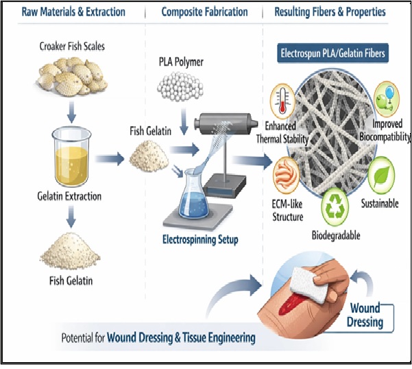
The poor performance of neat polylactic acid (PLA) and gelatin has driven the development of co- electrospun composites in biomaterials to achieve enhanced functional properties. In this study gelatin extracted from Crocker fish scale was co-electrospin with PLA. The composite fibres were fabricated at 2-17 wt.% gelatin. The electrospun fibres were evaluated via scanning electron microscope (SEM), Fourier transform spectroscopy (FTIR), differential scanning microscopy (DSC), thermogravimetric analysis (TGA), and Tensile test. FTIR analysis of PLA/gelatin fibres showed a peak growth at 1525cm-1 and a shift in the amide III band from 1239 cm-1 to 1192cm-1, indicating hydrogen bonding and chemical interaction between PLA and gelatin. The thermogram of the PLA/gelatin scaffold revealed an enhanced thermal stability with peak thermal stability (324oC) attained at 14 wt.% Gelatin. The DSC further confirms the interaction of PLA (Tg 75 OC) and gelatin (49 OC), forming a single glass transition temperature (Tg) at 70 OC. There was a slight increase in Tg of the composite fibres as the wight fraction of the gelatin increased. The SEM showed a good morphology resembling the native extracellular matrix (ECM) of the body. The ultimate tensile strength and percentage elongation of the fibres declined with increasing gelatin content. The introduction of this gelatin into PLA resulted in improved physiochemical properties of PLA/gelatin fibres due to chemical interaction. Thus, this composite fibre could serve as a potential wound dressing material.
Keywords:
PLA
; fish scale
; thermal stability
; wound dressing
; gelatin
1. Introduction
Fibrous structures at the nano and microscale can be produced from both natural and synthetic polymers via electrospinning. Electrospinning is an innovative and versatile technique that has been widely employed for fabricating fibrous materials because of its simplicity, continuous fibre production, precise control over fibre size, tunable porosity, and ease of surface functionalization [1].
Electrospun fibres with highly porous microstructures closely mimic the architecture of native extracellular matrices (ECMs), thereby enhancing cellular adhesion, proliferation and differentiation [2]. Consequently, electrospun scaffolds have found numerous applications in the biomedical and healthcare industries, particularly in Tissue engineering (TE) and Regenerative medicine (RM). Tissue engineering seeks to repair or regenerate defective tissues by combining biomaterials, cells, and bioactive factors [3]. Electrospun fibres are also widely explored for drug delivery, bone and cartilage regeneration, wound dressings, and advanced scaffolds for regenerative medicine [4,5,6].
More than 100 synthetic polymers have been successfully electrospun due to the simplicity and scalability of the process [7,8]. Among them, Poly-lactic acid (PLA) and its copolymer poly (lactic-co-glycolic acid) (PLGA) are the most widely used in biomedical application. Their popularity is attributed to their biocompatibility, biodegradability, non-toxicity, and mechanical stability [9]. PLA in particular is well recognized as a biodegradable polymer, with diverse applications, including medical implants and scaffold for tissue engineering. Its favourable properties include easy processability, excellent biocompatibility, a bioresorption rate that can be tuned to match tissue healing times, and the generation of non-toxic byproducts upon degradation [5]. However, PLA has limitations such as high hydrophobicity and local acidification during degradation impair cellular attachment, infiltration, and proliferation [10]. Co-spinning PLA with natural polymers, such as gelatin, is a promising strategy to overcome these drawbacks.
Gelatin, a natural polymer derived from collagen, exhibits excellent biocompatibility and biodegradability. It contains bioactive motifs, including arginine-glycine-aspartic acid (RGD) sequences, which promote cell adhesion, proliferation, and differentiation [11]. Nevertheless, gelatin’s poor mechanical strength, rapid degradation, and high hydrophilicity limit its use as a standalone scaffold material [12]. When blended with synthetic polymer like PLA, gelatin can enhance biological interactions while PLA provides the necessary mechanical stability and durability [1].
Several studies have investigated PLA/Gelatin composites and reported improved biological and physiochemical properties compared to PLA alone [12,13,15]. Gelatin, which is attractive due to its low cost and wide availability, is however, commercially sourced from Bovine and porcine tissues [16]. Concern over zoonotic diseases such as foot and mouth disease, as well as dietary and religious restrictions have driven the search for alternative gelatin sources. Fish-derived gelatin has emerged as a viable substitute offering comparable biocompatibility with fewer health and ethical concerns [8]. An et al. (2010) [8] successfully electrospun composite nanofibers from poly(l-lactide) (PLLA and gelatin extracted from catfish skin, achieving a scaffold with improved mechanical performance. More recently, gelatin has been extracted from various fish species, demonstrating promising results in biomedical applications [17,18].
In this study, gelatin was extracted from croaker fish scales of Nigerian origin and blended with PLA to fabricate electrospun scaffolds. To the best of our knowledge, electrospun PLA/gelatin composites derived from croaker fish scales have not been extensively reported. This research aligns with United Nations Sustainable Development Goals, particularly SDG 3(Good health and wellbeing), by advancing biocompatible wound dressing materials to support tissue regeneration and improved healthcare outcomes and SDG 12(responsible Consumption and Production),by transforming fish waste into sustainable materials, thereby promoting circular economy practices and reducing environmental impacts from marine by products. This study, therefore, seeks to explore their potential for biomedical applications, particularly in tissue engineering.
2. Materials and Methods
2.1. Materials
Polylactic acid (PLA) with an average molecular weight of 250,000 g/mol was obtained from Nature Works (Suzhou, China). Croaker fish scales were collected from Ojuelegba market, Lagos, Nigeria. Hydrochloric acid (HCl) and Dichloromethane (DCM), 95% purity were purchased from Sigma Alderich; while deionize water, was obtained from Metallurgical and Materials Engineering Laboratory, University of Lagos.
2.2. Methods
2.2.1. Extraction of Gelatin
The extraction of gelatin followed an acid pretreatment process. Approximately 200g of croaker fish scales were thoroughly washed with deionized water to remove surface impurities and oven dried at 70 oC to a constant weight. The dried scales were then immersed in 2.2 M of HCl, at a scale/solution ratio of 4:10 (w/v) for three hours (3 hrs) to decalcify and remove residual minerals. After decalcification, the samples were repeatedly washed with deionized water until a neutral pH was achieved. The scales were subsequently subjected to water bath extraction at 70 oC for 8 h using a fish scale/water ratio of 3:10 (w/v). The resulting gelatin solution was filtered through Hoffman filter paper (size 5) and freeze-dried for 24 hours to obtain powdered croaker fish gelatin.
2.2.2. Electrospinning of PLA/Gelatin
A 20% (w/v) of PLA solution was prepared by dissolving PLA pellets in dichloromethane (DCM) under magnetic stirring at 300 rpm until homogeneity was attained. Separately, gelatin was dissolved in 10 mL of deionize water at varying concentration (2 -17wt.%). The gelatin solutions were then added to the PLA solution in corresponding weight ratios and stirred for an additional 10 min to ensure uniform mixing. The prepared polymer solutions were loaded into a 10 mL syringe fitted with 21-gauge stainless steel needle. Electrospinning was carried out at a voltage of 15 KV, with the collector distance maintained at 12 cm. The fibres were collected on aluminium foil substrates and subsequently dried to remove residual solvents before characterisation.
2.3. Characterisation of Electrospun PLA/Gelatin Scaffolds
The morphological feature of PLA/gelatin was examined using a scanning microscope (Phenom Eindhoven, Netherlands). The samples were affixed on copper stub and was coated with gold for SEM observation. Functional group analysis via Fourier Transform Infrared Spectroscopy (FTIR) of the electrospun PLA/gelatin were done using Agilent Technologies Cary 630 spectrometer (India). The glass transition temperature (Tg) was observed using Mettler Toledo, DSC 1 star system. 5 mg of the samples were cut prior to the analysis. Thermal history of PLA/gelatin was carried out on TGA(Q500). 2mg of the fibres were heated to 750oC at 10oC/min. The tensile strength analysis of PLA/gelatin fibre was done using Instron Model 313. The electron samples were cut into 50 mm by 10 mm and were firmly held at both ends the samples were deformed until failure occurred.
3. Results and Discussion
3.1. Chemical Analysis of Electrospun PLA and PLA/Gelatin Scaffold
Electrospun neat PLA exhibits a weak asymmetric band of 1740cm-1 (Figure 1) that is attributed to C=O carbonyl stretching, with another absorption band of 1445cm-1, assigned to -CH stretching, which is approximately within the range reported by earlier studies [19–21]. The band at 1180 cm-1 is assigned to the ester group C-O, while 867 cm-1 is assigned to C-C stretching of the amorphous phase. Gelatin major peaks were found around 3304 cm-1, 1645 cm-1, 1527 cm-11239 cm-1, which are respectively assigned to OH group, C=O bending in amide I, C-H/C-H bending in amide II region, and amide III and 1058 [22].These peaks were also in the same range, with standard gelatin reported by other researchers [1,23–27]. The PLA/Gelatin electrospun composite showed more of the PLA spectral, with identical peaks irrespective of different weight fraction of gelatin; however, there is a peak growth in the band of 1525cm-1 for the PLA/Gelatin electrospun fibre. The amideI/II absorption band completely disappear as can be seen from the FTIR spectral of the electrospun fibre composite, which shows that PLA completely engulf the gelatin in the matrix. However, there was a slight peak shift in the amide III band (1239 cm-1) in gelatin to 1192cm-1 in the electrospun PLA/Gelatin composite, which established chemical interaction between PLA and gelatin. The peak shift is attributed to intermolecular hydrogen bonding within the functional groups [28]. The shift in peak has led to an enhanced C-H bond strength of electrospun PLA/Gelatin composite, which increased the stiffness and thermal stability of the composite fibre. Furthermore, another growth and broadening of peaks in the electrospun composite was observed, which were negligible in the spectra of gelatin and PLA. This could be attributed to the mechanism of superimposition of similar functional groups, leading to increment in the amount of the functional group present within the absorption band. The peak continued to grow as the percentage weight of gelatin increased from 2 to 17wt.%.
3.2. Thermal Properties of Electrospun PLA and PLA/Gelatin Scaffold
The thermogram of the electrospun PLA, gelatin, and its composites is shown in Figure 2. PLA exhibited a one-step decomposition, which agrees with Branco et al. (2018) and Nooeaid et al. (2020) reports [21,27]. Polylactide onset temperature is 302oC as seen in Table 1, with a maximum weight loss of 70 wt.% and decomposition temperature of 397 oC. The weight loss is as a result of the thermal decomposition of the electrospun PLA. However, gelatin onset temperature is 313 oC. It exhibits three-step decompositions, as reported by Nooeaid et al. (2020)[21]. Gelatin’s initial weight loss of 3 wt.% at 170 oC, is attributed to loss of moisture. The second stage is the decomposition of amino acids at 360 oC with about 25 wt.% loss and at 480oC decomposition of gelatin network [29]. The electrospun PLA/Gelatin composite fibre exhibited three stage decomposition characteristics of gelatin. The electrospun PLA/gelatin fibre mat becomes more thermally stable, compared to the individual polymer. The improved thermal stability is attributed to better interaction between PLA and Gelatin [30]. It is important to note that gelatin contains functional group such as the hydroxyl group and amine group, which can cross link with PLA molecules. This interaction has the capability to reduce the movement of the polymer chain, thus increasing the thermal stability of the electrospun PLA/Gelatin fibre mat. The improved thermal stability of the electrospun fibre mat varies from 316oC at 8 wt.% to peak of 324 oC at 14 wt.% gelatin. Thereafter there was a decline in the thermal stability to 271oC at 17 wt.%. Figure 3 shows the DTG of the individual polymer and the composite fibre mat. Gelatin showed maximum decomposition temperature at 397 oC, while PLA, PLA/gelatin (2 wt.%), PLA/gelatin (5 wt.%) demonstrated similar maximum decomposition temperature at 415 oC. Similarly, PLA/ gelatin (11 wt.%), PLA/gelatin (8 wt.%) exhibited maximum decomposition temperature of 385 oC. PLA/gelatin (14 wt.%) and PLA/gelatin (17 wt.%) has their decomposition temperature at 440 oC and 470 oC respectively.
3.3. Glass Transition Properties of Electrospun PLA and PLA/Gelatin Scaffold
The DSC of gelatin, PLA and PLA/gelatin composite is shown in Figure 4. Gelatin thermogram showed an endothermic peak. The glass transition and melting temperature were observed at 49oC and 53oC respectively. Similarly PLA glass transition and melting temperature was seen at 85oC and 172oC.which is similar to Nooeaid et al. (2020) report[21]. The electrospun composite exhibite more of the characteristic peak of gelatin with a Tg similar to that of gelatin, however there was a gradual shift in the Tg of the ellectrospun composite to higher value, with the reduction in the weight percent (8wt.%, 5wt.%, 3wt.%) of gelatin in the composite; this is assigned to increased chemical interacctions resulting in the restriction in molecular movement [31]. A single Tg occuring between the Tg of two polymers is an indication of polymer miscibiity. Two endothermic peak representting the melting temperature, was observed at 70 oC and 90 oC.The melting peak of gelatne undergoes significant increase in the melting temperature. The presence of two melting peak in the composite may be attributed to the formation of crystals of different sizes [32].
3.4. Morphological Evaluations of Electrospun PLA and PLA/Gelatin Scaffold
The morphology of electrospun PLA and gelatin is shown in Figure 5. The composite showed good fibre distribution, with enough pore spaces that allows tissue in growth and profilration. Similar morphology was also reported in the work done by Odili et al. (2025)[33]. The quantity of the fibres increased with increasing amount of gelatin that are present in the composite. The pore fibre diameter and pore size are shown in Figure 6. PLA/Gelatin (2 wt.%) has a fibre diameter and pore size of 6.3µm and 0.54 pore volume fraction respectively, while PLA/Gelatin (5 wt.%) showed fibre diameter and pore size of 2 µm and 0.42 pore volume fraction respectively. increasing the amount of gelatin leads to the thinning of the scaffold fibre.
3.5. Strength Characteristics of Electrospun PLA and PLA/Gelatin Scaffold
The tensile strength of PLA and PLA/Gelatin scaffolds are shown in Figure 7. The result reveals that the tensile strength of PLA is reduced, with increasing amount of gelatin. However, there was a spike at 14 wt.% of gelatin, thereafter the tensile strength declined. Similar observation was reported by earlier studies [8]19].The decline can be attributed to poor bonding between the PLA and gelatin matrix. Figure 8 also showed that the elongation properties of PLA was drastically reduced with the addition of gelatin. Despite the reduction, the elongation of PLA/gelatin scaffold, improved with incremental addition of gelatin, with the highest elongation attained at 14 wt.% of gelatin.
4. Conclusions
Gelatin from fish scale was successfully extracted, incorporated into PLA and electrospun into scaffolds, forming biodegradable composites suitable for biomedical use. The interaction between PLA and gelatin was confirmed through FTIR analysis, showing characteristic peak shifts that suggest hydrogen bonding between functional groups. Thermal analysis (TGA and DSC) revealed that the addition of gelatin enhances the thermal stability and miscibility of the composites, with peak performance observed at 14 wt.% gelatin. The SEM micrographs displayed interconnected fibrous networks with morphologies closely resembling native extracellular matrices, which are beneficial for tissue integration and nutrient transport. Although the tensile strength decreased with increasing gelatin content, the scaffold retained sufficient mechanical integrity for soft tissue application. These findings demonstrate that gelatin derived from croaker fish scale can effectively modify the physiochemical properties of PLA, providing ecofriendly and sustainable alternative to mammalian derived gelatin. The developed PLA/gelatin scaffold show promising potential for use as wound dressing materials and other tissue engineering applications, where biodegradability, hydrophilicity and biocompatibility are essential.
Author Contributions
Conceptualization, S.O.Adeosun and E.F.Ochulor.; methodology, C.C.Odili; software, C.C.Odili; validation, O.A. Olanrewaju, S.O.Adeosun and E.F.Ochulor; formal analysis, C.C.Odili.; investigation, O.A. Olanrewaju,.; resources, S.O.Adeosun; data curation, C.C.Odili.; writing—original draft preparation, C.C.Odili.; writing—review and editing, E.F.Ochulor S.O.Adeosun and O.A. Olanrewaju; visualization, C.C.Odili; supervision, S.O.Adeosun; project administration, E.F.Ochulor.; funding acquisition, E.F.Ochulor. All authors have read and agreed to this version of the manuscript.
Funding
This research received no external funding.
Data Availability Statement
Data are available from the corresponding author, upon reasonable request.
Conflicts of Interest
The authors declare that there is no conflicts of interest arising from this research.:.
Abbreviations
The following abbreviations are used in this manuscript:
| TE | Tissue engineering |
| RM | Regenerative medicine |
References
- Imani, F.; Karimi-Soflou, R.; Shabani, I.; Karkhaneh, A. PLA electrospun nanofibers modified with polypyrrole-grafted gelatin as bioactive electroconductive scaffold. Polymer 2021, 218. [Google Scholar] [CrossRef]
- Huang, Z.-M.; Zhang, Y.; Ramakrishna, S.; Lim, C. Electrospinning and mechanical characterization of gelatin nanofibers. Polymer 2004, 45, 5361–5368. [Google Scholar] [CrossRef]
- Murphy, S.V.; Atala, A. 3D bioprinting of tissues and organs. Nat. Biotechnol. 2014, 32, 773–785. [Google Scholar] [CrossRef]
- El-Seedi, H.R.; Said, N.S.; Yosri, N.; Hawash, H.B.; El-Sherif, D.M.; Abouzid, M.; Abdel-Daim, M.M.; Yaseen, M.; Omar, H.; Shou, Q.; et al. Gelatin nanofibers: Recent insights in synthesis, bio-medical applications and limitations. Heliyon 2023, 9, e16228. [Google Scholar] [CrossRef]
- Bahraminasab, M.; Doostmohammadi, N.; Talebi, A.; Arab, S.; Alizadeh, A.; Ghanbari, A.; Salati, A. 3D printed polylactic acid/gelatin-nano-hydroxyapatite/platelet-rich plasma scaffold for critical-sized skull defect regeneration. Biomed. Eng. Online 2022, 21, 1–25. [Google Scholar] [CrossRef] [PubMed]
- Xu, F.; Wang, H.; Zhang, J.; Jiang, L.; Zhang, W.; Hu, Y. A facile design of EGF conjugated PLA/gelatin electrospun nanofibers for nursing care of in vivo wound healing applications. J. Ind. Text. 2020, 51, 420S–440S. [Google Scholar] [CrossRef]
- Muthukrishnan, L. An overview on electrospinning and its advancement toward hard and soft tissue engineering applications. Colloid Polym. Sci. 2022, 300, 875–901. [Google Scholar] [CrossRef]
- An, K.; Liu, H.; Guo, S.; Kumar, D.; Wang, Q. Preparation of fish gelatin and fish gelatin/poly(l-lactide) nanofibers by electrospinning. Int. J. Biol. Macromol. 2010, 47, 380–388. [Google Scholar] [CrossRef]
- Zhao, W.; Li, J.; Jin, K.; Liu, W.; Qiu, X.; Li, C. Fabrication of functional PLGA-based electrospun scaffolds and their applications in biomedical engineering. Mater. Sci. Eng. C 2016, 59, 1181–1194. [Google Scholar] [CrossRef]
- Khorshidi, S.; Solouk, A.; Mirzadeh, H.; Mazinani, S.; Lagaron, J.M.; Sharifi, S.; Ramakrishna, S. A review of key challenges of electrospun scaffolds for tissue-engineering applications. J. Tissue Eng. Regen. Med. 2015, 10, 715–738. [Google Scholar] [CrossRef]
- Zhang, Y.Z.; Venugopal, J.; Huang, Z.-M.; Lim, C.T.; Ramakrishna, S. Crosslinking of the electrospun gelatin nanofibers. Polymer 2006, 47, 2911–2917. [Google Scholar] [CrossRef]
- Chen, H.; Zhang, H.; Shen, Y.; Dai, X.; Wang, X.; Deng, K.; Long, X.; Liu, L.; Zhang, X.; Li, Y.; et al. Instant in-situ Tissue Repair by Biodegradable PLA/Gelatin Nanofibrous Membrane Using a 3D Printed Handheld Electrospinning Device. Front. Bioeng. Biotechnol. 2021, 9. [Google Scholar] [CrossRef] [PubMed]
- Magiera, A.; Markowski, J.; Menaszek, E.; Pilch, J.; Blazewicz, S. PLA-Based Hybrid and Composite Electrospun Fibrous Scaffolds as Potential Materials for Tissue Engineering. J. Nanomater. 2017, 2017, 1–11. [Google Scholar] [CrossRef]
- Torricelli, P.; Gioffrè, M.; Fiorani, A.; Panzavolta, S.; Gualandi, C.; Fini, M.; Focarete, M.L.; Bigi, A. Co-electrospun gelatin-poly(l-lactic acid) scaffolds: Modulation of mechanical properties and chondrocyte response as a function of composition. Mater. Sci. Eng. C 2014, 36, 130–138. [Google Scholar] [CrossRef]
- Bogdanova, A.; Pavlova, E.; Polyanskaya, A.; Volkova, M.; Biryukova, E.; Filkov, G.; Trofimenko, A.; Durymanov, M.; Klinov, D.; Bagrov, D. Acceleration of Electrospun PLA Degradation by Addition of Gelatin. Int. J. Mol. Sci. 2023, 24, 3535. [Google Scholar] [CrossRef]
- Karim, A.A.; Bhat, R. Fish gelatin: properties, challenges, and prospects as an alternative to mammalian gelatins. Food Hydrocoll. 2009, 23, 563–576. [Google Scholar] [CrossRef]
- Feng, X.; Zhu, H.; Wang, Y.; Yu, Y.; Dai, H.; Ma, L.; Zhang, Y. Clean and sustainable extraction of gelatin: Effects of microwave and freeze-thaw on the crosslinking degree and hydrogen bond of fish skin collagen. Food Chem. 2024, 470, 142682. [Google Scholar] [CrossRef]
- Yu, E.; Pan, C.; Chen, W.; Ruan, Q.; Luo, X.; Lv, M.; Fang, Y.; Jiang, L.; Ma, H. Gelatin from specific freshwater and saltwater fish extracted using six different methods: Component interactions, structural characteristics, and functional properties. LWT 2023, 191. [Google Scholar] [CrossRef]
- Rashedi, S.; Afshar, S.; Rostami, A.; Ghazalian, M.; Nazockdast, H. Co-electrospun poly(lactic acid)/gelatin nanofibrous scaffold prepared by a new solvent system: morphological, mechanical and in vitro degradability properties. Int. J. Polym. Mater. Polym. Biomater. 2020, 70, 545–553. [Google Scholar] [CrossRef]
- Bagrov, D.V.; Nikishin, I.I.; Pavlova, E.R.; Klinov, D.V. Distribution of polylactide and gelatin in single electrospun nanofibers studied by Raman spectroscopy. STATE-OF-THE-ART TRENDS OF SCIENTIFIC RESEARCH OF ARTIFICIAL AND NATURAL NANOOBJECTS, STRANN-2018; LOCATION OF CONFERENCE, RussiaDATE OF CONFERENCE; p. 040001.
- Nooeaid, P.; Chuysinuan, P.; Pengsuk, C.; Dechtrirat, D.; Lirdprapamongkol, K.; Techasakul, S.; Svasti, J. Polylactic acid microparticles embedded porous gelatin scaffolds with multifunctional properties for soft tissue engineering. J. Sci. Adv. Mater. Devices 2020, 5, 337–345. [Google Scholar] [CrossRef]
- Švachová, V.; Vojtová, L.; Pavliňák, D.; Vojtek, L.; Sedláková, V.; Hyršl, P.; Alberti, M.; Jaroš, J.; Hampl, A.; Jančář, J. Novel electrospun gelatin/oxycellulose nanofibers as a suitable platform for lung disease modeling. Mater. Sci. Eng. C 2016, 67, 493–501. [Google Scholar] [CrossRef] [PubMed]
- Ahlawat, J.; Kumar, V.; Gopinath, P. Carica papaya loaded poly (vinyl alcohol)-gelatin nanofibrous scaffold for potential application in wound dressing. Mater. Sci. Eng. C 2019, 103, 109834. [Google Scholar] [CrossRef]
- Chiou, B.-S.; Jafri, H.; Avena-Bustillos, R.; Gregorski, K.S.; Bechtel, P.J.; Imam, S.H.; Glenn, G.M.; Orts, W.J. Properties of electrospun pollock gelatin/poly(vinyl alcohol) and pollock gelatin/poly(lactic acid) fibers. Int. J. Biol. Macromol. 2013, 55, 214–220. [Google Scholar] [CrossRef]
- Deng, L.; Li, Y.; Zhang, A.; Zhang, H. Characterization and physical properties of electrospun gelatin nanofibrous films by incorporation of nano-hydroxyapatite. Food Hydrocoll. 2020, 103. [Google Scholar] [CrossRef]
- Kotatha, D.; Hirata, M.; Ogino, M.; Uchida, S.; Ishikawa, M.; Furuike, T.; Tamura, H. Preparation and Characterization of Electrospun Gelatin Nanofibers for Use as Nonaqueous Electrolyte in Electric Double-Layer Capacitor. J. Nanotechnol. 2019, 2019, 1–11. [Google Scholar] [CrossRef]
- Caseiro, A.R.; Silva, D.M.; Amorim, I.; Rêma, A.; Pedrosa, S.S.; Branquinho, M.V.; Gomes, P.S.; Fernandes, M.H.R.; Santos, J.D.; Mauricio, A.C.; et al. Processing, Characterization, and in Vivo Evaluation of Poly(l-lactic acid)-Fish Gelatin Electrospun Membranes for Biomedical Applications. ACS Appl. Bio Mater. 2018, 1, 226–236. [Google Scholar] [CrossRef]
- Zia, I.; Mirza, S.; Jolly, R.; Rehman, A.; Ullah, R.; Shakir, M. Trigonella foenum graecum seed polysaccharide coupled nano hydroxyapatite-chitosan: A ternary nanocomposite for bone tissue engineering. Int. J. Biol. Macromol. 2019, 124, 88–101. [Google Scholar] [CrossRef]
- Correia, D.; Padrão, J.; Rodrigues, L.; Dourado, F.; Lanceros-Méndez, S.; Sencadas, V. Thermal and hydrolytic degradation of electrospun fish gelatin membranes. Polym. Test. 2013, 32, 995–1000. [Google Scholar] [CrossRef]
- Leonés, A.; Salaris, V.; Mujica-Garcia, A.; Arrieta, M.P.; Lopez, D.; Lieblich, M.; Kenny, J.M.; Peponi, L. PLA Electrospun Fibers Reinforced with Organic and Inorganic Nanoparticles: A Comparative Study. Molecules 2021, 26, 4925. [Google Scholar] [CrossRef]
- Ortega-Toro, R.; López-Córdoba, A.; Avalos-Belmontes, F. Epoxidised sesame oil as a biobased coupling agent and plasticiser in polylactic acid/thermoplastic yam starch blends. Heliyon 2021, 7, e06176. [Google Scholar] [CrossRef] [PubMed]
- Salaris, V.; García-Obregón, I.S.F.; López, D.; Peponi, L. Fabrication of PLA-Based Electrospun Nanofibers Reinforced with ZnO Nanoparticles and In Vitro Degradation Study. Nanomaterials 2023, 13, 2236. [Google Scholar] [CrossRef] [PubMed]
- Odili, C.C.; Gbenebor, O.P.; Obisike, R.U.; Adeosun, S.O. Strengthening propensity of hydroxyapatite-reinforced polylactide fibre. Discov. Polym. 2025, 2, 1–12. [Google Scholar] [CrossRef]
Figure 1.
FTIR spectral of electrospun PLA and PLA/Gelatin Composite.

Figure 2.
Thermogram of electrospun PLA and PLA/Gelatin Composite.

Figure 3.
DTG of electrospun PLA and PLA/Gelatin Composite.

Figure 4.
DSC of electrospun PLA and PLA/Gelatin Composite.

Figure 5.
a: PLA/Gelatin(2 w.%). b: PLA/Gelatin(5 wt.%). a and b: SEM morphology of electrospun PLA and PLA/Gelatin scaffold.
Figure 5.
a: PLA/Gelatin(2 w.%). b: PLA/Gelatin(5 wt.%). a and b: SEM morphology of electrospun PLA and PLA/Gelatin scaffold.

Figure 6.
A and B, Fibre diameter and pore size of PLA/Gelatin (2 wt.%). C and D, Fibre diameter and pore size of PLA/Gelatin (5 wt.%).
Figure 6.
A and B, Fibre diameter and pore size of PLA/Gelatin (2 wt.%). C and D, Fibre diameter and pore size of PLA/Gelatin (5 wt.%).

Figure 7.
Ultimate tensile strength of of electrospun PLA and PLA/Gelatin scaffold.

Figure 8.
Percentage elongation of electrospun PLA and PLA/Gelatin scaffold.


Table 1.
Thermal properties of electrospun PLA, gelatin and PLA/gelatin composite.
| Sample | On set | End set | Weight loss | First stage | 2nd stage | 3rd stage |
|---|---|---|---|---|---|---|
| PLA | 302 | 500 | 70 % | - | - | - |
| gelatin | 313 | 480 | 170 oc, & 2% | 360 oc, 25% | 43% | |
| PLA/2% gelatin | 321 | 502 | 203 oc ,3wt.% | 377oc, 39 wt.% | 502oc , 54 wt.% | |
| PLA/5% gelatin | 316 | 488 | 81wt.% | 187oc, 3wt.% | - | - |
| PLA/8% gelatin | 314 | 442 | 80WT.% | 161oc, 2wt.5 | - | - |
| PLA/11% gelatin | 322 | 430 | - | 158OC, 2WT.% | 361 oc, 26 wt.% | 44 wt.% |
| PLA/14% gelatin | 324 | 482 | -- | 173oc, 2wt.% | 357oc, 28 wt.% | 50 wt.% |
| PLA/17% gelatin | 271 | 533 | - | 466 | 38wt.% |
Disclaimer/Publisher’s Note: The statements, opinions and data contained in all publications are solely those of the individual author(s) and contributor(s) and not of MDPI and/or the editor(s). MDPI and/or the editor(s) disclaim responsibility for any injury to people or property resulting from any ideas, methods, instructions or products referred to in the content. |
© 2026 by the authors. Licensee MDPI, Basel, Switzerland. This article is an open access article distributed under the terms and conditions of the Creative Commons Attribution (CC BY) license (http://creativecommons.org/licenses/by/4.0/).
Copyright: This open access article is published under a Creative Commons CC BY 4.0 license, which permit the free download, distribution, and reuse, provided that the author and preprint are cited in any reuse.


