

Submitted:
10 March 2026
Posted:
11 March 2026
You are already at the latest version
Abstract
Background: Cancer cells exhibit metabolic reprogramming characterized by increased dependence on glutamine to sustain rapid proliferation and biosynthetic demands. Kidney-type glutaminase (KGA), which catalyzes the first and rate-limiting step of glutamine metabolism, represents a promising therapeutic target, particularly in triple-negative breast cancer (TNBC), an aggressive subtype lacking effective targeted therapies. This study evaluated 2-amino-4-boronobutyric acid (ABBA), a boronic acid-containing glutamine analog, as a potential KGA inhibitor with anticancer activity. Methods: KGA inhibition was assessed using a fluorometric enzymatic assay. Cytotoxic effects were examined in multiple TNBC cell lines. Covalent docking analysis was performed to characterize interactions between ABBA and the KGA active site. Results: ABBA potently inhibited KGA activity, with an IC₅₀ of approximately 1.0 μM, demonstrating greater efficacy than several non-proteinogenic amino acid analogs. ABBA induced dose-dependent cytotoxicity across multiple TNBC cell lines, with pronounced sensitivity observed in basal subtype cells. and Cellular sensitivity correlated with KGA expression levels. Expression of γ-glutamyl transpeptidase 1 (GGT1) was negligible, indicating that the observed anticancer effects are primarily mediated through KGA inhibition. Docking analysis predicted that ABBA forms a reversible covalent adduct with the catalytic Ser286 residue of KGA, adopting a boronate tetrahedral geometry consistent with transition-state mimics and stabilized by hydrogen bonding and electrostatic interactions. Conclusion: ABBA is a potent boron-based glutaminase inhibitor with therapeutic potential for targeting glutamine metabolism in TNBC. Further structural optimization and in vivo evaluation are warranted to advance ABBA toward therapeutic development.
Keywords:
triple-negative breast cancer
; 2-amino-4-boronobutyric acid
; glutaminase inhibitor
; boronic compounds
; non-proteinogenic amino acids
1. Introduction
Cancer cells exhibit profound metabolic reprogramming relative to normal tissue, characterized by elevated aerobic glycolysis and dysregulated amino acid uptake and metabolism [1]. Among amino acids, glutamine plays a central role in supporting tumor growth and proliferation [2,3,4]. Rapidly proliferating tumor cells undergo metabolic reprogramming and increased reliance on glutamine as a carbon and nitrogen source for the tricarboxylic acid (TCA) cycle anaplerosis, nucleotide and lipid biosynthesis, redox homeostasis, and ATP production. Although glutamine can be synthesized de novo from glutamate, it becomes conditionally essential during rapid proliferation or under metabolic stress conditions, resulting in increased dependency in many malignancies [5].
Cancer cells increase glutamine uptake via transporters such as SLC1A5 (ASCT2), followed by mitochondrial conversion to glutamate by glutaminase (GLS), the first and rate-limiting step in glutamine catabolism [5,6,7]. Glutamate subsequently contributes to cellular metabolism via aminotransferases, which replenish amino acid pools, or through glutamate dehydrogenase, which generates α-ketoglutarate to sustain the TCA cycle and anabolic growth [7,8,9]. This metabolic reliance on glutamine has led to the classification of malignancies including glioblastoma, leukemias, lymphomas, lung cancer, pancreatic cancer, and triple-negative breast cancer (TNBC) as “glutamine-addicted” tumors [6].
In humans, glutaminase exists as two major isozymes: kidney-type glutaminase (KGA), encoded by GLS1, and liver-type glutaminase (LGA), encoded by GLS2, both of which exhibit distinct tissue distributions and regulatory mechanisms [10,11]. GLS1 produces two primary splice variants: the full length KGA and the shorter glutaminase C (GAC) [12]. These variants share an identical N-terminal sequence (1–550) but differ in their C-terminal regions (residues 551–669 for KGA and 551–598 for GAC) [12]. Elevated GLS1 expression has been reported in basal-like/TNBC and HER2 positive breast cancers and is associated with poorer disease-free survival, particularly in positive lymph node metastasis [13]. Luminal B tumors also exhibit elevated GLS expression compared to luminal A tumors and correlates with poor clinical outcomes [13].
Given the importance of GLS in cancer metabolism, several glutaminase inhibitors have been developed and classified according to their binding sites. Competitive inhibitors, such as 6-diazo-5-oxo-L-norlucine (DON), Acivicin, DRP-104, and JHU-083 target the catalytic site and broadly inhibit glutamine-utilizing enzymes [14]. Although DON and Acivicin demonstrated antitumor activity, their lack of selectivity and dose-limiting toxicity halted clinical development [14]. Prodrug approaches, including DRP-104 and JHU-083, were designed to improve tumor selectivity and tolerance [15]. alongside other DON prodrugs including [16], Nedelcovych-13d [17], and Rais-5C [18]. In contrast, allosteric inhibitors such as BPTES bind to a hydrophobic regulatory site distinct from the catalytic center, inducing inactive conformations of GLS [19]. Improved derivatives, including CB-839 and IACS-6274 exhibit enhanced pharmacological properties, with IACS-6274 currently undergoing phase I clinical evaluation [14,20,21]. However, resistance to allosteric inhibitors, such as CB-839, has been reported in subsets of TNBC, limiting their clinical efficacy [22].
We previously demonstrated that hydroxyl arsinothricin (AST-OH) inhibits TNBC cell proliferation but is chemical instability due to oxidation of the trivalent arsenic center to the inactive pentavalent AST-OH [23]. Given the chemical similarities between arsenic and boron both are metalloids with analogous coordination properties. We explored boronic acid–based analogs as alternative glutaminase inhibitors. Boronic acids possess unique electronic and physicochemical properties including strong Lewis acidity and reversible covalent interactions with nucleophilic residues, making them attractive scaffolds in medicinal chemistry [24,25,26].
Here, we evaluate 2-amino-4-boronobutyric acid (ABBA), a glutamine analog boronic compound previously characterized as an inhibitor of gamma-glutamyl transpeptidase [27]. Using biochemical assays, cell-based studies, and covalent docking analysis, we demonstrate that ABBA potently inhibits KGA activity and suppresses TNBC cell viability. and forms a predicted covalent adduct with the catalytic Ser286 residue. These findings identify ABBA as a promising boron-based glutaminase inhibitor with potential therapeutic relevance in TNBC.
2. Materials and Methods
2.1. Chemicals
All chemicals and enzymes were purchased from Sigma-Aldrich Co., (St. Louis, MO, USA) unless otherwise specified. AST–OH was synthesized as previously described and used as a racemic mixture, with analyses focused on the L-enantiomer. Compound purity was confirmed by high-pressure liquid chromatography (series 2000, PerkinElmer, Waltham, MA, USA) coupled with inductively coupled plasma mass spectrometry (HPLC-ICP-MS, ELAN DRC-e; PerkinElmer). Trivalent R-AST–OH was obtained by chemically reduction of the pentavalent AST–OH. ABBA was obtained from Mcule, Inc. (Palo Alto, CA, USA).
2.2. KGA Inhibition Assay
Glutaminase inhibition was assessed using a commercial GLS1 inhibitor screening kit (Cat# ab283389, Abcam, Waltham, MA, USA). The assay was conducted as described in the user manual. Briefly, in a 96-well black plate, all inhibitors at a final concentration of 10 µM were pre-incubated with KGA for 15 minutes. The reaction was initiated by the addition of substrate glutamine. The inhibition of all compounds was monitored by fluorescence at 535 nm excitation and 587 nm emission wavelengths using BioTek Synergy Neo 2 plate reader for up to 30 minutes. The initial rate was calculated for each sample, and the percentage of inhibition was determined using the no-inhibitor condition as 0% and 2 µM CB-839 as 100%. It is assumed that 2 µM CB-839 fully inhibits GLS1. The dose-response curve for ABBA and R-AST-OH (0.01–100 μM) were generated using Sigma Plot and all experiments were performed in triplicate.
2.3. Cell Culture and Viability Assay
TNBC cell lines HCC70 (ATCC® CRL-2315TM), HCC38 (ATCC® CRL-2314TM), HCC1937 (ATCC® CRL-2336TM), and BT549 (ATCC® HTB-122TM) were obtained from ATCC (Manassas, VA, USA) and cultured under recommended conditions. These cell lines were maintained in the recommended media under 5% CO2 in a humidified incubator at 37 °C. Cells were seeded at 3.0 × 10⁴ cells/well in 96-well plates and incubated for 24 hours before being exposed to concentrations from 0.001 to 1000 µM of ABBA compounds for 72 hours. Cell viability was then assessed using a CellTiter-Glo Luminescent Cell Viability Assay (Promega). The assay measured relative luminescence as an indicator of viability. The results showed dose-dependent cytotoxic effects of ABBA compounds on TNBC cell lines, with half-maximal inhibitory concentration (IC50) values calculated to assess potency.
2.4. Covalent Docking
The crystal structure of the DON molecule bound to KGA was derived for docking from the protein data bank (4O7D) [28]. Water molecules and the DON molecule were removed before docking, and only one monomer was used for analysis. Flare software [29] facilitated protein and ligand preparation as well as docking evaluation. The SMILES of the ABBA compound were obtained from PubChem, and its three-dimensional structure was generated in Flare. The docking grid, with 6 Å dimensions, was centered around the oxygen atom of Ser286, where DON binds. The top-ranked conformations were selected for further analysis, with a binding energy of -6.513 kcal/mol for ABBA and KGA. Docking results were visualized using PyMOL [30].
3. Results
3.1. Structural Analysis of Glutamine Analogs Identifies ABBA as a Potent KGA Inhibitor
Pharmacological inhibition of KGA (GLS) has emerged as a promising therapeutic strategy against cancer cells that utilize glutamine as an energy source for rapid growth and proliferation. The mitochondrial conversion of glutamine to glutamate by GLS represents the first and rate-limiting step in glutamine utilization, and specifically in TNBC, elevated GLS expression has been associated with cancer growth [31]. Figure 1 illustrates the chemical structures of glutamine (Figure 1a), and related glutamine analog inhibitors (Figure 1b-f). All compounds share a four-carbon backbone, and 6-diazo-5-oxo-L-norleucine (DON) (Figure 1b) serving as a well-characterized reference inhibitor. DON inhibits KGA and its isoforms by binding to the catalytic active site, and functions as a glutamine antagonist, thereby disrupting nucleotide and protein synthesis pathways that rely on glutamine [28]. Structural studies of glutaminase from E. coli and B. subtilis in complex with DON have identified the critical role of a conserved serine residue as the catalytic nucleophile [32] which corresponds to Ser286 in human KGA where the catalytic dyad composed of Ser286 and Lys289 is critical for enzyme catalysis [28]. However, despite its potency, DON lacks selectivity and inhibits multiple glutamine-dependent enzymes, including amidotransferases and glutamine synthetase, resulting in unacceptable toxicity in animal models that precluded further clinical development [14].
Previously, we demonstrated that the non-proteinogenic amino acids 2-amino-4-phosphonobutyric acid (AP4, Figure 1c) and hydroxyl arsinothricin (AST-OH) exhibit improved inhibitory activity against KGA relative to DON [23]. Notably, trivalent AST-OH displays potent anti-cancer activity in TNBC cell-based assays. However, the trivalent arsenic atom of AST-OH is chemically unstable and readily oxidizes to the pentavalent form (R=AST-OH, Figure 1d), which significantly diminishes its biological efficacy [23].
To overcome this limitation, we explored substitution of the arsenic atom in AST-OH with boron, a metalloid that shares similar coordination chemistry. Boronic acids possess a unique ability to function as strong Lewis acids due to the electron-deficient boron center, enabling reversible covalent interactions with nucleophilic residues [25]. Based on these considerations, we evaluated ABBA a structural analog of AST-OH in which the arsenic atom is replaced by boron. ABBA is known as a potent inhibitor of GGT1, and crystallographic studies have shown that L-ABBA binds covalently to the active-site and forms a covalent bond with threonine residue of GGT1. [27] Given the shared catalytic serine nucleophile in KGA, we hypothesized that L-ABBA could similarly target the KGA active site and function as an effective glutaminase inhibitor with potential anti-cancer activity in TNBC.
3.2. ABBA Potently Inhibits KGA Activity
KGA inhibition by AP4, DON, AST-OH, R-AST-OH, and ABBA was evaluated using a fluorometric glutaminase assay. All compounds were initially tested at a final concentration of 10 μM, and the percentage of inhibition was calculated relative to the no-inhibitor control (Figure 2a). At 10 μM, both R-AST-OH and ABBA showed near-complete inhibition of KGA activity, whereas the AP4, DON, and AST-OH exhibited minimal inhibitory effects. Consistent with previous reports, the IC50 values of AP4, DON, and AST-OH have been reported to be approximately 0.5 mM, 0.9 mM, and 0.25 mM, indicating comparatively weak inhibition. To further quantify inhibitory potency, dose-response inhibition analyses were performed for R-AST-OH and ABBA across concentrations from 0.01 µM to 100 µM (Figure 2b). These experiments yielded IC50 values of 0.05 µM for R-AST-OH and 1.0 µM for ABBA. These results identify ABBA as a more potent KGA inhibitor than previously evaluated non-proteinogenic glutamine analogs.
3.3. ABBA Suppresses TNBC Cell Viability
To evaluate antiproliferative effects of ABBA on cancer cells, we performed cell viability assays using a panel of TNBC cell lines representing distinct molecular subtypes: HCC70 (basal B), HCC38 (basal A), HCC1937 (basal A), and BT549 (mesenchymal). Cells were treated with increasing concentrations of ABBA for 72 h, and viability was assessed using the CellTiter-Glo luminescent assay (Promega), and IC50 values were calculated for each cell line (Figure 3). ABBA treatment resulted in a dose-dependent reduction in cell viability across all TNBC cell lines tested. At 10 µM, ABBA caused near-complete loss of viability in all four cell lines. Among these, HCC70 cells exhibited the greatest sensitivity to ABBA. The calculated IC50 values were 0.04 µM for HCC70, 0.23 µM for HCC38, 1 µM for HCC1937, and 0.8 µM for BT549 cells, indicating differential sensitivity among TNBC subtypes. Basal subtype TNBC cells demonstrated greater sensitivity to ABBA compared to the mesenchymal subtype.
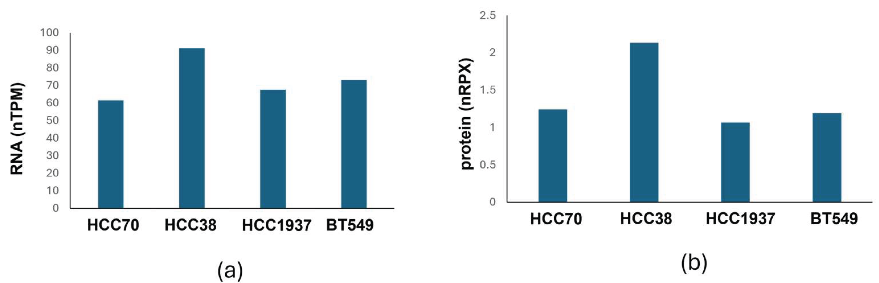
To assess whether this variability was associated with KGA expression, protein expression data for KGA/GLS1 in the four TNBC cell lines were obtained from the Human Protein Atlas (Figure 4) [33]. Comparison of KGA expression levels with ABBA sensitivity revealed a trend toward increased sensitivity in cell lines with higher KGA expression. For example, HCC38 exhibited slightly higher KGA expression than HCC1937, consistent with their lower IC50 values. Although preliminary, these findings suggest that ABBA-mediated suppression of TNBC cell viability may be linked to KGA expression levels. However, additional studies across a broader panel of cell lines and mechanistic analyses are required to determine whether KGA expression is a predictive determinant of ABBA sensitivity and to elucidate the underlying molecular mechanisms.
3.4. Covalent Docking Supports Targeting of Catalytic Ser286
Boronic acid-containing compounds can form reversible covalent bonds with nucleophilic serine residues in proteins, acting as transition state analogs through conversion from trigonal to tetrahedral geometry. DON has been shown to inhibit KGA by covalently modifying the catalytic Ser286 residue. During this process, the diazo group of DON is released, leaving 5-oxo-L-norleucine (ON) covalently bound to Ser286 and resulting in enzyme inhibition [28].
Based on this established mechanism, we hypothesized that ABBA could similarly interact with the catalytic Ser286 residue via formation of a boron-serine adduct. To evaluate this possibility, covalent docking of ABBA to KGA was performed in silico using Flare software. Covalent docking simulations predicted stable binding of ABBA within the KGA active site, with a calculated binding energy of -6.513 kcal/mol (Figure 5a). The docking model indicated that the boron atom of ABBA forms a covalent bond with the hydroxyl side chain of Ser286, adopting a tetragonal geometry, consistent with boronic acid–serine interactions (Figure 5b).
In addition to the covalent bond, multiple non-covalent contacts contributed to stabilization of the ABBA–KGA complex (Figure 5c). The amino group of ABBA forms hydrogen bonds with Gln285 and Glu381, while the hydroxyl group also engaged in hydrogen bonding with Glu381. The carbonyl group of ABBA established hydrogen bond interactions with Glu381, Asn388, and Tyr414. Furthermore, the positively charged Lys289 provided electrostatic stabilization of the negatively charged boronic species. Collectively, these data support a binding mode in which ABBA engages the catalytic center of KGA through a combination of covalent and non-covalent interactions, consistent with its potent enzymatic inhibition.
4. Discussion
Breast cancer remains a leading cause of cancer-related mortality among women worldwide. Clinically, breast tumors are classified based on the expression of estrogen receptor (ER), progesterone receptor (PR), and human epidermal growth factor receptor 2 (EGFR2/Her2), which guide prognosis and therapeutic decision-making [34]. ER/PR-positive tumors are generally responsive to endocrine therapies, whereas Her2-positive cancers benefit from Her2-targeted treatments [35]. In contrast, the triple-negative breast cancer (TNBC), defined by the absence of ER, PR, and HER2 expression, represents the most aggressive subtype and lacks effective targeted therapies [34]. TNBC accounts for approximately 10-20% of breast cancer cases, and is associated with increased metastatic potential, higher recurrence rates, and poorer clinical outcomes [36].
Cancer cells exhibit metabolic plasticity that enables adaptation to increased energetic and biosynthetic demands. Glutamine functions as a critical carbon and nitrogen source, supporting nucleotide synthesis, redox homeostasis, and anaplerotic flux into the tricarboxylic acid cycle. Accordingly, targeting glutamine metabolism has emerged as a promising therapeutic strategy, particularly for metabolically aggressive tumors such as TNBC. The KGA or GLS-1, which catalyzes the first and rate-limiting step in glutamine utilization, represent a key metabolic vulnerability in these cancers [6].
In this study, we demonstrate that the boron-containing non-proteinogenic amino acid ABBA inhibits KGA activity and suppresses TNBC cell viability. Compared to other glutamine analogs, ABBA exhibited enhanced inhibitory potency, consistent with its unique chemical properties. Boron, a metalloid distinct from carbon and silicon, exhibits strong Lewis acidity and readily forms reversible covalent interactions through trivalent coordination geometry. Upon interaction with nucleophilic residues, boron can transition from a trigonal planar to a tetrahedral sp3 configuration, enabling formation of stable yet reversible covalent adducts.
This chemical behavior has been widely exploited in the development of boron-based enzyme inhibitors, particularly those targeting serine residues within catalytic active sites. Our in silico covalent docking analysis supports a mechanism in which ABBA forms a tetrahedral boron–serine adducts with the catalytic Ser286 residue of KGA. Additional hydrogen bonding and electrostatic interactions further stabilize the ABBA–KGA complex, providing a structural basis for its potent inhibitory activity.
Functionally, ABBA induced marked cytotoxicity across multiple TNBC cell lines, with greater sensitivity observed in basal-like subtypes. Given that ABBA is a known inhibitor of GGT1, we assessed whether GGT1 inhibition contributed to the observed cellular effects. Analysis of transcriptomic and proteomic data demonstrated negligible GGT1 expression across all TNBC cell lines examined, indicating that the reduction in cell viability is unlikely to be mediated through GGT1 inhibition. These data suggest that KGA inhibition is the predominant mechanism underlying ABBA’s anticancer activity.
Together, our results identify ABBA as a promising boron-based glutaminase inhibitor that exploits glutamine dependency in TNBC. While further studies are warranted to evaluate in vivo efficacy, pharmacokinetic properties, selectivity, and potential therapeutic synergy with existing chemotherapeutic or metabolic-targeting strategies. This work further supports the development boron-containing glutamine analogs as a novel class of metabolic inhibitors for the treatment of aggressive breast cancers.
Author Contributions
V.S.N. designed and performed the research; V.S.N., D. D., T. V., B.P.R., and H.S.C., analyzed and reviewed the data; V.S.N., B.P.R., and H.S. C. wrote and edited the paper. All authors have read and agreed to the published version of the manuscript.
Acknowledgments
This work was supported by a pilot project grant from the Research Center in Minority Institutions, Florida International University (1U54MD012393-03), and Florida Breast Cancer Foundation (AWD 15620) to V.S.N. and NIH grant R35 GM136211 to B.P.R.
Abbreviations
The following abbreviations are used in this manuscript
KGA - Kidney-type glutaminase
GLS1 - Glutaminase 1
TNBC - Triple-negative breast cancer
ABBA - 2-amino-4-boronobutyric acid
GGT1 - γ-glutamyl transpeptidase 1
TCA - Tricarboxylic acid cycle
DON - 6-diazo-5-oxo-L-norlucine
AST-OH - Pentavalent hydroxyarsinothricin
R-AST-OH – Trivalent hydroxyarsinothricin
AP4 - 2-amino-4-phosphonobutyric acid
References
- Schiliro, C.; Firestein, BL. Mechanisms of Metabolic Reprogramming in Cancer Cells Supporting Enhanced Growth and Proliferation. Cells 2021, 10(5), 1056. [Google Scholar] [CrossRef]
- Jin, J; Byun, JK; Choi, YK; Park, KG. Targeting glutamine metabolism as a therapeutic strategy for cancer. Exp Mol Med. 2023, 55(4), 706–715. [Google Scholar] [CrossRef] [PubMed]
- Choi, YK; Park, KG. Targeting Glutamine Metabolism for Cancer Treatment. Biomol Ther (Seoul) 2018, 26(1), 19–28. [Google Scholar] [CrossRef]
- Rajagopalan, KN; DeBerardinis, RJ. Role of glutamine in cancer: therapeutic and imaging implications. J Nucl Med. 2011, 52(7), 1005–8. [Google Scholar] [CrossRef]
- Kumar, MA; Baba, SK; Khan, IR; Khan, MS; Husain, FM; Ahmad, S; Haris, M; Singh, M; Akil, ASA; Macha, MA; Bhat, AA. Glutamine Metabolism: Molecular Regulation, Biological Functions, and Diseases. MedComm (2020) 2025, 6(7), e70120. [Google Scholar] [CrossRef]
- Fan, Y; Xue, H; Li, Z; Huo, M; Gao, H; Guan, X. Exploiting the Achilles’ heel of cancer: disrupting glutamine metabolism for effective cancer treatment. Front Pharmacol. 2024, 15, 1345522. [Google Scholar] [CrossRef]
- Yoo, HC; Park, SJ; Nam, M; Kang, J; Kim, K; Yeo, JH; Kim, JK; Heo, Y; Lee, HS; Lee, MY; Lee, CW; Kang, JS; Kim, YH; Lee, J; Choi, J; Hwang, GS; Bang, S; Han, JM. A Variant of SLC1A5 Is a Mitochondrial Glutamine Transporter for Metabolic Reprogramming in Cancer Cells. Cell Metab 2020, 31(2), 267–283.e12. [Google Scholar] [CrossRef]
- Choi, I; Son, H; Baek, JH. Tricarboxylic Acid (TCA) Cycle Intermediates: Regulators of Immune Responses. Life (Basel) 2021, 11(1), 69. [Google Scholar] [CrossRef]
- Cluntun, A.A.; Lukey, M.J.; Cerione, R.A.; Locasale, J.W. Glutamine metabolism in cancer: understanding the heterogeneity. Trends Cancer 2017, 3, 169–180. [Google Scholar] [CrossRef] [PubMed]
- Katt, W.P.; Lukey, M.J.; Cerione, R.A. A tale of two glutaminases: homologous enzymes with distinct roles in tumorigenesis. Future medicinal chemistry 2017, 9(2), 223–243. [Google Scholar] [CrossRef] [PubMed]
- Altman, B.J.; Stine, Z.E.; Dang, C.V. From Krebs to clinic: glutamine metabolism to cancer therapy. Nature Reviews Cancer 2016, 16(10), 619–634. [Google Scholar] [CrossRef]
- Mates, J.M.; Segura, J.A.; Martin-Rufian, M.; Campos-Sandoval, J.A.; Alonso, F.J.; Marquez, J. Glutaminase isoenzymes as key regulators in metabolic and oxidative stress against cancer. Current molecular medicine 2013, 13(4), 514–534. [Google Scholar] [CrossRef]
- Vidula, N; Yau, C; Rugo, HS. Glutaminase (GLS1) gene expression in primary breast cancer. Breast Cancer 2023, 30(6), 1079–1084. [Google Scholar] [CrossRef]
- Xu, X.; Meng, Y.; Li, L.; Xu, P.; Wang, J.; Li, Z.; Bian, J. Overview of the development of glutaminase inhibitors: achievements and future directions. Journal of medicinal chemistry 2018, 62(3), 1096–1115. [Google Scholar] [CrossRef]
- Rais, R.; Lemberg, K.M.; Tenora, L.; Arwood, M.L.; Pal, A.; Alt, J.; Wu, Y.; Lam, J.; Aguilar, J.M.H.; Zhao, L.; Peters, D.E. Discovery of DRP-104, a tumor-targeted metabolic inhibitor prodrug. Science advances 2022, 8(46), eabq5925. [Google Scholar] [CrossRef] [PubMed]
- Hanaford, A.R.; Alt, J.; Rais, R.; Wang, S.Z.; Kaur, H.; Thorek, D.L.; Eberhart, C.G.; Slusher, B.S.; Martin, A.M.; Raabe, E.H. Orally bioavailable glutamine antagonist prodrug JHU-083 penetrates mouse brain and suppresses the growth of MYC-driven medulloblastoma. Translational oncology 2019, 12(10), 1314–1322. [Google Scholar] [CrossRef] [PubMed]
- Rais, R.; Jancarik, A.; Tenora, L.; Nedelcovych, M.; Alt, J.; Englert, J.; Rojas, C.; Le, A.; Elgogary, A.; Tan, J.; Monincova, L. Discovery of 6-diazo-5-oxo-l-norleucine (DON) prodrugs with enhanced CSF delivery in monkeys: a potential treatment for glioblastoma. Journal of medicinal chemistry 2016, 59(18), 8621–8633. [Google Scholar] [CrossRef]
- Nedelcovych, M.T.; Tenora, L.; Kim, B.H.; Kelschenbach, J.; Chao, W.; Hadas, E.; Jančařík, A.; Prchalová, E.; Zimmermann, S.C.; Dash, R.P.; Gadiano, A.J. N-(Pivaloyloxy) alkoxy-carbonyl Prodrugs of the Glutamine Antagonist 6-Diazo-5-oxo-l-norleucine (DON) as a Potential Treatment for HIV Associated Neurocognitive Disorders. Journal of medicinal chemistry 2017, 60(16), 7186–7198. [Google Scholar] [CrossRef]
- Robinson, M.M.; Mcbryant, S.J.; Tsukamoto, T.; Rojas, C.; Ferraris, D.V.; Hamilton, S.K.; Hansen, J.C.; Curthoys, N.P. Novel mechanism of inhibition of rat kidney-type glutaminase by bis-2-(5-phenylacetamido-1, 2, 4-thiadiazol-2-yl) ethyl sulfide (BPTES). Biochemical Journal 2007, 406(3), 407–414. [Google Scholar] [CrossRef] [PubMed]
- Gross, M.I.; Demo, S.D.; Dennison, J.B.; Chen, L.; Chernov-Rogan, T.; Goyal, B.; Janes, J.R.; Laidig, G.J.; Lewis, E.R.; Li, J.; MacKinnon, A.L. Antitumor activity of the glutaminase inhibitor CB-839 in triple-negative breast cancer. Molecular cancer therapeutics 2014, 13(4), 890–901. [Google Scholar] [CrossRef]
- Soth, M.J.; Le, K.; Di Francesco, M.E.; Hamilton, M.M.; Liu, G.; Burke, J.P.; Carroll, C.L.; Kovacs, J.J.; Bardenhagen, J.P.; Bristow, C.A.; Cardozo, M. Discovery of IPN60090, a clinical stage selective glutaminase-1 (GLS-1) inhibitor with excellent pharmacokinetic and physicochemical properties. Journal of medicinal chemistry 2020, 63(21), 12957–12977. [Google Scholar] [CrossRef] [PubMed]
- Xu, X.; Wang, J.; Wang, M.; Yuan, X.; Li, L.; Zhang, C.; Huang, H.; Jing, T.; Wang, C.; Tong, C.; Zhou, L. Structure-enabled discovery of novel macrocyclic inhibitors targeting glutaminase 1 allosteric binding site. Journal of Medicinal Chemistry 2021, 64(8), 4588–4611. [Google Scholar] [CrossRef]
- Sarkarai Nadar, V.; Yoshinaga-Sakurai, K.; Rosen, B.P. Anticancer Effects of the Trivalent Organoarsenical 2-Amino-4-(dihydroxyarsinoyl) Butanoate. Organometallics 2024, 43(10), 1137–1142. [Google Scholar] [CrossRef]
- Sekhon, BS. Metalloid compounds as drugs. Res Pharm Sci 2013, 8(3), 145–58. [Google Scholar] [PubMed] [PubMed Central]
- Ali, F; S Hosmane, N; Zhu, Y. Boron Chemistry for Medical Applications. Molecules 2020, 25(4), 828. [Google Scholar] [CrossRef] [PubMed] [PubMed Central]
- Alsenani, TA; Rodríguez, MM; Ghiglione, B; Taracila, MA; Mojica, MF; Rojas, LJ; Hujer, AM; Gutkind, G; Bethel, CR; Rather, PN; Introvigne, ML; Prati, F; Caselli, E; Power, P; van den Akker, F; Bonomo, RA. Boronic Acid Transition State Inhibitors as Potent Inactivators of KPC and CTX-M β-Lactamases: Biochemical and Structural Analyses. Antimicrob Agents Chemother 2023, 67, e00930-22. [Google Scholar] [CrossRef] [PubMed]
- Nguyen, L; Schultz, DC; Terzyan, ST; Rezaei, M; Songb, J; Li, C.; You, Y.; Hanigan, M.H. Design and evaluation of novel analogs of 2-amino-4-boronobutanoic acid (ABBA) as inhibitors of human gamma-glutamyl transpeptidase. Bioorganic & Medicinal Chemistry 2022, 73, 116986. [Google Scholar]
- Thangavelu, K.; Chong, Q.; Low, B.; Sivaraman, J. Structural Basis for the Active Site Inhibition Mechanism of Human Kidney-Type Glutaminase (KGA). Sci Rep 2014, 4, 3827. [Google Scholar] [CrossRef]
- Kuhn, M.; Firth-Clark, S.; Tosco, P.; Mey, A. S. J. S.; Mackey, M.; Michel, J. 2020 Assessment of Binding Affinity via Alchemical Free-Energy Calculations. J. Chem. Inf. Model. 60, 3120–3130. [CrossRef]
- The PyMOL Molecular Graphics System, Version 3.0. Schrödinger, LLC.
- Lukey, MJ; Wilson, KF; Cerione, RA. Therapeutic strategies impacting cancer cell glutamine metabolism. Future Med Chem. 2013, 5(14), 1685–700. [Google Scholar] [CrossRef] [PubMed]
- Brown, G.; Singer, A.; Proudfoot, M.; Skarina, T.; Kim, Y.; Chang, C.; Dementieva, I.; Kuznetsova, E.; Gonzalez, C.F.; Joachimiak, A.; Savchenko, A. Functional and structural characterization of four glutaminases from Escherichia coli and Bacillus subtilis. Biochemistry 2008, 47(21), 5724–5735. [Google Scholar] [CrossRef]
- Jin, H.; Zhang, C.; Zwahlen, M.; et al. Systematic transcriptional analysis of human cell lines for gene expression landscape and tumor representation. Nat Commun 2023, 14, 5417. [Google Scholar] [CrossRef]
- Foulkes, W. D.; Smith, I. E.; Reis-Filho, J. S. Triple-negative breast cancer. N Engl J Med 2010, 363, 1938–1948. [Google Scholar] [CrossRef] [PubMed]
- Yang, X; Yang, D; Qi, X; Luo, X; Zhang, G. Endocrine treatment mechanisms in triple-positive breast cancer: from targeted therapies to advances in precision medicine. Front Oncol. 2025, 14, 1467033. [Google Scholar] [CrossRef] [PubMed]
- Obidiro, O; Battogtokh, G; Akala, EO. Triple Negative Breast Cancer Treatment Options and Limitations: Future Outlook. Pharmaceutics 2023, 15(7), 1796. [Google Scholar] [CrossRef] [PubMed]
Figure 3.
Effect of ABBA on TNBC cell viability. (a) HCC1937, (b) HCC70, (c) HCC38, and (d) BT549 cells were seeded in 96-well plates and treated with increasing concentrations of ABBA for 72 h. Cell viability was assessed using the CellTiter-Glo luminescent assay (Promega). IC₅₀ values were calculated from dose–response curves. Data represent mean ± SD of triplicate experiments.
Figure 3.
Effect of ABBA on TNBC cell viability. (a) HCC1937, (b) HCC70, (c) HCC38, and (d) BT549 cells were seeded in 96-well plates and treated with increasing concentrations of ABBA for 72 h. Cell viability was assessed using the CellTiter-Glo luminescent assay (Promega). IC₅₀ values were calculated from dose–response curves. Data represent mean ± SD of triplicate experiments.
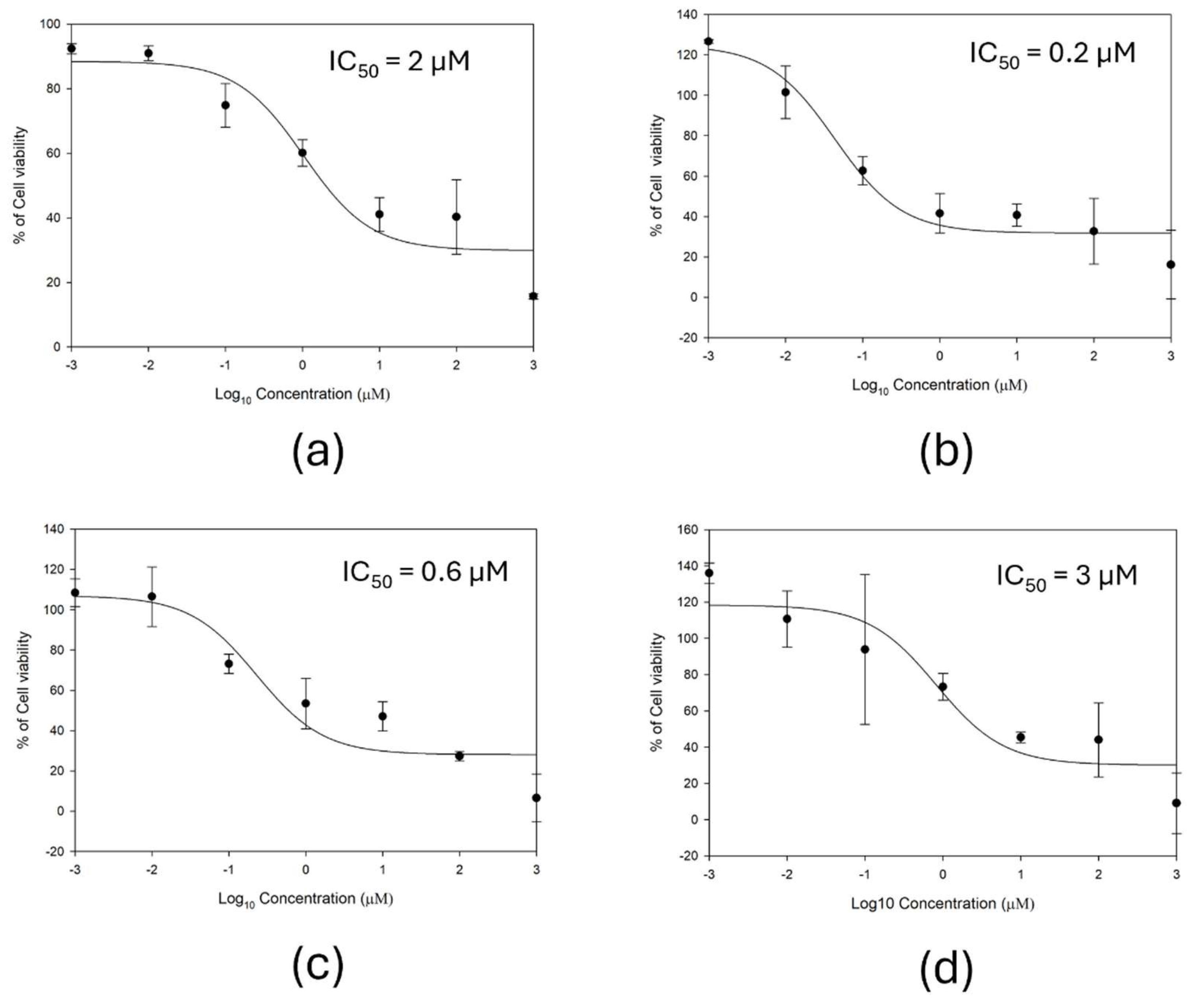
Figure 4.
KGA (GLS1) expression levels in TNBC cell lines. (a) RNA expression levels (normalized transcripts per million, nTPM). (b) Protein expression levels (normalized protein expression, nRPTX). Data were obtained from The Human Protein Atlas.
Figure 4.
KGA (GLS1) expression levels in TNBC cell lines. (a) RNA expression levels (normalized transcripts per million, nTPM). (b) Protein expression levels (normalized protein expression, nRPTX). Data were obtained from The Human Protein Atlas.
Disclaimer/Publisher’s Note: The statements, opinions and data contained in all publications are solely those of the individual author(s) and contributor(s) and not of MDPI and/or the editor(s). MDPI and/or the editor(s) disclaim responsibility for any injury to people or property resulting from any ideas, methods, instructions or products referred to in the content. |
© 2026 by the authors. Licensee MDPI, Basel, Switzerland. This article is an open access article distributed under the terms and conditions of the Creative Commons Attribution (CC BY) license (http://creativecommons.org/licenses/by/4.0/).
Copyright: This open access article is published under a Creative Commons CC BY 4.0 license, which permit the free download, distribution, and reuse, provided that the author and preprint are cited in any reuse.