Preprint
Review

This version is not peer-reviewed.

Emerging 4D-Printed pH-Responsive Nanofiber Implants for Spatiotemporal Breast Cancer Therapy: Design Principles, Tumour Microenvironment Modulation, Translational Barriers, and Future Perspectives

Submitted:

03 March 2026

Posted:

04 March 2026

You are already at the latest version

Abstract
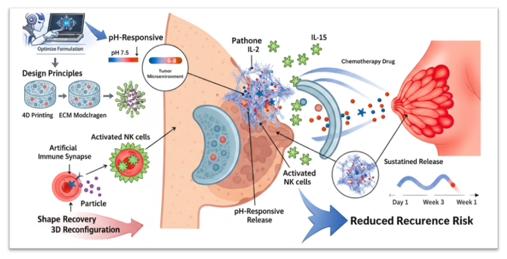
Breast cancer remains one of the leading causes of cancer-related morbidity and mortality worldwide, with tumour recurrence and therapeutic resistance largely driven by the immunosuppressive tumour microenvironment (TME). Conventional systemic chemotherapy and immunotherapy often suffer from poor tumour selectivity, systemic toxicity, and limited immune activation within the acidic and hypoxic TME. In this context, 4D-printed pH-responsive nanofiber implants have emerged as a next-generation platform capable of delivering spatiotemporally controlled therapy tailored to dynamic tumour conditions. Unlike static 3D constructs, 4D systems incorporate stimuli-responsive polymers that undergo programmed structural or functional transformations in response to environmental triggers such as acidic pH, enabling site-specific drug release. This review critically examines the design principles of pH-responsive nanofiber implants, including polymer selection, fabrication strategies, cytokine nano-assembly, and controlled release kinetics. Special emphasis is placed on TME modulation, highlighting how localised delivery of immune-stimulatory agents such as interleukin-15 and interleukin-2 can enhance natural killer cell activation, promote artificial immune synapse formation, and induce tumour apoptosis while minimising systemic toxicity. Furthermore, we analyse the translational challenges associated with manufacturing scalability, sterilisation, regulatory classification, and long-term implant safety. By integrating smart biomaterials engineering with immunotherapeutic strategies, 4D-printed nanofiber implants represent a transformative approach for localised breast cancer treatment. However, successful clinical translation will require interdisciplinary optimisation across materials science, pharmaceutical engineering, and regulatory frameworks. This review outlines future directions toward personalised, microenvironment-responsive cancer immunotherapy platforms.
Keywords: 
;  ;  ;  ;  ;  ;  ;  ;  ;  

1. Introduction

1.1. Global Burden and Therapeutic Limitations in Breast Cancer

Breast cancer is the most frequently diagnosed malignancy among women worldwide, accounting for approximately 2.3 million new cases annually and representing a leading cause of cancer-related mortality.[1,2]. Despite substantial advances in surgical resection, chemotherapy, radiotherapy, endocrine therapy, and molecularly targeted agents, locoregional recurrence and metastatic dissemination remain major determinants of disease-specific mortality. A persistent clinical challenge is the survival of residual tumour cells within the post-surgical cavity, where microscopic disease often escapes complete eradication. Furthermore, systemic therapies frequently fail to achieve sustained, therapeutically effective drug concentrations at the tumour bed without inducing dose-limiting systemic toxicities, thereby limiting long-term disease control.[3,4,5].
Conventional systemic immunotherapies, including recombinant cytokines and immune checkpoint inhibitors, often fail to elicit durable responses in breast tumours due to insufficient immune infiltration and an immunosuppressive tumour microenvironment (TME). Moreover, high-dose cytokine therapy is associated with severe systemic adverse effects, restricting its clinical applicability. These limitations underscore the urgent need for localized, microenvironment-responsive therapeutic platforms capable of providing sustained, spatiotemporally controlled drug delivery within the tumour resection cavity.[6,7].

1.2. Tumour Microenvironment as a Therapeutic Barrier and Opportunity

The breast TME is a dynamic, heterogeneous ecosystem composed of tumour cells, cancer-associated fibroblasts (CAFs), tumour-associated macrophages (TAMs), endothelial cells, extracellular matrix (ECM) components, and infiltrating immune cells.[8]. Metabolic reprogramming within tumours promotes aerobic glycolysis, leading to lactate accumulation and extracellular acidification (pH ~6.5–6.8). This acidic microenvironment not only enhances tumour invasion and drug resistance but also suppresses cytotoxic immune cell function.[9].
Immunosuppressive cytokines, hypoxia-driven signalling, and ECM stiffness collectively impair the activation of cytotoxic T lymphocytes and Natural killer cell populations. Consequently, therapeutic strategies that fail to account for TME-driven resistance often demonstrate suboptimal efficacy. However, the distinctive biochemical characteristics of the TME, particularly its acidity, also present an exploitable trigger for smart, stimuli-responsive drug delivery systems[10].

1.3. Emergence of 4D Printing in Drug Delivery

Three-dimensional (3D) printing has revolutionised the fabrication of biomedical devices by enabling precise architectural control and patient-specific customisation. Building upon this foundation, 4D printing introduces the dimension of time, allowing constructs to undergo programmed structural or functional changes in response to environmental stimuli such as pH, temperature, enzymatic activity, or redox gradients. In drug delivery, this concept enables implants that dynamically respond to pathological cues rather than passively releasing therapeutics.[11,12].
pH-responsive polymers, including PEG derivatives, poly(trimethylene carbonate) (PTMC), and other ionizable biomaterials, can be engineered into nanofiber scaffolds that degrade, swell, or alter porosity under acidic conditions. When implanted within a tumour resection cavity, such systems can enable controlled, localised release of chemotherapeutic or immunomodulatory agents precisely when and where needed.[13].

1.4. Rationale for Spatiotemporal Immunotherapy via 4D Nanofiber Implants

Localised cytokine delivery represents a promising strategy to overcome the systemic toxicity associated with high-dose immunotherapies such as Aldesleukin. Incorporation of immune-stimulatory cytokines, including Interleukin-15 and Interleukin-2, within pH-responsive nanofiber matrices may enable spatially confined activation of natural killer (NK) cells directly within the tumour niche. Such localised immunomodulation can facilitate the formation of artificial immune synapses, enhance granzyme-mediated cytotoxicity, and promote tumour apoptosis while minimising systemic exposure and off-target immune activation.[14,15].
By integrating stimuli-responsive biomaterials with immunotherapeutic nanoformulations, 4D-printed implants provide a multifunctional platform that exploits the acidic tumour microenvironment as a controlled-release trigger. This approach enables sequential or sustained cytokine delivery, supports remodelling of the immunosuppressive microenvironment, reduces systemic adverse effects, and enhances post-surgical local tumour control through sustained immune surveillance.[16].

1.5. Scope of This Review

This review critically examines the design principles of 4D-printed pH-responsive nanofiber implants for breast cancer therapy, with a particular focus on modulating the tumour microenvironment and spatiotemporal immunotherapy strategies.[17]. We analyse polymer selection, fabrication methodologies, cytokine nano-assembly approaches, release kinetics, and immune activation mechanisms. Furthermore, we address translational barriers, including manufacturing scalability, sterilisation, regulatory considerations, and long-term implant safety. Finally, we outline future directions toward personalised, microenvironment-adaptive cancer immunotherapy platforms.[18,19].

1.6. Literature Search Strategy and Study Selection Framework

This review does not aim to provide a quantitative meta-analysis but rather a critical and mechanistic synthesis of interdisciplinary evidence spanning materials science, immunology, and translational oncology.[20]. To enhance transparency and minimise selection bias, a structured literature search strategy guided by PRISMA principles was employed. The initial database search yielded 512 records. After removal of duplicates (n = 78), 434 articles were screened based on title and abstract relevance. Subsequently, 186 articles underwent full-text evaluation, and 112 studies were ultimately included to construct the thematic framework of this review.[21]. Given the emerging and rapidly evolving nature of 4D printing in oncology, the currently available literature remains limited and heterogeneous; therefore, a narrative synthesis approach was considered more appropriate than a strict meta-analytic framework. Additional foundational and conceptual references were incorporated where necessary to provide a mechanistic context and interdisciplinary background beyond the structured database search.[22].
Figure 1. PRISMA-guided literature search and study selection process. A structured search across major databases (2005–2025) yielded 512 records, which were systematically screened and filtered to include 112 studies relevant to 4D-printed pH-responsive nanofiber implants for breast cancer therapy.
Preprints 201196 g001
Figure 2. Schematic illustration of the programmed 4D-printed shape-memory nanofiber implant showing stimuli-triggered 2D-to-3D transformation, pH-responsive dual-drug release, and localized tumor-adaptive deployment for improved therapeutic effectiveness.
Preprints 201196 g002

2. Molecular and Metabolic Drivers of Breast Cancer: Implications for Microenvironment-Responsive Therapy

2.1. Carcinogenesis and Oncogenic Signalling in Breast Cancer

Breast cancer progression is a multistep process that typically evolves from ductal carcinoma in situ (DCIS) to invasive carcinoma, driven by cumulative genetic and epigenetic alterations. Key molecular subtypes, including hormone receptor–positive (ER/PR+), HER2-amplified, and triple-negative breast cancer (TNBC), exhibit distinct oncogenic signalling dependencies and therapeutic vulnerabilities.[23].
Genetic alterations commonly observed in breast cancer include TP53 mutations, HER2 amplification, dysregulation of PI3K/AKT/mTOR signalling, and BRCA1/2-associated DNA repair defects. Hyperactivation of receptor tyrosine kinases such as HER2 and EGFR promotes downstream activation of MAPK and AKT pathways, enhancing proliferation, survival, and therapeutic resistance.[24].
Transforming growth factor-β (TGF-β) exhibits a context-dependent role in breast cancer. While functioning as a tumour suppressor in early-stage disease, advanced tumours hijack TGF-β signalling to promote epithelial–mesenchymal transition (EMT), stromal activation, immune suppression, and metastatic dissemination. Crosstalk among the TGF-β, PI3K/AKT, and MAPK pathways further reinforces invasive phenotypes and contributes to resistance to systemic therapies[25].
Importantly, oncogenic signalling pathways also reshape the tumour immune microenvironment. HER2-driven tumours and TNBC frequently display altered antigen presentation, increased PD-L1 expression, and recruitment of immunosuppressive stromal populations, thereby limiting durable responses to immune checkpoint blockade.
Figure 3. Schematic representation of triple-negative breast cancer (TNBC) in breast cancer.
Preprints 201196 g003

2.2. Metabolic Reprogramming and Acidic Tumour Microenvironment

Breast cancer cells undergo profound metabolic reprogramming to sustain rapid proliferation under hypoxic and nutrient-deprived conditions characteristic of the tumour microenvironment (TME). A hallmark of this adaptation is enhanced aerobic glycolysis, commonly referred to as the Warburg effect, which results in excessive lactate accumulation and extracellular acidification, with tumour interstitial pH often declining to approximately 6.5–6.8. Central to this metabolic shift is hypoxia-inducible factor-1α (HIF-1α), a master transcriptional regulator that upregulates glycolytic enzymes, glucose transporters, and pro-angiogenic mediators to facilitate tumour survival and vascular adaptation. The consequent acidic and hypoxic milieu exerts multiple protumorigenic effects, including extracellular matrix (ECM) remodelling and increased stromal stiffness, enhanced tumour cell invasion and metastatic dissemination, and functional impairment of cytotoxic T lymphocytes and natural killer (NK) cells. Moreover, acidic stress has been associated with increased expression of immune checkpoint molecules such as PD-L1, thereby reinforcing immune escape mechanisms[26].
Beyond glycolysis, metabolic byproducts such as lactate directly suppress effector immune cell activity by disrupting intracellular pH homeostasis and mitochondrial oxidative metabolism, ultimately promoting T-cell exhaustion and diminished cytotoxic capacity. In parallel, breast cancer cells exploit adaptive pathways including autophagy and lipid metabolic reprogramming to maintain bioenergetic flexibility under sustained stress. Reciprocal metabolic crosstalk between tumour cells and cancer-associated fibroblasts (CAFs) further establishes a state of metabolic symbiosis that amplifies extracellular matrix deposition, stromal desmoplasia, and immune exclusion. Collectively, these interconnected metabolic adaptations not only drive tumour progression but also create a microenvironment that presents both a therapeutic barrier and an opportunity for pH-responsive, stimuli-adaptive implantable drug-delivery strategies.[27,28].

2.3. Implications for pH-Responsive 4D-Printed Implant Design

The molecular and metabolic reprogramming characteristic of breast cancer collectively establishes a hypoxic, acidic, and immunosuppressive tumour microenvironment (TME). Although these pathophysiological features contribute to therapeutic resistance and immune evasion, they simultaneously present biologically exploitable triggers for advanced stimuli-responsive drug delivery platforms. In particular, extracellular acidification within the tumour niche provides a predictable biochemical cue that activates ionizable polymers embedded within 4D-printed nanofiber implants, thereby enabling spatially confined and condition-dependent drug release. Concurrently, hypoxia-driven immune suppression underscores the need for localised immune reactivation strategies to restore cytotoxic T-cell and natural killer (NK) cell functionality directly at the tumour site. Furthermore, transforming growth factor-β (TGF-β)-mediated stromal remodelling and extracellular matrix (ECM) deposition highlight the importance of incorporating ECM-modulatory components to counteract desmoplastic barriers and enhance therapeutic penetration.[29,30].
By integrating pH-responsive biomaterials with localised cytokine delivery systems, 4D-printed implants have the potential to convert tumour-associated acidity from a pathological liability into a programmable therapeutic activation signal. Such a strategy reframes metabolic dysfunction as a design principle rather than merely a barrier, enabling spatiotemporally controlled immunomodulation tailored to the dynamic tumour microenvironment. Accordingly, a comprehensive understanding of breast cancer–specific molecular and metabolic adaptations provides a mechanistic foundation for the rational development of next-generation implantable platforms for precise, localised, and adaptive cancer therapy[31].

3. Fundamentals of 4D Printing in Drug Delivery

Four-dimensional (4D) printing represents an advanced evolution of additive manufacturing in which printed constructs are endowed with time-dependent or stimulus-responsive transformation capabilities. Unlike conventional three-dimensional (3D) printed systems that retain static geometries after fabrication, 4D-printed structures are engineered to undergo programmed structural, mechanical, or functional changes in response to predefined environmental triggers. This transformation is achieved by incorporating smart materials whose physicochemical properties enable reversible or irreversible reconfiguration over time. At the core of 4D printing lies the integration of stimuli-responsive polymers, shape-memory materials, and programmable architectures. These materials possess internal network arrangements that can store mechanical information during fabrication (the programming phase) and subsequently release this stored energy upon exposure to a specific trigger (the activation phase). Such dynamic behavior introduces a temporal dimension into biomedical device design, enabling constructs to adapt to physiological environments rather than remaining passive implants.[12,32,33].
Figure 4. Schematic overview illustrating the programmable transformation of a 4D-printed scaffold from a minimally invasive compact state to an expanded defect-conforming architecture. The scaffold integrates stimulus-responsive shape-memory polymers and drug-loaded nanocarriers to enable synchronized mechanical support, antibacterial activity, and regenerative enhancement in bone tissue engineering.
Preprints 201196 g004

3.1. Shape-Memory Mechanisms and Programming Strategies

Shape-memory polymers (SMPs) are among the most widely employed materials in 4D biomedical systems. These materials can be temporarily deformed into a programmed configuration and later recover their original shape when exposed to an external stimulus. The mechanism typically involves two key structural domains: (i) a switching segment responsible for fixing the temporary shape and (ii) a permanent network structure that defines the original geometry. The programming process generally includes deformation of the material above a transition temperature (or activation threshold), followed by cooling or chemical fixation to lock the temporary shape. Upon re-exposure to stimuli such as heat, pH variation, or solvent interactions, the polymer chains regain mobility, allowing structural recovery.[34,35].
In implantable systems, this behavior enables minimally invasive insertion in a compact configuration, followed by in situ expansion to conform to irregular tissue defects. Despite significant progress, precise control over recovery kinetics and mechanical fatigue resistance remains a challenge. Repeated swelling-deswelling cycles, especially in hydrated biological environments, may compromise structural integrity. Therefore, optimizing crosslink density, crystallinity, and degradation rates is critical for ensuring predictable long-term performance in vivo[36].
Table 1. Stimuli-Responsive Materials Used in 4D Printing.
Material type Mechanism of response Representative biomedical application Key advantages Primary limitations Reference
SMPs Thermally(or light) activated shape recovery self-expanding stents, deployable scaffolds, and minimally invasive implants strong shape recovery, tunable transition temp, printable by FDM/SLA fatigue over repeated cycles, limited biodegradability for some SMPs [37]
thermo-/photoresponsive hydrogels (e.g., PNIPAM blends) phase transition (swelling/deswelling) with temp or light (photothermal triggers when loaded with GNRs/UCNPs) on-demand drug release, wound dressings, and dynamic scaffolds excellent tissue-like hydration; compatible with cell encapsulation lower mechanical strength; may require reinforcement [38,39]
pH-responsive hydrogels/polyelectrolytes Protonation/deprotonation alters swelling or bond cleavage tumor-targeted DDS, GI tract targeting site-specific release (tumor/GI) narrow responsive window; variability in vivo [40]
biopolymers/biodegradable composites (PLA, PCL, bioceramics) enzyme/hydrolysis mediated degradation → structural change or release biodegradable implants, bone scaffolds established biocompatibility; tunable degradation slower response, mechanical trade-offs [41]
photochromic/photocleavable chemistries (azobenzene, spiropyran, o-nitrobenzyl esters) photoisomerization or bond cleavage on irradiation light-triggered release, microactuation in 4D constructs
high spatial/temporal control (when light accessible) limited tissue penetration (UV), possible phototoxicity [38,39]
magnetically responsive composites (Fe3O4, etc.) external magnetic fields induce force/heat → actuation or hyperthermia remote actuation (implants), magnetically guided carriers noncontact remote control; reversible actuation heating risk; long-term biosafety of magnetic NPs [42]
Figure 5. Swelling-induced shape evolution and drug release profiles of 4D-printed smart implant loaded with DOX, demonstrating programmable transformation and therapeutic effect (adapted from 4D printed multipurpose smart implants for breast cancer management, Int. J. Pharmaceutics, 2023, CC-BY).
Preprints 201196 g005

3.2. Conceptual Evolution from 3D to 4D Printing

Three-dimensional (3D) printing has transformed biomedical engineering by enabling layer-by-layer fabrication of complex, patient-specific constructs with precise spatial control. In pharmaceutical sciences, 3D printing has been applied to fabricate oral dosage forms, implants, and tissue scaffolds with customizable geometries and controlled drug distribution. However, conventional 3D-printed systems are inherently static; once fabricated, their structure and function remain largely unchanged under physiological conditions.[43].
The concept of 4D printing extends this paradigm by introducing the dimension of time, enabling printed constructs to undergo predefined structural, physicochemical, or functional transformations in response to environmental stimuli. These transformations may include swelling, shape morphing, degradation, pore size modulation, or triggered drug release. In drug delivery applications, this dynamic responsiveness enables systems to adapt to pathological microenvironments rather than functioning as passive depots[44].
The fourth dimension is typically activated by internal or external stimuli such as pH gradients, temperature fluctuations, enzymatic activity, light exposure, magnetic fields, or redox conditions. Among these, pH responsiveness is particularly attractive in oncology, given the well-established acidity of the tumour microenvironment (TME)[45].

3.3. Stimuli-Responsive Biomaterials for 4D Systems

The performance of a 4D-printed drug delivery platform is fundamentally governed by the selection of smart biomaterials that undergo predictable, reversible transformations. Stimuli-responsive polymers contain ionizable or cleavable functional groups that respond to environmental triggers through mechanisms such as protonation–deprotonation, hydrolysis, or conformational rearrangement.[46,47]. The performance of a 4D-printed drug delivery platform is fundamentally governed by the selection of smart biomaterials that undergo predictable, reversible transformations. Stimuli-responsive polymers contain ionizable or cleavable functional groups that respond to environmental triggers through mechanisms such as protonation–deprotonation, hydrolysis, or conformational rearrangement, enabling controlled release within the tumor microenvironment (Figure 6)[48].

3.3.1. pH-Responsive Polymers

pH-responsive polymers are typically engineered with ionizable functional moieties, including carboxylic acid (-COOH), amine (-NH₂), imidazole, and sulfonamide groups, which enable dynamic physicochemical transitions in response to extracellular pH variations. Under acidic conditions, protonation of these functional groups alters polymer charge distribution and intermolecular interactions, leading to structural swelling, enhanced hydrophilicity, accelerated hydrolytic degradation, or disruption of polymeric crosslinking networks. Within tumour-targeted delivery systems, such pH-dependent behaviour facilitates preferential activation in mildly acidic tumour tissues (pH 6.5–6.8), while preserving relative structural stability under physiological conditions (approximately pH 7.4). This differential responsiveness underpins the spatial selectivity of drug release and minimises premature systemic leakage[49].
Several biomaterials have been extensively explored for oncology-oriented 4D printing applications, including poly(ethylene glycol) (PEG) derivatives, poly(trimethylene carbonate) (PTMC), poly(β-amino esters), chitosan and its derivatives, and poly(lactic-co-glycolic acid) (PLGA)-based copolymers. Among these, PEG–PTMC hybrid systems have attracted particular interest owing to their favourable biocompatibility, tunable degradation kinetics, and compatibility with protein- and cytokine-based therapeutics. The synergistic integration of hydrophilic PEG domains with biodegradable PTMC segments enables fine control over mechanical integrity, drug encapsulation efficiency, and stimulus-responsive release behaviour, making such hybrid constructs especially suitable for 4D-printed nanofiber implants designed for spatiotemporally controlled oncological therapy.[50].

3.4. Mechanisms of Structural Transformation in 4D Implants

Structural evolution in 4D-printed systems is achieved through programmable material design. In pH-responsive nanofiber implants, the following transformation mechanisms are most relevant:

3.4.1. Swelling-Induced Porosity Modulation

Protonation of polymer chains in acidic environments increases osmotic pressure within the scaffold matrix, leading to swelling and expansion of pore networks. This enhances drug diffusion and accelerates release kinetics.[51].

3.4.2. Acid-Catalysed Hydrolytic Degradation

Ester or carbonate linkages within polymers such as PTMC may undergo accelerated cleavage under mildly acidic conditions, enabling controlled matrix erosion and sustained release of encapsulated agents.[52].

3.4.3. Shape Memory and Morphological Adaptation

Shape-memory polymers can be programmed to expand or conform to irregular post-surgical tumour cavities upon exposure to physiological temperature or pH triggers, improving tissue contact and therapeutic coverage.[53].

3.4.4. Multistage Release Programming

Layered or gradient-loaded nanofibers allow sequential drug release, enabling early-phase immunomodulation followed by sustained cytotoxic or cytokine-based therapy. These mechanisms enable precise spatiotemporal drug delivery, which is particularly critical in preventing local recurrence following tumour resection.[54].

3.5. Fabrication Technologies for 4D Nanofiber Implants

The fabrication method significantly influences fibre morphology, mechanical integrity, drug encapsulation efficiency, and responsiveness.

3.5.1. Extrusion-Based 4D Printing

Allows fabrication of macroscopic implant architectures with programmable geometries and gradient compositions[55].

3.5.2. Electrospinning 4D Hybrid Systems

Electrospinning produces nanofibrous matrices with high surface area-to-volume ratios, facilitating efficient drug loading and rapid environmental responsiveness. Integration with 4D-printable polymers enables dynamic transformation post-implantation[56].
Figure 7. Advanced functional hybrid nanofiber fabrication for 4D-printed implants. MOF-grown hybrid fibers, COF-coated porous nanofibers, polymer composition-dependent tuning, and electrospun inorganic–organic hybrid fibers illustrate the hierarchical design and fabrication workflow for pH-responsive spatiotemporal drug delivery.
Preprints 201196 g007

3.5.3. Digital Light Processing (DLP) and Stereolithography

Enable high-resolution constructs but require photopolymerizable materials, which may limit compatibility with sensitive biologics such as cytokines[57].
Among these approaches, hybrid electrospun–printed constructs offer a balance between nanoscale control and macroscopic architectural stability, making them particularly suitable for localised breast cancer therapy.[58].

3.6. Advantages of 4D Printing in Oncology-Oriented Drug Delivery

4D-printed pH-responsive nanofiber implants offer several conceptual and therapeutic advantages over conventional systemic drug delivery strategies. By leveraging microenvironment-triggered activation, these platforms enable condition-dependent drug release, selectively initiated within the acidic tumour niche rather than in normal tissues. Such spatial precision reduces systemic exposure and minimises off-target toxicities, while simultaneously achieving higher localised drug concentrations at the site of residual or recurrent disease. In addition, programmable multi-phase release kinetics can be engineered to allow sequential or sustained therapeutic delivery, supporting complex treatment regimens that may include immunomodulators, chemotherapeutics, or stromal-targeting agents. The structural adaptability inherent to 4D printing further enables improved conformity to irregular post-surgical tumour cavities, thereby enhancing implant–tissue integration and therapeutic coverage. Moreover, these systems provide a versatile platform for integrating combination therapy, enabling co-delivery of multiple agents with distinct temporal activation profiles[50,59,60].
In contrast to systemic cytokine administration, such as recombinant interleukin-based therapies, which are frequently associated with dose-limiting toxicities, localised 4D-printed systems have the potential to confine immune activation within the tumour microenvironment. This targeted immunostimulation may amplify antitumor cytotoxic responses while substantially mitigating systemic inflammatory adverse effects. Collectively, these attributes position 4D-printed pH-responsive implants as a promising next-generation strategy for achieving spatiotemporally controlled, site-specific breast cancer immunotherapy.[19,61].

3.7. Limitations and Current Knowledge Gaps

Despite significant promise, several challenges remain:
  • Limited long-term in vivo data
  • Variability in tumour pH among patients
  • Reproducibility of transformation kinetics
  • Protein stability during printing processes
  • Scale-up feasibility
A deeper understanding of material–immune interactions and standardised characterisation protocols will be essential to advance clinical translation.[62,63].

4. Design Principles of pH-Responsive Nanofiber Implants for Breast Cancer Therapy

4.1. Rational Polymer Selection for Microenvironment-Responsive Implants

The therapeutic performance of 4D-printed pH-responsive nanofiber implants is fundamentally governed by polymer composition, particularly in breast cancer applications, where multiple biological and mechanical constraints must be satisfied simultaneously. An optimal polymer system must demonstrate high biocompatibility with minimal immunogenic potential, exhibit controlled and predictable degradation kinetics, and maintain sufficient mechanical stability for post-surgical implantation within irregular lumpectomy cavities. In addition, compatibility with protein-based immunotherapeutics is essential to preserve cytokine bioactivity during fabrication, storage, and sustained release. Crucially, the material must retain pH-sensitive responsiveness in the mildly acidic tumour microenvironment to enable selective, spatially confined therapeutic activation.[64,65].
The extracellular acidity characteristic of breast tumours, typically ranging from pH 6.5 to 6.8, provides a physiologically relevant trigger for such stimulus-responsive systems. Polymers incorporating hydrolyzable ester or carbonate linkages, such as poly(trimethylene carbonate) (PTMC), offer tunable degradation behaviour under mildly acidic conditions, enabling programmable erosion and controlled payload release.[66]. When integrated with hydrophilic segments such as polyethene glycol (PEG), amphiphilic architectures can be engineered to enhance cytokine stabilisation, facilitate nano-scale assembly within fibrous matrices, and regulate diffusion dynamics. PEG–PTMC hybrid systems are particularly attractive for immunomodulatory implant design, as PEG domains improve protein compatibility and reduce nonspecific adsorption, while PTMC contributes structural integrity and adjustable degradation profiles. By modulating PEG chain length, molecular weight, and crosslink density, it becomes possible to precisely control swelling behaviour, pore expansion, and release kinetics, thereby aligning material performance with the spatiotemporal requirements of localised breast cancer therapy.[67,68].

4.2. Engineering Nanofiber Architecture for Controlled Release

Nanofiber morphology represents a critical determinant of drug loading capacity, release kinetics, and immune cell interaction within 4D-printed pH-responsive implants. Electrospinning-based fabrication strategies generate fibres with exceptionally high surface-area-to-volume ratios, thereby enhancing environmental sensitivity and facilitating rapid responsiveness to local physicochemical cues. In advanced 4D-printed systems, these nanofibrous architectures can be hierarchically integrated into larger programmable constructs, enabling simultaneous micro-scale diffusion control and macro-scale structural adaptation within post-surgical tumour cavities[69,70].
Architectural parameters such as fibre diameter (typically 100–800 nm), overall porosity, inter-fibre spacing, spatial drug distribution, and multilayer organisation collectively govern diffusion dynamics and degradation behaviour. In particular, core–shell nanofiber configurations allow spatial segregation of multiple therapeutic agents within a single filament. For instance, rapidly acting immunomodulators can be incorporated within the outer shell to initiate early immune priming. At the same time, sustained-release cytokines may be localised within the core to maintain prolonged activation. Such compartmentalised loading strategies enable multistage therapeutic programming and refined spatiotemporal immunomodulation.[71].
Importantly, pH-induced polymer swelling under mildly acidic tumour conditions increases inter-fibre spacing and enhances matrix permeability, thereby accelerating cytokine diffusion selectively within breast tumour tissues. Under physiological pH conditions, however, structural stability is relatively preserved, minimising premature release and off-target exposure. This morphology-dependent responsiveness underscores the central role of nanofiber architecture in aligning material design with tumour microenvironment–specific therapeutic objectives.[72].

4.3. Cytokine Nano-Assembly and Stabilisation Strategies

Localised immunotherapy requires preserving cytokine bioactivity during fabrication and implantation. Cytokines such as Interleukin-15 and Interleukin-2 possess potent immune-activating properties but are highly susceptible to denaturation, aggregation, and rapid systemic clearance.[73].
Nano-assembly approaches such as PEGylation or conjugation to biodegradable polymers can improve stability and prolong local retention. Amphiphilic PEG–PTMC conjugates can self-assemble into nano-aggregates capable of co-presenting cytokines in a multivalent configuration. Such co-binding architectures may enhance the efficiency of receptor engagement and promote synergistic immune activation[74].
Controlled immobilisation within nanofiber matrices further protects cytokines from premature degradation. Encapsulation methods must avoid harsh solvents, extreme temperatures, or photopolymerization conditions that compromise protein integrity. Low-temperature extrusion or post-printing loading strategies may improve preservation of bioactivity.[75].

4.4. Mechanistic Basis of NK Cell Activation and Artificial Immune Synapse Formation

The therapeutic objective of cytokine-loaded 4D implants is to enhance localized activation and persistence of natural killer (NK) cell populations within the tumour microenvironment (TME). NK cells exert cytotoxic activity through direct recognition of stress-induced ligands on malignant cells, followed by polarization of lytic granules and secretion of perforin and granzyme B, ultimately inducing apoptosis. However, within breast tumors, NK cell activity is frequently attenuated by immunosuppressive cytokines, metabolic constraints, and inhibitory receptor signaling.[76].
Interleukin-15 (IL-15) serves as a critical survival and proliferation factor for NK cells, whereas interleukin-2 (IL-2) promotes cytotoxic activation and clonal expansion. Spatially confined co-delivery of IL-15 and IL-2 may facilitate the formation of an artificial immune synapse (aIS) at the tumor–immune interface, promoting directed secretion of cytolytic mediators and enhancing tumor cell apoptosis. Localized cytokine gradients generated via pH-triggered implant release enable sustained NK activation within the tumour niche while minimizing systemic exposure and adverse effects associated with systemic cytokine therapies such as aldesleukin.[77].
Cytokine-based immunomodulatory strategies incorporating IL-15 and IL-2 signaling have demonstrated robust NK cell activation and enhanced antitumor efficacy. Representative nanomicelle-based systems designed to co-activate NK cells and induce targeted tumor apoptosis are illustrated in Figure 8. Although these platforms are primarily engineered for systemic administration, their mechanistic framework provides a conceptual basis for integrating cytokine signaling within 4D-printed implant architectures. By embedding cytokine reservoirs within pH-responsive nanofiber matrices, localized and temporally regulated immune activation may be achieved in post-resection cavities, thereby transforming immunologically “cold” tumors into immune-responsive phenotypes[78].

4.5. Spatiotemporal and Phased Release Strategies in 4D-Printed Implants

Spatiotemporal control represents the defining advantage of 4D-printed drug delivery systems, distinguishing them from conventional implantable platforms. Release kinetics can be precisely modulated through rational manipulation of material and structural parameters, including polymer molecular weight, crosslink density, hydrophilic–hydrophobic balance, fibre diameter, and drug–polymer binding interactions. Fine-tuning these variables enables dynamic control over diffusion rates, degradation behaviour, and stimulus responsiveness within the tumour microenvironment. Such engineering flexibility allows implants to be programmed to follow disease-specific biological timelines rather than relying solely on passive drug diffusion.[79].
A rational therapeutic design strategy may therefore incorporate a phased release architecture. An initial priming phase could deliver a mild, localised burst of immunomodulatory agents immediately following surgical tumour resection, facilitating early immune cell recruitment and activation. This may be followed by a sustained activation phase characterised by controlled cytokine release over days to weeks, aimed at maintaining natural killer (NK) cell cytotoxic function and preventing local tumour recurrence. Finally, a resolution phase involving gradual, predictable implant degradation would minimise the risk of chronic inflammation or long-term foreign-body reactions. Beyond temporal modulation, sequential release strategies can be engineered first to remodel the tumour microenvironment, such as by reprogramming tumour-associated macrophages or partially neutralising extracellular acidity, before initiating potent immune stimulation. Such hierarchical activation patterns reflect a biomimetic approach in which therapeutic intervention is aligned with the evolving immunobiology of the tumour niche, thereby reinforcing the translational potential of 4D-printed pH-responsive nanofiber implants for breast cancer therapy.[80].

4.6. Mechanical and Conformational Adaptation to Surgical Cavities

Post-lumpectomy breast cavities are inherently irregular in geometry, with substantial variability in size, depth, and contour depending on tumor location and surgical margins. This anatomical heterogeneity presents a significant challenge for conventional implantable drug delivery systems that lack adaptive structural properties. In contrast, 4D-printed nanofiber implants incorporating shape-memory characteristics can be introduced in a compact, minimally invasive configuration and subsequently undergo controlled expansion or geometric transformation upon exposure to physiological temperature and local pH conditions. Such adaptive conformability enables the implant to closely align with the cavity architecture, thereby improving spatial integration within the resection site[81].
Enhanced structural conformity promotes greater surface contact with residual tumour cells, facilitates more uniform cytokine or drug distribution across the surgical bed, reduces dead-space formation, and ultimately improves therapeutic coverage in areas at high risk of local recurrence. From a materials engineering perspective, mechanical tuning is critical to achieving an optimal balance between flexibility and structural persistence. The implant must retain sufficient elasticity to conform to irregular tissue surfaces while maintaining mechanical integrity to prevent premature collapse, fragmentation, or displacement. Careful modulation of polymer composition, crosslink density, and degradation kinetics is therefore essential to ensure both functional adaptability and sustained therapeutic performance in the post-surgical breast cancer setting.[82].

4.7. Critical Design Considerations and Trade-Offs

Despite the considerable design flexibility offered by 4D-printed pH-responsive implants, multiple interdependent trade-offs must be carefully optimized to achieve therapeutic reliability[83]. For instance, increased polymer swelling may accelerate drug release kinetics and enhance early bioavailability, yet excessive volumetric expansion can compromise mechanical integrity and structural stability within the surgical cavity. Similarly, accelerated degradation profiles may facilitate timely implant clearance and reduce long-term foreign body responses; however, overly rapid breakdown can limit sustained therapeutic exposure and diminish long-term efficacy. Dense crosslinking networks improve mechanical robustness and dimensional stability, but may simultaneously restrict cytokine diffusion and reduce release efficiency. In parallel, higher cytokine loading concentrations may enhance local immunostimulatory potency, although excessive dosing risks localized inflammation, tissue damage, or aberrant immune activation.[84].
These competing parameters underscore the necessity of a systems-level optimization framework that integrates materials science, tumor immunology, and pharmacokinetic modeling. Rather than focusing on isolated design variables, future development of 4D-printed nanofiber implants for breast cancer therapy must adopt a multidisciplinary strategy that balances structural performance, controlled release dynamics, and immune modulation to achieve predictable and clinically translatable outcomes[85].

5. Tumor Microenvironment Modulation by 4D-Printed pH-Responsive Implants

5.1. The Breast Tumor Microenvironment: A Dynamic Immunosuppressive Niche

The progression and therapeutic resistance of breast cancer are profoundly shaped by its tumor microenvironment (TME), a dynamic and adaptive ecosystem comprising malignant epithelial cells, cancer-associated fibroblasts (CAFs), tumor-associated macrophages (TAMs), endothelial cells, extracellular matrix (ECM) components, and diverse infiltrating immune populations.[86]. This complex cellular interplay establishes a structurally and metabolically restrictive niche that actively supports tumor survival and immune evasion. A hallmark feature of this microenvironment is metabolic reprogramming toward aerobic glycolysis, commonly referred to as the Warburg effect, which drives excessive lactate accumulation and extracellular acidification, with intratumoral pH frequently declining to approximately 6.5–6.8. The resulting acidic milieu exerts multiple protumorigenic effects, including suppression of cytotoxic T lymphocyte and natural killer (NK) cell activity, increased ECM stiffness and remodeling, enhanced tumor invasion and metastatic potential, and impaired penetration of therapeutic agents[87].
Beyond metabolic acidification, hypoxia-driven signaling pathways and immunosuppressive cytokines such as transforming growth factor-β (TGF-β) and interleukin-10 (IL-10) further dampen effective antitumor immune responses. Additional mechanisms, including indoleamine 2,3-dioxygenase (IDO)-mediated metabolic inhibition, stromal exclusion of effector lymphocytes, and progressive T-cell exhaustion, collectively reinforce a localized immunosuppressive state that systemic immunotherapies frequently fail to overcome. Importantly, these pathological characteristics also serve as actionable biochemical triggers for stimuli-responsive therapeutic systems. By harnessing tumor-associated acidity and hypoxia as programmable activation cues, 4D-printed pH-responsive nanofiber implants offer a rational strategy for localized, spatiotemporal immune reprogramming to restore intratumoral immunogenicity while minimizing systemic toxicity. Such an approach reframes the hostile TME from a barrier into a design-informed therapeutic opportunity.[88].
The breast tumor microenvironment is a highly dynamic and multicellular ecosystem composed of malignant epithelial cells, stromal fibroblasts, immune suppressive populations, and vascular components. Each cellular subset contributes uniquely to immune evasion, metabolic rewiring, angiogenesis, and therapeutic resistance. A systematic understanding of these components is essential for the rational design of spatiotemporally programmable 4D implants aimed at microenvironmental reprogramming[89]. The key cellular constituents and their functional roles in breast cancer progression, along with their therapeutic relevance to implant-mediated intervention, are summarized in Table 2.
Collectively, these cellular interactions establish an immunosuppressive and mechanically restrictive niche that limits immune infiltration and drug penetration. Therefore, next-generation implantable systems must not only deliver cytotoxic agents but also actively reprogram stromal, vascular, and immune compartments in a temporally coordinated manner.

5.2. Immune-Suppressive Tumour Microenvironment in Breast Cancer

Breast cancer, particularly triple-negative breast cancer (TNBC) and advanced metastatic subtypes, exhibits a profoundly immunosuppressive tumour microenvironment (TME) that limits durable responses to immunotherapy. Although certain subtypes exhibit moderate antigenicity, effective antitumor immunity is frequently constrained by dysfunctional antigen presentation, stromal barriers, metabolic reprogramming, and chronic immune checkpoint signalling.[97].
Reduced or heterogeneous expression of MHC class I molecules in breast tumour cells compromises antigen presentation and impairs CD8⁺ T-cell priming. While tumour-associated mutations generate neoantigens, these are often insufficient to elicit sustained cytotoxic responses due to pre-existing T-cell dysfunction and checkpoint-mediated inhibition. Dense extracellular matrix (ECM) deposition, primarily driven by cancer-associated fibroblasts (CAFs), further restricts immune cell trafficking into tumour cores, creating spatial immune exclusion[98].
Transforming growth factor-β (TGF-β) is a central immunosuppressive cytokine in the breast TME. Beyond its physiological role in tissue repair and immune homeostasis, TGF-β inhibits CD8⁺ T-cell proliferation, suppresses perforin and granzyme expression, and impairs dendritic cell maturation. Persistent TGF-β signalling reinforces immune tolerance and promotes immune evasion. Importantly, TGF-β also contributes to epithelial–mesenchymal transition (EMT) and metastatic dissemination, linking immune suppression with disease progression.[99].
Metabolic immune escape mechanisms are also prominent. Indoleamine 2,3-dioxygenase (IDO) expression by tumor cells and myeloid populations depletes tryptophan within the TME, disrupting T-cell proliferation and promoting T-cell anergy and apoptosis. Such metabolic reprogramming establishes a nutrient-deprived, hypoxic microenvironment, further aggravating immune dysfunction.[100].
Regulatory T cells (Tregs) accumulate within breast tumors and sequester IL-2, depriving effector T cells of critical growth signals. In parallel, tumor-associated macrophages (TAMs), particularly the M2-polarized phenotype, secrete IL-10 and VEGF, suppressing dendritic cell activation and enhancing angiogenesis. Myeloid-derived suppressor cells (MDSCs) further inhibit T-cell activation through arginase activity and the production of reactive oxygen species. Collectively, these cellular components establish a self-reinforcing immunosuppressive network.[92].

5.2.1. Antigenicity vs. Immunogenicity in Breast Cancer: Implications for Checkpoint Therapy

Antigenicity alone is insufficient to predict responsiveness to immune checkpoint blockade in breast cancer. Although triple-negative breast cancer (TNBC) generally exhibits a relatively higher tumor mutational burden compared with hormone receptor–positive subtypes, clinical responses to PD-1/PD-L1 inhibitors remain modest in unselected patient populations[98]. This discrepancy highlights the distinction between antigenicity and immunogenicity. While antigenicity reflects the presence of tumor-associated or neoantigens, immunogenicity denotes the capacity of these antigens to elicit effective and sustained antitumor immune responses. In breast cancer, immunogenicity is frequently compromised by multiple tumor microenvironment-mediated suppressive mechanisms. Transforming growth factor-β (TGF-β) signaling promotes immune exclusion by restricting T-cell infiltration, indoleamine 2,3-dioxygenase (IDO)-mediated metabolic reprogramming suppresses effector T-cell function, and the expansion of regulatory T cells (Tregs) and myeloid-derived suppressor cells (MDSCs) further dampens cytotoxic immunity. In parallel, dense stromal extracellular matrix deposition creates physical barriers that limit immune cell trafficking and drug penetration. Collectively, these factors impair T-cell priming, migration, and effector activity, thereby constraining the therapeutic efficacy of immune checkpoint inhibitors despite the presence of targetable antigens. Emerging evidence indicates that patients with preserved antigen-presenting machinery and pre-existing CD8⁺ T-cell infiltration demonstrate improved clinical responses, underscoring that restoration of intratumoral immunogenicity, rather than merely increasing antigen load, is essential for effective immune modulation. This insight provides a strong rationale for localised, spatiotemporally controlled immunoreprogramming strategies using 4D-printed pH-responsive implants[101].

5.2.2. T-Cell Exhaustion and Resistance to Immune Checkpoint Blockade

Chronic antigen exposure within the breast tumour microenvironment, coupled with persistent immunosuppressive signalling, drives the progressive exhaustion of cytotoxic T lymphocytes. Exhausted CD8⁺ T cells are characterised by sustained overexpression of inhibitory receptors, including programmed cell death protein-1 (PD-1), cytotoxic T-lymphocyte–associated protein 4 (CTLA-4), lymphocyte activation gene-3 (LAG-3), and T-cell immunoglobulin and mucin domain-containing protein-3 (TIM-3). This dysfunctional phenotype is reinforced by hypoxia, metabolic stress, transforming growth factor-β (TGF-β) signalling, and the accumulation of suppressive myeloid populations within the tumour microenvironment. Importantly, while early or transient exhaustion may remain partially reversible, terminally exhausted T cells exhibit epigenetic stabilisation of the dysfunctional state and markedly reduced proliferative capacity, thereby limiting the therapeutic benefit of checkpoint blockade as a monotherapy[102].
In addition to functional impairment, structural and cellular barriers further restrict effective antitumor immunity. The dense stromal architecture characteristic of many breast tumors physically limits T-cell infiltration, while defective antigen presentation compromises adequate priming and clonal expansion. Collectively, these converging mechanisms provide a biological explanation for the modest survival benefit observed with immune checkpoint inhibitors in metastatic breast cancer and underscore the need for localized strategies to restore intratumoral immune competence. Within this context, spatiotemporally controlled immunomodulation using 4D-printed pH-responsive implants represents a rational approach to counteract exhaustion, enhance immune infiltration, and recondition the tumor microenvironment[103].

5.2.3. Relevance to 4D-Printed pH-Responsive Nanofiber Implants

The immunosuppressive and spatially heterogeneous architecture of the breast tumour microenvironment underscores the intrinsic limitations of systemic immunotherapy. Diffuse drug distribution, dose-limiting toxicities, and inadequate intratumoral immune activation collectively constrain therapeutic efficacy. In this context, emerging 4D-printed pH-responsive nanofiber implants provide a rational platform for reprogramming the tumor microenvironment through precise spatiotemporal modulation. By leveraging tumor-associated extracellular acidity as a biological trigger, these systems can enable controlled, condition-dependent release of immunostimulatory cytokines such as interleukin-15 (IL-15), immune modulators, or inhibitors of the transforming growth factor-β (TGF-β) pathway directly within the tumor niche.[61,104].
Localised activation of such agents has the potential to enhance antigen presentation, promote activation of natural killer (NK) cells and CD8⁺ T cells, and counteract stromal stiffness–mediated immune exclusion. Furthermore, sustained intratumoral cytokine exposure may help reverse early-stage T-cell exhaustion and restore cytotoxic functionality before irreversible epigenetic fixation occurs. By confining immune modulation to the tumour bed, programmable 4D implants may significantly reduce systemic toxicity while amplifying local immunogenicity. Collectively, this strategy reframes immune checkpoint resistance not as an immutable barrier, but as a condition amenable to spatially controlled immune reconditioning, thereby positioning 4D-printed pH-responsive implants as a promising adjunct in breast cancer immunotherapy.[105].

5.3. Exploiting Acidic pH as a Therapeutic Activation Signal

Extracellular acidity within the tumour microenvironment (TME) constitutes one of the most consistent and exploitable hallmarks of solid malignancies, including breast cancer[106]. Rather than viewing this metabolic derangement solely as a driver of immune suppression and therapeutic resistance, it may be harnessed as a biological trigger to enable controlled drug activation. In this context, 4D-printed pH-responsive nanofiber implants are uniquely positioned to transform tumor-associated acidity into a programmable therapeutic advantage. Under mildly acidic conditions characteristic of tumor interstitium, protonation of ionizable functional groups within the polymer matrix increases hydrophilicity, induces scaffold swelling, expands pore networks, and accelerates hydrolytic degradation. These physicochemical transitions collectively facilitate the selective release of encapsulated cytokines or immunomodulatory agents within the tumor niche[107].
Importantly, relative structural stability is maintained at physiological pH (approximately 7.4), thereby minimising premature drug leakage and systemic exposure. The resulting localised, pH-triggered release establishes spatial cytokine gradients that preferentially enhance immune cell recruitment and activation within the tumour bed. Through this stimulus-adaptive behaviour, the TME is effectively redefined from a hostile barrier that limits therapeutic efficacy to an endogenous biochemical signal that guides spatiotemporal immune modulation. Such a paradigm underscores the rationale for integrating tumour-responsive materials into next-generation implantable immunotherapeutic platforms.[108].

5.4. Mechanisms of pH-Responsive Material Activation

pH-responsive polymers are rationally engineered with ionizable functional moieties such as carboxylic acids, amines, sulfonamides, and imidazole groups that undergo reversible protonation–deprotonation transitions in response to environmental pH fluctuations. Under physiological conditions (pH ~7.4), these functional groups maintain a stable ionization state, preserving network integrity through balanced electrostatic interactions and hydrogen bonding. In contrast, exposure to the mildly acidic tumor microenvironment (pH 6.5–6.8) or intracellular endosomal/lysosomal compartments (pH 5.0–6.0) perturbs this ionization equilibrium, thereby disrupting intermolecular forces and initiating physicochemical transformations such as volumetric swelling, conformational rearrangement, or cleavage of acid-labile linkages[109].
Drug release from pH-sensitive systems may be triggered through multiple complementary mechanisms. These include (i) swelling-induced mesh expansion that enhances diffusion kinetics, (ii) hydrolytic cleavage of acid-sensitive bonds such as hydrazone, acetal, or Schiff base linkages facilitating controlled drug liberation, and (iii) electrostatic repulsion–driven network expansion that increases porosity and permeability. Under acidic tumor conditions, protonation of pendant amine or carboxyl groups enhances polymer hydrophilicity and electrostatic chain repulsion, resulting in network expansion and enhanced diffusional transport. Consequently, pH-responsive hydrogels undergo protonation-driven swelling, enabling spatially confined and temporally regulated drug diffusion within tumor tissues (Figure 9)[110].
When incorporated into 4D-printed nanofiber or hydrogel architectures, these molecular-level chemical responses can be synergistically integrated with programmed mechanical deformation. For instance, asymmetric swelling in bilayer constructs may induce bending or folding, whereas isotropic volumetric expansion in hydrogel matrices can drive macroscopic shape transformation. This chemo-mechanical coupling allows synchronized structural adaptation and spatiotemporal drug release, thereby enhancing localized therapeutic precision following surgical implantation. Representative chemical structures of commonly employed pH-sensitive linkers and tumor-adaptive functional motifs are illustrated in Figure 9, highlighting the molecular design strategies underpinning microenvironment-responsive nanoformulations.[111].
Despite these advantages, excessive swelling may compromise mechanical robustness, particularly in load-bearing or implantable systems intended for post-resection breast cancer therapy. Therefore, precise optimization of crosslinking density, polymer composition, and network architecture is essential to balance environmental responsiveness with structural stability, degradation kinetics, and long-term functional performance.

5.5. Localised NK Cell Activation and Immune Synapse Enhancement

Effective immunotherapy requires reactivation of both innate and adaptive immune cells within the tumour microenvironment. Among these, Natural killer cell populations play a critical role in early tumour surveillance and cytotoxicity.[76].
Localised delivery of Interleukin-15 supports NK cell survival and proliferation, while Interleukin-2 enhances cytotoxic activation. Controlled co-presentation within the TME may promote the formation of an artificial immune synapse (aIS) between NK cells and tumour cells, thereby enabling the directed secretion of perforin and granzyme B.[76,112].
Importantly, localised cytokine gradients reduce the systemic toxicities observed with recombinant cytokine therapy such as Aldesleukin, which has historically been limited by vascular leak syndrome and dose-limiting adverse events.
Through spatiotemporal control, 4D implants create a confined immune-activating niche that shifts the balance from immune evasion toward immune-mediated tumour destruction.

5.6. Modulation of Tumour-Associated Macrophages and Immune Reprogramming

Tumor-associated macrophages (TAMs) within the breast tumor microenvironment predominantly acquire an M2-like phenotype characterized by pro-angiogenic, tissue-remodeling, and immunosuppressive functions. This polarization state is reinforced by persistent extracellular acidity, hypoxia-driven signaling pathways, and sustained exposure to anti-inflammatory cytokines, collectively stabilizing a pro-tumoral niche that supports immune evasion and disease progression. Targeted modulation of this macrophage phenotype, therefore, represents a critical objective for effective immune reconditioning strategies[113].
By enabling localized delivery of immunostimulatory cytokines and potentially integrating microenvironment-neutralizing agents within the same platform, 4D-printed pH-responsive nanofiber implants may facilitate macrophage reprogramming toward a more pro-inflammatory, M1-like phenotype. Such polarization shifts can enhance antigen-presenting capacity, promote the recruitment and activation of cytotoxic immune populations, and reduce the secretion of pro-tumoral cytokines that otherwise perpetuate immune suppression. Moreover, augmented activation of natural killer (NK) cells within the tumor bed may indirectly influence macrophage behavior through cytokine cross-talk and interferon-mediated signaling cascades, further amplifying intratumoral immune activation[114].
Through these coordinated mechanisms, the implant functions not merely as a passive drug reservoir, but as a localized immune-modulatory hub capable of reshaping cellular interactions within the tumor microenvironment. This systems-level immune reprogramming underscores the potential of 4D-printed pH-responsive implants to convert an immunosuppressive niche into a therapeutically permissive landscape in breast cancer.

5.7. ECM Remodelling and Improved Immune Penetration

The desmoplastic architecture of breast tumors represents a significant physical and immunological barrier to effective therapy. A dense extracellular matrix (ECM), enriched in crosslinked collagen fibers and associated with elevated mechanical stiffness, restricts both immune cell infiltration and drug diffusion within the tumor bed. This stromal rigidity not only limits cytotoxic lymphocyte trafficking but also impairs uniform therapeutic distribution, thereby contributing to treatment resistance and local recurrence following surgical resection[115,116].
In this context, 4D-printed pH-responsive nanofiber implants may exert secondary microenvironment-modulatory effects beyond their controlled drug-release function. Acid-triggered swelling and progressive scaffold degradation can locally perturb ECM organization by generating transient microchannels within the resection cavity, thereby facilitating immune cell infiltration and improving interstitial transport. The formation of such microarchitectural pathways may enhance the penetration of recruited cytotoxic T cells and natural killer (NK) cells while simultaneously sustaining intratumoral cytokine gradients that reinforce immune activation.
Importantly, programmable degradation kinetics enable temporal control of scaffold persistence, thereby reducing the risk of chronic foreign-body responses. By minimizing prolonged fibrotic encapsulation and secondary stromal stiffening, these systems may prevent re-establishment of an immunosuppressive niche. Collectively, this dual mechanical–immunological modulation positions 4D-printed nanofiber implants not only as drug-delivery platforms but also as dynamic regulators of tumor-stromal architecture, thereby improving immune accessibility and therapeutic responsiveness in breast cancer[117].

5.8. Metabolic Rewiring and Cholesterol Dynamics in Immune Activation

Emerging evidence suggests that immune cell activation is closely linked to metabolic remodeling, including cholesterol redistribution within cellular membranes. Enhanced membrane cholesterol availability can promote the stability of immune synapses and receptor clustering. Localized cytokine stimulation may influence cholesterol metabolic pathways in NK cells, facilitating membrane reorganization and improved synapse formation. When integrated into a spatiotemporal delivery framework, this metabolic modulation may enhance cytotoxicity against tumor cells. Although this mechanistic axis remains underexplored in implant-based systems, it represents a promising frontier for future immunoengineering strategies.[118].

5.9. Integrated Microenvironment Reprogramming: From Suppression to Activation

The cumulative impact of pH-triggered cytokine release, natural killer (NK) cell activation, macrophage repolarization, extracellular matrix (ECM) remodeling, and metabolic modulation culminates in a coordinated transition from an immunosuppressive to an immunostimulatory tumor microenvironment. Rather than targeting a single immune checkpoint or pathway, this strategy leverages the intrinsic biochemical characteristics of the tumor niche to initiate a multi-layered immune reconditioning process. By synchronizing biochemical, cellular, and mechanical cues, 4D-printed pH-responsive nanofiber implants facilitate spatiotemporally controlled immune activation directly within the tumor bed.[119].
Within this framework, these implants operate as dynamic therapeutic platforms with multifunctional roles. They act as microenvironmental sensors through pH-responsive activation mechanisms, as localized cytokine depots that sustain controlled immunostimulatory gradients, and as hubs for immune cell recruitment, thereby enhancing cytotoxic infiltration. Simultaneously, scaffold swelling and degradation contribute to the mechanical modulation of tumor architecture, while programmable release kinetics enable the temporal sequencing of therapeutic events. This integrated, multi-dimensional mode of action fundamentally distinguishes 4D systems from conventional passive implants or systemic nanoformulations, which primarily rely on diffusion-based delivery without adaptive responsiveness.[120].
Collectively, such programmable microenvironment reprogramming represents a paradigm shift from static drug delivery toward active immunoengineering of the tumor niche, offering a rational strategy to overcome immune exclusion and checkpoint resistance in breast cancer.

6. Spatiotemporal Immunotherapy Programming and Sequential Release Strategies

6.1. Rationale for Spatiotemporal Control in Cancer Immunotherapy

Effective immunotherapy requires not only activation of immune cells but precise coordination of timing, dosage, and spatial localization. Systemic cytokine administration often results in transient immune activation followed by exhaustion, while excessive exposure can induce severe toxicities. Therefore, immunotherapeutic success depends on maintaining a therapeutic window that supports sustained immune stimulation within the tumor microenvironment (TME) while avoiding systemic overactivation.[121].
4D-printed pH-responsive nanofiber implants offer a unique capability: they can program when, where, and how much immunostimulatory agent is released. By exploiting acidic tumor conditions as an activation trigger, these systems enable localized cytokine gradients that respond to microenvironmental cues.[122].

6.2. Multiphase Release Design: A Three-Stage Programming Model

A rational spatiotemporal therapeutic strategy for 4D-printed pH-responsive nanofiber implants may be conceptualized as a sequential, three-phase immunoengineering program. Such staged modulation is particularly relevant in post-surgical breast cancer settings, where the resection cavity represents both a high-risk site for recurrence and an opportunity for localized immune conditioning.[123].

6.2.1. Phase I: Immune Priming and Microenvironment Conditioning.

Immediately following implantation, a controlled initial burst release can initiate localized immune activation. This early phase is designed to recruit innate immune populations, stimulate inflammatory signaling cascades, and counteract residual immunosuppressive mediators within the tumor bed. By transiently enhancing antigen presentation and disrupting suppressive stromal cues, this priming window establishes a permissive microenvironment for subsequent cytotoxic engagement rather than allowing rapid re-establishment of immune exclusion.[124,125].

6.2.2. Phase II: Sustained NK Cell Activation.

Following priming, a prolonged and regulated release of cytokines such as interleukin-15 (IL-15) and interleukin-2 (IL-2) can support expansion and activation of natural killer (NK) cell populations within the local microenvironment. Sustained cytokine gradients promote immune synapse formation, enhance secretion of granzyme B and perforin, and facilitate direct tumor cell apoptosis. In contrast to systemic recombinant IL-2 therapies such as aldesleukin, localized sustained delivery minimizes peak plasma exposure, thereby reducing the risk of vascular leak syndrome and systemic inflammatory toxicities while preserving therapeutic potency at the target site.[126].

6.2.3. Phase III: Immune Memory Formation and Resolution.

As the implant undergoes programmable degradation, cytokine release tapers gradually, allowing the immune response to transition from active cytotoxicity toward memory stabilization and physiological resolution. This built-in decline in stimulatory signaling is critical to prevent chronic inflammation, foreign body reactions, and secondary fibrosis. The capacity to couple therapeutic escalation with controlled de-escalation fundamentally differentiates 4D-printed systems from continuous infusion approaches, which lack intrinsic resolution programming[127].
Collectively, this three-stage release model exemplifies how 4D-printed nanofiber implants can integrate temporal control with microenvironment-responsive activation, enabling structured immune reprogramming from suppression to durable antitumor surveillance in breast cancer.

6.3. Sequential Multi-Agent Loading Strategies

Beyond single-agent delivery, 4D-printed nanofiber platforms enable compartmentalized, gradient-based incorporation of multiple immunomodulatory agents within a single construct. Such architectural flexibility enables rational sequencing strategies that progressively recondition the tumor microenvironment. For instance, an initial release of acidity-neutralizing buffers may transiently elevate local pH, thereby improving immune cell viability and restoring cytotoxic function compromised by metabolic acidosis. This can be followed by the delivery of macrophage-reprogramming agents to shift tumor-associated macrophages from an M2-like, pro-tumoral phenotype toward a pro-inflammatory M1-like state. Subsequent sustained release of cytokines, including IL-15 or IL-2, may then support expansion and activation of natural killer (NK) cells, while delayed incorporation of immune checkpoint inhibitors can reinforce durable T-cell–mediated responses once adequate immune infiltration has been established.[128,129].
Technically, such spatiotemporal control can be achieved through layer-by-layer electrospinning approaches, gradient-loading strategies, or core–shell fiber architectures that physically segregate bioactive components. Differential polymer composition, crosslinking density, and degradation kinetics further regulate staged diffusion in response to environmental triggers, including pH fluctuations and enzymatic activity. By coupling structural compartmentalization with stimulus-responsive degradation, these systems enable programmed therapeutic cascades rather than simultaneous, uncontrolled co-release.
This level of architectural and functional integration exemplifies how 4D nanofiber implants can move beyond conventional combination therapy toward intelligently sequenced immunoengineering, aligning microenvironment conditioning, innate activation, and adaptive immune reinforcement within a single programmable platform.

6.4. Integration with Immune Checkpoint Blockade

Although immune checkpoint inhibitors have revolutionised oncology, their efficacy in breast cancer remains variable due to limited immune infiltration and TME suppression. Localised immune activation via 4D implants may synergise with checkpoint blockade by increasing tumour immunogenicity.[130].
For example, enhanced NK-mediated tumour lysis can increase antigen release, promoting dendritic cell activation and T cell priming. Subsequent systemic checkpoint inhibition may then sustain adaptive immune responses.
Such combinational strategies highlight the role of implants as immune amplifiers rather than standalone therapies.

6.5. Mathematical and Engineering Modelling of Release Kinetics

An essential component in the rational development of 4D-printed pH-responsive nanofiber implants is predictive modeling of spatiotemporal release behavior. Drug and cytokine release from nanofibrous scaffolds may be governed by diffusion-dominated transport, swelling-mediated permeability changes, degradation-driven erosion, or evolving hybrid multiphasic kinetics. In complex tumor microenvironments, these mechanisms often coexist, necessitating integrative computational frameworks rather than single-parameter approximations.[131].
Mathematical models incorporating Fickian diffusion principles, polymer swelling dynamics, and acid-triggered degradation rate constants can facilitate quantitative prediction of intratumoral cytokine concentration gradients and temporal exposure profiles. Such modeling approaches enable optimization of polymer composition, crosslinking density, fiber diameter, and implant geometry prior to experimental validation. Importantly, integrating patient-specific variables, including tumor pH heterogeneity, interstitial fluid dynamics, and post-surgical cavity geometry, may enable further refinement toward personalized implant configurations.[132].
Emerging computational strategies, including finite element modeling and machine learning–assisted optimization, offer additional opportunities to identify non-intuitive design parameters that maximize immune activation while minimizing off-target exposure. By coupling stimulus-responsive materials science with predictive analytics, 4D nanofiber systems can transition from empirically tuned constructs to data-driven, precision-engineered immunotherapeutic platforms capable of reproducible, patient-adapted performance in breast cancer[133].

6.6. Avoiding Immune Exhaustion and Cytokine Overload

A critical consideration in cytokine-based immunomodulation is the delicate balance between sustained immune activation and the risk of immune exhaustion. Prolonged exposure to high systemic concentrations of cytokines may lead to receptor downregulation, activation-induced cell death, or functional desensitization of effector lymphocytes, thereby paradoxically diminishing therapeutic efficacy. Moreover, excessive stimulation can amplify systemic inflammatory responses, increasing the likelihood of off-target toxicity.[134].
4D-printed pH-responsive nanofiber implants offer a strategy to mitigate these limitations by spatially confining and temporally regulating cytokine delivery[19]. By restricting release to the tumor microenvironment, systemic exposure is substantially reduced, minimizing peak plasma concentrations and associated toxicities. Gradual formation of localized concentration gradients enables sustained yet physiologically tolerable immune stimulation, while degradation-controlled tapering provides an intrinsic decline in cytokine availability as therapeutic objectives are achieved. Additionally, multiphase release programming allows sequential dosing within a single implant, eliminating the need for repeated systemic administrations and reducing fluctuations in immune activation intensity.[135].
Through these integrated control mechanisms, the implant functions as an autoregulated cytokine-delivery platform rather than a continuous infusion source. This capacity to couple immune stimulation with built-in resolution kinetics reinforces the safety profile of 4D systems while preserving their potential to induce durable antitumor immunity in breast cancer.

6.7. Clinical Implications of Spatiotemporal Programming

In the post-lumpectomy setting for breast cancer, residual microscopic disease within the surgical cavity remains a principal contributor to local recurrence. Despite advances in surgical precision and adjuvant radiotherapy, microscopic tumor foci and an immunosuppressive wound-healing microenvironment may facilitate tumor regrowth. A conformable 4D-printed pH-responsive nanofiber implant positioned within the resection cavity provides a localized strategy to counteract these early-recurrence-driving mechanisms. By sequentially priming the microenvironment, activating cytotoxic immune populations, and sustaining controlled immunostimulation, such systems may reinforce immune surveillance precisely at the site of highest relapse risk[136].
Localized spatiotemporal cytokine programming has the potential to reduce recurrence rates while minimizing systemic exposure and associated toxicities. Furthermore, by modulating immune infiltration and stromal architecture, these implants may synergize with adjuvant radiotherapy, which itself can enhance antigen release and immune visibility. Importantly, the capacity to tailor release kinetics according to tumor subtype, immune phenotype, and margin status introduces a level of therapeutic personalization not achievable with conventional systemic regimens.[137].
Collectively, these attributes position 4D-printed nanofiber implants as prospective adjuncts in personalized oncologic care, integrating surgical intervention with programmable immunoengineering to improve long-term disease control in breast cancer.

7. Translational and Regulatory Challenges

7.1. Manufacturing Scalability and GMP Compliance

Although laboratory-scale fabrication of 4D-printed pH-responsive nanofiber implants has yielded compelling proof-of-concept data, successful clinical translation necessitates scalable manufacturing under Good Manufacturing Practice (GMP) compliance. Reproducibility of fibre morphology, including diameter distribution and porosity, must be tightly controlled to ensure predictable release kinetics and mechanical stability. Similarly, uniform cytokine encapsulation efficiency, crosslinking density, and degradation behavior are critical determinants of therapeutic performance and must demonstrate minimal batch-to-batch variability.[138].
Electrospinning processes are inherently sensitive to environmental factors such as humidity, temperature, and solvent evaporation dynamics, posing significant challenges during scale-up from syringe-based laboratory systems to industrial production platforms. Implementation of closed-loop process control, real-time monitoring, and standardized operating parameters will therefore be essential to maintain structural and functional consistency.[139].
The incorporation of bioactive proteins further increases manufacturing complexity, as cytokines are susceptible to shear stress, thermal fluctuations, and solvent exposure during processing. Consequently, rigorous definition and validation of critical quality attributes (CQAs), including mechanical integrity, release kinetics, sterility, and preservation of cytokine bioactivity, will be indispensable for regulatory approval and clinical adoption.[140].

7.2. Sterilisation of Cytokine-Loaded Implants

Sterilization represents a critical translational hurdle for biologic-loaded 4D-printed nanofiber implants. Conventional sterilization modalities, including gamma irradiation, ethylene oxide exposure, and moist heat autoclaving, may adversely affect protein stability by inducing denaturation, oxidation, aggregation, or structural fragmentation. Such alterations can significantly compromise cytokine bioactivity and therapeutic efficacy.[141].
For protein-integrated 4D systems, alternative strategies must be carefully considered. Aseptic manufacturing under controlled GMP conditions may reduce reliance on terminal sterilization, while low-dose irradiation in the presence of stabilizing excipients may partially preserve structural integrity. Emerging techniques such as supercritical CO₂ sterilization offer reduced thermal and oxidative stress; however, their compatibility with complex polymer–protein matrices requires thorough validation. Post-sterilization cytokine loading presents another potential solution, but introduces additional process complexity and risks related to uniformity and contamination control.[142].
Each approach entails a balance between achieving acceptable sterility assurance levels and maintaining biological functionality. Accordingly, comprehensive validation studies assessing cytokine receptor-binding affinity, downstream signaling, and functional immune activation, such as NK cell proliferation and cytotoxicity assays, are essential to confirm post-sterilization bioactivity and ensure translational feasibility.

7.3. Stability and Storage Considerations

Long-term storage stability represents an additional translational challenge for cytokine-loaded 4D-printed nanofiber implants. Therapeutic proteins are inherently prone to aggregation, oxidative modification, moisture-induced degradation, and surface adsorption to polymer matrices, all of which may compromise structural integrity and bioactivity over time. To mitigate these risks, formulation strategies such as lyophilized incorporation, inclusion of cryo- and lyoprotectants, and chemical stabilization approaches (e.g., PEGylation) may enhance protein stability within the scaffold architecture[143].
Comprehensive stability evaluation under International Council for Harmonisation (ICH) guidelines will be necessary to establish shelf life, define acceptable storage conditions, and characterize degradation kinetics. Accelerated and long-term stability studies should assess not only physicochemical parameters but also functional cytokine activity. In parallel, packaging systems must provide robust protection against humidity, oxygen exposure, and temperature fluctuations while preserving sterility throughout the product lifecycle[144].
Collectively, integrating formulation stabilization strategies with validated storage and packaging controls will be essential to ensure clinical reliability and regulatory compliance of protein-loaded 4D implant systems.

7.4. Regulatory Classification: Combination Product Complexity

4D-printed cytokine-loaded nanofiber implants constitute a hybrid therapeutic platform integrating structural elements of an implantable medical device with the biological activity of cytokines such as interleukin-15 and interleukin-2, and potentially additional pharmacological agents. Owing to this multifunctional composition, such systems are likely to be regulated as combination products by authorities, including the U.S. Food and Drug Administration and comparable international agencies. Regulatory evaluation would therefore necessitate a comprehensive assessment across device and biologic domains, encompassing biocompatibility and mechanical integrity testing, validation of cytokine potency and bioactivity, pharmacokinetic and biodistribution profiling, as well as toxicology and immunogenicity studies[145].
A critical determinant of the approval pathway will be the designation of the primary mode of action (PMOA). If immune activation mediated by cytokine release is deemed the principal therapeutic mechanism, regulatory oversight may follow a biologics-centered framework, potentially increasing evidentiary and clinical requirements. Accordingly, early engagement with regulatory agencies during preclinical development is strongly advisable to clarify classification, define critical data packages, and streamline translational progression[146].

7.5. Preclinical Evaluation Challenges

Prior to clinical translation, rigorous in vivo validation is indispensable to establish the safety, efficacy, and immunomodulatory performance of 4D-printed cytokine-loaded implants. Replicating the structural and immunological complexity of the breast tumor microenvironment in animal models remains inherently challenging, necessitating careful experimental design. Critical evaluation parameters should include in vivo scaffold degradation kinetics, spatial and temporal cytokine release profiles, extent of local immune cell infiltration, systemic cytokine exposure, off-target inflammatory responses, and the incidence of fibrosis or foreign-body reactions[147].
Given interspecies differences in immune regulation, particularly in natural killer (NK) cell biology, model selection is of paramount importance. Immunocompetent and orthotopic breast tumor models are preferable to recapitulate tumor–immune interactions within the native microenvironment more accurately. Furthermore, long-term implantation studies are essential to assess chronic inflammatory sequelae, biodegradation byproducts, and sustained immunological effects. Such a comprehensive preclinical evaluation will be critical to generating clinically translatable data and de-risking early-phase human trials[148].

7.6. Safety Considerations and Immune-Related Risks

Although localized cytokine delivery substantially limits systemic exposure, potential safety risks remain and must be carefully addressed during preclinical development. Excessive local immune activation may provoke tissue damage, cytokine-mediated necrosis, or unintended amplification of inflammatory cascades. Additional concerns include autoimmune activation, chronic foreign body responses, and fibrotic encapsulation, all of which may compromise both therapeutic efficacy and implant functionality[149].
Achieving an optimal balance between immune stimulation and tissue tolerance is therefore critical. Dose-optimization studies are required to define safe cytokine loading thresholds that induce robust natural killer (NK) cell activation without triggering local cytotoxicity or sustained inflammatory injury. In parallel, the biocompatibility and clearance of polymer degradation byproducts must be thoroughly evaluated to exclude cumulative toxicity or systemic dissemination[150].
Comprehensive safety profiling integrating immunological, histopathological, and pharmacokinetic assessments will be essential to ensure that programmable immune activation remains controlled, reversible, and clinically acceptable.

7.7. Economic and Clinical Implementation Barriers

Beyond regulatory and technical challenges, economic considerations will substantially influence the translational viability of 4D-printed cytokine-loaded implants. The cost-intensive production of recombinant cytokines, scalability of stimuli-responsive polymer systems, requirement for specialized 4D printing infrastructure, and surgical implantation procedures collectively contribute to overall treatment expenditure. For successful integration into standard-of-care workflows, particularly in the post-lumpectomy management of breast cancer, these systems must demonstrate clear and measurable clinical advantages over established localized interventions, including radiotherapy and chemotherapeutic wafer-based implants[151].
Demonstration of superior recurrence control, reduced systemic toxicity, or improved quality-of-life outcomes will be essential to justify added complexity and cost. Ultimately, comprehensive health-economic evaluations incorporating manufacturing expenses, surgical logistics, long-term outcome data, and potential reductions in recurrence-related treatment costs will determine real-world adoption and reimbursement feasibility.

7.8. Bridging the Gap Between Innovation and Clinical Reality

The progression of 4D-printed cytokine-loaded nanofiber implants from experimental platforms to clinically applicable therapeutics will require coordinated interdisciplinary collaboration. Effective translation demands integration of expertise from pharmaceutical scientists, immunologists, biomedical engineers, regulatory specialists, and oncologic surgeons to align material design, immune modulation strategies, manufacturing controls, and clinical implementation pathways[152].
Standardized physicochemical and biological characterization protocols, coupled with robust predictive modeling tools, will be essential to ensure reproducibility and optimize spatiotemporal release profiles. Early-phase clinical investigations should prioritize safety, tolerability, and local immune biomarker assessment to establish mechanistic proof-of-concept in humans.
Although substantial scientific, regulatory, and economic challenges remain, continued rational design refinement and proactive regulatory alignment may ultimately position 4D-printed immunomodulatory implants as a novel class of programmable, localized cancer therapeutics capable of redefining post-surgical management of the tumor microenvironment[153].

8. Future Perspectives and Emerging Directions

8.1. Toward Personalised Microenvironment-Responsive Implants

One of the most compelling future directions for 4D-printed nanofiber platforms lies in the personalization of implant design. The breast tumor microenvironment exhibits substantial interpatient and subtype-specific heterogeneity, including variations in extracellular pH, immune cell infiltration density, stromal composition, and metabolic gradients. Such variability underscores the limitations of uniform implant configurations and highlights the need for biologically adaptive systems[154].
Next-generation 4D implants may therefore integrate patient-specific tumor pH mapping to calibrate stimulus sensitivity, programmable degradation kinetics tailored to anticipated healing and recurrence timelines, and adjustable cytokine loading guided by immune profiling data. Additionally, incorporating three-dimensional cavity imaging could enable geometry-matched scaffold fabrication, ensuring optimal conformability within post-resection defects. Integration with surgical planning software and intraoperative imaging modalities may further facilitate precise alignment between implant architecture and local tumor biology[155].
Through such convergence of biomaterials engineering, immunoprofiling, and digital surgical technologies, 4D-printed systems could evolve toward truly patient-specific immunomodulatory implants, advancing precision medicine in post-surgical breast cancer management.

8.2. AI-Guided Optimisation of Polymer and Release Profiles

Artificial intelligence (AI) and machine-learning–driven modeling frameworks present powerful opportunities to optimize 4D-printed nanofiber implant designs rationally. By integrating multivariate datasets encompassing polymer composition, crosslink density, degradation kinetics, and cytokine release behavior, predictive algorithms may facilitate data-guided material selection and formulation refinement. Computational models capable of incorporating tumor-specific parameters such as extracellular acidity gradients, cytokine diffusion coefficients, immune activation thresholds, and scaffold degradation constants could enable simulation of spatiotemporal release dynamics prior to physical fabrication[80,156].
Training such models on experimental and preclinical datasets may allow identification of optimal release profiles that balance immune stimulation with safety constraints. In this context, AI-assisted design strategies have the potential to reduce iterative experimental burden, accelerate developmental timelines, and enhance translational predictability. The convergence of computational intelligence with programmable biomaterials engineering may therefore represent a transformative step toward precision-optimized, clinically adaptable 4D immunomodulatory implants.

8.3. Integration of Biosensing and Feedback-Responsive Systems

Future iterations of 4D-printed nanofiber implants may progress beyond passive stimulus-responsive systems toward actively regulated, feedback-controlled therapeutic platforms. Integration of biosensing elements capable of monitoring local pH dynamics, inflammatory cytokine concentrations, immune cell density, or hypoxia-associated markers could enable real-time adaptation of cytokine release profiles. Such sensing modules, coupled with smart polymers exhibiting reversible conformational transitions, may permit dynamic modulation of therapeutic output in response to evolving tumor microenvironmental conditions[157].
In this closed-loop configuration, cytokine delivery would no longer rely solely on predefined degradation kinetics but could instead adjust to fluctuations in immune activation status or metabolic stress signals. By synchronizing biomaterial responsiveness with biological feedback, these systems could minimize overtreatment while sustaining effective immune stimulation. The development of autonomous, self-regulating immunotherapeutic implants therefore represents a forward-looking paradigm shift in localized cancer therapy, with the potential to transform tumor microenvironment modulation into an adaptive and precision-controlled intervention.

8.4. Expanding Beyond Cytokines: Gene Editing and Advanced Biologics

Future 4D-printed nanofiber platforms may expand beyond cytokine delivery to incorporate advanced biologic modalities, including mRNA-based therapeutics, CRISPR/Cas gene-editing systems, oncolytic viral agents, and bispecific immune engagers. Localized deployment of such technologies within the post-resection cavity could enable in situ reprogramming of tumor or immune cells, thereby directly modifying the immunological landscape at the site of highest risk of recurrence. In particular, spatially confined gene-editing approaches may enable selective enhancement of antitumor immunity or suppression of pro-tumoral signaling pathways without systemic exposure [158].
However, integration of these sophisticated biologics would substantially increase safety, containment, and regulatory complexity. Precise control over release kinetics, vector stability, and spatial confinement will be essential to prevent unintended gene expression, off-target editing, or systemic dissemination. Consequently, rigorous engineering of scaffold architecture, degradation dynamics, and bioactive payload stabilization must accompany parallel development of robust biosafety and regulatory frameworks[159].
If carefully implemented, the convergence of programmable biomaterials with gene-modulating technologies could redefine localized cancer immunotherapy, transforming resection cavities into sites of controlled biological re-engineering rather than passive post-surgical spaces.

8.5. Combination Strategies with Immune Checkpoint Modulation

Checkpoint inhibitors have demonstrated heterogeneous clinical efficacy in breast cancer, largely attributable to insufficient baseline immune infiltration and the prevalence of immunologically “cold” tumor phenotypes. Localized activation of natural killer (NK) cells through cytokine-loaded 4D-printed nanofiber implants may provide a strategy to overcome this limitation by enhancing early cytotoxic activity and promoting antigen release within the tumor bed. Increased tumor cell lysis can facilitate dendritic cell activation and antigen presentation, thereby improving subsequent T-cell priming and adaptive immune engagement[160].
A rational therapeutic sequence may therefore involve initial implant-mediated NK activation and localized tumor debulking, followed by enhanced antigen exposure and dendritic cell maturation, and ultimately systemic administration of checkpoint inhibitors to sustain and amplify T-cell–mediated immunity. Such temporally coordinated combination strategies have the potential to convert poorly infiltrated tumors into immunologically active, checkpoint-responsive phenotypes[161].
By integrating localized innate immune stimulation with systemic adaptive immune modulation, 4D-printed implants may function as immunological primers that expand the therapeutic window of checkpoint blockade in breast cancer.

8.6. Ethical and Long-Term Safety Considerations

As implantable immunomodulatory systems become more sophisticated, long-term surveillance of immune dysregulation, autoimmunity, and chronic inflammation will be necessary. Transparent patient consent and careful monitoring of immune biomarkers should accompany clinical trials. Balancing innovation with safety will determine the success of 4D-printed platforms in oncology[162].
Table 3. Tumour Microenvironment Targets Relevant to Breast Cancer and Potential Integration with 4D-Printed pH-Responsive Nanofiber Implants.
Target Pathway Representative Agents Mechanism of TME Modulation Clinical Status Main Challenges Potential Integration with 4D Implants Reference
FAK Signaling FAK inhibitors ± immune checkpoint inhibitors Inhibition of focal adhesion kinase reduces stromal fibrosis and enhances immune infiltration. Preclinical / Early clinical Limited clinical validation; stromal heterogeneity Localised co-delivery with cytokines to reduce ECM stiffness and improve immune cell penetration [163]
VEGFR / PDGFR / FGFR Nintedanib ± anti-PD-1 Suppresses angiogenesis and collagen deposition, improving CD8⁺ T-cell infiltration. Preclinical The translation of breast cancer is unclear Sequential release to normalise the vasculature before the NK activation phase [164]
TREM2–IL-1β Axis TREM2 modulation; IL-1β inhibitors Regulates macrophage-driven inflammatory signalling and TME immunosuppression. Preclinical Risk of excessive inflammation Controlled, localised release to reprogram TAMs while limiting systemic cytokine surge [165]
KRAS / Oncogenic Signalling KRAS inhibitors (e.g., MRTX-class agents) Suppresses oncogenic signalling and reverses CAF-mediated drug resistance. Preclinical / Early clinical Breast cancer subtype specificity Combined local immunotherapy with systemic targeted therapy [166]
Hyaluronan / ECM Density PEGylated hyaluronidase (PEGPH20) Degrades hyaluronan to reduce interstitial pressure and improve drug penetration. Clinical (Phase III, other cancers) Thromboembolic risk; no OS benefit Localised ECM modulation via pH-triggered enzyme release to enhance immune infiltration [167]
CXCR4–CXCL12 Axis Motixafortide ± pembrolizumab Enhances T-cell trafficking and reduces the number of immunosuppressive myeloid cells. Clinical (Phase II) Modest survival benefit; biomarker dependence Incorporation into a multi-phase nanofiber system for immune cell recruitment [168]
VEGFR Axitinib ± chemotherapy Inhibits tumour angiogenesis to enhance therapeutic delivery. Clinical (Phase III) Resistance to anti-angiogenic therapy Short-term vascular normalisation phase prior to cytokine release [169]
Mitochondrial Metabolism CPI-613 (devimistat) Induces metabolic stress in tumour cells. Early clinical Limited breast cancer data Sequential combination to sensitise tumour cells before immune activation [170]
Immune Activation (Cytokine Axis) IL-15 + IL-2 nano-assemblies Enhances NK activation and artificial immune synapse formation. Investigational Systemic toxicity if not localised Core component of 4D implant spatiotemporal immunotherapy [171]

9. Conclusion

4D-printed pH-responsive nanofiber implants represent a transformative convergence of smart biomaterials engineering, immunotherapy, and localised drug delivery. By exploiting the acidic tumour microenvironment as a biological trigger, these systems enable spatiotemporally programmed cytokine release, thereby enhancing NK cell activation while minimising systemic toxicity. Beyond serving as passive drug depots, such implants function as dynamic immunoengineering platforms that modulate tumour-associated macrophages, reshape extracellular matrix architecture, and convert immunosuppressive niches into immune-active environments. Despite significant translational challenges, including manufacturing scalability, sterilisation constraints, regulatory complexity, and long-term safety considerations, the rapid evolution of polymer science, computational modelling, and immunobiology provides a promising pathway toward clinical realisation. Future integration of AI-guided design, biosensing feedback systems, and personalised tumour profiling may further refine these platforms into adaptive, patient-specific therapeutic devices. With continued interdisciplinary collaboration, 4D-printed immunomodulatory implants have the potential to redefine localised therapy in breast cancer and establish a new paradigm in microenvironment-responsive oncology.

Author Contributions

Conceptualization, A.S.; methodology, A.S.; formal analysis, A.S.; investigation, A.S.; writing—original draft preparation, A.S.; writing—review and editing, R.S., M.A., B.S., V.T., and C.S.; supervision, C.S. and A.S.; software, A.S.; project administration, A.S.; visualization, A.S., R.S., M.A., B.S., V.T. All authors have read and agreed to the published version of the manuscript.

Funding

This research received no external funding.

Institutional Review Board Statement

Not applicable.

Informed Consent Statement

Not applicable.

Data Availability Statement

No new data were created or analyzed in this study.

Conflicts of Interest

The authors declare no conflicts of interest.

References

  1. Bray, F.; Laversanne, M.; Sung, H.; Ferlay, J.; Siegel, R.L.; Soerjomataram, I.; Jemal, A. Global cancer statistics 2022: GLOBOCAN estimates of incidence and mortality worldwide for 36 cancers in 185 countries. CA: a cancer journal for clinicians 2024, 74, 229–263. [Google Scholar] [CrossRef]
  2. Cai, Y.; Dai, F.; Ye, Y.; Qian, J. The global burden of breast cancer among women of reproductive age: a comprehensive analysis. Scientific Reports 2025, 15, 9347. [Google Scholar] [CrossRef]
  3. Kim, J.; Harper, A.; McCormack, V.; Sung, H.; Houssami, N.; Morgan, E.; Mutebi, M.; Garvey, G.; Soerjomataram, I.; Fidler-Benaoudia, M.M. Global patterns and trends in breast cancer incidence and mortality across 185 countries. Nature medicine 2025, 31, 1154–1162. [Google Scholar] [CrossRef]
  4. Deng, T.; Zi, H.; Guo, X.P.; Luo, L.S.; Yang, Y.L.; Hou, J.X.; Zhou, R.; Yuan, Q.Q.; Liu, Q.; Huang, Q. Global, regional, and national burden of breast cancer, 1990–2021, and projections to 2050: A systematic analysis of the global burden of disease study 2021. Thoracic Cancer 2025, 16, e70052. [Google Scholar] [CrossRef]
  5. Sha, R.; Kong, X.-m.; Li, X.-y.; Wang, Y.-b. Global burden of breast cancer and attributable risk factors in 204 countries and territories, from 1990 to 2021: results from the Global Burden of Disease Study 2021. Biomarker Research 2024, 12, 87. [Google Scholar] [CrossRef]
  6. Nagpal, D.; Verma, R.; Mittal, V.; Jeandet, P.; Kaushik, D. Targeted therapies against breast cancer: Clinical perspectives, obstacles and new opportunities. Journal of Drug Delivery Science and Technology 2023, 89, 105049. [Google Scholar] [CrossRef]
  7. Marei, H.E.; Bedair, K.; Hasan, A.; Al-Mansoori, L.; Caratelli, S.; Sconocchia, G.; Gaiba, A.; Cenciarelli, C. Current status and innovative developments of CAR-T-cell therapy for the treatment of breast cancer. Cancer Cell International 2025, 25, 3. [Google Scholar] [CrossRef]
  8. Harris, M.A.; Savas, P.; Virassamy, B.; O’Malley, M.M.; Kay, J.; Mueller, S.N.; Mackay, L.K.; Salgado, R.; Loi, S. Towards targeting the breast cancer immune microenvironment. Nature Reviews Cancer 2024, 24, 554–577. [Google Scholar] [CrossRef] [PubMed]
  9. Rodríguez-Bejarano, O.H.; Parra-López, C.; Patarroyo, M.A. A review concerning the breast cancer-related tumour microenvironment. Critical Reviews in Oncology/Hematology 2024, 199, 104389. [Google Scholar] [CrossRef] [PubMed]
  10. Monzavi-Karbassi, B.; Kelly, T.; Post, S.R. The tumor microenvironment and immune response in breast cancer. 2024, 25, 914. [Google Scholar] [CrossRef] [PubMed]
  11. Wu, W.; Wang, J.; Li, G. 3D/4D printing of stimuli-responsive polymers in biomedical engineering: materials, stimulations, and applications. Materials Science and Engineering: R: Reports 2025, 166, 101071. [Google Scholar] [CrossRef]
  12. Suryavanshi, P.; Wang, J.; Duggal, I.; Maniruzzaman, M.; Banerjee, S. Four-dimensional printed construct from temperature-responsive self-folding feedstock for pharmaceutical applications with machine learning modeling. Pharmaceutics 2023, 15, 1266. [Google Scholar] [CrossRef]
  13. Antezana, P.E.; Municoy, S.; Ostapchuk, G.; Catalano, P.N.; Hardy, J.G.; Evelson, P.A.; Orive, G.; Desimone, M.F. 4D printing: the development of responsive materials using 3D-printing technology. Pharmaceutics 2023, 15, 2743. [Google Scholar] [CrossRef]
  14. Guo, Y.; Li, Y.; Zhang, M.; Ma, R.; Wang, Y.; Weng, X.; Zhang, J.; Zhang, Z.; Chen, X.; Yang, W. Polymeric nanocarrier via metabolism regulation mediates immunogenic cell death with spatiotemporal orchestration for cancer immunotherapy. Nature communications 2024, 15, 8586. [Google Scholar] [CrossRef]
  15. Xu, Z.; Liu, S.; Li, Y.; Wu, Y.; Tu, J.; Chen, Q.; Sun, C. Engineering strategies of sequential drug delivery systems for combination tumor immunotherapy. Acta Pharmaceutica Sinica B 2025. [Google Scholar] [CrossRef]
  16. Zhou, H.; Zheng, H.; Yao, W.; Sun, H.; Yang, Y.-G.; Li, Z.; Song, D.; Zhang, Y.; Sun, T. Spatiotemporally controlled delivery of biomacromolecules via injectable hydrogels for precision modulation of the tumor immune microenvironment. Journal of Nanobiotechnology 2025. [Google Scholar] [CrossRef]
  17. Sajjad, R.; Chauhdary, S.T.; Anwar, M.T.; Zahid, A.; Khosa, A.A.; Imran, M.; Sajjad, M.H. A review of 4D printing–technologies, shape shifting, smart polymer based materials, and biomedical applications. Advanced Industrial and Engineering Polymer Research 2024, 7, 20–36. [Google Scholar] [CrossRef]
  18. Ramezani, M.; Mohd Ripin, Z. 4D printing in biomedical engineering: advancements, challenges, and future directions. Journal of functional biomaterials 2023, 14, 347. [Google Scholar] [CrossRef] [PubMed]
  19. Kanaujia, K.A.; Yadav, V.K.; Yadav, S.S.; Talha, M.; Saraf, S.A.; Kumar, S. 4D Printing in Healthcare: Innovations, Challenges, and Future Directions. In ACS Applied Bio Materials; 2025. [Google Scholar]
  20. Page, M.J.; McKenzie, J.E.; Bossuyt, P.M.; Boutron, I.; Hoffmann, T.C.; Mulrow, C.D.; Shamseer, L.; Tetzlaff, J.M.; Akl, E.A.; Brennan, S.E. The PRISMA 2020 statement: an updated guideline for reporting systematic reviews. 2021, 372. [Google Scholar]
  21. Liberati, A.; Altman, D.G.; Tetzlaff, J.; Mulrow, C.; Gøtzsche, P.C.; Ioannidis, J.P.; Clarke, M.; Devereaux, P.J.; Kleijnen, J.; Moher, D. The PRISMA statement for reporting systematic reviews and meta-analyses of studies that evaluate healthcare interventions: explanation and elaboration. Bmj 2009, 339. [Google Scholar] [CrossRef] [PubMed]
  22. Tripathi, D.; Ramar, M.; Lavudi, K.; Sharma, S.; Rajinikanth, P.; Pandey, P. The potential of fucoidans from ocean treasures to biomedical marvels: A review. International Journal of Biological Macromolecules 2025, 148979. [CrossRef]
  23. Miricescu, D.; Totan, A.; Stanescu-Spinu, I.-I.; Badoiu, S.C.; Stefani, C.; Greabu, M. PI3K/AKT/mTOR signaling pathway in breast cancer: from molecular landscape to clinical aspects. International journal of molecular sciences 2020, 22, 173. [Google Scholar] [CrossRef]
  24. Panda, V.K.; Mishra, B.; Mahapatra, S.; Swain, B.; Malhotra, D.; Saha, S.; Khanra, S.; Mishra, P.; Majhi, S.; Kumari, K. Molecular insights on signaling cascades in breast cancer: A comprehensive review. Cancers 2025, 17, 234. [Google Scholar] [CrossRef]
  25. Balkrishna, A.; Kumar, S.; Malik, R.; Mehra, K.S.; Chaturvedi, H.; Mittal, R. Unveiling role of oncogenic signalling pathways in complicating breast cancer. BioMedicine 2025, 15, 13. [Google Scholar] [CrossRef]
  26. Garg, P.; Singhal, G.; Horne, D.; Salgia, R.; Singhal, S.S. Metabolic reprogramming in breast cancer: Pathways driving progression, drug resistance, and emerging therapeutics. Biochimica et Biophysica Acta (BBA)-Reviews on Cancer 2025, 189396. [CrossRef] [PubMed]
  27. Liu, S.; Zhang, X.; Wang, W.; Li, X.; Sun, X.; Zhao, Y.; Wang, Q.; Li, Y.; Hu, F.; Ren, H. Metabolic reprogramming and therapeutic resistance in primary and metastatic breast cancer. Molecular cancer 2024, 23, 261. [Google Scholar] [CrossRef] [PubMed]
  28. Cappellesso, F.; Mazzone, M.; Virga, F. Acid affairs in anti-tumour immunity. Cancer cell international 2024, 24, 354. [Google Scholar] [CrossRef]
  29. Bhattacharya, S.; Prajapati, B.G.; Singh, S. A critical review on the dissemination of PH and stimuli-responsive polymeric nanoparticular systems to improve drug delivery in cancer therapy. Critical reviews in oncology/hematology 2023, 185, 103961. [Google Scholar] [CrossRef] [PubMed]
  30. Tan, R.Y.H.; Lee, C.S.; Pichika, M.R.; Cheng, S.F.; Lam, K.Y. PH responsive polyurethane for the advancement of biomedical and drug delivery. Polymers 2022, 14, 1672. [Google Scholar] [CrossRef]
  31. Liu, Y.; Si, L.; Jiang, Y.; Jiang, S.; Zhang, X.; Li, S.; Chen, J.; Hu, J. Design of pH-responsive nanomaterials based on the tumor microenvironment. International journal of nanomedicine 2025, 705–721. [CrossRef]
  32. Su, M.; Song, Y. Printable smart materials and devices: strategies and applications. Chemical reviews 2021, 122, 5144–5164. [Google Scholar] [CrossRef] [PubMed]
  33. Yu, C.; Zhao, J.; Cheng, F.; Chen, J.; Chen, J.; Xu, H.; Shi, K.; Xia, K.; Ding, S.; Wang, K. Silencing circATXN1 in aging nucleus pulposus cell alleviates intervertebral disc degeneration via correcting progerin mislocalization. Research 2024, 7, 0336. [Google Scholar] [CrossRef]
  34. Behl, M.; Lendlein, A. Shape-memory polymers. Materials today 2007, 10, 20–28. [Google Scholar] [CrossRef]
  35. Hu, J.; Zhu, Y.; Huang, H.; Lu, J. Recent advances in shape–memory polymers: Structure, mechanism, functionality, modeling and applications. Progress in polymer science 2012, 37, 1720–1763. [Google Scholar] [CrossRef]
  36. Miranda, A.; Lopez-Blanco, R.; Lopes-Nunes, J.; Melo, A.M.; Campello, M.P.C.; Paulo, A.; Oliveira, M.C.; Mergny, J.-L.; Oliveira, P.A.; Fernandez-Megia, E. Gallic acid–triethylene glycol aptadendrimers synthesis, biophysical characterization and cellular evaluation. Pharmaceutics 2022, 14, 2456. [Google Scholar] [CrossRef] [PubMed]
  37. Lin, C.; Zhang, L.; Liu, Y.; Liu, L.; Leng, J. 4D printing of personalized shape memory polymer vascular stents with negative Poisson’s ratio structure: A preliminary study. Science China Technological Sciences 2020, 63, 578–588. [Google Scholar] [CrossRef]
  38. Ghelardini, M.M.; Geisler, M.; Weigel, N.; Hankwitz, J.P.; Hauck, N.; Schubert, J.; Fery, A.; Tracy, J.B.; Thiele, J. 3D-printed hydrogels as photothermal actuators. Polymers 2024, 16, 2032. [Google Scholar] [CrossRef]
  39. Tran, T.S.; Balu, R.; Mettu, S.; Roy Choudhury, N.; Dutta, N.K. 4D printing of hydrogels: innovation in material design and emerging smart systems for drug delivery. Pharmaceuticals 2022, 15, 1282. [Google Scholar] [CrossRef]
  40. Parimita, S.; Kumar, A.; Krishnaswamy, H.; Ghosh, P. 4D printing of pH-responsive bilayer with programmable shape-shifting behaviour. European Polymer Journal 2025, 222, 113581. [Google Scholar] [CrossRef]
  41. Qu, G.; Huang, J.; Gu, G.; Li, Z.; Wu, X.; Ren, J. Smart implants: 4D-printed shape-morphing scaffolds for medical implantation. International Journal of Bioprinting 2023, 9, 764. [Google Scholar] [CrossRef] [PubMed]
  42. Zhang, F.; Wang, L.; Zheng, Z.; Liu, Y.; Leng, J. Magnetic programming of 4D printed shape memory composite structures. 2019. [Google Scholar] [CrossRef]
  43. Mandal, A.; Chatterjee, K. 4D printing for biomedical applications. Journal of Materials Chemistry B 2024, 12, 2985–3005. [Google Scholar] [CrossRef]
  44. Imam, S.S.; Hussain, A.; Altamimi, M.A.; Alshehri, S. Four-dimensional printing for hydrogel: theoretical concept, 4D materials, shape-morphing way, and future perspectives. Polymers 2021, 13, 3858. [Google Scholar] [CrossRef]
  45. Megdich, A.; Habibi, M.; Laperrière, L. A review on 4D printing: Material structures, stimuli and additive manufacturing techniques. Materials Letters 2023, 337, 133977. [Google Scholar] [CrossRef]
  46. Osouli-Bostanabad, K.; Masalehdan, T.; Kapsa, R.M.; Quigley, A.; Lalatsa, A.; Bruggeman, K.F.; Franks, S.J.; Williams, R.J.; Nisbet, D.R. Traction of 3D and 4D printing in the healthcare industry: from drug delivery and analysis to regenerative medicine. ACS Biomaterials Science & Engineering 2022, 8, 2764–2797. [Google Scholar] [CrossRef]
  47. Zhu, Y.; Haghniaz, R.; Hartel, M.C.; Mou, L.; Tian, X.; Garrido, P.R.; Wu, Z.; Hao, T.; Guan, S.; Ahadian, S. Recent advances in bioinspired hydrogels: materials, devices, and biosignal computing. ACS biomaterials science & engineering 2021, 9, 2048–2069. [Google Scholar]
  48. Basety, S.; Gudepu, R.; Velidandi, A. Smart Nanoformulations for Oncology: A Review on Overcoming Biological Barriers with Active Targeting, Stimuli-Responsive, and Controlled Release for Effective Drug Delivery. Pharmaceutics 2026, 18, 196. [Google Scholar] [CrossRef] [PubMed]
  49. Singh, J.; Nayak, P. pH-responsive polymers for drug delivery: trends and opportunities. Journal of polymer science 2023, 61, 2828–2850. [Google Scholar] [CrossRef]
  50. Veres, C.; Tănase, M.; Szabo, D.-A. Polymeric Materials in Biomedical Engineering: A Bibliometric Mapping. Polymers 2025, 17, 2886. [Google Scholar] [CrossRef] [PubMed]
  51. Zheng, X.; Duan, C.; Widenhoefer, R.A.; Craig, S.L. Improving Catalytic Enantioselectivity of Hydrogenation through Swelling-Induced Molecular Tension in Polymer Networks. Journal of the American Chemical Society 2025, 147, 31085–31090. [Google Scholar] [CrossRef]
  52. Barmatov, E.; La Terra, F.; Hughes, T. Mechanism of degradation of propargyl alcohol by acid-catalysed hydrolysis and corrosion inhibition efficiency of propargyl alcohol intermediates for carbon steel in hydrochloric acid. Materials Chemistry and Physics 2021, 272, 125048. [Google Scholar] [CrossRef]
  53. Sun, J.; Lerner, E.; Tighe, B.; Middlemist, C.; Zhao, J. Embedded shape morphing for morphologically adaptive robots. Nature communications 2023, 14, 6023. [Google Scholar] [CrossRef] [PubMed]
  54. Azizi, V.; Hu, G. A multi-stage stochastic programming model for the multi-echelon multi-period reverse logistics problem. Sustainability 2021, 13, 13596. [Google Scholar] [CrossRef]
  55. Shen, D.; Zhang, M.; Mujumdar, A.S.; Li, J. Advances and application of efficient physical fields in extrusion based 3D food printing technology. Trends in Food Science & Technology 2023, 131, 104–117. [Google Scholar]
  56. Vaseashta, A.; Demir, D.; Bolgen, N. Synergetic integration of electrospinning and additive 3D/4D printing process for biomedical applications. In Proceedings of the Nordic-Baltic Conference on Biomedical Engineering and Medical Physics, 2023; pp. 129–137. [Google Scholar]
  57. Li, W.; Wang, M.; Ma, H.; Chapa-Villarreal, F.A.; Lobo, A.O.; Zhang, Y.S. Stereolithography apparatus and digital light processing-based 3D bioprinting for tissue fabrication. Iscience 2023, 26. [Google Scholar] [CrossRef] [PubMed]
  58. Vlasa, A.; Bocanet, V.I.; Muntean, M.H.; Bud, A.; Dragomir, B.R.; Rosu, S.N.; Lazar, L.; Bud, E. Accuracy of three-dimensional printed dental models based on ethylene di-methacrylate-stereolithography (SLA) vs. digital light processing (DLP). Applied Sciences 2023, 13, 2664. [Google Scholar] [CrossRef]
  59. Friedman, L.R.; Stepp, H.E.; Lambdin, J.; Luna, A.; Rossi, A.; Lux, S.C.; Wach, M.M.; Luberice, K.; Ruff, S.M.; McDonald, J. An open-source normothermic perfusion system designed for research scientists. Journal of Visualized Experiments (JoVE) 2025, e68722. [CrossRef]
  60. Zolfagharian, A.; Kaynak, A.; Bodaghi, M.; Kouzani, A.Z.; Gharaie, S.; Nahavandi, S. Control-based 4D printing: Adaptive 4D-printed systems. Applied Sciences 2020, 10, 3020. [Google Scholar] [CrossRef]
  61. Sadraei, A.; Naghib, S.M.; Rabiee, N. 4D printing chemical stimuli-responsive hydrogels for tissue engineering and localized drug delivery applications–part 2. Expert Opinion on Drug Delivery 2025, 22, 491–510. [Google Scholar] [CrossRef]
  62. Gawne, P.J.; Ferreira, M.; Papaluca, M.; Grimm, J.; Decuzzi, P. New opportunities and old challenges in the clinical translation of nanotheranostics. Nature Reviews Materials 2023, 8, 783–798. [Google Scholar] [CrossRef]
  63. Skylaki, S.; Hilsenbeck, O.; Schroeder, T. Challenges in long-term imaging and quantification of single-cell dynamics. Nature Biotechnology 2016, 34, 1137–1144. [Google Scholar] [CrossRef]
  64. Zhang, J.; Zhuang, Y.; Sheng, R.; Tomás, H.; Rodrigues, J.; Yuan, G.; Wang, X.; Lin, K. Smart stimuli-responsive strategies for titanium implant functionalization in bone regeneration and therapeutics. Materials Horizons 2024, 11, 12–36. [Google Scholar] [CrossRef]
  65. Shi, Y.; Yu, Q.; Tan, L.; Wang, Q.; Zhu, W.H. Tumor Microenvironment-Responsive Polymer Delivery Platforms for Cancer Therapy. Angewandte Chemie 2025, 137, e202503776. [Google Scholar] [CrossRef]
  66. Peppicelli, S.; Bianchini, F.; Calorini, L. Extracellular acidity, a “reappreciated” trait of tumor environment driving malignancy: perspectives in diagnosis and therapy. Cancer and Metastasis Reviews 2014, 33, 823–832. [Google Scholar] [CrossRef]
  67. Kantamneni, D.; Gurbani, S.; Salvatore, M. The Role of Tumor pH in Breast Cancer Imaging: Biology, Diagnostic Applications, and Emerging Techniques. Diagnostics 2025, 16, 76. [Google Scholar] [CrossRef]
  68. Feng, W.; Wang, Z. Tailoring the swelling-shrinkable behavior of hydrogels for biomedical applications. Advanced Science 2023, 10, 2303326. [Google Scholar] [CrossRef] [PubMed]
  69. Ogueri, K.S.; Laurencin, C.T. Nanofiber technology for regenerative engineering. ACS nano 2020, 14, 9347–9363. [Google Scholar] [CrossRef] [PubMed]
  70. Ye, K.; Kuang, H.; You, Z.; Morsi, Y.; Mo, X. Electrospun nanofibers for tissue engineering with drug loading and release. Pharmaceutics 2019, 11, 182. [Google Scholar] [CrossRef] [PubMed]
  71. Kayali, Y.; Gleadall, A.; Silberschmidt, V.V. Geometric Characterisation of Stochastic Fibrous Networks: A Comprehensive Review. Fibers 2025, 13, 27. [Google Scholar] [CrossRef]
  72. Itano, K.; Choi, J.; Rubner, M.F. Mechanism of the pH-induced discontinuous swelling/deswelling transitions of poly (allylamine hydrochloride)-containing polyelectrolyte multilayer films. Macromolecules 2005, 38, 3450–3460. [Google Scholar] [CrossRef]
  73. Bandaru, S.; Arora, D.; Ganesh, K.M.; Umrao, S.; Thomas, S.; Bhaskar, S.; Chakrabortty, S. Recent advances in research from nanoparticle to nano-assembly: a review. Nanomaterials 2024, 14, 1387. [Google Scholar] [CrossRef]
  74. Zurletti, B.; Andreana, I.; Salaroglio, I.C.; Bincoletto, V.; Manzoli, M.; Rolando, B.; Milla, P.; Riganti, C.; Stella, B.; Arpicco, S. Tailoring the Composition of HA/PEG Mixed Nano-Assemblies for Anticancer Drug Delivery. Molecules 2025, 30, 1349. [Google Scholar] [CrossRef]
  75. Eer, K.X.; A Rahman, R.; Mat Alewi, N.A. Progress on encapsulation and entrapment of enzymes in electrospun nanofibers. Bioprocess and Biosystems Engineering 2025, 1–32. [CrossRef]
  76. Orange, J.S. Formation and function of the lytic NK-cell immunological synapse. Nature Reviews Immunology 2008, 8, 713–725. [Google Scholar] [CrossRef]
  77. Guo, P.; Zhu, B.; Bai, T.; Guo, X.; Shi, D.; Jiang, C.; Kong, J.; Huang, Q.; Shi, J.; Shao, D. Nanomaterial-Interleukin Combination for Boosting NK Cell-Based Tumor Immunotherapy. ACS Biomaterials Science & Engineering 2025, 11, 3126–3144. [Google Scholar]
  78. Yi, M.; Li, T.; Niu, M.; Zhang, H.; Wu, Y.; Wu, K.; Dai, Z. Targeting cytokine and chemokine signaling pathways for cancer therapy. Signal transduction and targeted therapy 2024, 9, 176. [Google Scholar] [CrossRef] [PubMed]
  79. Hu, X.; Yang, S.; Hu, Q.; Pu, Z.; Hu, Y.; Gong, W.; Wu, H.; Gao, Z.; Wang, J.; Yang, J. Spatiotemporal 4D-Printed Shape-Memory Scaffold with a Triple-Acting Liposomal Strategy for the Treatment of Infectious Bone Defects. Biomaterials 2026, 124020. [Google Scholar] [CrossRef] [PubMed]
  80. Chakrapani, G.; Vijay Kumar, V.; Zare, M. Next-Generation Biomaterials: Design Strategies, Clinical Translation, and the Rise of Intelligent Therapeutic Platforms. ACS omega 2025, 10, 46272–46293. [Google Scholar] [CrossRef]
  81. Cattapan, C.; Della Barbera, M.; Dedja, A.; Pavan, P.; Di Salvo, G.; Sabatino, J.; Avesani, M.; Padalino, M.; Guariento, A.; Basso, C. Mechanical and structural adaptation of the pulmonary root after Ross operation in a murine model. Journal of Clinical Medicine 2022, 11, 3742. [Google Scholar] [CrossRef] [PubMed]
  82. Yan, Y.; Chen, Y.; Huang, L.; Cai, M.; Yin, X.; Zhu, Y.Z.; Ye, L. Bioengineered In Situ-Forming Hydrogels as Smart Drug Delivery Systems for Postoperative Breast Cancer Immunotherapy: From Material Innovation to Clinical Translation. Journal of Functional Biomaterials 2025, 16, 381. [Google Scholar] [CrossRef]
  83. Sadraei, A.; Naghib, S.M. 4D printing of physical stimuli-responsive hydrogels for localized drug delivery and tissue engineering. Polymer Reviews 2025, 65, 104–168. [Google Scholar] [CrossRef]
  84. Nickel, J.; Hurst, A.; Duimering, P.R. Contextual influences on trade-offs in engineering design: A qualitative study. Design Science 2024, 10, e21. [Google Scholar] [CrossRef]
  85. Moroni, S.; Bingham, R.; Buckley, N.; Casettari, L.; Lamprou, D.A. 4D printed multipurpose smart implants for breast cancer management. International Journal of Pharmaceutics 2023, 642, 123154. [Google Scholar] [CrossRef] [PubMed]
  86. Wall, I.; Boulat, V.; Shah, A.; Blenman, K.R.; Wu, Y.; Alberts, E.; Calado, D.P.; Salgado, R.; Grigoriadis, A. Leveraging the dynamic immune environment triad in patients with breast cancer: tumour, lymph node, and peripheral blood. Cancers 2022, 14, 4505. [Google Scholar] [CrossRef]
  87. Piwocka, O.; Piotrowski, I.; Suchorska, W.M.; Kulcenty, K. Dynamic interactions in the tumor niche: how the cross-talk between CAFs and the tumor microenvironment impacts resistance to therapy. Frontiers in Molecular Biosciences 2024, 11, 1343523. [Google Scholar] [CrossRef] [PubMed]
  88. Dutta, N.; Halder, N.; Chini, E.N.; Kim, S.; Manna, A. Regulating the regulators via targeting CD38 in the tumor microenvironment. Frontiers in Immunology 2026, 17, 1745988. [Google Scholar] [CrossRef]
  89. Xiong, X.; Zheng, L.-W.; Ding, Y.; Chen, Y.-F.; Cai, Y.-W.; Wang, L.-P.; Huang, L.; Liu, C.-C.; Shao, Z.-M.; Yu, K.-D. Breast cancer: pathogenesis and treatments. Signal transduction and targeted therapy 2025, 10, 49. [Google Scholar] [CrossRef]
  90. Santana dos Santos, E.; Lallemand, F.; Petitalot, A.; Caputo, S.M.; Rouleau, E. HRness in breast and ovarian cancers. International Journal of Molecular Sciences 2020, 21, 3850. [Google Scholar] [CrossRef] [PubMed]
  91. Yang, S.; Guo, Y.; Jia, J.; Cui, W.; Zhang, X.; Wang, Y.; Xie, Z.; Fang, Y.; Ding, X.; Chang, L. Emerging role of cancer-associated fibroblasts in the premetastatic niche. Molecular Cancer 2025, 24, 250. [Google Scholar] [CrossRef]
  92. Xu, J.; Ding, L.; Mei, J.; Hu, Y.; Kong, X.; Dai, S.; Bu, T.; Xiao, Q.; Ding, K. Dual roles and therapeutic targeting of tumor-associated macrophages in tumor microenvironments. Signal transduction and targeted therapy 2025, 10, 268. [Google Scholar] [CrossRef]
  93. Bergenfelz, C.; Leandersson, K. The generation and identity of human myeloid-derived suppressor cells. Frontiers in oncology 2020, 10, 109. [Google Scholar] [CrossRef] [PubMed]
  94. Chen, W.; Jin, W.; Hardegen, N.; Lei, K.-j.; Li, L.; Marinos, N.; McGrady, G.; Wahl, S.M. Conversion of peripheral CD4+ CD25− naive T cells to CD4+ CD25+ regulatory T cells by TGF-β induction of transcription factor Foxp3. The Journal of experimental medicine 2003, 198, 1875–1886. [Google Scholar] [CrossRef]
  95. Salven, P.; Mustjoki, S.; Alitalo, R.; Alitalo, K.; Rafii, S. VEGFR-3 and CD133 identify a population of CD34+ lymphatic/vascular endothelial precursor cells. Blood, The Journal of the American Society of Hematology 2003, 101, 168–172. [Google Scholar] [CrossRef]
  96. Chiossone, L.; Dumas, P.-Y.; Vienne, M.; Vivier, E. Natural killer cells and other innate lymphoid cells in cancer. Nature Reviews Immunology 2018, 18, 671–688. [Google Scholar] [CrossRef]
  97. Fjørtoft, M.O.; Huse, K.; Rye, I.H. The tumor immune microenvironment in breast cancer progression. Acta Oncologica 2024, 63, 33008. [Google Scholar] [CrossRef]
  98. Jhunjhunwala, S.; Hammer, C.; Delamarre, L. Antigen presentation in cancer: insights into tumour immunogenicity and immune evasion. Nature Reviews Cancer 2021, 21, 298–312. [Google Scholar] [CrossRef]
  99. Xue, V.W.; Chung, J.Y.-F.; Córdoba, C.A.G.; Cheung, A.H.-K.; Kang, W.; Lam, E.W.-F.; Leung, K.-T.; To, K.-F.; Lan, H.-Y.; Tang, P.M.-K. Transforming growth factor-β: a multifunctional regulator of cancer immunity. Cancers 2020, 12, 3099. [Google Scholar] [CrossRef]
  100. Kwiatkowska, I.; Hermanowicz, J.M.; Przybyszewska-Podstawka, A.; Pawlak, D. Not Only Immune Escape—The Confusing Role of the TRP Metabolic Pathway in Carcinogenesis. Cancers 2021, 13, 2667. [Google Scholar] [CrossRef] [PubMed]
  101. Linderman, S.W.; DeRidder, L.; Sanjurjo, L.; Foote, M.B.; Alonso, M.J.; Kirtane, A.R.; Langer, R.; Traverso, G. Enhancing immunotherapy with tumour-responsive nanomaterials. Nature Reviews Clinical Oncology 2025, 22, 262–282. [Google Scholar] [CrossRef] [PubMed]
  102. Tseng, T.-Y.; Hsieh, C.-H.; Huang, H.-C.; Wu, Y.-C.; Hsu, C.; Hsu, C.-L.; Ou, D.-L.; Juan, H.-F. Transcriptional dynamics of CD8+ T-cell exhaustion in immune checkpoint inhibitor resistance at single-cell resolution. Molecular Cancer 2025, 24, 306. [Google Scholar] [CrossRef]
  103. Goff, S.L.; Danforth, D.N. The role of immune cells in breast tissue and immunotherapy for the treatment of breast cancer. Clinical breast cancer 2021, 21, e63–e73. [Google Scholar]
  104. Ulloa, L.; Doody, J.; Massagué, J. Inhibition of transforming growth factor-β/SMAD signalling by the interferon-γ/STAT pathway. Nature 1999, 397, 710–713. [Google Scholar] [CrossRef]
  105. Kothawade, S.; Singh, S. Recent Advancements in Stimuli-Responsive Polymeric Implants Fabricated via Additive Manufacturing: A Review. Current Pharmaceutical Design, 2025. [Google Scholar]
  106. Peppicelli, S.; Calorini, L.; Bianchini, F.; Papucci, L.; Magnelli, L.; Andreucci, E. Acidity and hypoxia of tumor microenvironment, a positive interplay in extracellular vesicle release by tumor cells. Cellular Oncology 2025, 48, 27–41. [Google Scholar] [CrossRef]
  107. Rahman, M.A.; Yadab, M.K.; Ali, M.M. Emerging role of extracellular pH in tumor microenvironment as a therapeutic target for cancer immunotherapy. Cells 2024, 13, 1924. [Google Scholar] [CrossRef]
  108. Shah, D.D.; Chorawala, M.R.; Raghani, N.R.; Patel, R.; Fareed, M.; Kashid, V.A.; Prajapati, B.G. Tumor microenvironment: recent advances in understanding and its role in modulating cancer therapies. Medical Oncology 2025, 42, 117. [Google Scholar] [CrossRef] [PubMed]
  109. Wang, Q.; Guo, Z.; Chen, Z.; Wang, C.; Yang, Y.; Shi, Q.; Gao, Q.; Li, H.; Zhang, D.; Liu, Y. pH-Responsive Materials for Therapy and Precision Biomedical Imaging. Chemical & Biomedical Imaging 2026. [Google Scholar]
  110. Andrade, F.; Roca-Melendres, M.M.; Duran-Lara, E.F.; Rafael, D.; Schwartz, S., Jr. Stimuli-responsive hydrogels for cancer treatment: The role of pH, light, ionic strength and magnetic field. Cancers 2021, 13, 1164. [Google Scholar] [CrossRef]
  111. Guo, J.; Zhang, R.; Zhang, L.; Cao, X. 4D printing of robust hydrogels consisted of agarose nanofibers and polyacrylamide. ACS Macro Letters 2018, 7, 442–446. [Google Scholar] [CrossRef]
  112. Verron, Q.; Forslund, E.; Brandt, L.; Leino, M.; Frisk, T.W.; Olofsson, P.E.; Önfelt, B. NK cells integrate signals over large areas when building immune synapses but require local stimuli for degranulation. Science Signaling 2021, 14, eabe2740. [Google Scholar] [CrossRef]
  113. Huang, R.; Kang, T.; Chen, S. The role of tumor-associated macrophages in tumor immune evasion. Journal of cancer research and clinical oncology 2024, 150, 238. [Google Scholar] [PubMed]
  114. Zhang, Q.; Sioud, M. Tumor-associated macrophage subsets: shaping polarization and targeting. International journal of molecular sciences 2023, 24, 7493. [Google Scholar] [CrossRef]
  115. Mai, Z.; Lin, Y.; Lin, P.; Zhao, X.; Cui, L. Modulating extracellular matrix stiffness: a strategic approach to boost cancer immunotherapy. Cell death & disease 2024, 15, 307. [Google Scholar]
  116. Winkler, J.; Abisoye-Ogunniyan, A.; Metcalf, K.J.; Werb, Z. Concepts of extracellular matrix remodelling in tumour progression and metastasis. Nature communications 2020, 11, 5120. [Google Scholar] [CrossRef]
  117. Zannat, A.; Lopez, A.; Cheng, Y.; Ma, J.; Delemeester, M.; Lin, S.; Jayaraman, K.; Liu, X. Mechanical strain-regulated hydrogel biodegradation for biological scaffolds with programmable lifetime. Journal of Materials Chemistry B 2025, 13, 10485–10499. [Google Scholar] [CrossRef]
  118. Lee, M.-S.; Bensinger, S.J. Reprogramming cholesterol metabolism in macrophages and its role in host defense against cholesterol-dependent cytolysins. Cellular & molecular immunology 2022, 19, 327–336. [Google Scholar]
  119. Datta, M.; Coussens, L.M.; Nishikawa, H.; Hodi, F.S.; Jain, R.K. Reprogramming the tumor microenvironment to improve immunotherapy: emerging strategies and combination therapies. In Proceedings of the American Society of Clinical Oncology educational book, 2019; American Society of Clinical Oncology. Annual Meeting; p. 165. [Google Scholar]
  120. Zhang, H.; Li, S.; Wang, D.; Liu, S.; Xiao, T.; Gu, W.; Yang, H.; Wang, H.; Yang, M.; Chen, P. Metabolic reprogramming and immune evasion: the interplay in the tumor microenvironment. Biomarker research 2024, 12, 96. [Google Scholar] [CrossRef]
  121. Lazebnik, T. Cell-level spatio-temporal model for a Bacillus Calmette–Guérin-based immunotherapy treatment protocol of superficial bladder cancer. Cells 2022, 11, 2372. [Google Scholar] [CrossRef] [PubMed]
  122. Huang, K.; Liu, L.; Huang, Y.; Wang, Y.; Zhou, Y.; Han, G. Remote control of cellular immunotherapy. Nature Reviews Bioengineering 2023, 1, 440–455. [Google Scholar] [CrossRef]
  123. Cheng, J.; Li, B.; Song, X.; Ji, X.; Zhang, Y.; Chen, J.; Xiang, H. Integrated surrogate model-based approach for aerodynamic design optimization of three-stage axial compressor in gas turbine applications. Energies 2025, 18, 4514. [Google Scholar] [CrossRef]
  124. Jagodinsky, J.C.; Morris, Z.S. Priming and propagating anti-tumor immunity: Focal hypofractionated radiation for in situ vaccination and systemic targeted radionuclide theranostics for immunomodulation of tumor microenvironments. In Proceedings of the Seminars in radiation oncology, 2020; pp. 181–186. [Google Scholar]
  125. Liu, C.; Yang, X.; Paoli-Bruno, J.; Sikes, D.; Marin-Ruiz, A.V.; Thomas, N.; Shane, R.; Har-Noy, M. Allo-Priming Reverses Immunosenescence and May Restore Broad Respiratory Viral Protection and Vaccine Responsiveness to the Elderly: Results of a Phase I/II Clinical Trial. Vaccines 2025, 13, 463. [Google Scholar] [CrossRef]
  126. Lee, S.-C.; Shimasaki, N.; Lim, J.S.; Wong, A.; Yadav, K.; Yong, W.P.; Tan, L.K.; Koh, L.P.; Poon, M.L.; Tan, S.H. Phase I trial of expanded, activated autologous NK-cell infusions with trastuzumab in patients with HER2-positive cancers. Clinical Cancer Research 2020, 26, 4494–4502. [Google Scholar]
  127. Lam, N.; Lee, Y.; Farber, D.L. A guide to adaptive immune memory. Nature Reviews Immunology 2024, 24, 810–829. [Google Scholar] [CrossRef]
  128. Xydas, E.; Marmaras, C.; Cipcigan, L.M. A multi-agent based scheduling algorithm for adaptive electric vehicles charging. Applied energy 2016, 177, 354–365. [Google Scholar]
  129. Poggi, A.; Zocchi, M.R. Natural killer cells and immune-checkpoint inhibitor therapy: Current knowledge and new challenges. Molecular Therapy-Oncolytics 2022, 24, 26–42. [Google Scholar] [CrossRef]
  130. Goswami, S.; Pauken, K.E.; Wang, L.; Sharma, P. Next-generation combination approaches for immune checkpoint therapy. Nature immunology 2024, 25, 2186–2199. [Google Scholar] [CrossRef] [PubMed]
  131. Mircioiu, C.; Voicu, V.; Anuta, V.; Tudose, A.; Celia, C.; Paolino, D.; Fresta, M.; Sandulovici, R.; Mircioiu, I. Mathematical modeling of release kinetics from supramolecular drug delivery systems. Pharmaceutics 2019, 11, 140. [Google Scholar] [CrossRef] [PubMed]
  132. Shishir, M.R.I.; Gowd, V.; Suo, H.; Wang, M.; Wang, Q.; Chen, F.; Cheng, K.W. Advances in smart delivery of food bioactive compounds using stimuli-responsive carriers: Responsive mechanism, contemporary challenges, and prospects. Comprehensive Reviews in Food Science and Food Safety 2021, 20, 5449–5488. [Google Scholar] [CrossRef]
  133. Gao, W.; Zhong, J.; Liu, X.; Bai, D.; Wu, M. Intelligent microstructure materials for diagnosis and treatment of osteoarthritis: progress and AI-enpowered future. Bone Research 2025, 13, 85. [Google Scholar] [CrossRef]
  134. Wu, Y.; Wu, Y.; Gao, Z.; Yu, W.; Zhang, L.; Zhou, F. Revitalizing T cells: breakthroughs and challenges in overcoming T cell exhaustion. Signal transduction and targeted therapy 2026, 11, 2. [Google Scholar]
  135. Chen, J.; Liang, R.; Jia, S.; Huang, J.; Liu, P.; Li, W.; Wang, J.; Cui, W.; Lin, J. Spatiotemporal Adaptation in 4D Bioprinting for Dynamic Bone and Cartilage Regeneration. Advanced Functional Materials 2025, e22357.
  136. Shibue, T.; Weinberg, R.A. EMT, CSCs, and drug resistance: the mechanistic link and clinical implications. Nature reviews Clinical oncology 2017, 14, 611–629. [Google Scholar] [CrossRef] [PubMed]
  137. Fowell, D.J.; Kim, M. The spatio-temporal control of effector T cell migration. Nature Reviews Immunology 2021, 21, 582–596. [Google Scholar] [CrossRef] [PubMed]
  138. Joshi, D.; Shukla, P.; Choudhary, N.K. Enhancing Pharmaceutical 3D Printing through Quality-by-Design (QbD): Challenges, Strategies, and Regulatory Insights. International Journal of Newgen Research in Pharmacy & Healthcare 2025, 158–168.
  139. Gonçalves, A.M.; Moreira, A.; Weber, A.; Williams, G.R.; Costa, P.F. Osteochondral tissue engineering: the potential of electrospinning and additive manufacturing. Pharmaceutics 2021, 13, 983. [Google Scholar] [CrossRef]
  140. Thakur, K.; Kaur, S.D.; Kak, D.; Kapoor, D.N. Quality by Design in parenteral drug development: Addressing formulation challenges and industrial insights. Drug Development and Industrial Pharmacy 2026, 1–23. [Google Scholar] [CrossRef]
  141. Hachim, D.; Iftikhar, A.; LoPresti, S.T.; Nolfi, A.L.; Ravichandar, S.; Skillen, C.D.; Brown, B.N. Distinct release strategies are required to modulate macrophage phenotype in young versus aged animals. Journal of Controlled Release 2019, 305, 65–74. [Google Scholar] [CrossRef]
  142. Zhou, Y.; Li, M.; Jin, K.; Wen, M.; Qin, H.; Xu, Y.; Wang, C.; Zhang, X.; Cao, X. The RNA-binding protein RRP1 brakes macrophage one-carbon metabolism to suppress autoinflammation. Nature Communications 2025, 16, 6880. [Google Scholar] [CrossRef]
  143. Moroni, S.; Casettari, L.; Lamprou, D.A. 3D and 4D Printing in the Fight against Breast Cancer. Biosensors 2022, 12, 568. [Google Scholar] [CrossRef]
  144. Ojha, A.; Bhargava, S. International council for harmonisation (ICH) guidelines. In Regulatory affairs in the pharmaceutical industry; Elsevier, 2022; pp. 47–74. [Google Scholar]
  145. Yuan, Z.; Bai, X.; Li, S.; Fu, Y.; Wan, Z.; Guo, X.; Zhai, M.; Yi, J.; Liu, Y.; Zhou, Y. Multimaterial and multidimensional bioprinting in regenerative medicine: advances, limitations, and future directions. Advanced Healthcare Materials 2025, 14, 2500475. [Google Scholar] [CrossRef]
  146. Leonard, W.J.; Lin, J.-X. Strategies to therapeutically modulate cytokine action. Nature Reviews Drug Discovery 2023, 22, 827–854. [Google Scholar] [CrossRef]
  147. Gutiérrez, J.M.; Solano, G.; Pla, D.; Herrera, M.; Segura, Á.; Vargas, M.; Villalta, M.; Sánchez, A.; Sanz, L.; Lomonte, B. Preclinical evaluation of the efficacy of antivenoms for snakebite envenoming: state-of-the-art and challenges ahead. Toxins 2017, 9, 163. [Google Scholar] [CrossRef] [PubMed]
  148. Parham, P.; Moffett, A. Variable NK cell receptors and their MHC class I ligands in immunity, reproduction and human evolution. Nature Reviews Immunology 2013, 13, 133–144. [Google Scholar] [CrossRef] [PubMed]
  149. Deckers, J.; Anbergen, T.; Hokke, A.M.; de Dreu, A.; Schrijver, D.P.; de Bruin, K.; Toner, Y.C.; Beldman, T.J.; Spangler, J.B.; de Greef, T.F. Engineering cytokine therapeutics. Nature Reviews Bioengineering 2023, 1, 286–303. [Google Scholar] [CrossRef]
  150. Ball, K.; Dovedi, S.J.; Vajjah, P.; Phipps, A. Strategies for clinical dose optimization of T cell-engaging therapies in oncology. In Proceedings of the MAbs, 2023; p. 2181016. [Google Scholar]
  151. Eisman, A.B.; Kilbourne, A.M.; Dopp, A.R.; Saldana, L.; Eisenberg, D. Economic evaluation in implementation science: making the business case for implementation strategies. Psychiatry research 2020, 283, 112433. [Google Scholar] [CrossRef]
  152. Bergkvist, L.; Karlsson, J. Bridging the Gap—From Great Ideas to Realized Innovations. In Service innovation for sustainable business: Stimulating, realizing and capturing the value from service innovation; World Scientific, 2019; pp. 225–252. [Google Scholar]
  153. Rodriguez-Manzano, J.; Subramaniam, S.; Uchea, C.; Szostak-Lipowicz, K.M.; Freeman, J.; Rauch, M.; Tinto, H.; Zar, H.J.; D'Alessandro, U.; Holmes, A.H. Innovative diagnostic technologies: navigating regulatory frameworks through advances, challenges, and future prospects. The Lancet Digital Health 2024, 6, e934–e943. [Google Scholar] [CrossRef]
  154. Keihan Shokooh, M.; Emami, F.; Jeong, J.-H.; Yook, S. Bio-inspired and smart nanoparticles for triple negative breast cancer microenvironment. Pharmaceutics 2021, 13, 287. [Google Scholar] [CrossRef] [PubMed]
  155. Yarali, E.; Mirzaali, M.J.; Ghalayaniesfahani, A.; Accardo, A.; Diaz-Payno, P.J.; Zadpoor, A.A. 4D printing for biomedical applications. Advanced Materials 2024, 36, 2402301. [Google Scholar] [CrossRef]
  156. Pugliese, R.; Regondi, S. Artificial intelligence-empowered 3D and 4D printing technologies toward smarter biomedical materials and approaches. Polymers 2022, 14, 2794. [Google Scholar] [CrossRef]
  157. Joo, J.; Kim, W.; Kim, D.H.; Lee, G.; Yoo, J.-Y.; Won, S.M. Materials and Systems for Closed-Loop Optical Actuation with Integrated Sensing Pathways. ACS Applied Optical Materials 2025, 4, 1–12. [Google Scholar] [CrossRef]
  158. Arias-Peregrino, V.M.; Tenorio-Barajas, A.Y.; Mendoza-Barrera, C.O.; Román-Doval, J.; Lavariega-Sumano, E.F.; Torres-Arellanes, S.P.; Román-Doval, R. 3D printing for tissue engineering: printing techniques, biomaterials, challenges, and the emerging role of 4D bioprinting. Bioengineering 2025, 12, 936. [Google Scholar] [CrossRef]
  159. Wu, Q.-X.; De Isla, N.; Zhang, L. Biomaterial-Based nucleic acid delivery systems for In situ tissue engineering and regenerative medicine. International Journal of Molecular Sciences 2025, 26, 7384. [Google Scholar] [CrossRef] [PubMed]
  160. Mc Neil, V.; Lee, S.W. Advancing cancer treatment: A review of immune checkpoint inhibitors and combination strategies. Cancers 2025, 17, 1408. [Google Scholar] [CrossRef] [PubMed]
  161. PEDARD, M.; Cantero, L.C.; Davanture, S.; Pernot, J.; Widmer, V.; Ghasemi, A.; Dutoit, V.; Migliorini, D. Adapting CAR T-cell therapy for glioblastoma to the post-surgical immunosuppressive microenvironment. 2026. [Google Scholar]
  162. Wang, J.; Arthanat, S.; Opuda, E.; LaRoche, D.; Hamilton, S.; Li, A.; Mitchell, C.; Woodward, A.; Li, G.; Begum, M. Ethical considerations in home monitoring technologies for persons living with cognitive impairment: a scoping review. The Gerontologist 2026, 66, gnaf261. [Google Scholar] [CrossRef] [PubMed]
  163. Yang, D.; Sun, X.; Wang, H.; Wistuba, I.I.; Wang, H.; Maitra, A.; Chen, Y. TREM2 depletion in pancreatic cancer elicits pathogenic inflammation and accelerates tumor progression via enriching IL-1β+ macrophages. Gastroenterology 2025, 168, 1153–1169. [Google Scholar] [CrossRef]
  164. Liu, X.; Yang, J.; Huang, S.; Hong, Y.; Zhu, Y.; Wang, J.; Wang, Y.; Liang, T.; Bai, X. Pancreatic cancer-derived extracellular vesicles enhance chemoresistance by delivering KRASG12D protein to cancer-associated fibroblasts. Molecular Therapy 2025, 33, 1134–1153. [Google Scholar] [CrossRef]
  165. Tempero, M.A.; Van Cutsem, E.; Sigal, D.; Oh, D.-Y.; Fazio, N.; Macarulla, T.; Hitre, E.; Hammel, P.; Hendifar, A.E.; Bates, S.E. HALO 109-301: A randomized, double-blind, placebo-controlled, phase 3 study of pegvorhyaluronidase alfa (PEGPH20)+ nab-paclitaxel/gemcitabine (AG) in patients (pts) with previously untreated hyaluronan (HA)-high metastatic pancreatic ductal adenocarcinoma (mPDA). 2020. [Google Scholar]
  166. Suzuki, N.; Hazama, S.; Iguchi, H.; Uesugi, K.; Tanaka, H.; Hirakawa, K.; Aruga, A.; Hatori, T.; Ishizaki, H.; Umeda, Y. Phase II clinical trial of peptide cocktail therapy for patients with advanced pancreatic cancer: VENUS-PC study. Cancer science 2017, 108, 73–80. [Google Scholar] [CrossRef]
  167. Bockorny, B.; Semenisty, V.; Macarulla, T.; Borazanci, E.; Wolpin, B.M.; Stemmer, S.M.; Golan, T.; Geva, R.; Borad, M.J.; Pedersen, K.S. BL-8040, a CXCR4 antagonist, in combination with pembrolizumab and chemotherapy for pancreatic cancer: the COMBAT trial. Nature Medicine 2020, 26, 878–885. [Google Scholar] [CrossRef]
  168. Bockorny, B.; Macarulla, T.; Semenisty, V.; Borazanci, E.; Feliu, J.; Ponz-Sarvise, M.; Abad, D.G.; Oberstein, P.; Alistar, A.; Munoz, A. Motixafortide and pembrolizumab combined to nanoliposomal irinotecan, fluorouracil, and folinic acid in metastatic pancreatic cancer: the COMBAT/KEYNOTE-202 trial. Clinical Cancer Research 2021, 27, 5020–5027. [Google Scholar] [CrossRef]
  169. Kindler, H.L.; Ioka, T.; Richel, D.J.; Bennouna, J.; Létourneau, R.; Okusaka, T.; Funakoshi, A.; Furuse, J.; Park, Y.S.; Ohkawa, S. Axitinib plus gemcitabine versus placebo plus gemcitabine in patients with advanced pancreatic adenocarcinoma: a double-blind randomised phase 3 study. The lancet oncology 2011, 12, 256–262. [Google Scholar] [CrossRef] [PubMed]
  170. Alistar, A.; Morris, B.B.; Desnoyer, R.; Klepin, H.D.; Hosseinzadeh, K.; Clark, C.; Cameron, A.; Leyendecker, J.; D'Agostino, R.; Topaloglu, U. Safety and tolerability of the first-in-class agent CPI-613 in combination with modified FOLFIRINOX in patients with metastatic pancreatic cancer: a single-centre, open-label, dose-escalation, phase 1 trial. The lancet oncology 2017, 18, 770–778. [Google Scholar] [CrossRef] [PubMed]
  171. Patnaik, A.; Pelster, M.; Hong, D.S.; Strickler, J.H.; Garrido-Laguna, I.; Aguirre, A.; Curran, D.; Woo, T.; Spira, A.I. A phase 1 trial evaluating the safety, tolerability, PK, and preliminary efficacy of QTX3034, an oral G12D-preferring multi-KRAS inhibitor, in patients with solid tumors with KRAS G12D mutation. 2024. [Google Scholar] [CrossRef]
Figure 6. Representative stimuli-responsive linkers and polymer systems used in smart nanoformulations for oncology and adapted from Basety S, Gudepu R, Velidandi A. *Smart Nanoformulations for Oncology: A Review on Overcoming Biological Barriers with Active Targeting, Stimuli-Responsive, and Controlled Release for Effective Drug Delivery.* Pharmaceutics 2026, 18(2), 196 (https://doi.org/10.3390/pharmaceutics18020196), distributed under the Creative Commons Attribution (CC BY) license.
Preprints 201196 g006
Figure 8. Schematic illustration of IL-15/IL-2-PEG-PTMC self-assembled nanomicelles for co-activation of natural killer (NK) cells and targeted tumor cell apoptosis. Amphiphilic PEG-PTMC copolymers undergo spontaneous self-assembly to encapsulate cytokines, enabling receptor-mediated co-engagement of IL-15 and IL-2 signaling pathways upon administration. Cytokine–receptor binding promotes NK cell proliferation, cytotoxic activation, and formation of an artificial immune synapse (aIS), facilitating polarized secretion of perforin and granzyme B toward malignant cells. Downstream modulation of metabolic pathways, including cholesterol homeostasis, further enhances NK effector function, culminating in tumor cell apoptosis. This strategy exemplifies immuno-nanocarrier-mediated precision therapy and provides a mechanistic foundation for localized cytokine delivery in 4D-printed implant platforms.3.5 Programming Spatiotemporal Release Kinetics.
Preprints 201196 g008
Figure 9. Schematic representation of pH-sensitive hydrogel swelling. Ionization of pendant functional groups under acidic conditions increases hydrophilicity and electrostatic chain repulsion, resulting in network expansion and controlled drug release. ECM, extracellular matrix; H⁺, hydrogen ions; OH⁻, hydroxide ions. Red circles indicate loaded therapeutic agents. Reproduced from [Author et al., Cancers, 2021, 13, 1164], distributed under the terms of the Creative Commons Attribution (CC BY 4.0) license.
Preprints 201196 g009
Table 2. Cellular Determinants of the Breast Tumor Microenvironment and Their Therapeutic Relevance to 4D Implant-Mediated Reprogramming.
Cell Type Origin Key Markers Major Secreted Factors Functional Role in Breast Cancer Potential Relevance to 4D Implants Reference
Breast Cancer Cells (TNBC / HR+) Epithelial HER2, ER, PR, p53, BRCA1/2 TGF-β, IL-6, lactate, exosomes Proliferation, EMT, immune evasion, metabolic reprogramming Targeted cytokine-triggered NK-mediated apoptosis [90]
Cancer-Associated Fibroblasts (CAFs) Mesenchymal FAP, α-SMA, PDPN Collagen, CXCL12, IL-6, ECM proteins Desmoplasia, ECM stiffening, chemoresistance, and immune exclusion Local ECM modulation via pH-responsive degradation [91]
Tumour-Associated Macrophages (M2-like) Monocytes CD163, CD206 IL-10, TGF-β, VEGF Immune suppression, angiogenesis, and metastasis promotion Cytokine-mediated macrophage reprogramming [92]
Myeloid-Derived Suppressor Cells (MDSCs) Myeloid lineage CD11b, CD33 Arginase, ROS, nitric oxide T-cell suppression, metabolic inhibition Local immune reactivation to overcome suppression [93]
Regulatory T Cells (Tregs) CD4⁺ T cells FoxP3, CD25 IL-10, TGF-β Immune tolerance, IL-2 sequestration Controlled IL-2 delivery to favour NK over Treg expansion [94]
Endothelial Cells Vascular VEGFR, CD31 VEGF, angiopoietins Tumour angiogenesis, nutrient supply Sequential vascular normalisation before cytokine release [95]
NK Cells Innate lymphoid cells CD56, NKG2D IFN-γ, perforin, granzyme B Direct tumor cytotoxicity The implant activates primary effector cells [96]
Disclaimer/Publisher’s Note: The statements, opinions and data contained in all publications are solely those of the individual author(s) and contributor(s) and not of MDPI and/or the editor(s). MDPI and/or the editor(s) disclaim responsibility for any injury to people or property resulting from any ideas, methods, instructions or products referred to in the content.
Copyright: This open access article is published under a Creative Commons CC BY 4.0 license, which permit the free download, distribution, and reuse, provided that the author and preprint are cited in any reuse.
Prerpints.org logo

Preprints.org is a free preprint server supported by MDPI in Basel, Switzerland.

Subscribe

Disclaimer

Terms of Use

Privacy Policy

Privacy Settings

© 2026 MDPI (Basel, Switzerland) unless otherwise stated