
Submitted:
11 February 2026
Posted:
15 February 2026
You are already at the latest version
Abstract
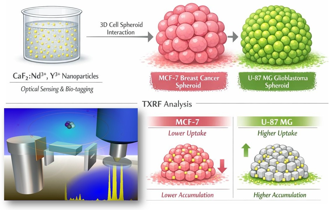
Understanding the interactions of nanomaterials with complex tumour models is essential for advancing their use in nanomedicine. Calcium fluoride nanoparticles doped with neodymium and yttrium (CaF₂:Nd3+, Y3+) exhibit promising properties for biomedical applications, particularly for optical sensing and tagging. This study investigates their interaction with 3D cell spheroids derived from breast cancer (MCF-7) and brain cancer (U-87 MG) cell lines as tumour models. Specific protocols have been developed in Total-reflection X-Ray Fluorescence (TXRF) to evaluate nanoparticles’ internalisation and diffusion within spheroids by quantifying the concentrations of Ca, Nd, and Y taken up by the cells. Minimal background interference enabled precise multi-element detection in low-volume biological samples, yielding very low detection limits and minimal uncertainties. The study demonstrates the effectiveness of TXRF for quantifying rare-earth-doped nanoparticles in 3D cancer models and reveals that, although both cell lines permit nanoparticle diffusion into cells, higher accumulation is observed in glioblastoma cell spheroids. A Weibull diffusion model was applied to help understand the observed internalisation kinetics of nanoparticles into U-87 MG and MCF-7 spheroids. The relevant differences suggest cell-line-dependent uptake behaviour, potentially influenced by differences in cellular architecture, the porosity of the generated spheroid, and its intercellular 3D microstructure. These findings highlight the importance of tumour-specific interactions in the investigation of nanoparticle systems for targeted cancer diagnostics and therapeutics.
Keywords:
TXRF
; nanoparticles
; CaF2: Nd3+
; Y3+
; cell spheroids
; adenocarcinoma
; MCF-7
; glioblastoma
; U-87 MG
; cellular uptake
; internalisation
; difussion
Copyright: This open access article is published under a Creative Commons CC BY 4.0 license, which permit the free download, distribution, and reuse, provided that the author and preprint are cited in any reuse.


