
Submitted:
11 February 2026
Posted:
12 February 2026
You are already at the latest version
Abstract
Background: Over the last two decades, there has been a substantial change in the understanding of post-traumatic hypopituitarism which is no longer regarded as a marginal phenomenon. Clinical manifestations of pituitary hormone deficiency are frequently nonspecific, with fatigue and cognitive dysfunction predominating. Given that head injuries currently constitute a global burden for healthcare systems, the aim of the present study was to determine whether self-reported post-mTBI symptoms that may indicate hypopituitarism reflect true pituitary insufficiency or are attributable to other hormonal aberrations. Objective: Assessment of the relationship between self-reported symptoms of post-traumatic hypopituitarism and hormonal test results following mild Traumatic Brain Injury (mTBI). Setting: Patients were recruited from a tertiary trauma center emergency department (ED) in northern Poland from January 2023 to October 2025. Participants: Adult (18>y.o.) individuals with mTBI meeting the inclusion criteria. Design: Prospective cohort study. During their post-head injury admission to the ED patients had a blood sample taken. The procedure was repeated consecutively after 3,6 and 12 months. After 6 and 12 months, patients were asked to complete a questionnaire. Method: Pituitary and thyroid hormones were measured using Chemiluminescence Immunoassay method (CLIA) and Heterogenous immunochemiluminescence method (CMIA). The questionnaire used was designed for the purposes of this study Questionnaire for the Assessment of Symptoms of Anterior Pituitary Insufficiency in Patients After Mild Traumatic Brain Injury (mTBI) Hospitalized in the Emergency Department. Results: Self-reported symptoms suggestive of anterior pituitary dysfunction following mTBI were not confirmed by laboratory assessment of pituitary hormones. However, after 6 months, a statistically significant correlation was found between the number of reported symptoms and prolactin levels (ρ = 0.730; p = 0.0013), whereas after 12 months a downward trend in fT3 levels was observed compared with baseline. Conclusion: Persistent symptoms observed in patients after mTBI at 6 and 12 months, particularly those related to increased fatigue and impaired concentration, appear to be associated primarily with prolactin levels (6-month observation) and with a decrease in fT3 levels (12-month observation) rather than true post-traumatic hypopituitarism (PTHP).
Keywords:
euthyroid sick syndrome
; low triiodothyronine
; mild Traumatic Brain Injury
; post-traumatic hypopituitarism
; prolactin
1. Introduction
Traumatic Brain Injury (TBI) is defined as non-congenital and non-degenerative brain injury sustained as a result of an external force, which may lead to transient or permanent neurological impairment [1,2]. Traditionally, brain injuries are classified according to the score obtained on the Glasgow Coma Scale (GCS) during post-traumatic assessment into mild (13–15 points), moderate (9–12 points), and severe (8 points or less) [3,4]. The most common type is mild TBI (mTBI), constituting from 80% to 90% of all cases [1,5]. Consistent with its designation, mTBI has traditionally been viewed as a benign injury with no expected long-term consequences. However, advances in neuroimaging, as well as in the understanding of molecular and biochemical processes occurring in the injured brain, undoubtedly necessitate a re-evaluation of this assumption since current evidence demonstrates that TBIs, even the mild ones, may as well be associated with numerous long-term, persistent sequelae [6,7] and the concept of TBI being a single injury event with time-limited recovery is no longer actual [8]. Moreover, current data suggests that up to 50% of patients after mTBI do not return to their pre-illness condition [5].
One of the consequences of traumatic brain injury that has gained increasing research attention over the past 25 years is post-traumatic hypopituitarism. Although the first case was described over 100 years ago, in 1918, it was considered a marginal phenomenon until the early 2000s [9]. Studies conducted since then, however, indicate that its prevalence is substantially higher than previously assumed [9]. The estimated occurrence rate of PTHP varies across studies, ranging from 15% to 60% in adults and up to 42% in children and adolescents [10]. Thus, assuming an estimated global incidence of approximately 50-60 million traumatic brain injuries per year [5], even the most conservative prevalence estimates imply that millions of individuals may be potentially affected by this long-term sequela of injury. Undiagnosed and untreated PTHP may lead to impaired quality of life and place a significant financial burden on healthcare system [11].
The postulated etiology of PTHP involves an overlap between primary brain injury (direct mechanical impact) and secondary brain injury mechanisms [12,13]. The primary, mechanical injury may lead to the damage of pituitary stalk, the gland itself or the supplying hypophyseal portal veins [12,14]. Secondary insult results from pathophysiological processes following TBI (e.g. hypoxia, ischemia, oedema, hemorrhage, alterations in cerebral metabolism)[12,15,16]Finally, the concept of post-traumatic neuroinflammation involving, among others, the tanacytes [17], has gained much attention in recent years. As a consequence of the inflammatory cascade, an autoimmune response is triggered, resulting in appearance of auto-antibodies: anti-pituitary antibodies (APA) and anti-hypothalamic antibodies (AHA) [18].
As indicated above, reported rates of PTHP differ between studies, which may be explained, among other factors, by the use of dynamic testing and the inclusion of patients representing all three brain injury severity categories [19]. On the whole, most studies consistently indicate that the somatotropic axis is the most commonly impaired in the long-term course following TBI [13]. Accurate diagnosis of PTHP remains difficult because the manifestations of pituitary insufficiency are nonspecific and may overlap with symptoms typical of post-concussion syndrome, which further hinders diagnostic evaluation [20]. The exact clinical phenotype depends on the extent and number of disrupted hormonal pathways [21] . Nevertheless, the most commonly reported symptoms associated with PTHP are fatigue, generalized weakness, and cognitive impairment. Although these symptoms are part of the clinical spectrum of growth hormone deficiency (GHD), they lack specificity and, in patients with traumatic brain injury, may result from multiple other factors and may mimic and/or overlap with post-concussion syndrome.
The accurate diagnosis of PTHP, and GHD in particular, is methodologically challenging and depends on dynamic stimulation tests, which are neither economically viable nor feasible for universal application in all patients reporting chronic fatigue following traumatic brain injury. This limitation is reflected in the available literature, as studies utilizing stimulation testing report a markedly lower prevalence of GHD compared with studies in which such tests were not performed [13]. This leads to certain discrepancies, as on the one hand the current literature reports a relatively frequent occurrence of PTHP, while on the other hand clinicians who routinely care for patients after head injury or with endocrine disorders do not observe such a substantial number of cases [22]. There is also no clear consensus in the existing literature regarding the prevalence of post-traumatic hypopituitarism (PTHP) across different levels of traumatic brain injury severity. Although PTHP is generally reported more frequently in patients with severe TBI, Schneider et al. suggested that this association may not apply to mild and moderate injuries, as a higher prevalence of PTHP was observed in patients with mild TBI [23].
Therefore, the authors of the present study aimed to investigate whether, in patients with mild traumatic brain injury (mTBI)—the most common form of brain injury from an epidemiological perspective—reported symptoms such as increased fatigue, reduced concentration, and cognitive impairment, which also fall within the post-concussion symptom spectrum, are associated with post-traumatic pituitary dysfunction or may instead be related to other hormonal disturbances.
2. Methods
Study Setting: This was a prospective single center study performed in Emergency Department in Poland. The ED is a part University Hospital and operates as a tertiary referral center. The annual admission rate to the ED is around 38000 patients.
Patients: Inclusion criteria for the study were: age>18 years old, meeting the American Congress of Rehabilitation Medicine’s (ACRM) definition of mTBI [24] and having no acute intracranial findings on initial CT. Exclusion criteria were as follows: pregnancy, abnormal post-injury neuroimaging findings, hospitalization >48 h due to TBI, age <18 y.o., lack of conscious consent. Patients were recruited from January 2023 until October 2025.
Clinical data: the following demographic and clinical data were collected for each patient: age, sex, past medical history concerning endocrinological diseases.
Clinical scales: Assessment of patient-reported symptoms of hypopituitarism was performed using a questionnaire developed specifically for the purposes of the present study: Questionnaire for the Assessment of Symptoms of Anterior Pituitary Insufficiency in Patients After Mild Traumatic Brain Injury (mTBI) Hospitalized in the Emergency Department. This questionnaire was based on article by Mele et al. [17] and consists of 18 items addressing symptoms related to dysfunction of the different pituitary axes. Full version of this questionnaire is available in Appendix 1.
Laboratory assessment: During the examination, the following laboratory assessments were carried out in the patients:
- -
- hormones: free Triiodothyronine (fT3), free Thyroxine (fT4), Thyroid-Stimulating Hormone (TSH), Growth Hormone (GH), Insuline-like Growth Factor 1 (IGF-1), Prolactine (PRL), Adrenocorticotropic Hormone (ACTH). Reference ranges: TSH 0.35–4.94 uU/ml; fT3 1.78–7.07pmol/l; fT4 9.01–19.05pmol/l, GH <8 ng/ml, IGF-1 89-290 ng/ml, PRL 108.8-557.1 mU/l, ACTH
- -
- Gonadotropic hormones were initially also assessed. However, due to the small size of the study cohort and the relatively high prevalence of confounding factors that could compromise the scientific validity of the collected data—including uncertain menopausal status, use of hormonal contraception, testosterone replacement therapy, and preparation for gender-affirming treatment—these data were excluded from the final analysis. Consequently, the scope of the study was restricted to hormones directly associated with symptoms that may mimic post-concussion syndrome, such as cognitive impairment, fatigue, and related mood fluctuations.
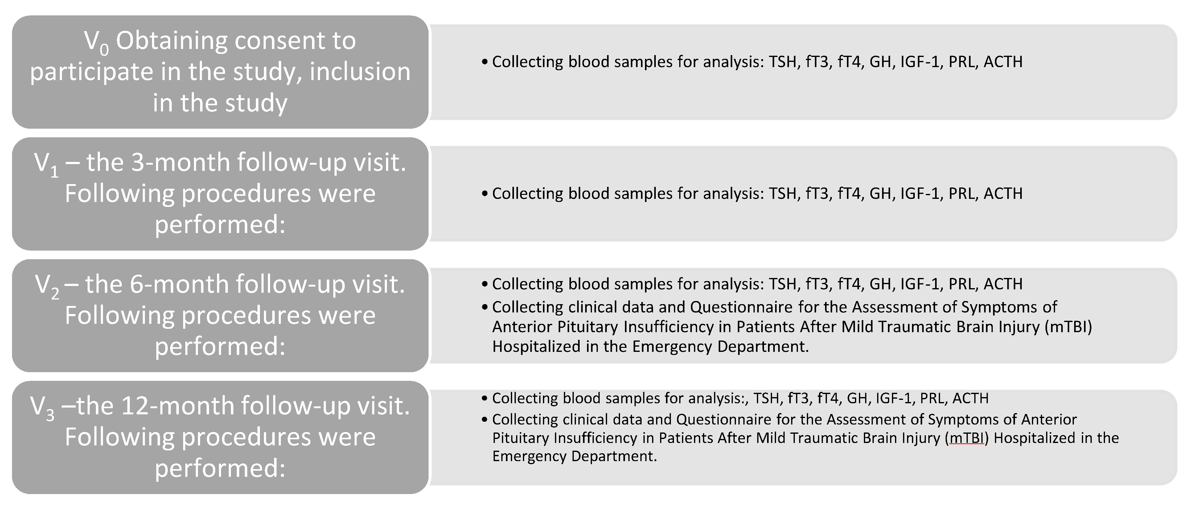
Study Protocol: the following visits were pre-scheduled for each patient (they are presented in Figure1):
V0 – the initial visit to the Emergency Department. Following procedures were performed:
- -
- Obtaining consent to participate in the study, inclusion in the study
- -
- Collecting blood samples for analysis: TSH, fT3, fT4, GH, IGF-1, PRL, ACTH
V1 – the 3-month follow-up visit. Following procedures were performed:
- -
- Collecting blood samples for analysis: TSH, fT3, fT4, GH, IGF-1, PRL, ACTH
V2 – the 6-month follow-up visit. Following procedures were performed:
- -
- Collecting blood samples for analysis: TSH, fT3, fT4, GH, IGF-1, PRL, ACTH
- -
- Collecting clinical data and Questionnaire for the Assessment of Symptoms of Anterior Pituitary Insufficiency in Patients After Mild Traumatic Brain Injury (mTBI) Hospitalized in the Emergency Department.
V3 –the 12-month follow-up visit. Following procedures were performed:
- -
- Collecting blood samples for analysis: TSH, fT3, fT4, GH, IGF-1, PRL, ACTH
- -
- Collecting clinical data and Questionnaire for the Assessment of Symptoms of Anterior Pituitary Insufficiency in Patients After Mild Traumatic Brain Injury (mTBI) Hospitalized in the Emergency Department.
Figure 1.
Study protocol.
Statistical Analysis
Data quality was verified by removing duplicate patient records and standardizing missing questionnaire entries. Values reported as below the detection limit (e.g., <x) were conservatively converted to x. Normality was assessed using the Shapiro–Wilk test (n ≥ 3) for baseline, follow-up, and paired differences. Changes over time were analyzed using paired t-tests or Wilcoxon signed-rank tests, as appropriate; variables with fewer than three paired observations were not interpreted. Associations between symptoms and laboratory parameters were assessed using Spearman’s rank correlation, while group comparisons were performed using Welch’s t-test or the Mann–Whitney U test. All tests were two-sided with a significance level of α = 0.05; raw p-values were reported, with false discovery rate correction (Benjamini–Hochberg) applied where appropriate.
The analyses were performed in the Python environment, version 3 (Python Software Foundation, USA), using the following libraries: pandas (pandas-dev, open-source, available at https://pandas.pydata.org), SciPy – the scipy.stats module (The SciPy Community, open-source, https://scipy.org), statsmodels (The statsmodels development team, open-source, https://www.statsmodels.org) openpyxl (open-source, https://openpyxl.readthedocs.io) – used for saving and organizing results in Excel spreadsheets. Additional calculations and table preparation were carried out in Microsoft Excel (Microsoft Corporation, Redmond, WA, USA).
3. Results
Patients were recruited from January 2023 till October 2025. A total of 140 individuals were enrolled in the study. However, in course of the study 8 participants were excluded (5 due to traumatic lesions identified on CT imaging, 1 due to pregnancy, 1 due to death, and 1 due to a subsequent severe head injury). Furthermore, 20 participants were excluded because of incomplete baseline data. Ultimately, the study group consisted of 56 participants. A total of 27 participants attended the laboratory follow-up at 3 months. At 6 months, 18 participants underwent follow-up, of whom 17 completed the questionnaire. After 12 months, 37 follow-up laboratory tests were performed, and 21 questionnaires were completed. The flow and characteristics of the patients are presented in Figure 2 and Table 1.
The 12-month analysis revealed a trend toward decreased fT3 levels (Figure 3, Figure 4 and Figure 5), however, the result does not reach statistical significance (paired t-test; p ≈ 0.0785; n = 37; median Δ = −0.150; alternatively, Wilcoxon test p ≈ 0.104). No association was found between the change in fT3 after 12 months and the severity of complaints reported in the questionnaire (Figure 6) (ΔfT3 vs. number of complaints Q_count: rho ≈ −0.064; p ≈ 0.705), nor were there differences in ΔfT3 between participants with and without complaints (p ≈ 0.931).
After 6 months, difficulty concentrating was the most frequently reported symptom, whereas after 12 months, increased fatigue was reported most often (Table 2). Assessment of symptoms and their frequency was done using a questionnaire designed by authors for the purpose of this study (Questionnaire is available in Appendix 1).
Although no statistically significant change in prolactin levels was observed after 6 months (Figure 7) compared with baseline values, a strong, positive, and statistically significant correlation was found between the number of reported symptoms and prolactin levels (ρ = 0.730; p = 0.0013). (Figure 8). After applying the FDR correction, the result remained statistically significant (p_FDR = 0.0173).
No statistically significant changes were found in the analysis of the remaining hormones (Figure 9).
4. Discussion
The occurrence of neuroendocrine disturbances as a response to injury or severe illness is well documented [25,26]. Some endocrine abnormalities, if unrecognized and untreated—such as glucocorticoid deficiency—can have life-threatening consequences in the acute phase [2].
Insufficiency of other hormones may be of prognostic relevance, influencing recovery and subsequent quality of life [11,27].
Thyroid hormones play a crucial role in brain development and are essential for its proper functioning, including the regulation of neuronal plasticity processes, stimulation of angiogenesis and neurogenesis, as well as modulation of cytoskeletal dynamics and intracellular transport processes.[28,29,30,31]. Even though the thyrotropic axis, due to the central location of thyrotrope cells within the anterior pituitary, is less frequently damaged after head trauma than the somatotropic or gonadotropic axes [32], numerous studies involving patients after traumatic brain injury have demonstrated abnormalities in thyroid hormone levels [33,34,35]. Consequently, the impact of thyroid hormones on post-TBI recovery and neurological outcome has already been the subject of various investigations [36,37,38,39,40] in which their beneficial effect on functional restoration has been proved. Aberrations in thyroid hormone levels in were first described in the 1970s, when critically ill patients—regardless of the underlying etiology—were observed to exhibit low serum concentrations of free triiodothyronine (fT3), or both free triiodothyronine (fT3) and free thyroxine (fT4), in the presence of normal thyroid-stimulating hormone (TSH) levels. This phenomenon is now referred to as Euthyroid Sick Syndrome (ESS) or Non-Thyroidal Illness Syndrome (NTIS) [41]. Since that time, transient disturbances in thyroid hormone parameters have been considered a part of the physiological stress response [9,42] and characteristic element of the laboratory manifestations of acute illness. However, the present study appears to suggest that fluctuations in fT3 levels may also occur long after head injury, thereby potentially affecting the recovery process.
As it was mentioned above, in the majority of studies conducted to date with long-term follow-up, chronic impairment of the pituitary–thyroid axis has been observed less frequently than dysfunction of the somatotropic or gonadotropic axes [2]. In this context, the findings of a recent study conducted exclusively in female athletes who sustained mild TBI are particularly noteworthy. In that cohort, central hypothyroidism was the most commonly diagnosed endocrine abnormality[43]. Notably, most previous investigations of post-traumatic hypopituitarism (PTHP) have predominantly included male populations [44], whereas in our study women constituted the majority of participants at all follow-up time points. It is therefore conceivable that in women the pituitary–thyroid axis may be more susceptible to post-traumatic injury, suggesting that strategies for post-traumatic monitoring of pituitary dysfunction should also take biological sex into account. The hypothesis of increased susceptibility of the female organism to deviations from physiological homeostasis is supported by a study conducted in a large cohort of women, which examined the relationship between trauma exposure, post-traumatic stress disorder (PTSD), and thyroid dysfunction. The authors demonstrated that PTSD was associated with an increased risk of hypothyroidism in a dose-dependent manner. Heightened clinical awareness of thyroid dysfunction may therefore be particularly important in women with PTSD [45]. Therefore, future research in female patients following mTBI appears warranted, with a special focus on fT3 level fluctuations.
No post-traumatic growth hormone deficiency was identified in any patient in our study. In the 12-month follow-up cohort, two participants had baseline growth hormone (GH) levels below the reference range. However, at the 12-month assessment, GH concentrations below the reference range were identified in three different participants. In all of these individuals, insulin-like growth factor 1 (IGF-1) levels remained within the normal range. These findings underscore the importance of appropriate timing in the assessment of growth hormone secretion and highlight the limitations of relying solely on a single hormone measurement for diagnostic purposes. Notably, in this study all affected participants were male. This observation is consistent with epidemiological data indicating a higher prevalence of GH deficiency in men [46]. Thus, it may suggest that the sex distribution of study populations may influence the reported prevalence of deficiencies of individual pituitary hormones. As noted above, most studies conducted to date on post-traumatic hypopituitarism (PTHP) have been characterized by a predominance of male participants, among whom growth hormone deficiency is epidemiologically more common. Consequently, this may have affected the final study outcomes, which in the great majority pointed to GH deficiency as the most common abnormality.
The study also provides an interesting observation regarding prolactin. The majority of previously published studies have reported hyperprolactinemia as a consequence of TBI [47,48], in line with the hypothesis of dopaminergic pathway injury and subsequent impairment of dopamine-mediated regulatory mechanisms as well as the anatomical damage of pituitary [48]. In this study, however, contrary to expectations, no significant prolactin elevation was observed. Despite this, after 6 months, a strong, positive, and statistically significant correlation was found between the number of reported symptoms and prolactin levels. It appears that in this case as well, the predominance of women in the current study may have influenced the result. After 6 months, the most common complaint was difficulty concentrating, followed by mood disturbances and mood fluctuations. The obtained findings may be supported by a study on prolactin levels in major depressive disorder, which demonstrated that prolactin levels are higher in women and in patients with major depressive disorder compared with the control group [49]. It appears, however, that the significance of prolactin after TBI has so far been underestimated and represents an area that clearly requires further research. In our study, during the long-term period following injury, higher prolactin levels were correlated with a greater number of adverse symptoms. In contrast, its role in the acute post brain injury period seems to be beneficial. Studies conducted in patients after non-traumatic brain injury (NTBI) have demonstrated the anti-inflammatory effects of prolactin through glial cell activation, as well as its ability to reduce calcium overload and exert protective effects against excitotoxicity [48]. It would therefore be worthwhile to consider expanding such studies to include patients with traumatic brain injury.
5. Conclusions
This pilot study provides an interesting perspective on patient-reported symptoms following mild traumatic brain injury (mTBI), conceptualizing them as a multifactorial and often difficult-to-classify problem. The findings suggest that post-mTBI symptoms do not necessarily have to be associated with major abnormalities resulting from pituitary damage, but may instead result from an overlap between post-concussion symptoms and subtle fluctuations in prolactin and thyroid hormone levels. Importantly, the present study included exclusively patients with mild TBI, with women constituting a substantially larger proportion of the study population. In contrast, the majority of previous studies focusing on post-traumatic hypopituitarism (PTHP), which indicated GH deficiency as the most common hormonal abnormality, predominantly included male patients and individuals with moderate to severe traumatic brain injury. This may suggest the need for future PTHP studies with a more balanced sex distribution among participants to verify whether differences exist between women and men in post-traumatic hormonal disorders, which could in turn imply differences in post-traumatic care.
6. Limitations
The main limitation of the present study is the small sample size, especially in terms of completing questionnaires, which renders the obtained results indicative of observed trends rather than definitive conclusions. Since the study took place in the emergency department, optimal timing of blood collection for hormonal assays could not always be ensured. Moreover, it was not possible to apply the dynamic tests recommended for the assessment of the somatotropic and corticotropic axes.
Additionally, concomitant medications taken by the patients, which could potentially influence thyroid hormone levels, were not accounted for in the final analysis.
Author Contributions
M.K.: conceptualization of the study, coordination of the study, literature search, preparation of the draft of the manuscript, preparation of the final version of the manuscript; M.S.: conceptualization of the study, literature selection, final editing of the manuscript, E.S.: data curation and analysis, literature search.
Insitutional Review Board Statement: This study protocol was reviewed and approved by Bioethics Committee of the Medical University of Gdansk, approval number NKBBN/406/2021.
Informed Consent Statement
The written informed consent was obtained from participants.
Conflicts of Interest Statement: The authors have no conflict of interest to declare.
Funding Sources: The authors did not receive any funding for any research relevant for this study.
Data Availability Statement
All data generated or analyzed during this study are included in this article. Further enquiries can be directed to the corresponding author.
Abbreviations
| ACTH | adrenocortycotropic hormone |
| CLIA | chemiluminescent immunoassay |
| CMIA | heterogenous immunochemiluminescence |
| ESS | euthyroid sick syndrome |
| fT3 | free triiodothyronine |
| GH | growth hormone |
| IGF-1 | insulin-like growth factor 1 |
| mTBI | mild traumatic brain injury |
| NTIS | non thyroidal illness syndrome |
| PRL | prolactin |
| PTHP | post traumatic hypopituitarism |
| T3 | triiodothyronine |
| TSH | thyroid-stimulating hormone |
| TH | thyroid hormones |
References
- Capizzi, A.; Woo, J.; Verduzco-Gutierrez, M. Traumatic Brain Injury: An Overview of Epidemiology, Pathophysiology, and Medical Management. Med Clin North Am 2020, 104, 213–238. [Google Scholar] [CrossRef]
- Kgosidialwa, O.; Agha, A. Hypopituitarism Post Traumatic Brain Injury (TBI): Review. Ir J Med Sci 2019, 188, 1201–1206. [Google Scholar] [CrossRef] [PubMed]
- Manley, G.T.; Dams-O’Connor, K.; Alosco, M.L.; Awwad, H.O.; Bazarian, J.J.; Bragge, P.; Corrigan, J.D.; Doperalski, A.; Ferguson, A.R.; Mac Donald, C.L.; et al. A New Characterisation of Acute Traumatic Brain Injury: The NIH-NINDS TBI Classification and Nomenclature Initiative. The Lancet Neurology 2025, 24, 512–523. [Google Scholar] [CrossRef]
- Brazinova, A.; Rehorcikova, V.; Taylor, M.S.; Buckova, V.; Majdan, M.; Psota, M.; Peeters, W.; Feigin, V.; Theadom, A.; Holkovic, L.; et al. Epidemiology of Traumatic Brain Injury in Europe: A Living Systematic Review. J Neurotrauma 2021, 38, 1411–1440. [Google Scholar] [CrossRef]
- Maas, A.I.R.; Menon, D.K.; Manley, G.T.; Abrams, M.; Åkerlund, C.; Andelic, N.; Aries, M.; Bashford, T.; Bell, M.J.; Bodien, Y.G.; et al. Traumatic Brain Injury: Progress and Challenges in Prevention, Clinical Care, and Research. Lancet Neurol 2022, 21, 1004–1060. [Google Scholar] [CrossRef]
- Wilson, L.; Stewart, W.; Dams-O’Connor, K.; Diaz-Arrastia, R.; Horton, L.; Menon, D.K.; Polinder, S. The Chronic and Evolving Neurological Consequences of Traumatic Brain Injury. Lancet Neurol 2017, 16, 813–825. [Google Scholar] [CrossRef]
- Pavlovic, D.; Pekic, S.; Stojanovic, M.; Popovic, V. Traumatic Brain Injury: Neuropathological, Neurocognitive and Neurobehavioral Sequelae. Pituitary 2019, 22, 270–282. [Google Scholar] [CrossRef]
- Dams-O’Connor, K.; Juengst, S.B.; Bogner, J.; Chiaravalloti, N.D.; Corrigan, J.D.; Giacino, J.T.; Harrison-Felix, C.L.; Hoffman, J.M.; Ketchum, J.M.; Lequerica, A.H.; et al. Traumatic Brain Injury as a Chronic Disease: Insights from the United States Traumatic Brain Injury Model Systems Research Program. Lancet Neurol 2023, 22, 517–528. [Google Scholar] [CrossRef]
- Gasco, V.; Cambria, V.; Bioletto, F.; Ghigo, E.; Grottoli, S. Traumatic Brain Injury as Frequent Cause of Hypopituitarism and Growth Hormone Deficiency: Epidemiology, Diagnosis, and Treatment. Frontiers in Endocrinology 2021, 12–2021. [Google Scholar] [CrossRef]
- Wexler, T.L. Neuroendocrine Disruptions Following Head Injury. Curr Neurol Neurosci Rep 2023, 23, 213–224. [Google Scholar] [CrossRef]
- Bensalah, M.; Donaldson, M.; Labassen, M.; Cherfi, L.; Nebbal, M.; Haffaf, E.M.; Abdennebi, B.; Guenane, K.; Kemali, Z.; Ould Kablia, S. Prevalence of Hypopituitarism and Quality of Life in Survivors of Post-Traumatic Brain Injury. Endocrinol Diabetes Metab 2020, 3, e00146. [Google Scholar] [CrossRef]
- Tan, C.L.; Hutchinson, P.J. A Neurosurgical Approach to Traumatic Brain Injury and Post-Traumatic Hypopituitarism. Pituitary 2019, 22, 332–337. [Google Scholar] [CrossRef]
- Kgosidialwa, O.; Hakami, O.; Muhammad Zia-Ul-Hussnain, H.; Agha, A. Growth Hormone Deficiency Following Traumatic Brain Injury. Int J Mol Sci 2019, 20. [Google Scholar] [CrossRef]
- Tanriverdi, F.; Schneider, H.J.; Aimaretti, G.; Masel, B.E.; Casanueva, F.F.; Kelestimur, F. Pituitary Dysfunction after Traumatic Brain Injury: A Clinical and Pathophysiological Approach. Endocr Rev 2015, 36, 305–342. [Google Scholar] [CrossRef]
- Karaboue, M.A.A.; Ministeri, F.; Sessa, F.; Nannola, C.; Chisari, M.G.; Cocimano, G.; Di Mauro, L.; Salerno, M.; Esposito, M. Traumatic Brain Injury as a Public Health Issue: Epidemiology, Prognostic Factors and Useful Data from Forensic Practice. Healthcare (Basel) 2024, 12. [Google Scholar] [CrossRef]
- De Bellis, A.; Bellastella, G.; Maiorino, M.I.; Costantino, A.; Cirillo, P.; Longo, M.; Pernice, V.; Bellastella, A.; Esposito, K. The Role of Autoimmunity in Pituitary Dysfunction Due to Traumatic Brain Injury. Pituitary 2019, 22, 236–248. [Google Scholar] [CrossRef]
- Mele, C.; Pingue, V.; Caputo, M.; Zavattaro, M.; Pagano, L.; Prodam, F.; Nardone, A.; Aimaretti, G.; Marzullo, P. Neuroinflammation and Hypothalamo-Pituitary Dysfunction: Focus of Traumatic Brain Injury. Int J Mol Sci 2021, 22. [Google Scholar] [CrossRef] [PubMed]
- The Autoimmune Basis of Hypopituitarism in Traumatic Brain Injury: Fiction or Reality? England, 2019; Vol. 33.
- Hacioglu, A.; Kelestimur, F.; Tanriverdi, F. Long-Term Neuroendocrine Consequences of Traumatic Brain Injury and Strategies for Management. Expert Rev Endocrinol Metab 2020, 15, 123–139. [Google Scholar] [CrossRef] [PubMed]
- Schneider, H.J.; Schneider, M.; Saller, B.; Petersenn, S.; Uhr, M.; Husemann, B.; von Rosen, F.; Stalla, G.K. Prevalence of Anterior Pituitary Insufficiency 3 and 12 Months after Traumatic Brain Injury. Eur J Endocrinol 2006, 154, 259–265. [Google Scholar] [CrossRef] [PubMed]
- Fleseriu, M.; Christ-Crain, M.; Langlois, F.; Gadelha, M.; Melmed, S. Hypopituitarism. Lancet 2024, 403, 2632–2648. [Google Scholar] [CrossRef]
- Klose, M.; Feldt-Rasmussen, U. Chronic Endocrine Consequences of Traumatic Brain Injury - What Is the Evidence? Nat Rev Endocrinol 2018, 14, 57–62. [Google Scholar] [CrossRef]
- Schneider, H.J.; Kreitschmann-Andermahr, I.; Ghigo, E.; Stalla, G.K.; Agha, A. Hypothalamopituitary Dysfunction Following Traumatic Brain Injury and Aneurysmal Subarachnoid Hemorrhage: A Systematic Review. JAMA 2007, 298, 1429–1438. [Google Scholar] [CrossRef]
- Silverberg, N.D.; Iverson, G.L.; Cogan, A.; Dams-O-Connor, K.; Delmonico, R.; Graf, M.J.P.; Iaccarino, M.A.; Kajankova, M.; Kamins, J.; McCulloch, K.L.; et al. The American Congress of Rehabilitation Medicine Diagnostic Criteria for Mild Traumatic Brain Injury. Arch Phys Med Rehabil 2023, 104, 1343–1355. [Google Scholar] [CrossRef] [PubMed]
- Woolf, P.D. Hormonal Responses to Trauma. Crit Care Med 1992, 20, 216–226. [Google Scholar] [CrossRef]
- Warner, M.H.; Beckett, G.J. Mechanisms behind the Non-Thyroidal Illness Syndrome: An Update. Journal of Endocrinology 2010, 205, 1–13. [Google Scholar] [CrossRef] [PubMed]
- Nourollahi, S.; Wille, J.; Weiß, V.; Wedekind, C.; Lippert-Grüner, M. Quality-of-Life in Patients with Post-Traumatic Hypopituitarism. Brain Inj 2014, 28, 1425–1429. [Google Scholar] [CrossRef] [PubMed]
- Talhada, D.; Santos, C.R.A.; Gonçalves, I.; Ruscher, K. Thyroid Hormones in the Brain and Their Impact in Recovery Mechanisms After Stroke. Front Neurol 2019, 10, 1103. [Google Scholar] [CrossRef]
- Liu, Y.-Y.; Brent, G.A. The Role of Thyroid Hormone in Neuronal Protection. Comprehensive Physiology 2021, 11, 2075–2095. [Google Scholar] [CrossRef]
- Mele, C.; De Marchi, L.; Pitino, R.; Costantini, L.; Cavigiolo, B.; Caputo, M.; Marzullo, P.; Aimaretti, G. The Interplay between Thyrotropic Axis, Neurological Complications, and Rehabilitation Outcomes in Patients with Traumatic Brain Injury. Best Pract Res Clin Endocrinol Metab 2025, 39, 102001. [Google Scholar] [CrossRef]
- Rovet, J.F. The Role of Thyroid Hormones for Brain Development and Cognitive Function. Endocr Dev 2014, 26, 26–43. [Google Scholar] [CrossRef]
- Fernández Rodriguez, E.; Villar Taibo, R.; Bernabeu, I. Hypopituitarism after Traumatic Brain Injury in Adults: Clinical Guidelines of the Neuroendocrinology Area of the Spanish Society of Endocrinology and Nutrition (SEEN). Endocrinol Diabetes Nutr (Engl Ed) 2023, 70, 584–591. [Google Scholar] [CrossRef]
- Malekpour, B.; Mehrafshan, A.; Saki, F.; Malekmohammadi, Z.; Saki, N. Effect of Posttraumatic Serum Thyroid Hormone Levels on Severity and Mortality of Patients with Severe Traumatic Brain Injury. Acta Med Iran 2012, 50, 113–116. [Google Scholar] [PubMed]
- Zhang, M.; Wei, J.-B.; Zhang, X.-B. Influence of Traumatic Brain Injury on Serum Levels of Pituitary, Thyroid, Adrenal and Gonadal Hormones. Medicine 2025, 104. [Google Scholar] [CrossRef]
- Mele, C.; Pagano, L.; Franciotta, D.; Caputo, M.; Nardone, A.; Aimaretti, G.; Marzullo, P.; Pingue, V. Thyroid Function in the Subacute Phase of Traumatic Brain Injury: A Potential Predictor of Post-Traumatic Neurological and Functional Outcomes. J Endocrinol Invest 2022, 45, 379–389. [Google Scholar] [CrossRef] [PubMed]
- Mele, C.; Bagnato, S.; De Tanti, A.; Lucca, L.F.; Saviola, D.; Marcuccio, L.; Moretta, P.; Scarponi, F.; Losavio, E.; Picciola, E.; et al. Free Thyroxine (fT4) as a Potential Biomarker of Neurological and Functional Outcome in Acquired Brain Injury: A Prospective Multicenter Cohort Study. J Clin Med 2023, 12. [Google Scholar] [CrossRef] [PubMed]
- Khandelwal, M.; Ying, Z.; Gomez-Pinilla, F. Thyroid Hormone T4 Alleviates Traumatic Brain Injury by Enhancing Blood–Brain Barrier Integrity. International Journal of Molecular Sciences 2025, 26. [Google Scholar] [CrossRef]
- Li, J.; Donangelo, I.; Abe, K.; Scremin, O.; Ke, S.; Li, F.; Milanesi, A.; Liu, Y.-Y.; Brent, G.A. Thyroid Hormone Treatment Activates Protective Pathways in Both in Vivo and in Vitro Models of Neuronal Injury. Molecular and cellular endocrinology 2017, 452, 120–130. [Google Scholar] [CrossRef]
- Crupi, R.; Paterniti, I.; Campolo, M.; Di Paola, R.; Cuzzocrea, S.; Esposito, E. Exogenous T3 Administration Provides Neuroprotection in a Murine Model of Traumatic Brain Injury. Pharmacological research 2013, 70, 80–89. [Google Scholar] [CrossRef]
- Lin, C.; Li, N.; Chang, H.; shen, Y.; Li, Z.; wei, W.; Chen, H.; Lu, H.; Ji, J.; Liu, N. Dual Effects of Thyroid Hormone on Neurons and Neurogenesis in Traumatic Brain Injury. Cell Death & Disease 2020, 11, 671. [Google Scholar] [CrossRef]
- Fliers, E.; Boelen, A. An Update on Non-Thyroidal Illness Syndrome. J Endocrinol Invest 2021, 44, 1597–1607. [Google Scholar] [CrossRef]
- Rolih, C.A.; Ober, K.P. The Endocrine Response to Critical Illness. Med Clin North Am 1995, 79, 211–224. [Google Scholar] [CrossRef]
- Eggertsdóttir Claessen, L.Ó.; Kristjánsdóttir, H.; Jónsdóttir, M.K.; Lund, S.H.; Unnsteinsdóttir Kristensen, I.; Sigurjónsdóttir, H.Á. Pituitary Dysfunction Following Mild Traumatic Brain Injury in Female Athletes. Endocr Connect 2024, 13, e230363. [Google Scholar] [CrossRef]
- Aljboor, G.S.; Tulemat, A.; Al-Saedi, A.R.; Radoi, M.P.; Toader, C.; Papacocea, T.M. Acute and Chronic Hypopituitarism Following Traumatic Brain Injury: A Systematic Review and Meta-Analysis. Neurosurg Rev 2024, 47, 841. [Google Scholar] [CrossRef]
- Jung, S.J.; Kang, J.H.; Roberts, A.L.; Nishimi, K.; Chen, Q.; Sumner, J.A.; Kubzansky, L.; Koenen, K.C. Posttraumatic Stress Disorder and Incidence of Thyroid Dysfunction in Women. Psychological Medicine 2019, 49, 2551–2560. [Google Scholar] [CrossRef] [PubMed]
- Stochholm, K.; Gravholt, C.H.; Laursen, T.; Jørgensen, J.O.; Laurberg, P.; Andersen, M.; Kristensen, L.Ø.; Feldt-Rasmussen, U.; Christiansen, J.S.; Frydenberg, M.; et al. Incidence of GH Deficiency - a Nationwide Study. Eur J Endocrinol 2006, 155, 61–71. [Google Scholar] [CrossRef] [PubMed]
- Magyar-Sumegi, Z.D.; Stankovics, L.; Lendvai-Emmert, D.; Czigler, A.; Hegedus, E.; Csendes, M.; Toth, L.; Ungvari, Z.; Buki, A.; Toth, P. Acute Neuroendocrine Changes after Traumatic Brain Injury. Brain and Spine 2024, 4, 102830. [Google Scholar] [CrossRef]
- Yousefvand, S.; Hadjzadeh, M.-A.-R.; Vafaee, F.; Dolatshad, H. The Protective Effects of Prolactin on Brain Injury. Life Sci 2020, 263, 118547. [Google Scholar] [CrossRef] [PubMed]
- Elgellaie, A.; Larkin, T.; Kaelle, J.; Mills, J.; Thomas, S. Plasma Prolactin Is Higher in Major Depressive Disorder and Females, and Associated with Anxiety, Hostility, Somatization, Psychotic Symptoms and Heart Rate. Compr Psychoneuroendocrinol 2021, 6, 100049. [Google Scholar] [CrossRef]
Figure 2.
Distribution of patients across different stages of the study.
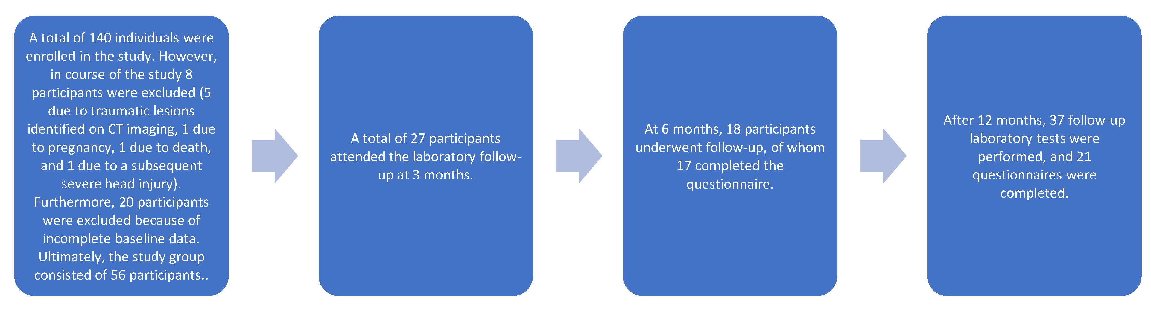
Figure 3.
Distribution of fT3 levels post-injury and at 12 months.
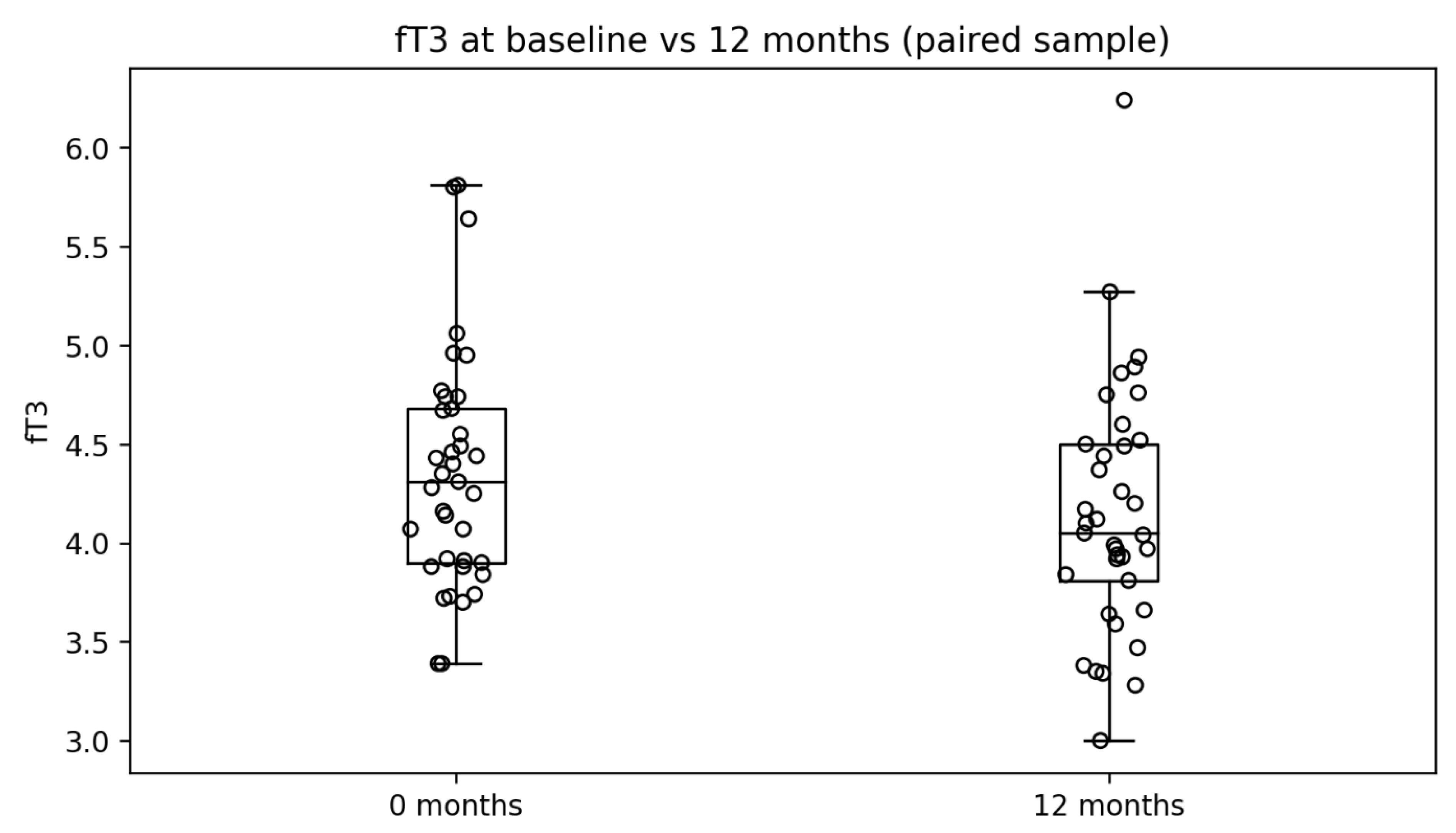
Figure 4.
Changes in the median fT3 over time.
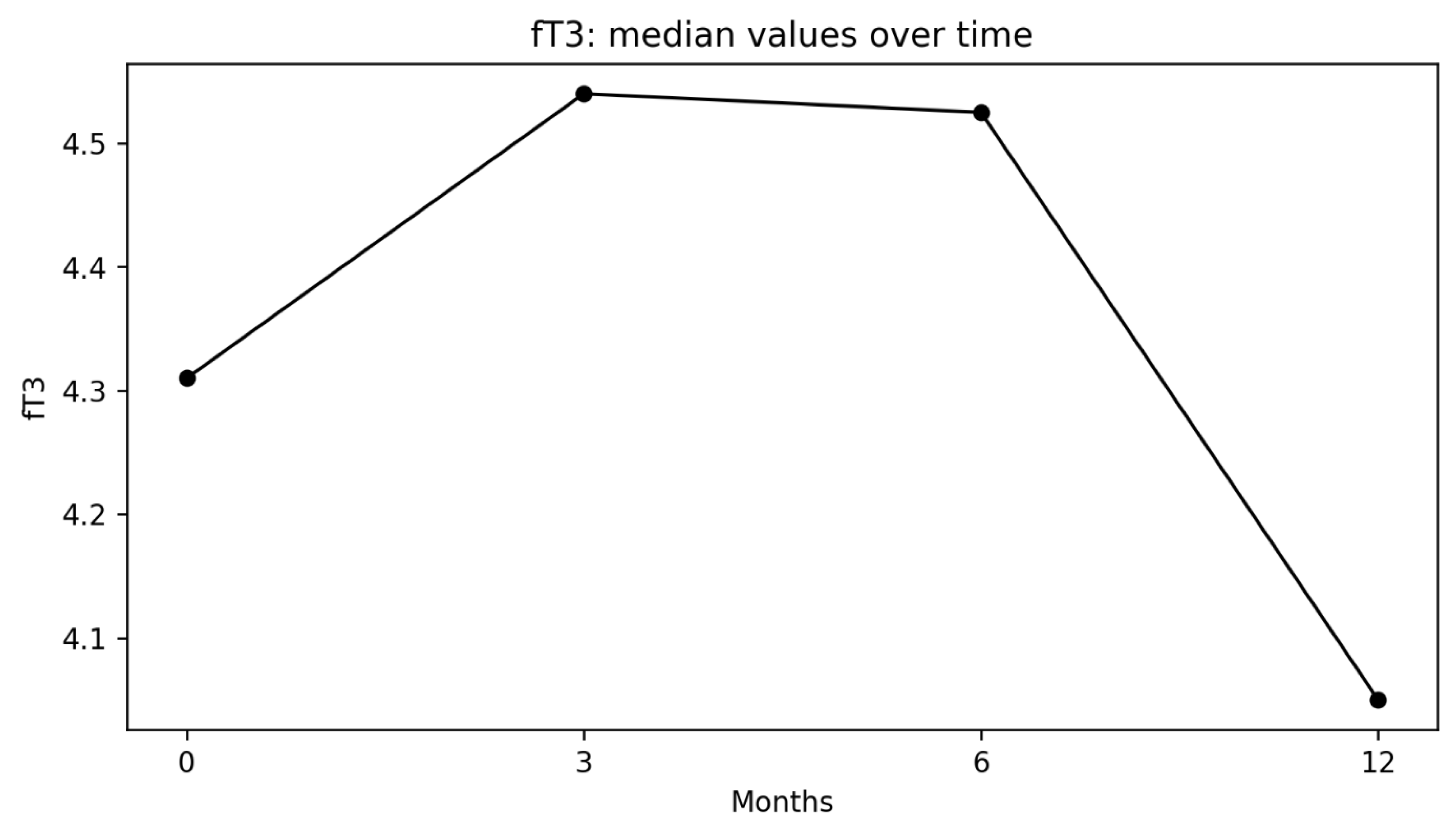
Figure 5.
Distribution of fT3 changes from baseline over time.
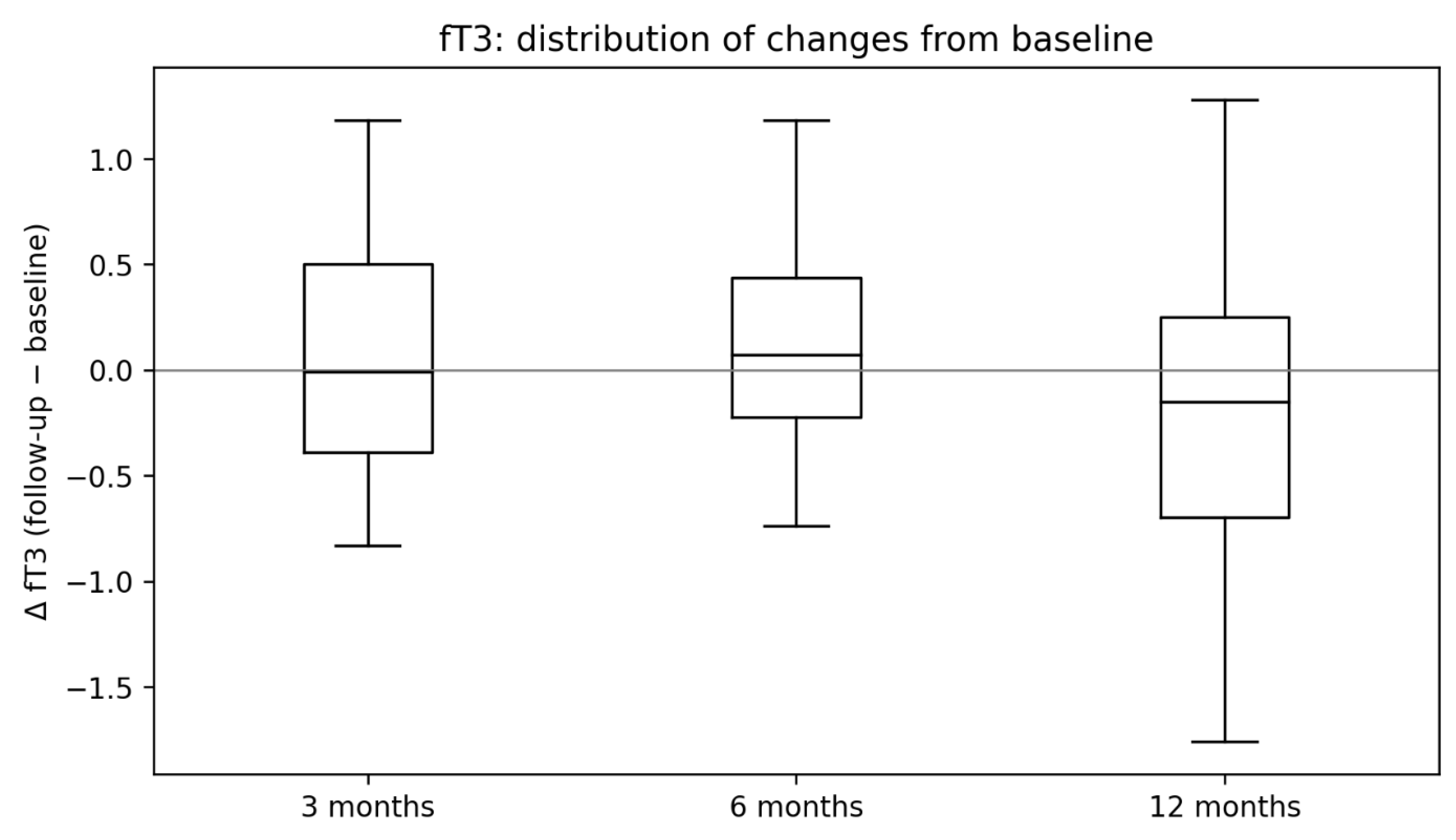
Figure 6.
Changes in fT3 levels vs. number of reported symptoms.
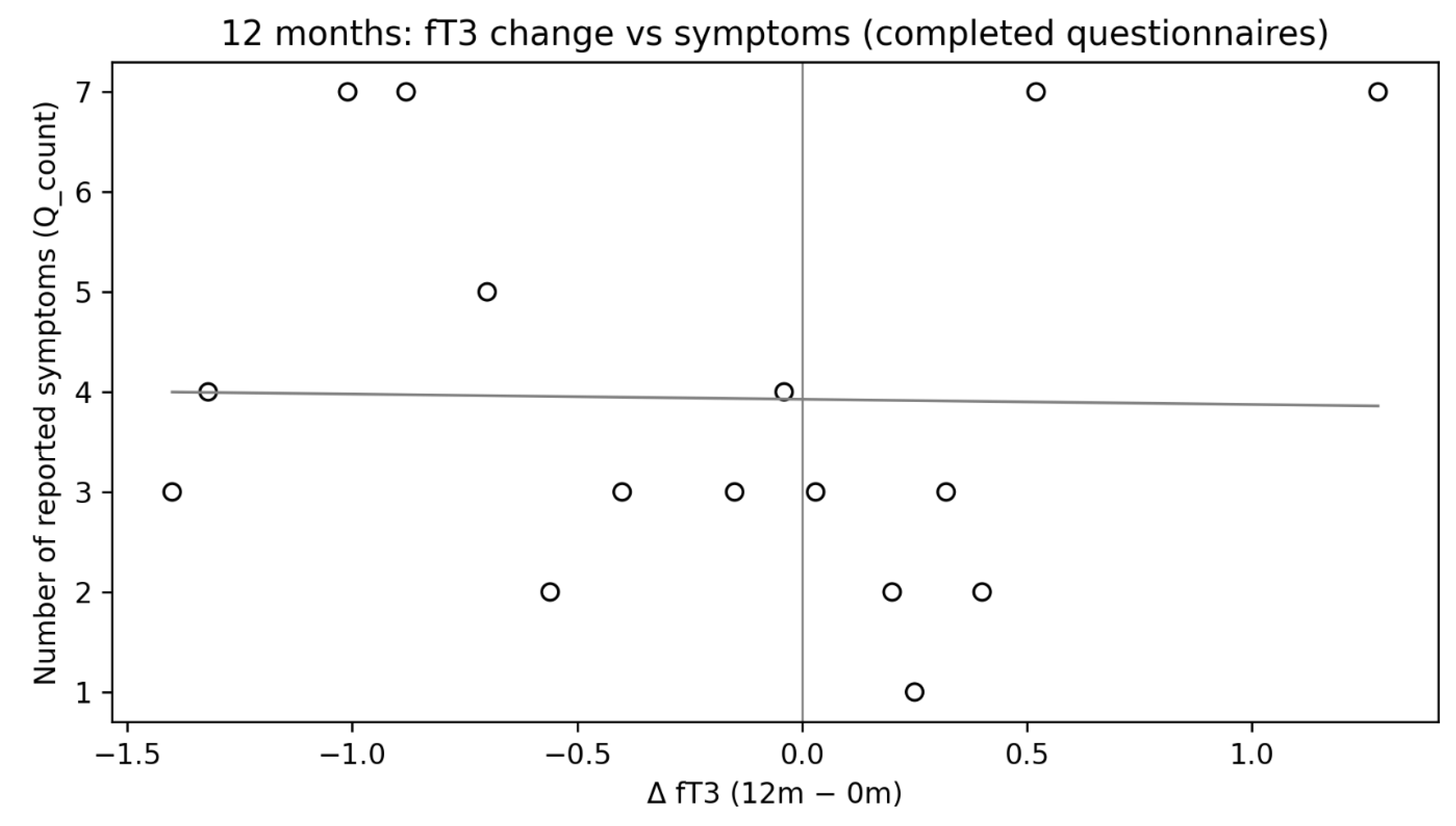
Figure 7.
Prolactin changes over time.
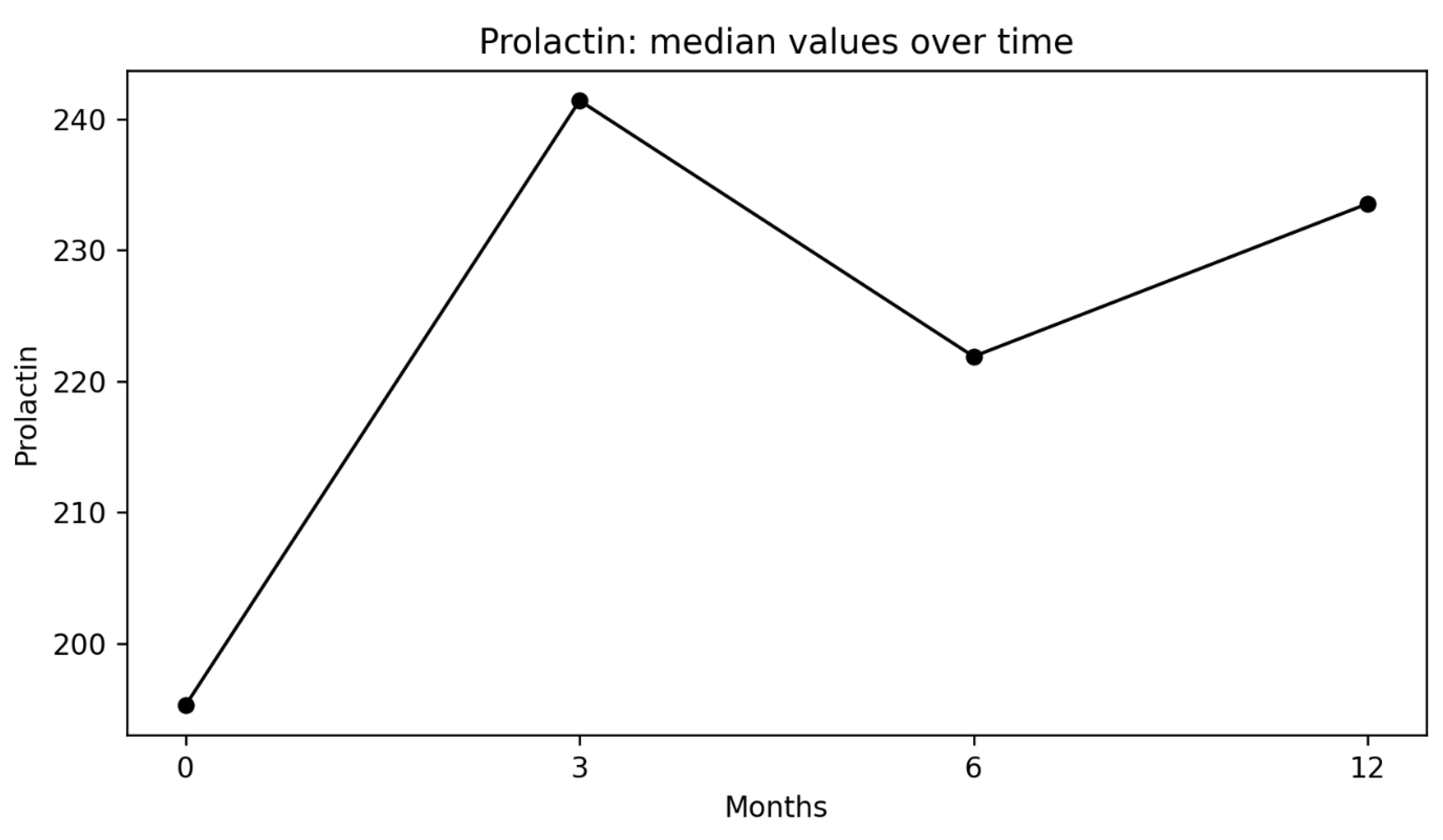
Figure 8.
Symptoms vs prolactin after 6 months.
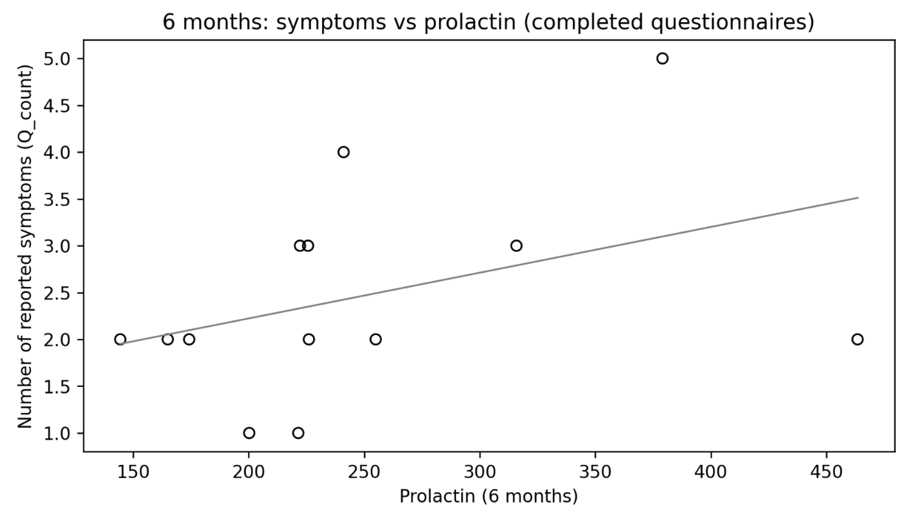
Figure 9.
hormonal changes after 12 months.
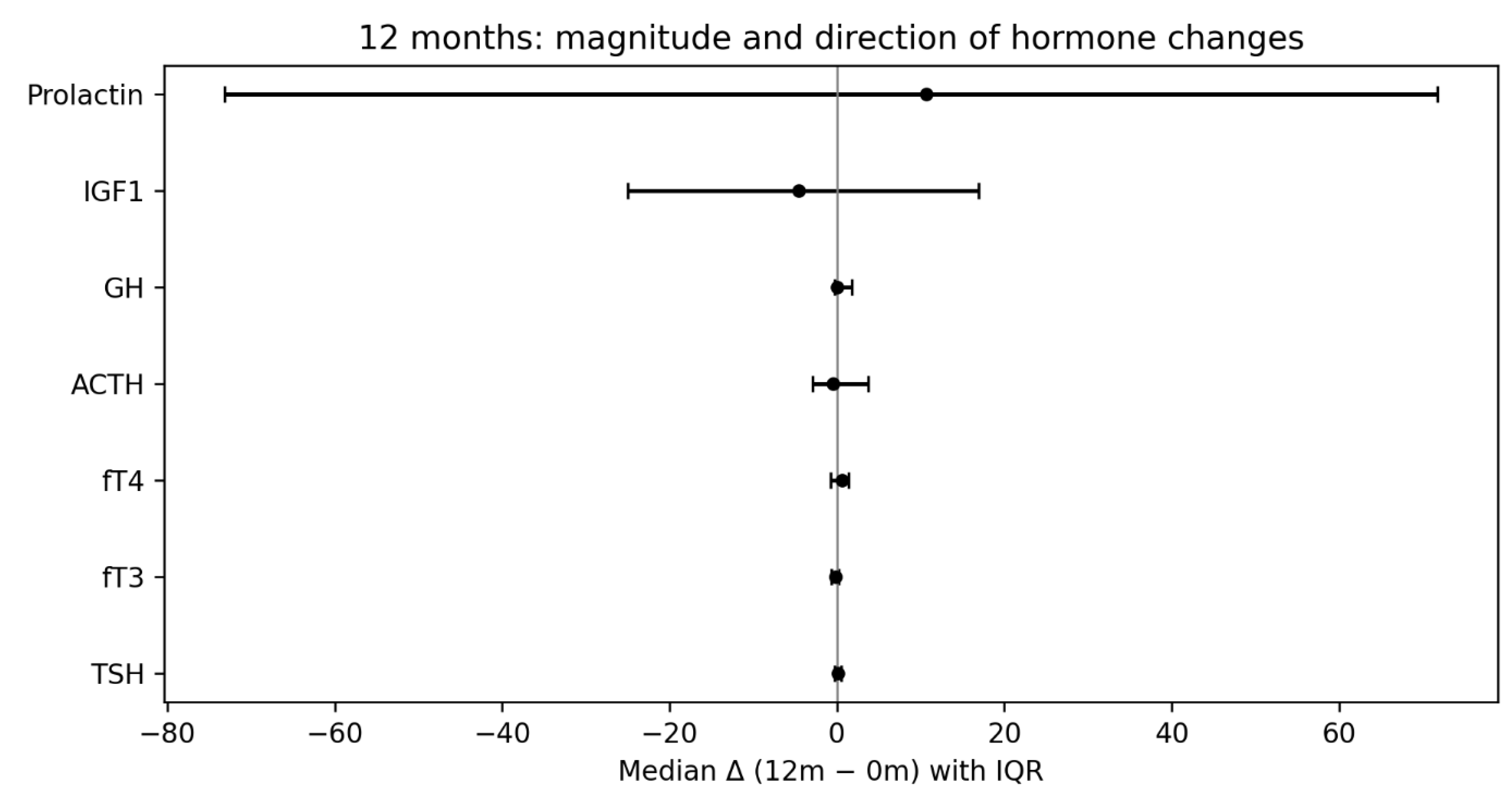

Table 1.
Patient characteristics.
| Clinical data | Value |
|---|---|
| Total number of patients (N) | 56 |
| Female sex, n (%) | 35 (62.5) |
| Male sex, n (%) | 21 (37.5) |
| Age, median (Q1–Q3), years | 45.0 (30.0–61.5) |
| Positive endocrinological history, n (%) | 15 (26.8) |
Table 2.
Frequency of symptoms at 6 and 12 months in the completed questionnaires.
| Symptom | Frequency at 6 months (%) | Frequency at 12 months (%) |
|---|---|---|
| Increased fatigue | 25 | 52,6 |
| Pallor | 0 | 5,3 |
| Decreased appetite and weight loss | 0 | 10,5 |
| Cold intolerance | 18,8 | 26,3 |
| Tendency toward constipation | 0 | 21,1 |
| Hoarseness | 12,5 | 10,5 |
| Increased hair loss and dry skin | 6,3 | 26,3 |
| Impaired concentration | 62,5 | 42,1 |
| Decreased libido | 6,3 | 5,3 |
| Mood fluctuations and/or depressive symptoms | 37,5 | 21,1 |
| Loss of hair in the genital area | 0 | 0 |
| Men: loss of facial hair and/or chest hair | 0 | 0 |
| Premenopausal women: menstrual irregularities or hypomenorrhea | 0 | 5,3 |
| Premenopausal women: difficulty conceiving | 0 | 0 |
| Body weight fluctuations | 0 | 15,8 |
| Memory impairment | 31,3 | 36,8 |
| Reduced muscle strength | 0 | 21,1 |
| Abdominal (central) obesity | 0 | 26,3 |
Disclaimer/Publisher’s Note: The statements, opinions and data contained in all publications are solely those of the individual author(s) and contributor(s) and not of MDPI and/or the editor(s). MDPI and/or the editor(s) disclaim responsibility for any injury to people or property resulting from any ideas, methods, instructions or products referred to in the content. |
© 2026 by the authors. Licensee MDPI, Basel, Switzerland. This article is an open access article distributed under the terms and conditions of the Creative Commons Attribution (CC BY) license.
Copyright: This open access article is published under a Creative Commons CC BY 4.0 license, which permit the free download, distribution, and reuse, provided that the author and preprint are cited in any reuse.


