Preprint
Article

This version is not peer-reviewed.

Four New Species and a New Record of Panaeolus from China, with Notes on the Taxonomy of Panaeolus rhombispermus

Submitted:

03 February 2026

Posted:

05 February 2026

You are already at the latest version

Abstract

Panaeolus is a genus of small, dark-spored agarics within the family Galeropsidaceae Singer. Based on the majority of specimens collected from China, this study investigated the genus Panaeolus and identified 17 species. These include four new species: Panaeolus bambusicola, P. latifolius, P. praecox, and P. ovinus; and one new record for China: P. fraxinophilus. The new species and the newly recorded species for China are morphologically described and illustrated. A multi-locus phylogenetic analysis (ITS, nrLSU, tef1-α, rpb2) was conducted using maximum likelihood and Bayesian inference. Combined morphological and phylogenetic evidence supports the reduction of the genus Crucispora to a subgenus within Panaeolus, accommodating P. rhombispermus.

Keywords: 
;  ;  ;  ;  ;  

1. Introduction

The genus Panaeolus (Fr.) Quél. was originally proposed as a subgenus of Agaricus L. by Fries (1836) [1], and subsequently unequivocally established at the generic level by Quélet (1872) [2]. As saprotrophic fungi, species of Panaeolus are globally distributed across temperate, tropical, and desert regions, typically inhabiting grasslands, herbivore dung, sandy soils, and wood chips. According to the Index Fungorum database (accessed January 5, 2026; https://www.indexfungorum.org), the genus encompasses 195 names, including 45 infraspecific taxa. Of the approximately 150 remaining names at the species level, several have been reassigned to other genera, such as Psathyrella (Fr.) Quél. and Psilocybe (Fr.) P. Kumm. However, within modern taxonomic frameworks, likely fewer than half are currently accepted as distinct species. Notably, several species within Panaeolus are known to contain psilocybin and other related psychoactive compounds, the ingestion of which can lead to poisoning characterized by hallucinogenic effects [3,4,5].
Historically, the taxonomic position of Panaeolus has been contentious, largely due to significant disparities in the methodological approaches and observational focuses of researchers. The concept of Panaeoloideae Singer was first proposed by Singer as a subfamily under Coprinaceae Overeem & Weese, encompassing Panaeolina Maire, Panaeolus (Fr.) Quél., Copelandia Bres., and Anellaria P. Karst., with Panaeolus (Fr.) Quél. as the type genus. In contrast, Olah (1969) reclassified the genus within Strophariaceae Singer & A.H. Sm., consolidating related species into a single generic name Panaeolus based primarily on chemical and cultural characteristics [6]. Singer (1976) challenged Olah’s conclusions, critiquing the methodology, biased character selection overemphasizing chemical traits, and the omission of key morphological features such as spore print color, solubility of spore pigments, and epicutis structure [7].
This historical debate illustrates the limitations of morphology-based classification and has been largely addressed by modern molecular phylogenetics. Molecular phylogenetic analyses have generated convergent insights. Tóth et al. (2013) demonstrated that the type species of Galeropsis Velen. (Galeropsis desertorum Velen. & Dvořák) clusters within the PanaeolusPanaeolina clade [8]. Subsequently, based on examinations of type specimens and molecular phylogenetic analyses, Malysheva et al. (2019) formally combined the type species of Galeropsis, G. desertorum, into Panaeolus [9]. In a review that synthesized these findings, Kalichman et al. (2020) further examined the taxonomic position of Panaeolus. They suggested that Galeropsidaceae Singer should be recognized as the correct familial designation for the clade historically treated as tribe Panaeoleae or subfamily Panaeoloideae Singer, positing it as the proper name for the family containing Copelandia Bres., Panaeolina Maire, Panaeolopsis Singer, and Panaeolus (Fr.) Quél., with the proviso that this grouping continues to be supported as distinct from Bolbitiaceae Singer [10]. More recently, this hypothesis was substantiated by He et al. (2026), who revised the taxonomic framework of Galeropsidaceae and, based on phylogenetic evidence, proposed a division of the genus Panaeolus into three subgenera: Panaeolus subg. Bresadolomyces, subg. Panaeolina, and subg. Panaeolus [11].
The taxonomic uncertainty extends to morphologically distinctive genera historically associated with Panaeolus. The genus Crucispora E. Horak was established in 1971 with Crucispora naucorioides E. Horak designated as its type species. However, its unique morphological characteristics, particularly the distinctive cruciform spores, precluded a clear familial assignment at the time [12]. Upon re-examining the type material, Singer acknowledged that its phylogenetic affinities remained unresolved but tentatively placed it within the Agaricaceae Chevall. based on a synthesis of morphological traits [7]. The species Panaeolina rhombispermus Hongo, originally described by Hongo (1973) [13], was subsequently transferred to Crucispora as Crucispora rhombisperma (Hongo) E. Horak, becoming the second species in the genus. The taxonomic status of this species remained uncertain for decades. Previous work by Ostuni et al. (2025) re-evaluated C. rhombisperma based on ITS and 28S phylogenetic data [14]. Their analysis revealed its close affinity with P. mexicanus, leading to the proposal of the new combination Panaeolus rhombispermus (Hongo) Birkebak, Voto & Ostuni. However, this conclusion was drawn mainly from molecular phylogenetic data. Therefore, the present study aims to re-evaluate the taxonomic status of P. rhombispermus by integrating novel morphological evidence from scanning electron microscopy (SEM) of spore morphology with multilocus phylogenetic analyses.
Previous records indicate over 30 species of Panaeolus in China [11,15,16,17,18,19,20], though some lack verifiable voucher specimens. Here, we report the findings from a four-year nationwide survey that yielded over 200 specimens. Using a combined morphological and phylogenetic approach, we identified 17 species, including four new species and one new record for China, which are described and illustrated in detail.

2. Materials and Methods

2.1. Sampling and Morphological Analyses

Specimens for this study were collected from different regions in China, including Inner Mongolia Autonomous Region, Xinjiang Uygur Autonomous Region and the provinces of Jilin, Zhejiang, Guangdong, and Yunnan. Habitat photographs were captured in the field (Figure 1), and specimens were deposited at the Fungarium of Jilin Agricultural University (FJAU). We described the colours of all major morphological structures (e.g., fresh basidiocarps, lamellae, spores) based on the Kornerup and Wanscher (1978) colour system [21]. The tissues of the specimens were treated with 5% KOH [22]. Microscopic observations were carried out with the aid of light microscopes (Carl Zeiss Primo Star, Jena, Germany; Olympus CX33, Tokyo, Japan). The basidiospore measurements do not include the apiculus and are presented as ‘(a)b–c(d) × e–f × g–h’, where ‘b–c’ represents the minimum of 90% of the measured values and ‘a’ and ‘d’ represent the extreme values. Due to the dorsiventral flattening of Panaeolus basidiospores, their dimensions are view-specific: ‘e–f’ represents the width in frontal view, and ‘g–h’ the thickness in side view. The main body (sterigmata not included) of the basidia, cheilocystidia, pleurocystidia, caulocystidia, pileocystidia and pileipellis were measured (if present). The notation (n/m/p) indicates that measurements were made on “n” randomly selected basidiospores from “m” basidiomes of “p” collections. Q is the ratio of length divided by width, and Qm represents the average quotient (length/width ratio) standard deviation.
For scanning electron microscopy (SEM) analysis, the lamellae of air-dried samples were first mounted on specimen stubs using double-sided conductive adhesive tape. Subsequently, the samples were sputter-coated with gold using an IXRF MSP-2S ion sputter coater and then observed under a Zeiss field-emission scanning electron microscope.

2.2. DNA Extraction, PCR Amplification, and Sequencing

Genomic DNA was extracted from the dried specimens using a NuClean Plant Genomic DNA kit (ComWin Biotech, CW0531M, Taizhou, China) according to the manufacturer’s instructions. The primer pairs ITS1F/ITS4 [23,24], LR0R/LR7 [25], EF1-983F/EF1-2212R [26], and RPB2-6F/RPB2-7.1R [27] were used to amplify the ITS, nrLSU, tef1-α, and rpb2 sequences, respectively. PCR amplifications were performed in 25 µL reactions containing: 2.0 µL template DNA, 12.5 µL 2× Es Taq Master Mix (Dye, ComWin Biotech, CW0690H, Taizhou, China.), 1.0 µL of each primer (10 pmol/µL), and 8.5 µL of ddH2O. PCR products were purified and sequenced by Sangon Biotech (Shanghai, China).

2.3. Phylogenetic Analyses

Newly generated sequences were deposited in the NCBI GenBank database, while other sequences were retrieved from NCBI (accession numbers are provided in Table 1). The ITS, nrLSU, tef1-α, and rpb2 sequences were aligned separately using the online MAFFT service with default parameters (https://mafft.cbrc.jp/alignment/server/) and subsequently refined manually in MEGA7 [28]. The resulting individual alignments were then concatenated into a multi-locus dataset using PhyloSuite [29]. The final concatenated dataset (ITS + nrLSU + tef1-α + rpb2) consisted of aligned sequences with lengths of 743 bp (ITS), 1351 bp (nrLSU), 1191 bp (tef1-α), and 731 bp (rpb2), resulting in a total concatenated length of 4,016 bp, with missing data coded as ‘?’. The best-fit partition and substitution models were selected using ModelFinder [30]. For the Maximum Likelihood (ML) analysis, the best-fit models selected under the Bayesian Information Criterion (BIC) were TPM2u+F+G4 for the ITS dataset, TIM3+F+R2 for the nrLSU dataset, SYM+I+G4 for the tef1-α dataset, and TNe+I+G4 for the rpb2 dataset. For the Bayesian Inference (BI) analysis, the best-fit models selected under the Akaike Information Criterion (AIC) were GTR+F+I+G4 for the ITS dataset, GTR+F+I+G4 for the nrLSU dataset, SYM+I+G4 for the tef1-α dataset, and SYM+I+G4 for the rpb2 dataset. The ML analysis was performed with IQ-TREE 3 under the selected models, with branch support assessed based on 1000 standard bootstrap replicates [31]. Bayesian inference was conducted with MrBayes v3.2.7a [32] under the partition model. Four independent Markov Chain Monte Carlo (MCMC) chains were run for 10,000,000 generations, sampling trees every 1000 generations. The first 25% of sampled trees were discarded as burn-in after the average standard deviation of split frequencies dropped below 0.01. Resulting consensus trees were visualized in FigTree v1.4.4 and prepared for publication using Adobe Illustrator 2021.

3. Results

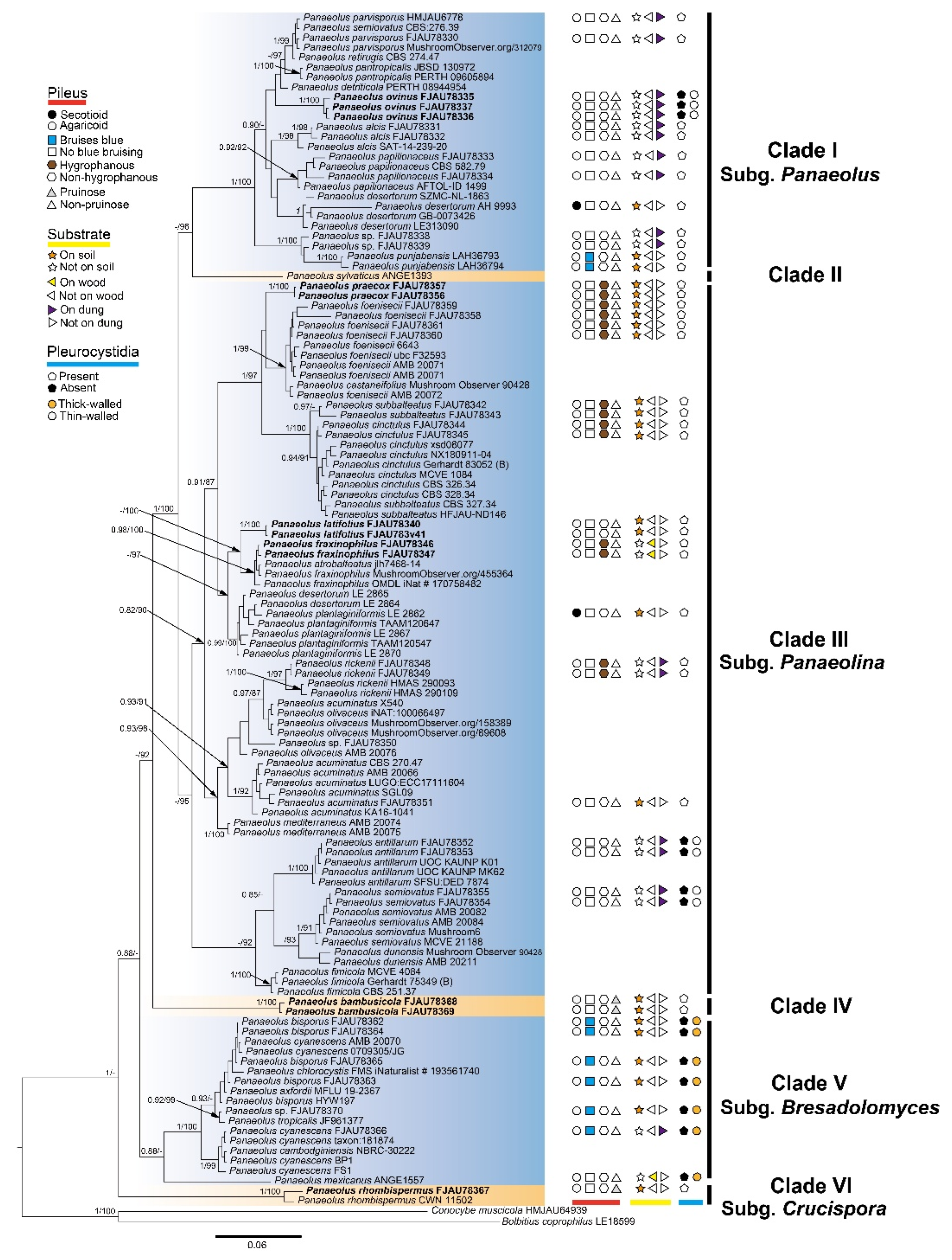
3.1. Molecular Phylogeny
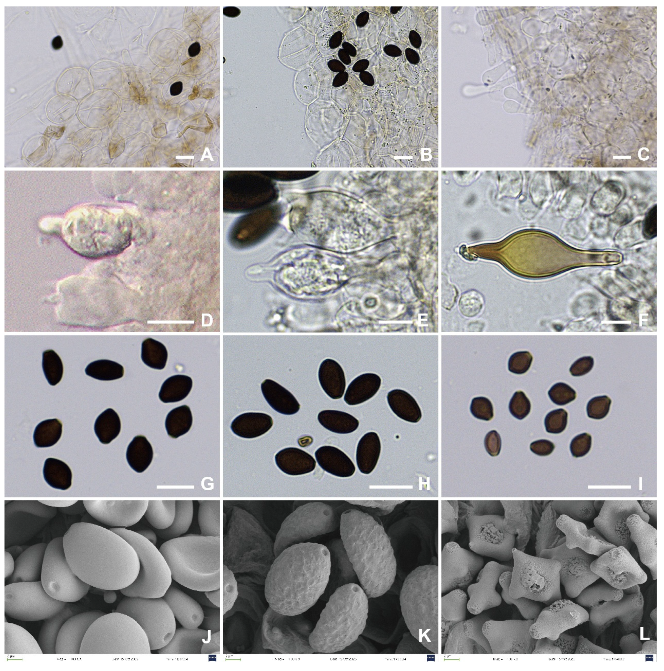
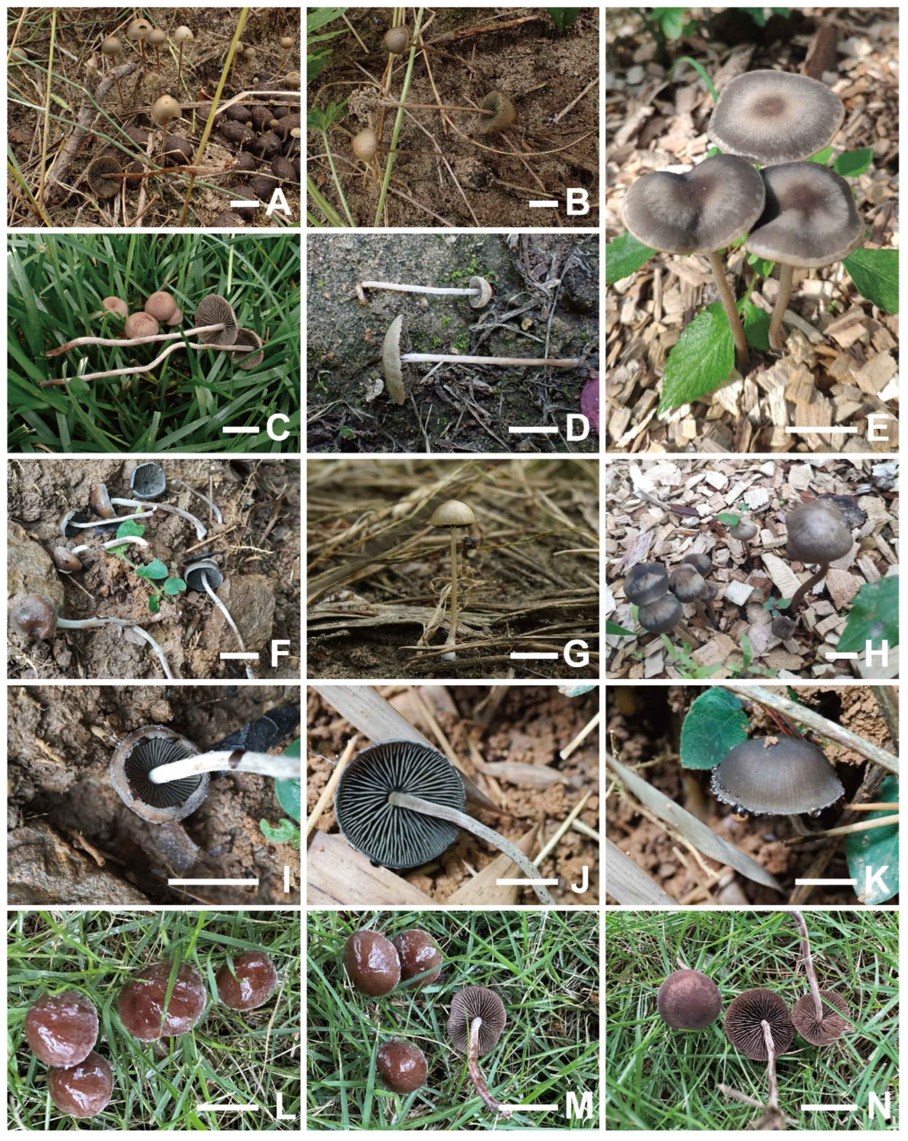
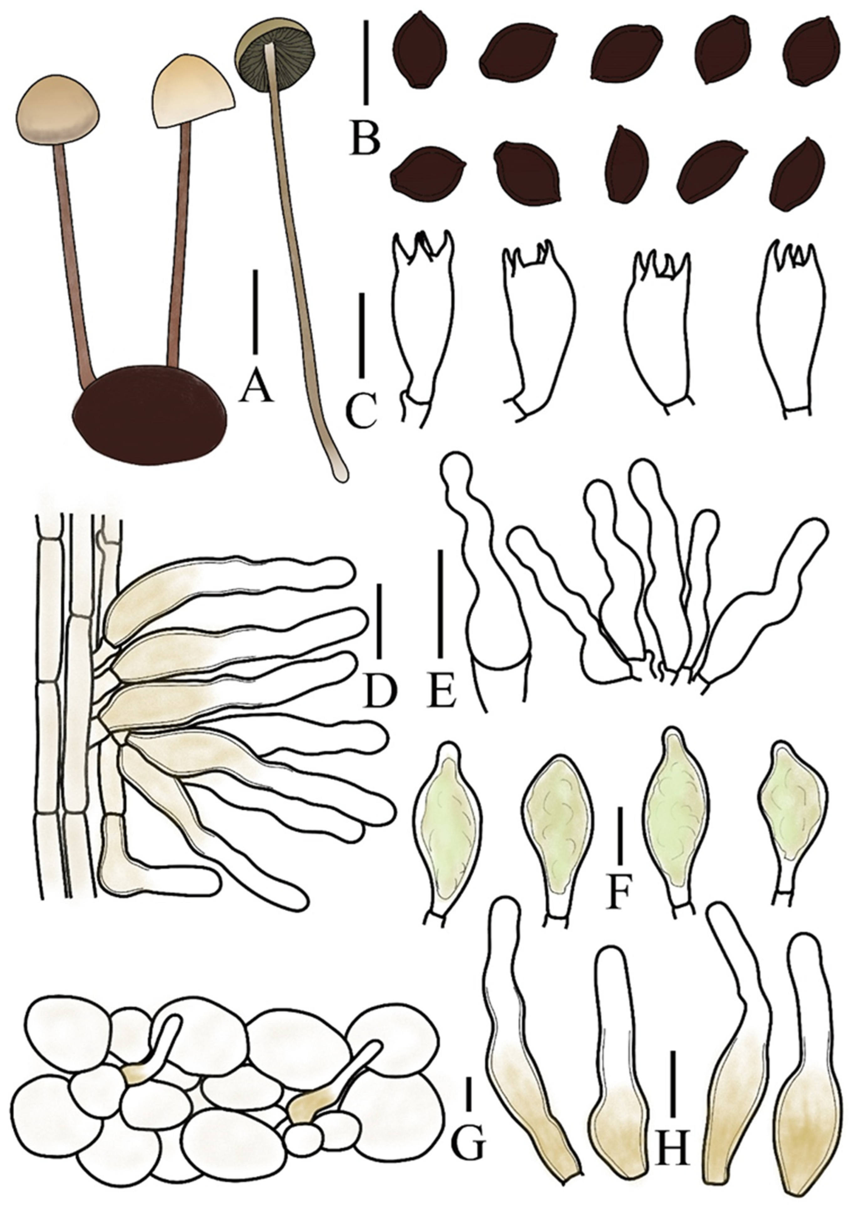
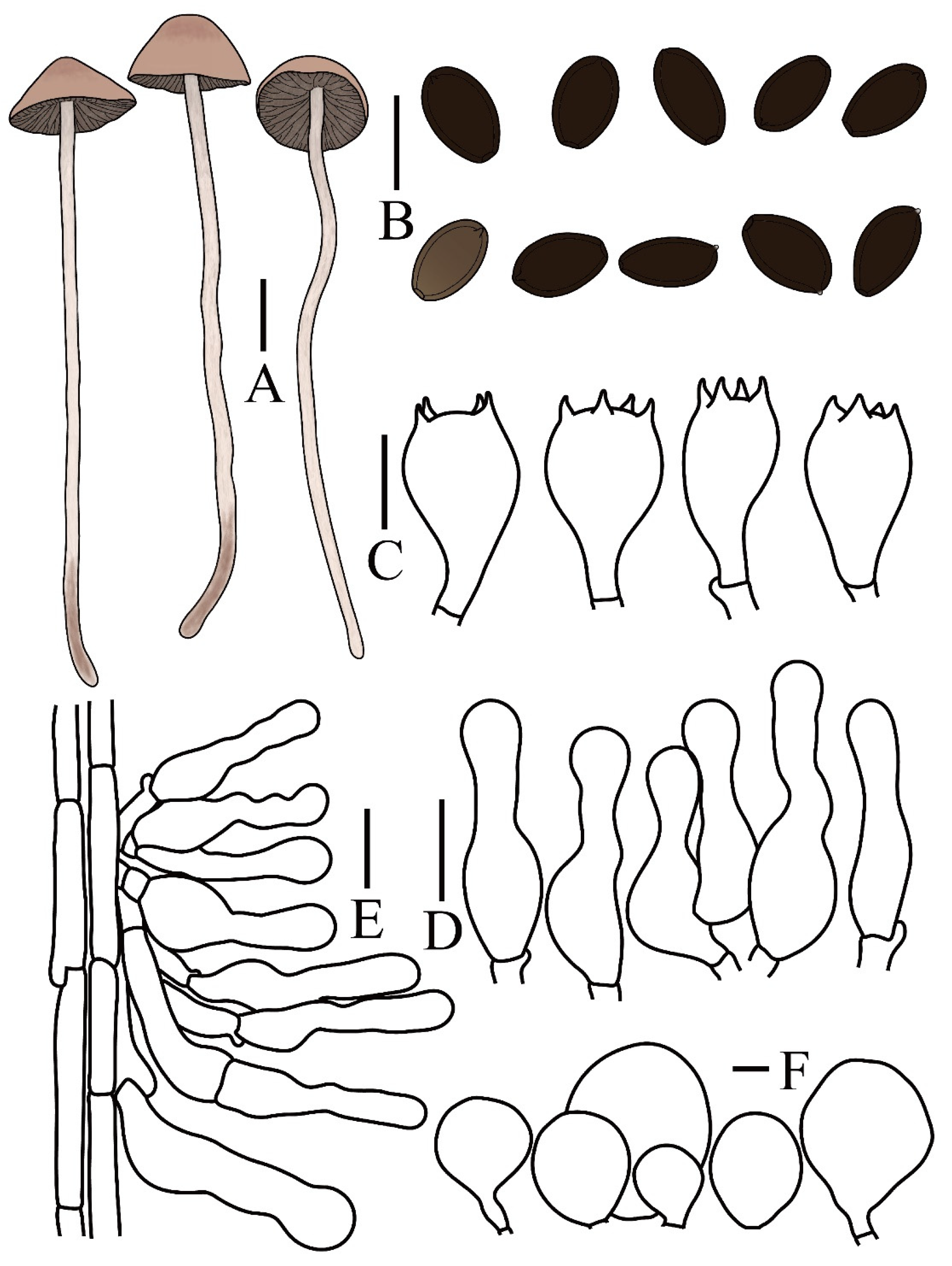
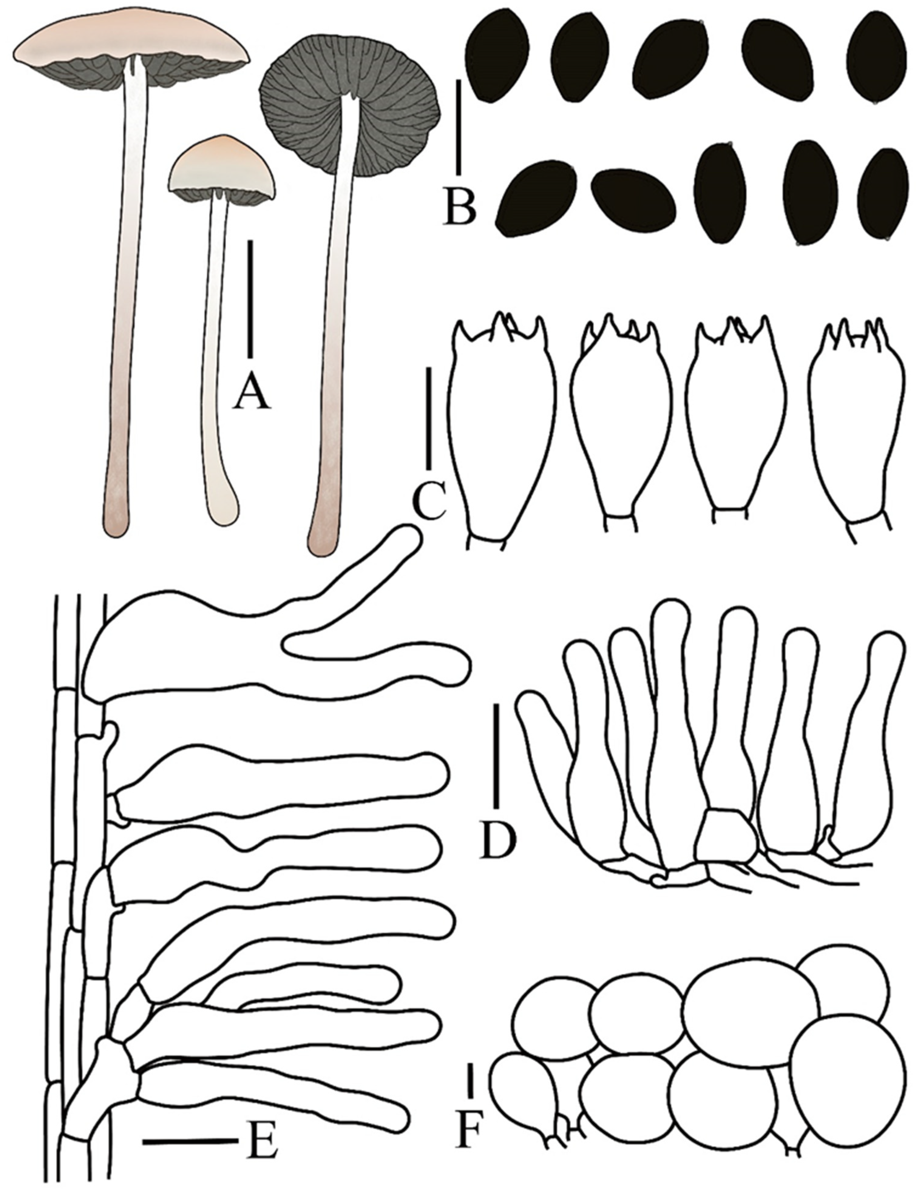
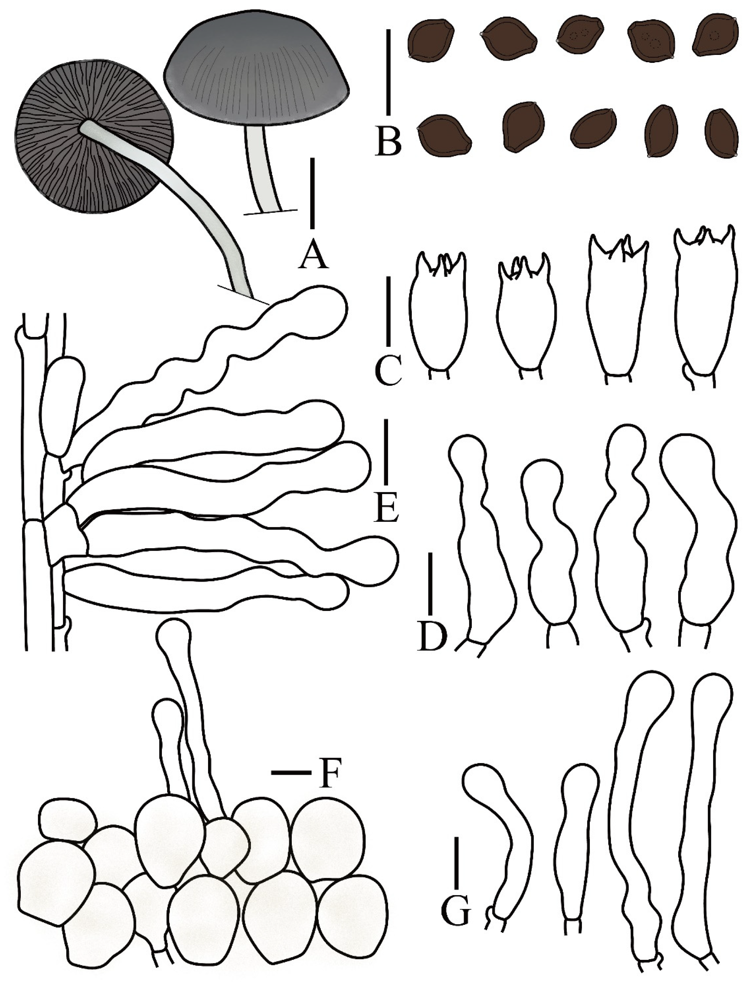
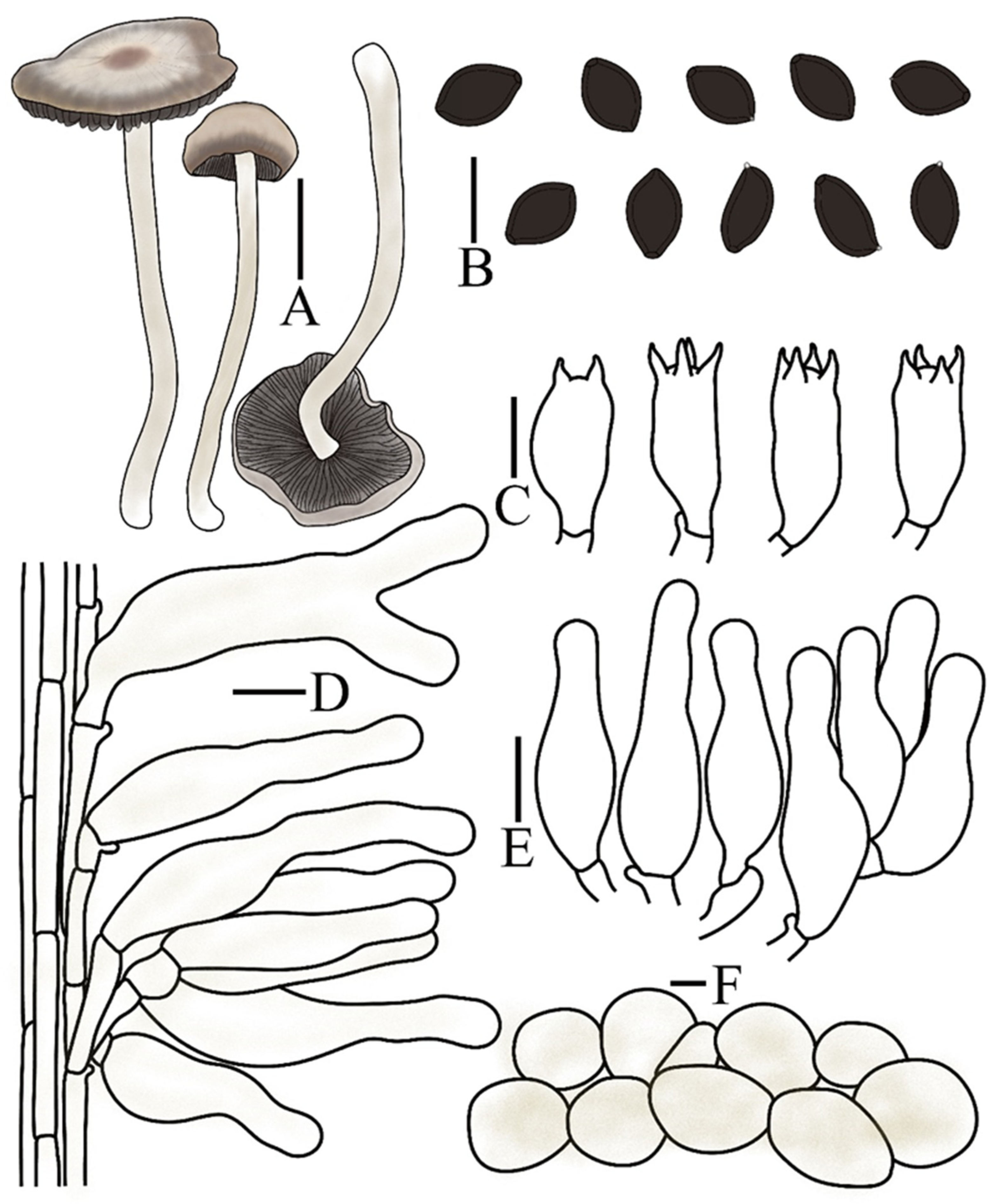
This study generated 95 new sequences (ITS: 40, nrLSU: 21, tef1-α: 18, rpb2: 16), which were combined with 120 sequences retrieved from GenBank, resulting in a final dataset of 120 taxa. Conocybe muscicola and Bolbitius coprophilus (Bolbitiaceae) were selected as the outgroup. The Bayesian inference (BI) and maximum likelihood (ML) analyses yielded largely congruent tree topologies. Only the BI tree is presented (Figure 2), with nodal support indicated as Bayesian posterior probabilities (PP) and ML bootstrap values (MLbs); nodes with PP ≥ 0.80 and MLbs ≥ 80% are shown.
The phylogenetic reconstruction did not support the traditional generic boundaries among Panaeolina, Copelandia, Anellaria, and Panaeolus sensu stricto, further supporting their consolidation into a single, unified genus. Within Panaeolus, six major clades (I–VI) were identified, each highlighted with a distinct background color in Figure 2.
Panaeolus ovinus (vouchers: FJAU78335–37) was placed within Clade I (subg. Panaeolus) and is distinguished by the presence of pleurocystidia, a trait not observed in other members of this subgenus. Within Clade III (subg. Panaeolina), three taxa were of particular interest: (1) P. praecox (vouchers: FJAU78356–57) was sister to P. foenisecii. These species are morphologically distinguished by spore ornamentation (P. foenisecii: verrucose; P. praecox: smooth) but share an exclusive occurrence on grassy lawns and hygrophanous pilei. (2) P. latifolius (vouchers: FJAU78340–41) and (3) the newly recorded P. fraxinophilus (vouchers: FJAU78346–47) formed a sister-group relationship. While sharing smooth-walled basidiospores, they differ in substrate preference and pileus characteristics: P. latifolius occurs on sandy soil and lacks hygrophany, whereas P. fraxinophilus grows on wood chips and exhibits a distinctly hygrophanous pileus. P. bambusicola (vouchers: FJAU78368–69) formed a distinct lineage, which is here designated as Clade IV. This species is unique among known Panaeolus taxa in its exclusive bamboo forest habitat and is further characterized by a persistently pruinose pileus, abundant pileocystidia, and relatively small basidiospores.
Notably, P. rhombispermus (vouchers: FJAU78367, CWN11502) formed an independent lineage identified as Clade VI (subg. Crucispora). This phylogenetic placement is corroborated by its distinctive cruciform basidiospores (Figure 8L). Consequently, we treat this lineage as a distinct subgenus within the genus Panaeolus.

3.2. Taxonomy

Panaeolus ovinus T. Bau & H. Cheng, sp. nov.
Mycobank No.: MB861990
Etymology. “ovinus” (Latin, pertaining to sheep) refers to its growth on sheep dung.
Holotypus. CHINA. Jilin Province, Baicheng City, Tongyu County, Xianghai National Nature Reserve, 25 August 2023, 122° 20′ 23” E, 45° 02′ 39” N, alt. 137 m, Liyang Zhu, Z2382506 (FJAU78336).
Diagnosis: Panaeolus ovinus basidiomata are relatively small, do not stain blue when bruised. Among the coprophilous Panaeolus species, it is macroscopically most similar to P. alcis M.M. Moser. However, it differs by the presence of pleurocystidia and its occurrence on sheep dung, which also distinguishes it from other Panaeolus species.
Pileus 0.5–1.5 cm in diameter, campanulate, paraboloid, or hemispherical, margin weakly hygrophanous, center blond (4C4), transitioning to greyish yellow (4B4) or pale yellow (4A4) toward the margin, drying brownish grey (5C2). Context thin, yellowish white (4A2) to grey (4B1), without a distinctive odor. Lamellae adnate to adnexed, moderately close, unequal, irregularly mottled with dark grey (1F1) or yellowish brown (5F8) to pale grey (1B1), edge even and remaining paler greyish. Stipe 4.0–6.5 cm long, 1.0–2.0 mm thick, cylindrical, slightly enlarged at the base, pale ochraceous grey, brownish toward the base, entirely pruinose, with the pruina more densely distributed on the middle and lower parts, and slightly longitudinally striated.
Spores (60/3/3) (7.8–)8.4–10.5(–11.5) × 6.1–8.2 × 5.0–6.5 µm, Q = 1.28–1.55, Qm = 1.39(0.07) in frontal view, limoniform to broadly limoniform, wall thick, smooth, Q = 1.55–1.82, Qm = 1.70(0.07) in side view, asymmetrical, pyriform, with a distinct germ pore, central, or slightly eccentric, spores appear dark brown (7F6) to black in a 5% KOH solution. Basidia (13–)16–23 × (7–)8–10 µm, nearly elliptical, clavate, 4-sterigmate, sterigmata 2–4 µm long. Cheilocystidia (15–)16–22(–25) × 3.5–5.5 µm, versiform, predominantly lageniform to narrowly utriform but with a ventricose base and an elongated, flexuose neck. Pleurocystidia (20–)23–27(–29) × (10–)11–12(–13) µm, thin-walled, functioning as chrysocystidia in KOH and containing yellow pigments, pedunculate or pedicellate, mostly with an obtuse apex. Caulocystidia (29–)32–40(–47) × (4.5–)5–7(–8.5) µm, versiform, predominantly narrowly cylindrical or less frequently geniculate, with both forms typically broadening toward the base, pale yellow (4A3) with slightly thickened walls on the medial and lower parts. Pileipellis hymeniform, composed predominantly of subglobose or globose elements, (15–)20–29(–40) × (14–)18–28(–36) µm, with yellow pigments at the base. Pileocystidia morphologically similar to caulocystidia, 32–46 × 8.5–10.5 µm. All tissues have clamp connections.
Habitat. Gregarious to scattered on sheep dung in early autumn.
Known distribution. Currently, only known in Jilin Province, China.
Additional specimens measured. CHINA. Jilin Province, Baicheng City, Tongyu County, Xianghai National Nature Reserve, 25 August 2023, Hanbing Song, S2382511 (FJAU78337), Shien Wang, E2308343 (FJAU78335).
Notes. Although a considerable number of species in Panaeolus grow on dung, current records indicate that only P. ovinus is specifically associated with sheep dung. A morphologically similar species, P. alcis [41], can be distinguished from P. ovinus by its elongate-ellipsoid spores and the absence of pleurocystidia. The fruiting bodies of P. papilionaceus (Bull.) Quél. are generally markedly larger than those of P. ovinus and typically bear distinctive white, pyramidal veil remnants at the pileus margin. Under nutrient-deficient conditions, however, developmental constraints and the abrasion of these remnants by rain or wind may cause P. papilionaceus to closely resemble P. ovinus. Adding to this morphological convergence, both species share lemon-shaped spores in frontal view. The absence of pleurocystidia in P. papilionaceus, nevertheless, provides a stable and definitive diagnostic character for differentiation.
Panaeolus praecox T. Bau & H. Cheng, sp. nov.
Mycobank No.: MB861989
Etymology. “praecox” (Latin, early-maturing or precocious) refers to its characteristic of fruiting earlier in the season compared to other Panaeolus species found in grassy habitats.
Holotypus. CHINA. Jilin Province, Changchun City, Jilin Agricultural University campus, 9 July 2023, 125°24′14” E, 43°48′35” N, alt. 352 m, Hong Cheng, C2370902 (FJAU78357).
Diagnosis: Panaeolus praecox is distinguished by its earlier fruiting phenology compared to other grassy Panaeolus species, a hygrophanous pileus, and smooth-walled, ellipsoid to elongate-ellipsoid basidiospores in frontal view.
Pileus 1.5–2.5 cm in diameter, conical, broadly conical, or obtusely conical, margin hygrophanous, center reddish brown (8D8), greyish red (8C5) to dull red (8C4), drying dull red (8B3) to reddish grey (8B2). Context thin, reddish white (8A2) to grey (8B1), without a distinctive odor. Lamellae adnate to adnexed, moderately close, unequal, irregularly mottled with dark grey (1F1) or brownish grey (8E2) to pale grey (1B1), edge smooth and remaining paler greyish. Stipe 7.0–9.0 cm long, 2.0–3.0 mm thick, cylindrical, erect to flexuous, slightly enlarged at the base, white (7A1) or pinkish (7A2), densely pruinose and slightly longitudinally striated, developing brownish discolorations where bruised or handled.
Spores (60/3/3) (9.5–)10.2–11.8(–14.0) × 6.2–7.5 × 5.3–6.5 µm, Q = 1.48–1.85, Qm = 1.63(0.09) in frontal view, ellipsoid to elongate-ellipsoid, wall thick, smooth, Q = 1.63–1.94, Qm = 1.80(0.09) in side view, elongate-ellipsoid, with a distinct germ pore, central, or slightly eccentric, spores appear dark brown (7F6) to black or brown (7E5) in a 5% KOH solution. Basidia (18–)19–22 × (10.5–)11–12.5 µm, clavate to broadly clavate, 4-sterigmate, sterigmata 2–4 µm long. Cheilocystidia (18–)19–24(–27) × 5.5–7.5 µm, versiform, predominantly narrowly utriform, often with a flexuous neck. Pleurocystidia absent. Caulocystidia (19–)23–33(–35) ×5.5–8.5(–9.2) µm, versiform, predominantly narrowly utriform with a flexuous neck to narrowly cylindrical. Pileipellis hymeniform, composed predominantly of subglobose or globose elements, (14–)28–33(–48) × (14–)26–32(–40) µm. Pileocystidia were not observed. All tissues have clamp connections.
Habitat. Gregarious to scattered on lawns during spring or early summer.
Known distribution. Currently, this species has been recorded only in Jilin and Hunan Provinces, China.
Additional specimens measured. CHINA. Hunan Province, Changsha City, 16 April 2024, Changzhuo Liu, C2441601 (FJAU78356).
Notes. Panaeolus foenisecii (Pers.) J. Schröt., P. cinctulus (Bolton) Sacc., and P. oligotrophus Ostuni & Voto are macroscopically similar to P. praecox and share comparable grassy habitats, all exhibiting a hygrophanous pileus. Spore morphology provides critical diagnostic characters for their differentiation. Only P. foenisecii possesses verrucose spores, whereas the others are smooth. The spores of P. cinctulus are predominantly lemon-shaped, while those of P. praecox and P. oligotrophus are ellipsoid to oval; additionally, the spores of P. praecox are generally more elongated than those of P. oligotrophus. Based on current knowledge, P. praecox also fruits earlier in the growing season than other documented Panaeolus species within China. P. subfoenisecii M.Q. He & R.L. Zhao can be further distinguished by its slightly larger spores compared to P. praecox and the absence of caulocystidia [11].
Panaeolus latifolius T. Bau & H. Cheng, sp. nov.
Mycobank No.: MB861988
Etymology. “latifolius” (Latin, broad-gilled) refers to its conspicuously wide lamellae that remain visible even in a frontal view of the basidioma.
Holotypus. CHINA. Inner Mongolia Autonomous Region, Tongliao City, Xar Moron Park, 19 July 2023, 122°15′28” E, 43°37′55” N alt. 260 m, Hong Cheng, C2371907 (FJAU78340).
Diagnosis: Panaeolus latifolius is distinguished by its convex to nearly applanate pileus, which causes the lamellae to appear conspicuously broad and ventricose. When the basidiomata are placed vertically and viewed frontally, the lamellae remain distinctly visible—a characteristic that distinguishes it from most other Panaeolus species. It further differs by its occurrence on sandy soil and non-hygrophanous pileus.
Pileus 0.8–1.8 cm in diameter, initially hemispherical, becoming convex to plano-convex at maturity, non-hygrophanous; center greyish orange (5B4) to greyish red (7B6), gradually transitioning to orange white (5A2) or reddish grey (7B2) toward the margin. Context thin, reddish white (7A2) to grey (7B1), without distinctive odor. Lamellae adnate to adnexed, moderately close, unequal, distinctly ventricose, irregularly mottled with dark grey (1F1) to pale grey (1B1), edge even and remaining paler greyish. Stipe 2.0–4.5 cm long, 1.0–2.0 mm thick, cylindrical, erect, slightly enlarged at the base, white (7A1), pruinose and slightly longitudinally striated, or brownish toward the base.
Spores (60/3/3) (9.5–)11.0–12.3(–13.7) × 6.0–7.1 × 5.3–6.4 µm, Q = 1.45–1.80, Qm = 1.58 (0.09) in frontal view, ovoid, ellipsoid to elongate-ellipsoid, wall thick, smooth, Q = 1.65–1.93, Qm = 1.76(0.08) in side view, elongate-ellipsoid, with a distinct germ pore, central, or slightly eccentric, spores appear dark brown (7F6) to black in a 5% KOH solution. Basidia (14.5–)16–20(–23) × 8–10 µm, nearly elliptical to clavate, 4-sterigmate, sterigmata 2–4 µm long. Cheilocystidia (14–)17–25(–27) × 5.0–7.0 µm, predominantly narrowly lageniform to narrowly utriform. Pleurocystidia absent. Caulocystidia (23–)35–45 × 7.5–8.5(–12.8) µm, versiform, predominantly narrowly lageniform to narrowly utriform, occasionally furcate. Pileipellis hymeniform, composed predominantly of subglobose or globose elements, (22–)24–36(–41) × (20–)23–30(–40) µm. Pileocystidia were not observed. All tissues have clamp connections.
Habitat. Scattered on sandy soil during summer and autumn.
Known distribution. Currently, this species is currently known only from Jilin Province and the Inner Mongolia Autonomous Region, China.
Additional specimens measured. Jilin Province, Baicheng City, Tongyu County, Xianghai National Nature Reserve, 26 August 2023, 122° 20′ 10” E, 44° 50′ 39” N, alt. 145 m, Liyang Zhu, Z2382617 (FJAU78341).
Notes. Panaeolus latifolius is characterized by its occurrence on sandy soil and small basidiomata with frontally visible lamellae. The species further exhibits a non-hygrophanous pileus that becomes convex to plano-convex at maturity, absence of pleurocystidia, and no blue bruising reaction. This unique combination of features allows clear differentiation from other Panaeolus species. P. fraxinophilus A.H. Sm., while also possessing a convex pileus, is distinguished by its fuscous-black pileus color, hygrophanous condition when moist, and lignicolous habit. These pronounced macroscopic differences allow for reliable separation from P. latifolius.
Panaeolus bambusicola T. Bau & H. Cheng, sp. nov.
Mycobank No.: MB861987
Figure 1F,I–K and Figure 6
Etymology. “bambusicola” (Latin, from bambusa ‘bamboo’ and -cola ‘inhabitant’) refers to its specific habitat in bamboo forests.
Holotypus. CHINA. Zhejiang Province, Huzhou City, Changxing County, 7 June 2024, 120°58′35” E, 30°57′44” N alt. 10 m, Zhuoluo Zhou, 1654 (FJAU78368).
Diagnosis: Panaeolus bambusicola is distinguished by its unique ecological habit of growing on bamboo forest soil, a pruinose pileus margin, absence of pleurocystidia, lack of a blue bruising reaction, and relatively small basidiospores measuring less than 10 µm in length.
Pileus 0.5–2.0 cm in diameter, initially campanulate, becoming broadly conical to convex at maturity, striate, pruinose at the margin; center brown (7E8 to 7E3) or grey (7E8), transitioning to reddish grey (7B2) or pale grey (7C1) toward the margin. Context thin, without distinctive odor. Lamellae adnate to adnexed, moderately close, unequal, distinctly ventricose, irregularly mottled with dark grey (1F1) to pale grey (1B1), edge even and remaining paler greyish. Stipe 2.0–4.0 cm long, 1.0–2.0 mm thick, cylindrical, erect to flexuous, slightly enlarged at the base; surface white (7A1) to pale grey (1B1), becoming brownish toward the base, densely pruinose and faintly longitudinally striate, developing reddish brown discolorations where bruised or handled.
Spores (60/3/3) (6.0–)6.5–7 × 4.8–5.8 × 4.0–5.0 µm, Q = 1.10–1.40, Qm = 1.23 (0.06) in frontal view, broadly limoniform to nearly subglobose, wall thick, smooth, Q = 1.38–1.65, Qm = 1.53(0.09) in side view, ellipsoid, with a distinct germ pore, central, spores appear dark brown (7F6) to black in a 5% KOH solution. Basidia (12–)13–16(–17.5) × 7.2–8.5 µm, nearly elliptical to clavate, 4-sterigmate, sterigmata 2–4 µm long. Cheilocystidia (22–)24–30(–34) × 6.0–7.5 µm, flexuose with obtuse apex. Pleurocystidia absent. Caulocystidia (30–)45–60 × 7.0–9(–10.5) µm, flexuose with obtuse apex. Pileipellis hymeniform, composed predominantly of subglobose or globose elements, (13–)18–22(–24) × (12–)15–20(–21) µm. Pileocystidia nearly narrowly cylindrical but curved, or flexuose with an obtuse apex. All tissues have clamp connections.
Habitat. Scattered on soil sections under bamboo forest during summer.
Known distribution. Currently, only known in Zhejiang Province, China.
Additional specimens measured. CHINA. Zhejiang Province, Huzhou City, Changxing County, 12 June 2025, 120°58′25” E, 30°57′18” N alt. 9 m, Zhuoluo Zhou, 1713 (FJAU78369).
Notes. Panaeolus bambusicola is characterized by its occurrence on soil in bamboo forests, a pruinose pileus margin, and relatively small spores. Although a pruinose pileus margin is also observed in P. rhombispermus, the latter is instantly distinguished by its unique cruciform basidiospores. This combination of ecological and micromorphological features reliably distinguishes P. bambusicola from all other known Panaeolus species.
Panaeolus fraxinophilus A.H. Sm.
Pileus 1–2.5 cm in diameter, initially campanulate, becoming convex to applanate or broadly conical with a subumbonate disc at maturity. Surface smooth or lacunose, hygrophanous when moist, with distinct striations; margin occasionally irregularly undulate, center fuscous black (6F1 to 6F2), transitioning to grey (6D1) or pale grey (6C1) toward the margin, then darkening again to fuscous black near the edge; drying uniformly to grey (6D1). Context thin, without distinctive odor. Lamellae adnate, moderately close, unequal, irregularly mottled with dark grey (1F1) to pale grey (1B1), edge even and remaining paler greyish. Stipe 2.0–8.0 cm long, 2.0–3.0 mm thick, cylindrical, erect to flexuous, slightly enlarged at the base; surface pale grey (1B1), becoming brownish grey toward the base, densely pruinose and faintly longitudinally striate.
Spores (60/3/3) 9.0–10.5(–13.5) × 6.5–7.5(–8.0) × 5.0–6.3 µm, Q = 1.32–1.63, Qm = 1.50 (0.07) in frontal view, ovate, ellipsoid, wall thick, smooth, Q = 1.67–2.07, Qm = 1.87(0.11) in side view, elongate-ellipsoid, asymmetrical, with a distinct germ pore, eccentric, spores appear dark brown (7F6) to black in a 5% KOH solution. Basidia 19–21 × 7.8–9.5 µm, nearly elliptical to clavate, 4(2)-sterigmate, sterigmata 2–4 µm long. Cheilocystidia (24–)29–32(–35) × 9.0–11.2 µm, narrowly utriform. Pleurocystidia absent. Caulocystidia (37–)40–60 × 7.0–10.0(–12.5) µm, narrowly utriform, or with an elongated neck. Pileipellis hymeniform, composed predominantly of subglobose or globose elements, (25–)32–55(–65) × (24–)30–54(–60) µm. Pileocystidia absent. All tissues have clamp connections.
Habitat. Gregarious to scattered on wood chip piles during spring.
Known distribution. Asia: China; North America: United States of America (Holotype), Canada; South America: Brazil.
Additional specimens measured. CHINA. Guangdong Province, Shenzhen City, 18 March 2024, Jianfeng Tan, C2431801(FJAU78346), Yunnan Province, Wenshan City, 18 May 2024, Xiangyang Li, C2452504 (FJAU78347).
Notes. Within the genus Panaeolus, lignicolous species are relatively uncommon. Panaeolus atrobalteatus Pegler & A. Henrici and P. fraxinophilus represent two such species that demonstrate remarkable morphological similarity. Both exhibit strongly hygrophanous pilei in moist conditions and possess closely overlapping spore size ranges [42]. It is noteworthy that in the protologue of P. atrobalteatus, Henrici did not undertake a comparative analysis with the morphologically similar P. fraxinophilus, nor was this taxon mentioned. Based on available herbarium material and literature, the basidiomata of P. atrobalteatus are generally larger in overall dimensions than those of P. fraxinophilus, a characteristic that may provide a practical morphological criterion for their differentiation.
Panaeolus subg. Crucispora (E. Horak) T. Bau & H. Cheng, comb. & stat. nov.
Mycobank No.: MB862146
Basionym. Crucispora E. Horak, New Zealand J. Bot. 9(3): 489 (1971)
Type species. Panaeolus naucorioides (E. Horak) T. Bau & H. Cheng
Description. emended based on E. Horak’s (1971) [12] concept of Crucispora: Basidiomata small to medium-sized, mycenoid or naucorioid, not staining blue when injured. Pileus hemispherical to conical when young, becoming convex to campanulate; surface dry, hygrophanous, colour deep brown, tobacco-brown; margin non-striate or finely striate, bearing a white, pruinose coating (veil remnants) when young, disappearing with age. Context thin, odour and taste not distinctive. Lamellae adnate to adnexed, ventricose, edge white. Stipe cylindrical, fistulose; no persistent cortina or annulus. Basidiospores in deposit brown to dark brown or black, cruciform-rhomboid.
Notes. Crucispora rhombispermus (Hongo) was transferred to Panaeolus (Fr.) Quél. by Birkebak, Voto & Ostuni based on phylogenetic analyses of ITS and nrLSU sequences [14]. Subsequently, He et al. placed P. rhombispermus into Panaeolus subg. Bresadolomyces M.Q. He & R.L. Zhao, mainly based on phylogenetic evidence [11]. However, our results indicate that this species is clearly distinct from other members of subg. Bresadolomyces. Furthermore, the type species of Crucispora, C. naucorioides E. Horak, also possesses cruciform spores. Based on these findings, we propose to reduce the genus Crucispora to a subgenus within Panaeolus.
Figure 8. Micro-morphological structures of Panaeolus species. (A–C) Pileipellis: (A) Panaeolus ovinus (FJAU78336). (B) P. latifolius (FJAU78340). (C) P. bambusicola (FJAU78368); (D–F) Pleurocystidia: (D) P. ovinus (FJAU78336). (E) P. semiovatus (FJAU78355). (F) P. bisporus (FJAU78362); (G–L) Basidiospores: (G) P. ovinus (FJAU78336). (H) P. latifolius (FJAU78340). (I) P. bambusicola (FJAU78368). (J) P. praecox (FJAU78357). (K) P. foenisecii (FJAU78361). (L) P. rhombispermus (FJAU78367). Scale bars: (A–I) = 10 μm; (J–L) = 2 μm.
Preprints 197343 g008
Panaeolus naucorioides (E. Horak) T. Bau & H. Cheng, comb. nov.
Mycobank No.: MB862188
Basionym. Crucispora naucorioides E. Horak, New Zealand J. Bot. 9(3): 489 (1971)
Notes. According to E. Horak’s description of Crucispora naucorioides, features such as its hygrophanous pileus, buff-brown lamellae with white edges, adnate to adnexed attachment, and a hymeniform pileipellis closely resemble those of species in the genus Panaeolus [12]. Furthermore, its spores are cruciform, as in P. rhombispermus. Based on this morphological congruence, we propose to transfer this species into the genus Panaeolus and place it within the subgenus Panaeolus subg. Crucispora.

4. Discussion

Our integrated taxonomic reconstruction of Panaeolus delineates six major clades (I–VI) and describes four new species and one new record from China. Notably, based on phylogenetic and morphological evidence (particularly the cruciform basidiospores), we propose recognizing Clade VI (P. rhombispermus) as a distinct subgenus, Panaeolus subg. Crucispora.
Among the newly described taxa, Panaeolus ovinus is currently the only known species in subg. Panaeolus that possesses pleurocystidia, with sheep dung as its sole known substrate. P. praecox is most closely related to P. foenisecii but is distinguished by its smooth spore surface. P. latifolius grows on sandy soil and, unlike the newly recorded species P. fraxinophilus (which grows on wood chips), we observed no hygrophany in the pileus of collected specimens—a characteristic that may be attributed to the species’ inherent traits or the relatively arid environment where it occurs. To our knowledge, P. bambusicola represents the first recorded species of Panaeolus found in bamboo forest habitats, characterized by relatively small spores and a frequently pruinose pileus. Correspondingly, this species exhibits abundant pileocystidia, whereas pileocystidia are generally sparse or even absent in other Panaeolus species. These findings enrich the known species diversity of Panaeolus in China and deepen our understanding of the habitat preferences and micromorphological characteristics within this genus.
In early taxonomy that relied predominantly on morphological characters, species of Panaeolus were historically classified into different genera or subgenera based on traits such as blue-bruising reaction upon injury, presence or absence of an annulus, spore ornamentation, and the morphology and occurrence of pleurocystidia [7]. In the present study, based on collected Panaeolus specimens and clear descriptions available in the literature, we summarized pileus characteristics, pleurocystidia types, and substrate preferences across the genus. The results indicate that these morphological features do not fully correspond to the six clades revealed by phylogenetic analysis; i.e., no shared derived characters (synapomorphies) useful for subdivision of the genus were identified. Because the phylogenetic framework obtained here is largely congruent with that of He et al. [11], we have adopted a similar subdivision of Panaeolus. However, in our phylogeny, Panaeolus rhombispermus (vouchers: FJAU78367, CWN 11502) form an independent lineage.
Based on scanning electron microscopy (SEM) observations of the spore ornamentation of Panaeolus rhombispermus and multi-locus phylogenetic analyses, we regard P. rhombispermus as distinct from other Panaeolus species (Figure 8). Considering that the type species of Crucispora, Crucispora naucorioides E. Horak, also possesses cruciform spores, we thus further propose to reduce Crucispora to a subgenus under Panaeolus. Previous work by Ostuni et al. [14] proposed transferring C. rhombispermus to the genus Panaeolus, arguing that its hymeniform pileipellis, mottled gills, and dark brown spores with a germ pore align with characteristics of Panaeolus. They further suggested that the distinctive cruciform spore shape of P. rhombispermus represents merely an extreme morphological variant within the known spore morphology range of Panaeolus, noting that a similar, albeit less pronounced, shape occurs in spores of P. mexicanus (Guzmán) Voto & Angelini. However, our observations indicate that the spore morphology and surface ornamentation of P. rhombispermus (Figure 8L) are fundamentally distinct from those of all known Panaeolus species. This is corroborated by light microscopy images from Chou et al. [40], which show pronounced spore surface ornamentation in P. rhombispermus, whereas spores of P. mexicanus appear smooth [38]. Furthermore, P. mexicanus possesses thick-walled pleurocystidia, a trait shared with other members of Clade V (Panaeolus subg. Bresadolomyces, including P. bisporus and P. cyanescens), while both currently recognized species of Panaeolus subg. Crucispora lack pleurocystidia. We note that in the original publications of the two Crucispora species, their spores were consistently described as “rhomboid” and “smooth”, with corresponding illustrations also depicting a smooth surface [12,13]. This conclusion, however, may have been constrained by the resolution limits of the microscopes available at the time, which likely prevented accurate recognition of the spore wall ornamentation. It should be emphasized that the rhomboid shape of the spores remains an accurate and stable diagnostic feature.
Our phylogenetic analysis reveals that Panaeolus bambusicola (FJAU78368, FJAU78369) and P. sylvaticus (ANGE1393) each form independent lineages, designated as Clade IV and Clade II, respectively, and neither is assigned to an existing subgenus. P. bambusicola is distinguished by a persistently pruinose pileus and broadly limoniform, relatively small basidiospores. Nevertheless, because its clade currently comprises only this species, we have not proposed a subgeneric placement for it. Conversely, the phylogenetic position of P. sylvaticus remains unstable, likely due to the current limitation to ITS sequences and a lack of additional sequence data for robust support. We expect that the future discovery of more Panaeolus species and the sharing of related molecular sequences will facilitate a more robust and stable subdivision of the genus.

Author Contributions

Conceptualization, T.B. and H.C.; methodology, H.C.; software, H.C.; validation, H.C. and T.B.; formal analysis, H.C.; investigation, H.C. and T.B.; resources, H.C. and T.B.; data curation, H.C. and T.B.; writing—original draft preparation, H.C.; writing—review and editing, H.C. and T.B.; visualization, H.C. and T.B.; supervision, T.B.; project administration, T.B.; funding acquisition, T.B. All authors have read and agreed to the published version of the manuscript.

Funding

This study was supported by the National Natural Science Foundation of China (32270001).

Institutional Review Board Statement

Not applicable.

Informed Consent Statement

Not applicable.

Data Availability Statement

All the sequences have been deposited in GenBank (https://www.ncbi.nlm.nih.gov) and MycoBank (https://www.mycobank.org). The data presented in this study are deposited in the Zenodo repository, accession number https://doi.org/10.5281/zenodo.18431687.

Acknowledgments

We sincerely thank our supervisor and the entire research team for their invaluable guidance and collaboration. We are also deeply grateful to Zhuoluo Zhou, Jianfeng Tan, Xiaofang Liao, SiAng Chen, and XiangYang Li for their generous assistance. Furthermore, we gratefully acknowledge the financial support provided by the National Natural Science Foundation of China (Grant No. 32270001).

Conflicts of Interest

The authors declare no conflict of interest.

References

  1. Fries, E. Epicrisis Systematis Mycologici: seu synopsis hymenomycetum; sumtibus auctoris: 1836; Volume 1.
  2. Quélet, L. Les champignons du Jura et des Vosges; Impr. et lithographie de H. Barbier, 1872. [Google Scholar]
  3. Pollock, S.H. A Novel Experience with Panaeolus A Case Study from Hawaii. Journal of Psychedelic Drugs 1974, 6, 85–89. [Google Scholar] [CrossRef]
  4. Watling, R. A Panaeolus poisoning in Scotland. Mycopathologia 1977, 61, 187–190. [Google Scholar] [CrossRef] [PubMed]
  5. Stijve, T. Psilocin, psilocybin, serotonin and urea in Panaeolus cyanescens from various origin. Persoonia-Molecular Phylogeny and Evolution of Fungi 1992, 15, 117–121. [Google Scholar]
  6. Oláh, G.-M. Le genre Panaeolus: Essai taxinomique et physiologique; Laboratoire de cryptogamie du Muséum national d’histoire naturelle, 1969. [Google Scholar]
  7. Shaffer, R.L. The Agaricales in modern taxonomy. 1976.
  8. Tóth, A.; Hausknecht, A.; Krisai-Greilhuber, I.; Papp, T.; Vágvölgyi, C.; Nagy, L.G. Iteratively refined guide trees help improving alignment and phylogenetic inference in the mushroom family Bolbitiaceae. PLoS One 2013, 8, e56143. [Google Scholar] [CrossRef] [PubMed]
  9. Malysheva, E.; Moreno, G.; Villarreal, M.; Malysheva, V.; Svetasheva, T. The secotioid genus Galeropsis (Agaricomycetes, Basidiomycota): a real taxonomic unit or ecological phenomenon? Mycological Progress 2019, 18, 805–831. [Google Scholar] [CrossRef]
  10. Kalichman, J.; Kirk, P.M.; Matheny, P.B. A compendium of generic names of agarics and Agaricales. Taxon 2020, 69, 425–447. [Google Scholar] [CrossRef]
  11. He, M.-Q.; Yang, W.-Q.; Phurbu, D.; Liu, F.; Li, J.-X.; Cao, B.; Zhao, R.-L. Systematic study of Panaeolus (Agaricales, Galeropsidaceae) sensu lato and psilocybin-producing traits of species from China. IMA Fungus 2026, 17, e167329. [Google Scholar] [CrossRef]
  12. Horak, E. Contributions to the knowledge of the Agaricales sl (Fungi) of New Zealand. New Zealand Journal of Botany 1971, 9, 463–493. [Google Scholar] [CrossRef]
  13. Hongo, T. Notulae Mycologicae (12): Auctore. Bulletin of the Faculty of Education, Shiga University. Natural Sciences 1973, 37–43. [Google Scholar]
  14. Ostuni, S.; Voto, P.; Birkebak, J.; Meyer, M.G.; Geurin, Z.; Slot, J.C. Panaeolus oligotrophus: A new species from central Florida, with notes on Panaeolus pumilus and Crucispora rhombisperma. Mycologia 2025, 117, 1057–1066. [Google Scholar] [CrossRef]
  15. Li, Y.; Li, T.H.; Yang, Z.L.; Dai, Y.C.; Bau, T. Atlas of Chinese macrofungal resources; Central China Farmers’ Publishing House: Zhengzhou, 2015. [Google Scholar]
  16. Bau, T.; Sarentoya. Strophariaceae of China (V) Panaeolus s.l. Journal of Fungal Research 2010, 8, 131–168. [Google Scholar] [CrossRef]
  17. He, X.-S. A new record of Panaeolina in China. Mycosystema 2004, 150. [Google Scholar] [CrossRef]
  18. Ma, T. Taxonomy of Psilocybe sl and Panaeolus in Yunnan, Southwest China, with notes on related genus Protostropharia. Beijing: Chinese Academy of Forestry 2014.
  19. Hu, Y.; Mortimer, P.E.; Karunarathna, S.C.; Raspe, O.; Promputtha, I.; Yan, K.; Xu, J.; Hyde, K.D. A new species of Panaeolus (Agaricales, Basidiomycota) from Yunnan, Southwest China. Phytotaxa 2020, 434, 22–34-22–34. [Google Scholar] [CrossRef]
  20. Cao, B.; Phurbu, D.; Ralaiveloarisoa, A.; Liimatainen, K.; Niskanen, T.; Ramírez-Cruz, V.; Bradshaw, A.J.; Dentinger, B.T.; Ramírez-Guillén, F.; Cortés-Pérez, A. Fungal diversity notes 1919–2016: taxonomic and phylogenetic contributions to fungal taxa. Fungal Diversity 2025, 1–227. [Google Scholar] [CrossRef]
  21. Kornerup, A.; Wanscher, J.H. Methuen handbook of colour. 1967.
  22. Weeks, R.A.; Singer, R.; Hearn, W.L. A new psilocybian species of Copelandia. Journal of Natural Products 1979, 42, 469–474. [Google Scholar] [CrossRef]
  23. Tj, W. Amplification and direct sequencing of fungal ribosomal RNA genes for phylogenetics. PCR protocols: a guide to methods and applications 1990, 315–322. [Google Scholar]
  24. Gardes, M.; Bruns, T.D. ITS primers with enhanced specificity for basidiomycetes-application to the identification of mycorrhizae and rusts. Molecular ecology 1993, 2, 113–118. [Google Scholar] [CrossRef]
  25. Moncalvo, J.-M.; Lutzoni, F.M.; Rehner, S.A.; Johnson, J.; Vilgalys, R. Phylogenetic relationships of agaric fungi based on nuclear large subunit ribosomal DNA sequences. Systematic biology 2000, 49, 278–305. [Google Scholar] [CrossRef]
  26. Rehner, S.A.; Buckley, E. A Beauveria phylogeny inferred from nuclear ITS and EF1-α sequences: evidence for cryptic diversification and links to Cordyceps teleomorphs. Mycologia 2005, 97, 84–98. [Google Scholar] [CrossRef]
  27. Matheny, P.B. Improving phylogenetic inference of mushrooms with RPB1 and RPB2 nucleotide sequences (Inocybe; Agaricales). Molecular phylogenetics and evolution 2005, 35, 1–20. [Google Scholar] [CrossRef]
  28. Kumar, S.; Stecher, G.; Tamura, K. MEGA7: molecular evolutionary genetics analysis version 7.0 for bigger datasets. Molecular biology and evolution 2016, 33, 1870–1874. [Google Scholar] [CrossRef]
  29. Zhao, D.; Ye, T.; Gao, F.; Jakovlić, I.; La, Q.; Tong, Y.; Liu, X.; Song, R.; Liu, F.; Lian, Z.m. PhyloSuite v2: The development of an all-in-one, efficient and visualization-oriented suite for molecular dating analysis and other advanced features. iMeta 2025, 4, e70095. [Google Scholar] [CrossRef] [PubMed]
  30. Kalyaanamoorthy, S.; Minh, B.Q.; Wong, T.K.; Von Haeseler, A.; Jermiin, L.S. ModelFinder: fast model selection for accurate phylogenetic estimates. Nature methods 2017, 14, 587–589. [Google Scholar] [CrossRef]
  31. Wong, T.K.; Ly-Trong, N.; Ren, H.; Baños, H.; Roger, A.J.; Susko, E.; Bielow, C.; De Maio, N.; Goldman, N.; Hahn, M.W. IQ-TREE 3: Phylogenomic Inference Software using Complex Evolutionary Models. 2025.
  32. Ronquist, F.; Teslenko, M.; Van Der Mark, P.; Ayres, D.L.; Darling, A.; Höhna, S.; Larget, B.; Liu, L.; Suchard, M.A.; Huelsenbeck, J.P. MrBayes 3.2: efficient Bayesian phylogenetic inference and model choice across a large model space. Systematic biology 2012, 61, 539–542. [Google Scholar] [CrossRef] [PubMed]
  33. Malysheva, E.F.; Malysheva, V.F.; Svetasheva, T.Y. Molecular phylogeny and taxonomic revision of the genus Bolbitius (Bolbitiaceae, Agaricales) in Russia. Mycological Progress 2015, 14, 64. [Google Scholar] [CrossRef]
  34. Song, H.-B.; Bau, T. Conocybe section Pilosellae in China: Reconciliation of taxonomy and phylogeny reveals seven new species and a new record. Journal of Fungi 2023, 9, 924. [Google Scholar] [CrossRef]
  35. Voto, P.; Angelini, C. Studies in the Panaeolus papilionaceus complex (Agaricales, Galeropsidaceae): two new species discovered in the Dominican Republic and Australia. 2024.
  36. Strauss, D.; Ghosh, S.; Murray, Z.; Gryzenhout, M. Global species diversity and distribution of the psychedelic fungal genus Panaeolus. Heliyon 2023, 9. [Google Scholar] [CrossRef]
  37. Consiglio G, M.M.C. Contributo alla conoscenza del genere Panaeolus sensu lato. Rivista di Micologia 2023, 66, 209–286. [Google Scholar]
  38. Voto, P.; Angelina, C. First record of Copelandia mexicana in Dominican Republic and notes on Panaeolus. Mycological Observations 2021, 1, 44–58. [Google Scholar]
  39. Asif, M.; Firdous, Q.; Izhar, A.; Niazi, A.R.; Sarwar, S.; Khalid, A.N. Molecular and morphological studies reveal a new species of Panaeolus (Agaricales, Basidiomycota) from Punjab, Pakistan. European journal of taxonomy 2023, 888, 77–96. [Google Scholar] [CrossRef]
  40. Chou, W.-N.; Yang, B.-H.; Hsieh, H.-M.; Ju, Y.-M. Crucispora rhombisperma newly recorded in Taiwan. Fungal Science 2021, 36, 33–36. [Google Scholar]
  41. Moser, M. Panaeolus alcidis, a new species from Scandinavia and Canada. Mycologia 1984, 76, 551–554. [Google Scholar] [CrossRef]
  42. Pegler, D.; Henrici, A. Panaeolus atrobalteatus sp. nov., a member of Panaeolus stirps Subbalteatus (Agaricales, Strophariaceae). 1998.
Figure 1. Basidiomata of Panaeolus species. (A,B) Panaeolus ovinus, (C) Panaeolus praecox, (D,G) Panaeolus latifolius, (E,H) Panaeolus fraxinophilus, (F,I–K) Panaeolus bambusicola, (L–N) Panaeolus rhombispermus. Scale bars = 1 cm.
Preprints 197343 g001
Figure 2. Phylogeny of Panaeolus inferred from Bayesian and maximum-likelihood analyses of a multi-locus dataset (ITS, nrLSU, tef1-α, rpb2). Nodal support values are shown. Key morphological features (pileus, pleurocystidia) and substrate preferences are summarized in the right panel.
Preprints 197343 g002
Figure 3. Panaeolus ovinus (FJAU78336) (A) basidiomata, (B) basidiospores, (C) basidia, (D) caulocystidia, (E) cheilocystidia, (F) pleurocystidia, (G) pileipellis element (H) pileocystidia. (A) Scale bar = 1 cm; (B–H) scale bars = 10 μm.
Preprints 197343 g003
Figure 4. Panaeolus praecox (FJAU78357) (A) basidiomata, (B) basidiospores, (C) basidia, (D) cheilocystidia, (E) caulocystidia and (F) pileipellis element. (A) Scale bar = 1 cm; (B–F) scale bars = 10 μm.
Preprints 197343 g004
Figure 5. Panaeolus latifolius (FJAU78340) (A) basidiomata, (B) basidiospores, (C) basidia, (D) cheilocystidia, (E) caulocystidia and (F) pileipellis element. (A) Scale bar = 1 cm; (B–F) scale bars = 10 μm.
Preprints 197343 g005
Figure 6. Panaeolus bambusicola (FJAU78368) (A) basidiomata, (B) basidiospores, (C) basidia, (D) cheilocystidia, (E) caulocystidia (F) pileipellis element (G) pileocystidia. (A) Scale bar = 1 cm; (B–G) scale bars = 10 μm.
Preprints 197343 g006
Figure 7. Panaeolus fraxinophilus (FJAU78346, FJAU78347) (A) basidiomata, (B) basidiospores, (C) basidia, (D) caulocystidia, (E) cheilocystidia and (F) pileipellis element. (A) Scale bar = 1 cm; (B–F) scale bars = 10 μm.
Preprints 197343 g007
Table 1. Information of DNA sequences used in the phylogenetic analyses. Sequences newly generated in this study are shown in bold. “ – ” means data not available.
Taxon Voucher/Strain Origin ITS nrLSU tef1 rpb2 References
Bolbitius coprophilus LE18599 Russia KR425526 KR425556 [33]
Conocybe muscicola HMJAU64939 China OQ758113 OQ758223 OQ758309 [34]
P. acuminatus FJAU78351 China PX868543 PX868580 This study
P. acuminatus CBS 270.47 not indicated MH856251 MH867783 [9]
P. acuminatus KA16-1041 Kyrgyzstan MK351680 [35]
P. acuminatus X540 Czech Republic MW352021 [35]
P. acuminatus LUGO:ECC17111604 Spain MW376698 [36]
P. acuminatus SGL09 not indicated OR035540 [35]
P. acuminatus AMB 20066 Italy PP447481 PP447517 [37]
P. alcis FJAU78331 China PX868551 PX868579 PX857983 PX852305 This study
P. alcis FJAU78332 China PX868540 This study
P. alcis SAT-14-239-20 USA(Alaska) MW597122 [35]
P. antillarum FJAU78352 China PX868546 PX868578 PX857978 This study
P. antillarum FJAU78353 China PX868537 PX868562 PX893654 PX852306 This study
P. antillarum UOC KAUNP MK62 Sri Lanka KP764810 [35]
P. antillarum UOC KAUNP K01 Sri Lanka KR867660 [35]
P. antillarum SFSU:DED 7874 Thailand MF497585 [35]
P. atrobalteatus jlh7468-14 USA PP808684
P. axfordii MFLU 19-2367 China NR169700 [19]
P. bambusicola FJAU78368 China PX868530 PX868563 PX893655 PX852307 This study
P. bambusicola FJAU78369 China PX868522 This study
P. bisporus FJAU78362 China PX868547 PX868577 This study
P. bisporus FJAU78363 China PX868550 This study
P. bisporus FJAU78365 China PX868532 PX868561 This study
P. bisporus HYW197 China OR035518 [35]
P. cambodginiensis NBRC-30222 Japan? AB158633 [35]
P. castaneifolius Mushroom Observer 90428 USA KX010428 [35]
P. chlorocystis FMS iNaturalist # 193561740 USA PQ678531
P. cinctulus FJAU78344 China PX868557 PX868570 PX857981 PX852303 This study
P. cinctulus FJAU78345 China PX868538 PX893653 PX852304 This study
P. cinctulus xsd08077 not indicated FJ478119 [37]
P. cinctulus CBS 326.34 not indicated MH855550 MH867055 [37]
Taxon Voucher/Strain Origin ITS nrLSU tef1 rpb2 References
P. cinctulus CBS 328.34 not indicated MH855552 MH867057 [37]
P. cinctulus NX180911-04 MN960188 [37]
P. cinctulus MCVE 1084 Italy PP447482 PP447518 [37]
P. cinctulus Gerhardt 83052 (B) Germany PP447483 PP447521 [37]
P. cyanescens FJAU78366 Timor-Leste PX868520 This study
P. cyanescens taxon:181874 not indicated HM035084 HM035084 [37]
P. cyanescens BP1 India MK855515 [37]
P. cyanescens FS1 India MK855516 [37]
P. cyanescens AMB 20070 Italy PP447484 PP447522 [37]
P. cyanescens 0709305/JG France PP447485 PP447523 [37]
P. desertorum SZMC-NL-1863 Hungary JX968154 JX968271 JX968387 [8]
P. desertorum LE 2865 Uzbekistan MH055383 [35]
P. desertorum LE 2864 Uzbekistan MH055384 [35]
P. desertorum AH 9993 Spain MK397543 MK397561 [35]
P. desertorum LE313090 Russia MK397566 MK397591 [37]
P. desertorum GB-0073426 Hungary PP447486 PP447524 [37]
P. detriticola PERTH 08944954 Australia NR199086 MT571659
P. dunensis AMB 20210 Italy PP447489 PP447527 [37]
P. dunensis AMB 20211 Italy PP447490 [37]
P. fimicola MCVE 4084 Italy JF908518 [37]
P. fimicola CBS 251.37 not indicated MH855904 MH867411 [37]
P. fimicola Gerhardt 75349 (B) Germany PP447492 PP447529 [37]
P. foenisecii FJAU78358 China PX868536 This study
P. foenisecii FJAU78359 China PX868542 This study
P. foenisecii FJAU78360 China PX868539 PX868560 PX893657 PX852302 This study
P. foenisecii FJAU78361 China PX868554 PX852298 This study
P. foenisecii 6643 Italy JF908520 [37]
P. foenisecii ubc F32593 Canada MG969989 [37]
P. foenisecii CBS 142.40 not indicated MH856067 MH867557 [37]
P. foenisecii AMB 20071 Italy PP447493 PP447530 [37]
P. foenisecii AMB 20072 Italy PP447494 PP447531 [37]
P. fraxinophilus FJAU78346 China PX868544 PX868575 PX857980 PX852308 This study
P. fraxinophilus FJAU78347 China PX868529 PX868574 PX857982 PX852310 This study
P. fraxinophilus MushroomObserver.org/455364 USA OL629088 [35]
Taxon Voucher/Strain Origin ITS nrLSU tef1 rpb2 References
P. fraxinophilus OMDL iNat # 170758482 USA OR987324 [35]
P. latifolius FJAU78340 China PX868523 This study
P. latifolius FJAU78341 China PX868533 PX868565 This study
P. mediterraneus AMB 20074 Italy PP447496 [37]
P. mediterraneus AMB 20075 Italy PP447497 PP447533 [37]
P. mexicanus ANGE1557 Dominican Republic MZ856314 OK546186 [38]
P. olivaceus MushroomObserver.org/158389 USA MF629829 [35]
P. olivaceus MushroomObserver.org/89608 USA MH285992 [35]
P. olivaceus iNAT:100066497 USA ON314881 [37]
P. olivaceus AMB 20076 Italy PP447498 PP447534 [37]
P. ovinus FJAU78335 China PX868559 This study
P. ovinus FJAU78336 China PX868534 PX868566 PX857973 PX852299 This study
P. ovinus FJAU78337 China PX868535 PX868567 PX857974 PX852297 This study
P. pantropicalis JBSD 130972 Dominican Republic PP590037 [35]
P. pantropicalis PERTH 09605894 Australia PP590039 [35]
P. papilionaceus FJAU78333 China PX868549 PX868573 PX857972 PX852300 This study
P. papilionaceus FJAU78334 China PX868525 This study
P. papilionaceus AFTOL-ID 1499 not indicated DQ182503 DQ470817 [37]
P. papilionaceus CBS 582.79 not indicated HM035081 HM035081 [37]
P. parvisporus FJAU78330 China PX868556 This study
P. parvisporus MushroomObserver.org/312079 USA MH101639 [35]
P. plantaginiformis LE 2867 Uzbekistan MK397575 MK397597 [9]
P. plantaginiformis LE 2870 Uzbekistan MK397576 MK397598 [9]
P. plantaginiformis LE 2862 Russia MK397577 MK397599 [9]
P. plantaginiformis TAAM120547 Uzbekistan PP447502 PP447537 [37]
P. plantaginiformis TAAM120647 Uzbekistan PP447503 [37]
P. praecox FJAU78356 China PX868528 PX868568 PX857975 PX852309 This study
P. praecox FJAU78357 China PX868558 PX868569 PX857976 This study
P. punjabensis LAH36793 Pakistan MZ265143 ON116490 [39]
P. punjabensis LAH36794 Pakistan MZ823627 ON116492 [39]
Taxon Voucher/Strain Origin ITS nrLSU tef1 rpb2 References
P. retirugis CBS 274.47 France MH856255 MH867787 [37]
P. rhombispermus FJAU78367 China PX868531 PX868564 PX926009 PX926010 This study
P. rhombispermus CWN 11502 China MZ782082 MZ781504 [40]
P. rickenii FJAU78348 China PX868548 PX868572 PX857979 PX852296 This study
P. rickenii FJAU78349 China PX868541 PX868571 This study
P. rickenii HMAS 290093 China MK966648
P. rickenii HMAS 290109 China MK966649
P. semiovatus FJAU78354 China PX868526 This study
P. semiovatus FJAU78355 China PX868524 This study
P. semiovatus MCVE 21188 Italy JF908515 [37]
P. semiovatus CBS 276.39 not indicated MH856012 [35]
P. semiovatus Mushroom6 China MT451924 [35]
P. semiovatus AMB 20084 Italy PP447509 PP447542 [37]
P. semiovatus AMB 20082 Italy PP447511 PP447541 [37]
Panaeolus sp. FJAU78338 China PX868555 PX893656 This study
Panaeolus sp. FJAU78339 China PX868545 This study
Panaeolus sp. FJAU78350 China PX868521 This study
Panaeolus sp. FJAU78364 China PX868553 This study
P. subbalteatus FJAU78342 China PX868552 PX868576 PX857977 PX852301 This study
P. subbalteatus FJAU78343 China PX868527 This study
P. subbalteatus CBS 327.34 USA MH855551 MH867056 [37]
P. subbalteatus HFJAU-ND146 China MN622762 [35]
P. sylvaticus ANGE1393 Dominican Republic OQ311002 [35]
P. tropicalis not indicated China JF961377
Disclaimer/Publisher’s Note: The statements, opinions and data contained in all publications are solely those of the individual author(s) and contributor(s) and not of MDPI and/or the editor(s). MDPI and/or the editor(s) disclaim responsibility for any injury to people or property resulting from any ideas, methods, instructions or products referred to in the content.
Copyright: This open access article is published under a Creative Commons CC BY 4.0 license, which permit the free download, distribution, and reuse, provided that the author and preprint are cited in any reuse.
Prerpints.org logo

Preprints.org is a free preprint server supported by MDPI in Basel, Switzerland.

Subscribe

Disclaimer

Terms of Use

Privacy Policy

Privacy Settings

© 2026 MDPI (Basel, Switzerland) unless otherwise stated