
Submitted:
30 January 2026
Posted:
02 February 2026
You are already at the latest version
Abstract
Environmental accumulation of fluoride poses a significant public health challenge globally, with syndrome of fluorosis affecting both humans and livestock through skeletal deformities, dental lesions, metabolic disruption and oxidative stress. Most studies are primarily carried on its impact on mankind. Limited studies on impact of fluoride exposure on livestock are available and none from the study region. Cattle calves are considered as ideal bio-indicators for fluoridated drinking water. We estimated impact of fluoride exposure in cattle population and investigated to analyze the therapeutic efficacy of Spirulina platensis (SP) in mitigating fluorosis in cattle calves. A total of 20 stall-fed calves were randomly and equally divided into one disease control (II) group, two test (III-IV) groups and one reference treatment group V. They were compared with group 1 as baseline healthy control. The study was suggestive of ameliorative potential of SP provided to test groups III and IV calves based on significant improvements in hemato-biochemical and oxidant-antioxidant profiles. Notably, SP at 650 mg/kg body weight per day supplementation in group IV significantly lowered serum and urinary fluoride levels comparable to reference treatment group V calves. These findings underscore the potential of drinking water SP as a sustainable, nutrient-rich intervention for managing livestock reared in endemic areas with fluoride-contaminated groundwater.
Keywords:
hydrofluorosis
; cattle
; metabolic dysfunction
; spirulina platensis
; therapeutic efficacy
; oxidative stress
1. Introduction
Fluoride is the thirteenth most common element found in earth’s crust. Most common sources of fluoride exposure to living organism are mainly through contaminated drinking water with geogenic origin [1]. Fluorine has high electronegativity and therefore exists in aqueous systems as fluoride anion and readily forms strong ionic bonds with cationic species, such as calcium and magnesium [2]. Fluoride is a beneficial mineral for the strengthening of both dental and skeletal tissues, however excessive exposure for pro-longed periods of time have serious health implication including disrupted metabolic processes and impaired mineralized in tissues [3,4]. The World Health Organization has established a maximum allowable limit of fluoride in drinking water at 1.5 mg/L [5]. However, it is estimated that over 200 million people in more than 25 countries, including India, take fluoride above 1.5 mg/L [6]. In India, some of the states that are most severely impacted include Rajasthan, Telangana, Bihar and Gujarat, where untreated ground water is the primary source of potable water [7]. Bihar situated in the middle Indo-Gangetic plain (MIGP) is designated as a Category III Fluoride Endemic State by Ministry of Jal Shakti. Fluoride contamination is reported across 11 districts as per the Bihar Economic Survey 2021-22 [8,9,10] with levels ranging from 1.16 mg/L to 2.32 mg/L and Sheikhpura district (Kumar et al. 2022). The effects of fluoride on animals have not received as much attention compared studies on human fluorosis. No study on animals is available from MIGP. Chronic exposure of livestock to fluoride causes mottling of dentin and osteosclerosis, resulting in joint stiffening, and irregularities in the blood or internal organs [11,12,13]. Systemically, Fluoride will create a broad spectrum of toxicological effects on multiple organ systems in addition to disrupting the normal functioning of the liver, kidneys, neurological system and thyroid gland [14]. Therefore, monitoring the effects of fluoride exposure in livestock and suitable adoptable remedial measures is needed for attracting planner attentions and awareness among farmers about its possible adverse effect on health and production. The problem may leveraged by focus on stall fed management, integration of livestock component with other enterprises and limited access to water quality monitoring and mitigation strategies for livestock.
While numerous public health efforts have been made to reduce fluoride exposure in human populations via community-level water treatment technologies, interventions for livestock remain largely absent [15,16]. This oversight is particularly concerning, as animals consume the same contaminated groundwater and fodder cultivated therein, yet remain unmonitored and untreated in most endemic zones. In recent years, nutraceuticals have emerged as a promising class of adjunct therapies for managing chronic toxicity. Spirulina platensis (SP) is a filamentous cyanobacterial member of the Oscillatoriaceae, family. It is a promising therapeutic agent due to its extensive bioactive and nutrient profile and protection against oxidative damage [17]. It also enhances the urinary excretion of fluoride and promotes its binding through trace elements [18]. The ameliorative potentials of supplementing @ 500 mg/Kg body weight/day in lambs of Morocco has been reported to prevent dental fluorosis [19]
Against this backdrop, we investigated the potential ameliorative effects of SP powder supplementation in naturally fluoride exposed cattle calves under field conditions. The study analyzed the pre- and post-supplementation changes in systemic fluoride burden, fluoride excretion dynamics, and associated hematological and biochemical indices, to evaluate its efficacy of the SP powder in mitigating fluoride induce adverse effects.
2. Materials and Methods
Study Area. The present study was conducted in the fluoride-endemic district of Sheikhpura, located in southern Bihar, India, with groundwater F- concentration exceeding the maximum permissible limits of 1.5 mg/L at several locations. The region spans a geographical area of 609.51 km2 and lies between 24°45′2″ to 25°45′2″ N latitude and 85°45′2″ to 86°45′2″ E longitude. The district comprises six administrative blocks and 360 villages. The study focused on three representative villages (Chorbar, Chordargah and Koinda) selected based on known groundwater F- levels and reported cases of endemic fluorosis [20].
Animals and study design. This study was carried out in 25 crossbred calves 5-8 months old of either sex. It included 20 clinically diagnosed fluorotic calves from the selected villages and five as baseline healthy controls from Patna district. The selection was based on visual assessment under daylight, focusing on characteristic signs of dental fluorosis, such as enamel mottling or discoloration of lower incisors. The severity of clinical fluorosis was evaluated based on dental deformities observed and scored from 0 to 4 using the standardized Dean’s Index grading system, corresponding with increasing severity [21]. Additional inclusion criteria included physical symptoms like joint stiffness, lameness, and palpable exostoses in the metacarpal, metatarsal, frontal, and costal bones. Based on their clinical fluorosis scores and locations (Figure 1), groups formed for the study are:
| Group I | Baseline healthy control calves | ICAR RCER, Patna Farm | Dean’s score>0.5 |
| Group II | Disease control calves without any interventions | Sheikhpura farmers field | Dean’s score<0.5 |
| Group III | Test Group calves + SP @350 mg per kg body weight | Sheikhpura farmers field | Dean’s score<0.5 |
| Group IV | Test Group calves + SP @650 mg per kg body weight | Sheikhpura farmers field | Dean’s score<0.5 |
| Group V | Reference control calves + dicalcium phosphate (DCP) @ 30g per day per calf [22] | Sheikhpura farmers field | Dean’s score<0.5 |
All calves were stall-fed and provided a uniform basal diet comprising concentrate feed mixture and maize leaves twice daily, along with ad libitum access to groundwater sourced from their respective locations. To ensure optimal palatability and intake, SPP and DCP were homogenized with 5% molasses before administration [23]. The animal study was approved by the Institutional Animal Ethics Committee (IAEC) at ICAR-RCER under the approval number IAEC/ICAR-RCER/20/08-Status of Fluorosis dated 08/01/2025.
Sample collection. Random groundwater samples (10 mL) were collected from hand pumps near to livestock sheds for fluoride estimation. Consent for participation was taken from all the livestock owner included in the study. Approximately 10 mL of blood was collected aseptically from the jugular vein using sterile clot activator vacutainers on day 0 and then on 15, 30, 45, and 60 of the experiment. Serum was collected and stored at –20 °C until analysis. Midstream urine (10mL) was collected in sterile polypropylene containers by the manual stimulation method and refrigerated at 4 °C before fluoride analysis.
Fluoride estimation. Fluoride concentration in serum and urine was quantified using an ion-selective electrode (ISE) method [24]. The system employed an Orion 96-09 fluoride-selective electrode and mV meter (Thermo Fisher Scientific, USA). To ensure consistent ionic strength and pH, all standards and samples were mixed with Total Ionic Strength Adjustment Buffer (TISAB) in a 1:1 ratio. A calibration curve was generated using sodium fluoride standards (0.1–20 µg/mL), and sample readings were taken after 1–2 min of stabilization.
Hematological analysis. Hematological indices were measured using an automated hematology analyzer (Nihon Kohden, Europe) outsourced to Bihar Veterinary College, Patna.
Liver function test. Serum total protein (TP) was determined by the biuret method [25] and albumin levels using the bromocresol green (BCG) assay [26]. Globulin was calculated by subtracting albumin from total protein. Aspartate transaminase (AST) and alanine transaminase (ALT) activities were determined as per the IFCC method [27], based on NADH oxidation. Serum alkaline phosphatase (ALP) activity was estimated using p-nitrophenyl phosphate (PNPP) substrate [28].
Kidney function test. Blood urea nitrogen (BUN) was calculated from serum urea levels estimated using the diacetyl monoxime method [29] and converted using a factor of 0.467. Creatinine was assessed via the alkaline picrate method [30].
Serum glucose. It was estimated spectrophotometrically using the GOD–POD enzymatic method [31].
Calcium and Phosphorus.Serum calcium levels were determined by the OCPC method [32], while phosphorus was estimated via the molybdate UV method [33].
Oxidative stress markers. Oxidant and antioxidant markers in serum samples were estimated using available standard procedures. GPx activity was measured based on the oxidation of NADPH [34]. The Catalase activity (µmol/min/mL) was determined by monitoring the rate of hydrogen peroxide decomposition [35]. Superoxide Dismutase (SOD) activity was quantified using MTT reduction inhibition assay [36]. Enzyme activity was expressed in U/mg protein based on percentage inhibition. Malondialdehyde (MDA) was measured to assess lipid peroxidation via the thiobarbituric acid reactive substances (TBARS) assay [37].
Statistical analysis. All numerical data were analyzed using one-way Analysis of Variance (ANOVA) and Tukey’s HSD post hoc test for intergroup comparisons at different time intervals. Results were expressed as mean ± standard error (SE), and significance was accepted at p ≤ 0.05. Statistical analyses were performed using IBM SPSS Statistics for Windows, Version 27.0 (IBM Corp., Armonk, NY).
3. Results
The ground water samples collected from hand pumps (n=23) near to animal sheds of the study area indicates fluoride contamination in the region with mean 1.496 ± 0.29 and range as 2.54-1.01 mg/L, respectively. The results provide understanding on the impact of slow intake of fluoride through contaminated groundwater under their natural ecosystem and provides ameliorative effect of supplementation of SP compared with DCP as a standard treatment. The results of the field trials are presented below under different subheads.
Serum and Urine Fluoride in calves
Baseline serum fluoride concentrations were significantly elevated (p ≤ 0.05) in all the calves of test groups compared to the baseline healthy control group (I) and remained persistently high in the untreated positive control group II throughout the study duration. In contrast, calves supplemented with SP (Groups III and IV) and in Group V exhibited a progressive and statistically significant reduction in serum F- concentration from day 15 onwards (Table 1). Among the test groups, group IV demonstrated the most pronounced F- clearance by day 60, outperforming the standard therapy in group V, thus indicating superior chelation and systemic detoxification efficacy. A comparable trend was observed in urinary F- excretion. Elevated urinary F- levels at baseline in test groups (II–V) persisted in group II calves but significantly declined in the treated groups (III-V) from day 15 onwards. Group IV consistently exhibited the highest urinary F- excretion, suggesting enhanced renal clearance of the toxicant and reflecting effective systemic mobilization of F- ions. Affected calves displaying classical signs of fluorosis, including mottled incisors, limb stiffness, and bony exostoses exhibited markedly elevated serum and urinary F- concentrations.
Hematological changes
Hematological parameters in these cattle calves under different groups indicate that exposure to F- and its bioaccumulation resulted in a significant reduction in hemoglobin (Hb), packed cell volume (PCV), total erythrocyte count (TEC) and total leucocyte count (TLC) as distinct from the baseline values of day 0. No changes in these indices were observed in groups I and II during the study (Table 2). However, SP-supplemented groups (III, IV) and group V on standard DCP supplement showed progressive hematological restoration from day 15 onwards. Group IV demonstrated the most substantial recovery across all hematological indices by day 60, with values nearing calves in group I. A transient rise in TLC was observed in groups IV and V on day 45, followed by normalization as in group I calves on day 60, indicating modulation of systemic inflammatory responses during recovery.
3.1. Effect on Hepatic and renal Function
The bioaccumulation of F- in body affected both liver and kidney function of calves of test group. Hepatic transaminases enzyme (AST, ALT) and ALP levels were significantly elevated in calves on day 0 of study in test groups II–V, indicative of hepatic stress and fluorosis-induced cytotoxicity. SP-supplemented test groups (III, IV) exhibited marked reductions from day 15 onward, with group IV showing the most pronounced decline in hepatic enzyme levels by day 60, suggesting hepatoprotective effects through anti-oxidative and anti-inflammatory mechanisms (Table 3). Serum TP, albumin, globulin and glucose concentrations were significantly decreased in fluorotic calves relative to controls throughout the initial phase (Figure 3a). Treatment with SP and DCP led to significant (p ≤ 0.05) recovery in all three parameters from day 15 onwards in groups III-V. However, on day 60, group IV registered the highest serum protein and near normal glucose concentrations, indicative of hepatic functional restoration and improved protein synthesis capacity under the intervention of SP.
BUN and serum creatinine were significantly elevated and glucose in fluorotic calves compared to controls (Table 3). While Group II showed no change, Groups III–V exhibited progressive decreases from Day 15 onwards. Group IV showed the most effective BUN reduction, whereas Group III demonstrated the sharpest creatinine decline, collectively indicating nephroprotective effects and improved nitrogenous waste clearance.
3.2. Mineral Homeostasis
Hypocalcemia was prominent in fluorotic groups, with significant improvement observed in all treatment groups (Figure 3b and table as supplementary file). Group IV achieved near-normal calcium levels by day 60, underscoring improved mineral metabolism. Serum phosphorus, elevated at baseline, showed a biphasic response in groups III and IV, with an initial decline through day 45 and mild resurgence by day 60. Group V, however, demonstrated a consistent downward trend, suggesting a differential mechanism of phosphate handling between SP and DCP as standard therapy.
3.3. Oxidative Stress Markers
Markers of oxidative stress showed significant dysregulation in fluorotic calves at baseline (Figure 4). GPx, CAT and SOD activities were significantly diminished, while MDA levels, a marker of lipid peroxidation were markedly elevated. SP supplementation led to significant upregulation of antioxidant enzymes (GPx, CAT, SOD) from Day 15 onwards. Group IV consistently exhibited the most robust antioxidant recovery, indicating enhanced cellular redox regulation. Concurrently, MDA concentrations declined significantly, particularly in Group IV, reflecting reduced oxidative membrane damage and improved systemic anti-oxidative defense.
3.4. Multivariate Heatmap and Cluster Analysis
A heatmap was generated to visualize complex temporal and treatment-based physiological trends (Figure 5a), integrating multiple biochemical and oxidative markers across all groups and time points. The heatmap revealed distinct temporal dynamics and “hot spot” zones—specifically during early stages (Day 0 and Day 15) in Groups II, III and V—characterized by high ALP, ALT, AST and BUN levels, suggesting acute hepatic and renal stress.
Group IV showed comparatively muted enzyme elevations, with delayed but more pronounced metabolic normalization evident by Day 30, supported by elevated glucose levels. Later time points (Days 30–60) demonstrated convergence toward normative ranges in ALP, ALT and AST in all treatment groups, further reinforcing progressive organ recovery.
Principal Component Analysis (PCA) of 17 key physiological and biochemical markers revealed four distinct clustering patterns, delineating the temporal and treatment-related dynamics of recovery in fluorosis-affected calves (Figure 5b and Figure 5c).
Cluster 1 consisted exclusively of samples from Group I (I0 to I60), which served as the healthy control group. These samples were characterized by consistently elevated levels of beneficial biomarkers such as glucose, TLC, TEC, calcium, SOD, globulin, GPx, TP and albumin. These variables showed strong positive loadings on the first principal component (PC1), indicating a physiologically stable and balanced systemic state.
Cluster 2 comprised samples from the early stages of all fluorotic groups, including Groups II, III, IV and V at baseline (Day 0) and extending up to Day 60 for the untreated Group II. These samples demonstrated high concentrations of stress-associated variables—namely ALT, AST, serum F-, urinary F-, BUN and MDA. These markers exhibited strong negative correlations with PC1, signifying a fluorosis-affected or metabolically compromised state.
Cluster 3 included samples from Groups III and IV at Days 45 and 60 (i.e., III45, III60, IV45, IV60). These samples represented an intermediate phenotype showing substantial improvement in hepatic and antioxidant parameters while moving away from the fluorotic cluster and aligning closer to the control cluster, indicating a partial but definitive trend toward physiological recovery under Spirulina supplementation.
Cluster 4 encompassed transitional stages in recovery across different treatment groups, specifically samples from Days 15 to 60 of Groups III, IV and V (IV15, III30, IV30, V30, V45, V60). These samples showed elevated levels of beneficial variables such as glucose, albumin, TEC and SOD but remained distinct from the healthy cluster, suggesting ongoing systemic improvement but incomplete normalization. Notably, samples within this cluster exhibited lower PC2 values compared to samples from Cluster 1, indicating residual metabolic stress despite significant therapeutic progress.
Collectively, the PCA results affirmed a clear separation between healthy and fluorotic states, with Spirulina treated groups, particularly at higher doses exhibited a more pronounced shift toward metabolic normalization over time.
4. Discussion
To our knowledge, this is the first field-based study to investigate systemic fluoride accumulation and the potential for therapeutic intervention of fluoride intoxication in naturally occurring fluoride-exposed cattle calves from the Middle Indo-Gangetic Plain (MIGP), addressing a major knowledge void in the epidemiology of livestock fluorosis [11,12]. Groundwater fluoride concentrations in this region are at mean levels of 1.77 mg/L, which facilitates the accumulation of fluoride in blood serum and urine (as urine fluoride is a biomarker for recent systemic exposure to fluoride and indicates systemic fluoride determinants), as well as increases in urine fluoride concentration as a result of ingesting feed and straw cultivated under contaminated irrigation [7,20]. Spirulina supplementation can reduce serum fluoride concentrations by facilitating urinary fluoride excretion, suggesting the detoxification process via bioavailability of previously deposited fluoride from tissues and enhanced renal elimination processes, indicating biological detoxification. This superior capacity to detoxify fluoride is evident at the 650 mg/kg dosage, which indicates a clear dose-dependent response to Spirulina by way of its high biological-value protein, phycocyanin-rich antioxidants and essential minerals, all of which work synergistically to promote erythropoiesis, maintain hepatoprotection and nephroprotection and maintain mineral homeostasis [18,39,40,41]. Similar study conducted on endemic exposed lambs in Morocco also reports ameliorative potential of SP @ 500 mg/kg body weight/day and reduced the plasmatic levels of fluoride, proteins, GSH, and MDA compared to control [19]. The hematological depression, increased levels of ALP; AST; BUN and creatinine; as well as the clinical signs observed are consistent with chronic fluoride poisoning due to extended duration of environmental exposure, not acute toxicity [41,42,43]. In addition to being a means to detoxify fluoride from the body, supplementation with spirulina has many additional health benefits systemically such as an increase in the antioxidant defense system, an increase in protein synthesis, correction of hypocalcemia and a return to normalized metabolic function overall [19,41,44]; thus, there is a significant benefit to the use of Spirulina as a multi-therapeutic agent. Also, the higher dose of Spirulina nutritional supplementation produced results comparable to those produced by conventional DCP therapies, making Spirulina a cost-effective, viable, and sustainable option for managing fluorosis in endemic scope livestock systems.
5. Conclusion and Future Directions
The findings indicate that supplying SPP to fluorosis-impacted calves at a dose of 650 mg/kg body weight produces positive therapeutic results. Specifically, SPP decreased the systemic burden of fluorine in these calves and improved their blood cell count and metabolism; improved and/or normalized their liver and kidney functions; and brought the oxidative status of these calves back to a normal state. In addition, the results from Group IV were often found to be as good as or better than what was observed in cattle treated with traditional Dicalcium Phosphate therapy.
The high level of nutrients contained in SPP along with its high content of antioxidants makes it a very good sustainable and biologically appropriate approach to treating livestock for fluorosis. Currently, there may be some cost barriers associated with purchasing commercial Spirulina; however, it may be possible to create localized areas for growing Spirulina as a crop and incorporating it into existing livestock management systems, thus reducing cost and improving access to it for livestock producers. Future research should focus on examining the longer-term effects of the use of Spirulina under field conditions, economic impacts of its use, and any potential synergistic relationship between Spirulina and conventional treatment for fluorosis so that it can be more widely utilized by rural animal health programs.
Author Contributions
All the authors contributed to conception and design. SS, PK, DM, AD conceptualized and designed the study. SS and BKS conducted field research and data collection among farmers. SS prepared the initial draft and tables. All authors contributed to revising and editing the manuscript. JS prepared graphs and GIS map. AD contributed in project administration and supervision. The final manuscript was approved by all authors.
Funding
This research was supported by intra-mural project of I.C.A.R.-R.C.E.R., Patna for contingency and chemical purchase.
Acknowledgments
The authors thank Dr. Kamal Sarma- Head, Division of Livestock and Fisheries Management, I.C.A.R R.C.E.R., Patna for providing the facilities to carry out this research work.
Competing Interests
The authors declare that they have no competing interests.
Ethical Approval
All authors have reviewed, understood and where applicable, adhered to the guidelines outlined in the “Ethical Responsibilities of Authors” provided in the Instructions for Authors. The animal study was approved by the Institutional Animal Ethics Committee (IAEC) at ICAR-RCER under the approval number IAEC/ICAR-RCER/20/08-Status of Fluorosis dated 08/01/2025.:
References
- Gupta S, Banerjee S, Saha R (2020) Fluoride contamination in groundwater and human health risk: A review. Environ Geochem Health 42:407–420.
- Chauhan N, Manjunath BC, Jadhav SK, Yadav V, Sabbarwal B (2022) Fluorosis—Prevalence and Management: A narrative review. Int J Dent Sci Clin Res 4(1):34–42.
- Wu S, Wang Y, Iqbal M, Mehmood K, Li Y, Tang Z, Zhang H (2022) Challenges of fluoride pollution in environment: Mechanisms and pathological significance of toxicity. Environ Pollut 304:119241. [CrossRef]
- Hussain J, Sharma KC, Hussain I (2004) Fluoride in drinking water in Rajasthan and its ill effects on human health. J Tissue Res 4:263–267.
- Duggal V, Sharma S (2022) Fluoride contamination in drinking water and associated health risk assessment in the Malwa Belt of Punjab, India. Environ Adv 8:100242. [CrossRef]
- Jha SK, Singh RK, Damodaran T, Mishra VK, Sharma DK, Rai D (2013) Fluoride in groundwater: Toxicological exposure and remedies. J Toxicol Environ Health B Crit Rev 16(1):52–66. [CrossRef]
- Choubisa SL (2023) A brief review of industrial fluorosis in domesticated bovines in India: Focus on socio-economic impacts on livestock farmers. J Biomed Res 4(1):8–15. [CrossRef]
- National Human Rights Commission (NHRC) (2022) Press release: Groundwater contamination in 31 of 38 districts in Bihar. NHRC, New Delhi. Available from: nhrc.nic.in (accessed 20 Jan 2026).
- Neeti K, Singh R, Ahmad S (2023) Fluoride detection in groundwater and its correlation with physicochemical parameters in Gaya Town, Bihar, India. In: Recent developments in energy and environmental engineering (TRACE 2022), vol 333. Springer, Singapore.
- Sulaiman MA, Zafar MM, Divya A, Singh SK, Kumar P (2024) Fluoride contamination in groundwater of the middle Gangetic plains of India: A comparative geochemical and health risk assessment. Groundw Sustain Dev 25:101112. [CrossRef]
- Shaurya S, Mondal D, Kumar M, Kumari RR, Sharma P, Das A, Kumar P (2025) Fluorosis among wild fauna of India: Assessing toxicity and ecosystem effects. Environ Monit Assess 197:766. [CrossRef]
- Choubisa SL (2015) Industrial fluorosis in goats, Rajasthan, India. Fluoride 48(2):105–112.
- Ulemale AH, Kulkarni MD, Yadav GB, Samant SR, Komatwar SJ, Khanvilkar AV (2010) Fluorosis in cattle. Vet World 3(11):526–527.
- Talpur BR, Nizamani ZA, Leghari IH, Tariq M, Rehman A, Kumbhar S (2022) Hepato-nephrotoxic effects of induced fluorosis in rabbits and broilers. J Anim Health Prod 10(2):214–220. [CrossRef]
- Jagtap S, Yenkie MK, Labhsetwar N, Rayalu S (2012) Fluoride in drinking water and defluoridation of water. Chem Rev 112(4):2454–2466. [CrossRef]
- Jamwal KD, Slathia D (2022) Water quality characterization and pollution source apportionment in the Himalayan River flowing through Jammu City, India using multivariate statistical approach and geospatial techniques. Environ Sci Pollut Res Int 29(51):76712–76727. [CrossRef]
- Fernandes R, Campos J, Serra M, Fidalgo López J, Almeida H, Casas A, Toubarro D, Barros A (2023) Exploring the benefits of phycocyanin: From Spirulina cultivation to widespread applications. Pharmaceuticals (Basel) 16:592. [CrossRef]
- Banji D, Banji O, Pratusha N, Annamalai AR (2013) Investigation on the role of Spirulina platensis in ameliorating behavioral changes, thyroid dysfunction and oxidative stress in offspring of rats exposed to fluoride. Food Chem 140:321–331. [CrossRef]
- Rahim A, Sibaoueih M, Essamadi A, El Amiri B (2023) An interventional clinical trial investigating the effects of Spirulina platensis on dental fluorosis and antioxidant system in lambs reared in endemic areas. Sci Rep 13(1): 16858. [CrossRef]
- Kumar R, Singh S, Kumar R, Sharma P (2022) Groundwater quality characterization for safe drinking water supply in Sheikhpura district of Bihar, India: A geospatial approach. 4:848018. [CrossRef]
- Dean HT (1942) The investigation of physiologic effects by epidemiological method. In: Moulton FR (ed) Fluorine and dental health. American Association for the Advancement of Science, Washington DC, pp 23–31. [CrossRef]
- Indian Standards Institution (1962) IS:1767–1961—Specification for dicalcium phosphate for dentifrice. Indian Standards Institution, New Delhi.
- Lemoufouet J, Tendonkeng F, Ngouopo NM, Miégoué E, Kana JR (2019) Effects of graded levels of Spirulina on ingestion and in vivo digestibility of rice straw associated with molasses in small ruminants. Agri Res Technol Open Access J 21(4):556171. [CrossRef]
- Bard AJ, Faulkner LR (2001) Electrochemical methods: Fundamentals and applications, 2nd edn. Wiley-Interscience, New York.
- Doumas BT (1975) Standards for total serum protein assays—A collaborative study. Clin Chem 21(8):1159–1166. [CrossRef]
- Doumas BT, Watson WA, Biggs HG (1971) Albumin standards and the measurement of serum albumin with bromcresol green. Clin Chim Acta 31(1):87–96. [CrossRef]
- Bergmeyer HU, Harder M, Rej R (1985) IFCC method for alanine aminotransferase (L-alanine:2-oxoglutarate aminotransferase, EC 2.6.1.2). J Clin Chem Clin Biochem 24:481–495.
- Henry RJ, Cannon DC, Winkelman WW (1974) Clinical chemistry: Principles and techniques, 2nd edn. Harper & Row, New York.
- Wybenga DR, Giorgia JD, Vincent JP (1971) Manual of automated methods for urea nitrogen measurement in whole serum. Clin Chem 17:891–895. [CrossRef]
- Frankel S, Reitman S, Sonnen AC (1970) A textbook on laboratory procedure and their interpretation. In: Grand-Wohl’s Clinical Laboratory Methods and Diagnosis. C.V. Mosby, St. Louis, pp 403–404.
- Kaplan LA (1984) Carbohydrates and metabolites. In: Kaplan LA, Pesce AJ (eds) Clinical chemistry: Theory, analysis and correlation. C.V. Mosby, St. Louis, pp 1032–1040.
- Bagainski ES (1973) Calcium estimation by OCPC method. Anal Biochem 18:521.
- Fiske CH, Subbarow Y (1925) The colorimetric determination of phosphorus. J Biol Chem 66:375–400. [CrossRef]
- Paglia DE, Valentine WN (1967) Studies on the quantitative and qualitative characterization of erythrocyte glutathione peroxidase. J Lab Clin Med 70:158–169. [CrossRef]
- Aebi H (1974) Catalase. In: Bergmeyer HU (ed) Methods of Enzymatic Analysis. Academic Press, New York, pp 673–684.
- Madesh M, Balasubramanian KA (1988) An unidentified inhibitor of lipid peroxidation in intestinal mucosa. Biochim Biophys Acta Lipids Lipid Metab 962(1):51–58. [CrossRef]
- Rehman SU (1984) Lead-induced regional lipid peroxidation in brain. Toxicol Lett 21(3):333–337. [CrossRef]
- Giri DK, Ghosh RC, Dey S, Mondal M, Kashyap DK, Dewanagan G (2013) Incidence of hydrofluorosis and its adverse effects on animal health. J Vet Public Health 11(1):1–6.
- Cenesiz S, Ozcan A, Kaya N, Baysu N, Karabulut A (2005) Chronic effects of fluoride in TUJ sheep on serum total protein, albumin, uric acid, nitric oxide and activities of lactate dehydrogenase and leucine aminopeptidase. Fluoride 38:52–56.
- Rodríguez RMM, Estrada-Beristain C, Metri-Ojeda J, Pérez-Alva A, Baigts-Allende DK (2021) Spirulina platensis protein as a sustainable ingredient for nutritional food product development. Sustainability 13(12):6849. [CrossRef]
- Deeb EMM, Abdel-Gawad M, Abdel-Hafez MAM, Ibrahim EMM (2021) Effect of adding Spirulina platensis algae to small ruminant rations on productive, reproductive traits and some blood components. Acta Sci Anim Sci 45:e57546.
- Youssef IMI, Saleh ESE, Tawfeek AAA, Abdel-Fadeel ARH, Abdel-Razik AH, Abdel-Daim ASA (2023) Effect of Spirulina platensis on growth, hematological, biochemical and immunological parameters of Nile tilapia (Oreochromis niloticus). Trop Anim Health Prod 55:275. [CrossRef]
- Maiti SK, Das PK (2004) Biochemical changes in endemic dental fluorosis in cattle. Indian J Anim Sci 74(2):169–171.
- Gargouri M, Soussi A, Akrouti A, Magné C, El Feki A (2018) Ameliorative effects of EXCLI J 17:215–232. [CrossRef]
Figure 1.
A spatial representation of Sheikhpura, Bihar and study sites.
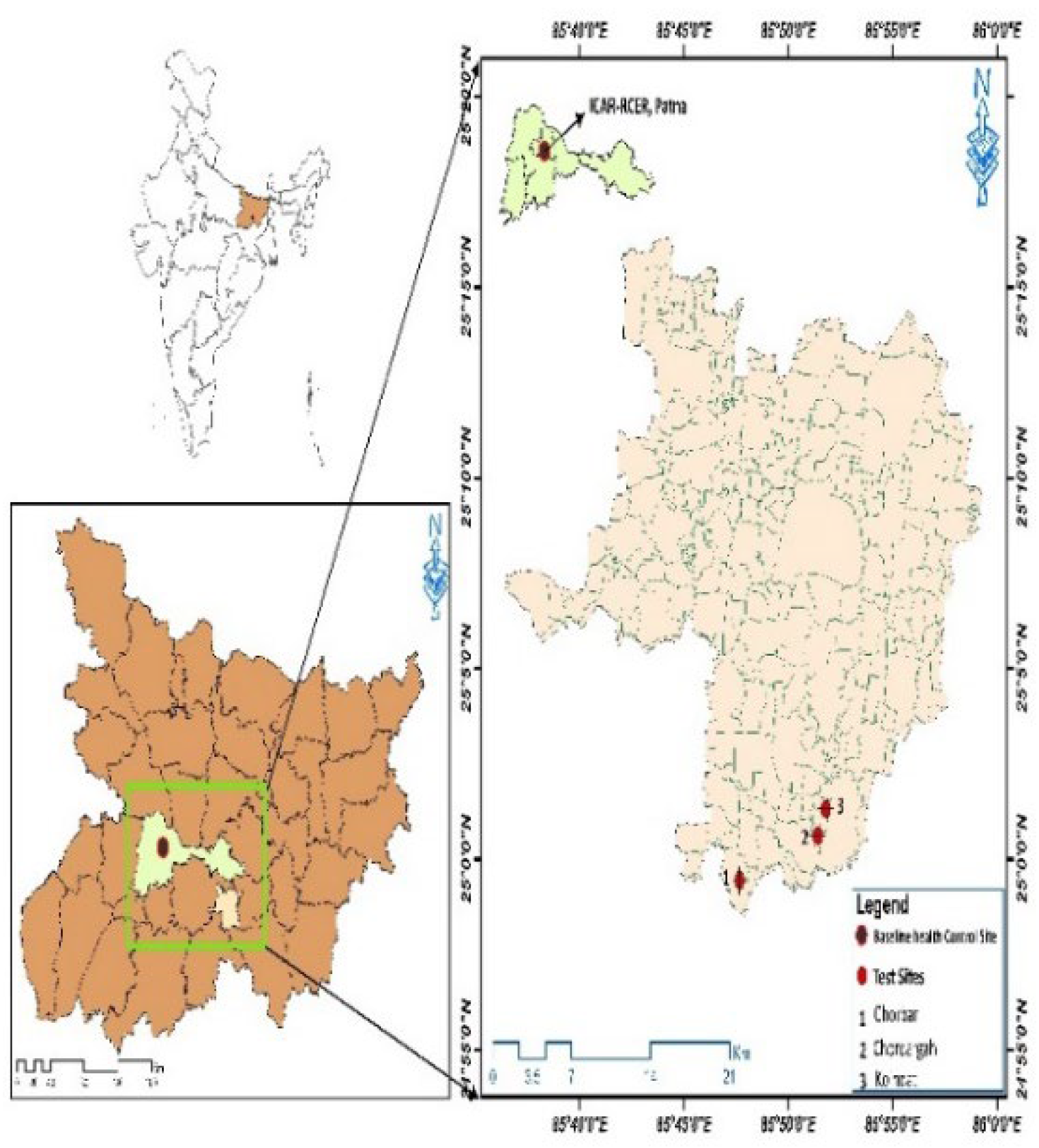
Figure 3.
Effect of treatments on serum levels in calves at different post-treatment intervals. a. Protein (g/dL) and glucose (mg/dL) b. Calcium and Phosphorus (mg/dL).
Figure 3.
Effect of treatments on serum levels in calves at different post-treatment intervals. a. Protein (g/dL) and glucose (mg/dL) b. Calcium and Phosphorus (mg/dL).
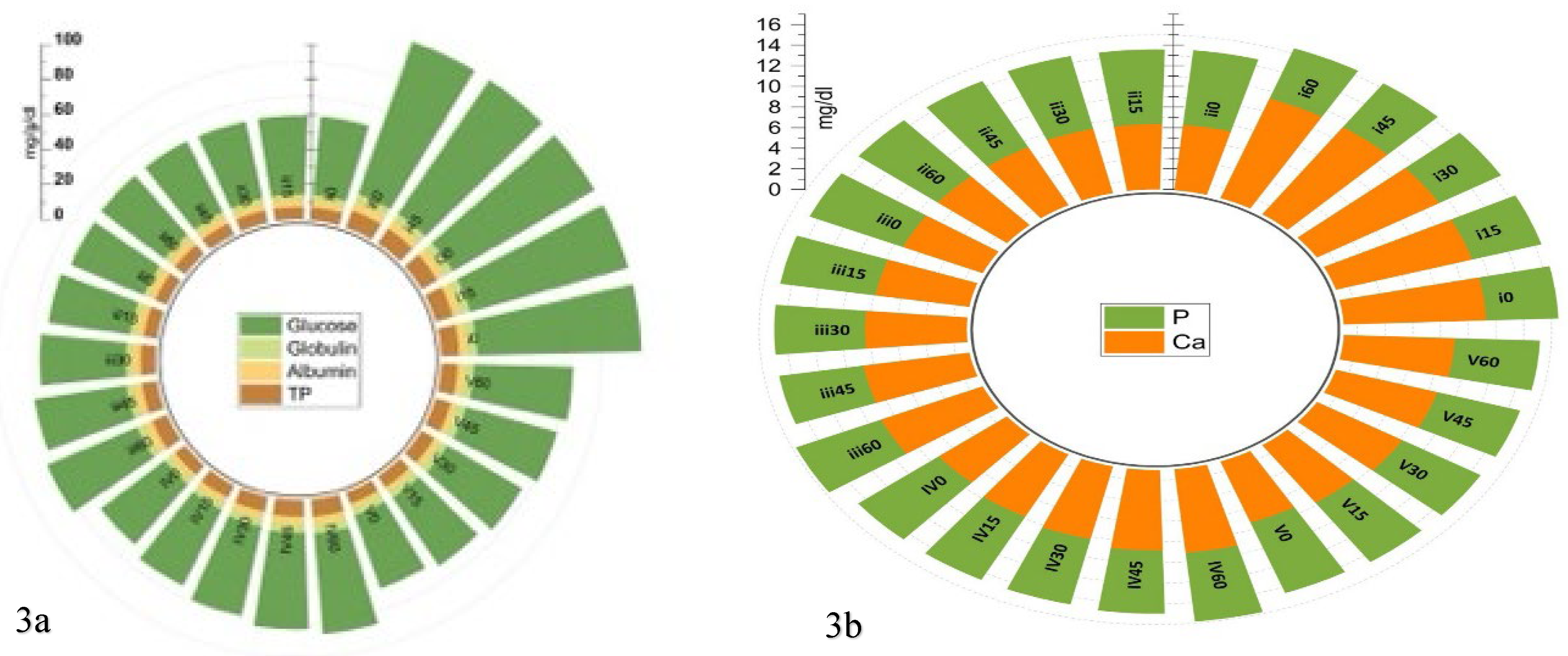
Figure 4.
Effect of treatments on oxidative stress markers in calves at different post-treatment.
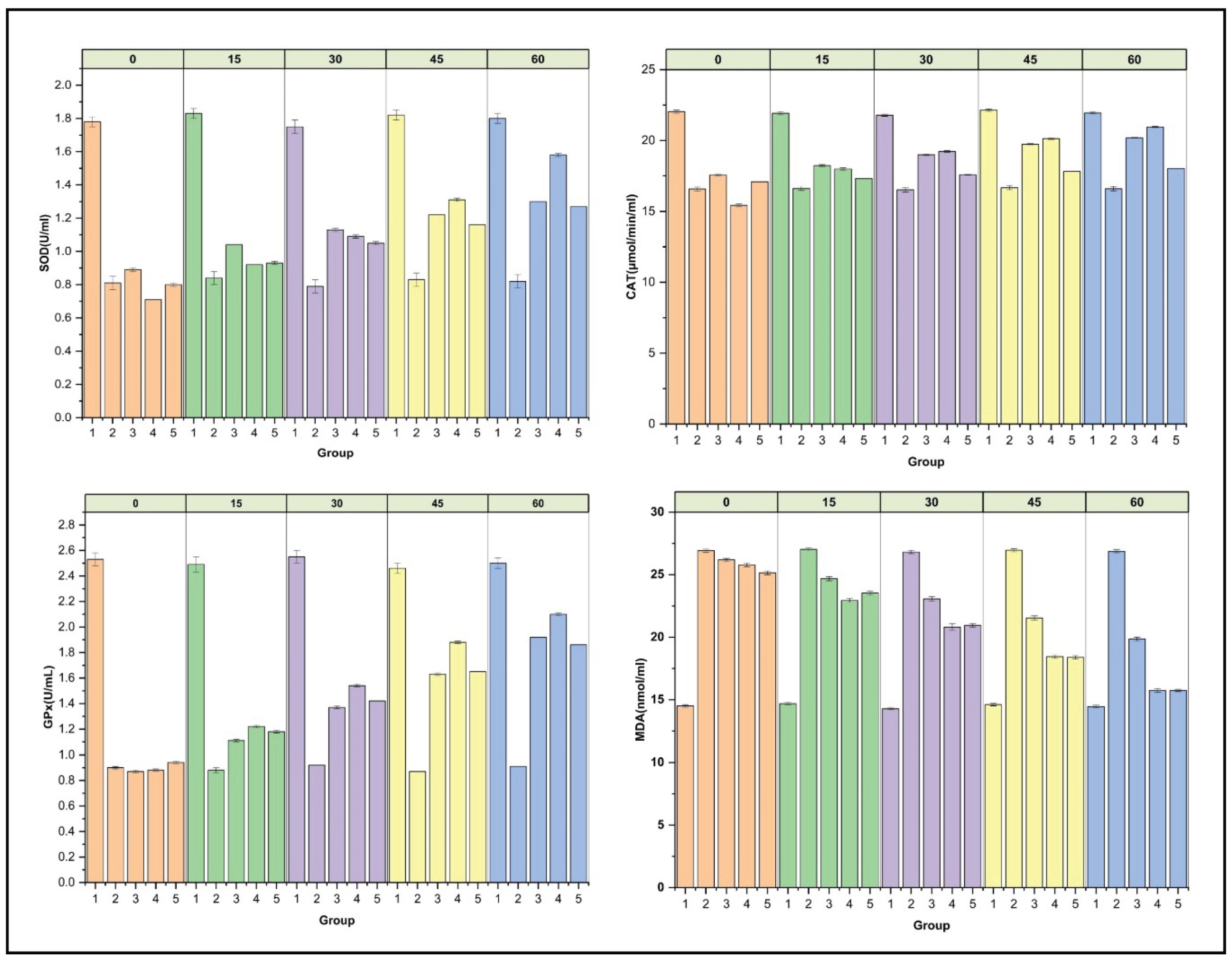
Figure 5.
Multivariate clustering of physiological responses at various treatment levels in calves with fluorosis. a. Heatmap showing how metabolic and oxidative stress markers change over time and in response to treatment. b. The distinction between the groups over time is depicted by the PCA score plot. c. PCA loading plot showing the main factors influencing the recovery and group separation paths.
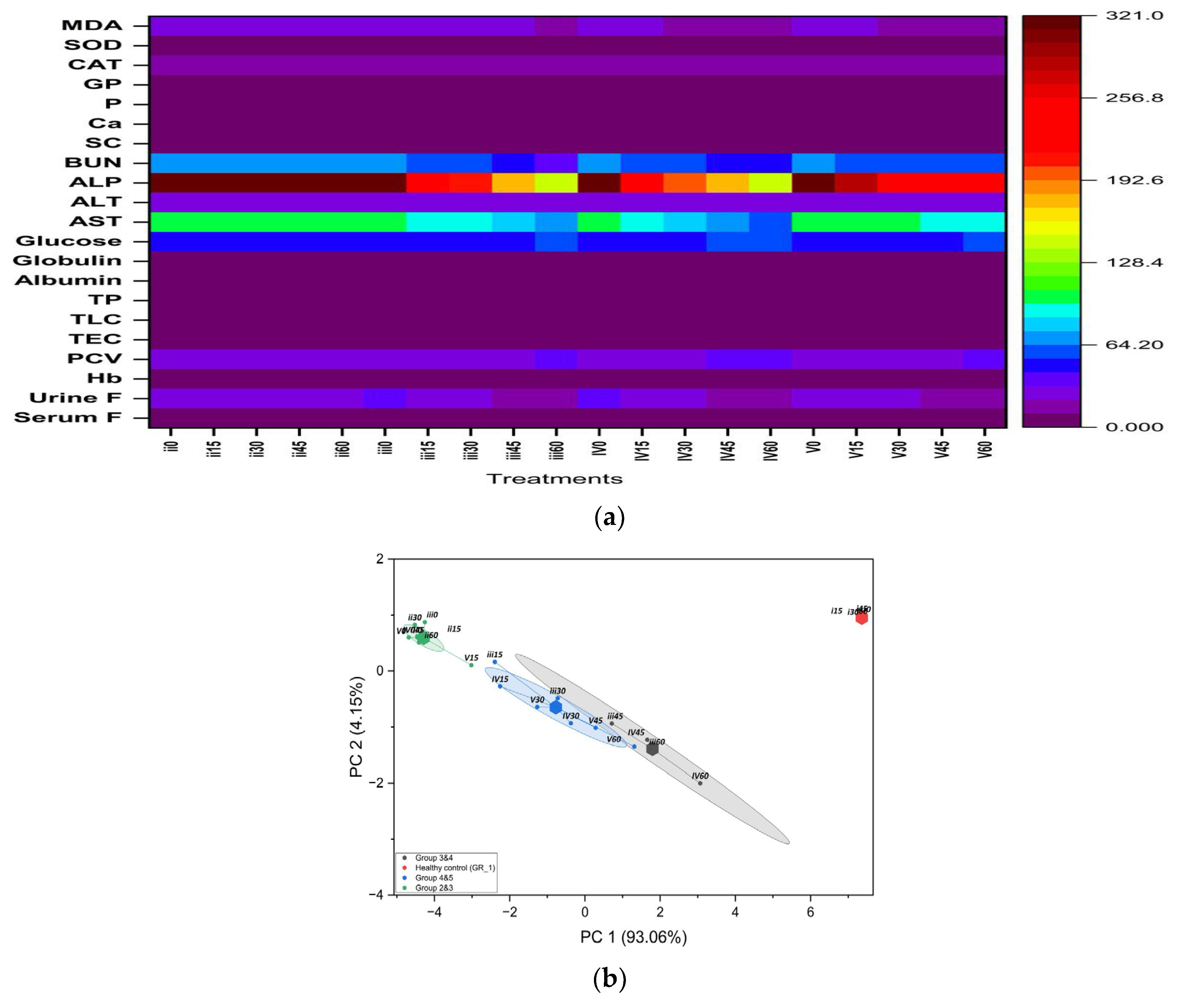
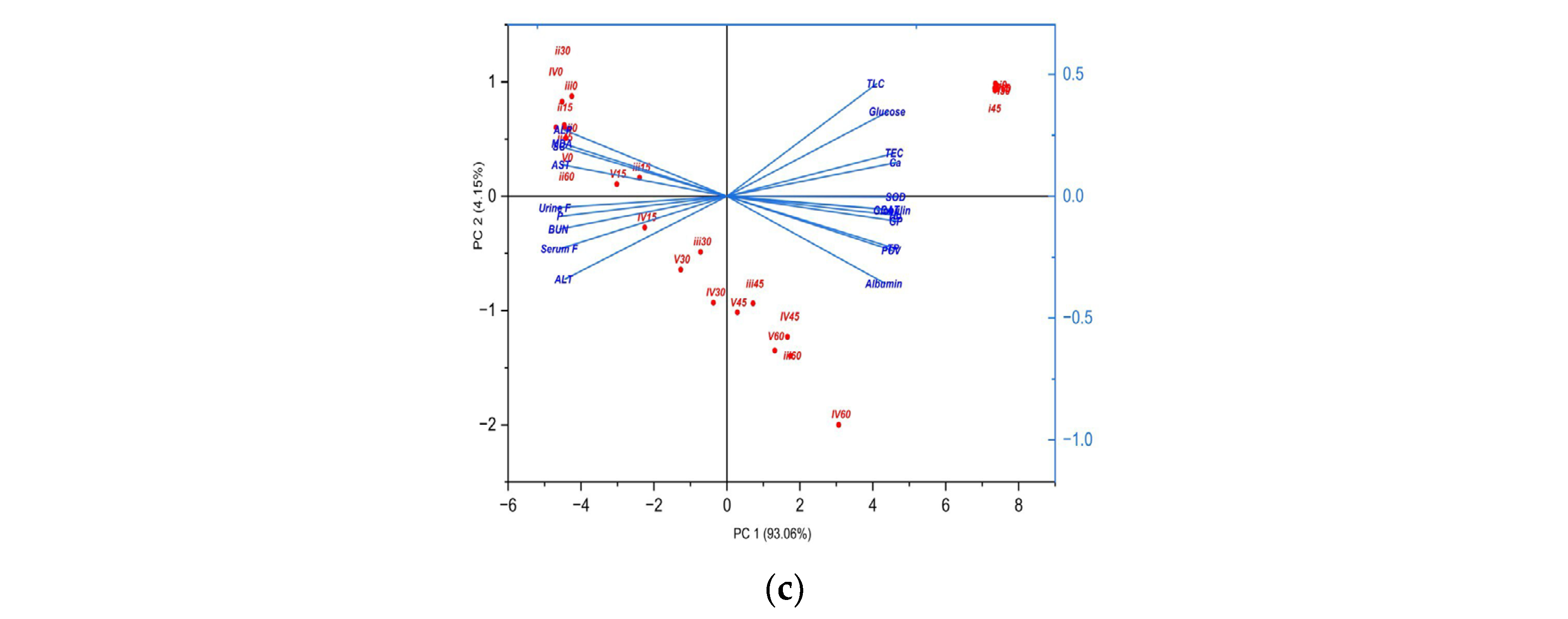

Table 1.
Effect of treatments on serum and urine fluoride (μg/mL) in calves at different post-treatment intervals.
Table 1.
Effect of treatments on serum and urine fluoride (μg/mL) in calves at different post-treatment intervals.
| Parameter | Day | Group I | Group II | Group III | Group IV | Group V |
| Serum F- | 0 | 0.143±0.00aA | 4.140±0.01aB | 4.136±0.0eB | 4.446±0.03eB | 4.160±0.02eB |
| 15 | 0.146±0.00aA | 4.159±0.01aD | 3.942±0.03dC | 3.910±0.02dC | 3.780±0.01dB | |
| 30 | 0.145±0.00aA | 4.178±0.01aD | 3.650±0.01cC | 3.350±0.01cB | 3.332±0.01cB | |
| 45 | 0.147±0.00aA | 4.129±0.01aD | 3.400±0.03bC | 3.100±0.03bB | 3.094±0.03bB | |
| 60 | 0.144±0.00aA | 4.132±0.01aD | 3.048±0.00aC | 2.350±0.03aB | 2.410±0.03aB | |
| Urine F- | 0 | 2.316±0.01aA | 32.068±0.03aC | 32.900±0.13eD | 33.320±0.14eD | 31.140±0.10eB |
| 15 | 2.282±0.02aA | 31.872±0.10aD | 28.880±0.10dC | 26.400±0.10dB | 28.560±0.10dC | |
| 30 | 2.264±0.02aA | 31.982±0.03aE | 23.420±0.12cC | 22.380±0.11cB | 24.340±0.10cD | |
| 45 | 2.649±0.02aA | 31.918±0.03aD | 20.200±0.07bC | 19.240±0.09bB | 20.200±0.07bC | |
| 60 | 2.252±0.02aA | 31.953±0.03aD | 17.300±0.10aC | 16.140±0.12aB | 17.360±0.13aC |
Values are expressed as Mean ± SE (n = 5). Means within a row and column bearing different superscripts differ significantly (p < 0.05; one-way ANOVA followed by Tukey’s post hoc test).
Table 2.
Effect of treatments on hematological parameters in calves at different post-treatment intervals.
Table 2.
Effect of treatments on hematological parameters in calves at different post-treatment intervals.
| Parameter | Day | Group I | Group II | Group III | Group IV | Group V |
| Hb (g/dL) | 0 | 11.44±0.02aC | 9.07±0.02aA | 9.05±0.06aA | 9.12±0.02aB | 9.17±0.04aB |
| 15 | 11.42±0.02aD | 9.04±0.02aA | 9.46±0.04bB | 9.66±0.04bBC | 9.57±0.05bC | |
| 30 | 11.41±0.02aC | 9.01±0.02aA | 9.87±0.05cB | 9.91±0.03cB | 10.03±0.06cB | |
| 45 | 11.47±0.02aC | 9.03±0.01aA | 10.15±0.07dB | 10.25±0.05dB | 10.26±0.05dB | |
| 60 | 11.39±0.02aC | 9.02±0.01aA | 10.38±0.05eB | 10.69±0.04eC | 10.61±0.04eC | |
| PCV (%) | 0 | 34.94±0.04aC | 25.95±0.04aA | 26.30±0.07aB | 26.08±0.07aAB | 26.01±0.08aA |
| 15 | 34.89±0.04aE | 25.89±0.04aA | 27.93±0.08bC | 28.50±0.14aD | 27.08±0.12bB | |
| 30 | 34.91±0.04aB | 25.86±0.04aA | 29.08±0.12cA | 28.11±0.95aA | 29.29±0.07cA | |
| 45 | 34.82±0.04aD | 25.92±0.04aA | 30.70±0.27dB | 32.41±0.14bC | 31.87±0.10dC | |
| 60 | 34.87±0.04aD | 25.83±0.04aA | 32.71±0.27eB | 34.24±0.16bC | 32.77±0.10eB | |
| TEC (x106/µL) |
0 | 5.59±0.01aB | 3.21±0.01aA | 3.22±0.02aA | 3.17±0.02aA | 3.14±0.01aA |
| 15 | 5.57±0.01aD | 3.19±0.01aA | 3.49±0.03bBC | 3.57±0.02bC | 3.45±0.03bB | |
| 30 | 5.56±0.01aD | 3.17±0.01aA | 3.60±0.03cB | 3.92±0.02cC | 3.62±0.02cB | |
| 45 | 5.54±0.01aD | 3.18±0.01aA | 3.87±0.02dB | 4.12±0.02dC | 3.92±0.02dB | |
| 60 | 5.58±0.01aD | 3.12±0.01aA | 4.02±0.04eB | 4.20±0.03eC | 4.00±0.03eB | |
| TLC (x103 cells/µL) | 0 | 6.22±0.02aD | 5.53±0.02aA | 5.76±0.00eC | 5.60±0.00cB | 5.59±0.00cAB |
| 15 | 6.18±0.02aC | 5.52±0.02aA | 5.72±0.00dB | 5.57±0.00bA | 5.51±0.00aA | |
| 30 | 6.20±0.02aC | 5.49±0.02aA | 5.69±0.00cB | 5.54±0.00aA | 5.53±0.00aA | |
| 45 | 6.21±0.02aD | 5.51±0.02aA | 5.66±0.00bB | 5.72±0.00eC | 5.60±0.00cC | |
| 60 | 6.19±0.02aD | 5.50±0.02aA | 5.63±0.00aC | 5.65±0.00dC | 5.56±0.00bB |
Values are expressed as Mean ± SE (n = 5). Means within a row and column bearing different superscripts differ significantly (p < 0.05; one-way ANOVA followed by Tukey’s post hoc test).
Table 3.
Effect of treatments on hepatic and renal function in calves at different post-treatment intervals.
Table 3.
Effect of treatments on hepatic and renal function in calves at different post-treatment intervals.
| Parameter | Day | Group I | Group II | Group III | Group IV | Group V |
| AST (IU/L) |
0 | 57.97±0.12aA | 106.06±0.19aB | 105.66±0.5eB | 105.91±0.40eB | 106.79±0.43eB |
| 15 | 57.89±0.14aA | 105.71±0.11aD | 95.21±0.66dB | 94.42±0.31dB | 102.19±0.32dC | |
| 30 | 57.96±0.14aA | 105.93±0.14aE | 86.28±0.24cC | 81.97±0.28cB | 98.10±0. 13cD | |
| 45 | 57.92±0.11aA | 105.81±0.10aE | 75.29±0.27bC | 73.21±0.11bB | 94.22±0.19bD | |
| 60 | 57.32±0.15aA | 105.61±0.11aB | 66.11±0.56aA | 62.82±0.30aA | 90.20±0.22aB | |
| ALT (IU/L) | 0 | 15.06±0.08aA | 24.16±0.13aB | 24.87±0.01eC | 24.62±0.00eC | 24.28±0.00eB |
| 15 | 14.97±0.08aA | 23.97±0.11aC | 23.89±0.01dC | 23.80±0.01dC | 23.50±0.00dB | |
| 30 | 15.05±0.09aA | 24.14±0.15aC | 23.09±0.01cB | 23.10±0.00cB | 23.00±0.00cB | |
| 45 | 15.03±0.08aA | 23.97±0.16aC | 22.49±0.01bB | 22.39±0.01bB | 22.50±0.00bB | |
| 60 | 15.04±0.10aA | 24.11±0.16aC | 22.01±0.01aB | 21.85±0.00aB | 22.23±0.00aC | |
| ALP (IU/L) |
0 | 128.58±0.07aA | 319.95±0.09aC | 320.85±0.01eD | 319.38±0.01eB | 319.84±0.11eC |
| 15 | 127.95±0.09aA | 319.84±0.13aE | 255.46±0.00dC | 250.89±0.01dB | 285.59±0.00dD | |
| 30 | 128.06±0.11aA | 319.90±0.13aE | 205.09±0.00cC | 200.54±0.01cB | 250.31±0.00cD | |
| 45 | 127.94±0.13aA | 319.92±0.13aD | 175.73±0.00bB | 175.74±0.03bB | 225.00±0.01bC | |
| 60 | 128.02±0.11aA | 319.87±0.13aE | 148.39±0.00aC | 142.03±0.01aB | 221.42±0.00aD | |
| BUN (mg/dL) | 0 | 25.86±0.05aA | 65.80±0.18aB | 65.8±0.01eB | 66.12±0.00eB | 65.91±0.00eB |
| 15 | 25.82±0.04aA | 66.06±0.10aE | 58.99±0.01dB | 60.88±0.00dC | 63.58±0.00dD | |
| 30 | 25.86±0.05aA | 66.03±0.12aE | 54.12±0.01cB | 55.40±0.00cC | 60.51±0.01cD | |
| 45 | 25.83±0.06aA | 66.04±0.08aE | 50.36±0.00bC | 50.14±0.00bB | 59.10±0.00bD | |
| 60 | 25.87±0.06aA | 66.02±0.13aE | 41.25±0.00aC | 43.96±0.00aB | 58.86±0.00aD | |
| Creatinine (mg/dL) | 0 | 0.51±0.00aA | 1.60 ±0.00aB | 1.64±0.01eC | 1.58±0.00eB | 1.60±0.00eB |
| 15 | 0.47±0.01aA | 1.62±0.00aD | 1.30±0.00dB | 1.34±0.00dC | 1.30±0.01dBC | |
| 30 | 0.48±0.00aA | 1.59±0.00aC | 1.04±0.03cB | 1.05±0.00cB | 1.04±0. 00cB | |
| 45 | 0.50±0.07aA | 1.61±0.00aD | 0.90±0.00bB | 0.95±0.00bB | 0.91±0.00bC | |
| 60 | 0.49±0.01aA | 1.59±0.12aD | 0.85±0.00aC | 0.76±0.00aB | 0.80±0.00aB |
Values are expressed as Mean ± SE (n = 5). Means within a row and column bearing different superscripts differ significantly (p < 0.05; one-way ANOVA followed by Tukey’s post hoc test).
Disclaimer/Publisher’s Note: The statements, opinions and data contained in all publications are solely those of the individual author(s) and contributor(s) and not of MDPI and/or the editor(s). MDPI and/or the editor(s) disclaim responsibility for any injury to people or property resulting from any ideas, methods, instructions or products referred to in the content. |
© 2026 by the authors. Licensee MDPI, Basel, Switzerland. This article is an open access article distributed under the terms and conditions of the Creative Commons Attribution (CC BY) license (http://creativecommons.org/licenses/by/4.0/).
Copyright: This open access article is published under a Creative Commons CC BY 4.0 license, which permit the free download, distribution, and reuse, provided that the author and preprint are cited in any reuse.


