Submitted:
30 January 2026
Posted:
03 February 2026
You are already at the latest version
Abstract
Introduction: To investigate if specific emergency physician(EP) admission diagnoses and/or neurological signs/symptoms on admission to the Emergency Department(ED) were associated to normal/not-informative emergency-electroencephalogram(emEEG). Methods: Data from consecutive patients admitted to the ED of our tertiary hospital during two-years period (1 Jan 2023-31 Dic 2024) were retrospectively analyzed. We evaluated the correlation between nor-mal/not-specific emEEG and EP admission diagnoses and neurological signs/symptoms on admission. Epileptic EEGs and EEGs showing triphasic morphology sharp-waves were considered as specific patterns. Results: A total of 2,008 patients underwent emEEG during the study-period. EmEEG was considered not-informative in 100% of global amnesia diagnosis, 100% of mild head trauma, 100% of migraine with aura, 98.3% of transient ischaemic attack(TIA), 95.6% of transient loss of consciousness(TLC) when seizure was not the primary suspected diagnosis and 92.7% of falls of unknown dynamics. Epileptic patterns were detected in 4% of patients presenting with TLC and in 2.4% of those with falls of unknown dynamics, with approximately half of these patients having a pre-existing diagnosis of epilepsy. Triphasic waves were detected in 4.9% pa-tients with falls of unknown dynamic, in 1.7% with TIA and in 0.4% with TLC. All these patients showed fe-ver/sepsis or metabolic/electrolyte disorders. Overall, across all clinical scenarios, emEEG was considered not-informative in 385(19.1%) patients who underwent emEEG. Conclusions: emEEG is almost not-informative in the diagnostic pathway of global amnesia, mild head trauma and migraine with aura, while in patients with TIA, TLC, or falls of unknown dynamics, EP can consider to safely avoid emEEG in the absence of previous epilepsy, fever/sepsis, metabolic/electrolyte disturbances or drug abuse.
Keywords:
Emergency EEG (emEEG)
; emergency department
; emergency diagnostic pathway
1. Introduction
Electroencephalography (EEG) is a technique that has been used for almost 70 years to study brain function [1]. Despite significant advancements in anatomical and functional neuroimaging, EEG remains essential for evaluating specific pathological conditions, particularly those affecting consciousness, such as coma. Its ability to provide real-time insights into brain activity makes it an indispensable tool in clinical practice.
In addition to its long-standing use in outpatient epilepsy management, EEG has recently become more widely used in emergency settings. This increase in use can be attributed to several factors: emergency EEG (emEEG) can be conveniently performed at the patient’s bedside; it is rapid, non-invasive and cost-effective. Most importantly, when neuroimaging results are negative, emEEG can significantly assist neurologists and emergency physicians in making a diagnosis. This capability is particularly vital in acute situations where timely intervention can alter patient outcomes [2,3]. Furthermore, EEG enables functional analysis of brain activity, complementing the anatomical assessments conducted through neuroimaging techniques. While neuroimaging provides detailed images of brain structures, EEG offers insight into the brain’s electrical activity, providing a more comprehensive understanding of a patient’s neurological status [2,3].
Although emEEG can provide valuable information about brain function and aid the diagnosis of several emergency conditions, including seizures and epileptic status (ES), both motor and non-convulsive [4,5,6,7,8], as well as confusional and altered mental states [9,10,11], there remains a significant gap in clinical practice due to the absence of international guidelines governing its use.
This results in high demand for emEEGs in everyday clinical practice, often related to its performance characteristics and rapid availability, or to the individual experience of the treating physician, rather than to its actual diagnostic power.
However, while the daily organization of a tertiary hospital can usually dedicate more than one neurophysiological technician and at least one EEG expert neurologist/neurophysiologist to recording and interpreting emEEGs during working hours, primary and secondary hospitals cannot meet such a high emEEG demand due to limited availability.
For this reason, in recent years authors have focused on the contribution of emEEGs to the diagnostic and therapeutic workup of patients admitted to the emergency department (ED), attempting to identify the reasons why ED attending physicians order emEEGs and the clinical conditions or neurological signs and symptoms on admission that indicate its greatest usefulness. This is intended to provide evidence to inform recommendations for the daily clinical practice of emEEGs [1,2,3,11,12,13,14].
Conversely, identifying clinical conditions usually associated with normal or not-informative EEGs performed as part of the emergency workup could also contribute to establishing recommendations that standardize its use in the ED setting.
In the study of Scarpino et al. [2], which was conducted on a large sample of ED patients, a normal emEEG was observed in 32.1% of cases, particularly when the initial diagnosis was global amnesia or mild head trauma. Notably, none of these patients exhibited specific EEG abnormalities, such as epileptic discharges or triphasic morphology sharp-waves. This suggests that an emEEG recording may not be necessary for patients with these initial diagnoses due to its non-specificity. Patients presenting to the ED following falls of unknown dynamics exhibited emEEG characteristics similar to those with global amnesia and mild head trauma. Only one patient with pre-existing epilepsy showed epileptic discharges, suggesting that emEEG was indicated in patients with this initial diagnosis only if they had a positive epilepsy history. However, despite the large sample size of this previous study [2], the main limitation was represented by the small number of patients admitted to the ED with these specific initial diagnoses.
Therefore, the current study aimed to evaluate the correlation between normal or not-specific EEG abnormalities and the emergency physician’s primary diagnosis at the time of emEEG request, as well as neurological signs and symptoms on admission to the ED, in a greater number of patients. The specific objective was to identify clinical scenarios in which emEEG may be safely omitted from the emergency diagnostic pathway due to its limited diagnostic yield.
2. Materials and Methods
The current study was an extension of the EMINENCE (ElectroencephalograMINEmergeNCydEpartment) study [2].
EMINENCE was an observational, monocentric, retrospective study conducted at Careggi Teaching Hospital, a tertiary care center in Florence, Italy. For the current study, we included all patients over the age of 18 who were admitted to the ED and underwent an emEEG between 1 January 2023 and 31 December 2024.
The main objective of the EMINENCE study [2] was to determine why ED physicians ordered an emEEG and its benefits, which were defined as changes in clinical diagnosis and the diagnostic-therapeutic management of patients, as well as subsequent discharge or hospitalization.
Clinical data were extracted from medical records, including demographic and clinical variables such as gender, age, neurological symptoms and signs, and the indications for emEEG. The clinical context, including a history of epilepsy or other conditions predisposing to ES or encephalopathy, was also recorded. Data on prior anti-seizure medications (ASMs) or other drugs/therapies that could predispose patients to neurological conditions assessed by emEEG, as well as records of neurological, neurosurgical, or resuscitator consultations and neuroimaging findings were also collected. Additional variables, including electrolyte or metabolic imbalances, fever, septic state, and a history of hypertension, diabetes, dyslipidaemia, cardiac disorders, and thyroid disorders, were documented. Discharge or hospitalization was also recorded. Finally, we recorded emEEGs performed both during and outside normal hospital hours.
2.1. EEG Recording and Classification
EEGs are available at our institution from 08:00 to 19:00 on weekdays and from 08:00 to 14:00 on Saturdays. Outside of these hours, EEG is available 24/7 for emergencies related to ES detection.
Standard 30-minute EEG recordings were performed using a digital system and an EEG cap with 19 electrodes (Fp1, Fp2, F7, F8, F3, F4, C3, C4, T3, T4, P3, P4, T5, T6, O1, O2, Fz, Cz and Pz), positioned according to the 10–20 international system. Recordings were acquired at a sampling rate of 128 Hz. During review, digital filters (low-pass filter = 30–70 Hz; time constant = 0.1–0.3 sec; notch filter = 50 Hz) and sensitivity gain (2 to 10 V/mm, standard gain = 7 V/mm) were adjusted to meet interpretative needs [15,16,17,18].
EEG recordings were typically conducted without sedation. Interpretation was performed by expert neurologists or neurophysiologists as promptly as possible, with ED requests given priority when necessary.
EEG classification followed the terminology set by the American Clinical Neurophysiology Society (ACNS) [19]. Descriptors included continuity, voltage, organization of the anterior–posterior gradient, reactivity, spontaneous variability of background activity, frequency, symmetry, epileptiform discharges, slow waves, periodic patterns, triphasic morphology sharp waves, and the presence of sleep-related patterns. Seizure patterns, whether motor or electrical, as well as motor ES or non-convulsive ES (NCES), were also reported. Further details can be found in Hirsch et al. [19].
2.2. Outcome Assessment
We evaluated the correlation between normal or non-specific EEG abnormalities and the initial diagnosis and/or the neurological signs and symptoms present upon admission to the ED. We considered epileptic EEGs (including epileptic discharges, periodic patterns, and seizure/epileptic status) and EEGs showing triphasic morphology sharp waves to be specific patterns because these EEG abnormalities allow a reliable diagnosis to be made, even in the absence of other instrumental or biochemical data. This is in contrast to other EEG abnormalities, such as bilateral or focal slow waves or minor slow waves (theta) sharply contoured, which, although they can contribute to reaching the correct diagnosis, always require the association with other instrumental or biochemical information. The principal aim of the study was to identify patients for whom an emEEG recording might not be indicated in the emergency diagnostic-therapeutic pathway due to its non-specificity.
2.3. Statistical Analysis
Descriptive statistics was employed to define the study population, with categorical variables expressed as numbers and percentages, and continuous variables as medians (interquartile range: 25th to 75th percentile).
Group comparisons were performed using chi-squared tests for categorical data and Kruskal–Wallis tests for continuous variables. Cramer’s V test was used to assess categorical correlations, with values between 0.30 and 0.49 indicating a moderate association, and values above 0.50 signifying a strong correlation. All statistical analyses were conducted using Jamovi (version 2.6.2). Two-tailed tests were applied, and p-values <0.05 have been considered statistically significant.
2.4. Standard Protocol Approvals, Registrations, and Patient Consent
The study complied with ethical principles and good clinical practice guidelines. Due to logistical constraints, it was not feasible to obtain informed consent from all patients. Approval from the competent ethics committee has been sought. The retrospective analysis of the patient database has been approved by the local ethics committee (Ethics Committee Area Vasta Centro27241, positive opinions of the Ethics Committee number 27241_oss positive opinion number).
3. Results
A total of 211.844 patients were evaluated in ED during the study period. Among them, 13.795 were admitted for neurological symptoms, of which 4610 for transient loss of consciousness (TLC).
A total of 2,008 patients were included in the analysis, of whom 1,018 (corresponding to the year 2023) were already included in our previous EMINENCE study [2]. The majority of the emEEGs (1,984; 98.9%) were performed during working hours.
The median age of the cohort was 70 years [interquartile range (IQR) 29]. One thousand and four patients (50.0%) were male. Of the patients, 566 (28.1%) had pre-existing epilepsy, of whom 353 had a structural aetiology and 213 had an unknown aetiology. Five hundred and ten patients were taking ASM.
One thousand and three hundred seventeen patients (65.5%) had no previous brain parenchymal damage. Of the remaining 691 patients, 238 (34.4%) had previously undergone neurosurgery. The most common causes of brain damage were: previous ischaemic stroke (185 patients; 26.7%); multi-infarct encephalopathy (137 patients; 19.8%); brain tumor (103 patients; 14.9%); previous traumatic brain injury (30 patients; 4.3%); and previous haemorrhagic stroke (25 patients; 3.6%). Three hundred sixty-four patients (18.1%) were admitted to the ED with metabolic or electrolyte disorders. Five hundred and seventy-six patients (28.7%) had a fever or sepsis, and 95 patients (4.7%) were admitted following drug abuse. Brain CT scans were performed on 1,844 patients (91.8%), showing acute pathology in 297 patients (16.1%). The most common type of brain damage was cerebral ischaemia (50 patients, 16.8%). The demographic characteristics of the patients are shown in Table 1.
On admission, neurological examinations revealed involuntary movements in 715 patients (35.6%), TLC in 687 patients (34.2%), cognitive/behavioural impairment in 672 patients (33.4%), speech disorder in 512 patients (25.4%), focal motor impairment in 282 patients (14.0%) and an altered level of consciousness in 267 patients (13.2%). Meanwhile, 1,296 patients (64.6%) showed a resolution of the symptoms that led to admission to the ED. Of the patients, 628 (31.3%) were subsequently admitted to hospital, 49 (2.4%) refused admission, and the remaining 1331 (66.3%) were discharged home after their ED visit. An emEEG recording and interpretation were performed by the neurologist/neurophysiologist with a median time of 90 minutes (IQR 75 min) after the ED attending physician’s request.
The most common clinical indications for an emEEG were seizures (520; 25.8%) or suspected seizures (263; 13.0%), followed by TLC (227; 11.3%), altered mental status (158; 7.8%), ischaemic or haemorrhagic stroke (124; 6.1%) (also in cases where in differential diagnosis with seizure or post-critical state; 111; 5.5%), and transient ischaemic attack (TIA) (118; 5.8%). The remaining clinical indications in order of frequency were represented by ES (61, 3.0%), global amnesia (55, 2.7%), mild head trauma (45, 2.1%), falls of unknown dynamics (41, 2.0%), and migraine with aura (15, 0.7%).
In 250 cases (12.4%), no defined clinical indication was given and patients underwent an emEEG depending on the presence of neurological symptoms or signs such as cognitive or behavioral impairment (215 cases, 86%), speech disorders (80 cases, 32%), and headache (41 cases, 16.4%).
Table 2 shows a comprehensive list of emEEG characteristics according to the ED admission diagnosis.
Overall, a normal emEEG was found in 653 patients (32.5%). The admission diagnoses most frequently associated with a normal emEEG were migraine with aura (12, 80%), global amnesia (42, 76.4%), TLC when seizures were not highly suspected (144, 63.4%), mild head trauma (25, 55.6%), TIA (62, 52.5%), and falls of unknown dynamics (17, 41.5%). Global amnesia (100%), mild head trauma (100%), migraine with aura (100%), transient ischaemic attack (TIA) (98.3%), TLC when seizures were not highly suspected (95.6%) and falls of unknown dynamics (92.7%) were the diagnoses most frequently associated with a not-specific emEEG. No patients admitted with global amnesia, mild head trauma or migraine with aura exhibited specific emEEG patterns. Epileptic patterns were detected in 4.0% of patients admitted for TLC and in 2.4% of patients admitted for falls of unknown dynamics, half of whom had pre-existing epilepsy. Triphasic morphology sharp waves were detected in 4.9% of patients with falls of unknown dynamics, 1.7% of patients admitted for TIAs, and 0.4% of subjects admitted for TLC. All of these patients presented with fever/sepsis, metabolic/electrolyte disorders or drug abuse.
Table 3 provides a comprehensive list of EEG characteristics according to the neurological signs and symptoms observed upon admission to the ED. According to our results, no neurological admission sign or symptom was associated with not-informative emEEGs when considered individually, except for patients showing hallucinations (all the neurological signs/symptoms except hallucinations were indeed associated at least with the presence of an epileptic emEEG pattern which accounted for more than 5% of the total).
Table 4 shows the distribution of specific and not-specific emEEGs and the respective final diagnoses, according to the ED admission diagnoses usually associated with a not-specific emEEG. For example, patients with an initial diagnosis of falls of unknown dynamic were discharged with different final diagnoses: 18 patients with confirmed falls of unknown aetiology, 3 with encephalopathy, and 17 with other diagnoses, when the emEEG was normal or not-specific. When the emEEG showed epileptic discharges (in one patient), the final diagnosis was seizure. In the two patients with triphasic morphology sharp waves, the final diagnosis was septicemia.
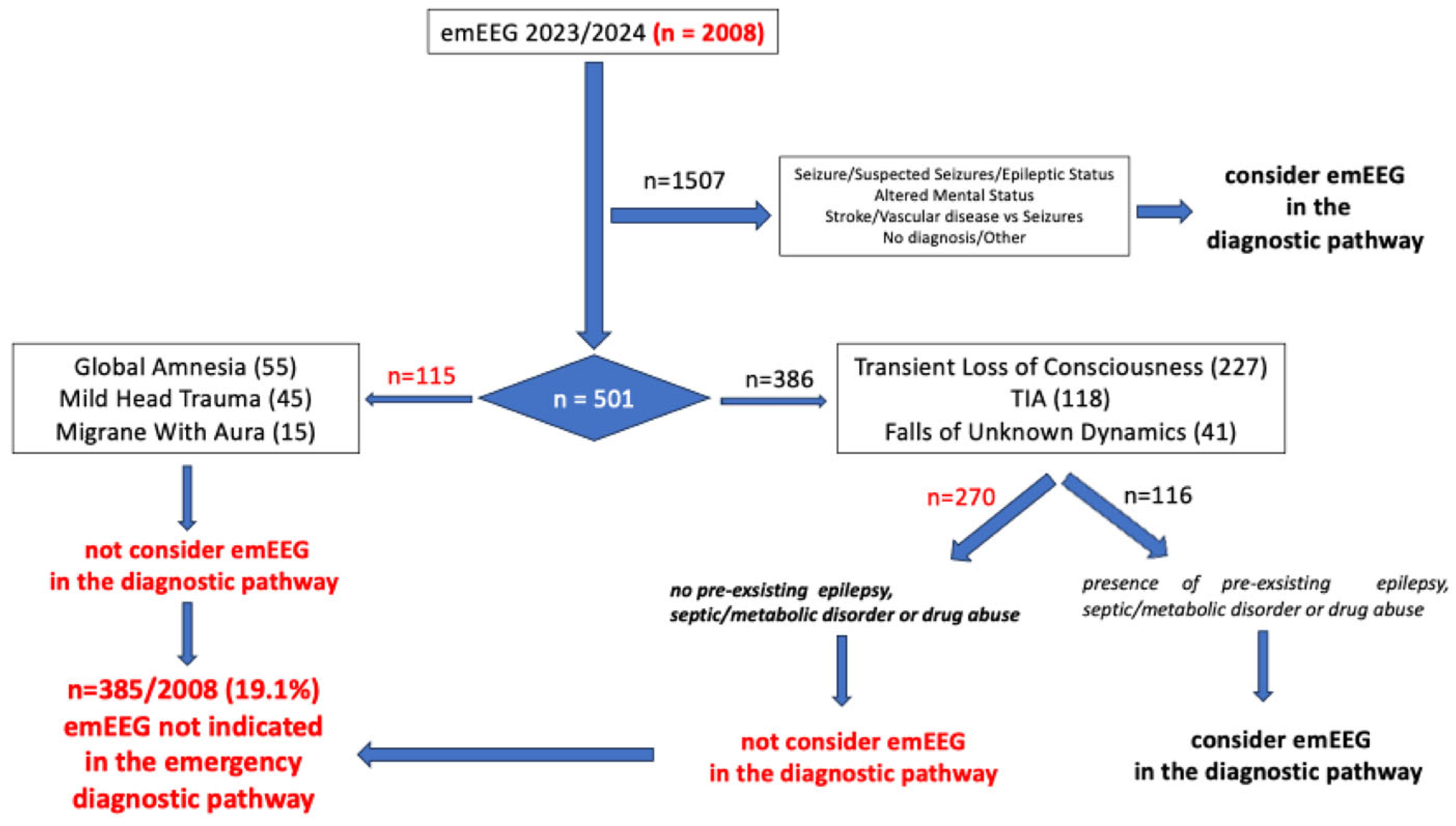
Figure 1 shows a practical suggestion for identifying patients for whom an emEEG would not be mandatory in an emergency diagnostic pathway. An emEEG recording is not mandatory for patients admitted for migraine with aura (n = 15), global amnesia (n = 55) or mild head trauma (n = 45) because none of them showed a specific emEEG. Of the remaining admission diagnoses, which are usually, but not constantly, associated with a not-informative emEEG (TLC without a high suspicion of seizures, TIA and falls of unknown dynamic), 66 of 227 (29.0%) patients admitted for TLC, 22 of 118 (18.6%) admitted for TIA and 28 of 41 (68.2%) admitted for falls of unknow dynamics had metabolic/septic derangements that could potentially cause encephalopathic disorders or pre-existing epilepsy. Thus, of the 386 patients with these three admission diagnoses, an emEEG could be indicated for 116 (30%). The probability of identifying specific EEG abnormalities in these patients is at least twice as high as in the overall patients admitted to the ED for TLC, TIA and falls of unknown dynamic. Thus, of the 2,008 patients, an emEEG could not be mandatory in the emergency pathway due to lack of informativeness in 385 patients (19.1%), corresponding to all 115 patients admitted for global amnesia, mild head trauma and migraine with aura, and 270 patients admitted for TLC, falls of unknown dynamics and TIA.
4. Discussion
This large retrospective study demonstrates that several ED scenarios in which emEEG is requested are consistently associated with not-informative findings. In particular, emEEG did not provide relevant information in the emergency diagnostic pathway of patients with global amnesia, mild head trauma, migraine with aura. In patients presenting with TLC, falls of unknown dynamics, or TIA, emEEG was rarely informative unless specific risk factors were present, such as a history of epilepsy or evidence of metabolic, septic disorder or drug abuse. These findings suggest that emEEG should be selectively employed rather than routinely requested in these scenarios.
The term ‘emergency’ refers to a life-threatening pathological condition that requires immediate treatment. Neurological disorders that cause an altered state of consciousness are usually included among emergent conditions and many of these (e.g., NCES, metabolic or septic encephalopathy and encephalitis) can be detected using an emEEG. The usefulness of emEEGs in emergency clinical practice is well established. Specifically, emEEG is beneficial during the first 24–48 hours after the onset of neurological symptoms. It is useful not only for the clinical management of ES, NCES, and ASM changes, but also for patients with loss of consciousness, acute altered mental status, mental confusion, and ischemic lesions. This is even the case when neuroimaging is normal [1,7,11,13,14,20,21,22]. However, there are still no universally accepted recommendations, except for the management of NCES and motor ES [1,4,5,6,7,8,12]. The lack of guidelines on the use of an emEEG is mainly due to scattered data in the literature and heterogeneous sample selection, recruitment and settings, which makes it difficult to compare reported results. The main limitations of the previous papers were the small sample sizes analyzed [1,7,11,13,23,24] and the heterogeneity of the settings chosen, with most studies [1,7,23,24] including intensive care units (ICUs). This resulted in several emEEGs being ordered for brain death detection or to predict the neurological outcomes of patients with severe brain injuries [15,18]. Even when the ICU was not involved, inpatient wards were included, resulting in EEG requests from the neurology and neurosurgery wards, where the attending physicians had more expertise in selecting neurophysiological tests [11].
Limiting the evaluation to the ED with the aim of suggesting recommendations for the use of EEG in the emergency diagnostic pathway would be important because the ED follows a specific daily workflow that differs from inpatient wards and is even more different from the ICU setting. The ED is indeed a unique setting in which attending physicians usually have to make rapid decisions about the diagnostic and therapeutic management of patients, often in the absence of the immediate availability of instrumental tests such as brain magnetic resonance imaging or prior neurological or neurosurgical consultation. At the same time, they must decide whether to discharge or hospitalize patients. For these reasons, not only confirming but also the ruling out of the initial diagnosis could be very important in this clinical setting for subsequent patient management. In the absence of universally accepted emEEG guidelines, the specific needs of the ED have resulted in high demand for neurophysiological tests in clinical practice. This demand is often related to the performance characteristics and rapid availability of the tests, or to the experience of ED attending physicians, rather than to the tests’ actual diagnostic power or a logical clinical approach. The widespread use of emEEGs in this clinical context has often led to over-prescription and misuse, reducing its diagnostic power and specificity. The high demand for emEEGs in clinical practice poses a significant challenge, particularly in primary and secondary care hospitals where resources are limited and there is an urgent need for highly qualified personnel.
Therefore, identifying admission diagnoses and/or neurological signs/symptoms that are most likely to be associated with informative/specific EEGs performed in emergencies is important in the ED clinical practice as well as the identification of admission diagnoses and/or neurological signs/symptoms that are usually associated with normal or non-specific EEG abnormalities. This would optimize the use of available instrumental tests, costs, and healthcare professional resources without negatively impacting the emergency diagnostic pathway of patients.
To the best of our knowledge, this is the first study in the literature to focus primarily on this topic. Only in a previous paper, Scarpino et al. [2] reported the ED admission diagnoses usually associated with a normal emEEG, especially in cases of global amnesia and mild head trauma. Notably, the emEEG ruled out seizures in all patients in these cases. Furthermore, none of the patients exhibited specific EEG abnormalities, such as epileptic discharges, periodic discharges, seizures, or triphasic morphology sharp waves. This suggests that an emEEG recording may not be indicated in subjects with these initial clinical diagnoses due to its not-specificity. Patients presenting to the ED following falls of unknown dynamics exhibited emEEG characteristics similar to those with global amnesia and mild head trauma. Only one patient with pre-existing epilepsy showed epileptic discharges, suggesting that emEEG was only indicated in patients with this initial diagnosis if they had a positive epilepsy history.
Although the study by Scarpino et al. [2] was conducted on a large patient sample, the main limitation was the small number of patients admitted to the ED with these specific initial diagnoses.
For this reason, we conducted a study with a larger sample size than in our previous work [2] to confirm our previous findings and strengthen our data, with the aim of more accurately identifying patients for whom an emEEG recording may not be indicated in the emergency diagnostic-therapeutic pathway due to its not-specificity.
We considered epileptic EEGs (including epileptiform discharges, periodic patterns, and seizure/epileptic status) and EEGs showing triphasic morphology sharp waves to be specific patterns because these EEG abnormalities allow a reliable diagnosis to be made even in the absence of other instrumental or biochemical data. This is in contrast to other EEG abnormalities, such as bilateral or focal slow waves or minor slow waves (theta) sharply contoured, which, although they could contribute to reaching the correct diagnosis, always require the association with other instrumental/biochemical data.
Our data analysis revealed that global amnesia (100%), mild head trauma (100%), migraine with aura (100%), transient ischaemic attack (TIA) (98.3%), TLC when seizures were not highly suspected (95.6%), and falls of unknown dynamics (92.7%) were the most common admission diagnoses associated with a not-specific EEG. Notably, all of these admission diagnoses were associated with either the absence or the presence of specific EEG patterns (both epileptic and triphasic), which accounted for less than 5% of the total. No patients admitted for global amnesia, mild head trauma or migraine with aura exhibited specific emEEG patterns. Epileptic patterns were detected in 4.0% of patients admitted for TLC and in 2.4% of patients admitted for falls of unknown dynamics; of these patients, half had pre-existing epilepsy. Triphasic morphology sharp waves were detected in 4.9% of patients with falls of unknown dynamics, 1.7% of patients admitted for TIA, and 0.4% of patients admitted for TLC. All of these patients presented with fever, sepsis, metabolic/electrolyte disorders or drug abuse.
Thus, according to our results, an emEEG could not be mandatory in the emergency diagnostic pathway for patients admitted to the ED with a diagnosis of global amnesia, mild head trauma or migraine with aura, as it was always not-informative. In patients admitted for TLC , in the absence of a high suspect of seizure, and for falls of unknown dynamics, an emEEG could be indicated only in the presence of a pre-existing epilepsy, whereas patients admitted for TIA, TLC or falls of unknown dynamics in the presence also of fever, sepsis, metabolic/electrolyte disturbance or drug abuse, could benefit from an emEEG because of they might show signs of brain involvement related to the presence of these medical disorders, such as septic or metabolic encephalopathy.
In particular, as shown in Figure 1, of the 2,008 patients included in the analysis, 501 (25%) showed an absence or alternatively presence of specific EEG patterns in less than 5% of cases. In more detail, an emEEG was always normal or not-specific in 115 (23%) subjects, representing the total number of patients admitted for global amnesia, mild head trauma and migraine with aura. Of the remaining 386 patients (77%), who were admitted for TLC, falls of unknown aetiology and TIA, an emEEG could only be informative in 116 (30%) of cases. This corresponded to patients with pre-existing epilepsy or septic/metabolic disorders upon admission to the ED.
Thus, according to our results, of the 2,008 patients, 385 (19.1%) could not have an emEEG as part of the emergency pathway because it would not be informative.
Our results demonstrate the importance for ED physicians of making the most accurate admission diagnosis possible based on admission symptoms and anamnestic data, even though this can often be difficult in an ED setting where initial information is frequently incomplete. However, an accurate admission diagnosis could significantly impact the benefit patients could derive from certain diagnostic tests. This is particularly true in primary and secondary care hospitals, where the availability of diagnostic tests and staff is limited. An illustrative example is the difference between an admission diagnosis of TLC in the absence of suspected seizures and an admission diagnosis of suspected seizures when not witnessed. According to our results, only 4% of patients with an admission diagnosis of TLC showed an epileptic pattern on emEEG, whereas the percentage of specific emEEGs with epileptic discharges increased to 48.7% when the admission diagnosis was suspected seizures.
On the other hand, taking again the example of TLC as admission diagnosis, the reduced usefulness of the emEEG, which usually, but not constantly, showed not-specific patterns, was evident also from its lower contribution to the ED final diagnosis of these patients (Table 4). Compared to most of the other admission diagnoses, where the emEEG result greatly influenced the final ED diagnosis, our data analysis revealed that 6 of the 215 patients admitted for TLC and exhibiting not-specific EEGs were discharged with a diagnosis of suspected seizures, while 5 were discharged with a diagnosis of infectious or metabolic/electrolyte disorders. At the same time, of the 9 patients showing epileptic emEEG, 7 were however discharged as TIA and only 2 as seizures. Similar results were observed also for patients admitted for TIA, of whom 75 of the 116 showing not-specific emEEGs were discharged with heterogenous final diagnoses, including seizures and infectious or metabolic/electrolyte. This suggests that ED physicians relied more on admission signs, symptoms, and anamnestic data than on the emEEG result for the final diagnosis of these patients.
According to our data, no neurological admission signs or symptoms, when considered individually, were associated with not-informative EEGs, except in patients who experienced hallucinations. This provides further evidence of the importance of making an accurate admission diagnosis for ED physicians by combining neurological and other clinical signs and symptoms of patients, as well as anamnesis. When a specific diagnosis could not be performed, cognitive/behavioral impairment, speech disorders, and headache were the most frequent neurological symptoms on admission for which an emEEG was usually requested, with 11.5% of these being specific for epileptic patterns. Therefore, according to our results, an emEEG recording performed in the emergency setting could contribute to the final diagnosis in patients for whom a reliable admission diagnosis could not be achieved.
Additionally, it is important to note that patients admitted for seizures or suspected seizures and who are actually subsequently discharged from the ED with an epileptic aetiology may exhibit an EEG characterized by the absence of epileptic discharges, with abnormalities that are more akin to those of the not-specific EEGs associated to the other diagnostic suspicions, or even normal emEEGs. This finding is consistent with previous literature indicating that the diagnostic contribution of an emEEG for witnessed or suspected seizures is just over 50% [5,25]. The substantial difference in the diagnostic pathway of these patients in who an emEEG recording is mandatory, compared to patients admitted for loss of consciousness in the absence of a high suspicion of seizure, is represented by the percentage of specific epileptic emEEGs that reaches 41.7% and 48.7% for seizures and suspected seizures, respectively, whereas it reaches <5% for patients admitted for TLC. These results highlight the importance for ED physicians of making an accurate admission diagnosis, not only to identify patients who could benefit most from an emEEG, but also to ensure the correct use of emEEG results, even when they are not-specific or normal.
Finally, our data demonstrated that, when an accurate admission diagnosis is made, ED physicians can feel confident enough to omit an EEG recording from the emergency pathway when specific neurological conditions are suspected, as the result of the neurophysiological test would usually not provide information that could impact patient management in the ED setting. In these cases, an EEG recording could be included in a fast-track outpatient program to complete the diagnostic workup, reducing the costs and length of hospital stays for ED patients related to EEG execution, waiting for the neurophysiological report, and waiting for the neurological consultation. It is important to emphasize that, while these costs and timeframes may not be too relevant in a third-level hospital, where resources and qualified personnel are sufficient, they could have a significant impact in first- and second-level hospitals, where resources are limited.
Even though the current study was the first to focus on ED admission diagnoses or neurological signs/symptoms usually associated with a normal/non-specific EEG, and thus contributed evidence to support recommendations for daily clinical emEEG practice, particularly in a specific setting such as the ED, it had some limitations. Mainly, it was retrospective, resulting in incomplete clinical and instrumental information collected from medical records. Another limitation was the monocentric setting of a tertiary hospital, which included many complex and severe cases that may not fully represent the spectrum of cases in smaller hospitals. Furthermore, the monocentric setting of a tertiary hospital, where at least two or three neurophysiological technicians and one EEG expert neurologist/neurophysiologist were exclusively dedicated to recording and interpreting emEEGs during working hours, limited the generalizability of the work, particularly with regard to primary and secondary hospitals where emEEG availability is limited.
This study which included a large sample of subjects and limited the evaluation to emEEGs performed only in the ED, attempted to identify patients for whom an emEEG recording may not be indicated in the emergency diagnostic-therapeutic pathway due to its not-specificity, thus not providing additional crucial information in an emergency setting that could significantly modify the emergency management of patients. By employing an appropriately sized sample, our study has the potential to be the first to comprehensively evaluate the not-informative emEEG in emergency settings. Previous research in this area has often been limited by small patient cohorts and conducted across heterogeneous environments, making it difficult to generalize findings to broader populations. By addressing these gaps, our study aims to clarify the utility of emEEGs in acute medical scenarios. In particular, it provides evidence to inform recommendations for daily clinical practice, particularly in specific settings such as the ED.
5. Conclusions
For EEG to be effectively integrated into emergency care, it is vital to establish international guidelines that standardize its use. These guidelines should provide a clear framework for when and how EEG should be utilized in emergency contexts, ensuring its application is efficient and beneficial. This includes cases in which the neurophysiological test is expected to be not-informative, meaning it is unnecessary to perform in an emergency setting.
It would be of great importance to develop these recommendations using well-established methodological tools, not only for the management of ES, but also focusing on non-informative emEEGs. This would be especially beneficial for primary and secondary hospitals, where emEEG availability is limited and resources are scarce, and highly qualified personnel are needed.
Author Contributions
Conceptualization, MS,AG, AN; Methodology, MS, AG, CM; investigation, FB,LB,AF; data curation, RF.; writing—original draft preparation, MS; writing—review and editing, AG,PN; supervision, AG,AN.; project administration, BP.
Funding
This research received no external funding.
Institutional Review Board Statement
The study was conducted in accordance with the Declaration of Helsinki, and approved by the Ethics Committee of Ethics Committee Area Vasta Centro, protocol code 27241 , 24-07-2024).
Informed Consent Statement
Patient consent was waived because due to logistical constraints, it was not feasible to obtain informed consent from all patients.
Data Availability Statement
The raw data supporting the conclusions of this article will be made available by the authors on request.
Acknowledgments
We acknowledge all technicians of Neurophysiopathology unit for their prompt response to calls from emergency doctors. and for the quality of recordings.
Conflicts of Interest
“The authors declare no conflicts of interest. The funders had no role in the design of the study; in the collection, analyses, or interpretation of data; in the writing of the manuscript; or in the decision to publish the results”.
Abbreviations
The following abbreviations are used in this manuscript:
| ACNS | American Clinical Neurophysiology Society |
| ASM | Antiseizure Medication |
| CT | Computed Tomograpgy |
| ED | emergency department |
| EEG | Electroencephalography |
| emEEG | emergency EEG |
| ES | Epileptic Status |
| ICU | intensive care unit |
| IQR | Interquartile Range |
| NCSE | non-convulsive Epileptic Status |
| TIA | transient ischaemic attack |
| TLC | transient loss of consciousness |
References
- Praline, J.; Grujic, J.; Corcia, P.; Lucas, B.; Hommet, C.; Autret, A.; et al. Emergent EEG in clinical practice. Clin. Neurophysiol. 2007, 118, 2149–2155. [Google Scholar] [CrossRef]
- Scarpino, M.; Verna, M.T.; Grippo, A.; Lolli, F.; Piccardi, B.; Nazerian, P.; et al. The role of EEG in the emergency department: Its contribution to the patient’s diagnostic-therapeutic pathway. The EMINENCE study. Clin. Neurophysiol. Pract. 2025, 10, 70–77. [Google Scholar] [CrossRef]
- Scarpino, M.; Grippo, A.; Verna, M.T.; Lolli, F.; Piccardi, B.; Nazerian, P.; et al. Contribution of the EEG in the Diagnostic Workup of Patients with Transient Neurological Deficit and Acute Confusional State at the Emergency Department: The EMINENCE Study. Diagnostics (Basel). 2025, 15, 863. [Google Scholar] [CrossRef]
- 4. ACEP Clinical Policies Committee Clinical Policies Subcommittee on Seizures (2004) Clinical policy: critical issues in the evaluation and management of adult patients present into the emergency department with seizures. Ann. Emerg. Med. 2004, 43, 605–625.
- Bellini, A.; Gusmeo Curti, D.; Cursi, M.; Cecchetti, G.; Agosta, F.; Fanelli, G.; et al. Predictors of seizure detection and EEG clinical impact in an Italian tertiary emergency department. J. of Neurology. 2024, 271, 5137–5145. [Google Scholar] [CrossRef]
- Feyissa, A.M.; Tatum, W.O.; Adult, E.E.G. Handbook of Clinical Neurology; Elsevier B.V: Amsterdam, 2019; pp. 103–124. [Google Scholar]
- Varelas, P.N.; Spanaki, M.V.; Hacein-Bey, L.; Hether, T.; Terranova, B. Emergent EEG Indications and diagnostic yield. Neurology 2003, 61, 702–704. [Google Scholar] [CrossRef]
- Vignatelli, L.; Tontini, V.; Meletti, S.; Camerlingo, M.; Mazzoni, S.; Giovannini, G.; et al. Clinical practice guidelines on the management of status epilepticus in adults: a systematic review. Epilepsia. 2024, 65, 1512–1530. [Google Scholar] [CrossRef]
- Conference de consensus sur les indications de l’EEG en urgence Rapport redigé par J Perret, president du jury, apres les presentations des experts. Neurophysiol. Clin. 1996, 28, 103–110.
- Kaplan, P.W. Assessing the outcomes in patients with nonconvulsive status epilepticus: nonconvulsive status epilepticus is underdiagnosed, potentially overtreated, and confounded by comorbidity. J. Clin. Neurophysiol. 1999, 16, 341–353. [Google Scholar] [CrossRef]
- Rodriguez Quintana, J.H.; Silvia Juliana Bueno, F.; Zuleta-Motta, J.L.; Ramos, M.F.; V́elez-van-Meerbeke, A. and the Neuroscience Research Group (NeuRos). Utility of Routine EEG in Emergency Department and Inpatient Service. Neurology Clinical Practice. 2021, e681 11, e677. [Google Scholar]
- Privitera, M.D.; Strawsburg, R.H. Electroencephalographic monitoring in the emergency department. Emerg. Med. Clin. North. Am. 1994, 12, 1089–1100. [Google Scholar] [CrossRef]
- Yigit, O.; Eray, O.; Mihci, E.; Yilmaz, D.; Arslan, S.; Eray, *!!! REPLACE !!!*. BThe utility of EEGin the emergency department. Emerg Med, J 2012, 29, 301–305. [Google Scholar] [CrossRef]
- Praline, J.; de Toffol, B.; Mondon, K.; Hommet, C.; Prunier, C.; Corcia, P.; et al. EEG d’urgence: indications reelles et resultats. Neurophysiol. Clin. 2004, 34, 175–181. [Google Scholar] [CrossRef]
- Scarpino, M.; Grippo, A.; Lanzo, G.; Lolli, F. The burden of clinical neurophysiology for the neurologicalprognosis of coma. Future Medicine. 2018, 13, 127–129. [Google Scholar]
- Scarpino, M.; Lolli, F.; Hakiki, B.; et al. The prognostic value of the EEG, according to the American Clinical Neurophysiology Society terminology, recorded at the postacute stage in patients with unresponsive wakefulness syndrome. Neurophysiol. Clin. 2019, 49, 317–327. [Google Scholar] [CrossRef] [PubMed]
- Scarpino, M.; Lolli, F.; Hakiki, B.; Lanzo, G.; Sterpu, R.; Atzori, T.; et al. for the Intensive Rehabilitation Unit Study Group of the IRCCS Don Gnocchi Foundation, Italy. EEG and Coma Recovery Scale-Revised prediction of neurological outcome in Disorder of Consciousness patients. Acta Neurol. Scand. 2020, 142, 221–228. [Google Scholar] [CrossRef] [PubMed]
- Scarpino, M.; Lanzo, G.; Hakiki, B.; Sterpu, R.; Maiorelli, A.; Cecchi, F.; et al. Acquired brain injuries: neurophysiology in early prognosis and rehabilitation pathway. Signa Vitae 2021, 2–11. [Google Scholar]
- Hirsch, L.J.; LaRoche, S.M.; Gaspard, N.; Gerard, E.; Svoronos, A.; Herman, S.T.; et al. American Clinical Neurophysiology Society’s standardized critical care EEG terminology: 2012 version. J. Clin. Neurophysiol. 2013, 30, 1–27. [Google Scholar] [CrossRef]
- Manez ˜ Miro, J.U.; Dıaz de Teran, F.J.; Alonso Singer, P.; Aguilar-Amat Prior, M.J. Uso de la electroencefalografıa urgente por el neurologo de guardia: utilidad en el diagnostico del estatusepilepticono convulsivo. Neurologıa. 2018, 33, 71–77. [Google Scholar] [CrossRef]
- Lozeron, P.; Tcheumeni, N.C.; Turki, S.; Amiel, H.; Meppiel, E.; Masmoudi, S.; et al. Contribution of EEG in transient neurological deficits. J. Neurol. 2018, 265, 89–97. [Google Scholar] [CrossRef]
- Prud’hon, S.; Amiel, H.; Zanin, A.; Revue, E.; Kubis, N.; Lozeron, P. EEG and acute confusional state at the emergency department. Neurophysiol. Clin. 2024, 54, 102966. [Google Scholar] [CrossRef] [PubMed]
- Khan, R.B.; Yerremsetty, P.K.; Lindstrom, D.; McGill, L.J. Emergency EEG and factors associated with nonconvulsive status epilepticus. J. Natl. Med. Assoc. 2001, 93, 359–362. [Google Scholar] [PubMed]
- Quigg, M.; Shneker, B.; Domer, P. Current practice in administration and clinical criteria of emergent EEG. J. Clin. Neurophysiol. 2001, 18, 162–165. [Google Scholar] [CrossRef]
- Zawar, I.; Briskin, I.; Hantus, S. Risk factors that predict delayed seizure detection on continuous electroencephalogram (cEEG) in a large sample size of critically ill patients. Epilepsia Open. 2022, 7, 131–143. [Google Scholar] [CrossRef] [PubMed]

Table 1.
Patient’s (n = 2008) Demographic Characteristics.
| Characteristics | |
|---|---|
| Age year, median (IQR) | 70 (IQR 29) |
| Male gender, n (%) | 1004 (50.0%) |
| Previous Epileptic Seizures | 566 (28.1%) |
| Unknown aetiology | 213 (10.6%) |
| Structural aetiology | 353 (17.6%) |
| Antiseizures medications | 510 (25.4%) |
| Under-dosed antiseizures medications | 122 (6.1%) |
| Fever | 262 (13.1%) |
| Sepsis | 314 (15.6%) |
| Metabolic disturbance | 129 (6.4%) |
| Electrolyte disturbance | 235 (11.7%) |
| Drug abuse | 95 (4.7%) |
| Previous Neurological history | 691 (34.4%) |
| Stroke | 159 (7.8%) |
| Neurosurgery | 238 (11.9%) |
| Cardiac disorders | 462 (23.0%) |
| Diabetes | 327 (16.3%) |
| Dyslipidemia | 540 (26.9%) |
| Thyroid disease | 218 (10.9%) |
| Brain CT | 1844 (91.8%) |
| Recent CT Lesions | 297 (14.7%) |
| Home discharge | 1331 (66.3%) |
| Hospitalization | 628 (31.3%) |
| Hospitalization Refused | 49 (2.4%) |
Table 2.
Emergency EEG characteristics and abnormalities according to the diagnosis on patient’s admission.
Table 2.
Emergency EEG characteristics and abnormalities according to the diagnosis on patient’s admission.
| Normal | Epileptiform | Triphasic sharp waves |
Frequency reduced |
No Reactivity | Asymmetry | Theta | Delta | |
|---|---|---|---|---|---|---|---|---|
| Cramer’s V ED ADMISSION DIAGNOSIS |
0.280 |
0.421 | 0.158 | 0.170 | 0.255 | 0.173 | 0.203 | 0.179 |
| Seizure (n=520) | 96 (18.5) | 217 (41.7) | 7 (1.3) | 197 (37.9) | 218 (41.9) | 111 (21.3) | 246 (47.3) | 270 (51.9) |
| Suspected Seizures (n=263) | 84 (31.9) | 64 (48.7) | 7 (2.6) | 99 (37.6) | 74 (28.1) | 50 (19.0) | 105 (39.9) | 17 (6.4) |
| Epileptic Status (n=61) | 3 (4.9%) | 41 (67.2) | 0 (0.0) | 34 (55.7) | 42 (68.8) | 30 (49.2) | 32 (52.5) | 50 (81.9) |
| Altered Mental Status (n=158) | 20 (18.4) | 24 (27.3) | 21 (13.2) | 109 (68.9) | 123 (77.8) | 28 (17.07) | 35 (22.1) | 80 (50.6) |
| TLC (n=227) | 144 (63.4) | 9 (4.0) | 1 (0.4) | 41 (18.0) | 19 (8.3) | 8 (3.6) | 32 (14.1) | 28 (12.3) |
| TIA (n=118) | 62 (52.5) | 0 (0.0) | 2 (1.7) | 26 (22.0) | 9 (7.9) | 4 (3.4) | 27 (22.9) | 22 (18.6) |
| Stroke (n=124) | 34 (27.4) | 11 (8.8) | 8 (6.4) | 57 (45.9) | 48 (35.8) | 24 (19.3) | 35 (28.2) | 56 (45.1) |
| Vascular disease vs seizures (n=111) | 24 (21.6) | 25 (22.5) | 0 (0.0) | 49 (44.1) | 36 (32.4) | 30 (27.0.) | 56 (50.5) | 53 (47.7) |
| Falls Of Unknow Dynamics (n=41) | 17 (41.5) | 1 (2.4) | 2 (4.9) | 19 (46.3) | 12 (29.3) | 2 (4.9) | 7 (17.1) | 12 (29.2) |
| Global Amnesia (n=55) | 42 (76.4) | 0 (0.0) | 0 (0.0) | 0 (0.0) | 2 (3.6) | 0 (0.0) | 8 (14.5) | 6 (10.9) |
| Mild head Trauma (n=45) | 25 (55.6) | 0 (0.0) | 0 (0.0) | 10 (22.2) | 8 (17.8) | 4 (8.9) | 11 (24.4) | 8 (17.7) |
| Migraine with aura (n=15) | 12 (80.0) | 0 (0.0) | 0 (0.0) | 0 (0.0) | 0 (0.0) | 0 (0.0) | 2 (13.3) | 1 (6.7) |
| No Diagnosis/Other (n=250) | 90 (36.0) | 28 (11.5) | 7 (2.9) | 99 (39.6) | 69 (27.6) | 40 (16.4) | 68 (27.2) | 82 (32.8) |
| Total (n=2008) | 653 (32.5) | 420 (20.9) | 55 (2.7) | 740 (36.8) | 660 (32.8) | 331 (16.4) | 664 (33.1) | 414 (20.6) |
TLC: Transient Loss of Consciousness; TIA: Transient Ischemic Attack; ES: Epileptic Status; ED: Emergency Department.
Table 3.
Emergency EEG characteristics and abnormalities according to neurologic signs/symptoms on patient’s admission.
Table 3.
Emergency EEG characteristics and abnormalities according to neurologic signs/symptoms on patient’s admission.
| Normal | Epileptiform | Triphasic sharp waves |
Frequency reduced |
No Reactivity |
Asymmetry | Theta | Delta | |
|---|---|---|---|---|---|---|---|---|
|
Neurological Signs/Symptoms on admission |
n (%) | n (%) | n (%) | n (%) | n (%) | n (%) | n (%) | n (%) |
| TLC (n=687) | 265 (38.6) | 142 (20.7) | 6 (0.9) | 232 (33.7) | 30 (4.4) | 85 (12.4) | 243 (35.4) | 232 (33.7) |
| Language Disorder (n=512) | 126 (24.6) | 92 (18.0) | 13 (2.5) | 238 (46.4) | 195 (38.1) | 114 (22.2) | 192 (37.5) | 235 (45.8) |
| Confusion (n=477) | 134 (28.1) | 81 (17.0) | 22 (4.6) | 230 (48.2) | 198 (41.5) | 74 (15.5) | 153 (32.1) | 197 (41.2) |
| Motor Manifestation (n=715) | 159 (22.2) | 264 (36.9) | 15 (2.1) | 212 (29.7) | 234 (32.7) | 97 (13.6) | 302 (42.2) | 365 (51.0) |
| Altered Mental Status (n=267) | 24 (9.0) | 72 (27.0) | 29 (10.8) | 206 (77.1) | 209 (78.2) | 60 (22.5) | 88 (33.0) | 159 (59.5) |
| Absence (n=124) | 34 (27.4) | 33 (26.6.) | 5 (4.0) | 50 (40.4) | 34 (33.0) | 23 (18.2) | 51 (41.1) | 53 (41.7) |
| Behavioural impairment (n=195) | 46 (23.5) | 41 (21.0) | 12 (6.1) | 116 (59.4) | 100 (51.2.) | 35 (17.9) | 73 (37.4.) | 94 (48.2) |
| Sensory symptoms (n=140) | 72 (51.4) | 25 (17.9) | 2 (1.4) | 17 (19.2) | 26 (18.6) | 20 (14.3) | 38 (27.1) | 38 (27.1) |
| Hypostenia (n=282) | 70 (24.8) | 62 (22.0) | 5 (1.7) | 104 (36.8) | 119 (42.1) | 77 (27.3) | 113 (40.1) | 134 (47.5) |
| Headache (n=148) | 77 (52.0) | 23 (15.5) | 1 (0.6) | 21 (14.1) | 33 (22.3) | 20 (13.5) | 46 (31.1) | 37 (25.0) |
| Asthenia (n=53) | 24 (45.3) | 4 (7.5) | 2 (3.7) | 13 (24.5) | 18 (33.9) | 8 (15.0) | 17 (32.1) | 17 (32.1) |
| Amnesia (n=197) | 107 (54.3) | 23 (11.7) | 1 (0.5) | 28 (14.2) | 30 (15.2) | 11 (5.5) | 57 (28.9) | 37 (18.7) |
| Hallucinations (n=22) | 7 (31.8) | 1 (4.5) | 1 (4.5) | 9 (40.9) | 7 (31.8) | 7 (31.8) | 5 (22.7) | 8 (36.3) |
| Catatonic state (n= 18) | 4 (22.2) | 3 (16.7) | 1 (5.5) | 9 (50.0) | 8 (44.4) | 2 (11.1) | 9 (50.0) | 6 (33.3) |
| Visual disturbance (n=66) | 18 (27.1) | 16 (24.6) | 0 (0.0) | 18 (27.2) | 29 (44.6) | 18 (27.2) | 23 (35.4) | 28 (42.4) |
| Morsus (n= 139) | 40 (28.8) | 46 (33.1) | 1 (0.7) | 36 (25.8) | 41 (29.5) | 13 (9.3) | 59 (42.4) | 59 (42.4) |
| Postural instability (n=39) | 17 (43.6) | 4 (10.3) | 0 (0.0) | 17 (43.6) | 13 (33.3) | 4 (10.3) | 10 (25.6) | 8 (20.6) |
TLC: Transient Loss of consciuousness.
Table 4.
Distribution of specific and non-specific emEEGs and the respective final diagnoses,.according to the ED admission diagnoses usually associated with a non-specific emEEG.
Table 4.
Distribution of specific and non-specific emEEGs and the respective final diagnoses,.according to the ED admission diagnoses usually associated with a non-specific emEEG.
| Discharge Diagnosis | ||||
|---|---|---|---|---|
| Diagnosi at patient admission (n) | Confirmed Diagnosis |
Seizures/ Suspected seizure |
Encephalopatic disease | Others Diagnosis |
| TLC (227) | ||||
| Normal/non-specific emEEG (N=217) | 178 | 6 | 5 | 26 |
| Epileptic emEEG (n=9) | 7 | 2 | 0 | 0 |
| Triphasic emEEG (n=1) | 0 | 0 | 1 | 0 |
| TIA (118) | ||||
| Normal/non-specific emEEG (n=116) | 41 | 6 | 2 | 67 |
| Epileptic emEEG (n=0) | 0 | 0 | 0 | 0 |
| Triphasic emEEG (n=2) | 0 | 0 | 2 | 0 |
| Falls Of Unknow Dynamics(41) | ||||
| Normal/non-specific emEEG (n=38) | 18 | 0 | 3 | 17 |
| Epileptic emEEG (n=1) | 0 | 1 | 0 | 0 |
| Triphasic emEEG (n=2) | 0 | 0 | 2 | 0 |
| Global Amnesia (55) | ||||
| Normal/non-specific emEEG (n=55) | 50 | 0 | 0 | 5 |
| Epileptic emEEG (n=0) | 0 | 0 | 0 | 0 |
| Triphasic emEEG (n=0) | 0 | 0 | 0 | 0 |
| Head Trauma (45) | ||||
| Normal/non-specific emEEG (n=45) | 37 | 0 | 0 | 8 |
| Epileptic emEEG (n=0) | 0 | 0 | 0 | 0 |
| Triphasic emEEG (n=0) | 0 | 0 | 0 | 0 |
| Migraine with aura(15) | ||||
| Normal/non-specific emEEG (n=15) | 14 | 0 | 0 | 1 |
| Epileptic emEEG (n=0) | 0 | 0 | 0 | 0 |
| Triphasic emEEG (n=0) | 0 | 0 | 0 | 0 |
TLC: Transient Loss of consciousness; ED: Emergency Department; TIA: Transient Ischemic Attack; ES: Epileptic Status.
Disclaimer/Publisher’s Note: The statements, opinions and data contained in all publications are solely those of the individual author(s) and contributor(s) and not of MDPI and/or the editor(s). MDPI and/or the editor(s) disclaim responsibility for any injury to people or property resulting from any ideas, methods, instructions or products referred to in the content. |
© 2026 by the authors. Licensee MDPI, Basel, Switzerland. This article is an open access article distributed under the terms and conditions of the Creative Commons Attribution (CC BY) license (http://creativecommons.org/licenses/by/4.0/).
Copyright: This open access article is published under a Creative Commons CC BY 4.0 license, which permit the free download, distribution, and reuse, provided that the author and preprint are cited in any reuse.


