Submitted:
27 January 2026
Posted:
28 January 2026
You are already at the latest version
Abstract
The axillary arch (AA), also known as Langer’s axillary arch, is the most common muscular variation encountered in the axilla. It typically arises from the latissimus dorsi and traverses the axillary fossa, often crossing anterior to the neurovascular structures before inserting into the pectoralis major or surrounding fascia. Although frequently asymptomatic, this variant may cause neurovascular compression or complicate surgical procedures involving the axilla. During routine cadaveric dissection, an axillary arch muscle was identified in a 97-year-old male donor. The muscular slip originated from the medial border of the latissimus dorsi and inserted into the trilaminar tendon of the pectoralis major, passing anterior to the axillary artery and median nerve. No additional muscular variations were observed. This report highlights the anatomical features and clinical relevance of the axillary arch and emphasizes the importance of recognizing this variation to avoid diagnostic confusion and surgical complications.
Keywords:
axillary arch
; Langer’s arch
; latissimus dorsi variation
; axillary anatomy
; Neurovascular compression
Introduction
The latissimus dorsi (LD) is a large, superficial muscle that plays an important role in movements of the shoulder and upper limb. Classically, it originates from the spinous processes of the lower thoracic vertebrae (T7–T12), thoracolumbar fascia, iliac crest, and lower ribs, and inserts into the floor of the intertubercular sulcus of the humerus (1). Variations of the LD are well documented, among which the axillary arch (AA) is the most frequent and clinically significant.
The axillary arch has been described under various terms, including Langer’s arch, axillopectoral muscle, pectorodorsal muscle, and arcus axillaris. Regardless of nomenclature, it refers to a muscular or fibromuscular slip arising from the latissimus dorsi and crossing the axilla, most commonly inserting into the pectoralis major tendon, coracobrachialis fascia, or adjacent structures (2,3). The innervation of the AA is variable and may arise from the thoracodorsal, medial pectoral, or lateral pectoral nerves, reflecting its mixed embryological origin (3,4).
The AA is regarded as the most common anatomical variation of the axilla, with reported prevalence ranging from approximately 0.25% to over 5% in cadaveric and imaging studies, and it is most often unilateral (3,5,6). Embryologically, it is thought to represent a remnant of the panniculus carnosus, a muscular sheet that is well developed in lower mammals but largely regresses in humans (7).
Although many individuals remain asymptomatic, the close relationship of the AA to the axillary neurovascular bundle means it can contribute to neurovascular compression, particularly during abduction and external rotation of the upper limb (8,9). Furthermore, the presence of an AA may obscure lymph nodes and complicate axillary surgical procedures, including sentinel lymph node biopsy and axillary lymph node dissection (5,6). This report describes a cadaveric case of an axillary arch muscle and discusses its anatomical characteristics and clinical relevance.
Case Presentation
During routine anatomical dissection, an axillary arch muscle was identified in a 97-year-old white male cadaver. The specimen was obtained through an institutional body donation program, and no medical history relevant to the axillary region was available.
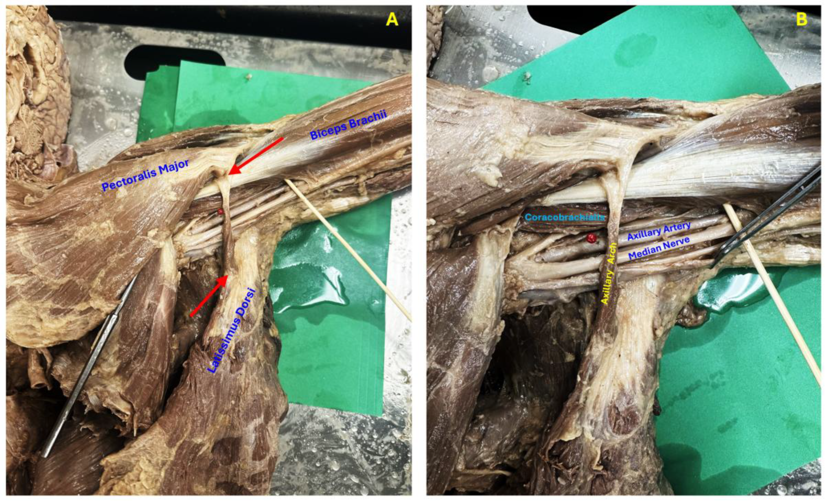
Dissection of the left axilla revealed a distinct muscular slip arising from the medial border of the latissimus dorsi muscle. The muscle coursed anteriorly across the axillary fossa and inserted into the trilaminar tendon of the pectoralis major muscle (Figure 1A). The axillary arch passed superficial and anterior to the axillary neurovascular structures (Figure 1B).
With simulated abduction of the upper limb, the axillary artery was observed immediately deep to the muscular slip, with the median nerve lying inferior to the artery. No gross evidence of compression, deformation, or entrapment of the neurovascular structures was noted in the neutral position. Examination of the thenar eminence showed no visible muscle atrophy. No additional muscular or neurovascular variations were identified in the ipsilateral or contralateral axilla or elsewhere in the body.
Discussion
The axillary arch was first described by Ramsay in 1795 and later detailed by Langer in 1864 (10). In its classical configuration, the muscle arises from the latissimus dorsi and crosses the base of the axilla to insert into the pectoralis major, placing it in close proximity to the axillary vessels and brachial plexus (4).
The axillary arch is commonly classified into complete and incomplete forms. In the complete form, the arch extends from the latissimus dorsi to the trilaminar tendon of the pectoralis major, as observed in the present case. In contrast, incomplete forms demonstrate variable insertions into structures such as the axillary fascia, coracobrachialis, pectoralis minor, biceps brachii, coracoid process, or first rib (3,8).
The variant identified in this study corresponds to the most frequently reported configuration of the axillary arch. Similar to descriptions by Loukas et al. (11) and cadaveric observations by Astaneh et al. (12), the muscular slip originated from the latissimus dorsi and inserted into the trilaminar tendon of the pectoralis major. This anatomy is consistent with established classifications of the complete axillary arch (2,3). Unlike more complex variants featuring multiple insertions or branching fibers, the present case demonstrated a single, well-defined musculotendinous band without bilateral occurrence or accessory slips.
Previous cadaveric and surgical reports have shown that the axillary arch may cross anterior to the axillary artery and elements of the brachial plexus, occasionally resulting in positional neurovascular compression during arm abduction or external rotation (8,13). Although no gross compression or deformation of the axillary artery or median nerve was observed in the neutral position in the present specimen, the close anatomical relationship mirrors configurations reported in surgical series where the axillary arch contributed to diagnostic confusion or operative difficulty (5,6). Large-scale cadaveric analyses further suggest that even anatomically subtle axillary arches may become clinically relevant under dynamic conditions or during surgical exposure (9).
Reported prevalence of the axillary arch varies widely. Earlier surgical studies suggested rates between 0.25% and 7% (5), whereas cadaveric and imaging-based investigations have reported substantially higher prevalence, in some populations exceeding 40% (9). This discrepancy likely reflects differences in study design and detection methods, as cadaveric studies are more likely to identify subtle muscular slips than intraoperative observations (3).
Clinically, the axillary arch may present as obliteration of the axillary fossa or as a palpable mass, potentially mimicking lymphadenopathy or soft-tissue tumors (4,8). In certain positions, particularly arm abduction and external rotation, the arch may contribute to compression of the brachial plexus or axillary vessels, producing symptoms resembling thoracic outlet or hyperabduction syndromes (9,14).
From a surgical standpoint, failure to recognize an axillary arch can complicate axillary procedures. The muscle may obscure lymph nodes or be mistaken for normal anatomical landmarks, potentially resulting in incomplete lymph node clearance or increased operative difficulty during sentinel lymph node biopsy or axillary lymph node dissection (5,6). The present case adds to the growing anatomical evidence emphasizing the importance of awareness of axillary arch variants among anatomists, radiologists, and surgeons.
Conclusion
The axillary arch is a common anatomical variation of the axilla with important clinical and surgical implications. Although often asymptomatic, its relationship to the axillary neurovascular structures may predispose to compression syndromes or complicate surgical procedures. Recognition of this variant during anatomical study and surgical practice is essential to avoid diagnostic errors and operative complications.
Funding
No external financial support was received for this study.
Acknowledgments
The authors would like to thank the Tilman J. Fertitta Family College of Medicine, University of Houston, for providing access to the donated cadaver and the cadaveric laboratory facilities that made this study possible.
Disclosure
The authors declare no conflicts of interest.
Ethical Clearance
The cadaver used in this study was obtained through an institutional body donation program in accordance with applicable laws and institutional guidelines. No identifiable personal data were available. Ethical committee approval was not required for this type of anatomical study.
References
- Gray H. Gray’s Anatomy: The Anatomical Basis of Medicine and Surgery. 38th ed. Churchill Livingstone; 1995. 782–783 p.
- Jelev L, Georgiev GP, Surchev L. Axillary arch in human: common morphology and variety. Definition of “clinical” axillary arch and its classification. Ann Anat. 2007; 189(5): 473–81. [CrossRef] [PubMed]
- Taterra D, Henry BM, Zarzecki MP, Sanna B, Pękala PA, Cirocchi R, Walocha JA, Tubbs RS, Tomaszewski KA. Prevalence and anatomy of the axillary arch and its implications in surgical practice: A meta-analysis. Surgeon. 2019;17(1): 43–51. [CrossRef] [PubMed]
- Bertone VH, Ottone NE, Tartaro ML, de Quirós NG, Dominguez M, Gonzalez D, Bonardi PL, Florio S, Lissandrello E, Blasi E, Medan C. The morphology and clinical importance of the axillary arch. Folia Morphol (Warsz). 2008; 67(4): 261–6. PMID: 19085866.
- Daniels IR, della Rovere GQ. The axillary arch of Langer--the most common muscular variation in the axilla. Breast Cancer Res Treat. 2000 Jan;59(1):77–80. [CrossRef] [PubMed]
- Yonkus JA, Jakub JW. Anterior Axillary Arch: An Anatomic Variant Every Surgeon Operating in the Axilla Should Be Aware of. J Surg Res. 2021; 259: 170–4. [CrossRef] [PubMed]
- Besana-Ciani I, Greenall MJ. Langer’s axillary arch: anatomy, embryological features and surgical implications. Surgeon. 2005; 3(5): 325–7. [CrossRef] [PubMed]
- Rai R, Iwanaga J, Loukas M, Oskouian RJ, Tubbs RS. The Role of the Axillary Arch Variant in Neurovascular Syndrome of Brachial Plexus Compression. Cureus. 2018; 10(6): e2875. [CrossRef] [PubMed] [PubMed Central]
- Weninger JT, Pruidze P, Didava G, Rossmann T, Geyer SH, Meng S, Weninger WJ. Axillary arch (of Langer): A large-scale dissection and simulation study based on unembalmed cadavers of body donors. J Anat. 2024; 244(3): 448–57. [CrossRef] [PubMed] [PubMed Central]
- Jung SJ, Lee H, Choi IJ, Lee JH. Muscular axillary arch accompanying variation of the musculocutaneous nerve: axillary arch. Anat Cell Biol. 2016; 49(2): 160–2. [CrossRef] [PubMed] [PubMed Central]
- Loukas M, Noordeh N, Tubbs RS, Jordan R. Variation of the axillary arch muscle with multiple insertions. Singapore Med J. 2009; 50(2): e88-90. PMID: 19296022.
- Astaneh ME, Rezaei-Tazangi F, Astaneh MR, Arefnezhad R. The observation of an axillary arch during dissection: A case report. Translational Research in Anatomy. 2023; 31: 100244. [CrossRef]
- Lyrtzis C, Paraskevas G, Anastasopoulos N, Greige P. Axillary arch as a rare variant of the latissimus dorsi: cadaveric case report and literature review. Acta Medica. 2025; 56(4): 271–4. [CrossRef]
- Jones MR, Prabhakar A, Viswanath O, Urits I, Green JB, Kendrick JB, Brunk AJ, Eng MR, Orhurhu V, Cornett EM, Kaye AD. Thoracic Outlet Syndrome: A Comprehensive Review of Pathophysiology, Diagnosis, and Treatment. Pain Ther. 2019; 8(1): 5–18. [CrossRef] [PubMed] [PubMed Central]
Figure 1.
(A) Anterior view of the axillary region showing the axillary arch muscle. The superior arrow indicates insertion into the trilaminar tendon of the pectoralis major, and the inferior arrow indicates origin from the medial border of the latissimus dorsi. (B) The axillary arch coursing anterior to the neurovascular contents of the axilla. The red pin marks the axillary artery; the median nerve lies immediately inferior to it.
Disclaimer/Publisher’s Note: The statements, opinions and data contained in all publications are solely those of the individual author(s) and contributor(s) and not of MDPI and/or the editor(s). MDPI and/or the editor(s) disclaim responsibility for any injury to people or property resulting from any ideas, methods, instructions or products referred to in the content. |
© 2026 by the authors. Licensee MDPI, Basel, Switzerland. This article is an open access article distributed under the terms and conditions of the Creative Commons Attribution (CC BY) license (http://creativecommons.org/licenses/by/4.0/).
Copyright: This open access article is published under a Creative Commons CC BY 4.0 license, which permit the free download, distribution, and reuse, provided that the author and preprint are cited in any reuse.


